


Abstract
Biopolymers—whether derived from natural sources or produced synthetically—combine biodegradability with biocompatibility, positioning them as essential materials for scaffolds and drug delivery systems in regenerative medicine. Although substantial progress has been made in synthesis methodologies and clinical evaluation, comprehensive and critically oriented reviews that systematically consolidate knowledge on biopolymer applications remain scarce. This review synthesizes evidence on natural polymers (collagen, cellulose, chitosan, hyaluronic acid, alginate) and synthetic polymers (polycaprolactone, polylactic acid, polyglycolic acid, their copolymers, and polyethylene glycol). We examine their classification, physicochemical properties, modification strategies, and applications—including tissue engineering, drug delivery, wound healing, and multi-tissue regeneration. Critically, rather than merely cataloging successful case studies, this review identifies and analyzes bottlenecks: the scarcity of head-to-head in vivo comparative studies, the lack of standardized preclinical protocols, poor predictability of long-term immune responses, and ambiguous criteria for material selection that hinder clinical decision-making. The final section discusses emerging hybrid constructs that integrate microcapsules for sustained therapeutic release, representing a paradigm shift from passive structural implants toward actively programmable therapeutic platforms. By bridging materials science, immunology, and translational medicine, this review provides a strategic roadmap for moving biopolymer research beyond proof-of-concept toward clinically viable, scalable, and immunologically smart regenerative systems.
1. Introduction
Regenerative medicine stands as one of the most rapidly advancing frontiers in contemporary biomedicine, aimed at restoring the structure and function of damaged tissues and organs. In contrast to traditional replacement therapies, the regenerative paradigm actively harnesses the body’s intrinsic reparative mechanisms through the synergistic application of cell-based technologies, signaling molecules, and—critically—biocompatible carrier matrices known as scaffolds [1,2].
Biopolymers—polymeric molecules of either natural or synthetic origin that are biodegradable, non-toxic, and tissue-compatible—play a pivotal role in the fabrication of such materials [3,4]. Natural biopolymers (collagen, chitosan, hyaluronic acid, alginate) mimic the native extracellular matrix and provide optimal biochemical cues for cell adhesion, proliferation, and differentiation [4]. In parallel, synthetic analogues (polycaprolactone, polylactic acid, polyglycolic acid, and others) afford precise tunability of mechanical properties and degradation kinetics [5]. Hybrid composites combining both classes offer the opportunity to synergistically integrate the advantages of each, while mitigating their respective limitations.
Despite considerable progress in the development of biopolymer-based materials, systematic analysis and critical comparison of the literature remain fragmented and incomplete. The existing body of work is dominated by isolated reports of successful experimental systems, whereas a holistic, problem-oriented perspective on unresolved challenges is conspicuously absent. Among the most pressing issues are:
- A paucity of direct comparative in vivo studies—the vast majority of investigations employ disparate animal models, implantation protocols, and evaluation criteria, rendering objective ranking of material performance effectively impossible;
- Lack of standardization—both at the level of synthesis and modification (molecular weight, degree of deacetylation, crosslinking density) and at the level of preclinical assessment (no unified protocols for biodegradation, biomechanical testing, or histomorphometric analysis);
- Poor predictability of the immune response—many polymers deemed “biocompatible” based on short-term assays provoke chronic inflammation, fibrosis, or capsule formation upon long-term implantation; however, systematic data on macrophage polarization, cytokine profiles, and the long-term fate of degradation products remain critically insufficient;
- Ambiguity in material selection criteria—for most clinical indications, there are no clear guidelines on which polymer class (natural, synthetic, or hybrid) is preferable, and existing reviews typically enumerate possibilities without systematically analyzing trade-offs and limitations.
In recent years, the volume of research dedicated to biopolymers has grown exponentially—encompassing everything from novel synthetic routes to preclinical and clinical validation. Nevertheless, comprehensive review articles that critically synthesize knowledge on their biomedical applications remain surprisingly scarce. This review aims to fill this gap by consolidating current knowledge on biopolymer properties and applications, and by critically examining the limitations and translational barriers that hinder their clinical adoption.
2. Biopolymers for Regenerative Medicine
2.1. General Classification Framework: Beyond Simple Categorization
Biopolymers constitute a broad and chemically diverse class of macromolecules unified by two cardinal properties—biodegradability and biocompatibility—that render them indispensable in tissue engineering and regenerative medicine [5,6]. By origin, biopolymers are traditionally categorized into two principal groups: natural (isolated from plant, animal, or microbial sources) and synthetic (produced via chemical polymerization of artificial monomers or through modification of natural analogues) (Figure 1) [4,6].
Figure 1.
Classification of major biopolymers into natural and synthetic categories (schematic representation).
However, this binary classification, while practically useful, obscures a more nuanced reality. As will become evident throughout this review, the distinction between “natural” and “synthetic” is neither absolute nor sufficient for predicting material performance in complex biological environments. Natural biopolymers, owing to their biochemical resemblance to components of the human extracellular matrix (ECM), exhibit high cellular recognition and favorable tissue interactions [7,8,9]. Yet, this very resemblance carries inherent risks—immunogenicity, batch-to-batch variability, and limited mechanical tunability—that complicate clinical translation. Synthetic biopolymers, in turn, afford precise control over mechanical properties, degradation kinetics, and batch-to-batch reproducibility—attributes critical for scalable manufacturing of medical devices [2,10]. However, their structural divergence from native ECM often results in poor cell adhesion, uncontrolled inflammatory responses, and accumulation of acidic degradation products that inhibit tissue regeneration.
A more productive framework for understanding biopolymer utility does not treat these classes as oppositional but rather as complementary, with hybrid composites offering the opportunity to synergistically integrate the advantages of each while mitigating their respective limitations. Within this framework, natural biopolymers are further subdivided into two major families—polysaccharides (cellulose, chitosan, hyaluronic acid, alginate) and proteins (collagen, gelatin, silk fibroin)—each possessing a distinctive repertoire of structural and bioactive properties.
Crucially, the classification of a biopolymer as “natural” or “synthetic” is increasingly being challenged by advances in biotechnology. Microbial fermentation, enzymatic synthesis, and genetic engineering now enable production of natural polymers with unprecedented control over molecular weight, stereochemistry, and functional group presentation—blurring the traditional boundaries and creating a continuum of materials that span from biologically derived to fully synthetic [11].
From a translational perspective, the choice between natural and synthetic biopolymers cannot be reduced to a simple binary decision. Instead, it requires systematic consideration of the target tissue’s biology, the required mechanical environment, the desired degradation timeline, the anticipated immune response, and—critically—the manufacturing and regulatory feasibility. As detailed in subsequent sections, each polymer class presents a distinct profile of trade-offs rather than a hierarchy of superiority. This review adopts this trade-off centered framework, analyzing not only what each material can do, but also what it cannot—and why that matters for clinical success.
2.2. Natural Biopolymers: Between Biological Inspiration and Clinical Reality
Natural biopolymers are macromolecules synthesized by living organisms—including plants, animals, fungi, bacteria, and algae. Their structural architecture is defined by repeating monomeric units, typically amino acids (proteins) or monosaccharides (polysaccharides), arranged in specific sequences or glycosidic linkages that dictate their biological functions [8,12,13]. This inherent biological origin confers upon natural biopolymers a fundamental advantage over their synthetic counterparts: evolutionary optimization for interaction with biological systems. Proteins and polysaccharides of natural origin are recognized by cell surface receptors, degraded by endogenous enzymes, and integrated into metabolic pathways—attributes that synthetic polymers, regardless of their chemical sophistication, struggle to replicate.
The promise of natural biopolymers rests on three pillars:
- Biocompatibility—derived from biochemical homology with native extracellular matrix components, minimizing foreign body responses [7];
- Biodegradability—enabling enzymatic clearance and gradual replacement by newly formed tissue, obviating the need for surgical removal [8];
- Bioactivity—actively modulating cell behavior through receptor-mediated signaling, rather than serving as passive structural supports [14].
Yet this very biological origin, which underpins their clinical promise, also constitutes their Achilles’ heel. As will become evident in the detailed analysis of individual polymers below, natural biopolymers are plagued by batch-to-batch variability (influenced by species, season, and extraction methods), limited mechanical tunability (restricted by their native conformation), risk of immunogenicity (particularly for xenogeneic proteins), and high production costs associated with purification under GMP conditions. These limitations, often underemphasized in the promotional literature, have historically constrained the translation of natural polymers from laboratory success to widespread clinical adoption.
Critically, natural biopolymers are not immunologically inert; they actively engage with the host immune system through pattern recognition receptors (Toll-like receptors, mannose receptors) and complement pathways. This interaction can be either beneficial (promoting M2 macrophage polarization, resolving inflammation) or detrimental (triggering chronic inflammation, fibrosis, or capsule formation), depending on the polymer’s purity, molecular weight, degree of modification, and degradation products [15]. This dual nature—simultaneously bioactive and potentially immunogenic—positions natural biopolymers as active participants in the regenerative process rather than passive scaffolds, a theme that will be explored in depth in Section 3.
Within this framework, natural biopolymers for regenerative medicine are conventionally subdivided into two major families: proteins (collagen, gelatin, silk fibroin) and polysaccharides (cellulose, chitosan, hyaluronic acid, alginate). Each family possesses a distinctive structural, mechanical, and bioactive profile, dictating its suitability for specific tissue engineering applications. The following sections provide a critical, balanced analysis of the five most clinically relevant natural biopolymers—collagen, cellulose, chitosan, hyaluronic acid, and alginate—examining not only their established advantages but also their translational barriers and the strategies being developed to overcome them.
2.2.1. Collagen
Collagen is the most abundant structural protein in the human body, constituting approximately 25–35% of total protein mass and serving as the primary architectural component of skin, bone, tendons, ligaments, cartilage, and other connective tissues [16,17]. Its distinctive triple-helical structure, composed of repeating Gly-X-Y amino acid sequences—where X and Y are frequently proline and hydroxyproline—confers both mechanical resilience and biological recognition. In its native state, collagen exists as a hierarchical assembly, from individual α-chains to fibrils and fibres, that provides structural integrity while remaining accessible to cellular receptors such as integrins and discoidin domain receptors [18]. Denatured collagen, known as gelatin, retains bioactivity but loses fibrillar organisation, offering distinct processing advantages for certain applications.
For biomedical applications, collagen is primarily extracted from mammalian sources—bovine skin and tendon, porcine tissues—and, increasingly, from marine organisms, including fish skin, scales, and bones [16,19]. Marine-derived collagen has attracted growing interest due to its lower immunogenicity (attributable to shorter telopeptide regions), reduced risk of zoonotic disease transmission, and absence of religious restrictions. However, marine collagen typically exhibits lower hydroxyproline content and denaturation temperatures (e.g., 34.4–36.1 °C for tilapia collagen versus 38.2 °C for calf collagen), which can compromise thermal stability at physiological temperature. This trade-off between reduced immunogenicity and thermal stability exemplifies the broader challenge of optimising collagen for specific clinical applications.
Collagen-based materials have achieved remarkable clinical translation across multiple domains. They are widely employed as injectable suspensions for dermal contouring [20], haemostatic sponges [21], wound dressings [22], three-dimensional scaffolds [23], hydrogel [24], nanofibres [25], and cartilage repair matrices [26]. Collagen-based wound dressings have been extensively investigated for burn and chronic wound healing, including diabetic ulcers, with demonstrated efficacy in promoting granulation tissue formation, re-epithelialisation, and angiogenesis [27,28]. For controlled drug delivery, collagen conjugates with antibiotics (ciprofloxacin), antioxidants (quercetin, curcumin), and growth factors (bFGF) have shown accelerated healing in diabetic wound models [29,30,31].
Collagen scaffolds have been developed for regeneration of diverse tissues—skin, bone, cartilage, tendons, and nerves—often combined with stem cells, growth factors, and bioactive molecules to enhance regenerative capacity [23,32,33]. Recent advances have focused on creating oriented microchannel structures within collagen hydrogels to provide physical contact guidance for directional cell migration and deep tissue infiltration [24]. As illustrated in Figure 2, the combination of topographical cues from aligned microchannels with NIR-triggered release of immunomodulatory agents (spermidine) enables synergistic regulation of both cellular behavior and the inflammatory microenvironment, representing a promising strategy for chronic wound healing and tissue regeneration. In bone tissue engineering, collagen scaffolds functionalised with osteogenic factors and mineral components (hydroxyapatite, bioglass) mimic the native bone extracellular matrix and stimulate osteoblast differentiation and mineralization [34,35]. Collagen coatings on metallic implants improve osteoblast adhesion and enhance osseointegration, facilitating stable bone–implant interfaces [36,37]. For cartilage and osteochondral regeneration, biphasic collagen scaffolds with distinct regions mimicking articular cartilage and subchondral bone have been developed, with several products advancing to clinical trials and regulatory approval. In neural tissue engineering, collagen scaffolds serve as carriers for stem cells (bone marrow-derived mesenchymal stromal cells, neural stem cells, induced pluripotent stem cells) and neurotrophic factors (brain-derived neurotrophic factor, nerve growth factor, glial cell line-derived neurotrophic factor), promoting cell survival, migration, and neuronal differentiation while providing structural support for axonal growth.
Figure 2.
Schematic illustration for the preparation of collagen-based hydrogel with oriented microchannel structure, NIR-activated photothermal performance and drug release behavior, fibroblast infiltration promotion, antibacterial activity and anti-inflammatory activity by the directional freeze–thaw process. Reprinted from Ref. [24].
Collagen faces persistent challenges related to raw material source. The tissue source of type I collagen—whether bovine, porcine, or rodent—critically affects molecular characteristics (e.g., lysine content) and macroscopic hydrogel properties (e.g., swelling ratio and compression modulus), which in turn influence scalability and regulation-friendly clinical translation of medical device prototypes. Selecting a commercial collagen source that satisfies both manufacturability and regulatory requirements remains challenging [38]. For marine collagen, environmental temperature during the organism’s life directly affects thermal stability—a factor rarely considered in material selection. Immunogenicity risks persist despite rigorous purification, as residual telopeptides (especially in mammalian collagen) can trigger immune responses, chronic inflammation, or fibrosis in susceptible patients [39,40]. Marine collagen offers reduced immunogenicity but at the cost of lower thermal stability. Native collagen also exhibits low mechanical strength in the hydrated state and rapid enzymatic degradation, necessitating crosslinking or composite fabrication; however, crosslinking often compromises bioactivity, creating a fundamental trade-off between stability and biological performance [41].
To address the fundamental limitations of animal-derived collagen, recombinant collagen production has emerged as a transformative approach. Recombinant human collagens—produced in bacterial, yeast (Pichia pastoris), insect, mammalian, or plant systems—offer human-sequence design, reduced immunogenicity, and batch-to-batch consistency. Yeast and bacterial systems are scalable and cost-effective but require co-expression of prolyl-4-hydroxylase to achieve proper hydroxylation and thermal stability (denaturation temperature of approximately 40 °C). Insect and mammalian cell systems produce more native-like post-translational modifications but at higher cost. Recombinant type III collagen has attracted particular attention for its role in scarless wound healing—fetal skin rich in type III collagen exhibits minimal scarring, and scaffolds enriched with recombinant type III collagen promote softer, more regenerative healing by mimicking fetal repair mechanisms; indeed, the type I/III collagen ratio is positively correlated with scar severity, suggesting that therapeutic modulation of this ratio could improve clinical outcomes. Other emerging directions include black phosphorus-based collagen composites for photothermal antibacterial therapy and stimuli-responsive collagen scaffolds that release therapeutics in response to enzymatic or pH cues. Plant-based recombinant collagen production platforms are also being explored, offering reduced animal-origin risk and potential for scalable production.
2.2.2. Cellulose
Cellulose is one of the most abundant and renewable biopolymers on Earth, distinguished by a unique combination of biocompatibility, non-toxicity, and biodegradability. In addition to natural photosynthesis and chemical synthesis, cellulose can be produced enzymatically in vitro or via biosynthesis using various producers—algae, fungi, and bacteria [42,43,44]. Bacterial cellulose (BC), in particular, has emerged as a high-purity biomaterial synthesised by microorganisms of the genera Acetobacter, Gluconacetobacter, Achromobacter, Rhizobium, Pseudomonas, Sarcina ventriculi, and Agrobacterium [42,45].
Bacterial cellulose in the form of hydrogels, films, and nanoparticles is an effective platform for controlled and targeted drug delivery. Its high water-retention capacity and biocompatibility enable local administration of pharmaceuticals, growth factors, and other therapeutic agents to target tissues, enhancing therapeutic efficacy while minimising systemic side effects [46]. Cellulose-based materials have demonstrated potent antibacterial activity in wound healing: composites with lysostaphin, silver nanoparticles (AgNPs), and silver sulfadiazine exhibited pronounced bactericidal properties against Escherichia coli and Staphylococcus aureus [47,48,49].
Extensive work has focused on cellulose-based hydrogels and scaffolds. Modified cellulose—particularly carboxymethylcellulose (CMC)—offers promising avenues for biomedical applications. The hydrophilic nature of CMC allows blending with other materials (synthetic polymers, natural polymers, and inorganic materials) to create innovative wound dressings. In one study, UV-crosslinked cellulose–acrylic acid hydrogels enhanced neovascularisation and re-epithelialisation in burn wound healing [50]. A CMC-based hydrogel (CPPFe@TA) exhibited photothermal antibacterial activity, accelerating infected wound healing, and also functioned as a flexible skin biosensor for monitoring physiological signals (Figure 3). Despite the need for improved freeze–thaw resistance and reduced hygroscopicity, this material holds substantial promise for regenerative wound therapy and “smart” sensing systems [51]. Using a cyclic freeze–thaw method, Shin and colleagues fabricated a hydrogel from sodium carboxymethylcellulose, polyvinyl acetate, and PEG 400. The material exhibited high porosity, enhanced strength and swelling capacity, and promoted cell growth and accelerated wound re-epithelialisation [52]. Employing 3D printing technology, Díaz-Gómez and coworkers developed a CMC-based scaffold for diabetic wound therapy. Impregnation with platelet-rich plasma (PRP) induced secretion of growth factors, which in turn stimulated angiogenesis, granulation, and re-epithelialisation [53].
Figure 3.
Schematic illustration of the integrated platform of intelligent collaborative photothermal wound therapy based on CPPFe@TA hydrogel. Reprinted with permission from Ref. [51] © 2024 Wiley-VCH GmbH.
In the context of diabetic wound healing, multifunctional composite hydrogels have been designed by combining BC with conductive polymers and therapeutic agents. One recent study developed a polypyrrole/bacterial cellulose/platelet-rich plasma (PBP) composite hydrogel that mimics the natural repair cascade. The BC hydrogel serves as a scaffold for PRP encapsulation, protecting growth factors from protease degradation while enabling sustained release. The capacitive polypyrrole component provides potent antibacterial activity through electric field-mediated charge storage and promotes electrical signal transduction to regulate growth factor release kinetics. In diabetic wound models, treatment with the electroactivated PBP composite hydrogel demonstrated a marked reduction in inflammatory responses, accelerated vascular regeneration, enhanced collagen deposition, and overall improvement in wound healing [54]. Similarly, copper-doped borosilicate bioactive glass coatings on BC nanofibers have been developed, conferring excellent antibacterial properties, anti-inflammatory activities, cytocompatibility, and angiogenic potential, with in vivo results confirming effective inhibition of MRSA-induced infection and accelerated wound healing [55].
Beyond established applications, recent research has focused on BC’s emerging roles in immunomodulation and mixed-biofilm control. BC’s stiffness, porosity, and nanoscale architecture collectively influence cell phenotype through mechanotransduction pathways—substrate stiffness regulates cytoskeletal organisation, nuclear deformation, and gene expression, mechanisms directly relevant to BC scaffolds, which can be tuned from soft hydrogel-like matrices to stiffer load-bearing constructs [56]. This cell-instructive property positions BC not only as a passive substrate but also as a mechanically instructive, biologically interactive scaffold capable of shaping cellular responses and guiding regenerative outcomes. For example, citrate-modified BC scaffolds have demonstrated improved mechanical resilience and osteoconductivity, making the material more suitable for bone regeneration. Furthermore, BC’s ability to control mixed-biofilm formation has been explored in the context of chronic wound infections. Since a single bacterial species is rarely responsible for chronic wound infections, BC-based dressings that modulate the polymicrobial community structure—rather than simply eradicating planktonic bacteria—represent a promising strategy for managing wound biofilms and preventing antibiotic resistance [56].
Modified cellulose derivatives have also demonstrated utility beyond wound healing. A conductive anisotropic hydrogel scaffold based on CMC and polyvinyl alcohol (PVA) was developed for ligament repair, exhibiting excellent anisotropic mechanical properties, electrical conductivity, and self-powered capacity for monitoring body motion without an external power supply [57]. In another approach, composite gels based on type I collagen and CMC demonstrated superior rigidity and stability compared to pure collagen gels, with preserved native collagen fibril structure and improved proliferation and migration of human mesenchymal stromal cells [58]. These findings highlight the synergistic potential of combining cellulose derivatives with other biopolymers to overcome the limitations of individual materials.
Despite these promising developments, biomedical translation of cellulose materials is constrained by several factors. A key limitation is that native cellulose lacks endogenous degradation pathways in the human body. Cellulose-degrading enzymes (cellulases) are not produced by human cells, and the highly crystalline structure of native cellulose makes it inaccessible to enzymatic hydrolysis. As a result, unmodified cellulose implanted in vivo is essentially non-resorbable unless cellulases are exogenously supplied or the material is chemically modified to become enzyme-susceptible. Yadav et al. showed that unmodified bacterial cellulose implanted subcutaneously in mice degraded poorly, whereas a metabolically engineered cellulose–chitin copolymer with reduced crystallinity was completely degraded within 20 days [59]. Similarly, Bastard et al. noted that nanocellulose does not degrade in the human body, and therefore developed casein microparticles to locally deliver cellulase, achieving nanocellulose hydrogel degradation over 21 days [60]. For temporary scaffolds that require complete replacement by new tissue, this distinction is critical: unmodified cellulose may remain at the implantation site rather than being resorbed in step with tissue regeneration [61]. Although BC exhibits good biocompatibility, its high hygroscopicity may lead to excessive swelling and altered mechanical properties [62]. Moreover, the dense network of intra- and intermolecular hydrogen bonds makes chemical modification challenging, requiring harsh reaction conditions that risk compromising the nanofibrillar structure and limiting controlled incorporation of bioactive molecules [63]. Finally, the high cost of bacterial synthesis compared to plant-derived cellulose, combined with the absence of scalable GMP processes, currently prevents BC from competing with simpler polymers for mass-produced wound dressings.
Scalability of BC production remains a significant bottleneck, as fermentation-based production is resource-intensive, costly, and challenging to scale for clinical demands. Variability in culture conditions, nutrient availability, and bacterial strain performance can lead to differences in BC’s physical properties, which in turn affect scaffold performance. Additionally, while BC is biodegradable, its in vivo degradation rate is relatively slow and often insufficient for applications requiring timely resorption [64]. From a translational perspective, the lack of clinical trial results reported in the literature on the safety and efficacy of cellulose-based products in healthcare applications remains a significant gap. While cellulose nanomaterials—including cellulose nanocrystals (CNCs), cellulose nanofibers (CNFs), and bacterial nanocellulose (BNC) have demonstrated promise in drug delivery, wound dressings, and tissue engineering, their physiological stability and long-term biocompatibility require further investigation [64,65,66]. Despite these challenges, cellulose—particularly in its bacterial and nanocellulose forms—continues to offer a versatile, renewable, and biocompatible platform for next-generation regenerative medicine applications, from smart wound dressings to multifunctional tissue engineering scaffolds.
2.2.3. Chitosan
Chitosan is a linear polysaccharide obtained by deacetylation of chitin—a natural polymer found in crustacean shells, insect exoskeletons, and fungal cell walls [67]. Key features underpinning its biomedical potential include non-toxicity, non-immunogenicity, biodegradability, antimicrobial activity, and wound-healing acceleration. Notably, chitosan possesses a pronounced hemostatic effect, making it particularly attractive for blood-stopping wound dressings and hemostatic devices [68,69]. Owing to its high biocompatibility, biodegradability, and elasticity, chitosan-containing materials have found broad application in biomedicine—particularly for skin wound repair [70], bone regeneration [71], and targeted drug delivery [71].
Chitosan exhibits potent hemostatic activity independent of the classical coagulation cascade [72]. Its hemostatic effect is primarily attributed to electrostatic interactions between cationic chitosan groups and negatively charged erythrocyte membranes, inducing rapid cell aggregation and platelet plug formation at the injury site. Additional mechanisms include stimulation of platelet adhesion and aggregation, as well as binding to plasma proteins and coagulation factors, potentially initiating the contact pathway of coagulation [73]. Chitosan displays broad-spectrum antimicrobial activity against a wide range of microorganisms—including bacteria (both Gram-positive and Gram-negative), fungi [74]. This property renders it particularly attractive for biomedical applications, including wound dressings, antibacterial scaffolds, and drug delivery systems [75].
Chitosan can be processed into various forms—membranes, hydrogels, fibres, and sponges [76]. Nanotechnology has opened new avenues for antibacterial biomaterials. Incorporation of silver nanoparticles (AgNPs) into chitosan-based hydrogels as bactericidal and bacteriostatic agents has shown promise. In vitro evaluation against wound infections caused by S. aureus and E. coli confirmed pronounced antibacterial effects [77]. Kim and colleagues developed a chitosan–catechol-conjugated hemostatic sponge for patients with coagulopathy. The proposed synergistic mechanism—combining catechol groups with cationic chitosan charges—enables rapid coagulation even in clotting disorders, with catechol-modified chitosan outperforming native chitosan in film-forming capacity and hemostatic efficacy [78]. Wu and coworkers developed a chitosan-based hemostatic sponge modified with mercaptosuccinic acid and AgNPs (Figure 4), achieving rapid hemostasis, sustained antimicrobial activity, and reduced cytotoxicity through controlled AgNP release [79]. Chitosan fibres incorporated into wound dressings demonstrate superior hemostatic properties compared to gauze, reducing bleeding time and effectively controlling blood loss, as confirmed in a femoral artery model and clinical practice [80].
Figure 4.
Design and mechanism of the chitosan-based hemostatic sponge modified with mercaptosuccinic acid and silver nanoparticles (AgNPs). Reprinted from Ref. [79].
Beyond wound healing, chitosan has emerged as a promising platform for bone tissue regeneration and immunomodulation. A chitosan-silica hybrid dressing enhanced with bioactive glass has demonstrated excellent biocompatibility, no inflammatory response, and strong hemostatic properties, with controlled degradation over 3–6 months and significantly enhanced bone regeneration through ion release (Si4+, Ca2+) [81]. In the context of cancer immunotherapy, multimodal chitosan-based systems are being developed to enable precise targeting, controlled release, and enhanced immune activation. Advanced chitosan derivatives—including trimethyl chitosan, carboxymethyl chitosan, fluorinated chitosan, and thiolated chitosan—are being explored for integration with chemotherapy, radiotherapy, phototherapy, and gene therapy, capitalising on chitosan’s intrinsic immunomodulatory properties [82].
Despite its unique combination of hemostatic and antimicrobial properties, clinical translation of chitosan faces substantial challenges. First, its antibacterial activity is strongly dependent on the degree of deacetylation (DD) and molecular weight. In general, higher DD is associated with stronger activity, while high-molecular-weight fractions have limited solubility at physiological pH, which narrows the range of effective concentrations [83,84]. Second, the same strong cationic nature that underlies its hemostatic effect—driving erythrocyte aggregation and platelet adhesion—may, at high local concentrations, also compromise blood cell integrity, while its interactions with plasma proteins and the immune system raise additional biocompatibility concerns that require careful evaluation [73]. Third, the mechanical strength of chitosan hydrogels in the hydrated state is often insufficient for load-bearing tissue replacement, necessitating crosslinking with synthetic polymers or chemical reagents (e.g., glutaraldehyde) that may themselves be cytotoxic [85]. Finally, raw material instability (chitin content varies with crustacean season, species, and farming conditions) poses reproducibility challenges critical for medical device certification. The importance of standardisation has been further emphasised in the recent literature, with researchers highlighting that endotoxin contamination (a critical quality attribute) must be reduced to ≤0.5 EU/mL to meet FDA and EMA requirements for wound care biomaterials [86]. Additionally, variability in DD and molecular weight due to biological source, seasonal variation, and extraction techniques necessitates stringent material controls, including robust Certificates of Analysis covering MW, DD, viscosity, microbial load, heavy metals, moisture, and endotoxin levels [86]. These standardisation challenges have also been identified as a major obstacle preventing the translation of chitosan-based vaccines and immunotherapies to the market [87].
2.2.4. Hyaluronic Acid
Hyaluronic acid (HA) is a natural polysaccharide and a major component of the human and animal extracellular matrix. It is widely distributed in connective tissue, synovial fluid, vitreous humor, skin, and cartilage. Since its first isolation from bovine vitreous humor in the 1930s [88], HA has consistently attracted research interest due to its unique combination of physicochemical properties and biological functions—namely, outstanding viscoelasticity, high water-retention capacity, biocompatibility, and biodegradability [89,90]. Structurally, HA is a linear, unbranched polysaccharide composed of repeating disaccharide units of D-glucuronic acid and N-acetyl-D-glucosamine, with a molecular weight that can reach up to 2 × 10⁷ Da. Its carboxyl groups confer a negative charge and strong hydrophilicity, enabling the formation of viscous networks at high molecular weights that hydrate the extracellular matrix, preserve tissue balance, and withstand compressive forces [91].
HA can be isolated from a wide spectrum of biological sources—from microorganisms and invertebrates to mammals and humans. It has been found in Pseudomonas mucus [92]; Ascaris bodies [93]; and rat, rabbit [94], and human tissues [95]—specifically skin [95], aorta, cartilage [94], and brain [96]. Critically, after purification, the chemical structure of HA is independent of the species or tissue source. Currently, HA is produced either by extraction from rooster combs or through recombinant bacterial fermentation using Streptococcus strains, with both methods yielding materials that exhibit different rheological characteristics. Since the early 1980s, HA has been extensively studied as a raw material for intraocular lenses, becoming an essential product in ophthalmology, and has since found applications in orthopaedics, wound healing, and soft tissue augmentation. Since the late 1980s, HA has been utilised in drug delivery systems, with ongoing efforts to develop HA-based carriers to improve therapeutic effectiveness [91].
HA is actively investigated for bone tissue engineering, serving as an effective molecular carrier or structural scaffold that stimulates osteoregeneration. Numerous studies confirm successful HA application for calvarial and alveolar bone defect repair [97,98]. A recent systematic review of clinical evidence concluded that HA positively influences bone regeneration outcomes, with the most consistent benefits observed in alveolar ridge preservation and ridge augmentation procedures, including increased new bone formation, improved bone density, enhanced bone maturation, and reduced dimensional bone loss. However, findings regarding maxillary sinus augmentation were less consistent, and the current evidence remains limited, with insufficient mechanistic data to fully elucidate HA’s biological effects in bone regeneration [99].
HA-based hydrogels modified with N-cadherin-mimetic peptides have been shown to mimic pro-osteogenic niches. Such biomodification enhances osteogenic differentiation of human mesenchymal stem cells, increasing expression of osteogenic markers, alkaline phosphatase activity, type I collagen synthesis, and matrix mineralization in vitro and in vivo [100]. HA-based materials have demonstrated significant potential in osteochondral tissue engineering for simultaneous regeneration of cartilage and subchondral bone (Figure 5) [101]. HA-based hydrogels, especially injectable and 3D-bioprinted forms, provide a biomimetic ECM environment that supports cell viability, proliferation and chondrogenesis while reducing inflammatory responses [101]. Commercial HA-based products such as Hyalgan®, Synvisc®, Orthovisc®, Monovisc® and Supartz® are used in clinical practice for the treatment of pain in osteoarthritis of the knee. Separately, HA-based hydrogels can serve as cell carriers for the encapsulation of stem cells or chondrocytes and their delivery to lesion sites, creating a favourable microenvironment for proliferation, differentiation, and in situ cartilage regeneration [102,103].
Figure 5.
Hyaluronic acid-based hydrogels for cartilage tissue engineering: mechanisms and applications. Reprinted from Ref. [101].
HA is a critical regulator of the angiogenic phase of wound healing, participating in modulation of inflammation, neutralisation of reactive oxygen species, and receptor-mediated communication with cells driving tissue regeneration [104]. HA-based hydrogels have demonstrated significant potential in advancing wound healing by regulating inflammatory responses, enhancing angiogenesis, and participating in extracellular matrix remodeling [91]. Biocompatible polyelectrolyte films based on chitosan and HA have been shown suitable as vascular substitutes [105]. Concurrently, HA-containing nanofibrous matrices stimulate endothelial cell proliferation without inducing haemolysis or thrombosis [106]. Notably, thermosensitive injectable HA-based gels incorporating corn silk extract and AgNPs exhibit optimal mechanical properties, biocompatibility, antibacterial activity, and accelerated wound healing in vitro, positioning them as promising wound dressings [107].
Beyond these established applications, recent research has focused on HA’s emerging role in immunomodulation. The molecular weight of HA critically determines its biological function: high-molecular-weight HA (>1000 kDa) generally suppresses pro-inflammatory responses and promotes an anti-inflammatory M2 macrophage phenotype, while low-molecular-weight HA fragments (<60 kDa) bind to TLR4 and stimulate pro-inflammatory M1 polarisation. This differential response results from the multivalent binding of long HA polymers to CD44 receptors, which induces distinct signalling cascades, whereas smaller fragments have fewer binding sites and dissociate more readily. Interestingly, recent studies have demonstrated that while soluble HA polarises macrophages in a molecular weight-dependent manner in 2D culture, the chemical crosslinking of HA within 3D hydrogel microstructures circumvents this effect, highlighting the importance of HA presentation in biomaterial design [108]. This dual nature—simultaneously bioactive and potentially immunogenic—positions HA as an active participant in the regenerative process rather than a passive scaffold.
Despite these promising developments, HA faces fundamental limitations that constrain its clinical translation. The foremost is extremely rapid enzymatic degradation by hyaluronidases, which in tissues with high enzyme activity (skin, joints) occurs within hours to 2–3 days, rendering native HA unsuitable for long-term therapeutic retention or scaffold volume maintenance [109]. Chemical crosslinking extends residence time but reduces bioavailability for CD44 receptors and may provoke chronic inflammation due to degradation products of the modified polymer [110]. Second, the high cost of medical-grade purified HA—particularly from bacterial producer strains—limits its widespread application in combination delivery systems. Finally, poor tissue adhesiveness necessitates additional functionalisation for fixation at the implantation site, especially under continuous mechanical load.
To address these limitations, extensive research has focused on HA modification strategies. Chemical modification approaches—including esterification, amidation, thiolation, and crosslinking—aim to enhance mechanical properties, control degradation kinetics, and introduce stimuli-responsive functionalities [101]. Despite these challenges, HA continues to offer a versatile, biocompatible, and biologically active platform for next-generation regenerative medicine applications—from drug delivery and wound healing to bone and cartilage tissue engineering.
2.2.5. Alginate
Alginate, an anionic and hydrophilic polysaccharide, ranks among the most widely biosynthesised biomaterials used in biomedicine [111]. This natural polymer is traditionally extracted from brown seaweed and also produced microbiologically using Pseudomonas and Azotobacter strains [112]. Key industrial sources include various marine algae species: Laminaria hyperborea, Laminaria digitata, Macrocystis pyrifera, Ascophyllum nodosum, Eclonia maxima, Laminaria japonica, Lessonia nigrescens, Durvillea antarctica, and Sargassum spp. [113]. Structurally, alginate is a linear copolymer composed of (1→4)-linked β-D-mannuronic acid (M) and α-L-guluronic acid (G) residues, arranged in homopolymeric M-blocks, G-blocks, and alternating MG-sequences. The relative content and distribution of these blocks—particularly the M/G ratio—critically determine the polymer’s physicochemical properties, including gelation capacity, mechanical stiffness, and degradation behaviour. Notably, the M/G ratio varies significantly with algal species, harvest season, and geographic location, directly influencing batch-to-batch reproducibility [114].
Combining valuable characteristics—hydrophilicity, biocompatibility, biodegradability, non-toxicity, mechanical elasticity, and chelating activity—alginate and its hydrogels have secured a firm position in tissue engineering [115,116]. The biocompatibility of alginate has been extensively validated, and its safety profile has been recognised by the FDA, which has approved alginate as a generally recognised as safe (GRAS) substance for use in food and pharmaceutical applications. However, the polymer’s potential is constrained by certain shortcomings, including high solution viscosity, low solubility in physiological media, and slow degradation [117]. To overcome these limitations, additional natural or synthetic components are incorporated into alginate matrices, enabling directed modulation of properties and expansion of functional capabilities [117].
Alginate can be combined with other materials to produce various three-dimensional scaffolds—hydrogels, sponges, microcapsules, microspheres, and fibres [118]. In regenerative medicine, alginate-based scaffolds have found extensive application for regeneration of both hard and soft tissues, including skin, bone, cartilage, myocardium, and blood vessels [119]. Sodium alginate–pectin hydrogel films containing simvastatin significantly accelerated diabetic wound healing in type 1 diabetic rats [120]. Alginate microspheres and composite scaffolds (alginate–chitosan and alginate–chitosan–chitooligosaccharide systems) are actively applied as effective skin substitutes, characterised by enhanced biocompatibility and the ability to nearly fully recapitulate the native skin microenvironment [121]. Collagen–alginate scaffolds containing nanocurcumin (Figure 6) actively promote improved wound healing [122].
Figure 6.
Synthesis of PEG-modified nanocurcumin, which was incorporated into a COL/ALG composite and used for wound skin treatment. Reprinted from [122].
Infection represents a serious threat in wound healing, with potential for severe complications. Tao and colleagues developed an alginate matrix with immobilised hydroxyapatite, zinc oxide, metal ions, and chitosan (as crosslinker). This system not only stimulates collagen deposition and skin restoration but also—through pH reduction (via metal ions)—accelerates healing. Importantly, the combination of Ca2+, Zn2+, and Cu2+ with chitosan exhibits pronounced antimicrobial activity against E. coli and S. aureus (Figure 7) [123].
Figure 7.
Schematic representation of an alginate matrix with immobilized hydroxapatite, zinc oxide, metal ions, and chitosan. Reprinted from Ref. [123].
In recent years, alginate has emerged as a preferred bioink for three-dimensional (3D) bioprinting due to its mild gelation conditions, high cell viability, tuneable mechanical properties, and capacity for functionalisation that can mimic tissue-specific stiffness. With the rapid advancement of bioprinting technology, alginate has increasingly become a preferred bioink selection for fabricating various tissue substitutes. Notably, through emerging modifications or blending with other biomaterials, alginate bioinks can achieve substantial enhancements in printability and physicochemical properties, in turn providing a favourable microenvironment for seed cells and achieving the integrated structural and functional repair of target tissues [124]. Furthermore, alginate-based bioinks have been progressively used to mimic the functional complexity of tumour microenvironments for cancer modelling, demonstrating enhanced physiological relevance through improved cellular organisation, gene expression profiles, and drug response characteristics compared to conventional systems [125].
For targeted bone regeneration, a hybrid delivery system was developed: an electrospun nanofibrous mesh tube encapsulating peptide-modified alginate hydrogel for controlled growth factor release. This construct provided transport of recombinant human bone morphogenetic protein-2 (rhBMP-2) and successfully promoted healing of critical-size segmental bone defects in a rat model [126]. Additionally, 3D-printed sodium alginate–chitosan scaffolds with optimally sized pores were fabricated for osteoplasty and bone regeneration, creating favourable conditions for cell adhesion and proliferation while exhibiting high tensile strength, tunable swelling and degradation, and excellent biological properties [127].
Alginate hydrogels with stiffness matching brain tissue have been successfully applied for 3D culture of neural cells, with anisotropic forms directing axonal growth and promoting spinal cord healing [128,129]. Implantation of such hydrogels into rat spinal cord injury sites did not elicit inflammatory responses, preserved anisotropic structure, and stimulated directed axonal regeneration. Inclusion of neural progenitor cells in alginate matrices enhanced regenerative potential, positioning these hydrogels as promising tools for directed nerve repair following spinal cord injury [129].
Alginate nanosystems provide stable encapsulation of anticancer drugs, improved targeting, low toxicity, and high efficacy—particularly for hydrophobic compounds. Alginate nanogels with Artemisia annua extract exhibited anti-proliferative and apoptotic effects on AGS gastric cancer cells [130]. pH-sensitive alginate nanohybrids with high doxorubicin encapsulation efficiency effectively penetrated osteosarcoma cells and exhibited higher cytotoxicity than free drug [131]. Magnetic alginate–chitosan nanomaterials containing curcumin [132] demonstrated enhanced uptake, controlled and prolonged release, high bioavailability, and cytotoxicity against MDA-MB-231 human breast adenocarcinoma cells. Their magnetic-field-directed curcumin delivery enabled pronounced cytotoxicity against cancer cells [132]. Alginate-driven nanotherapeutics have also been explored for liver cancer therapy, with alginate-based nanocarriers—including nanoparticles, nanogels, microspheres, nanocapsules, micelles, and hybrid nanocomposites—demonstrating enhanced drug loading, tumour targeting, and reduced systemic toxicity [133].
Despite its popularity in tissue engineering, alginate faces limitations that complicate certain clinical scenarios. A primary drawback is its slow degradation under physiological conditions: alginate dissolves slowly in the body, primarily through an exchange reaction in which the Ca2+ cross-linking agents are released and replaced by monovalent cations (e.g., Na+) present in body fluids [134]. Ionically crosslinked hydrogels often suffer from poor mechanical strength, limited long-term stability due to uncontrolled ion exchange, and potential for premature drug release [135]. Second, poor cell adhesion—due to the absence of RGD sequences and other active centres in alginate—requires introduction of additional adhesive motifs (e.g., peptides) or combination with collagen/fibronectin, complicating the system and increasing cost [136]. Furthermore, divalent cation release during gelation may be toxic to certain cell lines at high concentrations, while raw material variability (mannuronic-to-guluronic acid ratio) makes reproducibility of mechanical and biological properties a challenging task. The M/G ratio of alginate from different species and geographic locations shows distinct values, leading to different gel properties and batch inconsistency [114].
To address these limitations, extensive research has focused on alginate modification strategies. The development of alginate-based “smart” hydrogels responsive to pH, light, temperature, or magnetic stimuli has enabled controlled release of bioactive molecules, promoting angiogenesis and tissue regeneration. Computational modelling and AI-driven design are also being explored to optimise scaffold performance and predict drug release kinetics with greater accuracy. Composite material construction, nano-reinforcement, and dynamic crosslinking are being employed to optimise mechanical properties, functionalisation, and biological characteristics. Future directions focus on personalised scaffolds, sustainable algal sourcing, and scalable bioprinting to accelerate clinical translation [137]. Despite these challenges, alginate continues to offer a versatile, renewable, and biocompatible platform for next-generation regenerative medicine applications—from drug delivery and wound healing to bone, cartilage, and neural tissue engineering.
2.2.6. Critical Synthesis of Natural Biopolymers: Common Themes and Unresolved Challenges
The preceding analysis of the five most clinically relevant natural biopolymers—collagen, cellulose, chitosan, hyaluronic acid, and alginate—reveals a consistent pattern: biological inspiration comes at a cost. While each polymer offers distinct advantages rooted in its evolutionary optimisation for interaction with biological systems, each also exhibits fundamental limitations that constrain its universal applicability and complicate clinical translation. A comparative overview of their structural features, key applications, advantages, and critical limitations is summarised in Table 1.
Table 1.
Comparative characteristics of structural features, key application areas, advantages, and significant limitations of natural polymers.
Several common themes emerge from this comparative analysis. Bioactivity and cell recognition are the defining strengths of natural polymers. Collagen provides integrin-binding motifs and a native ECM architecture; chitosan offers cationic interactions with cell membranes and pathogens; hyaluronic acid signals through CD44 and TLR receptors; alginate provides a hydrated, biocompatible environment; and cellulose offers mechanical resilience and high water content. This bioactivity, however, is intrinsically linked to batch-to-batch variability—a consequence of biological sourcing that persists despite advances in purification and microbial production.
Second, degradation behaviour is a double-edged sword. Enzymatic degradation ensures biocompatibility and gradual replacement by new tissue, yet the kinetics are often poorly matched to clinical requirements: collagen degrades too rapidly in some contexts; hyaluronic acid is cleared within hours to days; cellulose is virtually non-resorbable in humans; alginate degrades slowly and incompletely; and chitosan’s degradation depends on molecular weight and degree of deacetylation. This heterogeneity in degradation profiles complicates material selection for specific applications and underscores the need for predictive models of in vivo performance.
Third, mechanical performance remains a persistent limitation. Natural hydrogels typically exhibit low stiffness and strength in the hydrated state, necessitating crosslinking, composite fabrication, or reinforcement with synthetic polymers—strategies that often compromise bioactivity, introduce cytotoxicity, or increase manufacturing complexity. The fundamental trade-off between mechanical integrity and biological functionality is a recurring challenge across all five polymers.
Fourth, immunogenicity and purity are non-trivial concerns. Residual antigenic determinants (collagen telopeptides), endotoxins (chitosan, alginate, bacterial cellulose), and pathogen-associated molecular patterns can trigger chronic inflammation, fibrosis, or allergic reactions. While microbial production offers enhanced purity and reduced immunogenicity, it introduces new challenges related to endotoxin control, post-translational modification fidelity, and scalability.
Fifth, manufacturing and regulatory hurdles separate laboratory success from clinical adoption. The high cost of GMP-compliant extraction and purification, the lack of standardised characterisation protocols, and the absence of unified preclinical testing frameworks collectively impede the translation of natural polymer-based products. As summarised in Table 1, each polymer presents a unique constellation of advantages and limitations, yet no single material satisfies all clinical requirements.
These observations lead to a critical conclusion: the future of natural biopolymers in regenerative medicine lies not in identifying a “superior” polymer, but in developing hybrid and functionalised systems that strategically combine the strengths of individual materials while mitigating their weaknesses. This perspective aligns with the framework introduced in Section 2.1, where natural and synthetic polymers are viewed as complementary rather than oppositional. The incorporation of bioactive molecules (growth factors, peptides, drugs), the integration of immunomodulatory components, the engineering of controlled degradation profiles, and the advancement of scalable biofabrication technologies are all essential steps toward realising the full therapeutic potential of natural biopolymers.
In this context, the transition from passive scaffolds to actively programmable therapeutic platforms represents the next frontier. The following section (Section 2.3) examines synthetic biopolymers and their contributions to regenerative medicine, setting the stage for the subsequent discussion of hybrid systems and the critical role of immunomodulation in shaping material performance in vivo.
2.3. Synthetic Biopolymers: Precision Engineering and Its Trade-Offs
Synthetic biopolymers constitute a class of biocompatible and bioresorbable materials characterised by reproducible physicochemical properties that can be precisely tailored to specific applications. The most prominent representatives include polycaprolactone (PCL), polylactic acid (PLA), polyglycolic acid (PGA), their copolymers (PLGA), and polyethylene glycol (PEG) [138]. Polyurethanes and polyvinyl alcohol have also attracted increasing attention, though their clinical application lags behind the aforementioned polymers in terms of FDA-approved products and volume of clinical data.
Unlike natural analogues, synthetic polymers offer a fundamentally different value proposition: precision, reproducibility, and tunability. The ability to control molecular weight, stereochemistry, copolymer composition, and end-group functionality enables the design of materials with precisely defined mechanical properties, degradation kinetics, and processing characteristics. This level of control is particularly advantageous for applications requiring predictable performance across large-scale manufacturing batches, such as surgical sutures, drug delivery devices, and implantable fixation systems [2,10].
However, this precision comes at a cost. Synthetic polymers lack the biochemical complexity and cell-recognition motifs inherent to natural polymers, resulting in poor cell adhesion, limited bioactivity, and uncontrolled inflammatory responses. Degradation of polyester-based polymers (PLA, PGA, PLGA, PCL) generates acidic byproducts—lactic and glycolic acid, as well as 6-hydroxyhexanoic acid in the case of PCL—that can create local acidosis, inhibit osteogenesis, and provoke chronic inflammation. These limitations are not mere inconveniences but fundamental constraints that have historically restricted the clinical performance of synthetic scaffolds, particularly in load-bearing and immune-sensitive applications.
To address these deficiencies, extensive research has focused on functionalisation strategies designed to “naturalise” synthetic materials. These include surface immobilisation of bioactive motifs (RGD peptides, fibronectin, collagen), bulk incorporation of growth factors, and the development of hybrid composites that combine the mechanical strength of synthetic polymers with the bioactivity of natural ones [139]. Such approaches aim to achieve the optimal balance—synthetic reproducibility with natural bioactivity—though they introduce additional manufacturing complexity and regulatory considerations.
The synthetic polymers discussed in this section share a common set of translational advantages: FDA approval for numerous medical devices, well-established GMP manufacturing processes, and extensive documentation of safety and efficacy. Yet, they also confront recurring challenges—acidic degradation products, limited cell adhesion, and the need for functionalisation—that require careful consideration during material selection.
Within this framework, the following subsections examine the five most clinically significant synthetic biopolymers: polycaprolactone (PCL), polylactic acid (PLA), polyglycolic acid (PGA), their copolymer PLGA, and polyethylene glycol (PEG). A comparative overview of their chemical structures, key applications, advantages, and critical limitations is provided in Table 2. For each polymer, we critically assess not only its established clinical roles but also the barriers that have prevented broader adoption and the emerging strategies—including functionalisation, composite fabrication, and advanced processing techniques—that aim to overcome these limitations.
2.3.1. Polycaprolactone (PCL)
Polycaprolactone is a synthetic polyester consisting of a chain of ε-caprolactone units, obtained by ring-opening polymerisation [140]. Being biocompatible, biodegradable, and bioresorbable, PCL is characterised by a degradation period ranging from 2 to 4 years, which is determined by its molecular weight and degree of crystallinity [141]. This slow degradation profile makes PCL particularly suitable for long-term implantation applications, yet it also represents a fundamental limitation for applications requiring rapid tissue replacement.
PCL has found diverse applications in medicine and bioengineering [142]. Its slow degradation rate in physiological conditions and the ability to fabricate porous scaffolds make PCL applicable for bone and cartilage tissue regeneration, skin wound healing, and other tissue engineering applications [143,144]. Notably, PCL’s processability—enabling fabrication via electrospinning, freeze-drying, and blending with natural polymers—has positioned it as one of the most extensively studied synthetic polymers in tissue engineering [145].
Surgical sutures were among the first medical devices fabricated from synthetic bioresorbable polymers, including PCL. Their key advantage lies in predictably slow degradation in tissues, with the PCL–PGA copolymer having been successfully used across various surgical fields for many years [146]. This suture material maintains high tensile strength after implantation and elicits minimal tissue reaction, confirming its safety and reliability in clinical practice.
PCL is an attractive material for long-term drug delivery due to its combination of high permeability for many drugs, biocompatibility, and slow biodegradation. Various therapeutic agents—from anticancer to anti-inflammatory drugs—can be encapsulated in PCL microcapsules, enabling controlled release profiles [147,148,149,150]. Recent advances have extended this capability to 3D-printed PCL scaffolds reinforced with drug-loaded microspheres, enabling sustained and localised delivery of osteogenic compounds such as icariin for bone regeneration.
Wound dressings based on PCL scaffolds are widely used due to their high porosity, which protects wounds from infection, and the possibility of incorporating bioactive components. Composite fibrous PCL scaffolds with antibiotics (tetracycline, cephalexin) and biopolymers (chitosan, cellulose, gelatin) have demonstrated pronounced antibacterial activity against S. aureus and E. coli, while stimulating fibroblast proliferation [151,152,153].
In bone tissue engineering, PCL scaffolds have been extensively investigated due to their mechanical properties and slow degradation. To enhance bioactivity, PCL is frequently combined with natural polymers (gelatin, collagen, chitosan); synthetic polymers (PLA, PLGA, PEG); and inorganic components such as hydroxyapatite, bioglass, and metal oxides [145]. For instance, composites of PCL with bioglass and zinc have demonstrated superior mechanical properties, controllable degradation rates, and enhanced cytocompatibility compared to pure PCL [154]. In guided bone regeneration, biomimetic PCL membranes with wave-patterned microgrooves have been shown to promote osteogenesis while suppressing osteoclastogenesis, highlighting the potential of surface topography to direct cell behaviour [155].
In parallel with composite modifications, advanced fabrication techniques have expanded the design space for PCL-based scaffolds. A particularly promising approach is the combination of topology optimization with melt electrowriting (MEW) for abdominal wall reconstruction. This strategy enables data-driven scaffold design with precisely controlled fibre architecture, resulting in mechanical properties that meet clinical requirements. The topology-optimised (TO) PCL scaffold, fabricated via MEW with fibres approximately 10 µm in diameter, achieved an abdominal tensile strength of 1.85 ± 0.02 N/cm—39% superior to conventional designs. This value represents the measured strength of the isolated optimised scaffold configuration under the specific testing conditions employed. Notably, when the multilayer TO-C scaffold (300 layers) was tested ex vivo on a porcine abdominal wall model after suturing to a 1 cm × 1 cm defect, its tensile strength reached approximately 22 N/cm, exceeding the 16 N/cm clinical recommendation (Figure 8). Collagen coating further enhanced cell adhesion, proliferation, and deep penetration into thick multilayer scaffolds, demonstrating the synergistic potential of structural optimisation and biological functionalisation for soft tissue repair [156].
Figure 8.
Topology-optimised PCL scaffold: mechanical performance, cellular integration, and ex vivo validation. (A) Tensile strength (green) and cellular fluorescence intensity (pink) of collagen-coated (TO-C) and uncoated (TO-P) scaffolds over 28 days of culture. (B) Comparative tensile strength of TO-C and TO-P scaffolds; dashed red line indicates acellular TO-C scaffold strength. (C) Correlation between cell density and tensile strength for both scaffold types. (D) Ex vivo mechanical test setup on porcine abdominal wall specimens. (E) Tensile strength of TO-C scaffolds in ex vivo model; dashed red line indicates the clinical benchmark of 16 N/cm. Reprinted from Ref. [156].
Nerve tissue engineering has also benefited from PCL’s processability. Gradient-magnetised conductive nerve conduits fabricated from PCL have been developed, enabling wireless electrical stimulation for peripheral nerve regeneration. These conduits integrate passive magnetic guidance and active electromagnetic stimulation, promoting Schwann cell proliferation and axonal myelination [157].
Despite its widespread use, PCL has several fundamental limitations that substantially constrain its clinical potential. The primary challenge is its anomalously slow degradation in physiological conditions: a two-year orthotopic porcine tracheal study found that 3D-printed PCL constructs did not degrade as rapidly as hypothesized, particularly in terms of mass loss, indicating that PCL may persist for extended periods in vivo and is therefore unsuitable for tissues requiring rapid remodeling, raising concerns about prolonged foreign body presence [158]. A second significant drawback is the accumulation of acidic degradation products (caprolactone) which can cause local pH reduction and aseptic inflammation, particularly in enclosed bone cavities, as confirmed in large animal studies [159]. Third, the hydrophobicity of PCL leads to poor cell adhesion and limited capacity to retain hydrophilic therapeutic agents without surface functionalisation or the introduction of amphiphilic additives [160]. Furthermore, technological challenges in producing highly porous scaffolds with controlled pore sizes (50–500 µm) without toxic solvents significantly limit the scalability of PCL products compared to less expensive polyesters such as PLGA. The intrinsic hydrophobicity and poor bioactivity of PCL have motivated extensive research into composite modifications, blending with natural polymers (gelatin, chitosan, collagen, alginate) to enhance cell affinity and nutrient permeability while retaining greater mechanical strength than scaffolds composed solely of natural materials [145].
Future directions for PCL-based materials include the development of smart scaffolds with stimulus-responsive release mechanisms; the integration of computational modelling and artificial intelligence for scaffold design; and the adoption of sustainable, green manufacturing processes. The convergence of advanced fabrication techniques—electrospinning, 3D printing, and freeze-drying—with composite modifications offers promising avenues to overcome current limitations and realise the clinical potential of PCL in personalised and functional tissue regeneration [145].
2.3.2. Polylactic Acid (PLA)
Polylactic acid (PLA) is a biodegradable polymer composed of lactic acid monomers. PLA is obtained by fermentation of environmentally friendly materials such as corn syrup, potato, and sugar cane, and then synthesised by polymerisation of lactic acid [161]. Compared to other biopolymers, PLA offers biocompatibility, biodegradability, and plasticity, making it suitable for medical applications. It has found use in tissue engineering, drug delivery systems, and the production of implants and sutures [161,162].
PLA exists in three stereoisomeric forms: poly(L-lactic acid) (PLLA), poly(D-lactic acid) (PDLA), and poly(D,L-lactic acid) (PDLLA), each exhibiting distinct crystallinity and degradation behaviour [161]. PLLA and PDLA are semi-crystalline and provide high mechanical strength, making them advantageous for structural applications such as orthopaedic implants, while PDLLA is amorphous and more desirable for drug delivery systems due to its homogeneous degradation behaviour [163]. The stereochemical composition directly influences degradation rate, as higher crystallinity results in slower hydrolysis due to reduced water permeability.
The attractiveness of PLA for biomedicine is driven by three factors: non-toxicity and non-carcinogenicity (its degradation product, lactic acid, is metabolised by the body) [164]; controllable resorption rate, adjustable through molecular weight and stereochemistry (from hours to months) [165]; and technological flexibility, enabling the fabrication of films, fibres, particles, and porous scaffolds [166].
PLA and its composites are actively investigated in various drug delivery approaches, including nanosystems, hydrogels, films, electrospun nanofibres, and fibrous matrices, due to the ability to control release rates and ensure prolonged drug action [167,168]. Electrospun PLA nanofibres, in particular, have attracted significant attention due to their high surface-area-to-volume ratio, excellent porosity, and ability to replicate the extracellular matrix architecture, making them suitable for tissue engineering, drug release control, and disposable medical supplies (Figure 9) [162]. This diversity of forms allows PLA-based materials to be adapted to specific clinical applications—from systemic delivery of anticancer agents to local antibiotic administration at infection sites [169,170,171].
Figure 9.
Chemical structure and biomedical applications of PLA. Reprinted from Ref. [162].
In orthopaedics and dentistry, PLA-based materials are widely used as biodegradable fixation devices—pins and screws—for reconstructive surgery of fractures and bone defects [144]. The key advantage of such implants is their gradual degradation in the body, eliminating the need for secondary surgical removal [144,172]. PLA is recognized by the FDA as a suitable material for surgical implants, as reflected in FDA-recognized consensus standards (e.g., ASTM F2579 and F1925) for poly(lactide) resins used in implantable devices.
In tissue engineering, PLA-based biomaterials open new possibilities for regeneration of bone, ligament, and cartilage, serving as temporary scaffolds that guide cell growth and differentiation while gradually being replaced by regenerating tissue [173]. Modern technologies, including 3D printing and electrospinning, allow the fabrication of PLA scaffolds with precisely controlled porosity, architecture, and mechanical properties that match specific defects, significantly enhancing regeneration efficiency [174]. For bone tissue engineering, PLA is frequently combined with inorganic components such as hydroxyapatite (HAp) to enhance osteoconductivity. PLA/HAp composite scaffolds fabricated by digital light processing (DLP) 3D printing have demonstrated high structural stability under physiological loads, with interconnected pores of 350 µm and cell viabilities reaching up to 86.1%, exhibiting minimal cytotoxicity according to ISO 10993-5 [175].
Modern fabrication techniques, particularly 3D printing, have revolutionised PLA-based scaffold design. This technology enables the fabrication of patient-specific constructs with precisely controlled architecture, pore size, and mechanical properties. As shown in Figure 10, 3D-printed PLA scaffolds can be further functionalised with bioactive molecules—such as VEGF—to simultaneously promote osteogenesis and angiogenesis, addressing two critical challenges in bone tissue engineering. Advanced strategies also include surface functionalisation with polydopamine and polyethyleneimine-VEGF nanoplexes, which have been shown to promote the formation of a greater number of blood vessels in regenerated bone in rat calvarial critical bone defect models [176]. Emerging multifunctional PLA composites incorporating photothermal agents such as lanthanum hexaboride (LaB6) enable near-infrared-responsive shape memory behaviour and photothermal therapy, expanding PLA’s potential for treating complex bone defects, including those with irregular shapes or tumour-induced defects [177].
Figure 10.
3D-printed functionalised PLA scaffold for bone regeneration. (a) Overview of the fabrication process: (i) 3D printing of PLA scaffold followed by coating with PEI-pVEGF for controlled VEGF delivery. (ii,iii) SEM images showing the porous architecture of 3D-printed PLA scaffolds with different infill densities. (b) Micro-CT imaging of regenerated bone (yellow circle) 30 days post-implantation, demonstrating effective bone defect repair. Reprinted from Ref. [176].
PLA is also successfully applied in surgery for the production of sutures that provide tensile strength comparable to traditional synthetic sutures and maintain tissue approximation during wound healing, gradually degrading in the body over time and eliminating the need for suture removal [178]. Such sutures are well tolerated by the body, elicit minimal inflammatory reactions, and do not require removal, reducing trauma and improving clinical outcomes [179,180].
Thermal instability during sterilisation (autoclaving and irradiation lead to chain scission and reduced molecular weight) significantly complicates the production of sterile finished forms and precludes the use of these otherwise simple and cost-effective methods, necessitating alternative sterilisation approaches such as ethylene oxide treatment (which requires extensive degassing) or aseptic manufacturing [181,182]. Finally, variability in crystallinity (from amorphous to highly crystalline, depending on stereoisomerism—PLLA, PDLA, PDLLA) results in a wide range of degradation rates and mechanical properties even within a single polymer class, complicating standardisation and comparative evaluation of results across different research groups. In thick scaffolds exceeding 500 µm, passive diffusion often leads to core necrosis, further highlighting the need for improved scaffold design and vascularisation strategies. The convergence of PLA with bioactive natural polymers and advanced fabrication techniques continues to expand its clinical potential.
2.3.3. Polyglycolic Acid (PGA)
Polyglycolic acid (PGA) is one of the synthetic biodegradable polymers widely used in medical applications due to its biocompatibility and toxicological safety, as it is a metabolic molecule present in the human body [183]. PGA possesses the simplest chemical structure among aliphatic polyesters, consisting of repeating glycolic acid units linked by ester bonds. This simplicity, however, comes with a significant limitation: PGA exhibits a high degree of crystallinity and a high melting point, which contribute to its mechanical strength but also render the material inherently brittle [184].
A substantial drawback of PGA is its rapid degradation, leading to loss of mechanical strength [185]. Nevertheless, PGA has found wide application in biomedicine and tissue engineering [186]. The rapid degradation profile is a double-edged sword: while it limits use in load-bearing applications, it makes PGA highly suitable for temporary scaffolds and short-term implants where fast resorption is clinically desirable.
PGA has proven itself as one of the most effective biodegradable polymers for creating barrier membranes for bone and cartilage regeneration [187]. Furthermore, application of a self-reinforced PGA membrane for orbital floor reconstruction did not lead to clinical complications related to membrane hydrolysis [188]. Recent clinical studies have extended the utility of PGA beyond traditional applications. In oral surgery, PGA sheets combined with fibrin glue have been successfully applied as wound coverings after partial glossectomy and other mucosal resections, demonstrating effective wound protection and accelerated healing [189]. Novel cotton-like bone graft materials combining β-tricalcium phosphate, poly-L-lactic acid, and PGA (β-TCP/PLLA/PGA) have demonstrated excellent handling characteristics, three-dimensional shapeability, and clinical safety in jawbone defect reconstruction following cystectomy, with significant bone formation observed at 6 months postoperatively and no serious adverse events [190].
In recent times, PGA has been actively applied for wound healing due to its ability to stimulate neoepithelialisation and regulate inflammation (Figure 11) [191]. In one study, covering wounds with PGA sheets with autologous fibrin glue provided more effective healing and accelerated epithelialisation [192]. Beyond wound dressings, PGA-based nanofibrous scaffolds have been explored for bone regeneration. Three-dimensional nanostructured PGA fibres have been developed for novel bone regenerative medicine applications, capitalising on PGA’s high mechanical strength and controlled degradation to support osteogenesis [193]. Furthermore, PGA has been incorporated into blended electrospun membranes with poly(4-hydroxybutyrate) for multi-modal synergistic prevention of abdominal adhesions, where PGA’s enhanced hydrophilicity and ability to suppress fibroblast-to-myofibroblast differentiation contributed to reduced excessive collagen deposition and restored fibrinolytic balance [194].
Figure 11.
Schematic illustration of the preparation process of PGA/silk fibroin-deferoxamine (PGA/SF-DFO) nanofibrous scaffolds, the possible mechanism of wound-healing action, and their application in diabetic wound healing. (A) Preparation process: (a) dissolution, (b) electrospinning, and (c) crosslinking. (B) Possible mechanisms: antibacterial activity, angiogenesis promotion, anti-inflammatory effects, and re-epithelialisation. Reprinted from Ref. [191].
Moreover, the degradation rate is poorly controlled through molecular weight or crystallinity, making the prediction of PGA product behaviour in different anatomical zones extremely difficult [195]. A second serious drawback is the massive release of glycolic acid (a more acidic product than lactic acid), which creates a sharp local pH drop and pronounced inflammation, especially in bone tissue and joints, as repeatedly confirmed in in vivo studies [196]. The acidic degradation products have been shown to activate complement pathways and trigger inflammatory responses, further complicating the use of PGA in sensitive applications [196]. Additionally, PGA is hygroscopic and rapidly loses mechanical properties in wet environments, requiring special storage conditions and preoperative preparation. These limitations explain why in modern practice PGA is used almost exclusively in the form of copolymers with PLA (PLGA) or in fast-resorbing sutures, but not as an independent material for three-dimensional scaffolds. Recent reviews on high-molecular-weight PGA highlight that its development remains constrained by the intrinsic coupling between molecular design, processing feasibility, and application requirements, and emphasise that future research should move beyond the pursuit of ever-higher molecular weight toward application-oriented molecular and process integration [184].
2.3.4. PLGA Copolymer
PLGA is a copolymer composed of lactic acid and glycolic acid monomer units. The copolymer is typically synthesised by ring-opening polymerisation of lactide and glycolide—the cyclic diesters of lactic and glycolic acids, respectively [197]. Among synthetic biopolymers, PLGA has attracted considerable interest due to its properties: high biocompatibility, biodegradability, non-toxicity, and mechanical strength [198]. As a result, the copolymer has found broad application in medicine. PLGA is approved by the FDA for drug delivery applications [199,200]. The versatility of PLGA stems from its tunable degradation kinetics, which can be precisely controlled by adjusting the lactic-to-glycolic acid ratio, molecular weight, and end-group chemistry. PLGA copolymers with LA:GA ratios of 50:50, 65:35, 75:25, and 85:15 are most commonly employed, providing degradation rates ranging from weeks to months [163].
The copolymer is widely used to create various biodegradable devices, such as microparticles, nanoparticles, and implants. Of particular interest are nanoparticles, which provide prolonged and targeted delivery of a wide range of agents—drugs, proteins, peptides, and plasmid DNA—due to their ability to protect genetic material from degradation in endolysosomes [201,202,203]. PLGA nanosystems have evolved from simple drug carriers to sophisticated theranostic platforms that combine targeted therapy with diagnostic capabilities, including multimodal imaging for in vivo image-guided drug delivery [204]. Beyond traditional nanoparticles, PLGA block copolymers with PEG, PCL, or other segments offer enhanced circulation time, improved passive targeting via the enhanced permeability and retention (EPR) effect, and active targeting through surface ligand conjugation [204].
PLGA is a promising biomaterial for treating diabetic wounds due to its biocompatibility, biodegradability, and the ability to precisely tune properties through variation in the lactide/glycolide ratio, molecular weight, and end-group chemistry. The copolymer is applied in the form of nanoparticles, microspheres, hydrogels, nanofibrous scaffolds, and three-dimensional constructs, providing prolonged and local release of antibiotics, growth factors (VEGF), anti-inflammatory agents, and anti-diabetic drugs. Hybrid and multilayer systems are considered the most promising, enabling simultaneous targeting of infection, chronic inflammation, angiogenesis, and defective extracellular matrix [205]. Recent advances have demonstrated innovative approaches such as anti-VEGFR2 antibody-coated PLGA nanoparticles for ocular delivery combining photothermal and chemotherapy to treat corneal neovascularisation, and PLGA-PEG nanoparticles encapsulating doxorubicin and superparamagnetic iron oxide nanoparticles (SPIONs) exhibiting superior cytotoxicity, reduced cardiotoxicity, and dual-mode MRI-fluorescence imaging [204].
PLGA is widely used for the production of various biomedical devices, including grafts, sutures, implants, and prostheses [199]. To improve bone regeneration, PLGA is often combined with inorganic materials to create hybrid scaffolds that better mimic the natural structure of bone. Studies have shown that PLGA composites with mesoporous bioglass enhance the angiogenic and osteogenic potential of cells, while the addition of hydroxyapatite improves osteogenic differentiation and proliferation compared to pure PLGA scaffolds [206,207]. Thus, composite PLGA-based scaffolds demonstrate superior potential for bone defect healing. In bone tissue engineering, 3D-printed PLGA scaffolds with hydroxyapatite microspheres have shown increased compressive strength by 30% compared to pure PLGA [208]. Recent studies have also explored PLGA/TCP composite scaffolds for dual “tumour inhibition-bone repair” functions in osteosarcoma models [209]. The addition of inorganic components not only enhances the biological performance of PLGA scaffolds but also alters their surface morphology. As shown in Figure 12, scanning electron microscopy reveals distinct differences between PLGA/TCP and PLGA/TCP/Mg scaffolds. While both compositions maintain the porous architecture essential for cell infiltration and bone ingrowth, the incorporation of magnesium particles results in a more developed surface texture with evenly distributed Mg particles embedded in the PLGA/TCP matrix. This morphological modification contributes to improved mechanical properties and enhanced osteogenic potential, as magnesium ions are known to stimulate osteoblast activity and promote new bone formation [210].
Figure 12.
Pictures and SEM images of PLGA/TCP/Mg and PLGA/TCP scaffolds. (a1) PT10M scaffold; (a2) PT scaffold; (b1) SEM image of PT10M scaffold, in which the red arrow indicates the Mg particle; (b2) SEM image of PT scaffold, in which the white arrow indicates the TCP particle. Scale bar is 400 μm. Reprinted from Ref. [210].
Despite being considered a “gold standard” among synthetic resorbable materials, PLGA also has systemic limitations that are often underestimated. The most significant is the high sensitivity of degradation rate to the lactide/glycolide ratio, molecular weight, end-groups, and crystallinity: changing any of these parameters can alter the resorption time from 2 weeks to 18 months, making reproducibility between batches a serious challenge for commercial products [211,212]. A second concern is the acidic degradation products (lactic and glycolic acids), which can create a local acidic microenvironment at the implantation site—with pH dropping below 3 in vitro—and trigger inflammatory reactions [213]. The acid environment generated during PLGA degradation has been shown to decrease cell viability, increase cell apoptosis, and convert macrophages to the pro-inflammatory (M1) polarisation state, upregulating pro-inflammatory cytokines [214]. Furthermore, medical-grade PLGA remains costly due to stringent controls on residual monomers and catalysts. The recent literature emphasizes precise microstructure control: regio- and stereoselective ring-opening polymerisation enables the synthesis of alternating PLGA with linear degradation and controlled drug release behavior [215,216,217]. Formulation challenges such as burst release, organic solvent use in preparation, batch-to-batch variability, and potential degradation of acid-sensitive payloads within the acidic microenvironment of the polymer matrix present ongoing hurdles for clinical translation. Additionally, PLGA undergoes bulk hydrolysis, and in denser or larger constructs, this leads to internal accumulation of acidic degradation products and autocatalytic degradation, which can further compromise structural integrity and release profiles [218].
Future directions for PLGA-based materials include the development of stimulus-responsive systems that exploit the acidic degradation microenvironment as an endogenous trigger for on-demand drug release, the incorporation of immunomodulatory agents to shift macrophage polarisation from M1 to M2, and the advancement of AI-driven scaffold design for patient-specific bone regeneration [208]. Multifunctional scaffolds integrating drug delivery (e.g., BMP-2 combined with chemotherapeutic agents) and diagnostic capabilities (e.g., pH-responsive nanoparticles for infection detection) are also promising directions [208]. The convergence of PLGA with biofunctional natural polymers, advanced fabrication techniques (3D printing, electrospinning, microfluidics), and precision polymer synthesis continues to expand its clinical potential, though challenges in long-term safety validation, cost reduction, and large-scale manufacturing remain to be addressed.
2.3.5. Polyethylene Glycol (PEG)
Polyethylene glycol (PEG) is a synthetic hydrophilic polymer of significant medical importance. Due to its biocompatibility, non-toxicity, and ability to resist protein adsorption, PEG is widely used in biomedicine for drug delivery and tissue engineering [219,220]. PEG is approved by the FDA for various medical applications, including for drug delivery systems, for wound dressings, and as a component of hydrogels for tissue engineering. The hydrophilicity of PEG arises from its repeating ethylene oxide units, which form hydrogen bonds with water molecules, creating a hydrated corona that effectively shields surfaces from protein adsorption and cellular recognition [221].
Over the past decade, PEG-based hydrogels have attracted increasing attention in various areas of biomedicine due to their unique physicochemical properties. Although their clinical application for bone defects remains limited, PEG hydrogels demonstrate potential as structural scaffolds capable of supporting damaged sites and stimulating bone tissue regeneration through endogenous healing mechanisms [222]. PEG hydrogels can be fabricated through various crosslinking strategies, including radical polymerisation of PEG diacrylates, Michael-type addition of thiols to PEG-vinyl sulfones [223]. These chemistries provide high spatiotemporal control over crosslinking and enable tunable network properties, though photoinitiated crosslinking strategies may be limited by harmful wavelengths and cytotoxic reagents. PEG-based hydrogels can also be fabricated as injectable granular systems—microporous annealed particle (MAP) scaffolds composed of hydrogel microparticles—which offer advantages over bulk hydrogels, including injectability independent of sheer thinning behaviour, enhanced porosity for cellular motility, and versatility in incorporating diverse payloads [224].
PEG hydrogels acquire osteoinductive properties when combined with various materials that stimulate bone tissue growth [225]. The addition of hydroxyapatite directly to PEG gels enhances their osteogenic functions and mineralisation capacity [226]. Composite injectable PEG gels with calcium phosphate nanoparticles enhanced extracellular matrix mineralisation and alkaline phosphatase activity in mouse embryonic osteoblast precursor cells, while also promoting healing of critical calvarial defects in rats [227]. The combination of nanoparticles with PEG matrices provides a synergistic approach where bioactive nanoparticles provide osteoinductive and antimicrobial properties, while the PEG matrix acts as structural support and a favourable microenvironment for cell adhesion and proliferation. Notably, recent studies have highlighted that the interaction between nanoparticles and PEG matrices can positively influence angiogenesis, a fundamental process for successful large-scale bone regeneration [228].
PEG is widely used in biomedicine for drug delivery systems due to its biocompatibility, hydrophilicity, and modifiability. In the form of hydrogels, PEG enables controlled release of various agents—growth factors, antibiotics, and pharmaceuticals—providing local delivery (Figure 13) [229]. The drug release properties of PEG hydrogels can be tuned by adjusting crosslink density, molecular weight, and the incorporation of hydrolytically or enzymatically degradable segments [229].
Figure 13.
Modification of PEG-based hydrogels and creation of drug delivery systems for enhanced bone tissue regeneration. Schematic illustration showing the various modification strategies for PEG hydrogels—including functionalisation with bioactive molecules (RGD peptides, growth factors), incorporation of nanoparticles (hydroxyapatite, calcium phosphate), and tuning of crosslinking density—to create controlled drug delivery systems that promote osteogenesis and bone regeneration. Reprinted from Ref. [229].
The combination of elasticity and biocompatibility makes PEG a promising material for developing self-healing and bioadhesive hydrogels for accelerated wound healing [230,231]. Hybrid PEG hydrogels with CuS nanoparticles effectively combined antibacterial action and regeneration stimulation, representing a promising approach to wound treatment [232]. Hybrid injectable self-healing hydrogels based on chitosan and POSS-PEG with antibacterial activity accelerated diabetic wound healing in mice by stimulating re-epithelialisation, angiogenesis, and collagen deposition [233].
Despite its status as one of the most biocompatible synthetic polymers, PEG has disadvantages that limit its use as a standalone scaffold material. It is important to distinguish three distinct processes that are often conflated under the term “non-biodegradability.” First, the PEG backbone itself is hydrolytically non-degradable when the molecular weight exceeds approximately 20 kDa. Second, the hydrogel network can nevertheless be designed to degrade if cleavable linkages are incorporated into the crosslinks; for example, tetra-PEG hydrogels crosslinked with β-eliminative linkers dissolve at tunable rates, whereas hydrogels with non-cleavable crosslinks remained intact for at least 68 days after subcutaneous implantation in mice [234]. Third, renal clearance of PEG chains is strongly molecular-weight-dependent: PEGs with MW between 1.5 and 4 kDa are rapidly cleared by glomerular filtration, whereas PEG 20 kDa and 40 kDa exhibit markedly reduced renal clearance efficiencies (42.4% and 14.7% of injected dose at 24 h, respectively). After subcutaneous injection in mice, four-armed PEGs with Mw ≥ 20 kg/mol stagnated at the injection site and in deep adipose tissue over 168 h, whereas PEG ≤ 10 kg/mol diffused more readily [235]. A second serious problem is the near-complete absence of cell adhesion: PEG is inert towards cells, requiring mandatory functionalisation with RGD peptides, fibronectin, or other adhesive signals, without which cells do not migrate into the hydrogel or synthesise extracellular matrix [236]. Third, oxidative stress during degradation of some PEG derivatives (particularly when ester bonds are hydrolysed, acids are formed that lower pH) and the risk of toxic low-molecular-weight product accumulation when purification is inadequate have been reported [237]. The issue of immunogenicity has become increasingly prominent, with pre-existing anti-PEG antibodies detected in a significant portion of the treatment-naive population, potentially leading to accelerated blood clearance (ABC) and complement activation-related pseudoallergy (CARPA), as illustrated by the pegnivacogin experience in the REGULATE-PCI program [238]. These immunological liabilities have been shown to be particularly problematic in repeat-dose, chronic-exposure contexts where modern nanomedicines and biologic conjugates are most needed [238]. Finally, the high hydrophilicity of PEG hydrogels leads to significant swelling in physiological fluids (up to 200–500%), disrupting scaffold architecture and mechanical stability, especially under load [229].
2.3.6. Critical Synthesis of Synthetic Biopolymers: Precision, Trade-Offs, and Unresolved Challenges
The preceding analysis of the five most clinically significant synthetic biopolymers—polycaprolactone (PCL), polylactic acid (PLA), polyglycolic acid (PGA), their copolymer PLGA, and polyethylene glycol (PEG)—reveals a consistent paradigm: precision engineering comes with biological compromises. Unlike their natural counterparts, synthetic polymers offer unparalleled control over mechanical properties, degradation kinetics, and batch-to-batch reproducibility. Yet, this control is achieved at the cost of biological inertness, inflammatory byproducts, and a persistent gap between laboratory design and in vivo performance. A comparative overview of their chemical structures, key applications, advantages, and critical limitations is summarised in Table 2.
Table 2.
Comparative characteristics of structural features, key application areas, advantages, and significant limitations of synthetic polymers.
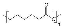
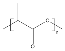
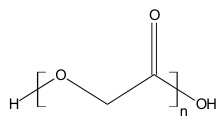
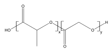
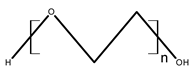
Several common themes emerge from this comparative analysis. First, degradation behaviour is a double-edged sword that defines clinical utility. PCL degrades too slowly (2–4 years) for soft tissue applications, PGA degrades too rapidly (4–6 months) for load-bearing bone scaffolds, PLGA offers tunability but at the cost of batch-to-batch sensitivity, PLA suffers from premature strength loss, and PEG is non-biodegradable at high molecular weights. This heterogeneity in degradation profiles—ranging from weeks to years—complicates material selection and underscores the critical need for predictive models that correlate degradation kinetics with tissue-specific regeneration timelines.
Second, acidic degradation products represent a potential biological barrier. Polyester-based polymers such as PLA, PGA and PLGA generate lactic and/or glycolic acid during hydrolysis. PCL, in contrast, degrades primarily to 6-hydroxycaproic acid and related oligomers rather. Creating local acidification that inhibits osteogenesis, promotes M1 macrophage polarisation, and triggers chronic inflammation. The acid environment has been shown to decrease cell viability, increase apoptosis, and upregulate pro-inflammatory cytokines. This challenge is particularly pronounced in thick scaffolds exceeding 500 µm, where passive diffusion leads to core necrosis and autocalytic acceleration of degradation. Neutralisation strategies—such as β-tricalcium phosphate or chitosan coatings—have been explored but often compromise mechanical strength or have short efficacy windows.
Third, cell adhesion and bioactivity remain persistent limitations. PEG is biologically inert and requires mandatory functionalisation with RGD peptides or fibronectin; PCL’s hydrophobicity leads to poor cell attachment; and PLA, PGA, and PLGA lack the cell-recognition motifs inherent to natural polymers. Surface functionalisation strategies—including peptide immobilisation, protein adsorption, and composite fabrication—can partially address these deficiencies, but they introduce additional manufacturing complexity, regulatory hurdles, and cost.
Fourth, manufacturing and regulatory challenges separate laboratory success from clinical adoption. While synthetic polymers offer theoretical batch-to-batch reproducibility, the reality is more nuanced: PLGA degradation is highly sensitive to subtle changes in molecular weight, end-group chemistry, and lactide/glycolide ratio; PLA crystallinity varies with stereoisomerism; and PGA is hygroscopic and requires special storage. The high cost of GMP-grade production, the lack of standardised characterisation protocols, and the absence of unified preclinical testing frameworks collectively impede translation. For PEG, pre-existing anti-PEG antibodies in the treatment-naive population introduce immunogenicity risks that complicate repeat-dose and chronic-exposure applications.
Fifth, thermomechanical instability during processing is a recurring issue. PLA undergoes depolymerisation during autoclaving and irradiation, requiring expensive aseptic manufacturing; PCL requires toxic solvents for scaffold fabrication; and the processing of PGA is complicated by its high crystallinity and hygroscopicity. These challenges have motivated the development of advanced fabrication techniques—melt electrowriting, 3D printing, electrospinning, and freeze-drying—that offer improved control over scaffold architecture while minimising thermal and solvent-induced damage.
These observations lead to a critical conclusion: the future of synthetic biopolymers in regenerative medicine lies not in identifying a single “superior” polymer, but in strategically combining synthetic precision with biological inspiration. This perspective aligns with the framework introduced in Section 2.1, where natural and synthetic polymers are viewed as complementary rather than oppositional. The development of hybrid and functionalised systems—combining synthetic polymers with bioactive natural components, growth factors, immunomodulatory agents, and inorganic reinforcements—represents the most promising pathway to overcome the limitations of individual materials. Advanced fabrication techniques that enable patient-specific design, such as 3D bioprinting and melt electrowriting, are increasingly capable of integrating multiple material classes into spatially organised, functionally graded constructs that recapitulate the complexity of native tissues.
In this context, the transition from passive synthetic scaffolds to actively programmable therapeutic platforms represents the next frontier. The convergence of synthetic polymer chemistry, biofunctionalisation, immunomodulation, and advanced biofabrication is progressively transforming synthetic polymers from inert structural substitutes into intelligent, responsive systems capable of directing tissue regeneration. The following section (Section 3) examines the critical role of immunomodulation in shaping the performance of both natural and synthetic biopolymers in vivo, setting the stage for the subsequent discussion of hybrid systems and the barriers to clinical translation (Section 5).
3. Immunomodulation by Biopolymers: From Passive Compatibility to Active Regeneration Control
For a long time, the biocompatibility of biopolymers was viewed primarily as the absence of toxicity and a minimal inflammatory response. However, contemporary research demonstrates that the immune response to an implanted material is not merely an obstacle to be minimised but a key regulatory mechanism that determines the success or failure of regeneration [239]. The immune system governs the processes of inflammation, proliferation, and tissue remodelling, with macrophages acting as central coordinators of these events [239,240]. Understanding how biopolymers interact with immune cells opens the possibility of transitioning from passive, “inert” materials to actively acting immunomodulatory platforms capable of directing regeneration along the desired path.
The key role in this process belongs to macrophages—cells that exhibit pronounced functional plasticity. Depending on the microenvironment, they can polarise towards a pro-inflammatory M1 phenotype or an anti-inflammatory M2 phenotype [240]. M1 macrophages, activated by bacterial lipopolysaccharides and interferon-gamma, secrete pro-inflammatory cytokines (TNF-α, IL-1β, IL-6), produce reactive oxygen species, and are essential for eliminating pathogens and debris in the early stages of healing. However, their prolonged activity leads to chronic inflammation and fibrosis. M2 macrophages, induced by interleukins IL-4 and IL-13, secrete anti-inflammatory cytokines (IL-10, TGF-β) and stimulate angiogenesis, extracellular matrix synthesis, and tissue regeneration. Successful healing requires a timely transition from an M1-dominant profile in the early stages to an M2-dominant profile during proliferation and remodelling [239,240]. The balance between these phenotypes is critical: premature M2 polarisation can lead to excessive fibrosis and capsule formation around the implant, while prolonged M1 activity impedes regeneration.
Immunomodulatory hydrogels can influence macrophage phenotype through both their polymeric network and their therapeutic payload, and distinguishing these contributions is important for rational design. A representative example is the injectable hydrogel HP-PVA@MH/Fe-Que (Figure 14), which consists of a phenylboronic acid-modified hyaluronic acid/PVA boronate ester network loaded with minocycline and Fe-quercetin nanoparticles [241]. In this system, macrophage polarization is typically assessed in two complementary in vitro models: LPS stimulation, which drives M1-associated polarization, and IL-4 stimulation, which drives M2-associated polarization. Under LPS stimulation, iNOS expression increases, and in the reported system, iNOS was significantly reduced in the Fe-Que-containing formulations (HP-PVA@Fe-Que and HP-PVA@MH/Fe-Que), whereas the unloaded network (HP-PVA) and the minocycline-only formulation (HP-PVA@MH) did not differ significantly from the LPS control. Under IL-4 stimulation, CD206 expression increases, and again, CD206 was significantly enhanced in the Fe-Que-containing formulations, while HP-PVA and HP-PVA@MH did not differ significantly from the IL-4 control [241]. These observations illustrate that the nanoparticle payload, rather than the polymeric network alone, can be the principal driver of marker changes, and that marker modulation in LPS- and IL-4-stimulated cultures reflects attenuation of M1-associated and enhancement of M2-associated responses in their respective contexts.
Figure 14.
HP-PVA@MH/Fe-Que hydrogels inhibit the polarization of macrophages toward the M1 phenotype while facilitating their polarization toward the M2 phenotype. (A) Schematic diagram showing the effect of the HP-PVA@MH/Fe-Que hydrogels on macrophage polarization. (B) Microscopic view of macrophages. IF images of (C) M1 (iNOS) and (E) M2 (CD206) macrophages maker. Flow cytometry analysis of (D) M1 (CD86) and (F) M2 (CD206) macrophage markers. Reprinted from Ref. [241].
Biopolymers can actively modulate macrophage polarisation through several mechanisms. First, physical properties of the material—stiffness, surface topography, porosity, and hydrophilicity—play a crucial role. Macrophages can “sense” the mechanical properties of the substrate through mechanosensory pathways, and these signals directly influence their phenotype. For example, substrates with high stiffness often promote M1 polarisation, while soft hydrogels can induce the M2 phenotype, which is particularly important for soft tissue regeneration [242].
Second, the chemical composition and degradation products of the polymer can act as signalling molecules. Acidic degradation products of polyesters (PLA, PGA, PLGA, PCL) can create local acidosis, which generally enhances the inflammatory response and M1 polarisation. Conversely, some natural polysaccharides, such as fucoidan, demonstrate intrinsic anti-inflammatory activity and promote M2 polarisation, making them valuable components of immunomodulatory hydrogels. In particular, hydrazone-crosslinked fucoidan hydrogels have been shown to provide an anti-inflammatory environment through the inherent bioactivity of fucoidan, offering tunable mechanical properties and injectability [243].
Third, emerging evidence highlights the role of biomaterial chirality in directing the immune response. A recent study demonstrated that levorotatory-chiral hydroxyapatite (L-CHAP) nanoplates coated onto polylactic acid electrospun membranes induce macrophage M2 polarisation by enhancing macrophage efferocytosis, thereby alleviating inflammatory damage to tendon stem cells through downregulated IL-17–NF-κB signalling and promoting tenogenic differentiation [244].
Of particular interest is the use of microorganisms for the synthesis of biopolymers with specified immunomodulatory properties. Microbial synthesis allows more precise control over molecular structure, molecular weight, and purity of the final product, as well as removal of immunogenic components at the purification stage [245]. Bacterial cellulose, for example, can enhance anti-inflammatory responses, unlike plant cellulose, which is more often associated with pro-inflammatory reactions. Genetic modification of microorganisms opens the possibility of “programming” the synthesis of biopolymers with specified immunological properties, which represents a promising direction for future research.
No less important, although less studied, is the adaptive immune response to biopolymers, mediated by T and B lymphocytes. Implanted materials can influence the recruitment and function of T-helper cells (Th1, Th2, Th17), which in turn determines the nature of interaction with macrophages and the final regeneration profile [246]. For example, decellularised matrices promote muscle healing through induction of Th2 immunity, while synthetic polyesters can have differentiated effects on the B-cell response and fibrosis. The physicochemical properties of the material—its shape, size, topography, stiffness, and degradation dynamics—directly correlate with the nature of the adaptive immune response, opening opportunities for targeted design of materials capable of inducing the desired immune profile [246].
In clinical practice, consideration of the immunomodulatory properties of biopolymers is becoming a critical factor for success. For bone tissue regeneration, materials are preferred that cause moderate M1 inflammation at the early stage (stimulating stem cell recruitment) and then provide a rapid transition to the M2 phenotype, promoting osteogenesis [240]. For wound healing, on the other hand, rapid suppression of inflammation and induction of M2 polarisation to stimulate angiogenesis and re-epithelialisation are more important [239]. It is important to note that individual characteristics of the patient’s immune status (genetic polymorphism, age, comorbidities) can significantly affect the response to the same material, making the development of personalised approaches to the selection of biopolymer constructs relevant [247].
Thus, immunomodulation by biopolymers is not just an additional characteristic of the material but a fundamental aspect of its clinical efficacy. The transition from the paradigm of “inert biocompatibility” to the paradigm of “active immunoregulation” requires the inclusion of immunological parameters in standard preclinical evaluation protocols (assessment of M1/M2 polarisation, cytokine profile, T-cell response) and the development of materials capable of directionally controlling the immune microenvironment at all stages of regeneration.
4. Advanced Approaches to Biopolymer Matrices with Programmable Drug Delivery Functions
Despite the diversity of biopolymers described above, their clinical application is often limited to the passive role of a scaffold. Contemporary research in regenerative medicine is increasingly shifting focus towards actively acting therapeutic systems capable not only of functioning as temporary scaffolds but also of controllably delivering bioactive molecules directly to the injury site. A key direction of this approach is the creation of biopolymer matrices with integrated micro- and nanocapsules for prolonged drug release, which allows maintaining a local therapeutic concentration over an extended period, minimising systemic side effects and the need for frequent drug administration. Such systems are finding ever-wider application in the treatment of acute and chronic wounds, diabetic ulcers, bone and muscle defects, as well as for targeted regeneration of nerve and cartilage tissue.
The fabrication of such programmable systems often relies on microgels—particles with diameters ranging from 100 to 1000 μm that are widely used in controlled drug release, tissue regeneration, and immuno-isolation of therapeutic cells, as their larger size helps reduce foreign-body responses and fibrosis compared to smaller particles. Key factors in engineering injectable microgels include the drug release rate and dosage. These parameters depend on the particle’s volumetric surface area, mesh size, and the interactions between cargo molecules and the gel matrix. Mesh size—the spacing between cross-linked polymer chains—governs liquid uptake and solute diffusion, and is influenced by the polymer chain’s molecular weight, the type of cross-linker, and the cross-linking density. For example, varying the ratios of stable and cleavable cross-linkers enables compositional control over payload release, as cleavable cross-linkers degrade more rapidly to increase the encapsulant release rate. Other strategies include modifying polymer chains with functional groups to adjust the hydrogel’s affinity for encapsulated drugs, further tuning release characteristics [248].
A particularly elegant strategy for creating programmable release profiles is the use of ion-exchange methods in alginate microgels. Researchers have developed a simple approach to prepare alginate microgels with tunable release profiles by initially cross-linking them with Ca2+ and subsequently transferring them to various salt solutions for ion exchange. By adjusting the type and concentration of cations used during this process, one can precisely control the cross-linking density and swollen mesh size of the microgels. This approach enables the development of microgels with tunable release performance that is compatible with payloads of varying molecular weights and charge characteristics, all without resorting to complex cross-linking chemistries and polymer designs. Notably, these alginate microgels with different release profiles can be further combined in modular delivery systems with programmable dosing control, shedding light on new biocompatible routes for engineering microcapsule-based therapeutics [249].
The versatility of this ion-exchange approach has been further extended to jet-printed microgel particles, where precise control over cation type and concentration enables systematic modulation of release rates for model compounds of varying molecular weights. As shown in Figure 15, mixing fast-releasing and slow-releasing microgel populations in defined ratios enables the programming of complex temporal release profiles for a single cargo, with mathematical modeling confirming the predictability of the release behaviour. Importantly, this study used model dyes and dextrans as cargo; it demonstrates release programmability and predictability, not therapeutic dosing or regenerative efficacy in a biological model. This modular strategy—where individual microgel populations serve as interchangeable “release modules”—therefore represents a methodological platform that could, in principle, be adapted for therapeutic payloads, but such adaptation and its biological validation remain to be demonstrated [249].
Figure 15.
Programmable drug release from modular microgel systems. The figure demonstrates a strategy for achieving tunable, stepwise release by mixing jet-printed alginate microgels with different cross-linking densities, prepared via an in situ ion-exchange method. (a) Schematic illustration of microgel mixing for programmed release. (b) Deconvoluted release profiles of model cargoes (Rhodamine B and Methylene Blue) from individual microgel populations. (c) Release kinetics of individual microgel populations with different cation compositions. (d–g) Mathematical modeling of release behaviour using modified Korsmeyer–Peppas and Weibull models. (h) Programmed stepwise release of Rhodamine B from a mixture of fast-releasing (IX-5Ca5Mg) and slow-releasing (IX-10Ca5Na) microgels, demonstrating temporal control over drug delivery. Reprinted from Ref. [249].
Hybrid microcapsule deposition onto fibrous scaffolds represents another promising approach that is gaining traction. Studies have demonstrated that polymer and hybrid microcapsules can be prepared and used as drug carriers, which are then deposited onto polymer microfiber scaffolds such as polycaprolactone (PCL), poly(3-hydroxybutyrate) (PHB), and PHB doped with conductive polyaniline (PHB-PANi). The immobilisation of microcapsules loaded with different bioactive molecules onto the scaffold surface enabled multimodal triggering by physical (ultrasound, laser radiation) and biological (enzymatic treatment) stimuli, providing controllable release of the cargo from scaffolds. Importantly, the immobilised microcapsules did not influence cell growth, viability, or proliferation on the scaffolds. In proof-of-concept studies, two bioactive compounds—the antibiotic ceftriaxone sodium and the osteogenic factor dexamethasone—were loaded into the microcapsules. The modified scaffolds demonstrated prolonged release of these agents, which provided an improved antimicrobial effect, as well as enhanced osteogenic differentiation and mineralisation compared to individual scaffolds soaked in drug solutions [250]. This approach provides a simple, convenient way to incorporate bioactive compounds onto polymer scaffolds, making these multimodal materials suitable for personalised drug therapy and bone tissue engineering.
In the area of wound healing, multilayer hydrogel dressings with integrated microspheres have been developed for the treatment of chronic diabetic wounds. A three-layer hydrogel dressing was designed in which the middle layer contains PLGA microspheres with mangiferin for prolonged release. The upper layer includes silver nanoparticles for rapid antibacterial action, while the lower layer made of gelatin methacrylate provides structural support. Such multilayer constructs ensure sustained release of mangiferin over 72 h, effectively suppress bacterial growth, and reduce levels of pro-inflammatory cytokines and reactive oxygen species [250]. The spatial separation of functional components in such layer-by-layer designs offers distinct advantages in controlling release kinetics and reducing interferences between antibacterial agents and therapeutic payloads. Similarly, in bone tissue engineering, modular systems of PLGA-based microcapsules have been developed to enable sequential release of three agents: minocycline (early antibacterial protection), VEGF-A (angiogenesis), and BMP-2 (osteogenesis). Varying the molecular weight of PLGA and the thickness of the capsule shell allows precise control over the release of each component, with the system demonstrating prolonged growth factor release over approximately 60 days in a critical-size bone defect model in mice. Such multi-stage approaches, mimicking the natural phases of bone healing, represent a promising strategy for creating multifunctional scaffolds [251].
For cartilage tissue engineering, biodegradable cellulose scaffolds with integrated PLGA microspheres containing kartogenin (KGN), an inducer of chondrogenesis, have been developed. Microspheres with high encapsulation efficiency provide prolonged KGN release, stimulating the migration of mesenchymal stem cells and their chondrogenic differentiation. In a rat cartilage defect model, such composite scaffolds with implanted stem cells demonstrated active secretion of extracellular matrix and improved cartilage tissue regeneration [252].
Emerging strategies also focus on hydrogel systems with programmable release profiles, in which the release kinetics are predefined at the design stage through material composition and network architecture. PEG-azlactone hydrogels illustrate this principle: cross-linking density and the PEG-diol to PEG-diamine ratio regulate network stability, hydrolytic degradation, and release kinetics, enabling phased, multicargo release of small molecules and antibodies from the same hydrogel. In such systems, the release programme is fixed prior to implantation and cannot be externally adjusted thereafter [253]. A distinct category comprises on-demand release systems, in which the release kinetics can be actively modulated after implantation by external or physiological triggers. pH- and magnetic-responsive alginate hydrogel cargoes exemplify this approach: magnetic actuation significantly increases the effective diffusion coefficient compared to pH-assisted release alone, providing spatiotemporal control that is not achievable with a predefined profile alone. The essential distinction is that programmable systems encode the release schedule in the material itself, whereas on-demand systems retain the capacity for external or trigger-mediated adjustment of dosing and timing [254].
Thus, matrices based on both natural and synthetic biopolymers with integrated microcapsules and microparticles represent an effective approach for prolonged delivery of therapeutic agents in the treatment of wounds, diabetic ulcers, bone and cartilage defects. The combination of the biocompatibility of natural polymers with the controlled release kinetics of synthetic analogues opens up opportunities for creating next-generation hybrid systems adapted to specific clinical applications. Future directions include the integration of immune-modulating components, the development of magnetic or ultrasound-triggered release systems, and the use of advanced biofabrication techniques such as 3D bioprinting and electrospinning to create spatially organised, multifunctional constructs.
To better position the described systems within the broader landscape of programmable delivery, Table 3 compares representative microcapsule- and microgel-based platforms according to cargo type, release duration, triggering mechanism, and level of biological validation. This comparison highlights a clear progression: from methodological studies using model dyes to demonstrate release programmability [249], through systems validated in vitro with therapeutic payloads [250], to multi-stage platforms confirmed in vivo in bone and cartilage defect models [251,252].
Table 3.
Comparison of representative programmable delivery systems by payload, release kinetics, triggering strategy, and level of biological evidence (from model compounds to in vivo validation).
5. Barriers to Clinical Translation of Biopolymer Materials
The diversity of laboratory developments described in previous sections creates an impression of vast opportunities for biopolymers in regenerative medicine. However, a critical analysis of the literature and clinical trial data reveals that the overwhelming majority of these systems never reach the stage of widespread clinical application. This gap between fundamental research and practical medicine is systemic in nature and is explained by a complex of interconnected barriers that are often underestimated at early stages of development. Technological and biological barriers form the first group of challenges. Even with successful demonstration of biocompatibility in standard in vitro tests, material behaviour in a living organism often proves unpredictable. Classical two-dimensional cell cultures do not reflect the complex three-dimensional microenvironment, the dynamics of blood supply, and the constant renewal of the extracellular matrix, leading to systematic discrepancies between laboratory results and data obtained from animal models. For instance, hydrogels based on polylactide-co-glycolide-polyethylene glycol-polylactide-co-glycolide copolymers, which had already undergone clinical trials, demonstrated pronounced cytotoxicity in standard 2D tests, attributed to the accumulation of acidic degradation products in a closed culture system; however, this effect was not reproduced in vivo due to the constant washout of metabolites by blood flow [255]. Similarly, for a number of polyurethane implants, it was shown that accelerated hydrolytic degradation tests could not predict their behaviour in the clinic due to the complex morphology of the material and the influence of enzymatic processes, leading to unexpectedly early or, conversely, delayed loss of mechanical integrity [256]. Some studies report a complete absence of biodegradation of polyglycolic acid polymers in certain anatomical zones, resulting in prolonged foreign body retention, fibrous capsule formation, and chronic inflammation, rather than the expected replacement by new tissue [257]. The intrinsic trade-off between material stability and biological performance—where chemical crosslinking improves mechanical properties but often reduces bioactivity and introduces cytotoxicity—remains a fundamental challenge that complicates the design of clinically viable materials [41].
Manufacturing reproducibility and scalability represent a second critical hurdle. The behaviour of many biopolymers, particularly those derived from natural sources, is highly dependent on source material, processing history, and formulation context. For marine-derived polymers such as chitosan, alginate, and carrageenan, the major translational barrier lies not in the lack of potential, but in the difficulty of achieving the required degree of control, reproducibility, and manufacturability for reliable use as pharmaceutical excipients. The presence of these materials in proof-of-concept studies does not mean they are ready for routine application, as their functional benefits can be compromised by variability in source, structure, and processing history [258].
Economic and production constraints often become an insurmountable obstacle to commercialisation. Preclinical studies on animal models require colossal financial investments: according to a systematic review on polymeric scaffolds for nerve regeneration, the total cost of all preclinical work in this area was estimated at over $61 million USD, with no direct correlation found between the volume of funding and the success of material transition to clinical practice [259]. Many academic groups develop materials without considering the complexity and cost of scaling production to GMP standards, making the product economically unfeasible, even subject to regulatory approval. This problem is particularly acute for natural polymers (collagen, chitosan, hyaluronic acid), where raw material variability and multi-stage purification lead to the final medical device costing tens of times more than synthetic analogues with comparable functional characteristics. For synthetic polymers, even though theoretical reproducibility is higher, the problem of controlling molecular weight distribution and monomer ratio in copolymers (e.g., in PLGA) persists, making each large batch unique and requiring additional validation for each clinical application, significantly increasing the production cycle cost [211].
Regulatory and strategic barriers constitute the third group of challenges. Researchers in academic environments traditionally focus on proving that a new material “works” in laboratory conditions—that is, on its effectiveness—whereas regulatory bodies (FDA, EMA) primarily require evidence of safety, standardisability, and long-term predictability of behaviour. This fundamental divergence in priorities leads to many promising developments not even passing the initial stages of regulatory analysis.
Furthermore, for the vast majority of biopolymer systems, there are no unified protocols for preclinical assessment, validated methods for testing biodegradation in vivo, or standardised criteria for comparing results from different research groups, which complicates objective evaluation and comparison of materials, as well as creating difficulties in preparing regulatory dossiers [260]. Three-dimensional printed constructs present a particular problem: despite their obvious potential for personalised regeneration, they lack clear regulatory protocols for clinical application due to the complexity of quality control at each printing stage and the absence of standardised approaches to sterilisation and validation [261]. No less important is the absence of industry standards for long-term monitoring of patients after implantation of biopolymer devices, making it impossible to accumulate statistically significant data on rare but serious complications (e.g., delayed hypersensitivity reactions or late fibrosis) that may manifest only years after the intervention.
Thus, the clinical success of a biopolymer product is determined not so much by its biological activity or mechanical strength as by its ability to achieve sustainable, scalable, and economically viable production while strictly complying with regulatory requirements and having validated assessment methods at all stages of the life cycle. Overcoming the described barriers requires not just the improvement of the materials themselves, but a fundamental shift in the thinking of researchers—a transition from optimising biological effects to prioritising translational design, which involves close collaboration with industry, regulators, and clinicians at the earliest stages of development. Only such a comprehensive approach can reduce the gap between promising laboratory developments and real medical products capable of changing clinical practice.
6. Conclusions
The conducted review demonstrates that modern biopolymers for regenerative medicine have reached a high level of technological sophistication: numerous modifications of natural and synthetic polymers have been developed, hybrid systems with controlled release of growth factors have been created, and methods of 3D printing and microfluidic encapsulation have been mastered. However, a critical analysis of the literature reveals a fundamental mismatch between the extensive growth in the number of laboratory developments and the limited number of truly functioning clinical products. This mismatch is not so much technological as methodological and institutional in nature.
Each of the considered classes of biopolymers has fundamental disadvantages limiting their universal application. Natural polymers provide a biomimetic environment and cellular recognition but suffer from raw material variability, scaling complexity, and, in some cases, immunogenicity or excessively rapid degradation. Synthetic polymers offer control over mechanical properties and resorption kinetics, but their acidic degradation products create local acidosis that inhibits regeneration and provokes chronic inflammation. Hybrid systems combining both classes are considered the most promising; however, their complexity creates additional regulatory and production barriers, and evidence of their superiority over monocomponent systems in direct comparative in vivo studies remains fragmentary.
The key conclusion of the review is that further progress is determined not so much by the creation of new materials as by rethinking the criteria for their evaluation and implementing a translation-oriented approach at all stages of development. This requires standardisation of preclinical assessment protocols with mandatory inclusion of immunological parameters (M1/M2 macrophage polarisation, cytokine profile), development of technologically mature and economically justified production processes from the very beginning of research work, as well as the creation of systems with programmable sequential release of therapeutic agents corresponding to the natural phases of healing.
The immune response to biopolymers deserves particular attention, which has long been considered only an obstacle. Modern research shows that targeted immunomodulation through control of the physical properties of the material and the inclusion of immunoactive components can become a key mechanism for improving regeneration. However, clinical data on the long-term immunological consequences of biopolymer implantation are practically absent, and the high cost of development and production of such products, especially of natural origin, makes them inaccessible for widespread use in healthcare systems with limited resources.
With regard to the advantage of capsule integration over direct loading into the scaffold, comparative data point to several key differences. Direct physical immobilisation of growth factors within the scaffold bulk is typically accompanied by rapid burst release and poorly controlled kinetics, whereas incorporation into micro- or nanoparticles provides more precise control over the release rate and, where necessary, enables sequential release of multiple agents. In addition, encapsulation allows functional components to be spatially separated: for example, in multilayer constructs, an antibacterial agent can be localised in one layer and an osteoinductive factor in another, which reduces their mutual interference and enables independent release profiles to be programmed. Direct soaking of the scaffold in a drug solution, as a rule, does not provide such modularity and often leads to uncontrolled initial burst followed by rapid depletion of the local dose.
Thus, capsule integration is justified in three situations: protection of a labile therapeutic agent during fabrication and in vivo is required; release kinetics unattainable by passive diffusion from the scaffold are needed; or spatiotemporal separation of multiple agents is desired. In cases where simple local delivery of a stable low-molecular-weight compound over a short period is sufficient, direct loading into the scaffold may remain a simpler and more cost-effective alternative.
The translational barriers identified in this review—the gap between in vitro and in vivo performance, manufacturing reproducibility challenges, high GMP costs, and regulatory complexity—represent systemic obstacles that cannot be overcome by improving material properties alone. The clinical success of a biopolymer product is determined not so much by its biological activity or mechanical strength as by its ability to achieve sustainable, scalable, and economically viable production while strictly complying with regulatory requirements. Overcoming these barriers requires a fundamental shift in the thinking of researchers—a transition from optimising biological effects to prioritising translational design, which involves close collaboration with industry, regulators, and clinicians at the earliest stages of development.
Several strategic directions emerge for the future of biopolymer research:
- First, the development of hybrid and functionalised systems that strategically combine the strengths of natural and synthetic polymers while mitigating their individual weaknesses. The integration of immunomodulatory components, the engineering of controlled degradation profiles, and the advancement of scalable biofabrication technologies are all essential steps toward realising the full therapeutic potential of biopolymers.
- Second, the transition from passive scaffolds to actively programmable therapeutic platforms. The convergence of advanced fabrication techniques—3D bioprinting, electrospinning, and microfluidics—with composite modifications and stimuli-responsive release mechanisms offers promising avenues to overcome current limitations and realise the clinical potential of biopolymers in personalised and functional tissue regeneration.
- Third, the integration of immunological parameters into standard preclinical evaluation protocols. The transition from the paradigm of “inert biocompatibility” to the paradigm of “active immunoregulation” requires mandatory assessment of M1/M2 polarisation, cytokine profiles, and T-cell responses, as well as the development of materials capable of directionally controlling the immune microenvironment at all stages of regeneration.
- Fourth, the adoption of translational design principles from the earliest stages of material development. This includes consideration of manufacturing scalability, cost-effectiveness, regulatory requirements, and long-term safety validation—not as afterthoughts but as integral components of the design process. Early engagement with regulatory agencies and industry partners is essential to ensure compliance, mitigate development delays, and facilitate successful market translation.
Thus, the future of biopolymers in regenerative medicine will be determined not by technological breakthroughs in synthesis but by a systemic approach to translation that unites fundamental science, engineering solutions, regulatory science, clinical practice, and economic evaluation. Only with such a comprehensive view can promising laboratory developments become real medical products that change clinical practice. The challenge ahead is not to create more materials, but to create the right materials—with the right properties, at the right cost, with the right regulatory pathway, for the right patients.
Author Contributions
Conceptualization, A.I.L.; Writing—Original Draft Preparation, A.I.L. and Y.V.L.; Writing—Review & Editing, R.V.G. and V.A.A.; Project Administration, T.V.K. All authors have read and agreed to the published version of the manuscript.
Funding
This research was funded by the Ministry of Science and Higher Education of the Russian Federation within the framework of a state assignment on the topic “Chemical, biological and nanostructural modification of functional materials as a control strategy, research of properties and approaches to the practical application of microbial biofilms” (FEWG-2024-0004) and the Tula State University development program for 2025–2036 under the “Priority-2030” strategic academic leadership program (Agreement No. 075-15-2026-058 dated 3 April 2026; IGK 000000Ю807526QXN0002).
Institutional Review Board Statement
Not applicable.
Informed Consent Statement
Not applicable.
Data Availability Statement
No new data were created or analyzed in this study. Data sharing is not applicable to this article.
Conflicts of Interest
The authors declare no conflicts of interest.
References
- Sharma, K.; Porat, Z.; Gedanken, A. Designing Natural Polymer-Based Capsules and Spheres for Biomedical Applications—A Review. Polymers 2021, 13, 4307. [Google Scholar] [CrossRef] [Scilit] [PubMed]
- Grasman, J.M.; Zayas, M.J.; Page, R.L.; Pins, G.D. Biomimetic Scaffolds for Regeneration of Volumetric Muscle Loss in Skeletal Muscle Injuries. Acta Biomater. 2015, 25, 2–15. [Google Scholar] [CrossRef] [Scilit] [PubMed]
- Edo, G.I.; Ndudi, W.; Ali, A.M.; Yousif, E.; Jikah, A.N.; Isoje, E.F.; Igbuku, U.A.; Mafe, A.N.; Opiti, R.A.; Madueke, C.J.; et al. Biopolymers: An Inclusive Review. Hybrid Adv. 2025, 9, 100418. [Google Scholar] [CrossRef] [Scilit]
- Grabowski, M.; Gmyrek, D.; Żurawska, M.; Trusek, A. Biopolymers in Biotechnology and Tissue Engineering: A Comprehensive Review. Macromol 2025, 5, 34. [Google Scholar] [CrossRef] [Scilit]
- Das, A.; Ringu, T.; Ghosh, S.; Pramanik, N. A Comprehensive Review on Recent Advances in Preparation, Physicochemical Characterization, and Bioengineering Applications of Biopolymers. Polym. Bull. 2023, 80, 7247–7312. [Google Scholar] [CrossRef] [Scilit] [PubMed]
- Prete, S.; Dattilo, M.; Patitucci, F.; Pezzi, G.; Parisi, O.I.; Puoci, F. Natural and Synthetic Polymeric Biomaterials for Application in Wound Management. J. Funct. Biomater. 2023, 14, 455. [Google Scholar] [CrossRef] [Scilit] [PubMed]
- Abolhasani, S.; Ahmadi, Y.; Rostami, Y.; Baravar, E.; Fattahi, D. Biomaterials in Tissue Repair and Regeneration: Key Insights from Extracellular Matrix Biology. Front. Med. Technol. 2025, 7, 1565810. [Google Scholar] [CrossRef] [Scilit] [PubMed]
- Hu, T.; Fang, J.; Shen, Y.; Li, M.; Wang, B.; Xu, Z.; Hu, W. Advances of Naturally Derived Biomedical Polymers in Tissue Engineering. Front. Chem. 2024, 12, 1469183. [Google Scholar] [CrossRef] [Scilit] [PubMed]
- Moholkar, D.N.; Sadalage, P.S.; Peixoto, D.; Paiva-Santos, A.C.; Pawar, K.D. Recent Advances in Biopolymer-Based Formulations for Wound Healing Applications. Eur. Polym. J. 2021, 160, 110784. [Google Scholar] [CrossRef] [Scilit]
- Reddy, M.S.B.; Ponnamma, D.; Choudhary, R.; Sadasivuni, K.K. A Comparative Review of Natural and Synthetic Biopolymer Composite Scaffolds. Polymers 2021, 13, 1105. [Google Scholar] [CrossRef] [Scilit] [PubMed]
- Gazeau, B.; Zaman, A.; Pacini, H.; Khan, M.A. Clarifying the Taxonomy of Plastics and Bioplastics: Toward a ‘Zero-Trace Plastic’ (ZTP) Material Framework. Sustainability 2025, 17, 6763. [Google Scholar] [CrossRef] [Scilit]
- Bealer, E.J.; Onissema-Karimu, S.; Rivera-Galletti, A.; Francis, M.; Wilkowski, J.; Salas-de La Cruz, D.; Hu, X. Protein–Polysaccharide Composite Materials: Fabrication and Applications. Polymers 2020, 12, 464. [Google Scholar] [CrossRef] [Scilit] [PubMed]
- Chen, W.-H.; Chen, Q.-W.; Chen, Q.; Cui, C.; Duan, S.; Kang, Y.; Liu, Y.; Liu, Y.; Muhammad, W.; Shao, S.; et al. Biomedical Polymers: Synthesis, Properties, and Applications. Sci. China Chem. 2022, 65, 1010–1075. [Google Scholar] [CrossRef] [Scilit] [PubMed]
- Satchanska, G.; Davidova, S.; Petrov, P.D. Natural and Synthetic Polymers for Biomedical and Environmental Applications. Polymers 2024, 16, 1159. [Google Scholar] [CrossRef] [Scilit] [PubMed]
- Cao, W.; Li, X.; Zuo, X.; Gao, C. Migration of Endothelial Cells into Photo-Responsive Hydrogels with Tunable Modulus under the Presence of pro-Inflammatory Macrophages. Regen. Biomater. 2019, 6, 259–267. [Google Scholar] [CrossRef] [Scilit] [PubMed]
- Nurilmala, M.; Suryamarevita, H.; Husein Hizbullah, H.; Jacoeb, A.M.; Ochiai, Y. Fish Skin as a Biomaterial for Halal Collagen and Gelatin. Saudi J. Biol. Sci. 2022, 29, 1100–1110. [Google Scholar] [CrossRef] [Scilit] [PubMed]
- Chevallay, B.; Herbage, D. Collagen-Based Biomaterials as 3D Scaffold for Cell Cultures: Applications for Tissue Engineering and Gene Therapy. Med. Biol. Eng. Comput. 2000, 38, 211–218. [Google Scholar] [CrossRef] [Scilit] [PubMed]
- Matinong, A.M.E.; Chisti, Y.; Pickering, K.L.; Haverkamp, R.G. Collagen Extraction from Animal Skin. Biology 2022, 11, 905. [Google Scholar] [CrossRef] [Scilit] [PubMed]
- Gómez-Guillén, M.C.; Giménez, B.; López-Caballero, M.E.; Montero, M.P. Functional and Bioactive Properties of Collagen and Gelatin from Alternative Sources: A Review. Food Hydrocoll. 2011, 25, 1813–1827. [Google Scholar] [CrossRef] [Scilit]
- Chan, L.; Li, A.; Schlesinger, T. Permanent Fillers—What Is Their Role. Dermatol. Rev. 2023, 4, 121–126. [Google Scholar] [CrossRef] [Scilit]
- Xu, H.; Liu, Y.; Qiu, L.; Mangue, A.L.M.M.; Zhang, J.; Wei, B.; Zhu, L.; Xu, C.; Shirshin, E.A.; Wang, H. Preparation and Application of Collagen-Based Hemostatic Materials: A Review. Collagen Leather 2025, 7, 11. [Google Scholar] [CrossRef] [Scilit]
- Chattopadhyay, S.; Raines, R.T. Collagen-based Biomaterials for Wound Healing. Biopolymers 2014, 101, 821–833. [Google Scholar] [CrossRef] [Scilit] [PubMed]
- Dong, C.; Lv, Y. Application of Collagen Scaffold in Tissue Engineering: Recent Advances and New Perspectives. Polymers 2016, 8, 42. [Google Scholar] [CrossRef] [Scilit] [PubMed]
- Chen, H.; Xu, Y.; Zhou, Y.; Xu, Z.; Guo, J.; Mu, C.; Li, D.; Ge, L. Near-Infrared Light-Activated Collagen-Based Hydrogel with Oriented Microchannel Structure Promoting Fibroblast Infiltration and Inflammatory Resolution. Collagen Leather 2026, 8, 35. [Google Scholar] [CrossRef] [Scilit]
- Law, J.X.; Liau, L.L.; Saim, A.; Yang, Y.; Idrus, R. Electrospun Collagen Nanofibers and Their Applications in Skin Tissue Engineering. Tissue Eng. Regen. Med. 2017, 14, 699–718. [Google Scholar] [CrossRef] [Scilit] [PubMed]
- Ivaskiene, T.; Viskelis, J.; Streimikyte, P.; Savickaitė, M.; Mobasheri, A.; Kaspute, G. Collagen Supplementation and Regenerative Health: Advances in Biomarker Detection and Smart Material Integration. Front. Nutr. 2025, 12, 1716166. [Google Scholar] [CrossRef] [Scilit] [PubMed]
- Jinno, C.; Morimoto, N.; Ito, R.; Sakamoto, M.; Ogino, S.; Taira, T.; Suzuki, S. A Comparison of Conventional Collagen Sponge and Collagen-Gelatin Sponge in Wound Healing. BioMed Res. Int. 2016, 2016, 1–8. [Google Scholar] [CrossRef] [Scilit] [PubMed]
- Almazrooa, S.A.; Noonan, V.; Woo, S.-B. Resorbable Collagen Membranes: Histopathologic Features. Oral Surg. Oral Med. Oral Pathol. Oral Radiol. 2014, 118, 236–240. [Google Scholar] [CrossRef] [Scilit] [PubMed]
- Puoci, F.; Piangiolino, C.; Givigliano, F.; Parisi, O.I.; Cassano, R.; Trombino, S.; Curcio, M.; Iemma, F.; Cirillo, G.; Spizzirri, U.G.; et al. Ciprofloxacin-Collagen Conjugate in the Wound Healing Treatment. J. Funct. Biomater. 2012, 3, 361–371. [Google Scholar] [CrossRef] [Scilit] [PubMed]
- Gomathi, K.; Gopinath, D.; Rafiuddin Ahmed, M.; Jayakumar, R. Quercetin Incorporated Collagen Matrices for Dermal Wound Healing Processes in Rat. Biomaterials 2003, 24, 2767–2772. [Google Scholar] [CrossRef] [Scilit] [PubMed]
- Gopinath, D.; Ahmed, M.R.; Gomathi, K.; Chitra, K.; Sehgal, P.K.; Jayakumar, R. Dermal Wound Healing Processes with Curcumin Incorporated Collagen Films. Biomaterials 2004, 25, 1911–1917. [Google Scholar] [CrossRef] [Scilit] [PubMed]
- Sowbhagya, R.; Muktha, H.; Ramakrishnaiah, T.N.; Surendra, A.S.; Sushma, S.M.; Tejaswini, C.; Roopini, K.; Rajashekara, S. Collagen as the Extracellular Matrix Biomaterials in the Arena of Medical Sciences. Tissue Cell 2024, 90, 102497. [Google Scholar] [CrossRef] [Scilit] [PubMed]
- Zhu, L.; Yu, Z.-L.; Li, S.; Xu, C.-Z.; Hou, Y.-J.; Liao, L.-X.; Xu, Y.-L.; Zhang, J.-T.; Wei, B.-M.; Wen, W.; et al. Recent Advances on Collagen Biomaterial: From Extraction, Cross-Linking to Tissue Regeneration. Polym. Rev. 2024, 64, 1031–1059. [Google Scholar] [CrossRef] [Scilit]
- Kido, H.; Gabbai-Armelin, P.; Magri, A.; Fernandes, K.; Cruz, M.; Santana, A.; Caliari, H.; Parisi, J.; Avanzi, I.; Daguano, J.; et al. Bioglass/Collagen Scaffolds Combined with Bone Marrow Stromal Cells on Bone Healing in an Experimental Model in Cranial Defects in Rats. J. Biomater. Appl. 2023, 37, 1632–1644. [Google Scholar] [CrossRef] [Scilit] [PubMed]
- Yang, W.; Ni, W.; Yu, C.; Gu, T.; Ye, L.; Sun, R.; Ying, X.; Yik, J.H.N.; Haudenschild, D.R.; Yao, S.; et al. Biomimetic Bone-Like Composite Hydrogel Scaffolds Composed of Collagen Fibrils and Natural Hydroxyapatite for Promoting Bone Repair. ACS Biomater. Sci. Eng. 2024, 10, 2385–2397. [Google Scholar] [CrossRef] [Scilit] [PubMed]
- Zhang, D.; Wu, X.; Chen, J.; Lin, K. The Development of Collagen Based Composite Scaffolds for Bone Regeneration. Bioact. Mater. 2018, 3, 129–138. [Google Scholar] [CrossRef] [Scilit] [PubMed]
- Veronesi, F.; Torricelli, P.; Martini, L.; Tschon, M.; Giavaresi, G.; Bellini, D.; Casagranda, V.; Alemani, F.; Fini, M. An Alternative Ex Vivo Method to Evaluate the Osseointegration of Ti–6Al–4V Alloy Also Combined with Collagen. Biomed. Mater. 2021, 16, 025007. [Google Scholar] [CrossRef] [Scilit] [PubMed]
- Brooker, C.; Tronci, G. Effect of Mammalian Tissue Source on the Molecular and Macroscopic Characteristics of UV-Cured Type I Collagen Hydrogel Networks. Prosthesis 2022, 4, 1–14. [Google Scholar] [CrossRef] [Scilit]
- Oikonomou, G.; Vareltzis, P. Cold-Water versus Warm-Water Fish Collagen (Type I): Comparative Functional Properties and Implications for Food Industry Applications. Appl. Food Res. 2026, 6, 102068. [Google Scholar] [CrossRef] [Scilit]
- Zhai, X.; Geng, X.; Li, W.; Cui, H.; Wang, Y.; Qin, S. Comprehensive Review on Application Progress of Marine Collagen Cross-Linking Modification in Bone Repairs. Mar. Drugs 2025, 23, 151. [Google Scholar] [CrossRef] [Scilit] [PubMed]
- Cheng, C.-C.; Chang, F.-C.; Kao, W.-Y.; Hwang, S.-M.; Liao, L.-C.; Chang, Y.-J.; Liang, M.-C.; Chen, J.-K.; Lee, D.-J. Highly Efficient Drug Delivery Systems Based on Functional Supramolecular Polymers: In Vitro Evaluation. Acta Biomater. 2016, 33, 194–202. [Google Scholar] [CrossRef] [Scilit] [PubMed]
- Chen, C.; Ding, W.; Zhang, H.; Zhang, L.; Huang, Y.; Fan, M.; Yang, J.; Sun, D. Bacterial Cellulose-Based Biomaterials: From Fabrication to Application. Carbohydr. Polym. 2022, 278, 118995. [Google Scholar] [CrossRef] [Scilit] [PubMed]
- Mahima; Rani, V.; Vishal, K.; Doharey, P.K.; Tanveer, A.; Yadav, P.K.; Yadav, D. Fungal Cellulases: A Comprehensive Review on Production, Innovations, and Applications. Arch. Microbiol. 2026, 208, 291. [Google Scholar] [CrossRef] [Scilit] [PubMed]
- Li, R.; Xing, R.; Li, H.; Ren, J.; Chen, L.; Yu, Z.; Zhang, H.; Wu, W.; Li, C.; Zhu, L.; et al. From Terrestrial Plants to Marine Macroalgae: A Comprehensive Review of Cell Wall Component-Properties, Extraction, Modification, and Application of Algal Cellulose. J. Agric. Food Chem. 2025, 73, 23759–23782. [Google Scholar] [CrossRef] [Scilit] [PubMed]
- Bae, S.O.; Shoda, M. Production of Bacterial Cellulose by Acetobacter Xylinum BPR2001 Using Molasses Medium in a Jar Fermentor. Appl. Microbiol. Biotechnol. 2005, 67, 45–51. [Google Scholar] [CrossRef] [Scilit] [PubMed]
- Cacicedo, M.L.; Islan, G.A.; León, I.E.; Álvarez, V.A.; Chourpa, I.; Allard-Vannier, E.; García-Aranda, N.; Díaz-Riascos, Z.V.; Fernández, Y.; Schwartz, S.; et al. Bacterial Cellulose Hydrogel Loaded with Lipid Nanoparticles for Localized Cancer Treatment. Colloids Surf. B Biointerfaces 2018, 170, 596–608. [Google Scholar] [CrossRef] [Scilit] [PubMed]
- Miao, J.; Pangule, R.C.; Paskaleva, E.E.; Hwang, E.E.; Kane, R.S.; Linhardt, R.J.; Dordick, J.S. Lysostaphin-Functionalized Cellulose Fibers with Antistaphylococcal Activity for Wound Healing Applications. Biomaterials 2011, 32, 9557–9567. [Google Scholar] [CrossRef] [Scilit] [PubMed]
- Maneerung, T.; Tokura, S.; Rujiravanit, R. Impregnation of Silver Nanoparticles into Bacterial Cellulose for Antimicrobial Wound Dressing. Carbohydr. Polym. 2008, 72, 43–51. [Google Scholar] [CrossRef] [Scilit]
- Wen, X.; Zheng, Y.; Wu, J.; Yue, L.; Wang, C.; Luan, J.; Wu, Z.; Wang, K. In Vitro and in Vivo Investigation of Bacterial Cellulose Dressing Containing Uniform Silver Sulfadiazine Nanoparticles for Burn Wound Healing. Prog. Nat. Sci. Mater. Int. 2015, 25, 197–203. [Google Scholar] [CrossRef] [Scilit]
- Mohamad, N.; Mohd Amin, M.C.I.; Pandey, M.; Ahmad, N.; Rajab, N.F. Bacterial Cellulose/Acrylic Acid Hydrogel Synthesized via Electron Beam Irradiation: Accelerated Burn Wound Healing in an Animal Model. Carbohydr. Polym. 2014, 114, 312–320. [Google Scholar] [CrossRef] [Scilit] [PubMed]
- Dang, X.; Fu, Y.; Wang, X. Versatile Biomass-Based Injectable Photothermal Hydrogel for Integrated Regenerative Wound Healing and Skin Bioelectronics. Adv. Funct. Mater. 2024, 34, 2405745. [Google Scholar] [CrossRef] [Scilit]
- Shin, J.; Lee, D.Y.; Kim, B.; Yoon, J.I. Effect of Polyethylene Glycol Molecular Weight on Cell Growth Behavior of Polyvinyl Alcohol/Carboxymethyl Cellulose/Polyethylene Glycol Hydrogel. J. Appl. Polym. Sci. 2020, 137, 49568. [Google Scholar] [CrossRef] [Scilit]
- Diaz-Gomez, L.; Gonzalez-Prada, I.; Millan, R.; Da Silva-Candal, A.; Bugallo-Casal, A.; Campos, F.; Concheiro, A.; Alvarez-Lorenzo, C. 3D Printed Carboxymethyl Cellulose Scaffolds for Autologous Growth Factors Delivery in Wound Healing. Carbohydr. Polym. 2022, 278, 118924. [Google Scholar] [CrossRef] [Scilit] [PubMed]
- Wang, H.; He, P.; Tang, G.; Qi, F.; Lu, X.; Zhang, M.; Zheng, R.; Li, X.; Shi, Z.; Zhang, Y.; et al. Skin Repairing Procedure Inspired Polypyrrole/Bacterial Cellulose/Platelet Rich Plasma Composite Hydrogel as Diabetes Wound Dressing. J. Bioresour. Bioprod. 2026, 11, 100224. [Google Scholar] [CrossRef] [Scilit]
- Ao, H.; Wang, D.; Ma, L.; Wang, M.; Zhang, D.; Xun, X.; Wu, X.; Zhang, M.; Zong, J. Copper-doped Borosilicate Bioactive Glass Coating Modified Bacterial Cellulose Functional Dressing for Diabetic Ulcer. BMEMat 2026, 4, e70042. [Google Scholar] [CrossRef] [Scilit]
- Luong, A.D.; Maruthapandi, M.; Luong, J.H.T. Bacterial Cellulose Scaffolds for Advanced Wound Care: Immunomodulation, Mixed Biofilms, and Smart Regenerative Dressings. Macromol 2026, 6, 23. [Google Scholar] [CrossRef] [Scilit]
- Wang, X.; Song, X.; Lu, J.; Chen, Y.; Guo, Y. A Conductive Anisotropic Hydrogel Based on Carboxymethyl Cellulose and Polyvinyl Alcohol for Intelligent Ligament Scaffold. Polymer 2026, 344, 129477. [Google Scholar] [CrossRef] [Scilit]
- Nashchekina, Y.; Chabina, A.; Konson, V.; Nashchekin, A.; Mikhailova, N. Carboxymethyl Cellulose in Composite Collagen Gel Improves Mesenchymal Stromal Cell Proliferation and Migration. Int. J. Biol. Macromol. 2025, 319, 145418. [Google Scholar] [CrossRef] [Scilit] [PubMed]
- Yadav, V.; Paniliatis, B.J.; Shi, H.; Lee, K.; Cebe, P.; Kaplan, D.L. Novel In Vivo-Degradable Cellulose-Chitin Copolymer from Metabolically Engineered Gluconacetobacter xylinus. Appl. Environ. Microbiol. 2010, 76, 6257–6265. [Google Scholar] [CrossRef] [Scilit] [PubMed]
- Bastard, C.; Schulte, J.C.-T.; Asaduzzaman, M.; Hohn, C.; Kittel, Y.; De Laporte, L.; Gebhardt, R. Casein Microparticles Filled with Cellulase to Enzymatically Degrade Nanocellulose for Cell Growth. Biomater. Sci. 2025, 13, 2435–2443. [Google Scholar] [CrossRef] [Scilit] [PubMed]
- Sannino, A.; Demitri, C.; Madaghiele, M. Biodegradable Cellulose-Based Hydrogels: Design and Applications. Materials 2009, 2, 353–373. [Google Scholar] [CrossRef] [Scilit]
- Zhang, M.; Chen, S.; Sheng, N.; Wang, B.; Yao, J.; Wu, Z.; Wang, H. A Strategy of Tailoring Polymorphs and Nanostructures to Construct Self-Reinforced Nonswelling High-Strength Bacterial Cellulose Hydrogels. Nanoscale 2019, 11, 15347–15358. [Google Scholar] [CrossRef] [Scilit] [PubMed]
- Gayathri, V.; Lobo, N.P.; Vikash, V.L.; Kamini, N.R.; Samanta, D. Functionalization of Bacterial Cellulose and Related Surfaces Using a Facile Coupling Reaction by Thermoresponsive Catalyst. ACS Biomater. Sci. Eng. 2023, 9, 625–641. [Google Scholar] [CrossRef] [Scilit] [PubMed]
- Bulkina, A.; Prilepskii, A. Bacterial Cellulose: Is It Really a Promising Biomedical Material? Carbohydr. Polym. 2025, 357, 123427. [Google Scholar] [CrossRef] [Scilit] [PubMed]
- Krontiras, P.; Gatenholm, P.; Hägg, D.A. Adipogenic Differentiation of Stem Cells in Three-Dimensional Porous Bacterial Nanocellulose Scaffolds: Adipogenic Differentiation of Stem Cells In 3d Porous Scaffolds. J. Biomed. Mater. Res. B Appl. Biomater. 2015, 103, 195–203. [Google Scholar] [CrossRef] [Scilit] [PubMed]
- Martínez Ávila, H.; Schwarz, S.; Feldmann, E.-M.; Mantas, A.; Von Bomhard, A.; Gatenholm, P.; Rotter, N. Biocompatibility Evaluation of Densified Bacterial Nanocellulose Hydrogel as an Implant Material for Auricular Cartilage Regeneration. Appl. Microbiol. Biotechnol. 2014, 98, 7423–7435. [Google Scholar] [CrossRef] [Scilit] [PubMed]
- Gabriel Kou, S.; Peters, L.M.; Mucalo, M.R. Chitosan: A Review of Sources and Preparation Methods. Int. J. Biol. Macromol. 2021, 169, 85–94. [Google Scholar] [CrossRef] [Scilit] [PubMed]
- Zhang, W.; Geng, X.; Qin, S.; Xie, Z.; Li, W.; Li, J. Research Progress and Application of Chitosan Dressings in Hemostasis: A Review. Int. J. Biol. Macromol. 2024, 282, 136421. [Google Scholar] [CrossRef] [Scilit] [PubMed]
- Su, J.; Liu, C.; Sun, A.; Yan, J.; Sang, F.; Xin, Y.; Zhao, Y.; Wang, S.; Dang, Q. Hemostatic and Antimicrobial Properties of Chitosan-Based Wound Healing Dressings: A Review. Int. J. Biol. Macromol. 2025, 306, 141570. [Google Scholar] [CrossRef] [Scilit] [PubMed]
- Wang, T.; Zheng, Y.; Shen, Y.; Shi, Y.; Li, F.; Su, C.; Zhao, L. Chitosan Nanoparticles Loaded Hydrogels Promote Skin Wound Healing through the Modulation of Reactive Oxygen Species. Artif. Cells Nanomed. Biotechnol. 2018, 46, 138–149. [Google Scholar] [CrossRef] [Scilit] [PubMed]
- Gaweł, J.; Milan, J.; Żebrowski, J.; Płoch, D.; Stefaniuk, I.; Kus-Liśkiewicz, M. Biomaterial Composed of Chitosan, Riboflavin, and Hydroxyapatite for Bone Tissue Regeneration. Sci. Rep. 2023, 13, 17004. [Google Scholar] [CrossRef] [Scilit] [PubMed]
- Malette, W.G.; Quigley, H.J.; Gaines, R.D.; Johnson, N.D.; Rainer, W.G. Chitosan: A New Hemostatic. Ann. Thorac. Surg. 1983, 36, 55–58. [Google Scholar] [CrossRef] [Scilit] [PubMed]
- Zhang, S.; Lei, X.; Lv, Y.; Wang, L.; Wang, L.-N. Recent Advances of Chitosan as a Hemostatic Material: Hemostatic Mechanism, Material Design and Prospective Application. Carbohydr. Polym. 2024, 327, 121673. [Google Scholar] [CrossRef] [Scilit] [PubMed]
- Eickelpasch, K.; Lemke, P.; Sreekumar, S.; Chilukoti, N.; Moerschbacher, B.M.; Richter, C. A Bioactivity Matrix for Antimicrobial Activities of Chitosans: A Review. Int. J. Biol. Macromol. 2025, 299, 140740. [Google Scholar] [CrossRef] [Scilit] [PubMed]
- Salim, A.; Sathishkumar, P. Therapeutic Efficacy of Chitosan-Based Hybrid Nanomaterials to Treat Microbial Biofilms and Their Infections—A Review. Int. J. Biol. Macromol. 2024, 283, 137850. [Google Scholar] [CrossRef] [Scilit] [PubMed]
- Ahmed, S.; Ikram, S. Chitosan Based Scaffolds and Their Applications in Wound Healing. Achiev. Life Sci. 2016, 10, 27–37. [Google Scholar] [CrossRef] [Scilit]
- Rodríguez-Acosta, H.; Tapia-Rivera, J.M.; Guerrero-Guzmán, A.; Hernández-Elizarraráz, E.; Hernández-Díaz, J.A.; Garza-García, J.J.O.; Pérez-Ramírez, P.E.; Velasco-Ramírez, S.F.; Ramírez-Anguiano, A.C.; Velázquez-Juárez, G.; et al. Chronic Wound Healing by Controlled Release of Chitosan Hydrogels Loaded with Silver Nanoparticles and Calendula Extract. J. Tissue Viability 2022, 31, 173–179. [Google Scholar] [CrossRef] [Scilit] [PubMed]
- Kim, K.; Ryu, J.H.; Koh, M.-Y.; Yun, S.P.; Kim, S.; Park, J.P.; Jung, C.-W.; Lee, M.S.; Seo, H.-I.; Kim, J.H.; et al. Coagulopathy-Independent, Bioinspired Hemostatic Materials: A Full Research Story from Preclinical Models to a Human Clinical Trial. Sci. Adv. 2021, 7, eabc9992. [Google Scholar] [CrossRef] [Scilit] [PubMed]
- Wu, Z.; Zhou, W.; Deng, W.; Xu, C.; Cai, Y.; Wang, X. Antibacterial and Hemostatic Thiol-Modified Chitosan-Immobilized AgNPs Composite Sponges. ACS Appl. Mater. Interfaces 2020, 12, 20307–20320. [Google Scholar] [CrossRef] [Scilit] [PubMed]
- Wang, Y.-W.; Liu, C.-C.; Cherng, J.-H.; Lin, C.-S.; Chang, S.-J.; Hong, Z.-J.; Liu, C.-C.; Chiu, Y.-K.; Hsu, S.-D.; Chang, H. Biological Effects of Chitosan-Based Dressing on Hemostasis Mechanism. Polymers 2019, 11, 1906. [Google Scholar] [CrossRef] [Scilit] [PubMed]
- Lin, Y.-C.; Ramanathan, S.; Wang, H.-Y.; Lin, Y.-C.; Liu, W.-C.; Jones, J.R.; Cho, N.-J.; Hu, C.-C.; Chung, R.-J. Engineered Bioactive Glass-Chitosan Hybrid for Dual Tissue and Bone Regeneration Multifunctional Healing. Biomater. Adv. 2025, 176, 214340. [Google Scholar] [CrossRef] [Scilit] [PubMed]
- Safarzadeh, S.; Naghib, S.M.; Takdehghan, G.; Forooshani, R.S.; Roozbahani, M.H.; Sharifianjazi, F.; Tavamaishvili, K. Multimodal Chitosan-Based Materials for Combination Immunotherapy in Cancers: Structural Engineering, Immune Regulatory Mechanisms and Synergistic Therapeutic Applications. Carbohydr. Polym. 2025, 369, 124245. [Google Scholar] [CrossRef] [Scilit] [PubMed]
- Tamer, T.M.; Hassan, M.A.; Omer, A.M.; Valachová, K.; Eldin, M.S.M.; Collins, M.N.; Šoltés, L. Antibacterial and Antioxidative Activity of O-Amine Functionalized Chitosan. Carbohydr. Polym. 2017, 169, 441–450. [Google Scholar] [CrossRef] [Scilit] [PubMed]
- Yu, J.; Hu, N.; Hou, L.; Hang, F.; Li, K.; Xie, C. Effect of Deacetylation of Chitosan on the Physicochemical, Antioxidant and Antibacterial Properties Activities of Chitosan–Mannose Derivatives. J. Sci. Food Agric. 2023, 103, 6394–6405. [Google Scholar] [CrossRef] [Scilit] [PubMed]
- Krishna, V.S.; Subashini, V.; Hariharan, A.; Chidambaram, D.; Raaju, A.; Gopichandran, N.; Nanthanalaxmi, M.P.; Lekhavadhani, S.; Shanmugavadivu, A.; Selvamurugan, N. Role of Crosslinkers in Advancing Chitosan-Based Biocomposite Scaffolds for Bone Tissue Engineering: A Comprehensive Review. Int. J. Biol. Macromol. 2024, 283, 137625. [Google Scholar] [CrossRef] [Scilit] [PubMed]
- Reay, S.L.; Jackson, E.L.; Salthouse, D.; Ferreira, A.M.; Hilkens, C.M.U.; Novakovic, K. Effective Endotoxin Removal from Chitosan That Preserves Chemical Structure and Improves Compatibility with Immune Cells. Polymers 2023, 15, 1592. [Google Scholar] [CrossRef] [Scilit] [PubMed]
- Duymaz, D.; Kebabci, A.O.; Kizilel, S. Harnessing the Immunomodulatory Potential of Chitosan and Its Derivatives for Advanced Biomedical Applications. Int. J. Biol. Macromol. 2025, 307, 142055. [Google Scholar] [CrossRef] [Scilit] [PubMed]
- Laurent, T.C.; Laurent, U.B.; Fraser, J.R. Functions of Hyaluronan. Ann. Rheum. Dis. 1995, 54, 429–432. [Google Scholar] [CrossRef] [Scilit] [PubMed]
- Necas, J.; Bartosikova, L.; Brauner, P.; Kolar, J. Hyaluronic Acid (Hyaluronan): A Review. Veterinární Medicína 2008, 53, 397–411. [Google Scholar] [CrossRef] [Scilit]
- Dechert, T.A.; Ducale, A.E.; Ward, S.I.; Yager, D.R. Hyaluronan in Human Acute and Chronic Dermal Wounds. Wound Repair Regen. 2006, 14, 252–258. [Google Scholar] [CrossRef] [Scilit] [PubMed]
- Wang, L.; Zhou, F.; Xie, W. Advances in Hyaluronic Acid-Based Biomaterials: Applications in Cancer Therapy, Wound Healing, and Disease Management. J. Mater. Sci. Mater. Med. 2025, 36, 91. [Google Scholar] [CrossRef] [Scilit] [PubMed]
- Brown, M.R.W.; Foster, J.H.S.; Clamp, J.R. Composition of Pseudomonas aeruginosa Slime. Biochem. J. 1969, 112, 521–525. [Google Scholar] [CrossRef] [Scilit] [PubMed][Green Version]
- Rahemtulla, F.; Løvtrup, S. The Comparative Biochemistry of Invertebrate Mucopolysaccharides—II. Nematoda; Annelida. Comp. Biochem. Physiol. Part B Comp. Biochem. 1974, 49, 639–646. [Google Scholar] [CrossRef] [Scilit] [PubMed]
- Yamada, K. Effects of Novel (Streptomyces) Hyaluronidase Digestion upon Some Mucosaccharide Stainings of the Cartilages and Aortas in the Rabbit and Rat. Histochemie 1971, 27, 277–289. [Google Scholar] [CrossRef] [Scilit] [PubMed]
- Fleischmajer, R.; Perlish, J.S.; Gaisin, A. Comparative Study of Dermal Glycosaminoglycans. J. Investig. Dermatol. 1973, 61, 1–6. [Google Scholar] [CrossRef] [Scilit] [PubMed]
- Singh, M.; Chandrasekaran, E.V.; Cherian, R.; Bachhawat, B.K. Isolation and characterization of glycosaminoglycans in brain of different species. J. Neurochem. 1969, 16, 1157–1162. [Google Scholar] [CrossRef] [Scilit] [PubMed]
- Park, H.-J.; Jin, Y.; Shin, J.; Yang, K.; Lee, C.; Yang, H.S.; Cho, S.-W. Catechol-Functionalized Hyaluronic Acid Hydrogels Enhance Angiogenesis and Osteogenesis of Human Adipose-Derived Stem Cells in Critical Tissue Defects. Biomacromolecules 2016, 17, 1939–1948. [Google Scholar] [CrossRef] [Scilit] [PubMed]
- Subramaniam, S.; Fang, Y.-H.; Sivasubramanian, S.; Lin, F.-H.; Lin, C. Hydroxyapatite-Calcium Sulfate-Hyaluronic Acid Composite Encapsulated with Collagenase as Bone Substitute for Alveolar Bone Regeneration. Biomaterials 2016, 74, 99–108. [Google Scholar] [CrossRef] [Scilit] [PubMed]
- Hennebelle, L.; Reis, C.; Relvas, M.; Salazar, F.; Costa, R.; Cabral, C.; Vinhas, A.S. Hyaluronic Acid as an Adjunct in Bone Regeneration—A Systematic Review. Biomedicines 2026, 14, 1514. [Google Scholar] [CrossRef] [Scilit] [PubMed]
- Zhu, M.; Lin, S.; Sun, Y.; Feng, Q.; Li, G.; Bian, L. Hydrogels Functionalized with N-Cadherin Mimetic Peptide Enhance Osteogenesis of hMSCs by Emulating the Osteogenic Niche. Biomaterials 2016, 77, 44–52. [Google Scholar] [CrossRef] [Scilit] [PubMed]
- Wang, M.; Deng, Z.; Guo, Y.; Xu, P. Designing Functional Hyaluronic Acid-Based Hydrogels for Cartilage Tissue Engineering. Mater. Today Bio 2022, 17, 100495. [Google Scholar] [CrossRef] [Scilit] [PubMed]
- Bowman, S.; Awad, M.E.; Hamrick, M.W.; Hunter, M.; Fulzele, S. Recent Advances in Hyaluronic Acid Based Therapy for Osteoarthritis. Clin. Transl. Med. 2018, 7, e6. [Google Scholar] [CrossRef] [Scilit] [PubMed]
- Deng, S.; Cao, H.; Lu, Y.; Shi, W.; Chen, M.; Cui, X.; Liang, J.; Fan, Y.; Wang, Q.; Zhang, X. Injectable dECM-Enhanced Hyaluronic Microgels with Spatiotemporal Release of Cartilage-Specific Molecules to Improve Osteoarthritic Chondrocyte’s Function. Collagen Leather 2024, 6, 14. [Google Scholar] [CrossRef] [Scilit]
- Perng, C.-K.; Wang, Y.-J.; Tsi, C.-H.; Ma, H. In Vivo Angiogenesis Effect of Porous Collagen Scaffold with Hyaluronic Acid Oligosaccharides. J. Surg. Res. 2011, 168, 9–15. [Google Scholar] [CrossRef] [Scilit] [PubMed]
- Dennaoui, H.; Chouery, E.; Rammal, H.; Abdel-Razzak, Z.; Harmouch, C. Chitosan/Hyaluronic Acid Multilayer Films Are Biocompatible Substrate for Wharton’s Jelly Derived Stem Cells. Stem Cell Investig. 2018, 5, 47. [Google Scholar] [CrossRef] [Scilit] [PubMed]
- Kang, L.; Jia, W.; Li, M.; Wang, Q.; Wang, C.; Liu, Y.; Wang, X.; Jin, L.; Jiang, J.; Gu, G.; et al. Hyaluronic Acid Oligosaccharide-Modified Collagen Nanofibers as Vascular Tissue-Engineered Scaffold for Promoting Endothelial Cell Proliferation. Carbohydr. Polym. 2019, 223, 115106. [Google Scholar] [CrossRef] [Scilit] [PubMed]
- Makvandi, P.; Ali, G.W.; Della Sala, F.; Abdel-Fattah, W.I.; Borzacchiello, A. Biosynthesis and Characterization of Antibacterial Thermosensitive Hydrogels Based on Corn Silk Extract, Hyaluronic Acid and Nanosilver for Potential Wound Healing. Carbohydr. Polym. 2019, 223, 115023. [Google Scholar] [CrossRef] [Scilit] [PubMed]
- Oates, T.C.; Boyd, J.; Dolan, L.; De Kergariou, C.; Zhu, J.; Toye, A.; Perriman, A.W.; Boussahel, A. The Immobilization of Hyaluronic Acid in 3D Hydrogel Scaffolds Modulates Macrophage Polarization. Adv. Biol. 2025, 9, e00682. [Google Scholar] [CrossRef] [Scilit] [PubMed]
- Wollina, U.; Goldman, A. Spontaneous and Induced Degradation of Dermal Fillers: A Review. J. Cutan. Aesthetic Surg. 2023, 17, 273–281. [Google Scholar] [CrossRef] [Scilit] [PubMed]
- Wolf, K.J.; Kumar, S. Hyaluronic Acid: Incorporating the Bio into the Material. ACS Biomater. Sci. Eng. 2019, 5, 3753–3765. [Google Scholar] [CrossRef] [Scilit] [PubMed]
- Sahoo, D.R.; Biswal, T. Alginate and Its Application to Tissue Engineering. SN Appl. Sci. 2021, 3, 30. [Google Scholar] [CrossRef] [Scilit]
- Ertesvåg, H. Alginate-Modifying Enzymes: Biological Roles and Biotechnological Uses. Front. Microbiol. 2015, 6, 523. [Google Scholar] [CrossRef] [Scilit] [PubMed]
- Fernando, I.P.S.; Lee, W.; Han, E.J.; Ahn, G. Alginate-Based Nanomaterials: Fabrication Techniques, Properties, and Applications. Chem. Eng. J. 2020, 391, 123823. [Google Scholar] [CrossRef] [Scilit]
- Foudah, A.I.; Ahmad, A.; Alam, A. Next-Generation Green Marine Pharmaceutical Excipients: Integrating Multifunctionality and Precision Drug Delivery. Green Chem. Lett. Rev. 2026, 19, 2652200. [Google Scholar] [CrossRef] [Scilit]
- Iravani, S.; Varma, R.S. Alginate-Based Micro- and Nanosystems for Targeted Cancer Therapy. Mar. Drugs 2022, 20, 598. [Google Scholar] [CrossRef] [Scilit] [PubMed]
- Venkatesan, J.; Bhatnagar, I.; Manivasagan, P.; Kang, K.-H.; Kim, S.-K. Alginate Composites for Bone Tissue Engineering: A Review. Int. J. Biol. Macromol. 2015, 72, 269–281. [Google Scholar] [CrossRef] [Scilit] [PubMed]
- Farshidfar, N.; Iravani, S.; Varma, R.S. Alginate-Based Biomaterials in Tissue Engineering and Regenerative Medicine. Mar. Drugs 2023, 21, 189. [Google Scholar] [CrossRef] [Scilit] [PubMed]
- Sun, J.; Tan, H. Alginate-Based Biomaterials for Regenerative Medicine Applications. Materials 2013, 6, 1285–1309. [Google Scholar] [CrossRef] [Scilit] [PubMed]
- Neves, M.I.; Moroni, L.; Barrias, C.C. Modulating Alginate Hydrogels for Improved Biological Performance as Cellular 3D Microenvironments. Front. Bioeng. Biotechnol. 2020, 8, 665. [Google Scholar] [CrossRef] [Scilit] [PubMed]
- Rezvanian, M.; Ng, S.-F.; Alavi, T.; Ahmad, W. In-Vivo Evaluation of Alginate-Pectin Hydrogel Film Loaded with Simvastatin for Diabetic Wound Healing in Streptozotocin-Induced Diabetic Rats. Int. J. Biol. Macromol. 2021, 171, 308–319. [Google Scholar] [CrossRef] [Scilit] [PubMed]
- Suamte, L.; Tirkey, A.; Babu, P.J. Design of 3D Smart Scaffolds Using Natural, Synthetic and Hybrid Derived Polymers for Skin Regenerative Applications. Smart Mater. Med. 2023, 4, 243–256. [Google Scholar] [CrossRef] [Scilit]
- Mobaraki, M.; Bizari, D.; Soltani, M.; Khshmohabat, H.; Raahemifar, K.; Akbarzade Amirdehi, M. The Effects of Curcumin Nanoparticles Incorporated into Collagen-Alginate Scaffold on Wound Healing of Skin Tissue in Trauma Patients. Polymers 2021, 13, 4291. [Google Scholar] [CrossRef] [Scilit] [PubMed]
- Sukhodub, L.; Kumeda, M.; Sukhodub, L.; Bielai, V.; Lyndin, M. Metal Ions Doping Effect on the Physicochemical, Antimicrobial, and Wound Healing Profiles of Alginate-Based Composite. Carbohydr. Polym. 2023, 304, 120486. [Google Scholar] [CrossRef] [Scilit] [PubMed]
- Liu, F.; Jiang, J.; Zhe, M.; Yu, P.; Xing, F.; Xiang, Z. Alginate-Based 3D Bioprinting Strategies for Structure–Function Integrated Tissue Regeneration. J. Mater. Chem. B 2025, 13, 12765–12811. [Google Scholar] [CrossRef] [Scilit] [PubMed]
- Rastin, H.; Abousalman-Rezvani, Z. 3D Bioprinting of Alginate for Cancer Therapy: A Review. Int. J. Biol. Macromol. 2025, 320, 145494. [Google Scholar] [CrossRef] [Scilit] [PubMed]
- Kolambkar, Y.M.; Dupont, K.M.; Boerckel, J.D.; Huebsch, N.; Mooney, D.J.; Hutmacher, D.W.; Guldberg, R.E. An Alginate-Based Hybrid System for Growth Factor Delivery in the Functional Repair of Large Bone Defects. Biomaterials 2011, 32, 65–74. [Google Scholar] [CrossRef] [Scilit] [PubMed]
- Singh, A.K.; Pramanik, K. Fabrication and Investigation of Physicochemical and Biological Properties of 3D Printed Sodium Alginate-chitosan Blend Polyelectrolyte Complex Scaffold for Bone Tissue Engineering Application. J. Appl. Polym. Sci. 2023, 140, e53642. [Google Scholar] [CrossRef] [Scilit]
- Karvinen, J.; Joki, T.; Ylä-Outinen, L.; Koivisto, J.T.; Narkilahti, S.; Kellomäki, M. Soft Hydrazone Crosslinked Hyaluronan- and Alginate-Based Hydrogels as 3D Supportive Matrices for Human Pluripotent Stem Cell-Derived Neuronal Cells. React. Funct. Polym. 2018, 124, 29–39. [Google Scholar] [CrossRef] [Scilit]
- Prang, P.; Müller, R.; Eljaouhari, A.; Heckmann, K.; Kunz, W.; Weber, T.; Faber, C.; Vroemen, M.; Bogdahn, U.; Weidner, N. The Promotion of Oriented Axonal Regrowth in the Injured Spinal Cord by Alginate-Based Anisotropic Capillary Hydrogels. Biomaterials 2006, 27, 3560–3569. [Google Scholar] [CrossRef] [Scilit] [PubMed]
- Rahimivand, M.; Tafvizi, F.; Noorbazargan, H. Synthesis and Characterization of Alginate Nanocarrier Encapsulating Artemisia Ciniformis Extract and Evaluation of the Cytotoxicity and Apoptosis Induction in AGS Cell Line. Int. J. Biol. Macromol. 2020, 158, 338–357. [Google Scholar] [CrossRef] [Scilit] [PubMed]
- Gonçalves, M.; Figueira, P.; Maciel, D.; Rodrigues, J.; Qu, X.; Liu, C.; Tomás, H.; Li, Y. pH-Sensitive Laponite®/Doxorubicin/Alginate Nanohybrids with Improved Anticancer Efficacy. Acta Biomater. 2014, 10, 300–307. [Google Scholar] [CrossRef] [Scilit] [PubMed]
- Song, W.; Su, X.; Gregory, D.A.; Li, W.; Cai, Z.; Zhao, X. Magnetic Alginate/Chitosan Nanoparticles for Targeted Delivery of Curcumin into Human Breast Cancer Cells. Nanomaterials 2018, 8, 907. [Google Scholar] [CrossRef] [Scilit] [PubMed]
- Desai, T.; Rawat, E.; Haswani, N.G.; Wahab, S.; Alshehri, S.A.; Kapoor, D.U. Alginate-Driven Nanotherapeutics in Liver Cancer: Design, Applications, and Future Perspectives. Int. J. Pharm. 2026, 696, 126844. [Google Scholar] [CrossRef] [Scilit] [PubMed]
- Ahmad Raus, R.; Wan Nawawi, W.M.F.; Nasaruddin, R.R. Alginate and Alginate Composites for Biomedical Applications. Asian J. Pharm. Sci. 2021, 16, 280–306. [Google Scholar] [CrossRef] [Scilit] [PubMed]
- Liang, X.; Chen, S.; Liang, Y.; Wang, M.; Wang, Q.; Chen, D.; Ma, X.; Ding, H.; Zhong, H.-J. Alginate-Based Hydrogels: Recent Progress in Preparation, Property Tuning, and Multifunctional Applications. Gels 2026, 12, 182. [Google Scholar] [CrossRef] [Scilit] [PubMed]
- Andersen, T.; Auk-Emblem, P.; Dornish, M. 3D Cell Culture in Alginate Hydrogels. Microarrays 2015, 4, 133–161. [Google Scholar] [CrossRef] [Scilit] [PubMed]
- Varaprasad, K.; Jayaramudu, T. A Review of Smart Alginate-Based Biomaterials: Innovations and Challenges in Tissue Engineering and Regenerative Medicine. Int. J. Biol. Macromol. 2026, 337, 149518. [Google Scholar] [CrossRef] [Scilit] [PubMed]
- S, S.; R G, A.P.; Bajaj, G.; John, A.E.; Chandran, S.; Kumar, V.V.; Ramakrishna, S. A Review on the Recent Applications of Synthetic Biopolymers in 3D Printing for Biomedical Applications. J. Mater. Sci. Mater. Med. 2023, 34, 62. [Google Scholar] [CrossRef] [Scilit] [PubMed]
- Williams, D.F. Biocompatibility Pathways and Mechanisms for Bioactive Materials: The Bioactivity Zone. Bioact. Mater. 2022, 10, 306–322. [Google Scholar] [CrossRef] [Scilit] [PubMed]
- Manoukian, O.S.; Sardashti, N.; Stedman, T.; Gailiunas, K.; Ojha, A.; Penalosa, A.; Mancuso, C.; Hobert, M.; Kumbar, S.G. Biomaterials for Tissue Engineering and Regenerative Medicine. In Encyclopedia of Biomedical Engineering; Elsevier: Amsterdam, The Netherlands, 2019; pp. 462–482. ISBN 978-0-12-805144-3. [Google Scholar]
- Chen, T.; Cai, T.; Jin, Q.; Ji, J. Design and Fabrication of Functional Polycaprolactone. e-Polymers 2015, 15, 3–13. [Google Scholar] [CrossRef] [Scilit]
- Malikmammadov, E.; Tanir, T.E.; Kiziltay, A.; Hasirci, V.; Hasirci, N. PCL and PCL-Based Materials in Biomedical Applications. J. Biomater. Sci. Polym. Ed. 2018, 29, 863–893. [Google Scholar] [CrossRef] [Scilit] [PubMed]
- Mohammed, R.; Balakrishnan, R. Advancements and Applications of Polycaprolactone and Its Composites in Biomedical Engineering: A Comprehensive Review. In Biocompatible Nanocomposites; Balakrishnan, R., Mohammed, R., Thomas, N.G., Dalvi, Y.B., Eds.; Advanced Structured Materials; Springer Nature Singapore: Singapore, 2025; Volume 239, pp. 117–148. ISBN 978-981-96-7449-7. [Google Scholar]
- Middleton, J.C.; Tipton, A.J. Synthetic Biodegradable Polymers as Orthopedic Devices. Biomaterials 2000, 21, 2335–2346. [Google Scholar] [CrossRef] [Scilit] [PubMed]
- Wang, Y.; Zhang, W.; Karamergenova, A.; Lin, L. Fabrication and Application of Polycaprolactone-Based Composite Scaffolds in Tissue Engineering: A Review. Mater. Today Commun. 2025, 49, 113821. [Google Scholar] [CrossRef] [Scilit]
- Bezwada, R.S.; Jamiolkowski, D.D.; Lee, I.-Y.; Agarwal, V.; Persivale, J.; Trenka-Benthin, S.; Erneta, M.; Suryadevara, J.; Yang, A.; Liu, S. Monocryl® Suture, a New Ultra-Pliable Absorbable Monofilament Suture. Biomaterials 1995, 16, 1141–1148. [Google Scholar] [CrossRef] [Scilit] [PubMed]
- Sinha, V.R.; Bansal, K.; Kaushik, R.; Kumria, R.; Trehan, A. Poly-ϵ-Caprolactone Microspheres and Nanospheres: An Overview. Int. J. Pharm. 2004, 278, 1–23. [Google Scholar] [CrossRef] [PubMed]
- Kurakula, M.; Rao, G.S.N.K.; Yadav, K.S. Fabrication and Characterization of Polycaprolactone-Based Green Materials for Drug Delivery. In Applications of Advanced Green Materials; Elsevier: Amsterdam, The Netherlands, 2021; pp. 395–423. ISBN 978-0-12-820484-9. [Google Scholar]
- Dordunoo, S.K.; Jackson, J.K.; Arsenault, L.A.; Oktaba, A.M.C.; Hunter, W.L.; Burt, H.M. Taxol Encapsulation in Poly(ɛ-Caprolactone) Microspheres. Cancer Chemother. Pharmacol. 1995, 36, 279–282. [Google Scholar] [CrossRef] [PubMed]
- Carreras, N.; Acuña, V.; Martí, M.; Lis, M.J. Drug Release System of Ibuprofen in PCL-Microspheres. Colloid Polym. Sci. 2013, 291, 157–165. [Google Scholar] [CrossRef] [Scilit]
- Liao, N.; Unnithan, A.R.; Joshi, M.K.; Tiwari, A.P.; Hong, S.T.; Park, C.-H.; Kim, C.S. Electrospun Bioactive Poly (ɛ-Caprolactone)–Cellulose Acetate–Dextran Antibacterial Composite Mats for Wound Dressing Applications. Colloids Surf. Physicochem. Eng. Asp. 2015, 469, 194–201. [Google Scholar] [CrossRef] [Scilit]
- Bakhsheshi-Rad, H.R.; Ismail, A.F.; Aziz, M.; Akbari, M.; Hadisi, Z.; Daroonparvar, M.; Chen, X.B. Antibacterial Activity and in Vivo Wound Healing Evaluation of Polycaprolactone-Gelatin Methacryloyl-Cephalexin Electrospun Nanofibrous. Mater. Lett. 2019, 256, 126618. [Google Scholar] [CrossRef] [Scilit]
- Oh, G.-W.; Ko, S.-C.; Je, J.-Y.; Kim, Y.-M.; Oh, J.; Jung, W.-K. Fabrication, Characterization and Determination of Biological Activities of Poly(ε-Caprolactone)/Chitosan-Caffeic Acid Composite Fibrous Mat for Wound Dressing Application. Int. J. Biol. Macromol. 2016, 93, 1549–1558. [Google Scholar] [CrossRef] [Scilit] [PubMed]
- Das, T.; Panda, S.; Dey, R.; Das, M. Bioglass and Zinc Incorporated PCL Based Composite as Biodegradable Implants for Bone Fixation. Biomed. Phys. Eng. Express 2026, 12, 035069. [Google Scholar] [CrossRef] [Scilit] [PubMed]
- Weng, X.Z.; Guan, X.; Lei, Y.X.; Lin, Y.X.; Wang, W.X.; Zhang, L.; Su, J.J.; Lai, Y.Z. Dual Effects of Biomimetic Wave-Microgrooved Polycaprolactone Membranes: Enhancement of Osteogenesis and Suppression of Osteoclastogenesis. Dent. Mater. J. 2026, 45, 325–337. [Google Scholar] [CrossRef] [Scilit] [PubMed]
- Liu, Y.; Von Witzleben, M.; Duin, S.; Bernhardt, A.; Gelinsky, M. Topology-Optimized Melt-Electrowritten PCL Patch for Abdominal Wall Reconstruction. Bioact. Mater. 2026, 55, 529–545. [Google Scholar] [CrossRef] [Scilit] [PubMed]
- Wang, B.; Zhang, S.; Liu, S.; Tang, Y.; Chen, F.; Zhu, L.-F.; Chang, M.-W. Gradient-Magnetized Conductive Nerve Conduits Enable Enhanced Wireless Electrical Stimulation for Peripheral Nerve Regeneration. Chem. Eng. J. 2026, 547, 181409. [Google Scholar] [CrossRef] [Scilit]
- Ramaraju, H.; Verga, A.S.; Steedley, B.J.; Kowblansky, A.P.; Green, G.E.; Hollister, S.J. Investigation of the Biodegradation Kinetics and Associated Mechanical Properties of 3D-Printed Polycaprolactone during Long-Term Preclinical Testing. Biomaterials 2025, 321, 123257. [Google Scholar] [CrossRef] [Scilit] [PubMed]
- Zuo, Y.; Yang, B.; Zou, Q.; Li, L.; Li, J.; Man, Y.; Li, Y. Effect of Ultrafine Poly(ε-Caprolactone) Fibers on Calcium Phosphate Cement: In Vitro Degradation and in Vivo Regeneration. Int. J. Nanomed. 2016, 11, 163–177. [Google Scholar] [CrossRef] [Scilit] [PubMed]
- Corvaglia, I.; Fiorilli, S.; Vitale-Brovarone, C. Electrospun Matrices for Sustained Drug Release Made by a PCL-Chitosan Blend Shell and a PVA Core. Mater. Lett. 2024, 371, 136894. [Google Scholar] [CrossRef] [Scilit]
- Li, G.; Zhao, M.; Xu, F.; Yang, B.; Li, X.; Meng, X.; Teng, L.; Sun, F.; Li, Y. Synthesis and Biological Application of Polylactic Acid. Molecules 2020, 25, 5023. [Google Scholar] [CrossRef] [Scilit] [PubMed]
- Yang, Z.; Yin, G.; Sun, S.; Xu, P. Medical Applications and Prospects of Polylactic Acid Materials. iScience 2024, 27, 111512. [Google Scholar] [CrossRef] [Scilit] [PubMed]
- Jumadi, J.; Harun, W.S.W.; Kadirgama, K.; Samylingam, L.; Aslfattahi, N.; S., C.K.; Kok, C.K.; Kiai, M.S. A Comparative Review of Polylactic Acid and Poly(Lactic-Co-Glycolic Acid) Biomaterials: Optimizing Drug Delivery. BioNanoScience 2026, 16, 19. [Google Scholar] [CrossRef] [Scilit]
- Da Silva, D.; Kaduri, M.; Poley, M.; Adir, O.; Krinsky, N.; Shainsky-Roitman, J.; Schroeder, A. Biocompatibility, Biodegradation and Excretion of Polylactic Acid (PLA) in Medical Implants and Theranostic Systems. Chem. Eng. J. 2018, 340, 9–14. [Google Scholar] [CrossRef] [Scilit] [PubMed]
- Mainil-Varlet, P.; Curtis, R.; Gogolewski, S. Effect Ofin Vivo Andin Vitro Degradation on Molecular and Mechanical Properties of Various Low-Molecular-Weight Polylactides. J. Biomed. Mater. Res. 1997, 36, 360–380. [Google Scholar] [CrossRef] [Scilit]
- Saini, P.; Arora, M.; Kumar, M.N.V.R. Poly(Lactic Acid) Blends in Biomedical Applications. Adv. Drug Deliv. Rev. 2016, 107, 47–59. [Google Scholar] [CrossRef] [Scilit] [PubMed]
- Nagiah, N.; Murdock, C.J.; Bhattacharjee, M.; Nair, L.; Laurencin, C.T. Development of Tripolymeric Triaxial Electrospun Fibrous Matrices for Dual Drug Delivery Applications. Sci. Rep. 2020, 10, 609. [Google Scholar] [CrossRef] [Scilit] [PubMed]
- Pan, X.; Jiang, Q.; Zhong, Y.; Cui, P.; Hao, W.; Yang, W. Biomedical Applications of Poly(Lactic Acid) Electrospun Nanofibers: A Review. Int. J. Biol. Macromol. 2025, 322, 146977. [Google Scholar] [CrossRef] [Scilit] [PubMed]
- Pinto, M.; Silva, V.; Barreiro, S.; Silva, R.; Remião, F.; Borges, F.; Fernandes, C. Brain Drug Delivery and Neurodegenerative Diseases: Polymeric PLGA-Based Nanoparticles as a Forefront Platform. Ageing Res. Rev. 2022, 79, 101658. [Google Scholar] [CrossRef] [Scilit] [PubMed]
- Hou, Y.; Jin, J.; Duan, H.; Liu, C.; Chen, L.; Huang, W.; Gao, Z.; Jin, M. Targeted Therapeutic Effects of Oral Inulin-Modified Double-Layered Nanoparticles Containing Chemotherapeutics on Orthotopic Colon Cancer. Biomaterials 2022, 283, 121440. [Google Scholar] [CrossRef] [Scilit] [PubMed]
- Di Mascolo, D.; Palange, A.L.; Primavera, R.; Macchi, F.; Catelani, T.; Piccardi, F.; Spanò, R.; Ferreira, M.; Marotta, R.; Armirotti, A.; et al. Conformable Hierarchically Engineered Polymeric Micromeshes Enabling Combinatorial Therapies in Brain Tumours. Nat. Nanotechnol. 2021, 16, 820–829. [Google Scholar] [CrossRef] [Scilit] [PubMed]
- Hamad, K.; Kaseem, M.; Yang, H.W.; Deri, F.; Ko, Y.G. Properties and Medical Applications of Polylactic Acid: A Review. Express Polym. Lett. 2015, 9, 435–455. [Google Scholar] [CrossRef] [Scilit]
- Narayanan, G.; Vernekar, V.N.; Kuyinu, E.L.; Laurencin, C.T. Poly (Lactic Acid)-Based Biomaterials for Orthopaedic Regenerative Engineering. Adv. Drug Deliv. Rev. 2016, 107, 247–276. [Google Scholar] [CrossRef] [Scilit] [PubMed]
- Akhila, B.; Abhijith, V.; Sreedharan, M.; Ravindran, L.; Sathian, A.; Thomas, S.; Sadasivan, S.M. Innovations in Core–Shell Electrospinning: A Comprehensive Review in Recent Advances of Core–Shell Electrospun Polylactic Acid Nanocomposite Fibers for Potential Biomedical Applications. ACS Biomater. Sci. Eng. 2025, 11, 3826–3857. [Google Scholar] [CrossRef] [Scilit] [PubMed]
- Hernández, V.J.G.; Campos, D.S.; Santos, C.V.; Villaseñor-Cerón, L.S.; Villafuerte-Segura, R.; Rodríguez Lugo, V. PLA/Hydroxyapatite Composite Scaffolds Fabricated by Digital Light Processing for Bone Regeneration. J. Mater. Sci. Mater. Med. 2026, 37, 92. [Google Scholar] [CrossRef] [Scilit] [PubMed]
- Saberi, A.; Kouhjani, M.; Mohammadi, M.; Hosta-Rigau, L. Novel Scaffold Platforms for Simultaneous Induction Osteogenesis and Angiogenesis in Bone Tissue Engineering: A Cutting-Edge Approach. J. Nanobiotechnol. 2023, 21, 351. [Google Scholar] [CrossRef] [Scilit] [PubMed]
- Guo, W.; Li, B.; Wu, Y.; Xu, C.; Wei, Y.; Pang, Y.; Zhang, Y.; Wang, S.; Mai, H.; Long, Y. Multi-Functional 3D-Printed PLA/LaB6 Composite Bone Scaffolds with Photothermal-Mediated Shape Memory and Thermotherapy. J. Rare Earths 2026. [Google Scholar] [CrossRef] [Scilit]
- Moshkbid, E.; Cree, D.E.; Bradford, L.; Zhang, W. Biodegradable Alternatives to Plastic in Medical Equipment: Current State, Challenges, and the Future. J. Compos. Sci. 2024, 8, 342. [Google Scholar] [CrossRef] [Scilit]
- Liu, S.; Wu, G.; Chen, X.; Zhang, X.; Yu, J.; Liu, M.; Zhang, Y.; Wang, P. Degradation Behavior In Vitro of Carbon Nanotubes (CNTs)/Poly(Lactic Acid) (PLA) Composite Suture. Polymers 2019, 11, 1015. [Google Scholar] [CrossRef] [Scilit] [PubMed]
- Liu, S.; Yu, J.; Li, H.; Wang, K.; Wu, G.; Wang, B.; Liu, M.; Zhang, Y.; Wang, P.; Zhang, J.; et al. Controllable Drug Release Behavior of Polylactic Acid (PLA) Surgical Suture Coating with Ciprofloxacin (CPFX)—Polycaprolactone (PCL)/Polyglycolide (PGA). Polymers 2020, 12, 288. [Google Scholar] [CrossRef] [Scilit] [PubMed]
- Behera, K.; Chang, Y.-H.; Chiu, F.-C.; Yang, J.-C. Characterization of Poly(Lactic Acid)s with Reduced Molecular Weight Fabricated through an Autoclave Process. Polym. Test. 2017, 60, 132–139. [Google Scholar] [CrossRef] [Scilit]
- Bernkopf, M. Sterilisation of Bioresorbable Polymer Implants. Med. Device Technol. 2007, 18, 26, 28–29. [Google Scholar]
- Ueda, H.; Tabata, Y. Polyhydroxyalkanonate Derivatives in Current Clinical Applications and Trials. Adv. Drug Deliv. Rev. 2003, 55, 501–518. [Google Scholar] [CrossRef] [Scilit] [PubMed]
- Wu, Q.; Tan, H.; Zhao, X.; Zhang, X.; Su, Z. Recent Advances in High-Molecular-Weight Polyglycolic Acid: Controlled Synthesis, Tailored Modification, and Emerging Applications. Mater. Today Chem. 2026, 51, 103333. [Google Scholar] [CrossRef] [Scilit]
- Ulery, B.D.; Nair, L.S.; Laurencin, C.T. Biomedical Applications of Biodegradable Polymers. J. Polym. Sci. Part B Polym. Phys. 2011, 49, 832–864. [Google Scholar] [CrossRef] [Scilit] [PubMed]
- Budak, K.; Sogut, O.; Aydemir Sezer, U. A Review on Synthesis and Biomedical Applications of Polyglycolic Acid. J. Polym. Res. 2020, 27, 208. [Google Scholar] [CrossRef] [Scilit]
- Dimitriou, R.; Mataliotakis, G.I.; Calori, G.M.; Giannoudis, P.V. The Role of Barrier Membranes for Guided Bone Regeneration and Restoration of Large Bone Defects: Current Experimental and Clinical Evidence. BMC Med. 2012, 10, 81. [Google Scholar] [CrossRef] [Scilit] [PubMed]
- McVicar, I.; Hatton, P.V.; Brook, I.M. Self-Reinforced Polyglycolic Acid Membrane: A Bioresorbable Material for Orbital Floor Repair. Initial Clinical Report. Br. J. Oral Maxillofac. Surg. 1995, 33, 220–223. [Google Scholar] [CrossRef] [Scilit] [PubMed]
- Kouketsu, A.; Nogami, S.; Yamada-Fujiwara, M.; Nagai, H.; Yamauchi, K.; Mori, S.; Miyashita, H.; Kawai, T.; Matsui, A.; Kataoka, Y.; et al. Clinical Evaluations of Complete Autologous Fibrin Glue, Produced by the CryoSeal® FS System, and Polyglycolic Acid Sheets as Wound Coverings after Oral Surgery. J. Cranio-Maxillofac. Surg. 2017, 45, 1458–1463. [Google Scholar] [CrossRef] [Scilit] [PubMed]
- Doigami, T.; Matsuda, Y.; Somoto, M.; Morioka, R.; Sonoyama-Osako, R.; Tatsumi, H.; Fujioka-Kobayashi, M.; Kanno, T. Clinical Evaluation of a Novel Cotton-like β-Tricalcium Phosphate, Poly-L-Lactic Acid, and Polyglycolic Acid Bone Graft Material for Jawbone Defects Following Cystectomy. J. Korean Assoc. Oral Maxillofac. Surg. 2026, 52, 128–138. [Google Scholar] [CrossRef] [Scilit] [PubMed]
- Zha, S.; Utomo, Y.K.S.; Yang, L.; Liang, G.; Liu, W. Mechanic-Driven Biodegradable Polyglycolic Acid/Silk Fibroin Nanofibrous Scaffolds Containing Deferoxamine Accelerate Diabetic Wound Healing. Pharmaceutics 2022, 14, 601. [Google Scholar] [CrossRef] [Scilit] [PubMed]
- Kouketsu, A.; Shimizu, Y.; Nogami, S.; Yamada-Fujiwara, M.; Nagai, H.; Yamauchi, K.; Miyashita, H.; Saito, H.; Odashima, K.; Yanagisawa, Y.; et al. Wound Healing Effect of Autologous Fibrin Glue and Polyglycolic Acid Sheets in a Rat Back Skin Defect Model. Transfus. Apher. Sci. 2021, 60, 103144. [Google Scholar] [CrossRef] [Scilit] [PubMed]
- Hayashi, I.; Nakamura-Yamada, S.; Abe, K.; Ito, E.; Katagiri, W.; Miyazaki, T.; Yamada, Y. The Utility of 3D Nanostructured Polyglycolic Acid Fibers for Novel Bone Regenerative Medicine. J. Dent. Sci. 2025. [Google Scholar] [CrossRef] [Scilit]
- Wang, M.; Li, J.; Pan, W.; Li, J.; Xu, W.; Ding, Z.; Yu, Q.; Gan, Z.; Li, Z.; Jiang, N. Poly(4-Hydroxybutyrate)/Polyglycolic Acid Blend Membrane Loading Sodium Diclofenac for Multi-Modal Synergistic Prevention of Abdominal Adhesion. Chem. Eng. J. 2026, 527, 172201. [Google Scholar] [CrossRef] [Scilit]
- Zhang, J.; Xie, B.; Xi, Z.; Zhao, L.; Cen, L.; Yang, Y. A Comparable Study of Polyglycolic Acid’s Degradation on Macrophages’ Activation. Mater. Sci. Eng. C 2020, 109, 110574. [Google Scholar] [CrossRef] [Scilit] [PubMed]
- Ceonzo, K.; Gaynor, A.; Shaffer, L.; Kojima, K.; Vacanti, C.A.; Stahl, G.L. Polyglycolic Acid-Induced Inflammation: Role of Hydrolysis and Resulting Complement Activation. Tissue Eng. 2006, 12, 301–308. [Google Scholar] [CrossRef] [Scilit] [PubMed]
- Dechy-Cabaret, O.; Martin-Vaca, B.; Bourissou, D. Controlled Ring-Opening Polymerization of Lactide and Glycolide. Chem. Rev. 2004, 104, 6147–6176. [Google Scholar] [CrossRef] [Scilit] [PubMed]
- Hua, Y.; Su, Y.; Zhang, H.; Liu, N.; Wang, Z.; Gao, X.; Gao, J.; Zheng, A. Poly(Lactic-Co-Glycolic Acid) Microsphere Production Based on Quality by Design: A Review. Drug Deliv. 2021, 28, 1342–1355. [Google Scholar] [CrossRef] [Scilit] [PubMed]
- Jain, R.A. The Manufacturing Techniques of Various Drug Loaded Biodegradable Poly(Lactide-Co-Glycolide) (PLGA) Devices. Biomaterials 2000, 21, 2475–2490. [Google Scholar] [CrossRef] [Scilit] [PubMed]
- Jain, R.; Shah, N.H.; Malick, A.W.; Rhodes, C.T. Controlled Drug Delivery by Biodegradable Poly(Ester) Devices: Different Preparative Approaches. Drug Dev. Ind. Pharm. 1998, 24, 703–727. [Google Scholar] [CrossRef] [Scilit] [PubMed]
- Panyam, J.; Labhasetwar, V. Biodegradable Nanoparticles for Drug and Gene Delivery to Cells and Tissue. Adv. Drug Deliv. Rev. 2003, 55, 329–347. [Google Scholar] [CrossRef] [Scilit] [PubMed]
- Panyam, J.; Zhou, W.; Prabha, S.; Sahoo, S.K.; Labhasetwar, V. Rapid Endo-lysosomal Escape of Poly(DL-lactide-CoGlycolide) Nanoparticles: Implications for Drug and Gene Delivery. FASEB J. 2002, 16, 1217–1226. [Google Scholar] [CrossRef] [Scilit] [PubMed]
- Prabha, S.; Labhasetwar, V. Critical Determinants in PLGA/PLA Nanoparticle-Mediated Gene Expression. Pharm. Res. 2004, 21, 354–364. [Google Scholar] [CrossRef] [Scilit] [PubMed]
- Li, Y.; Tao, T.; Xiong, Y.; Guo, W.; Liang, Y. Multifunctional PLGA Nanosystems: Enabling Integrated Diagnostic and Therapeutic Strategies. Front. Pharmacol. 2025, 16, 1670397. [Google Scholar] [CrossRef] [Scilit] [PubMed]
- Martinho, I.; Cunha, J.; Seiça, R.; Ribeiro, A.J. Poly (Lactic-Co-Glycolic Acid) as a Macromolecular Biomaterial in Nanotechnology for Diabetic Wound Healing. J. Drug Deliv. Sci. Technol. 2026, 120, 108253. [Google Scholar] [CrossRef] [Scilit]
- Liu, Y.; Zhang, S.; Zhang, X.; Ji, L.; Yu, H.; Wang, J.; Liu, C. Porous PLGA/MBG Scaffold Enhanced Bone Regeneration through Osteoimmunomodulation. Compos. Part B Eng. 2024, 272, 111202. [Google Scholar] [CrossRef] [Scilit]
- Wenzhi, S.; Dezhou, W.; Min, G.; Chunyu, H.; Lanlan, Z.; Peibiao, Z. Assessment of Nano-Hydroxyapatite and Poly (Lactide-Co-Glycolide) Nanocomposite Microspheres Fabricated by Novel Airflow Shearing Technique for in Vivo Bone Repair. Mater. Sci. Eng. C 2021, 128, 112299. [Google Scholar] [CrossRef] [Scilit] [PubMed]
- Yang, H.; Lu, C.; Xu, B.; Shi, Y.; Xin, X.; Wang, Z.; Chen, Z.; Yang, F. Research Progress of 3D-Printed PLGA Scaffolds for the Treatment of Bone Defects. Biomed. Eng. OnLine 2025, 25, 14. [Google Scholar] [CrossRef] [Scilit] [PubMed]
- Lu, Y.; Wan, Y.; Gan, D.; Zhang, Q.; Luo, H.; Deng, X.; Li, Z.; Yang, Z. Enwrapping Polydopamine on Doxorubicin-Loaded Lamellar Hydroxyapatite/Poly(Lactic-Co-Glycolic Acid) Composite Fibers for Inhibiting Bone Tumor Recurrence and Enhancing Bone Regeneration. ACS Appl. Bio Mater. 2021, 4, 6036–6045. [Google Scholar] [CrossRef] [Scilit] [PubMed]
- Ma, R.; Lai, Y.; Li, L.; Tan, H.; Wang, J.; Li, Y.; Tang, T.; Qin, L. Bacterial Inhibition Potential of 3D Rapid-Prototyped Magnesium-Based Porous Composite Scaffolds–an in Vitro Efficacy Study. Sci. Rep. 2015, 5, 13775. [Google Scholar] [CrossRef] [Scilit] [PubMed]
- Mayer, J.B.; Patil, S.M.; Shin, S.-H.; Yoo, J.; Won, Y.-Y. Internal Water-Induced Acceleration, Chemical Pathways, and Contributing Factors in the Degradation of Poly(Lactic-Co-Glycolic Acid) (PLGA) Microparticles and Devices. ACS Biomater. Sci. Eng. 2025, 11, 3932–3948. [Google Scholar] [CrossRef] [Scilit] [PubMed]
- Wan, B.; Bao, Q.; Zou, Y.; Wang, Y.; Burgess, D.J. Effect of Polymer Source Variation on the Properties and Performance of Risperidone Microspheres. Int. J. Pharm. 2021, 610, 121265. [Google Scholar] [CrossRef] [Scilit] [PubMed]
- Liu, H.; Slamovich, E.B.; Webster, T.J. Less Harmful Acidic Degradation of Poly(Lactic-Co-Glycolic Acid) Bone Tissue Engineering Scaffolds through Titania Nanoparticle Addition. Int. J. Nanomed. 2006, 1, 541–545. [Google Scholar] [CrossRef] [Scilit] [PubMed]
- Ma, S.; Feng, X.; Liu, F.; Wang, B.; Zhang, H.; Niu, X. The Pro-inflammatory Response of Macrophages Regulated by Acid Degradation Products of Poly(Lactide-co-glycolide) Nanoparticles. Eng. Life Sci. 2021, 21, 709–720. [Google Scholar] [CrossRef] [Scilit] [PubMed]
- Yang, F.; Stahnke, R.; Lawal, K.; Mahnen, C.; Duffy, P.; Xu, S.; Durig, T. Development of Poly (Lactic-Co-Glycolic Acid) (PLGA) Based Implants Using Hot Melt Extrusion (HME) for Sustained Release of Drugs: The Impacts of PLGA’s Material Characteristics. Int. J. Pharm. 2024, 663, 124556. [Google Scholar] [CrossRef] [Scilit] [PubMed]
- Huang, Y.; Huang, H.; Cheng, J.; Xie, M.; Feng, L.; Cai, Z.; Zhu, J. A Regio- and Stereoselective Ring-Opening Polymerization Approach to Isotactic Alternating Poly(Lactic-Co-glycolic Acid) with Stereocomplexation. Angew. Chem. Int. Ed. 2025, 64, e202422147. [Google Scholar] [CrossRef] [Scilit] [PubMed]
- Guo, X.; Ahmed, H.; Xu, G.; Wang, Q. Catalyst Improved Stereoselectivity and Regioselectivity Control to Access Completely Alternating Poly(Lactic-co-glycolic Acid) with Enhanced Properties. Angew. Chem. Int. Ed. 2025, 64, e202417075. [Google Scholar] [CrossRef] [Scilit] [PubMed]
- Popescu, B.A.; Belu, I.; Mocanu, A.G.; Ciocîlteu, M.V.; Calina, D.; Manda, C.V.; Neamțu, J.; Nicolaescu, O.E.; Stoian, A.-C.; Pîrvu, A.S. Poly(Lactic-Co-Glycolic Acid)-Based Systems in Implantology: Advances in Biomaterial Design, Drug Delivery, and Tissue Regeneration. Polymers 2026, 18, 1113. [Google Scholar] [CrossRef] [Scilit] [PubMed]
- Sanchez Armengol, E.; Unterweger, A.; Laffleur, F. PEGylated Drug Delivery Systems in the Pharmaceutical Field: Past, Present and Future Perspective. Drug Dev. Ind. Pharm. 2022, 48, 129–139. [Google Scholar] [CrossRef] [Scilit] [PubMed]
- López-Estevez, A.M.; Gref, R.; Alonso, M.J. A Journey through the History of PEGylated Drug Delivery Nanocarriers. Drug Deliv. Transl. Res. 2024, 14, 2026–2031. [Google Scholar] [CrossRef] [Scilit] [PubMed]
- Gan, M.; Zhou, Q.; Ge, J.; Zhao, J.; Wang, Y.; Yan, Q.; Wu, C.; Yu, H.; Xiao, Q.; Wang, W.; et al. Precise In-Situ Release of microRNA from an Injectable Hydrogel Induces Bone Regeneration. Acta Biomater. 2021, 135, 289–303. [Google Scholar] [CrossRef] [Scilit] [PubMed]
- Wang, J.-Z.; You, M.-L.; Ding, Z.-Q.; Ye, W.-B. A Review of Emerging Bone Tissue Engineering via PEG Conjugated Biodegradable Amphiphilic Copolymers. Mater. Sci. Eng. C 2019, 97, 1021–1035. [Google Scholar] [CrossRef] [Scilit] [PubMed]
- Phelps, E.A.; Enemchukwu, N.O.; Fiore, V.F.; Sy, J.C.; Murthy, N.; Sulchek, T.A.; Barker, T.H.; García, A.J. Maleimide Cross-Linked Bioactive PEG Hydrogel Exhibits Improved Reaction Kinetics and Cross-Linking for Cell Encapsulation and In Situ Delivery. Adv. Mater. 2012, 24, 64–70. [Google Scholar] [CrossRef] [Scilit] [PubMed]
- Shofolawe-Bakare, O.; Leipzig, N.D. Design and Tissue Engineering Applications of Microporous Annealed Particle (MAP) Scaffolds. Acta Biomater. 2026, 213, 103–119. [Google Scholar] [CrossRef] [Scilit] [PubMed]
- Liu, W.; Jing, X.; Xu, Z.; Teng, C. PEGDA/HA Mineralized Hydrogel Loaded with Exendin4 Promotes Bone Regeneration in Rat Models with Bone Defects by Inducing Osteogenesis. J. Biomater. Appl. 2021, 35, 1337–1346. [Google Scholar] [CrossRef] [Scilit] [PubMed]
- Nejadnik, M.R.; Mikos, A.G.; Jansen, J.A.; Leeuwenburgh, S.C.G. Facilitating the Mineralization of Oligo(Poly(Ethylene Glycol) Fumarate) Hydrogel by Incorporation of Hydroxyapatite Nanoparticles. J. Biomed. Mater. Res. A 2012, 100A, 1316–1323. [Google Scholar] [CrossRef] [Scilit] [PubMed]
- Cheng, R.; Xin, T.; Liu, L.; Wang, F.; Ye, T.; Deng, L.; Cui, W. A “Three-in-One” Injectable Hydrogel Platform with Osteogenesis, Angiogenesis and Antibacterial for Guiding Bone Regeneration. Appl. Mater. Today 2020, 20, 100763. [Google Scholar] [CrossRef] [Scilit]
- Machado, E.A.M.; Da Silva Rocha, A.C.; De Menezes, L.R. Applicability of Hydrogels as Platforms for Bone Regeneration: A Mini-Review. Polym. Adv. Technol. 2025, 36, e70336. [Google Scholar] [CrossRef] [Scilit]
- Sun, S.; Cui, Y.; Yuan, B.; Dou, M.; Wang, G.; Xu, H.; Wang, J.; Yin, W.; Wu, D.; Peng, C. Drug Delivery Systems Based on Polyethylene Glycol Hydrogels for Enhanced Bone Regeneration. Front. Bioeng. Biotechnol. 2023, 11, 1117647. [Google Scholar] [CrossRef] [Scilit] [PubMed]
- Wang, Z.; Cui, H.; Liu, M.; Grage, S.L.; Hoffmann, M.; Sedghamiz, E.; Wenzel, W.; Levkin, P.A. Tough, Transparent, 3D-Printable, and Self-Healing Poly(Ethylene Glycol)-Gel (PEGgel). Adv. Mater. 2022, 34, 2107791. [Google Scholar] [CrossRef] [Scilit] [PubMed]
- Rodriguez-Rivera, G.J.; Green, M.; Shah, V.; Leyendecker, K.; Cosgriff-Hernandez, E. A User’s Guide to Degradation Testing of Polyethylene Glycol-based Hydrogels: From in Vitro to in Vivo Studies. J. Biomed. Mater. Res. A 2024, 112, 1200–1212. [Google Scholar] [CrossRef] [Scilit] [PubMed]
- Zhou, L.; Chen, F.; Hou, Z.; Chen, Y.; Luo, X. Injectable Self-Healing CuS Nanoparticle Complex Hydrogels with Antibacterial, Anti-Cancer, and Wound Healing Properties. Chem. Eng. J. 2021, 409, 128224. [Google Scholar] [CrossRef] [Scilit]
- Li, C.; Jiang, T.; Zhou, C.; Jiang, A.; Lu, C.; Yang, G.; Nie, J.; Wang, F.; Yang, X.; Chen, Z. Injectable Self-Healing Chitosan-Based POSS-PEG Hybrid Hydrogel as Wound Dressing to Promote Diabetic Wound Healing. Carbohydr. Polym. 2023, 299, 120198. [Google Scholar] [CrossRef] [Scilit] [PubMed]
- Ishikawa, S.; Kato, M.; Si, J.; Chenyu, L.; Kimura, K.; Katashima, T.; Naito, M.; Kurita, M.; Sakai, T. Molecular Weight-Dependent Diffusion, Biodistribution, and Clearance Behavior of Tetra-Armed Poly(Ethylene Glycol) Subcutaneously Injected into the Back of Mice. ACS Macro Lett. 2023, 12, 510–517. [Google Scholar] [CrossRef] [Scilit] [PubMed]
- Du, B.; Jiang, X.; Huang, Y.; Li, S.; Lin, J.C.; Yu, M.; Zheng, J. TailoringKidney Transport of Organic Dyes with Low-Molecular-WeightPEGylation. Bioconjug. Chem. 2020, 31, 241–247. [Google Scholar] [CrossRef] [Scilit] [PubMed]
- Yoon, H.Y.; Koo, H.; Choi, K.Y.; Lee, S.J.; Kim, K.; Kwon, I.C.; Leary, J.F.; Park, K.; Yuk, S.H.; Park, J.H.; et al. Tumor-Targeting Hyaluronic Acid Nanoparticles for Photodynamic Imaging and Therapy. Biomaterials 2012, 33, 3980–3989. [Google Scholar] [CrossRef] [Scilit] [PubMed]
- Shu, N.; Lorentzen, L.G.; Davies, M.J. Reaction of Quinones with Proteins: Kinetics of Adduct Formation, Effects on Enzymatic Activity and Protein Structure, and Potential Reversibility of Modifications. Free Radic. Biol. Med. 2019, 137, 169–180. [Google Scholar] [CrossRef] [Scilit] [PubMed]
- Mihindukulasooriya, S.P.; Wang, Z.; Li, S.; Saini, A.; Wilson, K.; Plebanski, M.; Kent, S.J.; Ju, Y. Anti-PEG Antibodies in Nanomedicine: Mechanisms, Risks, and Opportunities. Adv. Drug Deliv. Rev. 2026, 236, 115925. [Google Scholar] [CrossRef] [Scilit] [PubMed]
- Biglari, N.; Razzaghi, M.; Afkham, Y.; Azimi, G.; Gross, J.D.; Samadi, A. Advanced Biomaterials in Immune Modulation: The Future of Regenerative Therapies. Int. J. Pharm. 2025, 682, 125972. [Google Scholar] [CrossRef] [Scilit] [PubMed]
- Yang, T.; Fang, Z.; Zhang, J.; Zheng, S. Physical Cues in Biomaterials Modulate Macrophage Polarization for Bone Regeneration: A Review. Front. Bioeng. Biotechnol. 2025, 13, 1640560. [Google Scholar] [CrossRef] [Scilit] [PubMed]
- Zhu, Y.; Xiu, Z.; Jiang, X.; Zhang, H.; Li, X.; Feng, Y.; Li, B.; Cai, R.; Li, C.; Tao, G. Injectable Hydrogels with ROS-Triggered Drug Release Enable the Co-Delivery of Antibacterial Agent and Anti-Inflammatory Nanoparticle for Periodontitis Treatment. J. Nanobiotechnol. 2025, 23, 205. [Google Scholar] [CrossRef] [Scilit] [PubMed]
- Nie, R.; Zhang, Q.-Y.; Feng, Z.-Y.; Huang, K.; Zou, C.-Y.; Fan, M.-H.; Zhang, Y.-Q.; Zhang, J.-Y.; Li-Ling, J.; Tan, B.; et al. Hydrogel-Based Immunoregulation of Macrophages for Tissue Repair and Regeneration. Int. J. Biol. Macromol. 2024, 268, 131643. [Google Scholar] [CrossRef] [Scilit] [PubMed]
- Siddiqua Prova, O.; Afrin, S.; Tabish, T.A.; Rizwan, M. Fucoidan Based Hydrogel Biomaterials for Tissue Engineering. Biomater. Sci. 2025, 13, 6572–6597. [Google Scholar] [CrossRef] [Scilit] [PubMed]
- Luo, G.; Li, J.; Chen, S.; Yuan, Z.; Sun, Z.; Lou, T.; Chen, Z.; Liu, H.; Zhou, C.; Fan, C.; et al. Polylactic Acid Electrospun Membranes Coated with Chiral Hierarchical-Structured Hydroxyapatite Nanoplates Promote Tendon Healing Based on a Macrophage-Homeostatic Modulation Strategy. Bioact. Mater. 2025, 47, 460–480. [Google Scholar] [CrossRef] [Scilit] [PubMed]
- Bhattacharyya, A.; Noh, I. Control of Immunogenic Responses of Microorganism-Synthesized Biopolymers for 3D Bioprinting Tissue Engineering and Regenerative Medicine. Bioact. Mater. 2026, 58, 590–611. [Google Scholar] [CrossRef] [Scilit] [PubMed]
- Lee, J.-H.; Shin, S.-J.; Lee, J.H.; Knowles, J.C.; Lee, H.-H.; Kim, H.-W. Adaptive Immunity of Materials: Implications for Tissue Healing and Regeneration. Bioact. Mater. 2024, 41, 499–522. [Google Scholar] [CrossRef] [Scilit] [PubMed]
- Říhová, B. Biocompatibility and Immunocompatibility of Water-Soluble Polymers Based on HPMA. Compos. Part B Eng. 2007, 38, 386–397. [Google Scholar] [CrossRef] [Scilit]
- Cai, Y.; Xu, D.; Chong, Z.E.; Chen, E.S.A.; Gan, S.W.; Bok, A.S.K.; Lu, W.F.; Zhai, W.; Yen, C.-C. Multifunctional Composite Microgels: From Structural Design to Biomedical Applications. Mater. Today Bio 2026, 38, 103127. [Google Scholar] [CrossRef] [Scilit] [PubMed]
- Ma, R.; Sun, J.; Park, S.; Nikolla, F.; Liu, A.T. Programmable Cargo Release from Jet-Printed Microgel Particles via an In Situ Ionic Exchange Method. Chem. Bio Eng. 2025, 2, 312–321. [Google Scholar] [CrossRef] [Scilit] [PubMed]
- Timin, A.S.; Muslimov, A.R.; Zyuzin, M.V.; Peltek, O.O.; Karpov, T.E.; Sergeev, I.S.; Dotsenko, A.I.; Goncharenko, A.A.; Yolshin, N.D.; Sinelnik, A.; et al. Multifunctional Scaffolds with Improved Antimicrobial Properties and Osteogenicity Based on Piezoelectric Electrospun Fibers Decorated with Bioactive Composite Microcapsules. ACS Appl. Mater. Interfaces 2018, 10, 34849–34868. [Google Scholar] [CrossRef] [Scilit] [PubMed]
- Yuan, L.; Liang, R.; Wang, M.; Zhao, W.; Wang, M.; Chen, C.; Xie, H. Multi-Component PLGA Microcapsules System for Temporal Control in Bone Regeneration: Regulating Release Timing and Rate. Mater. Des. 2025, 260, 115034. [Google Scholar] [CrossRef] [Scilit]
- Yu, X.; Wang, J.; Zhou, S.; Pan, P.; Chen, T.; Wang, X.; Liu, W. Biodegradable Cellulose Scaffolds Embedding Kartogenin-Loaded Microspheres for Cartilage Regeneration. Int. J. Biol. Macromol. 2025, 306, 141344. [Google Scholar] [CrossRef] [Scilit] [PubMed]
- Rasmussen, E.; Mohammad, S.K.A.; Kelly, K.; Bounds, A.G.; Hulugalla, K.; Jankoski, P.; Stacy, E.; Jarrett, W.L.; Clemons, T.; Smith, A.E.; et al. Tunable PEG-Azlactone Hydrogels Enable Programmable Multiphase Drug Delivery. Biomacromolecules 2026, 27, 3675–3685. [Google Scholar] [CrossRef] [Scilit] [PubMed]
- Banerjee, U.; Ganguly, S.; Tang, X.S.; Mitra, S.K. Controlled Release of Dyes from Hydrogel Cargoes through pH and Magnetic Stimuli. Langmuir 2025, 41, 30691–30702. [Google Scholar] [CrossRef] [Scilit] [PubMed]
- Stewart, C.L.; Hook, A.L.; Zelzer, M.; Marlow, M.; Piccinini, A.M. PLGA-PEG-PLGA Hydrogels Induce Cytotoxicity in Conventional in Vitro Assays. Cell Biochem. Funct. 2024, 42, e4097. [Google Scholar] [CrossRef] [Scilit] [PubMed]
- Padsalgikar, A.; Cosgriff-Hernandez, E.; Gallagher, G.; Touchet, T.; Iacob, C.; Mellin, L.; Norlin-Weissenrieder, A.; Runt, J. Limitations of Predicting in Vivo Biostability of Multiphase Polyurethane Elastomers Using Temperature-Accelerated Degradation Testing: Temperature-Accelerated Degradation Testing. J. Biomed. Mater. Res. B Appl. Biomater. 2015, 103, 159–168. [Google Scholar] [CrossRef] [Scilit] [PubMed]
- Park, H.-J.; Yu, S.J.; Yang, K.; Jin, Y.; Cho, A.-N.; Kim, J.; Lee, B.; Yang, H.S.; Im, S.G.; Cho, S.-W. Paper-Based Bioactive Scaffolds for Stem Cell-Mediated Bone Tissue Engineering. Biomaterials 2014, 35, 9811–9823. [Google Scholar] [CrossRef] [Scilit] [PubMed]
- Khan, M.U.A.; Yousaf, S.; Abdal-Hay, A.; Abdullah, M.F.B.; Madani, S.; Zafar, M.S.; Stojanović, G.M.; Tayebi, L. Recent Insight and Perspective of Marine-Inspired Biopolymers for Wound Healing Applications—A Review. Mater. Today Sustain. 2026, 34, 101327. [Google Scholar] [CrossRef] [Scilit]
- Cousin, M.A.; Greenberg, A.J.; Koep, T.H.; Angius, D.; Yaszemski, M.J.; Spinner, R.J.; Windebank, A.J. The Value of Systematic Reviews in Estimating the Cost and Barriers to Translation in Tissue Engineering. Tissue Eng. Part B Rev. 2016, 22, 430–437. [Google Scholar] [CrossRef] [Scilit] [PubMed]
- Liu, Z.; Li, J.; Chen, W.; Liu, L.; Yu, F. Light and Sound to Trigger the Pandora’s Box against Breast Cancer: A Combination Strategy of Sonodynamic, Photodynamic and Photothermal Therapies. Biomaterials 2020, 232, 119685. [Google Scholar] [CrossRef] [Scilit] [PubMed]
- Taylor, S.; Mueller, E.; Jones, L.R.; Makela, A.V.; Ashammakhi, N. Translational Aspects of 3D and 4D Printing and Bioprinting. Adv. Healthc. Mater. 2024, 13, 2400463. [Google Scholar] [CrossRef] [Scilit] [PubMed]
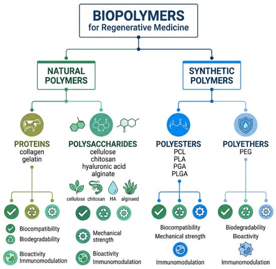
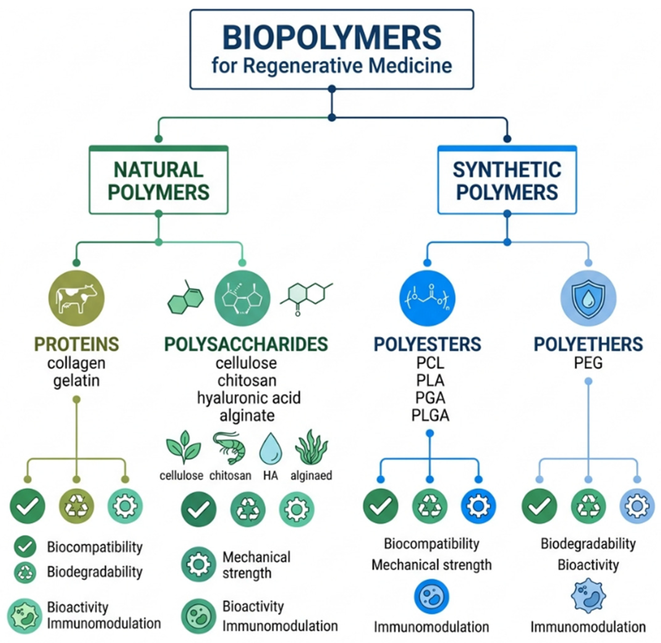
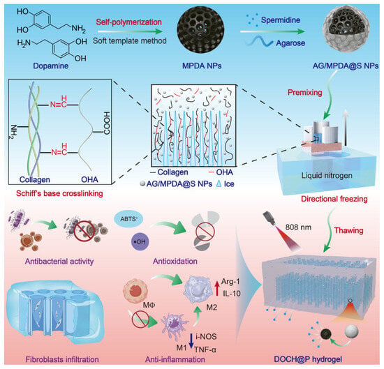
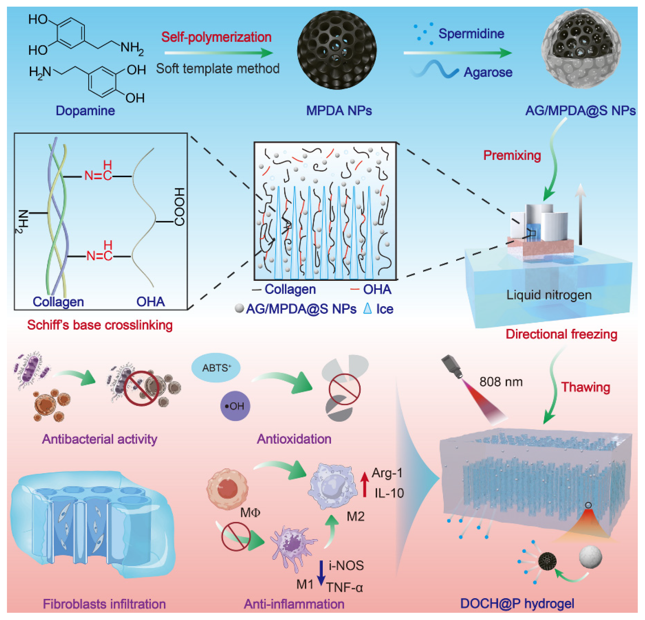
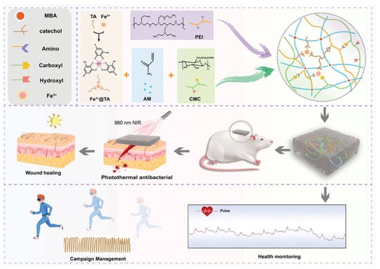
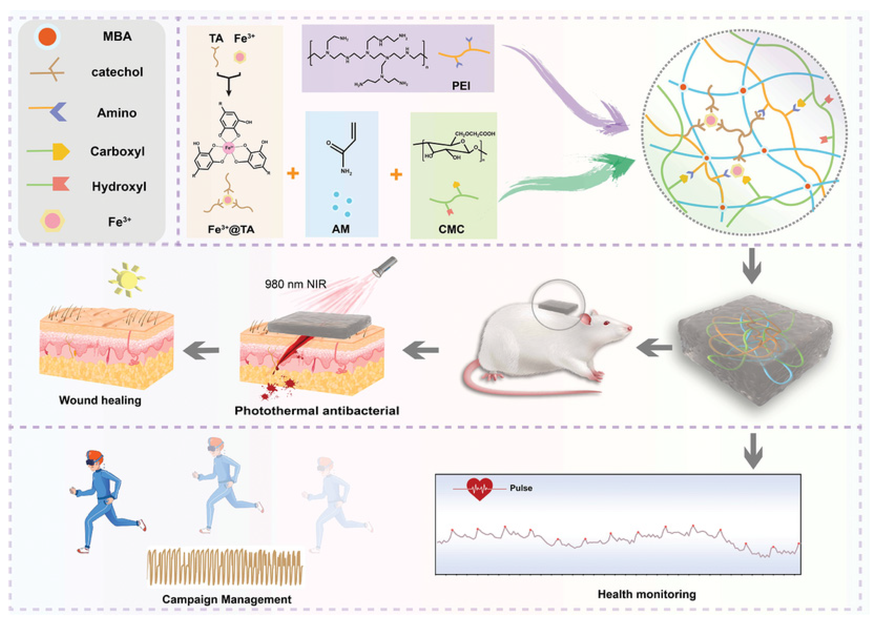
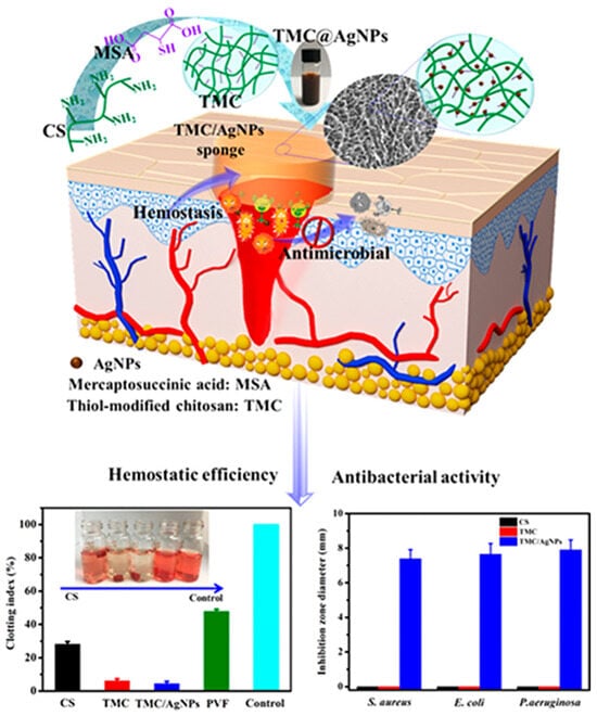
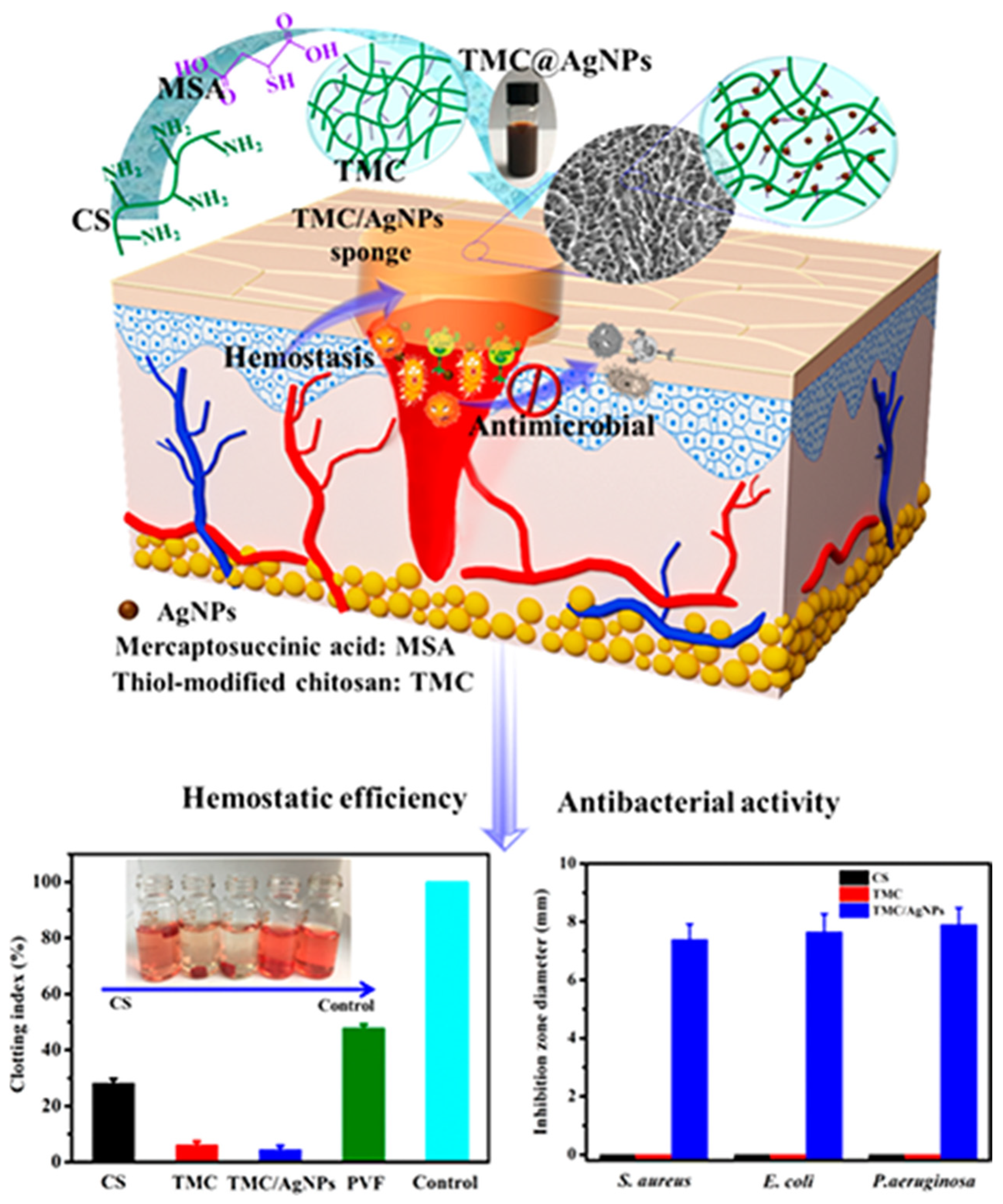
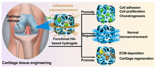
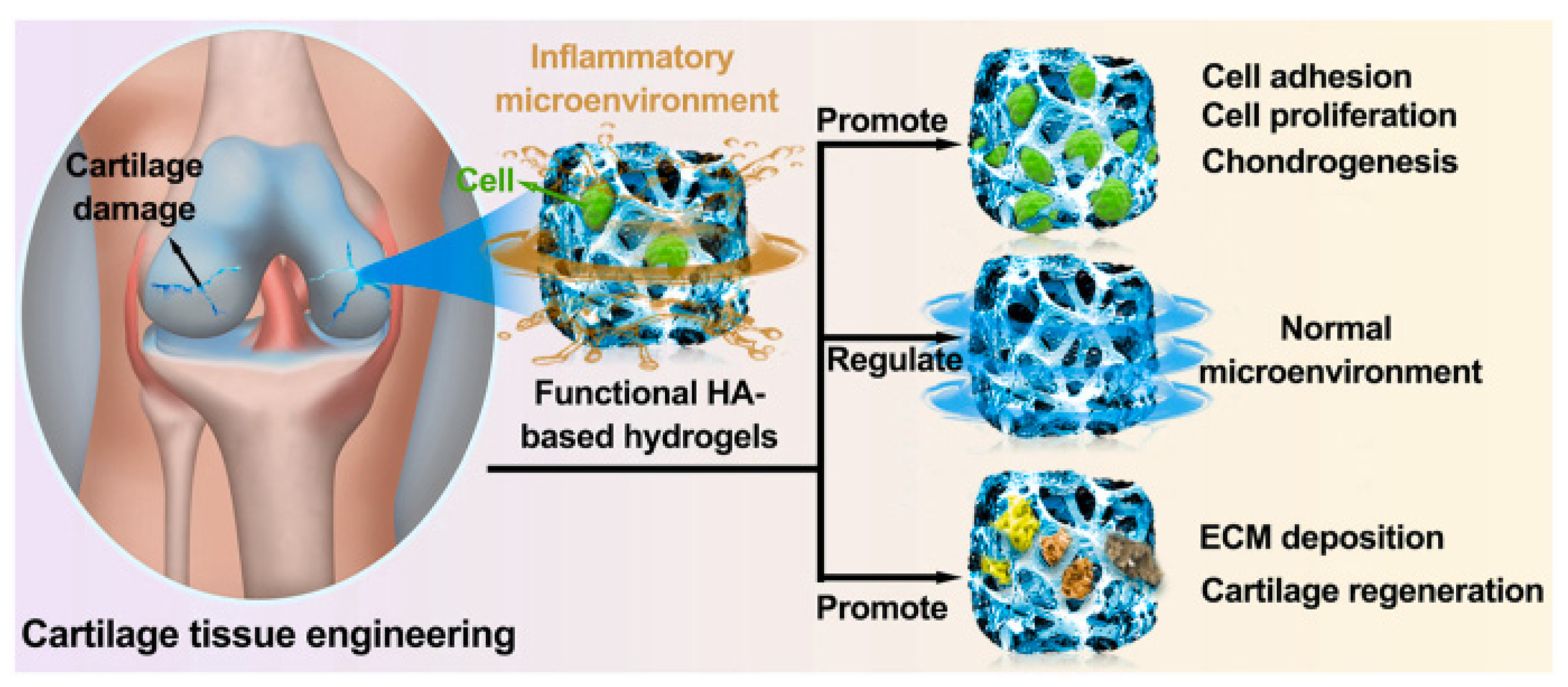
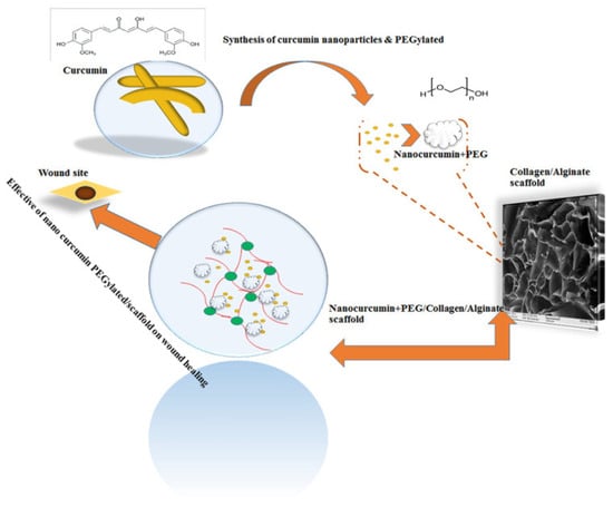
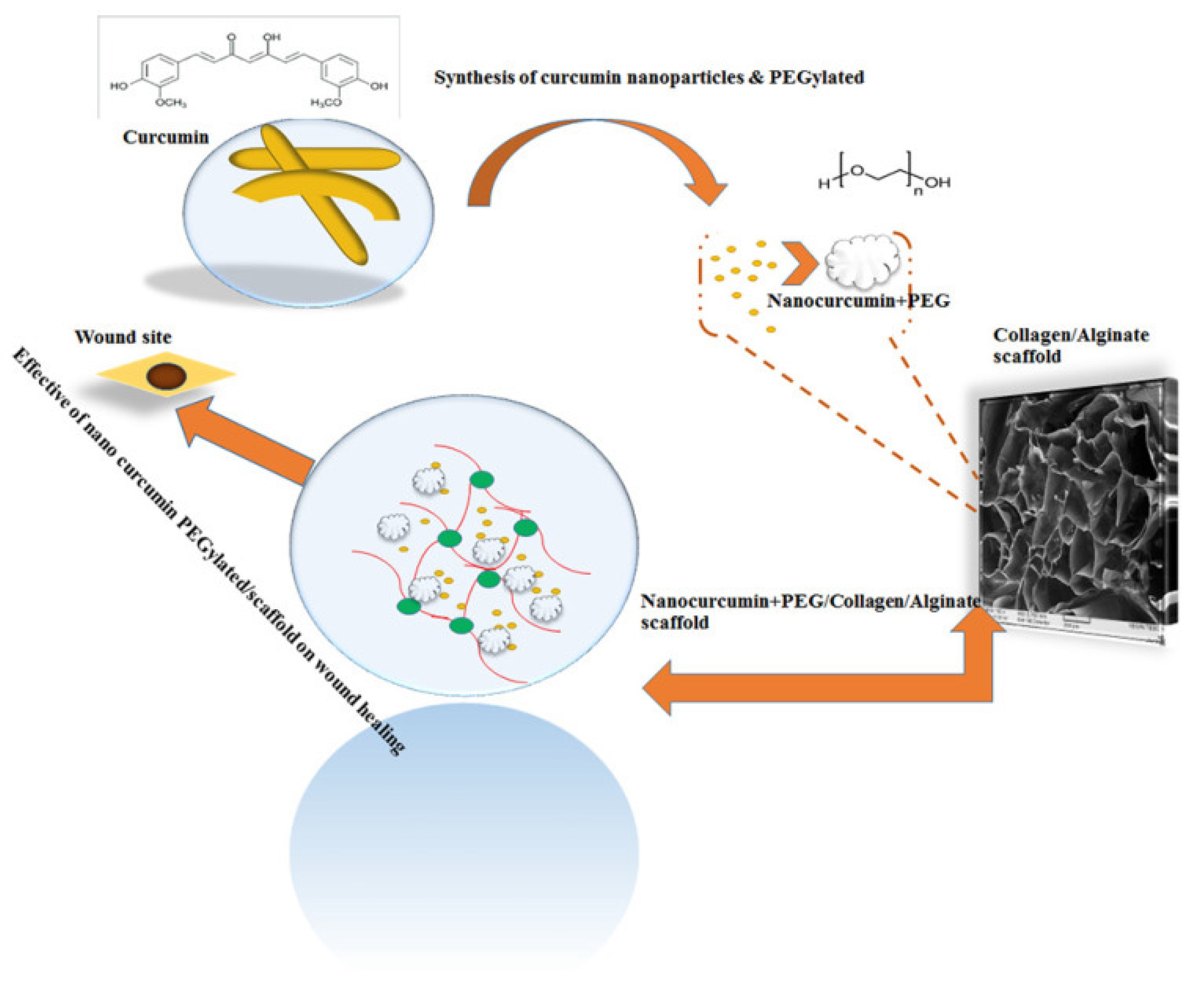
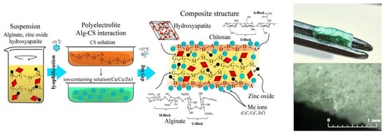
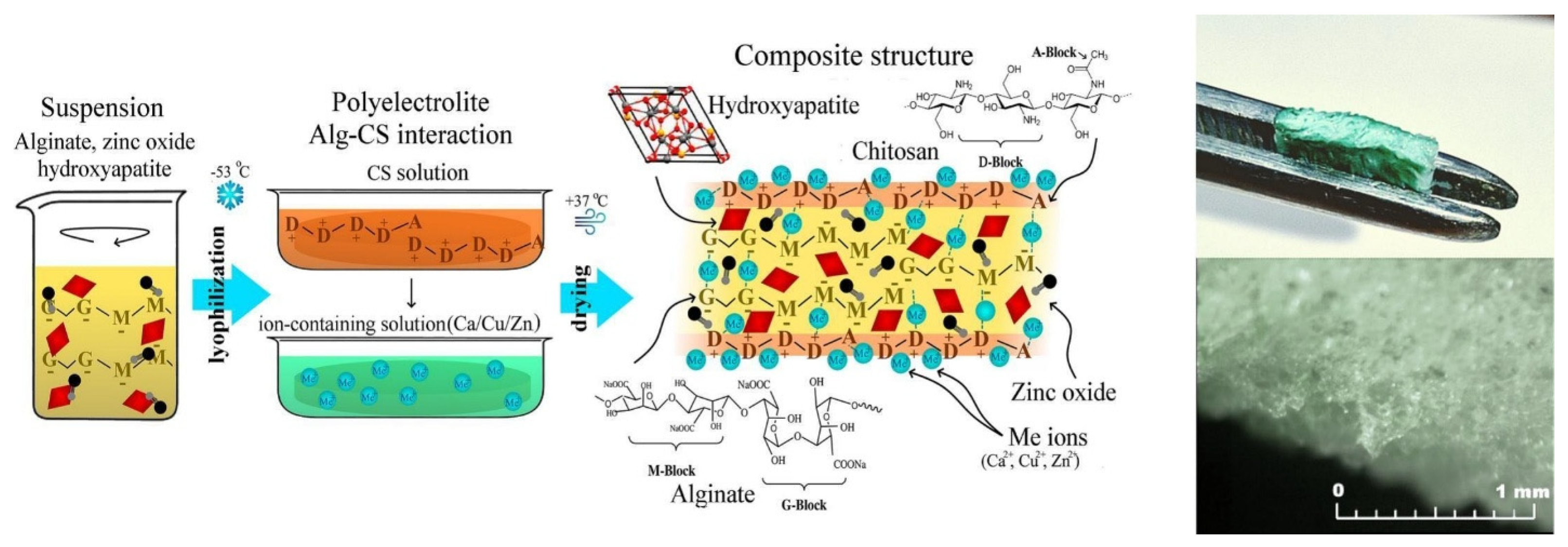
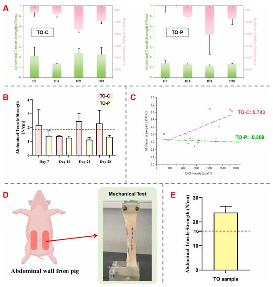
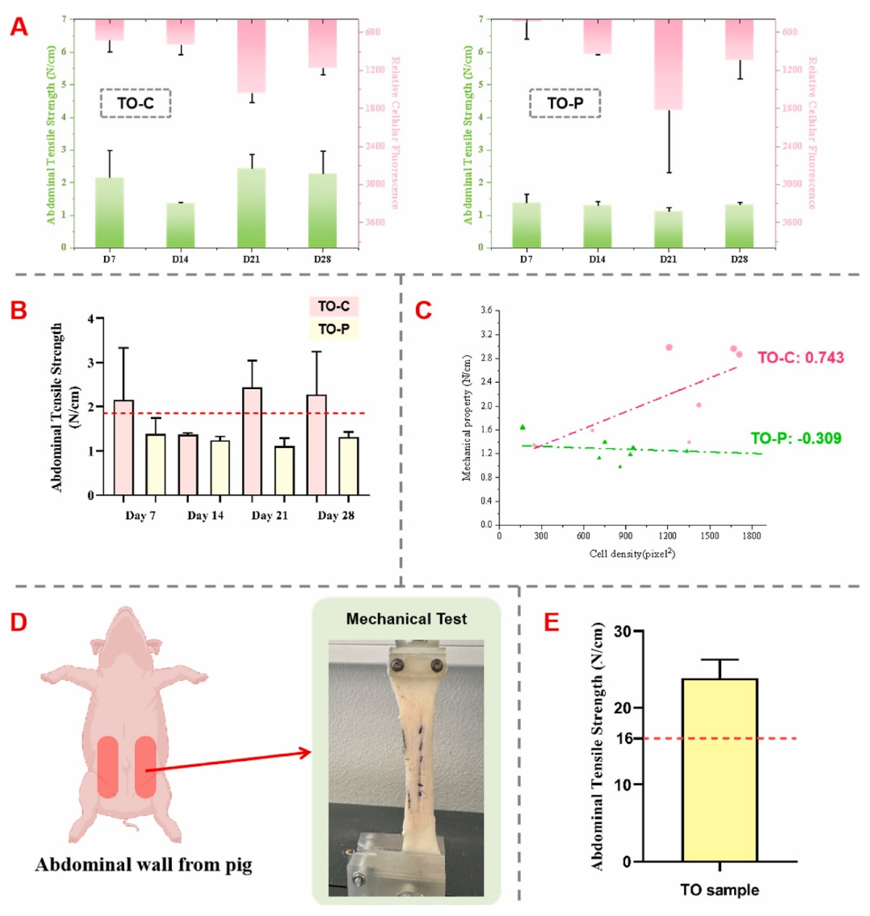
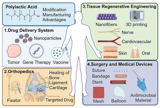
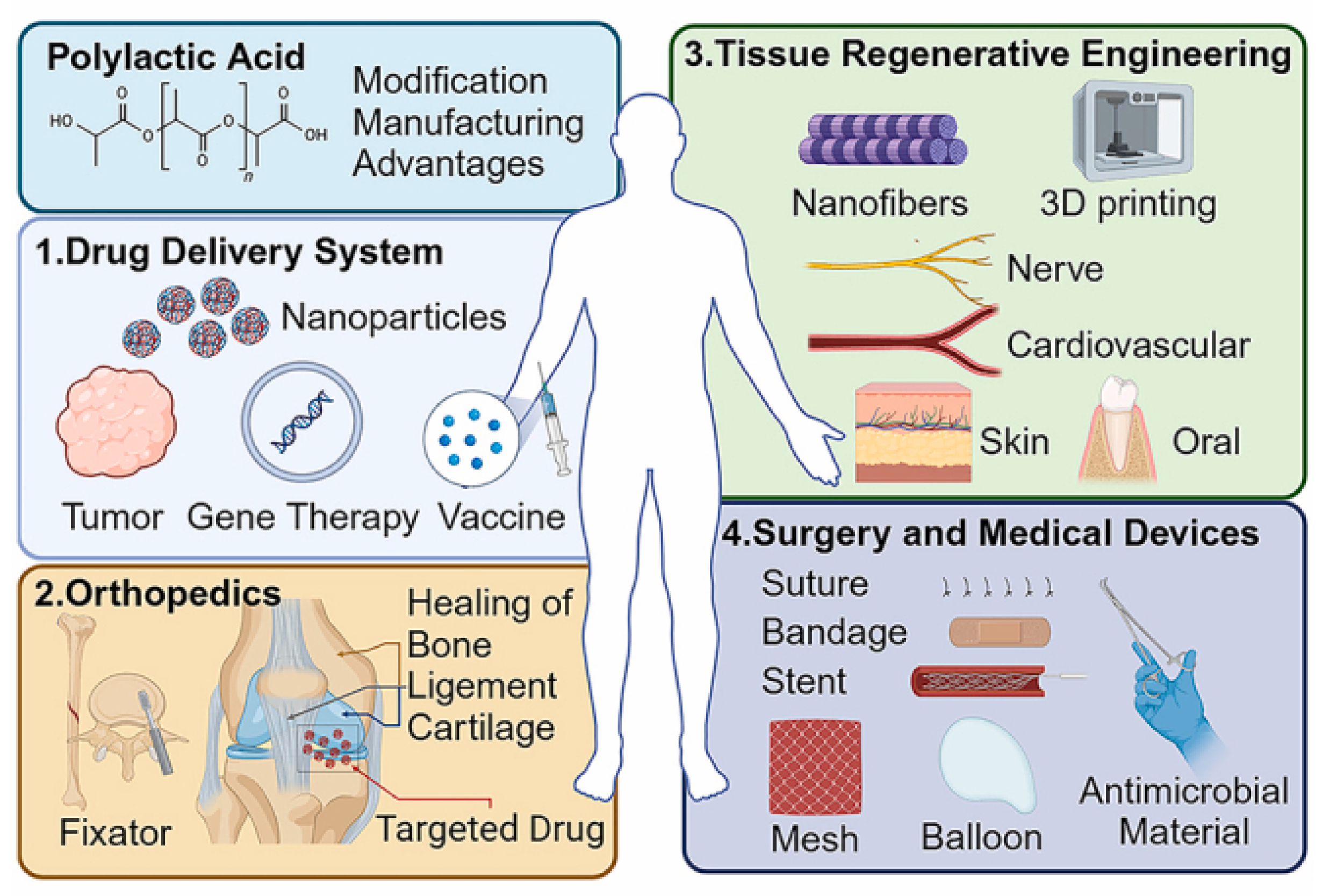
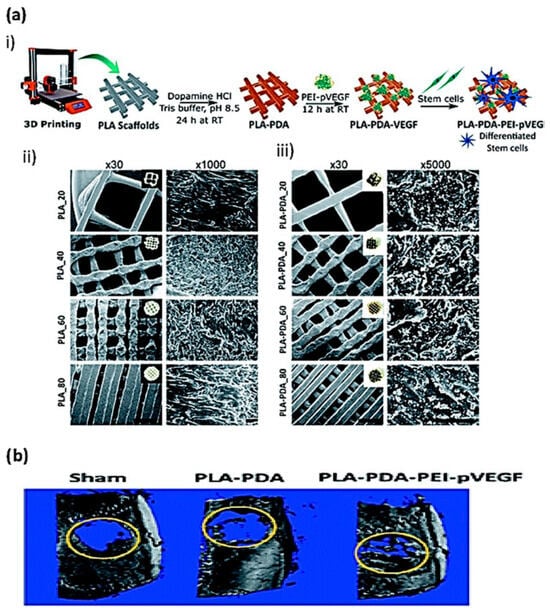
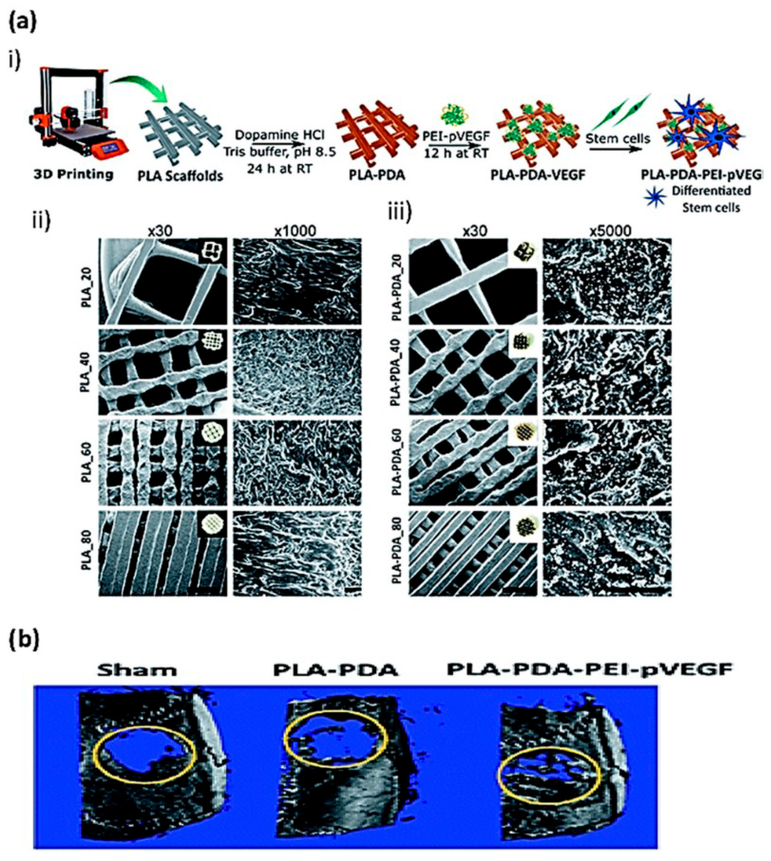
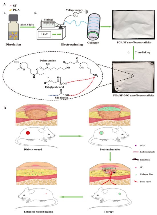
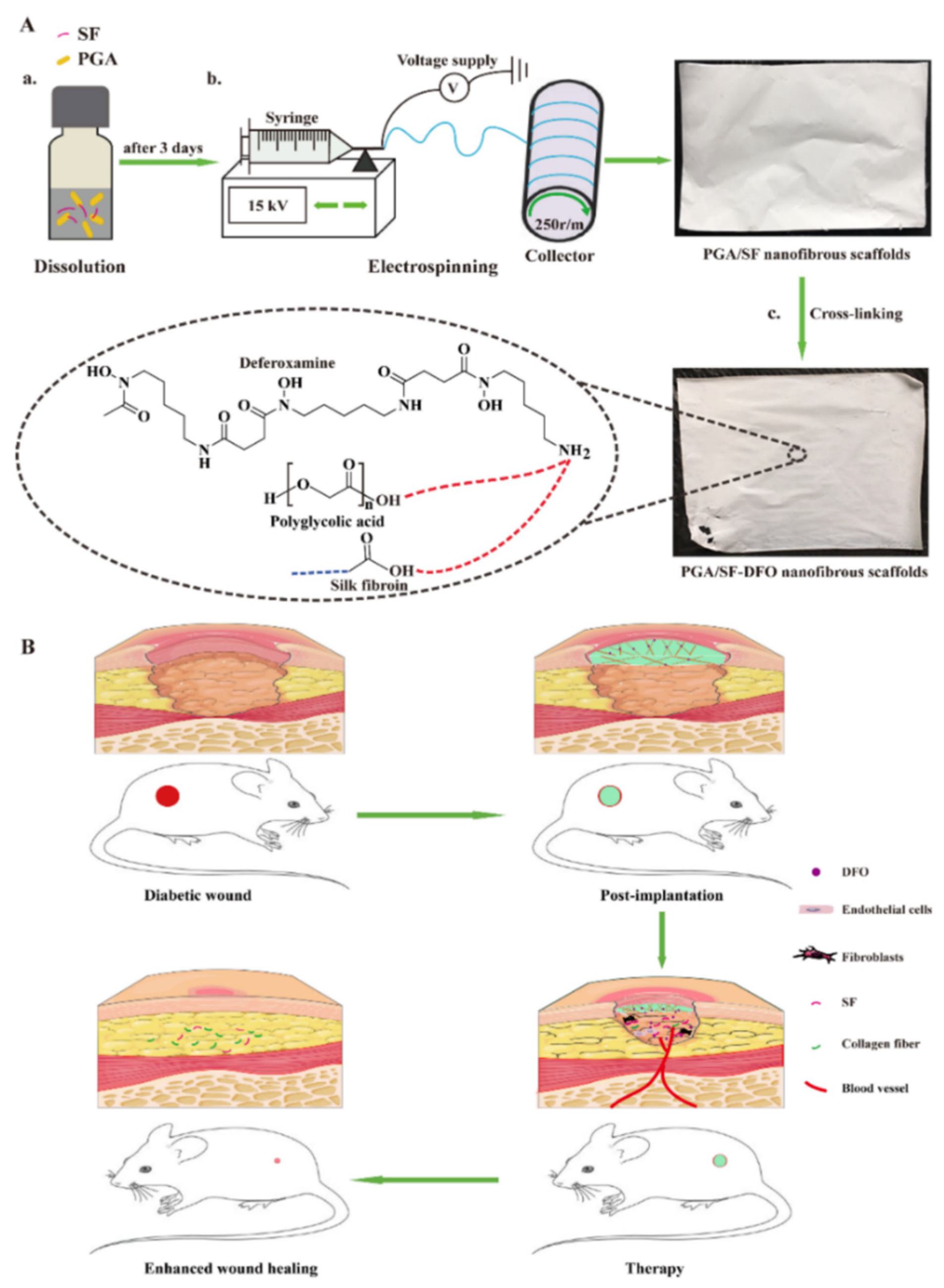
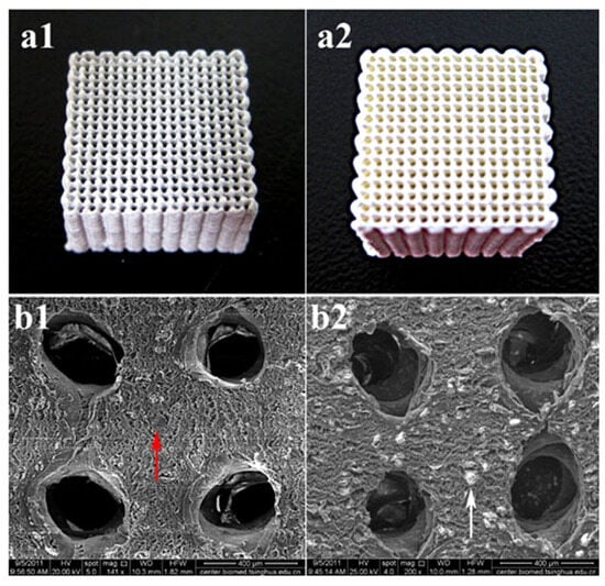
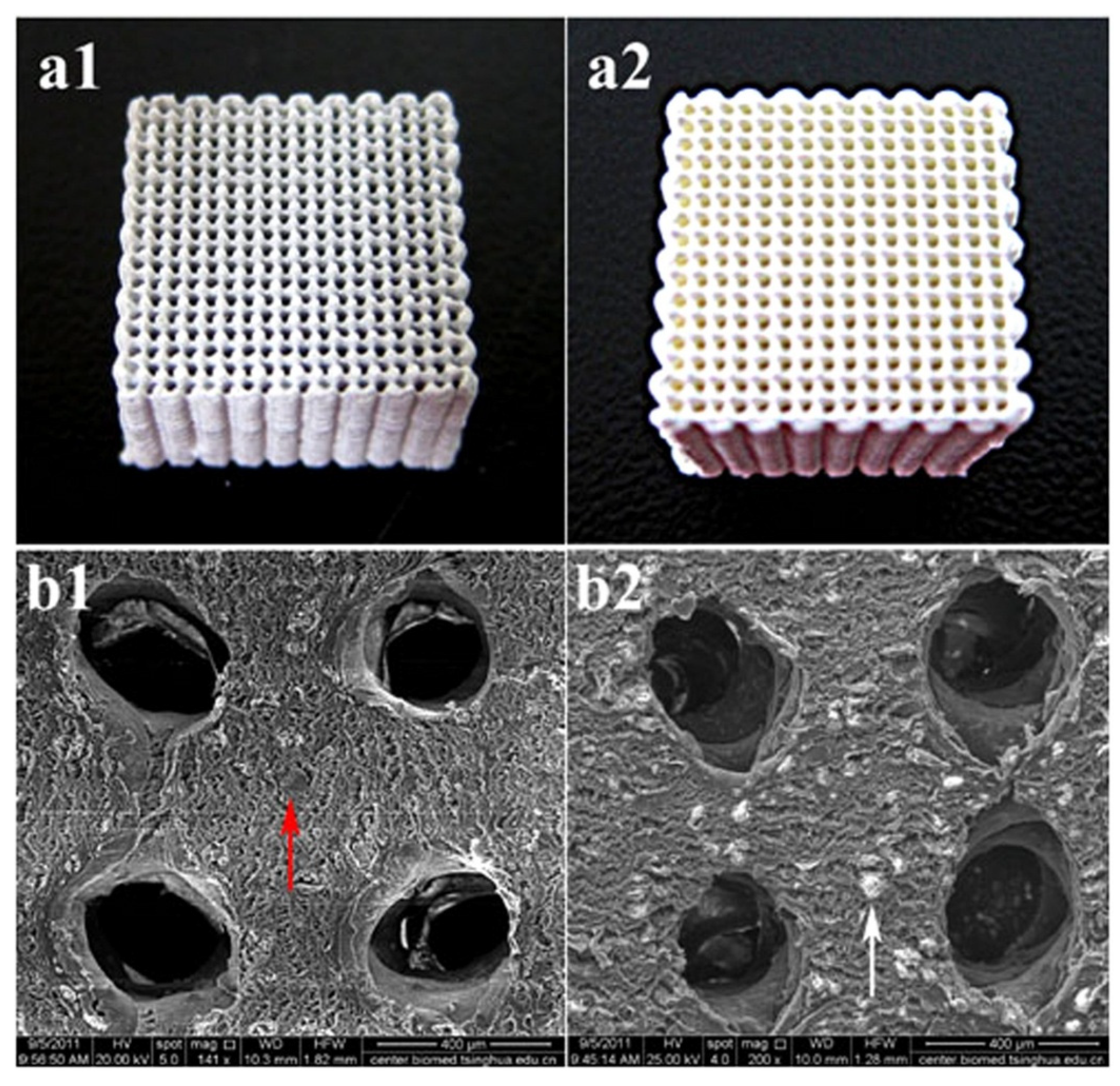
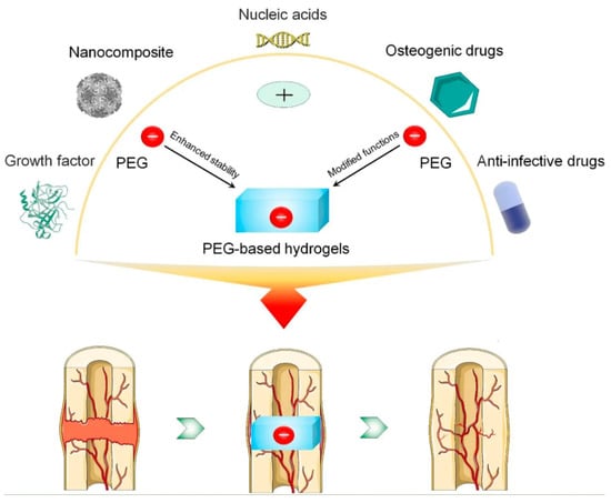
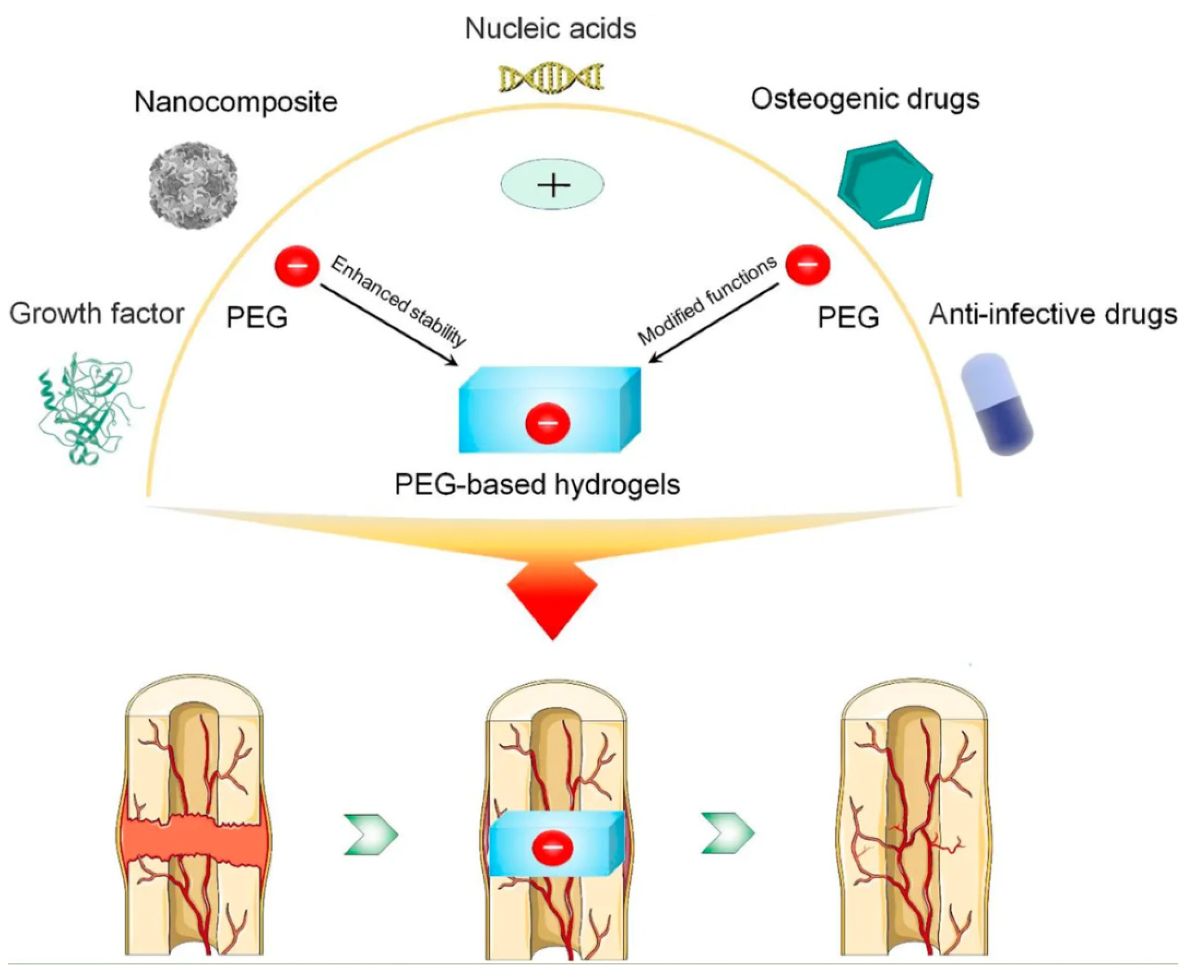
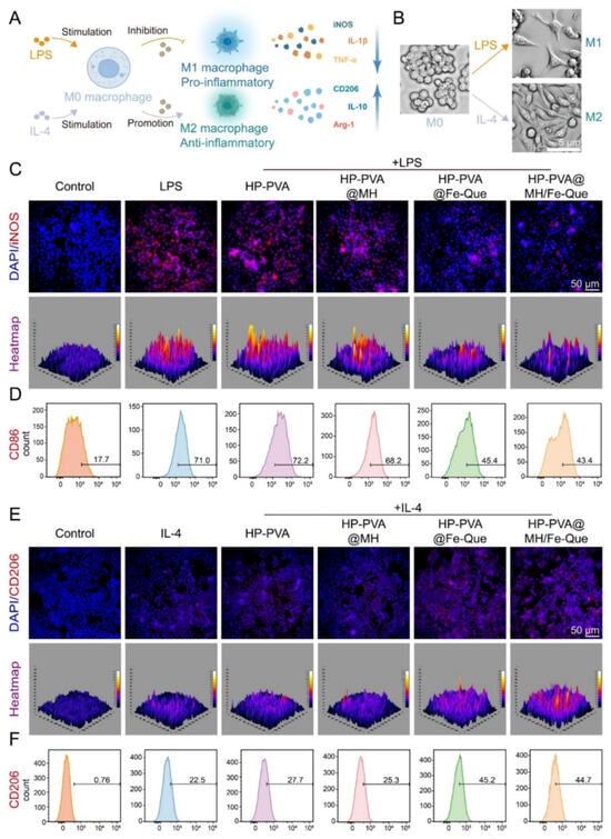
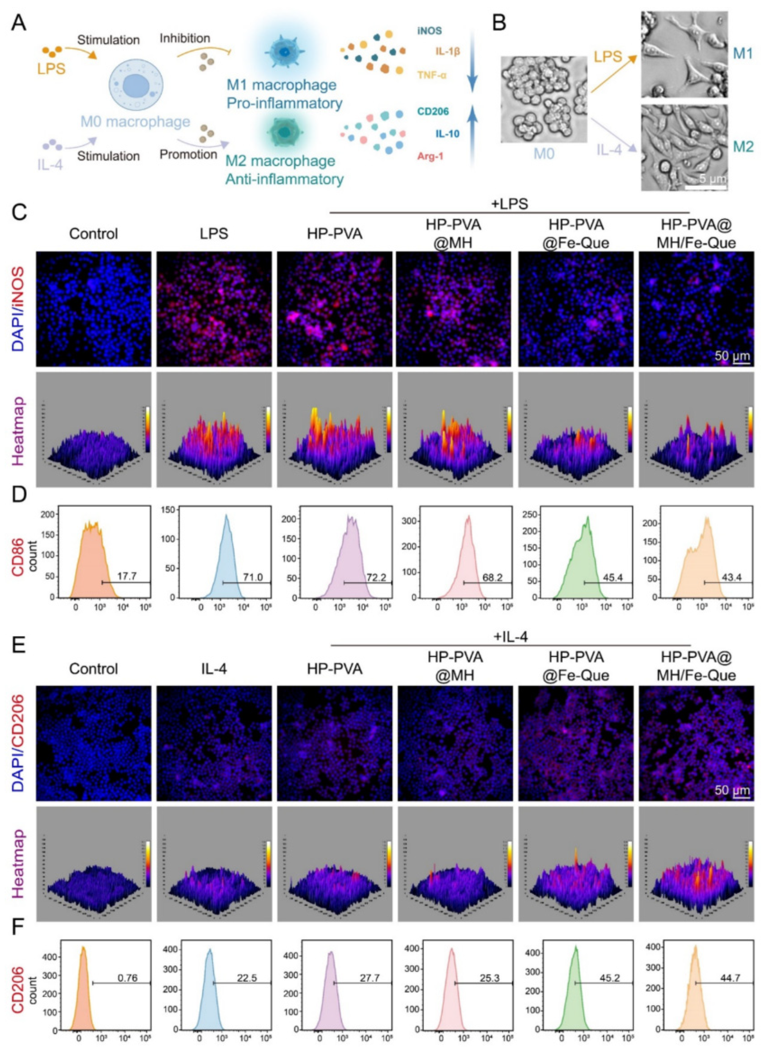
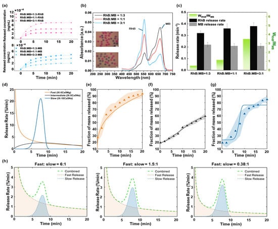
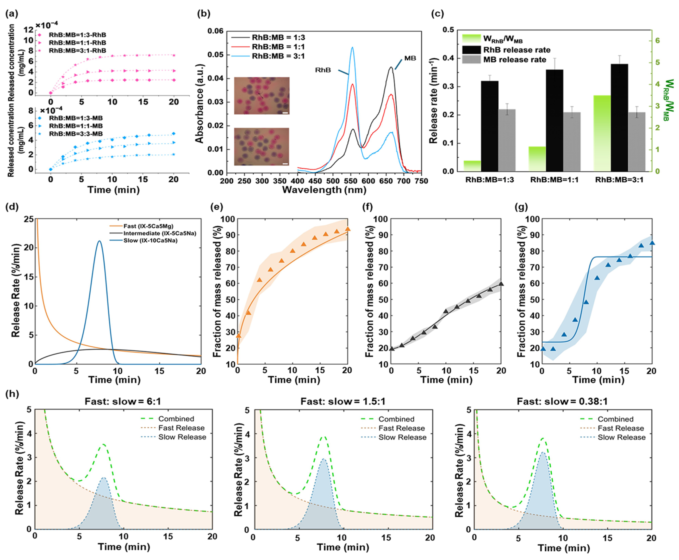

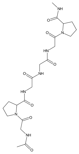
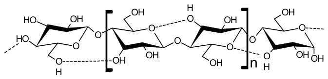
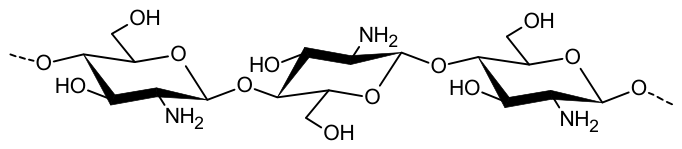
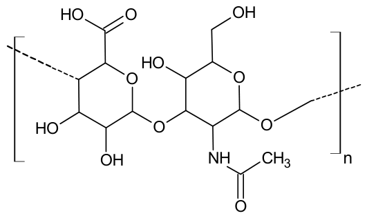
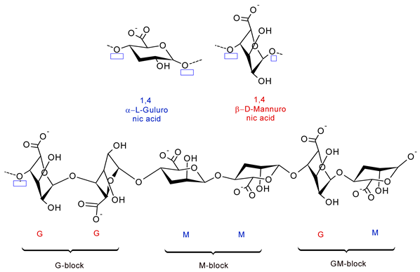
Disclaimer/Publisher’s Note: The statements, opinions and data contained in all publications are solely those of the individual author(s) and contributor(s) and not of MDPI and/or the editor(s). MDPI and/or the editor(s) disclaim responsibility for any injury to people or property resulting from any ideas, methods, instructions or products referred to in the content. |
© 2026 by the authors. Licensee MDPI, Basel, Switzerland. This article is an open access article distributed under the terms and conditions of the Creative Commons Attribution (CC BY) license.