


Abstract
Introduction: Smoking cigarettes is widely recognized as a significant risk factor for tooth loss. Objectives: The aim of this study was to assess the association between cigarette smoking, smoking cessation, and the number of remaining teeth in an adult population. Methods: Data from the Bialystok Plus population study, collected between November 2018 and January 2024, were analyzed. Participants were stratified into young (20–44 years), middle-aged (45–64 years), and older (65 years and above) groups. The outcome was the number of lost teeth. Smoking status (non-smoker vs. ever smoker and former smoker vs. current smoker) and smoking duration were the main independent variables. Additional variables included age, sex, dental habits and medical history. Risk factor analysis was done with generalized linear models using the negative binomial family. Results: Ever smoking was significantly associated with tooth count. Differences in the number of remaining teeth between former and current smokers were observed across age groups but not in the overall population. Current smokers exhibited a significantly greater decline in the number of teeth with an increasing fraction of life spent smoking compared to former smokers. Negative binomial regression models showed that ever smoking, when adjusted for age and diabetes, was a risk factor of tooth loss, but a protective effect of smoking cessation was not confirmed. Conclusions: This study confirmed that tobacco addiction is associated with tooth loss. Being a current smoker remained non-significant as a risk factor for tooth loss when compared to former smoker status.
1. Introduction
The loss of permanent teeth is considered one of the key indicators of oral health and serves as a proxy for overall health status, as well as a risk marker of mortality [1,2]. The extent of tooth loss depends on the progression of oral diseases and the effectiveness of their management. The number of remaining teeth reflects the availability and quality of dental services in a population. The etiology of tooth loss is multifactorial and includes biological factors such as caries and its complications, periodontal disease, trauma, and oral cancer. Non-biological determinants include causes related to access to dental care, patient preferences, and the method of financing dental treatment. Risk factors of tooth loss include socioeconomic determinants (age, sex, education, standard of living), general health, stimulants, hygiene and dietary habits [3,4,5,6,7,8].
Smoking cigarettes has numerous harmful effects on the oral cavity and is well established as a major risk factor for tooth loss [4,9,10,11,12]. Tobacco use contributes to loss of teeth through several pathways. By reducing the amount of oxygen, it promotes the development of cariogenic and periodontal microbiota [13,14,15]. Although nicotine initially stimulates saliva production, chronic exposure leads to its suppression. A diminished flow rate, along with lower buffering capacity, reduced pH, and elevated concentrations of calcium and phosphorus ions, promotes the accumulation of dental plaque and calculus [14,16]. Smoking modulates the inflammatory mediators and sustains a chronic inflammatory process that leads to the destruction of periodontal ligament and alveolar bone, ultimately resulting in tooth loss [17]. Smokers exhibit higher rates of chronic periodontitis progression and tooth loss, as well as poorer outcomes following both non-surgical and surgical periodontal therapies, compared with non-smokers [18,19,20,21].
The aim of this study was to assess the association between cigarette smoking, smoking cessation, and the number of remaining teeth in the adult population of Bialystok, Poland.
2. Materials and Methods
Approval for the study was granted by the Bioethics Committee at the Medical University of Bialystok, No. APK.002.147.2024.
2.1. Study Population
We used retrospective data from the Bialystok Plus population study conducted by the Population Research Center of the Medical University of Bialystok, Poland. All patients provided written informed consent prior to enrollment. The STROBE guidelines [22] were applied and presented in Appendix A—Table A1. The cohort was selected to reflect the demographic structure of Bialystok residents aged 20 to 79 (at the time of selection). Data collected between November 2018 and January 2024 were included. The minimum sample size was established at 384 participants based on the following assumptions: the population of Bialystok in 2018—297,459; the proportion of smokers—50%; confidence level—95%; and maximum error—5%. The inclusion criteria comprised availability of data from medical and dental examinations and the availability of data on smoking status, frequency of tooth brushing, and dental visits. The exclusion criterion was the absence of any of the following: dental and medical examinations and questionnaire survey. The study population was stratified into three age groups: young people (from 20 years to 44 years and 11 months), middle-aged people (from 45 years to 64 years and 11 months), and elderly people (65 years and above).
Smoking status was assessed based on participants’ declarations. The population was divided into non-smokers, i.e., those who declared that they had never smoked cigarettes, and ever smokers, i.e., participants who stated that they had smoked cigarettes at some point in their lives. Ever smokers were further categorized as former and current smokers according to their smoking status at the time of participation in the project. For ever smokers who reported the duration of their addiction, the length of smoking in years and the fraction of life spent smoking, expressed as a percentage of years of smoking in relation to age, were determined. Daily exposure to cigarette smoking was assessed on the basis of responses to the following question: “What was/is the highest number of cigarettes you smoke in a day?”
The number of remaining teeth was determined through an oral examination carried out in a dental office under artificial lighting, using a dental mirror and a UNC15 periodontal probe. Third molars were not included in the assessment. A tooth without pathology, a carious or filled tooth, a tooth with a prosthetic crown (single or being part of a bridge), and a dental root were classified as present in the oral cavity. Teeth replaced with an implant, pontics, or removable denture were not considered remaining teeth.
The questionnaire data included the participant’s age, sex, and education; the frequency of tooth brushing; and the time of last dental visit. Diabetes was diagnosed on the basis of medical history and confirmed by medication taken or by the following parameters: fasting blood sugar level of 200 mg/dL or higher, blood sugar level of 200 mg/dL or higher after 2 h in an oral glucose tolerance test (OGTT), or a glycated hemoglobin (HbA1c) value ≥ 6.5. Obesity was diagnosed using the Body Mass Index (BMI) calculated as the ratio of body weight in kilograms to height in meters squared. A BMI ≥ 30 was classified as obesity.
2.2. Statistical Analysis
The number of remaining teeth was treated as a count variable. Descriptive tables were reported as median [Q1–Q3]. Pairwise comparisons (never vs. ever; former vs. current) were performed with the two-sided Mann–Whitney U test, and comparisons of more than two groups were performed with the Kruskal–Wallis test. For categorical summaries, group differences were assessed with the χ2 test. When expected cell counts were small, the two-sided Fisher’s exact test was used. For covariate-stratified tables, p-values were adjusted for multiple testing within each variable using the Holm’s method across the following age groups: total, from 20 years to 44 years and 11 months (20–44), from 45 years to 64 years and 11 months (45–64), and 65 years and more (≥65). Otherwise, unadjusted p-values were shown. The risk factor analysis was done with generalized linear models using the negative binomial family (log link, fixed dispersion α = 1.0). For each predictor, two variants were fitted: a crude model (single predictor) and a model adjusted for age and diabetes. Categorical predictors used treatment coding with the advantageous category as a reference. The “current smoker” effect was evaluated only among ever smokers. Coefficients were assessed using Wald tests, and effects were presented as incidence rate ratios (IRRs) with 95% confidence intervals. Two-sided p < 0.05 was considered statistically significant. Analyses were conducted in Python 3.12 using SciPy 1.13.1 and statsmodels.
3. Results
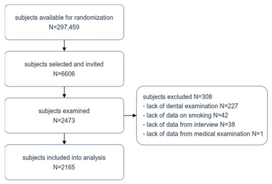
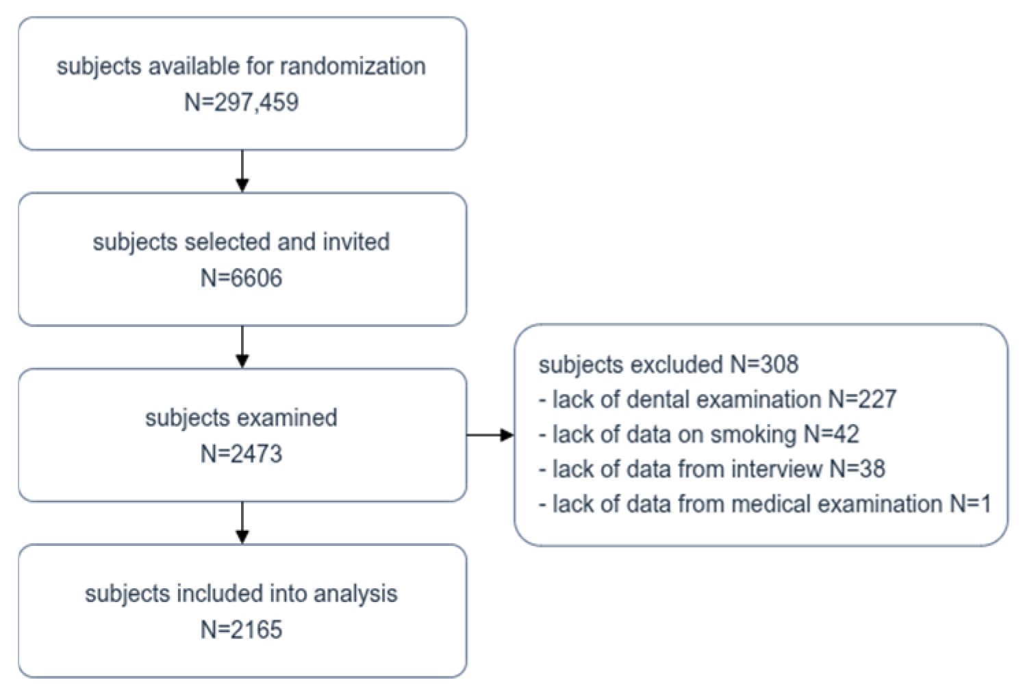
Among the 6606 residents of Bialystok invited to take part in the study, 2473 people participated, giving a response rate of 37.43%. Based on the exclusion criteria, data from 308 individuals were removed from the analysis. The final size of the study population was 2165 individuals. Figure 1 shows a detailed flow chart of the selection of the study population.
Figure 1.
Flow chart of study population selection.
The age of the participants ranged from 20 to 82 years (mean 49.2 ± 15.2). The total group included 1213 (56%) women, and 1132 (52.3%) participants had completed higher education. The number of people in each age group was as follows: 914 young people (20–44 years and 11 months old), 809 middle-aged people (45–64 years and 11 months old), and 442 seniors (65 years old and above). The sex structure in the age groups was as follows: 488 (53.4%) women and 426 (46.6%) men in the youngest group, 471 (58.2%) women and 338 (41.8%) men in the middle-aged group, and 254 women (57.5%) vs. 188 men (42.5%) in the senior group. The age groups did not differ statistically based on sex.
The median number of remaining teeth was 25.0 (19.0–28.0). For women, it was 25.0 (19.0–27.0), while men had an average of 25.0 (20.0–28.0) teeth (p = 0.11, Mann–Whitney U test). The mean number of teeth in the young population was 28.0 (26.0–28.0); in the middle-aged group, it was 23.0 (19.0–26.0); and it was 14.0 (6.0–20.0) in the elderly group (p < 0.001, Kruskal–Wallis test). The mean number of teeth in women in particular age groups was as follows: 28.0 (26.0–28.0), 23.0 (18.0–26.0), 14.0 (6.2–20.0) (p < 0.001, Kruskal–Wallis test). The mean number of teeth in men was as follows: 28.0 (26.0–28.0), 24.0 (19.2–26.0) and 14.0 (6.0–20.0) (p < 0.001, Kruskal–Wallis test).
In the total cohort, 1227 (56.7%) participants had ever smoked cigarettes. There were 398 (18.4%) current smokers and 829 (38.3%) former smokers. The prevalence of being an ever smoker was 56.8% (519 people) in the youngest group, 55.6% (450 people) in the middle-aged group and 58.4% (258 people) among seniors, and the prevalence of being a current smoker among these age groups was 19.4% (177 people), 18.9% (153 people), and 15.4% (68 people), respectively. In comparisons across age groups, the differences in the proportions of ever versus never smokers, as well as former versus current smokers, were not statistically significant (p = 0.6621 and p = 0.0653, χ2). The mean age of people who had ever smoked was 48.8 ± 15.4 and did not differ significantly from never smokers—49.5 ± 15.1 (p = 0.302, Mann–Whitney U test). Current smokers were, on average, 48.0 ± 14.8 years old, whereas former smokers were 50.3 ± 15.2 years old (p < 0.05, Mann–Whitney U test). Former smokers reported a smoking duration of 13.9 ± 10.8 years, while current smokers used them for 24.5 ± 14.3 years, and the fraction of life spent smoking was 26.7% ± 17.3 and 47.9% ± 18.6, respectively. Statistically significant differences between former and current smokers were observed across age groups with respect to smoking duration and the fraction of life spent smoking (p < 0.001).
Table 1 presents the characteristics of the population according to smoking status. In the total cohort, a statistically significant difference in the count of remaining teeth was observed according to smoking status (never smokers vs. ever smokers), but no such associations were found when comparing former and current smokers. Across all age groups, both ever smokers and current smokers had a significantly lower number of remaining teeth. Sex distribution differed significantly between never and ever smokers (p < 0.001), and no differences were identified between former and current smokers. Ever smokers had a lower education level (p < 0.001) and had dental visits less frequently (p < 0.001) than never smokers. Additionally, ever smokers showed a higher prevalence of obesity and diabetes (p = 0.004). Current smokers, compared with former smokers, reported significantly lower educational level (p < 0.001) and were more likely to brush teeth irregularly (p = 0.043).
Table 1.
Characteristics of the population according to smoking status (a test U Mann–Whitney; b χ2) (median (IQR 25–75), N (%)).
Table 2 summarizes data on the teeth count in the whole sample and in the age groups by smoking status. Being an ever smoker had a statistically significant effect on teeth count, and this held at the total sample level regardless of sex, time of last dental visit, diabetes, regular frequency of tooth brushing and lack of obesity. In the youngest group, the lower number of remaining teeth was recorded among male smokers, people who brushed their teeth regularly, those whose last dental visit was over a year ago, participants with a BMI < 30, and individuals without diabetes. In the middle-aged group, ever smokers consistently presented a statistically significant teeth count across all strata. Among seniors, ever smokers, irrespective of sex, and those with secondary or lower education, regular tooth brushing habits, those who visited the dentist over a year ago, and individuals without diabetes and obesity had a lower mean number of teeth.
Table 2.
Number of remaining teeth according to smoking status (never smokers vs. current smokers).
No significant association between smoking cessation and a higher number of teeth was observed in the overall sample, except among participants without obesity (Table 3). In the youngest age group, a significant association between smoking cessation and greater tooth retention was identified only among individuals with a BMI < 30. Participants aged 45–64 years demonstrated the strongest pattern of associations between the covariates and the number of teeth with regard to quitting smoking vs. continuing tobacco use. Current smokers who were men, less educated, irregularly attended dental visits (>1 year since last visit), had a BMI < 30, and did not have diabetes had fewer remaining teeth than former smokers. Signals by tooth brushing frequency were mixed rather than uniform. In the oldest age group, smoking cessation was associated with a higher number of remaining teeth only among participants without obesity and diabetes.
Table 3.
Number of remaining teeth according to smoking status (former smokers vs. current smokers).
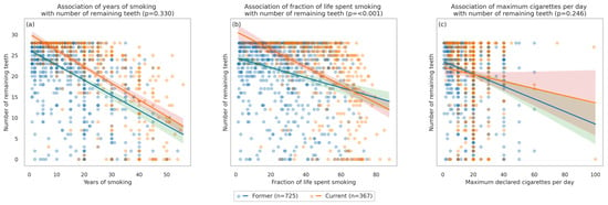
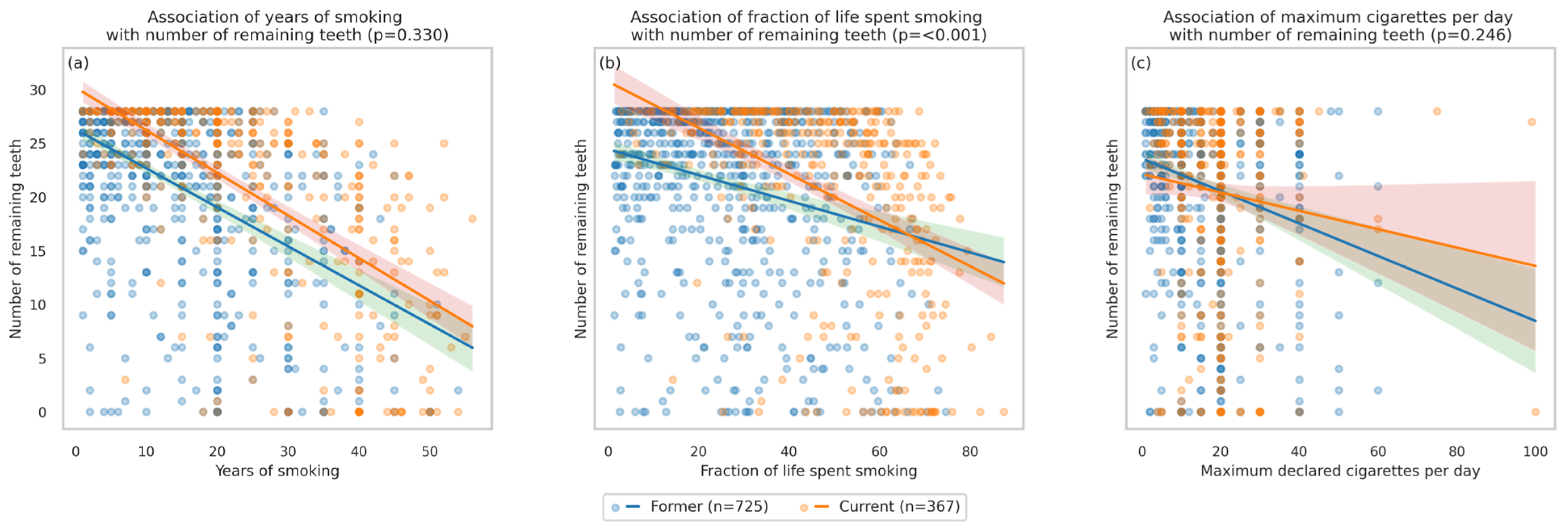
Figure 2 presents the association between the number of remaining teeth, the duration of smoking and the maximum daily exposure to cigarettes. Among ever smokers, both exposure metrics of smoking duration showed clear inverse associations with the number of remaining teeth. Years of smoking were negatively related to teeth count in both former and current smokers, but the rate of decline per additional year did not differ among groups (p = 0.330). This indicates a broadly similar year-by-year loss pattern in former and current smokers at the sample level. In contrast, the fraction of life spent smoking differed by group. Although the tooth count decreased with increasing fraction in both groups, the decrease was significantly steeper among current smokers (p < 0.001). The association of declared maximum cigarettes per day with the tooth count was not significant.
Figure 2.
Association between number of remaining teeth and exposure to cigarettes: (a) duration of smoking as years of smoking (p = 0.330), (b) duration of smoking as fraction of life spent smoking (p < 0.001), (c) maximum cigarettes per day (p = 0.246).
Details of negative binomial regression models identifying risk factors of losing teeth are presented in Table 4. In the total cohort, neither sex nor current smoker status was confirmed as a risk factor of tooth loss. In the model adjusted for age and diabetes, the following factors had a significant impact on the number of remaining teeth: secondary or lower education (β = −0.151, p = 0.001), last dental visit more than a year ago (β = −0.144, p = 0.003), and ever smoking (β = −0.094, p = 0.035). Brushing frequency and obesity were no longer significant, and sex and current smoking remained non-significant among ever smokers. For ever smokers, the basic model indicated age (β = −0.021, p < 0.001), secondary or lower education (β = −0.249, p < 0.001), last dental visit over one year ago (β = −0.226, p < 0.001), obesity ≥ 30 (β = −0.165, p = 0.012), diabetes (β = −0.473, p < 0.001), and cumulative exposure measures like years of smoking (β = −0.019, p < 0.001) and fraction of life spent smoking (β = −0.066, p < 0.001) as risk factors. Brushing frequency was not significant, and neither sex nor current smoking reached significance. In the age- and diabetes-adjusted model for ever smokers, significant factors included: secondary or lower education (β = −0.146, p = 0.016), last dental visit over one year ago (β = −0.180, p = 0.004), years of smoking (β = −0.009, p = 0.003), and fraction of life spent smoking (β = −0.004, p = 0.017). BMI ≥ 30 and current smoking were not significant after adjustment, and sex remained non-significant.
Table 4.
Risk factor analysis for total population and in ever smokers. Negative binomial regression (crude model and adjusted by age and diabetes).
4. Discussion
A key result of the present study is that smoking cigarettes represents an important modifiable factor associated with tooth loss. This finding is in accordance with data from the literature [4,8,9,12,19,23,24,25,26,27,28,29]. Depending on the study, the risk of tooth extraction in smokers was estimated between 1.4 and 2.49 times higher than in non-smokers [4,30,31]. However, not all evidence is consistent, as some studies have reported no statistically significant association between smoking and tooth loss [32,33,34,35,36,37,38,39,40,41,42,43].
According to the literature, smoking cessation improves tooth retention, but a recent meta-analysis indicates that the risk of tooth loss among former smokers remains higher than in never smokers [12,19,24,26,27,29,35,36]. Our results concerning the evidence regarding the impact of smoking cessation on tooth loss were inconclusive. Within each age group, smoking cessation was associated with a reduction in the number of extractions compared to continuing to smoke, and a longer fraction of life spent smoking had a negative impact on the number of teeth when comparing former and current smokers. However, there were no differences in the number of remaining teeth between former and current smokers at the total population level. Moreover, the negative binomial regression models did not identify being a current smoker as a risk factor of tooth loss. This may be explained by the long-term effect of smoking on oral health, even after quitting cigarettes. The period of nicotine abstinence required for the risk of tooth loss to return to that of a non-smoker is 10 to 20 years [24,36,37]. In cross-sectional studies, participants who have only recently quit smoking may influence the reduction in the effect of smoking cessation on the preservation of teeth [28]. The inclusion of such subjects in the analysis may have had an impact on the results. Undoubtedly, the most desirable health behavior is never initiating smoking. Despite the inconsistency of our findings regarding the benefits of smoking cessation for tooth preservation, quitting smoking should be treated as a priority in dental patients [37].
This study provides further support for the multifactorial etiology of tooth loss, with biological factors (age and general diseases), behavioral factors (hygiene habits and dental check-ups), and socio-economic factors (education) playing important roles as risk factors. In our study, negative binomial regression did not identify sex as a risk factor for tooth loss, which is contrary to previous studies [28,35,38,39]. Proper oral hygiene and regular dental care are essential for preventing oral diseases that lead to tooth loss [8,40]. In the SHIP study, last dental visit over 12 months ago, poor general health and poor interdental health were revealed as risk factors of a high number of missing teeth [41]. In elderly Brazilians, last dental visit over 12 months ago and over 24 months ago resulted in prevalence ratios of 2.39 and 2.76 for edentulism [26]. Also, young Finnish adults with improper oral habits were at a greater risk of losing more than six teeth [42]. In our models, a dental visit that took place more than a year ago was a significant predictor of tooth loss, whereas irregular tooth brushing showed no significant correlation. Although previous studies have reported links between BMI and tooth loss, in our cohort, obesity remained insignificant after adjusting for age and diabetes [8,43,44,45].
In negative binomial regression models, age and diabetes were determined as controlling factors. In a crude model, both variables significantly influenced the number of remaining teeth in the overall population and among ever smokers (p < 0.001). The chance of preserving teeth decreases with age, and tooth loss peaks in the seventh decade [46,47]. However, a marked decline in extractions is now being observed, particularly among younger adults [26,35,39]. Patients with uncontrolled diabetes are more prone to severe tooth loss and edentulism, but some studies suggest that well-controlled disease may not significantly affect the number of teeth extracted [45,48,49,50,51,52,53]. In the present study, patients were not differentiated according to the type of diabetes (type I and II), the level of disease management, or whether they were newly diagnosed or had been treated for many years. All of the above aspects can affect the severity of tooth loss, which is why diabetes was included as a controlling factor.
A strength of this study is the age-stratified analysis of factors associated with tooth loss. There were only a few variables associated with the number of remaining teeth in ever smokers vs. never smokers in the 20–44 age group. In contrast, a broader range of factors was linked to tooth loss in middle-aged and older individuals with a history of smoking. On the other hand, only a few statistically significant associations were observed for smoking cessation, irrespective of age group.
When comparing young never and ever smokers, a higher number of missing teeth was found among smoking males, those who brushed their teeth regularly, those who visited a dentist irregularly, and those who were not obese and did not have diabetes. In contrast, with the exception of BMI < 30, no associations were found between the variables and the number of preserved teeth among young current and former smokers. In a comparable age group of Japanese individuals, the factors that correlated with tooth loss were being a current smoker regardless of sex, and in women, correlations were found with irregular tooth brushing and being overweight [54]. According to Modin et al. [21], smokers aged ≥25 years with severe periodontal disease had a 3.6-fold higher incidence risk of tooth loss.
Among individuals aged 45–64, ever smokers had fewer teeth compared to never smokers, regardless of other covariates. This finding suggests that in middle-aged adults, who have likely smoked since early adulthood, the detrimental effects of smoking accumulate over time. Similä et al. [37] reported that the majority of middle-aged current smokers had been smoking cigarettes for more than 10 years. They also found that a positive association between time since smoking cessation and tooth count was evident in men but not in women. In the present study, among participants aged 45–64, a comparison between former smokers and current smokers revealed that the only factors significantly associated with tooth loss were a lower level of education and a BMI below 30.
Regarding the oldest age group, no differences in the number of remaining teeth between never and ever smokers were observed among individuals with higher education, those reporting regular dental visits, and those with poor oral hygiene. Hsu at el. [25] found that in elderly subjects, both higher education level and proper oral health behaviors had better general health (i.e., absence of obesity and diabetes), and these factors were associated with a higher number of remaining teeth when comparing ever smokers with never smokers, as well as current smokers with former smokers. These findings may suggest that, in healthy elderly individuals, smoking has a particularly detrimental effect on tooth preservation.
This study has some limitations. First, the analysis was restricted to conventional cigarette smoking, without including alternative nicotine delivery systems such as electronic cigarettes or heated tobacco products. Alternative methods of nicotine delivery have only been available on the market for a few years, and their impact on oral health remains unclear, particularly in comparison with traditional cigarettes [55]. A recent meta-analysis by Thiem et al. [55] suggests that e-cigarettes may be less harmful to periodontal tissues than conventional tobacco products. Since the effect of nicotine on tooth loss is long-term, it is unlikely that the exclusion of alternative methods of nicotine delivery would have a significant impact on the current results. Many studies have shown a dose–response relationship between smoking and tooth loss [19,27,42,56]. The inability to calculate pack-years (resulting from the phrasing of the question on daily cigarette use) should be acknowledged as a limitation of the present study. Instead, the severity of tobacco exposure was estimated on the basis of smoking duration in years and the fraction of life spent smoking, and it was found to be a significant risk factor for tooth loss.
5. Conclusions
The results of this study show an association between tobacco addiction and the ability to maintain dentition. Being an ever smoker increased the risk of tooth loss regardless of age and diabetes, but being a current smoker remained non-significant as a risk factor for tooth loss in comparison to former smokers. This study highlights the important role that dentists can play in tobacco prevention by informing patients about the greater risk of tooth loss due to cigarette smoking.
Author Contributions
Conceptualization, J.B., K.Z. and W.Ł.; methodology, J.B., I.K. and K.K.; investigation, J.B., K.Z., I.K., Z.S., M.D., N.S. and S.S.; data curation, W.Ł.; writing—original draft preparation, J.B., K.Z. and W.Ł.; writing—review and editing, I.K., Z.S., M.D., N.S., S.S. and K.K.; visualization, W.Ł.; supervision, K.K.; project administration, Z.S., M.D. and N.S.; funding acquisition, J.B. All authors have read and agreed to the published version of the manuscript.
Funding
This research was funded by the Medical University of Bialystok, Poland, grant no. B.SUB.24.509, B.SUB.25.509 and B.SUB.26.509.
Institutional Review Board Statement
This study was conducted in accordance with the Declaration of Helsinki and approved by the Bioethics Committee at the Medical University of Bialystok, No. APK.002.147.2024, date of approval: 22 April 2024.
Informed Consent Statement
Written informed consent has been obtained from the patients to publish this paper.
Data Availability Statement
The original contributions presented in this study are included in the article. Further inquiries can be directed to the corresponding author.
Acknowledgments
The authors would like to acknowledge each person who participated in the project, Marek Bagiński for performing linguistic editing of the manuscript, and Ewa Rodakowska for her contribution to the Białystok PLUS project.
Conflicts of Interest
The authors declare no conflicts of interest.
Abbreviations
The following abbreviations are used in this manuscript:
| OGTT | oral glucose tolerance test |
| HbA1c | hemoglobin A1C |
| BMI | body mass index |
| IRR | incidence rate ratios |
Appendix A
Table A1.
STROBE checklist.
References
- Holmlund, A.; Holm, G.; Lind, L. Number of teeth as a predictor of cardiovascular mortality in a cohort of 7,674 subjects followed for 12 years. J. Periodontol. 2010, 81, 870–876. [Google Scholar] [CrossRef] [Scilit]
- Peng, J.; Song, J.; Han, J.; Chen, Z.; Yin, X.; Zhu, J.; Song, J. The relationship between tooth loss and mortality from all causes, cardiovascular diseases, and coronary heart disease in the general population: Systematic review and dose-response meta-analysis of prospective cohort studies. Biosci. Rep. 2019, 39, BSR20181773. [Google Scholar] [CrossRef] [Scilit] [PubMed]
- Ali, D. Reasons for extraction of permanent teeth in a university dental clinic setting. Clin. Cosmet. Investig. Dent. 2021, 24, 51–57. [Google Scholar] [CrossRef] [Scilit] [PubMed]
- Passarelli, P.C.; Pagnoni, S.; Piccirillo, G.B.; Desantis, V.; Benegiamo, M.; Liguori, A.; Papa, R.; Papi, P.; Pompa, G.; D’Addona, A. Reasons for tooth extractions and related risk factors in adult patients: A Cohort Study. Int. J. Environ. Res. Public Health 2020, 17, 2575. [Google Scholar] [CrossRef] [Scilit] [PubMed]
- Salim, N.A.; Sawair, F.A.; Abusidu, Z.; Sharaireh, A.M.; Satterthwaite, J.D.; Hassona, Y. Pattern, frequency and causes of tooth extraction among adult Syrian refugees. Spec. Care Dent. 2023, 43, 588–596. [Google Scholar] [CrossRef] [Scilit]
- Felton, D.A. Edentulism and comorbid factors. J. Prosthodont. 2009, 18, 88–96. [Google Scholar] [CrossRef] [Scilit]
- Osunde, O.D.; Efunkoya, A.A.; Omeje, K.U. Reasons for loss of the permanent teeth in patients in Kano, North western Nigeria. J. West Afr. Coll. Surg. 2017, 7, 47–64. [Google Scholar]
- Alzahrani, S.G.; Rijhwani, K.; Sabbah, W. Health-risk behaviours co-occur among adults with tooth loss. Int. J. Dent. Hyg. 2024, 22, 857–862. [Google Scholar] [CrossRef] [Scilit]
- Carson, S.J.; Burns, J. Impact of smoking on tooth loss in adults. Evid. Based Dent. 2016, 17, 73–74. [Google Scholar] [CrossRef] [Scilit]
- McCormick, K.M.; Mejia, G.; Luzzi, L.; Jamieson, L. Mapping Tooth Loss at the Tooth Level: Associations with Demographic, Health, and Behavioral Factors. JDR Clin. Transl. Res. 2025. Online ahead of print. [Google Scholar] [CrossRef] [Scilit]
- Similä, T.; Virtanen, J.I. Association between smoking intensity and duration and tooth loss among Finnish middle-aged adults: The Northern Finland Birth Cohort 1966 Project. BMC Public Health 2015, 15, 1141. [Google Scholar] [CrossRef] [Scilit] [PubMed]
- Baumeister, S.E.; Listl, S.; Holtfreter, B.; Nascimento, G.G.; Leite, F.R.M. Causal effect of smoking and cessation on tooth loss. J. Clin. Periodontol. 2025, 52, 539–546. [Google Scholar] [CrossRef] [Scilit] [PubMed]
- Johnson, G.K.; Slach, N.A. Impact of tobacco use on periodontal status. J. Dent. Educ. 2001, 65, 313–321. [Google Scholar] [CrossRef] [Scilit] [PubMed]
- Ozturk, O.; Fidanci, I.; Unal, M. Effects of smoking on oral cavity. J. Exp. Clin. Med. 2017, 34, 3–7. [Google Scholar]
- Wu, J.; Peters, B.A.; Dominianni, C.; Zhang, Y.; Pei, Z.; Yang, L.; Ma, Y.; Purdue, M.P.; Jacobs, E.J.; Gapstur, S.M.; et al. Cigarette smoking and the oral microbiome in a large study of American adults. ISME J. 2016, 10, 2435–2446. [Google Scholar] [CrossRef] [Scilit]
- Rad, M.; Kakoie, S.; Niliye Brojeni, F.; Pourdamghan, N. Effect of long-term smoking on whole-mouth salivary flow rate and oral health. J. Dent. Res. Dent. Clin. Dent. Prospects 2010, 4, 110–114. [Google Scholar]
- Ye, D.; Gajendra, S.; Lawyer, G.; Jadeja, N.; Pishey, D.; Pathagunti, S.; Lyons, J.; Veazie, P.; Watson, G.; McIntosh, S.; et al. Inflammatory biomarkers and growth factors in saliva and gingival crevicular fluid of e-cigarette users, cigarette smokers, and dual smokers: A pilot study. J. Periodontol. 2020, 91, 1274–1283. [Google Scholar] [CrossRef] [Scilit]
- Kinane, D.F.; Peterson, M.; Stathopoulou, P.G. Environmental and other modifying factors of the periodontal diseases. Periodontol. 2000 2006, 40, 107–119. [Google Scholar] [CrossRef] [Scilit]
- Mai, X.; Wactawski-Wende, J.; Hovey, K.M.; LaMonte, M.J.; Chen, C.; Tezal, M.; Genco, R.J. Associations between smoking and tooth loss according to the reason for tooth loss: The Buffalo OsteoPerio Study. J. Am. Dent. Assoc. 2013, 144, 252–265. [Google Scholar] [CrossRef] [Scilit]
- Zhang, Y.; He, J.; He, B.; Huang, R.; Li, M. Effect of tobacco on periodontal disease and oral cancer. Tob. Induc. Dis. 2019, 17, 40. [Google Scholar] [CrossRef] [Scilit]
- Modin, C.; Cederlund, A.; Gustafsson, A.; Yucel-Lindberg, T.; Jansson, L.E. Factors influencing tooth loss over 9–11 years in young individuals with periodontitis. J. Periodontol. 2026, 97, 207–219. [Google Scholar] [CrossRef] [Scilit] [PubMed]
- von Elm, E.; Altman, D.G.; Egger, M.; Pocock, S.J.; Gøtzsche, P.C.; Vandenbroucke, J.P. STROBE Initiative. The Strengthening the Reporting of Observational Studies in Epidemiology (STROBE) statement: Guidelines for reporting observational studies. BMJ 2007, 335, 806–808. [Google Scholar] [CrossRef] [Scilit] [PubMed]
- Bomfim, R.A. Last dental visit and severity of tooth loss: A machine learning approach. BMC Res. Notes 2023, 16, 347. [Google Scholar] [CrossRef] [Scilit]
- Dietrich, T.; Walter, C.; Oluwagbemigun, K.; Bergmann, M.; Pischon, T.; Pischon, N.; Boeing, H. Smoking, smoking cessation, and risk of tooth loss: The EPIC-Potsdam Study. J. Dent. Res. 2015, 94, 1369–1375. [Google Scholar] [CrossRef] [Scilit] [PubMed]
- Hsu, K.J.; Yen, Y.Y.; Lan, S.J.; Wu, Y.M.; Lee, H.E. Impact of oral health behaviours and oral habits on the number of remaining teeth in older Taiwanese dentate adults. Oral Health Prev. Dent. 2013, 11, 121–130. [Google Scholar]
- Jordan, A.R.; Stark, H.; Nitschke, I.; Micheelis, W.; Schwendicke, F. Epidemiological trends, predictive factors, and projection of tooth loss in Germany 1997–2030: Part I. Missing teeth in adults and seniors. Clin. Oral Investig. 2021, 25, 67–76. [Google Scholar] [CrossRef] [Scilit]
- Mundt, T.; Polzer, I.; Samietz, S.; Grabe, H.J.; Dören, M.; Schwarz, S.; Kocher, T.; Biffar, R.; Schwahn, C. Gender-dependent associations between socioeconomic status and tooth loss in working age people in the Study of Health in Pomerania (SHIP), Germany. Community Dent. Oral Epidemiol. 2011, 39, 398–408. [Google Scholar] [CrossRef] [Scilit]
- Ribeiro, C.G.; Cascaes, A.M.; Silva, A.E.; Seerig, L.M.; Nascimento, G.G.; Demarco, F.F. Edentulism, severe tooth loss and lack of functional dentition in elders: A study in southern Brazil. Braz. Dent. J. 2016, 27, 345–352. [Google Scholar] [CrossRef] [Scilit]
- Chen, Z.; Kaur, G.; Kaunein, N.; Hanadoko, C.; Peres, M.A.; Russell, M.; Singh, A. The Effect of Tobacco Smoking on Tooth Loss: A Systematic Review and Meta-Analysis. J. Clin. Periodontol. 2026, 1–22. [Google Scholar] [CrossRef] [Scilit]
- Queiroz, A.C.; Herkrath, F.J.; Araújo, A.R.; Smith, C.V.; Herkrath, A.P.C.Q. Tobacco smoking and tooth loss in adults and elderly in Brazil: A populational-based cross-sectional study. Community Dent. Health 2024, 41, 183–188. [Google Scholar]
- Lee, C.T.; Zhang, K.; Li, W.; Tang, K.; Ling, Y.; Walji, M.F.; Jiang, X. Identifying predictors of tooth loss using a rule-based machine learning approach: A retrospective study at university-setting clinics. J. Periodontol. 2023, 94, 1231–1242. [Google Scholar] [CrossRef] [Scilit]
- Jiang, L.; Li, J.; Yang, Z.; Huang, X.; Zhong, X.; Huang, Y.; Liu, B.; Wu, L.; Huang, S.; Fan, W. Analysis of epidemiological trends of and associated factors for tooth loss among 35- to 44-year-old adults in Guangdong, Southern China, 1995–2015: A population-based cross-sectional survey. BMC Oral Health 2023, 23, 74. [Google Scholar] [CrossRef] [Scilit] [PubMed]
- Mehr, K.; Olszanecka-Glinianowicz, M.; Chudek, J.; Szybalska, A.; Mossakowska, M.; Zejda, J.; Wieczorowska-Tobis, K.; Grodzicki, T.; Piotrowski, P. Dental status in Polish senior population and its correlates—Results of national survey PolSenior. Gerodontology 2018, 35, 398–406. [Google Scholar] [CrossRef] [Scilit] [PubMed]
- Yoshino, K.; Ito, K.; Kuroda, M.; Sugihara, N. Tooth loss in problem-oriented, irregular, and regular attenders at dental offices. Bull. Tokyo Dent. Coll. 2016, 57, 11–19. [Google Scholar] [CrossRef] [Scilit] [PubMed]
- Schwendicke, F.; Nitschke, I.; Stark, H.; Micheelis, W.; Jordan, R.A. Epidemiological trends, predictive factors, and projection of tooth loss in Germany 1997–2030: Part II. Edentulism in seniors. Clin. Oral Investig. 2020, 24, 3997–4003. [Google Scholar] [CrossRef] [Scilit]
- Souto, M.L.S.; Rovai, E.S.; Villar, C.C.; Braga, M.M.; Pannuti, C.M. Effect of smoking cessation on tooth loss: A systematic review with meta-analysis. BMC Oral Health 2019, 19, 245. [Google Scholar] [CrossRef] [Scilit]
- Similä, T.; Auvinen, J.; Timonen, M.; Virtanen, J.I. Long-term effects of smoking on tooth loss after cessation among middle-aged Finnish adults: The Northern Finland Birth Cohort 1966 Study. BMC Public Health 2016, 24, 867. [Google Scholar] [CrossRef] [Scilit]
- Moreira Rda, S.; Nico, L.S.; Barrozo, L.V.; Pereira, J.C. Tooth loss in Brazilian middle-aged adults: Multilevel effects. Acta Odontol. Scand. 2010, 68, 269–277. [Google Scholar] [CrossRef] [Scilit]
- Peres, M.A.; Barbato, P.R.; Reis, S.C.; Freitas, C.H.; Antunes, J.L. Tooth loss in Brazil: Analysis of the 2010 Brazilian Oral Health Survey. Rev. Saude Publica 2013, 47, 78–89. [Google Scholar] [CrossRef] [Scilit]
- Sheiham, A.; Watt, R.G. The common risk factor approach: A rational basis for promoting oral health. Community Dent. Oral Epidemiol. 2000, 28, 399–406. [Google Scholar] [CrossRef] [Scilit]
- Mundt, T.; Schwahn, C.; Mack, F.; Polzer, I.; Samietz, S.; Kocher, T.; Biffar, R. Risk indicators for missing teeth in working-age Pomeranians—An evaluation of high-risk populations. J. Public Health Dent. 2007, 67, 243–249. [Google Scholar] [CrossRef] [Scilit] [PubMed]
- Ylöstalo, P.; Sakki, T.; Laitinen, J.; Järvelin, M.R.; Knuuttila, M. The relation of tobacco smoking to tooth loss among young adults. Eur. J. Oral Sci. 2004, 112, 121–126. [Google Scholar] [CrossRef] [Scilit] [PubMed]
- Vallim, A.C.; Gaio, E.J.; Oppermann, R.V.; Rösing, C.K.; Albandar, J.M.; Susin, C.; Haas, A.N. Obesity as a risk factor for tooth loss over 5 years: A population-based cohort study. J. Clin. Periodontol. 2021, 48, 14–23. [Google Scholar] [CrossRef] [Scilit] [PubMed]
- Natarajan, P.; Choudhury, M.; Seenivasan, M.; Jeyapalan, K.; Natarajan, S.; Vaidhyanathan, A. Body Mass Index and tooth loss: An epidemiological study in a sample of suburban South Indian population. J. Pharm. Bioallied Sci. 2019, 11, S402–S406. [Google Scholar] [CrossRef] [Scilit]
- Parker, M.L.; Thornton-Evans, G.; Wei, L.; Griffin, S.O. Prevalence of and changes in tooth loss among adults aged ≥50 years with selected chronic conditions—United States, 1999–2004 and 2011–2016. Morb. Mortal Wkly. Rep. 2020, 69, 641–646. [Google Scholar] [CrossRef] [Scilit]
- Marcenes, W.; Kassebaum, N.J.; Bernabé, E.; Flaxman, A.; Naghavi, M.; Lopez, A.; Murray, C.J. Global burden of oral conditions in 1990–2010: A systematic analysis. J. Dent. Res. 2013, 92, 592–597. [Google Scholar] [CrossRef] [Scilit]
- Stock, C.; Jürges, H.; Shen, J.; Bozorgmehr, K.; Listl, S. A comparison of tooth retention and replacement across 15 countries in the over-50s. Community Dent. Oral Epidemiol. 2016, 44, 223–231. [Google Scholar] [CrossRef] [Scilit]
- Greenblatt, A.P.; Salazar, C.R.; Northridge, M.E.; Kaplan, R.C.; Taylor, G.W.; Finlayson, T.L.; Qi, Q.; Badner, V. Association of diabetes with tooth loss in Hispanic/Latino adults: Findings from the Hispanic Community Health Study/Study of Latinos. BMJ Open Diabetes Res. Care 2016, 4, e000211. [Google Scholar] [CrossRef] [Scilit]
- Ong, G. Periodontal disease and tooth loss. Int. Dent. J. 1998, 48, 233–238. [Google Scholar] [CrossRef] [Scilit]
- Weijdijk, L.P.M.; Ziukaite, L.; Van der Weijden, G.A.F.; Bakker, E.W.P.; Slot, D.E. The risk of tooth loss in patients with diabetes: A systematic review and meta-analysis. Int. J. Dent. Hyg. 2022, 20, 145–166. [Google Scholar] [CrossRef] [Scilit]
- Wu, Y.Y.; Zhang, W.; Woodworth, F.; Mattheus, D. Smoking modifies the effect of diabetes on severe tooth loss: Cross-sectional analysis of Hawai’i BRFSS data from 2012 to 2020. Asia Pac. J. Public Health 2025, 37, 489–496. [Google Scholar] [CrossRef] [Scilit]
- Vu, G.T.; Little, B.B.; Lai, P.C.; Cheng, G.L. Tooth loss and uncontrolled diabetes among US adults. J. Am. Dent. Assoc. 2022, 153, 542–551. [Google Scholar] [CrossRef] [Scilit]
- Kamijo, H.; Oyama, A.; Suzuki, S.; Onose, Y.; Eguchi, T.; Yoshino, K.; Takayanagi, A.; Takahashi, Y.; Sugihara, N. Impact of impaired fasting or diabetes compared with that of smoking habit on tooth loss. Bull. Tokyo Dent. Coll. 2026, 67, 27–32. [Google Scholar] [CrossRef] [Scilit]
- Ojima, M.; Hanioka, T.; Tanaka, K.; Aoyama, H. Cigarette smoking and tooth loss experience among young adults: A national record linkage study. BMC Public Health 2007, 2, 313. [Google Scholar] [CrossRef] [Scilit]
- Thiem, D.G.E.; Donkiewicz, P.; Rejaey, R.; Wiesmann-Imilowski, N.; Deschner, J.; Al-Nawas, B.; Kämmerer, P.W. The impact of electronic and conventional cigarettes on periodontal health-a systematic review and meta-analysis. Clin. Oral Investig. 2023, 27, 4911–4928. [Google Scholar] [CrossRef] [Scilit]
- Okamoto, Y.; Tsuboi, S.; Suzuki, S.; Nakagaki, H.; Ogura, Y.; Maeda, K.; Tokudome, S. Effects of smoking and drinking habits on the incidence of periodontal disease and tooth loss among Japanese males: A 4-yr longitudinal study. J. Periodontal. Res. 2006, 41, 560–566. [Google Scholar] [CrossRef] [Scilit]

Disclaimer/Publisher’s Note: The statements, opinions and data contained in all publications are solely those of the individual author(s) and contributor(s) and not of MDPI and/or the editor(s). MDPI and/or the editor(s) disclaim responsibility for any injury to people or property resulting from any ideas, methods, instructions or products referred to in the content. |
© 2026 by the authors. Licensee MDPI, Basel, Switzerland. This article is an open access article distributed under the terms and conditions of the Creative Commons Attribution (CC BY) license.