


Abstract
Hypertensive disorders of pregnancy (HDP) are associated with future cardiovascular disease (CVD), which may be mediated by diminished cardiorespiratory fitness (CRF). In this systematic review and meta-analysis, we summarize evidence linking CRF with HDP before, during, and after pregnancy. We searched relevant databases to identify observational or randomized studies that measured CRF (VO2 max or peak, VO2 at anaerobic threshold, or work rate at peak VO2) in women with and without HDP. We pooled results using random effects models. Fourteen studies (n = 2406 women) reporting on CRF before, during, and after pregnancy were included. Before pregnancy, women who developed HDP had lower CRF (e.g., VO2max < 37 vs. ≥37 mL O2/min) than those without HDP (two studies, 811 women). VO2max at 14–18 weeks of pregnancy was marginally lower among women who developed preeclampsia vs. normotensive women (three studies, 275 women; mean difference 0.43 mL/kg/min [95% CI 0.97, 0.10]). Postpartum, there was a trend towards lower VO2peak in women with previous preeclampsia (three studies, 208 women; 0.26 mL/kg/min [−0.54, 0.02]). While exploratory, our findings raise the possibility that CRF can identify women at risk for HDP, and furthermore, that HDP confers a hit to a woman’s cardiorespiratory reserve.
1. Introduction
Hypertensive disorders of pregnancy (HDP), including gestational hypertension (GH) and preeclampsia (PE), are leading causes of maternal and neonatal morbidity and mortality [1,2]. Beyond the immediate implications for mother and child, numerous studies have demonstrated an association between HDP and future cardiovascular disease (CVD) [3,4,5,6]. However, whether HDP contributes causally to CVD, merely highlights pre-existing CVD risk, or both [7] and what specific mechanisms interact to lead to HDP and future CVD [5] are still uncertain.
Cardiorespiratory fitness (CRF) reflects the combined functional capacity of the cardiovascular and respiratory systems to provide and maintain an adequate oxygen supply to working skeletal muscles over prolonged periods of physical activity [8,9]. Pregnancy alters both the cardiovascular and respiratory systems during rest and exercise, acting as a natural cardiorespiratory stress test [10]. A recent meta-analysis in normotensive women showed that prenatal exercise interventions help improve maternal CRF, indicating that it is an actionable and modifiable metric that has the potential to improve cardiovascular health during and after pregnancy [11].
Focusing on peripartum CRF offers a dynamic and mechanistic approach to understanding the interplay between potential risk factors for HDP and their roles in the trajectory towards CVD [12,13]. However, it is not known whether women who develop HDP have reduced CRF, or if HDP accelerates CRF decline after pregnancy. We therefore conducted this systematic review to summarize the literature on CRF before, during, and after HDP, compared with pregnant women without HDP, with CRF assessed using maximal or peak oxygen consumption (VO2), VO2 at anaerobic threshold (VO2AT), and work rate or test distance or duration.
2. Materials and Methods
2.1. Study Eligibility and Inclusion Criteria
The Preferred Reporting Items for Systematic Reviews and Meta-Analyses (PRISMA) guidelines were used to conduct this systematic review and meta-analysis based on an a priori protocol [14,15]. The protocol was registered and can be accessed at the International Prospective Register of Systematic Reviews (PROSPERO; Registration No. CRD42019126663).
We included observational (case–control, prospective and retrospective cohort, and cross-sectional) and interventional (RCT or non-randomized intervention) studies, with no date or language restrictions. Eligible studies were performed in women of any age and parity. Conference abstracts were included only if a corresponding article published in a peer-reviewed journal was not found. Animal studies, case reports and series, comments and editorial letters, and reviews were excluded, as were study protocols. The exposure of interest was HDP of any severity (including PE and gestational hypertension), which needed to be clearly defined either via direct clinical assessment, medical chart review, or by self-report. Studies were only included if there was a control group of unexposed women without HDP, thereby enabling between-group comparisons of outcomes. The outcome in all eligible studies was CRF, measured using any type of cardiopulmonary exercise test or aerobic fitness field test. CRF measures were reported as VO2max (mL/kg/min) or VO2peak (mL/kg/min), predicted or estimated VO2max (mL/kg/min), VO2AT (mL/kg/min), and work rate (watts) or aerobic test distance (metres) or duration (seconds). We considered all studies with a measure of HDP and of CRF, irrespective of the timing of these measurements or the presence of other gestational disorders. We reported and analyzed antepartum and postpartum measurements separately.
2.2. Data Sources and Search Strategy
An initial MEDLINE strategy was developed by a research librarian (T.L.) based on a scoping search to identify relevant search terms and was then modified with input from the research team. The final MEDLINE strategy was adapted for other databases, with vocabulary and syntax tailored to enable optimal digital searches up to July 2018 of the following databases: MEDLINE (via Ovid and PubMed), EMBASE, Cochrane Library (CENTRAL and CDSR), and Scopus. Search strategies complied with the Institute of Medicine standards [16] and were not limited by language restrictions. The reference lists of included articles and relevant systematic reviews were checked manually for potentially relevant articles. ClinicalTrials.gov and WHO’s International Clinical Trials Registry Platform (ICTRP) Search Portal were used to identify clinical trials recently completed, and associated publications were then retrieved. A final MEDLINE search was performed to identify additional references published through 14 January 2021 (L.H.). Search terms and results are shown in Appendix A.
2.3. Study Selection
Two reviewers (F.A. and G.D.S.) independently selected studies using the specific eligibility criteria. The first screening was based on titles and abstracts of identified publications. All studies identified by at least one reviewer as potentially relevant were retrieved for full-text evaluation. Both F.A. and G.D.S. independently evaluated full-text references, and reasons for exclusion were recorded. Disagreements were resolved by a third reviewer (N.D.). If studies were found using the same study population, the most recent or most complete publication was selected [17,18,19], unless both publications contained unique, potentially relevant data, in which case both were included but counted as one study in the flow diagram. Citations of relevant systematic reviews and meta-analyses were searched manually (F.A. and G.D.S.), and eligible full texts were retrieved. Authors were contacted when studies reported using cardiopulmonary exercise testing but did not report CRF. For example, studies reporting other cardiorespiratory health measures such as blood pressure and heart rate in response to exercise were excluded if no other CRF values were recorded. References excluded with reasons can be found in Appendix B.
2.4. Data Extraction
2.4.1. Data Collection Process
Data were extracted from full-text articles using a data extraction spreadsheet in Microsoft Excel by two independent reviewers (F.A. and G.D.S.), with disagreements resolved through discussion and consultation (N.D. and M.H.D.). For the meta-analysis, data were entered into ReviewManager v5.3 (Cochrane Collaboration, Copenhagen, Denmark) by one reviewer and verified by the other (F.A. and G.D.S., respectively).
2.4.2. Data Retrieved
Study characteristics extracted included study design, year and country of publication, definition and subtype of HDP (i.e., GH or PE), method of CRF assessment, and type of exercise intervention (if applicable). From each included study, we extracted sample characteristics including number of participants in each exposure group (GH/PE vs. no GH/PE), age, ethnicity, parity, length of time between pregnancy and CRF measurement, whether participants were pregnant or postpartum, history of underlying conditions, CRF measurements, and other obstetric outcomes as available (e.g., gestational age at delivery, Caesarean delivery).
2.4.3. Assessment of Risk of Bias
Reviewers (F.A. and G.D.S.) independently assessed the quality of each included study with the resolution of disagreements through discussion or involvement of N.D. Risk of bias was assessed using the Joanna Briggs Institute Critical Appraisal Checklist for Cohort and Analytical Cross Sectional Studies [20], which was used for intervention studies as well, as only pre-intervention data were included. All publications meeting inclusion criteria were included regardless of quality. Given the small number of studies obtained for subgroup analysis and meta-regression, it was not possible to examine the impact of study quality or publication bias on pooled outcomes.
2.5. Data Analysis
Statistical analyses were conducted using ReviewManager. Effect measures were reported as mean differences and standard deviations (SD) in CRF between groups. For studies using different exercise or field test modes to estimate CRF, standardised mean differences (SMD) were calculated instead. If studies reported standard error (SE), SD values were calculated from the SE by multiplying by the square root of the sample size [21]. If results were reported as absolute CRF, authors were contacted to obtain CRF relative to weight.
Meta-analyses were conducted using inverse variance random-effects models. If there were at least 2 studies using the same type of variable for CRF, results were pooled, with a p-value < 0.05 considered statistically significant. The I-squared statistic was used to evaluate heterogeneity, with results pooled only for studies with I2 < 80%. A priori subgroup analyses were carried out for type of CRF measurement (VO2peak, estimated VO2max, VO2AT, work rate, test distance, or test time) and timing of CRF measurement (before, during, or after pregnancy). If data were not suitable for meta-analysis or relevant data were missing or unclear, authors were contacted to obtain additional information. Data were qualitatively synthesized if authors were unable to provide additional numerical data.
3. Results
3.1. Literature Search
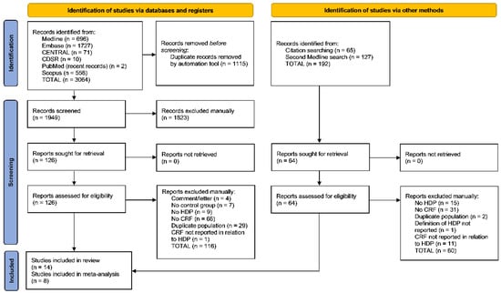
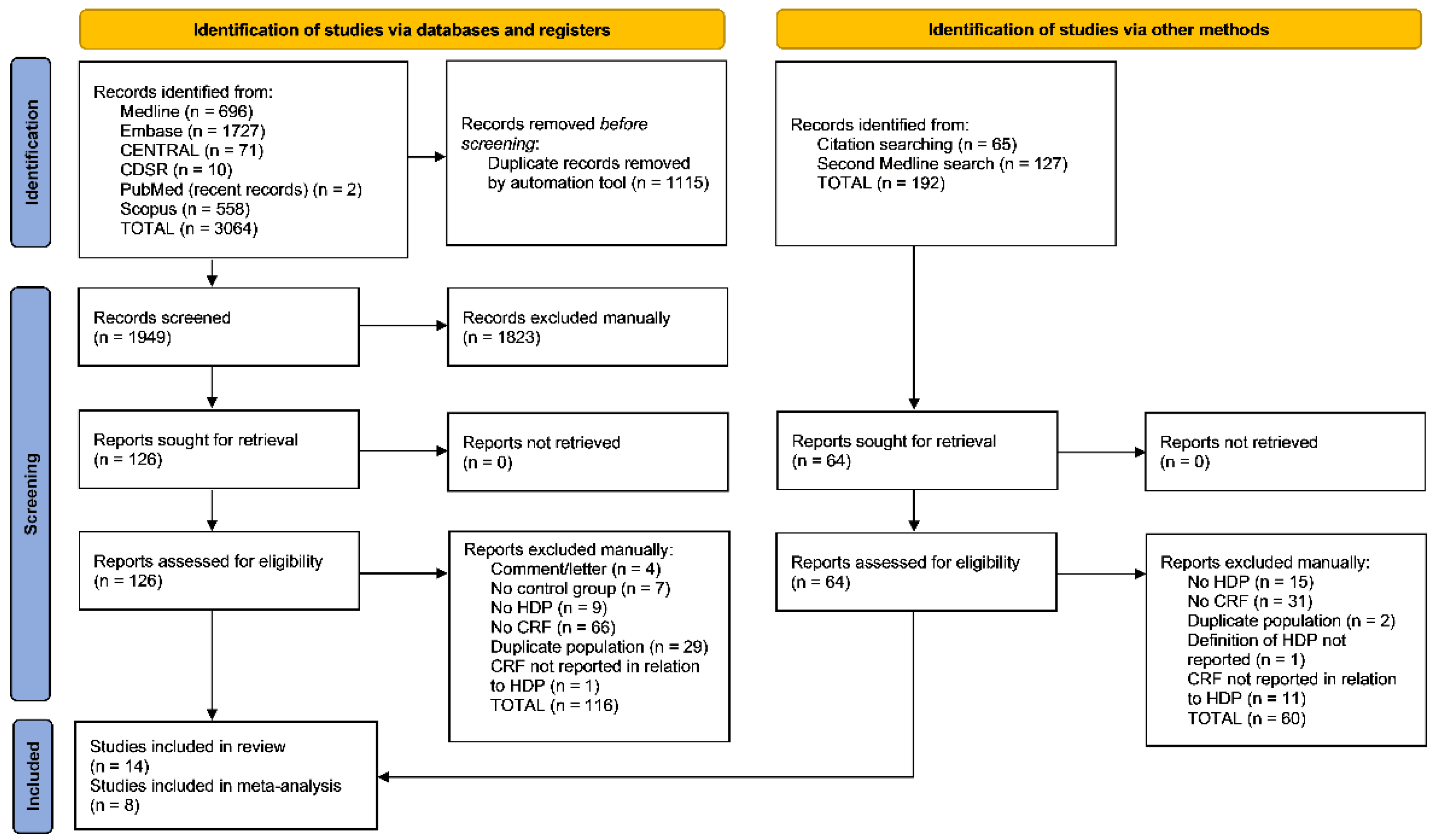
The initial literature search yielded 1949 records for title and abstract screening after removal of duplicate results, of which 126 records were retrieved and assessed for eligibility. A total of 116 reports were excluded for reasons including no measure of any HDP or CRF, no normotensive comparison group, CRF not reported in relation to HDP, duplicate study population, or inappropriate study design (Figure 1). Ten studies were included in the review [17,18,19,22,23,24,25,26,27,28] and five in the meta-analysis [17,19,22,23,28]. Manual citation searching of included articles and relevant systematic reviews yielded 65 additional references, of which 3 were included in our systematic review [29,30,31] (2 in the meta-analysis [29,31]). Finally, the second Medline search conducted on 14 January 2021 yielded 127 additional records for title and abstract screening, among which 13 additional eligible reports were assessed, and 1 [32] was included in the meta-analysis. Authors were contacted to request study data for nine studies for which data relevant to our review were potentially available but not reported [18,19,23,25,27,28,29,30,32]; authors of seven studies responded and provided relevant data [19,23,25,28,29,30,32]. A total of 14 studies met final inclusion criteria including 2406 women: 560 with HDP GH/PE and 1846 without GH/PE.
Figure 1.
PRISMA flow diagram.
3.2. Study Characteristics
Eight cohort studies [18,22,25,26,27,28,31,32], four RCTs [19,23,29,30], one cross-sectional study [24], and one non-randomized intervention study [17] were included (Table 1). The number of included participants in each study ranged from 26 to 768, while the number of participants with GH/PE ranged from 3 to 139. Estimated VO2max was measured in four studies [18,19,28,29], VO2peak in three studies [17,22,32] and VO2AT in one study [30]. Of those that did not measure VO2, two studies used a Balke treadmill test [26,28], one used a cycle ergometer test (pulse-rate-controlled submaximal 6 min exercise test with target heart rate ≥ 140 bpm) [31], and three used a walk or run test (6 min walk test, 4-m walk test, 2-mile walk or run test) [23,24,25]. CRF was examined before [18,27], during [19,23,24,28,29,30,31], and after [17,22,23,25,26,32] pregnancy in two, seven, and six studies, respectively (one study examined CRF both during pregnancy and postpartum [23]). Among studies that examined CRF during pregnancy, CRF was measured before diagnosis of HDP in four studies [19,23,28,29] (range 12 to 18 weeks gestation) and after diagnosis of HDP in one study [31] (35.5 to 37.7 weeks gestation), while one study included serial measurements of CRF [30] (14 and 28 weeks gestation). Eight studies contained sufficient data to be included in the meta-analysis [17,19,22,23,28,29,31,32]; results from the remaining six studies were reported qualitatively [18,24,25,26,27,30]. Of the four RCTs, two [29,30] reported clinical trial registration.
Table 1.
Study and participant characteristics.
3.3. Study Quality
Study quality ratings are shown in Table 2. Appropriate conduct of analysis and follow-up was documented in nearly all studies. Exposure measurement was considered valid and reliable in 12 of 14 studies. One study was presented as an abstract, and women with previous severe PE were compared to controls, but assessment of PE was not well-described [22]. The second study reported mean watts produced but did not report VO2 [23]. The outcome was measured in a valid and reliable way in 8 [17,19,22,23,28,29,30,32] of 14 studies. Unreliable outcome measures for the purposes of our review included self-reported HDP [27], assessment of HDP without reporting diagnostic criteria [18], and measurement of work rate [31], distance travelled [24,25], or exercise test duration [26] without presentation of sufficient data to calculate VO2.
Table 2.
Study quality ratings based on Briggs Institute Critical Appraisal Tools.
Confounding factors such as maternal ethnicity, smoking, education, and income were identified in 13 of 14 studies, and strategies to deal with confounding factors were stated in 9 of these studies. However, none of the studies included adjusted analyses of the association between HDP and CRF.
3.4. Study Description
3.4.1. CRF before Pregnancy and Subsequent HDP
Two studies [18,27] examined CRF before pregnancy in relation to future GH and/or PE. Morris et al. [18] compared women with VO2max < 37 vs. ≥37 mL O2/min and found that pre-pregnancy CRF was low in 100% of women with GH or PE (n = 10) compared to 57.6% (19/33) of women without GH or PE (p = 0.01). Lane-Cordova et al. [27] divided study participants into three fitness tertiles based on performance on a treadmill test and found that GH was diagnosed in 21%, 19%, and 12% of women in the lowest, middle, and highest fitness tertiles, respectively (p = 0.03). However, the study did not report rates of PE.
3.4.2. CRF during Pregnancy and Subsequent GH or PE
Seven studies [19,23,24,28,29,30,31] examined CRF during pregnancy. Bisson et al. [30] found a mean VO2AT of 15.0 and 15.7 mL/kg/min among women who developed GH and normotensive women, respectively, at 14 weeks gestation, with a mean VO2AT of 14.9 and 15.0 mL/kg/min in the two groups, respectively, at 28 weeks gestation. Da Silva et al. [24] found women who had been diagnosed with PE completed a shorter distance on the 6 min walk test compared to normotensive women (421 m vs. 497 m, p = 0.001, median gestational age at measurement 37 weeks in both groups, interquartile range 34–38 among women with PE, 33–38 for normotensive women).
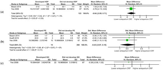
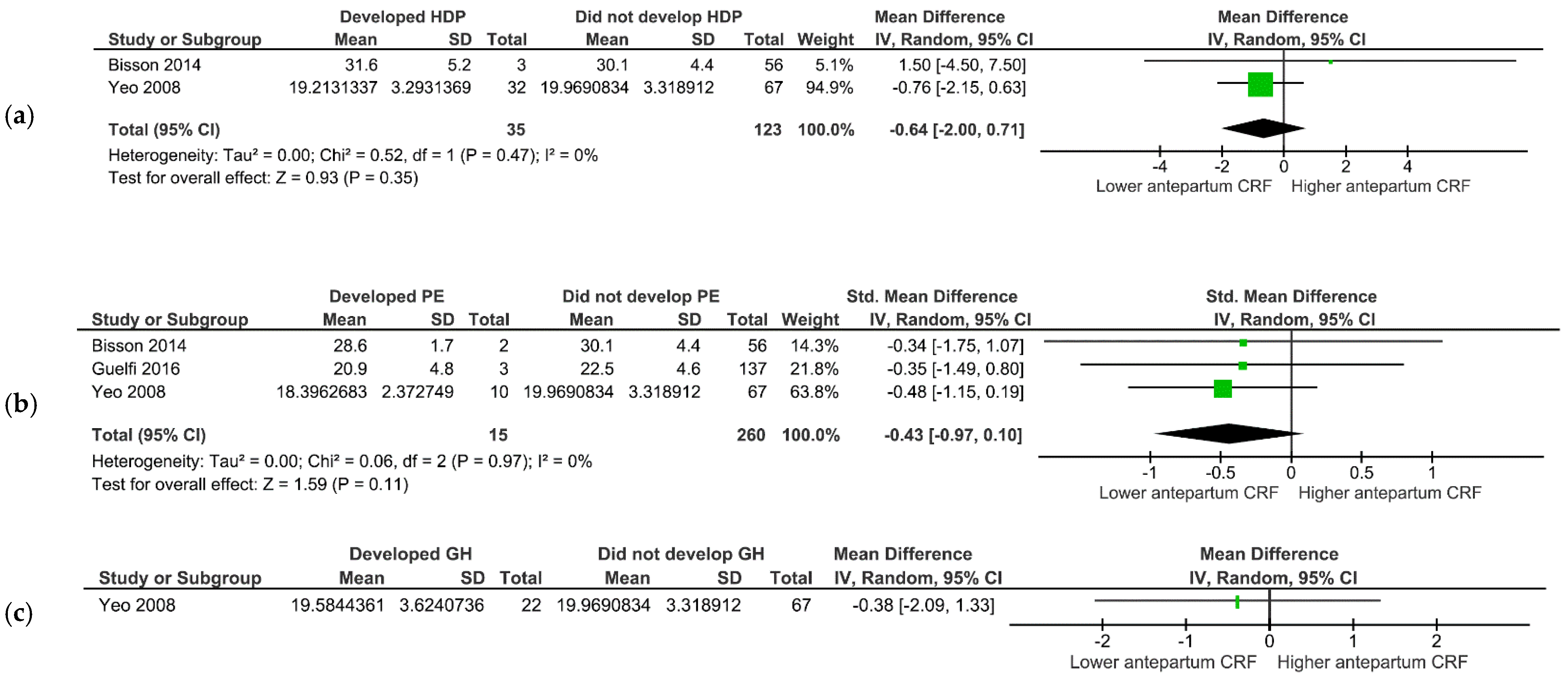
Estimated VO2max measured during pregnancy did not significantly differ between women who developed GH and/or PE combined and those who did not (two studies [19,28], 158 women, gestational age at measurement 16–18 weeks; −0.64 mL/kg/min [95% CI −2.00, 0.71]; p = 0.35) (Figure 2a). Similarly, no difference in VO2max was found between women who developed GH only vs. non-HDP women (one study [19], 146 women, gestational age at measurement 18 weeks; −0.38 mL/kg/min [95% CI −2.09, 1.33]; p = 0.66) (Figure 2c). However, a trend toward lower VO2max was observed for women who developed PE only vs. non-HDP women (three studies [19,28,29], 275 women, gestational age at measurement 14–18 weeks; −0.43 mL/kg/min [−0.97, 0.10]; p = 0.11) (Figure 2b).
Figure 2.
Mean differences in VO2max during pregnancy and subsequent development of preeclampsia and/or gestational hypertension: (a) Weight-adjusted VO2max (in mL/kg/min) and preeclampsia or gestational hypertension [19,28]; (b) Weight-adjusted VO2max (in mL/kg/min) and preeclampsia [19,28,29]; (c) Weight-adjusted VO2max (in mL/kg/min) and gestational hypertension [19].
The mean work rate did not significantly differ between groups (two studies [23,31], 88 women; −0.08 watts [−1.01, 0.85]; p = 0.17; Figure S2), nor did lactate threshold (one study [30], 47 women; −0.70 mL/kg/min [−2.25, 0.85] at 14 weeks gestation; −0.10 mL/kg/min [−1.49, 1.29] at 28 weeks gestation).
Three studies measured absolute VO2max during pregnancy without adjustment for weight, rendering values challenging to interpret. Absolute VO2max was significantly higher in women with GH or PE vs. those without (two studies [19,28], 163 women, gestational age at measurement 16–18 weeks; 0.19 L/min [0.03, 0.35]; p = 0.02) and in women with GH only vs. those without (one study [19], 94 women, gestational age at measurement 18 weeks; 0.54 L/min [0.08, 1.01]; p = 0.02), while it was marginally higher in women with PE only vs. those without (three studies [19,28,29], 277 women, gestational age at measurement 14–18 weeks; 0.96 mL/kg/min [−0.23, 2.14]; p = 0.11) (Figure S1).
3.4.3. HDP and Postpartum CRF
CRF was examined after pregnancy in six studies [17,22,23,25,26,32]. Duration since delivery at the time of CRF measurement ranged from 6 weeks to 20 years. There was a trend towards lower VO2peak in women with prior PE compared with controls (three studies [17,22,32], 208 women; −0.26 mL/kg/min [−0.54, 0.02], p = 0.07; Figure 3). Cottrill et al. [26] found that women with previous GH and/or PE had a lower mean duration of exercise on a modified Balke treadmill test compared to women with previous non-HDP pregnancy (115 women; 660 vs. 738 s; SMD −0.42 [−0.80, 0.05]; p = 0.03; measurement of CRF 4–6 years following pregnancy). Harville et al. [25] found that women with GH and/or PE took longer to complete a 4-m walk test compared to women with no previous GH or PE (1329 women; 4.11 vs. 3.95 s; 0.18 [−0.00, 0.36]; p = 0.05), with similar distances completed on a 6 min walk test (1246 women; 409.7 vs. 413.3 m; −0.05 [−0.24, 0.15]; p = 0.61) (median age at last pregnancy 28.0, median age at interview 48.4).
Figure 3.
Mean differences in VO2peak (in mL/kg/min) in postpartum women with and without previous preeclampsia [17,22,32].
4. Discussion
In this systematic review and meta-analysis of 14 studies, we identified a pattern of lower preconception CRF in women who were subsequently diagnosed with GH and/or PE. While no significant differences were noted in weight-adjusted CRF during pregnancy, results suggest that women who developed PE may have lower CRF earlier in pregnancy, as well as months to years after delivery.
It is widely known that higher CRF improves cardiovascular health and lowers cardiovascular mortality [33,34]. Previous studies have shown HDP, especially preeclampsia, to be a risk factor for later CVD [3,4], with a two-fold increased risk of ischemic heart disease 10 to 15 years following the pregnancy [5]. Our findings suggest that reduced CRF may be one pathway connecting HDP to future adverse cardiovascular outcomes. Specifically, lower postpartum CRF in HDP-affected women suggests a possible “hit” to cardiorespiratory reserve brought about by HDP—one that is potentially actionable.
Our study adds to the large body of literature suggesting that pregnancy is a “stress test” identifying women at risk for future cardiovascular risk factors and overt CVD [35]. Our analyses suggest reduced cardiorespiratory capacity both in women who went on to develop GH or PE and in those with previous HDP. Prior systematic reviews have demonstrated that engaging in physical activity during pregnancy reduces the odds of developing HDP by ~40% [36]. That this observed benefit seems mediated by improved CRF is perhaps not surprising but highlights the importance of offsetting the complex cardiovascular adaptations of both normotensive and hypertensive pregnancy with exercise. It is therefore likely that CRF is a metric that can be used both to identify higher-risk individuals and also to personalize safe and targeted exercise programs during and after pregnancy.
Our study highlights the need for the standardization of exercise tests used in research on pregnant and postpartum women in order to facilitate meaningful comparison of results between studies. Our findings also have implications for clinical practice. The American Heart Association has recommended that CRF be used as a risk marker for morbidity and mortality in the general population [12], and our review suggests that CRF may be particularly valuable as a clinical vital sign in the peripartum period and during reproductive years. CRF may also prove useful in clinical prediction of maternal morbidity, as adverse cardiovascular outcomes make up a substantial proportion of severe maternal morbidity [37,38]. Finally, our findings suggest that dedicated postpartum cardiovascular rehabilitation after HDP may be worthwhile, using a patient-centred approach sensitive to the needs of new mothers.
In light of evidence for the benefits of physical activity during pregnancy [39], the appropriateness of physical activity in women with established HDP is an area under study. Current GH is a relative contraindication to physical activity, whereas PE is an absolute contraindication [40,41]. Our review was not designed to assess whether current guideline recommendations regarding physical activity for women with HDP are appropriate, and further studies to elucidate the safe CRF threshold customization of physical activity regimens for women with HDP under the guidance of obstetrician-gynecologists or other obstetric care providers, such as kinesiologists, are essential [42].
Our review is subject to several limitations. While the included studies showed high quality ratings with regards to selection of participants and measurement of exposures and covariates, many studies used CRF measurements that did not allow calculation of VO2. Accordingly, our ability to combine results from different studies in meta-analysis was limited by the variety of CRF measures employed, as well as by the heterogeneity of study designs. Standardization of CRF measures in perinatal research, including adjustment for BMI, will be essential to understanding the role of CRF in normotensive and hypertensive pregnancy, including its lasting effects in the postpartum period. In addition, most of the included studies did not explicitly aim to measure HDP in relation to CRF and examined HDP as a secondary outcome. Furthermore, the majority of studies examining CRF during pregnancy measured CRF before HDP diagnosis, limiting our ability to draw conclusions about the effects of HDP on CRF in a current pregnancy. Finally, while preeclampsia is understood as a heterogeneous disorder with different phenotypes during pregnancy (i.e., early-onset vs. late-onset) [43], the majority of studies included in this review did not report on the timing or severity of HDP. In order to plan tailored postpartum rehabilitation programs, further studies should evaluate CRF according to the timing and severity of HDP.
HDP often occurs in women with existing comorbidities, such as pregestational diabetes and obesity, which may impact cardiovascular fitness. Women with pre-pregnancy obesity are particularly at elevated risk for PE and CVD and also tend to have reduced CRF. While our analyses focused on weight-adjusted CRF, several of the studies included in our review examined VO2 without adjustment for maternal weight, which tends to be higher in women with HDP. Additional studies using weight-adjusted VO2 values before, during, and after HDP are needed to analyze CRF changes with respect to HDP and to shed light on the interplay between HDP and CVD risk factors.
Our analyses may also be subject to confounding by age, which is known to be linked with CRF decline. However, the relatively narrow age range of child-bearing women likely minimizes the importance of this bias. Finally, we were not able to address persistence and duration of hypertension after delivery. However, PE generally normalizes within one to two weeks postpartum in 70% of individuals [44], whereas the earliest postpartum CRF measurements in the included studies were at six weeks.
Despite these limitations, our study was focused on CRF as an actionable risk marker rather than an independent causal factor. Furthermore, our aim was to identify knowledge gaps to guide future research in the prospective evaluation of CRF. Eventually, studies evaluating CRF-guided rehabilitation programs at key periods in women’s reproductive trajectory will be needed to contribute evidence-based recommendations for optimal cardiovascular health around the time of pregnancy.
5. Conclusions
In summary, in the first systematic review to date on CRF before, during, and after HDP suggested that CRF is a valuable marker of perinatal cardiovascular risk, and that postpartum measurement of CRF may shed light on the cardiovascular sequelae of hypertensive pregnancy. Our findings raise the possibility that HDP may impair cardiorespiratory reserve and suggest that dedicated postpartum cardiovascular rehabilitation programs may be indicated. Additional studies using standardized measures of VO2 are needed to quantify the strength and temporality of the association between CRF and HDP.
Supplementary Materials
The following are available online at https://www.mdpi.com/article/10.3390/jcm11154364/s1. Figure S1: Mean weight-unadjusted differences in VO2max during pregnancy and subsequent development of preeclampsia and/or gestational hypertension; Figure S2: Mean difference in work rate (Watts) during pregnancy and development of preeclampsia and/or gestational hypertension.
Author Contributions
F.A.-H. led the literature search, data extraction, and analysis. G.D.S. contributed to the literature search, data extraction, and analysis. F.A.-H. and G.D.S. drafted the manuscript. M.H.D. and M.B. provided substantive guidance and revised the manuscript. N.D. supervised all aspects of the review and revised the manuscript. All authors have read and agreed to the published version of the manuscript.
Funding
This research received no external funding.
Institutional Review Board Statement
No ethical approval was required for this study, as it consisted of secondary analysis of data from previous published studies.
Informed Consent Statement
Informed consent was obtained by primary investigators of the studies reviewed herein.
Data Availability Statement
Data available on request.
Acknowledgments
The authors thank Tara Landry and Lindsay Hales for assistance with the literature search.
Conflicts of Interest
The authors declare no conflict of interest.
Appendix A
Table A1.
Database search strategy.
Appendix B
Table A2.
References excluded with reasons.
References
- World Health Organization. WHO Recommendations for Prevention and Treatment of Pre-Eclampsia and Eclampsia; World Health Organization: Geneva, Switzerland, 2011. [Google Scholar]
- Steegers, E.A.P.; von Dadelszen, P.; Duvekot, J.J.; Pijnenborg, R. Pre-eclampsia. Lancet 2010, 376, 631–644. [Google Scholar] [CrossRef] [Scilit]
- Wu, P.; Haththotuwa, R.; Kwok, C.S.; Babu, A.; Kotronias, R.A.; Rushton, C.; Zaman, A.; Fryer, A.A.; Kadam, U.; Chew-Graham, C.A.; et al. Preeclampsia and Future Cardiovascular Health: A Systematic Review and Meta-Analysis. Circ. Cardiovasc. Qual. Outcomes 2017, 10, e003497. [Google Scholar] [CrossRef] [Scilit] [PubMed]
- Welters, S.; Teunissen, P.; Alma, L.; Hermes, W.; Ravelli, A.; De Groot, C. Higher women’s cardiovascular mortality in their forties years after hypertensive disease of pregnancy. Reprod. Sci. 2018, 25, 238A–239A. [Google Scholar]
- Bellamy, L.; Casas, J.-P.; Hingorani, A.D.; Williams, D.J. Pre-eclampsia and risk of cardiovascular disease and cancer in later life: Systematic review and meta-analysis. BMJ 2007, 335, 974. [Google Scholar] [CrossRef] [Scilit] [PubMed]
- Brown, M.C.; Best, K.E.; Pearce, M.S.; Waugh, J.; Robson, S.C.; Bell, R. Cardiovascular disease risk in women with pre-eclampsia: Systematic review and meta-analysis. Eur. J. Epidemiol. 2013, 28, 1–19. [Google Scholar] [CrossRef] [Scilit]
- Seely, E.W.; Tsigas, E.; Rich-Edwards, J.W. Preeclampsia and future cardiovascular disease in women: How good are the data and how can we manage our patients? Semin. Perinatol. 2015, 39, 276–283. [Google Scholar] [CrossRef] [Scilit]
- Vivian, H.; Heyward, A.L.G. Advanced Fitness Assessment & Exercise Prescription, 7th ed.; Human Kinetics: Champaign, IL, USA, 2014; p. 79. [Google Scholar]
- Caspersen, C.J.; Powell, K.E.; Christenson, G.M. Physical activity, exercise, and physical fitness: Definitions and distinctions for health-related research. Public Health Rep. 1985, 100, 126–131. [Google Scholar]
- Davenport, M.H.; Skow, R.J.; Steinback, C.D. Maternal Responses to Aerobic Exercise in Pregnancy. Clin. Obstet. Gynecol. 2016, 59, 541–551. [Google Scholar] [CrossRef] [Scilit]
- Cai, C.; Ruchat, S.M.; Sivak, A.; Davenport, M.H. Prenatal Exercise and Cardiorespiratory Health and Fitness: A Meta-analysis. Med. Sci. Sports Exerc. 2020, 52, 1538–1548. [Google Scholar] [CrossRef] [Scilit]
- Ross, R.; Blair, S.N.; Arena, R.; Church, T.S.; Després, J.-P.; Franklin, B.A.; Haskell, W.L.; Kaminsky, L.A.; Levine, B.D.; Lavie, C.J.; et al. Importance of Assessing Cardiorespiratory Fitness in Clinical Practice: A Case for Fitness as a Clinical Vital Sign: A Scientific Statement From the American Heart Association. Circulation 2016, 134, e653–e699. [Google Scholar] [CrossRef] [Scilit]
- Meah, V.L.; Backx, K.; Davenport, M.H.; International Working Group on Maternal Hemodynamics. Functional hemodynamic testing in pregnancy: Recommendations of the International Working Group on Maternal Hemodynamics. Ultrasound Obstet. Gynecol. 2018, 51, 331–340. [Google Scholar] [CrossRef] [Scilit] [PubMed]
- Moher, D.; Liberati, A.; Tetzlaff, J.; Altman, D. Preferred Reporting Items for Systematic Reviews and Meta-Analyses: The PRISMA Statement. PLoS Med. 2009, 6, e1000097. [Google Scholar] [CrossRef] [Scilit]
- Page, M.J.; Moher, D.; Bossuyt, P.M.; Boutron, I.; Hoffmann, T.C.; Mulrow, C.D.; Shamseer, L.; Tetzlaff, J.M.; Akl, E.A.; Brennan, S.E.; et al. PRISMA 2020 explanation and elaboration: Updated guidance and exemplars for reporting systematic reviews. BMJ 2021, 372, n160. [Google Scholar] [CrossRef]
- Institute of Medicine Committee on Standards for Systematic Reviews of Comparative Effectiveness Research. Finding What Works in Health Care: Standards for Systematic Reviews; Eden, J., Levit, L., Berg, A., Morton, S., Eds.; National Academies Press: Washington, DC, USA, 2011. [Google Scholar]
- Scholten, R.R.; Hopman, M.T.; Lotgering, F.K.; Spaanderman, M.E. Aerobic Exercise Training in Formerly Preeclamptic Women: Effects on Venous Reserve. Hypertension 2015, 66, 1058–1065. [Google Scholar] [CrossRef] [Scilit] [PubMed]
- Morris, E.; McBride, C.A.; Badger, G.J.; Bernstein, I.M. Prepregnancy fitness and risk of hypertensive disorders of pregnancy. Am. J. Obstet. Gynecol. 2017, 216 (Suppl. S1), S488–S489. [Google Scholar] [CrossRef] [Scilit]
- Yeo, S.; Davidge, S.; Ronis, D.L.; Antonakos, C.L.; Hayashi, R.; O’Leary, S. A comparison of walking versus stretching exercises to reduce the incidence of preeclampsia: A randomized clinical trial. Hypertens. Pregnancy 2008, 27, 113–130. [Google Scholar] [CrossRef] [Scilit] [PubMed]
- Moola, S.; Munn, Z.; Tufanaru, C.; Aromataris, E.; Sears, K.; Sfetcu, R.; Currie, M.; Qureshi, R.; Mattis, P.; Lisy, K. Chapter 7: Systematic reviews of etiology and risk. In Joanna Briggs Institute Reviewer’s Manual; The Joanna Briggs Institute: Adelaide, Australia, 2017; Volume 5. [Google Scholar]
- Higgins, J.P.; Thomas, J.; Chandler, J.; Cumpston, M.; Li, T.; Page, M.J.; Welch, V.A. Cochrane Handbook for Systematic Reviews of Interventions; John Wiley & Sons: Chichester, UK, 2019. [Google Scholar]
- Gronningsaeter, L.; Estensen, M.E.; Langesaeter, E.; Edvardsen, E. Cardiorespiratory fitness in women with previous preeclampsia. Eur. Heart J. 2016, 37 (Suppl. S1), 130. [Google Scholar]
- Price, B.B.; Amini, S.B.; Kappeler, K. Exercise in pregnancy: Effect on fitness and obstetric outcomes-a randomized trial. Med. Sci. Sports Exerc. 2012, 44, 2263–2269. [Google Scholar] [CrossRef] [Scilit] [PubMed]
- da Silva, E.G.; de Godoy, I.; de Oliveira Antunes, L.C.; da Silva, E.G.; Peracoli, J.C. Respiratory parameters and exercise functional capacity in preeclampsia. Hypertens. Pregnancy 2010, 29, 301–309. [Google Scholar] [CrossRef] [Scilit]
- Harville, E.W.; Chen, W.; Guralnik, J.; Bazzano, L.A. Reproductive history and physical functioning in midlife: The Bogalusa Heart Study. Maturitas 2018, 109, 26–31. [Google Scholar] [CrossRef] [Scilit] [PubMed]
- Cottrill, C.M.; Kotchen, J.M.; Guthrie, G.; Kotchen, T. Cardiovascular responses to exercise following adolescent hypertensive pregnancy. J. Adolesc. Health Care 1980, 1, 91–95. [Google Scholar] [CrossRef] [Scilit]
- Lane-Cordova, A.D.; Carnethon, M.R.; Catov, J.M.; Montag, S.; Lewis, C.E.; Schreiner, P.J.; Dude, A.; Sternfeld, B.; Badon, S.E.; Greenland, P.; et al. Cardiorespiratory fitness, exercise haemodynamics and birth outcomes: The Coronary Artery Risk Development in Young Adults Study. BJOG 2018, 125, 1127–1134. [Google Scholar] [CrossRef] [Scilit] [PubMed]
- Bisson, M.; Rheaume, C.; Bujold, E.; Tremblay, A.; Marc, I. Modulation of blood pressure response to exercise by physical activity and relationship with resting blood pressure during pregnancy. J. Hypertens. 2014, 32, 1450–1457, discussion 1457. [Google Scholar] [CrossRef] [Scilit] [PubMed]
- Guelfi, K.J.; Ong, M.J.; Crisp, N.A.; Fournier, P.A.; Wallman, K.E.; Grove, J.R.; Doherty, D.A.; Newnham, J.P. Regular Exercise to Prevent the Recurrence of Gestational Diabetes Mellitus: A Randomized Controlled Trial. Obstet. Gynecol. 2016, 128, 819–827. [Google Scholar] [CrossRef] [Scilit] [PubMed]
- Bisson, M.; Almeras, N.; Dufresne, S.S.; Robitaille, J.; Rheaume, C.; Bujold, E.; Frenette, J.; Tremblay, A.; Marc, I. A 12-Week Exercise Program for Pregnant Women with Obesity to Improve Physical Activity Levels: An Open Randomised Preliminary Study. PLoS ONE 2015, 10, e0137742. [Google Scholar] [CrossRef] [Scilit] [PubMed]
- Rauramo, I.; Forss, M. Effect of exercise on placental blood flow in pregnancies complicated by hypertension, diabetes or intrahepatic cholestasis. Acta Obstet. Gynecol. Scand. 1988, 67, 15–20. [Google Scholar] [CrossRef] [Scilit] [PubMed]
- Ersbøll, A.S.; Bojer, A.S.; Hauge, M.G.; Johansen, M.; Damm, P.; Gustafsson, F.; Vejlstrup, N.G. Long-term cardiac function after peripartum cardiomyopathy and preeclampsia: A Danish nationwide, clinical follow-up study using maximal exercise testing and cardiac magnetic resonance imaging. J. Am. Heart Assoc. 2018, 7, e008991. [Google Scholar] [CrossRef] [Scilit]
- Al-Mallah, M.H.; Sakr, S.; Al-Qunaibet, A. Cardiorespiratory Fitness and Cardiovascular Disease Prevention: An Update. Curr. Atheroscler. Rep. 2018, 20, 1. [Google Scholar] [CrossRef] [Scilit]
- Sui, X.; Sarzynski, M.A.; Lee, D.C.; Kokkinos, P.F. Impact of Changes in Cardiorespiratory Fitness on Hypertension, Dyslipidemia and Survival: An Overview of the Epidemiological Evidence. Prog. Cardiovasc. Dis. 2017, 60, 56–66. [Google Scholar] [CrossRef] [Scilit]
- Coutinho, T.; Lamai, O.; Nerenberg, K. Hypertensive Disorders of Pregnancy and Cardiovascular Diseases: Current Knowledge and Future Directions. Curr. Treat Options Cardiovasc. Med. 2018, 20, 56. [Google Scholar] [CrossRef] [Scilit] [PubMed]
- Davenport, M.H.; Ruchat, S.M.; Poitras, V.J.; Jaramillo Garcia, A.; Gray, C.E.; Barrowman, N.; Skow, R.J.; Meah, V.L.; Riske, L.; Sobierajski, F.; et al. Prenatal exercise for the prevention of gestational diabetes mellitus and hypertensive disorders of pregnancy: A systematic review and meta-analysis. Br. J. Sports Med. 2018, 52, 1367–1375. [Google Scholar] [CrossRef] [Scilit] [PubMed]
- Bateman, B.T.; Mhyre, J.M.; Hernandez-Diaz, S.; Huybrechts, K.F.; Fischer, M.A.; Creanga, A.A.; Callaghan, W.M.; Gagne, J.J. Development of a comorbidity index for use in obstetric patients. Obstet. Gynecol. 2013, 122, 957–965. [Google Scholar] [CrossRef] [Scilit] [PubMed]
- Dayan, N.; Shapiro, G.D.; Luo, J.; Guan, J.; Fell, D.B.; Laskin, C.A.; Basso, O.; Park, A.L.; Ray, J.G. Development and internal validation of a model predicting severe maternal morbidity using pre-conception and early pregnancy variables: A population-based study in Ontario, Canada. BMC Pregnancy Childbirth 2021, 21, 679. [Google Scholar] [CrossRef] [Scilit]
- May, L.E.; Knowlton, J.; Hanson, J.; Suminski, R.; Paynter, C.; Fang, X.; Gustafson, K.M. Effects of Exercise During Pregnancy on Maternal Heart Rate and Heart Rate Variability. PM&R 2016, 8, 611–617. [Google Scholar] [CrossRef] [Scilit]
- Mottola, M.F.; Davenport, M.H.; Ruchat, S.M.; Davies, G.A.; Poitras, V.; Gray, C.; Garcia, A.J.; Barrowman, N.; Adamo, K.B.; Duggan, M.; et al. No. 367-2019 Canadian Guideline for Physical Activity throughout Pregnancy. J. Obstet. Gynaecol. Can. 2018, 40, 1528–1537. [Google Scholar] [CrossRef] [Scilit]
- Meah, V.L.; Davies, G.A.; Davenport, M.H. Why can’t I exercise during pregnancy? Time to revisit medical ‘absolute’ and ‘relative’ contraindications: Systematic review of evidence of harm and a call to action. Br. J. Sports Med. 2020, 54, 1395–1404. [Google Scholar] [CrossRef] [Scilit] [PubMed]
- ACOG Committee Members. Physical Activity and Exercise During Pregnancy and the Postpartum Period: ACOG Committee Opinion, Number 804. Obstet. Gynecol. 2020, 135, e178–e188. [Google Scholar] [CrossRef] [Scilit]
- Masini, G.; Foo, L.F.; Tay, J.; Wilkinson, I.B.; Valensise, H.; Gyselaers, W.; Lees, C.C. Preeclampsia has two phenotypes which require different treatment strategies. Am. J. Obstet. Gynecol. 2022, 226, S1006–S1018. [Google Scholar] [CrossRef] [Scilit]
- Podymow, T.; August, P. Postpartum course of gestational hypertension and preeclampsia. Hypertens. Pregnancy 2010, 29, 294–300. [Google Scholar] [CrossRef] [Scilit]



Publisher’s Note: MDPI stays neutral with regard to jurisdictional claims in published maps and institutional affiliations. |
© 2022 by the authors. Licensee MDPI, Basel, Switzerland. This article is an open access article distributed under the terms and conditions of the Creative Commons Attribution (CC BY) license (https://creativecommons.org/licenses/by/4.0/).