


Abstract
Neuroinflammation is a multifaceted process in which specific biomolecules (such as pro-inflammatory cytokines and signaling proteins) act as primary mediators of neuropsychiatric disorders, including depression. Despite growing evidence, the precise interconnected cascades within these neuroinflammatory signaling pathways and their regulatory proteins remain to be fully elucidated, which presents a significant unmet need for targeted therapeutic strategies with clear mechanistic validation. Accordingly, the present review analyzes the impact of neuroinflammation on the pathophysiology of depression in various rodent models. We explore the regulatory mechanisms by which factors such as oxidative stress, calcium signaling, and inflammasome-mediated inflammation affect neurotransmitter synthesis and neuronal integrity. Furthermore, we elucidate the therapeutic potential of synthetic compounds and naturally derived agents targeting these specific molecular pathways. Based on a systematic literature search, this review focuses exclusively on pharmacological agents with clearly defined molecular mechanisms and rigorous depression-associated in vivo validation, thereby providing promising therapeutic strategies for the restoration of neurobiological homeostasis.
1. Introduction
Major depressive disorder (MDD) affects nearly 3.9% of the global population and represents the most critical paradigms due to their unparalleled disease burden with structural impact on the brain, and profound clinical severity [1,2]. While alterations in monoaminergic neurotransmission have long been implicated in the pathophysiology of MDD, accumulating evidence suggests that neuroimmune dysregulation is an important contributor to depressive pathophysiology, prompting a shift toward broader neurobiological mechanisms [3]. Central to this evolving perspective is neuroinflammation, a multifaceted process that responds to injuries, infections, and pathological events. Characterized by the involvement of diverse biomolecules and cellular responses, it is posited not as a primary etiology, but rather as a potential contributing or modulating factor in the pathophysiology of neuropsychiatric disorders, including depression, schizophrenia, and bipolar disorder [3,4,5,6].
To understand neuropsychiatric disorders such as depression, various pathophysiological mechanisms have been proposed and studied. In particular, various studies have emphasized that the onset of neuroinflammation is promoted by the production of reactive oxygen species (ROS) and mobilization/circulation of pro-inflammatory mediators, notably interleukin (IL)-1β, IL-6, and tumor necrosis factor (TNF)-α [7,8,9]. These inflammatory molecules act as pivotal orchestrators, initiating downstream signaling pathways such as mitogen-activated protein kinase (MAPK) pathways, particularly the extracellular signal-regulated kinase (ERK) cascade within glial cells, thereby promoting pro-inflammatory cytokine production and sustaining neuroinflammatory responses [9,10,11].
The sustained activation of inflammatory signaling pathways leads to profound transcriptional and functional remodeling of microglia, characterized by enhanced inflammatory signaling and altered interactions with neurons and astrocytes [12,13,14,15]. This microglial remodeling promotes excessive oxidative/nitrosative stress and the maturation of the inflammasome complex, further augmenting the feedback loop of inflammation [16]. Moreover, several studies have reported that oxidative/nitrosative stress leads to lipid peroxidation, reduced glutathione (GSH) levels, DNA damage and an attenuated enzymatic reduction of oxidative stress, all of which are involved in the pathophysiology of depression and anxiety [17,18,19,20]. Crucially, these inflammatory molecules are active participants in disrupting the structural integrity of the blood–brain barrier (BBB), thereby inducing enhanced permeability to peripheral immunocytes and disrupting the homeostatic environment necessary for neuronal health [16].
In the absence of homeostatic control, the feed-forward loop of oxidative damage and inflammasome-mediated neuroinflammation not only impairs neuronal integrity but also is increasingly recognized to contribute to the manifestation of depressive symptoms in patients and depression-like behaviors (DLBs) in animal models (Figure 1). The present review, therefore, provides a focused evaluation of the key biomolecules and cytokines that constitute this neuroinflammatory milieu, highlighting their potential roles in modulating depression-related pathophysiology. By evaluating established and emerging evidence regarding their mechanisms of action, we highlight how these discrete molecular signatures converge to modulate the pathophysiology of depression in various stress- or agent-induced experimental models of the depressive disorder. Furthermore, we elucidate the therapeutic potential of diverse synthetic compounds and naturally derived agents that target these specific molecular nodes, offering promising targets and expanded strategies to attenuate the neuroinflammatory drive and restore neurobiological homeostasis and behavioral equilibrium.
Figure 1.
Schematic illustration of interconnected neuroinflammatory signaling pathways contributing to the pathophysiology of depression and their potential therapeutic targets. Neuroinflammation is initiated by diverse clinical and pathological factors, including chronic stress, infection, metabolic dysfunction, and tissue injury, as well as by experimental inducers commonly used in animal models. These stimuli promote peripheral inflammatory responses and BBB dysfunction, facilitating glial activation and the release of pro-inflammatory mediators. Sustained neuroinflammatory signaling, characterized by excessive cytokine production, oxidative/nitrosative stress, and inflammasome activation, contributes to neuronal and synaptic dysfunction, ultimately promoting depressive symptoms in patients and DLBs in animal models. Therapeutic interventions targeting these interconnected neuroinflammatory pathways—including synthetic compounds and naturally derived agents—may attenuate inflammatory responses, preserve neuronal homeostasis, and restore behavioral function. Solid arrows indicate established relationships, whereas dotted arrows indicate potential or indirect relationships rather than definitive causal links. Abbreviation: DLBs, depression-like behaviors; BBB, blood–brain barrier; IL, interleukin; TNF-α, tumor necrosis factor-α; ROS, reactive oxygen species; RNS, reactive nitrogen species; NLRP3, NOD-like receptor family pyrin domain containing 3; 5-HT, serotonin; DA, dopamine; NE, norepinephrine; LPS, lipopolysaccharide; CUMS, chronic unpredicted mild stress; TSG, total saponins of Panax ginseng.
2. Methodology and Inclusion Criteria
This review comprised studies retrieved from PubMed and focused on the effects of various factors, such as oxidative stress and calcium (Ca2+) signaling, on neuroinflammation and depression. In particular, we focused on depression models, signaling proteins, and the potential of pharmacological agents (synthetic and natural-derived agents) as treatment strategies. Furthermore, the current review explored the relationship between diverse disease models and depression, but also elucidated a molecular regulatory mechanism underlying neuroinflammation-driven depressive disorders.
2.1. Search Strategy and Literature Selection
A literature search was conducted on PubMed for articles published between 1972 and 2026. The complete search string was structured as follows: oxidative stress or nitrosative stress, Ca2+, neuroinflammation, depression, neuronal apoptosis, and hyperglycemia.
2.2. Inclusion/Exclusion Criteria and Screening Process
Studies were selected according to predefined eligibility criteria.
2.2.1. Inclusion Criteria
- (1)
- Study design and models: Peer-reviewed original research articles utilizing in vivo animal models of depression and disease-related models presenting depression-like behavioral or neuroinflammatory outcomes;
- (2)
- Signaling pathways: Studies evaluating key regulatory proteins that act as initiators and amplifiers of inflammation, mediators of oxidative stress and cell death, or regulators of ion dyshomeostasis, representing core convergence points of anti-depressant efficacy;
- (3)
- Pharmacological interventions: Studies investigating pharmacological agents (synthetic and natural-derived) sharing a common therapeutic axis—specifically, the attenuation of neuroinflammatory cascades, suppression of cellular stress pathways (e.g., ER stress and ferroptosis), and the restoration of microglial homeostasis.
2.2.2. Exclusion Criteria
- (1)
- In vitro-only studies without accompanying in vivo validation;
- (2)
- Non-peer-reviewed publications or conference abstracts.
2.2.3. Screening Process
Title, abstract, and full-text screening was performed independently by four reviewers. Discrepancies were resolved through group discussion and consensus. After applying the eligibility criteria, the selected original studies were included in this review.
3. Emerging Candidate Signaling Proteins in Depression and Neuroinflammation
This section explores the intracellular components induced by neuroinflammation to elucidate the molecular mechanisms underlying depression. In this context, we highlight a diverse array of emerging candidate signaling molecules categorized by their primary pathological axes: initiators and amplifiers of the inflammation [NOD-like receptor (NLR) pyrin domain-containing proteins (NLRPs) and sterile α and TIR motif containing 1 (SARM1)], oxidative stress and induction of cell death [peroxiredoxins (Prdxs), nitric oxide synthase (NOS), and glutathione peroxidase 4 (GPx4)], and ion dyshomeostasis [Na+, K+-ATPase (NKA), sarco/endoplasmic reticulum Ca2+-ATPase 2 (SERCA2), synaptojanin-2-binding protein (SYNJ2BP), and endophilin A1 (EPA1)]-encompassing additional regulatory factors that collectively drive synaptic dysfunction and neuronal impairment within the depressive brain. To facilitate understanding, these targets and their preliminary supporting evidence in depression and disease-related models are summarized in Table 1.
3.1. The Initiators and Amplifiers of the Inflammation
3.1.1. NOD-like Receptor Pyrin Domain-Containing Protein
NLRs are intracellular pattern recognition receptors that monitor various pathogen-associated molecular patterns and damage-associated molecular patterns and are able to form multimeric inflammasome complexes [21,22]. In the central nervous system, NLRP1 is principally expressed within cortical and spinal neurons, as well as oligodendrocytes, with lower levels detected in other glial populations [23]. The NLRP1 inflammasome promotes activation of caspase (Casp)1, and this interaction leads to pro-Casp1 auto-cleavage, forming biologically functional p10/p20 heterodimers [24]. Subsequently, activation of Casp1 results to maturation and secretion of pro-IL-18 and pro-IL-1β. Consequently, activation of IL-18 and IL-1β mediates innate and adaptive immune systems [25]. Among NLRP isoforms, NLRP3, known as cryopyrin and NALP3, is the best characterized component of inflammasome [21] which is formed by stimulation of toll-like receptor (TLR) activation, cytokine, and others [26]. NLRP3 expression is founded in neutrophils, monocytes, and dendritic cells, which are thought to be high producers of IL-1β [27]. Specifically, activation of NLRP3 inflammasome is induced through enhanced the generation of ROS and thioredoxin-interacting protein (TXNIP) by chronic hyperglycemia; subsequently, the ROS/TXNIP axis promotes the activation and secretion of Casp1-dependent IL-1β [28,29]. Moreover, hyperglycemia enhances permeability of the blood–brain barrier and neutrophil infiltration and subsequently induces brain ischemic injury [30]. Streptozotocin (STZ), known as a chemical for inducer of type 1 diabetes mellitus, affects insulin-producing pancreatic β-cells through its toxic effect and has been demonstrated to mimic diabetes-associated depression mouse models [31,32,33,34,35]. To mimic the hyperglycemic mouse model, microglia is stimulated by high glucose [36]. DLBs, NLRP3 activation, and neuroinflammation are ameliorated by the pharmacological inhibition of NLRP3 using MCC950 in the hippocampal region of a STZ-elicited hyperglycemic rodent model [36]. Neuroinflammation is enhanced through activated nuclear factor kappa-light-chain-enhancer of activated B cells (NF-κB)-NLRP3 pathway and phosphorylated double-stranded RNA-dependent protein kinase (PKR)-TXNIP-ROS-NLRP3 signaling by treatment with high glucose in mouse microglia [36].
3.1.2. Sterile α and TIR Motif Containing 1
Sterile α and TIR motif containing 1 (SARM1), as a NAD+ hydrolase, possesses crucial functions in immune responses and neuronal degeneration [37]. A study recently showed that knockdown of SARM1 induces the phosphorylated form of c-Jun N-terminal kinase (JNK) and subsequently induces DLBs and synaptic impairment in mice [38]. Genetic and pharmacological deletion of SARM1 exacerbates neuroinflammation through the activation of the JNK/stimulator of interferon genes (STING)/TANK-binding kinase (TBK)1 and NLRP3 signaling pathways in both hippocampal tissues and neuronal cells [38]. Notably, these pro-inflammatory effects and the concomitant production of ROS are attenuated by the JNK inhibitor SP600125, indicating that the SARM1-deficiency-elicited inflammatory response is mediated via the JNK/STING/TBK1/NLRP3 signaling axis [38].
3.2. Oxidative Stress and Induction of Cell Death
3.2.1. Peroxiredoxins
Peroxiredoxins (Prdxs) are a family of conserved antioxidant enzymes that scavenge excessive ROS and oxidative stress [39,40]. A recent study addressed that LPS-elicited depression models exhibit significant behavioral and molecular alterations [41]. Systemic administration of LPS not only triggers depression-like symptoms but also exacerbates neuroinflammation and ROS production within the hippocampal region and microglial cells. In response to the amplified oxidative stress, the expression levels of Prdxs members, including Prdx 1, 2, 4, and 5, are significantly upregulated, suggesting a localized antioxidant defense mechanism in the LPS-stimulated neuroenvironment [41].
3.2.2. Nitric Oxide Synthase
Several studies report that pro-inflammatory cytokines, which interact with various pathophysiologic targets concerning depressive disorder, including dysfunction of neurotransmitter metabolism, neuroendocrine function, and synaptic plasticity [42,43], are upregulated in MDD patients [42,44]. NOSs have three isoforms, comprising neuronal (nNOS), inducible (iNOS), and endothelial NOS (eNOS), and possess biological functions, including regulation of vascular homeostasis, neurotransmission, and immunological surveillance [45]. Among them, iNOS mediates NO overproduction, thereby leading to neurotoxicity [46,47]. Moreover, NO is produced through L-arginine oxidation by NOS in mammalian cells, and functions as a pivotal regulatory factor of diverse physiological homeostasis in the immune, cardiovascular, and nerve systems [46]. Neuroinflammation is attenuated through NO-cGMP signaling via inhibition of NOS and phosphodiesterase (PDE) 5 by treatment with Nω-nitro-L-arginine methyl ester hydrochloride (L-NAME), aminoguanidine or sildenafil, respectively, thereby decreasing DLBs in the hippocampal region of LPS-elicited rodent models of depression [48]. Moreover, antioxidant effects are enhanced through increased GSH levels by treatment with L-NAME, aminoguanidine or sildenafil in the hippocampal region of LPS-elicited rodent models of depression [48].
3.2.3. Glutathione Peroxidase 4
Recent advances highlighting the role of ferroptosis—induced cell death by iron-dependent lipid peroxidation—suggest that this oxidative microenvironment directly triggers neuroinflammation in depression. Specifically, recent evidence from LPS-elicited neuroinflammation models demonstrates that the downregulation of glutathione peroxidase 4 (GPx4) elevates the susceptibility of microglia to oxidative degradation of lipid and subsequent ferroptosis [49]. Notably, pharmacological or metabolic restoration of GPx4—such as through nicotinamide mononucleotide administration—significantly replenishes GSH pools, suppresses pro-inflammatory cytokine transcription, and ameliorates microglia activation [49]. This intricate coupling between GPx4 depletion and microglial activation sustains a vicious cycle between oxidative stress and neuroinflammation, underscoring the need to expand traditional antioxidant paradigms (e.g., Prdxs and NOS) toward ferroptosis-targeted interventions. Moreover, the modulatory approach of GPx4 should be studied in depression-associated models in the coming years.
3.3. Ion Dyshomeostasis
3.3.1. Na+, K+-ATPase
Na+, K+-ATPase (NKA), found in all animal cells, is a cellular membrane-localized Na+ pump and comprises two distinct subunits: a catalytic α-subunit harboring the ion-transporting domains, and β-subunit, characterized as a monotopic glycoprotein [50,51]. The electrochemical gradient (e.g., Na+ and K+) is crucial in regulation of ionic homeostasis [52]. In pathological conditions, it has been known that free radicals and NO disrupt NKA activity [53]. In the brain, NKA activity is essential for maintenance of neuronal function, and inhibition of NKA activity by pharmaceutical agents and gene deletion is involved in aging [54], neurological disease including Parkinson’s disease (PD) and Alzheimer’s disease (AD) [55], and mood disorders [56,57]. In rats, stimulation of repeated restraint stress promotes neuroinflammation and oxidative stress by enhancing activator peptide-1, iNOS, and cyclooxygenase (COX)-2 activity, while simultaneously inducing neuronal degeneration and a reduction in α2,3-NKA activity in the frontal cortex, which is an emotional and cognitive region [58]. Additionally, decline in NKA function is further validated in the reserpine-elicited depression rat model, where a substantial decrease in enzyme activity is observed [59]. Briefly, reserpine, known as an antihypertensive agent [60], is used to elicit depression in animal models to assess the pathological manifestations of depressive disorder [61]. In rat models of bipolar disorder, it has been demonstrated that DLBs accompany neuroinflammation following the administration of ouabain, a potent NKA inhibitor [62]. These findings collectively suggest that NKA serves as more than just an ion pump; it acts as a molecular bridge between biochemical stressors and behavioral manifestations. Consequently, the pathoelectrophysiological role of NKA should be highlighted as a fundamental mechanism that links ionic dyshomeostasis and neuroinflammatory cascades in the neuropsychiatric environment. Although direct clinical trials evaluating specific NKA activators in neuropsychiatric disorders are still lacking, growing correlative evidence from brain samples and biomarker studies might support NKA as a viable therapeutic target.
3.3.2. Sarco/Endoplasmic Reticulum Ca2+-ATPase 2
The endoplasmic reticulum (ER) is responsible for maintenance of Ca2+ homeostasis, modulation of apoptosis, lipid synthesis, and protein folding [63]. When ER Ca2+ homeostasis is disrupted, misfolded proteins are accumulated and the unfolded protein response is activated [64]. Persistent ER stress leads to cell death via C/EBP homologous protein (CHOP) and downstream effectors like Casp3 and Casp12 [65,66]. The sarco/endoplasmic reticulum Ca2+-ATPase 2 (SERCA2) performs a vital function in ER-Ca2+ regulation and pumps cytosolic Ca2+ into the ER lumen [67]. SERCA2 dysregulation mediates ER Ca2+ depletion, followed by protein misfolding, ER stress, and apoptosis [68]. Diminished ER-released intracellular Ca2+ peak is induced through impaired SERCA2-mediated Ca2+ reuptake and reduced ER-Ca2+ content, subsequently leads to enhanced ER stress in the hippocampal region of STZ-elicited hyperglycemia mouse models and high glucose-treated human neuroblastoma SH-SY5Y cells [69]. Enhanced ER stress by thapsigargin accelerates cell death through enhanced expression of CHOP, cleaved Casp12, and apoptosis markers, such as Bcl2-associated X (Bax) and cleaved Casp3, thereby contributing to the development of DLBs, including anhedonia, despair, and anxiety, in the hippocampal region of STZ-elicited hyperglycemia mouse models and high glucose-treated human neuroblastomas [69]. Thus, as the modulatory strategy on ER stress, SERCA2 activation with tauroursodeoxycholic acid (TUDCA) reduces ER stress and DLBs in STZ-elicited hyperglycemia mouse models [69]. Moreover, TUDCA is currently undergoing clinical evaluation for various neurodegenerative diseases, including amyotrophic lateral sclerosis (ALS), AD, and PD, owing to its neuroprotective effects [70,71].
3.3.3. Synaptojanin-2-Binding Protein
Importantly, the Ca2+ dyshomeostasis mediated by SERCA2 dysfunction closely converges with novel structural modulators at the endoplasmic reticulum (ER)–mitochondria interface. Recent studies have identified synaptojanin-2-binding protein (SYNJ2BP) as a crucial regulator of ER–mitochondria tethering and synaptic plasticity in depression models [72]. The downregulation of SYNJ2BP exacerbates Ca2+-dependent mitochondrial apoptosis and neuroinflammation via the disruption of the phosphatidylinositol 4,5-bisphosphate (PIP2)/inositol 1,4,5-trisphosphate signaling axis, positioning the SERCA2-SYNJ2BP crosstalk as a potential nexus for stress-induced neurodegeneration [72]. Although clinical translation targeting SYNJ2BP remains unclear, these findings highlight the SERCA2-SYNJ2BP axis as a potential biomarker for stress-induced neurodegenerative disorders.
3.3.4. Endophilin A1
Endophilin A1 (EPA1) is localized in the synaptic terminals involved in synaptic organization such as endocytosis of synaptic vesicle and actin polymerization [73,74]. A recent study showed that the synaptic-associated protein EPA1 expression is enhanced in the hippocampal region of chronic unpredicted mild stress (CUMS)-elicited rodent models of depression [75]. DLBs and neuronal degeneration are reduced by the production of synaptic-associated proteins, comprising mature neuron marker microtubule-associated protein 2, synaptophysin (SYN), brain-derived neurotrophic factor (BDNF), growth-associated protein-43, and postsynaptic density protein 95 (PSD95), which are upregulated following the suppression of EPA1 in CUMS-elicited rodent models of depression [75]. EPA1 expression is associated with intracellular Ca2+ contents and ROS production [76]. In the CUMS-elicited rodent model of depression, the levels of Ca2+/ROS are attenuated by suppressing EPA1 in the hippocampal region [75]. Moreover, hippocampal inflammation is exacerbated via the upregulation of the EPA1-NLRP1 inflammasome/apoptosis-associated speck-like protein containing a CARD (ASC)/Casp1 signaling axis in a CUMS-induced rodent model of depression [75]. Thus, mitochondrial structure and hippocampal neuronal ultrastructure are damaged through the involvement of EPA1-mediated Ca2+-ROS-NLRP1/ASC/Casp1 signaling in CUMS-elicited rodent models of depression [75].
Table 1.
Effects of signaling proteins in depression and disease-related models presenting depression-like behavioral or neuroinflammatory outcomes.
4. Modulation of Neuroinflammation and Therapeutic Approaches
Collectively, the pharmacological agents discussed in this section—ranging from natural-derived phytochemicals to synthetic molecules—converge on a shared therapeutic axis: the attenuation of neuroinflammatory cascades, suppression of cellular stress pathways (e.g., ER stress and ferroptosis), and the restoration of microglial homeostasis. Rather than acting through isolated mechanisms, these diverse compounds functionally counteract neuroinflammation by cooperatively suppressing inflammasome activation, oxidative/nitrosative stress, and BBB dysfunction. The following subsections synthesize how these distinct molecular entities orchestrate multi-target protection in preclinical models of depression and neuroinflammation. Notably, while these compounds broadly target the shared neuroinflammatory pathways described above, most are supported primarily by preclinical evidence; among them, only curcumin and mangiferin have reported clinical outcomes to date. To provide clear translation context, these distinctions in preclinical models and clinical interpretations are categorized and summarized in Table 2.
4.1. Doxycycline
The pathophysiology of MDD is mediated by excessive oxidative/nitrosative stress, including overproduced peroxide and NO as well as involvement of the BDNF signaling pathway [20,77,78,79]. Furthermore, downregulated levels of an antioxidant defense, such as GSH content, have been demonstrated in the post-mortem MDD brain [80]. Doxycycline (6-Deoxy-5-hydroxytetracycline) is a tetracycline antibiotic used for treating infections elicited by both Gram-negative and Gram-positive bacteria [81] and possesses various pharmacological functions, such as reduction of oxidative stress [82] and anti-inflammatory properties [83] in the central nervous system (CNS). Neuroinflammation is reduced by treatment with doxycycline in brain area of LPS-elicited rodent models of depression, thereby attenuating DLBs [84]. In addition, treatment with doxycycline induces through enhanced GSH levels an antioxidant effect in the brain area of LPS-elicited rodent models of depression [84].
4.2. Curcumin
Curcumin is a bioactive phytochemical derived from Curcuma longa and possesses diverse biological fuctions, such as mitigation of inflammation [85], and anti-carcinogenic [86], antioxidant [87], neuroprotective [88], and anti-depressant properties [89]. The neuroinflammation is downregulated through reduced expression of inflammatory cytokines, iNOS, and COX-2 via NF-κB signaling, thereby ameliorating microglial activation and associated behavioral impairments following curcumin treatment in the hippocampal region and prefrontal cortex (PFC) of LPS-elicited rodent models of depression [90,91]. Recent studies have moved beyond the monoamine hypothesis, positioning curcumin as a multi-target agent that simultaneously regulates the hypothalamic–pituitary–adrenal (HPA) axis, neuroinflammation, and oxidative stress pathways to alleviate depressive symptoms [92,93,94]. Additionally, nanoparticle platforms have been applied to enhance its availability. For instance, curcumin-coated iron oxide nanoparticles restore the activity of monoaminergic neurotransmission as well as protect against oxidative stress in reserpine-elicited depression rat models [95]. Consequently, while clinical evidence supports the therapeutic potential of curcumin in reducing depressive symptoms, its utility remains constrained by heterogeneous formulations and limited bioavailability [96,97]. Addressing these challenges through standardized clinical validation and advanced carrier platforms would be crucial to bridge the gap between experimental findings and psychiatric practice.
4.3. Mangiferin
Several studies have reported that oxidative/nitrosative stress promotes oxidative degradation of lipid, impaired DNA, decrease in GSH level, and reduced activity of antioxidant enzymes, thereby leading to the pathophysiology of depression and anxiety [17,18,19,20]. Mangiferin, derived from the bark, leaves, and root of Mangifera indica, is a major phytochemical that is a natural C-glucosylxanthone [98]. Mangiferin has various biological functions, including reduction of oxidative stress [99], and cardioprotective [100], anti-inflammatory [101], hepatoprotective [102], anti-tumor [103], anti-diabetic [104], and monoamine oxidase inhibition [105] properties. Treatment with mangiferin attenuates neuroinflammation by restoring BDNF expression and enhancing antioxidant activities, including superoxide dismutase (SOD), catalase, and GSH levels in the hippocampal region and PFC, thereby preventing DLBs and anxiety-like behaviors (ALBs) in LPS-elicited rodent models of depression [106]. Additionally, mechanistic investigations have demonstrated that mangiferin suppresses the maturated NLRP3 inflammasome within the hippocampal region, consequently inhibiting the secretion of major pro-inflammatory agents, including IL-1β, which performs a critical role in the pathogenesis of stress-elicited depression [107]. Mangiferin was investigated in postpartum depression, where it was shown to alleviate DLBs by inhibiting MAPK signaling and preventing excessive microglial activation under in vivo and in vitro conditions [108]. In recent double-blind, placebo-controlled trials, extracts of mango leaf (Zynamite® S) acutely improves mood states for example by reducing tension and depression, as well as improves mental clarity in humans [109]. Overall, because human clinical data remain restricted to healthy cohorts receiving crude mango leaf extracts rather than purified mangiferin [109], transitioning from mechanistic preclinical studies to rigorous clinical trials using purified formulations is imperative to establish the therapeutic efficacy and safety of mangiferin in MDD patients.
4.4. Etazolate
Several studies reported that etazolate is a selective inhibitor of the PDE4 enzyme and its pharmacological effects include preserving memory loss [110,111,112], and anxiolytic [113] and anti-depressant-like effects [114]. Treatment with etazolate attenuates DLBs, including anhedonia and neuroinflammation, by suppressing the expression of IL-1β and PDE4 isozymes (specifically PDE4A, 4B, and 4D), while simultaneously enhancing cyclic adenosine monophosphate (cAMP)/phospho (p)-cAMP response element-binding protein (CREB)/BDNF signaling in the LPS-elicited rodent model of depression [115]. Furthermore, the application of etazolate to post-traumatic stress disorder models has shown that it effectively prevents memory impairment and ALBs by restoring cAMP-mediated signaling pathways [116]. Additionally, current research trends point toward the ability of etazolate to modulate myelin-related gene expression, offering a strategic opening for treating depression linked to subtle neurodegenerative damage [117]. Taken together, these findings characterize etazolate as a versatile therapeutic candidate capable of modulating the complex interplay between neuroinflammation and structural brain plasticity in the context of MDD.
4.5. 1-Methyl-3-(Phenylselanyl)-1H-Indole
Selenium, known as a crucial mineral for human vitality, induces various physiological characteristics, including antioxidant [118], anti-depressant-like [119,120], anti-genotoxic [121], neuroprotective, and anti-apoptotic effects [122]. Neuroinflammation and oxidative stress are decreased through the reduced expression of inflammatory cytokines and iNOS by treatment with 1-methyl-3-(phenylselanyl)-1H-indole (MFSind), an organoselenium compound, in the PFC and hippocampal region of an STZ-elicited diabetic mouse model [31]. In addition, serotonin production is enhanced through the reduced expression of indoleamine-2,3-dioxygenase (IDO) by MFSind treatment in the PFC and the hippocampal region of the STZ-elicited diabetic mouse model [31]. Treatment with MFSind inhibits the increase in blood glucose level through increased insulin receptor substance-1/glucose transporter (GLUT)-4 signaling, thereby suppressing DLBs in the STZ-elicited diabetic mouse model [31].
4.6. Fenretinide
Fenretinide (Fen) is a synthesized retinoid 4-hydroxy(phenyl) retinamide and elicits various pharmaceutical effects, such as anti-cancer, anti-inflammatory, and antioxidant effects [123,124,125]. In an LPS-elicited brain injury mouse model, Fen treatment inhibits oxidative stress and neuroinflammation by upregulating the Nrf2 signaling and downregulating the NF-κB, pro-inflammatory agents, and vascular cell adhesion molecule-1 (VCAM-1) [126]. These molecular changes lead to the alleviation of brain impairment and cell death, subsequently attenuating BBB dysfunction through upregulated expression levels of tight junction proteins, comprising occludin and zonula occludens-1, thereby improving brain injury and DLBs in the LPS-elicited brain injury mouse model [126].
4.7. Ginsenoside Rb1
Ginsenoside Rb1 (GRb1) is a class of active phytochemical isolated from Ginseng and has been reported to induce various neuropharmaceutical effects, including regulation of monoamine neurotransmitters, maintenance of the HPA axis, and anti-inflammatory, anti-depressant, and neuroprotective activities [127,128,129,130,131]. In chronic mild stress (CMS)-exposed mice and LPS-stimulated primary microglia, treatment with GRb1 stimulates the peroxisome proliferator-activated receptor (PPAR)γ signaling pathway, which inhibits neuroinflammation by stimulating the conversion of microglia from an M1 (pro-inflammatory) to an M2 (an anti-inflammatory)-like phase [132]. Although the dynamic and continuous spectrum of microglial activation driven by local signals extends beyond the classical, rigid “M1/M2” binary framework, this microglial shift also leads to the upregulation of anti-inflammatory agents in the hippocampal region and cortex of CMS-exposed mice and LPS-stimulated primary microglia [132]. Consequently, this modulated microenvironment enhances neurogenesis, thereby alleviating DLBs, including anhedonia, in CMS-exposed rodent models of depression [132].
4.8. Sericin
Silk sericin is a natural hydrophilic polymer produced by silkworms [133] and possesses different pharmacological properties including antioxidant [134], anti-tumor [135], anti-inflammatory [136], anti-bacterial [137], and wound healing [138] effects. In the brain, sericin has been reported to be involved in inhibition of neuronal apoptosis in the hippocampal region of rats with type 2 diabetes mellitus [139], reduced oxidative degradation of lipids, and augmentation of enzymatic antioxidant activities in both the brain and peripheral tissues of AD-elicited and oxidative stress-elicited hypercholesterolemic rats [140,141]. Moreover, treatment with sericin inhibits oxidative stress production by enhancing antioxidant activities, including SOD and GPx activity, thereby reducing neuroinflammation through downregulated production of pro-inflammatory agents and NF-κB and increased mitochondrial membrane potential (MMp) level in the PFC and hippocampal region of restraint stress-stimulated depressive (RSSD) rodent models [142]. The restored mitochondrial biology by sericin attenuates apoptosis through enhanced anti-apoptotic signaling, including B-cell lymphoma (Bcl)-2, and suppressed apoptotic signaling, such as Bax, cleaved-Casp9, cleaved-Casp3, and cytosolic cytochrome c in the PFC and hippocampal region of RSSD rodent models [142]. In addition, levels of serum corticosterone (CORT), which contributes to the development and persistence of depressive symptoms [143,144,145], as well as ALBs and DLBs are improved by treatment with sericin in RSSD rodent models [142].
4.9. Edaravone
Edaravone (EDA) is used as a scavenger of free radicals and possesses potent pharmacological effects, such as mitigation of inflammation as well as antioxidant and neuroprotective effects [146]. Moreover, several studies have described its therapeutic potential in acute ischemic stroke [147], acute cerebral infarction [148], ALS progression [149], reduction of oxidative stress and mitigation of inflammation effects by the Nrf2/heme oxygenase (HO)-1 signaling pathway in cerebral infarction and bronchial asthma [150], and alleviation of DLBs [151,152]. Treatment with EDA inhibits neuroinflammation by downregulating the TLR4/NF-κB pathway and subsequent inflammatory cytokine expression, thereby attenuating neuronal dysfunction, microglial activation, and astrocyte dysfunction through the augmented expression levels of triggering receptors expressed on myeloid cells 2 (TREM2), connexin (Cx)43, and Cx30 in the hippocampal region and medial PFC (mPFC) of a chronic social defeat stress (CSDS)-stimulated rodent model of depression [153]. TREM2, found in microglia, is a pattern recognition receptor [154,155,156] and has an anti-inflammatory function [157]. In addition, Cx43 and Cx30, mainly found in astrocytes, are astrocytic network proteins that conduct regulation of ion interaction for activation and secretion of signaling modules in astroglial cells [158,159]. In the hippocampal region and mPFC of the CSDS model, oxidative stress-mediated mitochondrial damage is inhibited through upregulated enzymatic antioxidant activities, including SOD and GPx activities, while decreased energy metabolites, such as guanosine monophosphate, guanosine diphosphate, and adenosine monophosphate, are restored by EDA treatment [153]. Consequently, ALBs and DLBs are ameliorated by EDA treatment via the activated Sirtuin1/Nrf2/HO-1/GPx4 pathway in the CSDS-elicited depression model [153].
4.10. Fuzi and Ganjiang Extraction
Chinese medicines Fuzi and Ganjiang (FG), isolated from the branch roots of Aconitum carmichaelii Debx. and Zingiber officinale Rosc. [160], respectively, are used in combination for treating inflammation and heart failure [160,161]. Cancer-related fatigue (CRF) represents a debilitating and unyielding state of physical, psychological, or cognitive exhaustion that directly impairs daily functioning in oncology patients [162]. Various cancer therapies, including radiotherapy, chemotherapy, and hormonal or biological therapies, exacerbate fatigue levels, which could be elevated prior to the onset of cancer therapy [163]. In particular, neuroinflammation-elicited extreme fatigue contributes to exacerbation of depression-like symptoms in patients with CRF [163]. Treatment with FG reduces NO production and prostaglandin (PGE)2 secretion by inhibiting iNOS and COX-2 expression, respectively, thereby attenuating neuroinflammation via downregulated NF-κB signaling and activated Nrf2/HO-1 signaling in LPS-treated mouse microglia [164]. Consistent with in vitro experiments, DLBs are improved through decreased iNOS and COX-2 expression levels by FG treatment in CRF-elicited rodent models of depression [164].
4.11. Total Saponins of Panax Ginseng (TSG)
Total saponins of Panax ginseng (TSG), as an active phytochemical isolated from Ginseng (Panax ginseng C. A. Meyer), induces alleviation of depressive behaviors in patients [165,166,167] and possesses various biological properties, including anti-inflammatory [168] and neuroprotective effects [169]. Treatment with TSG attenuates neuroinflammation through downregulated production of major pro-inflammatory agents, comprising TNF-α, IL-6 and IL-1β, thereby decreasing oxidative stress production through inhibition of the C-X3-C motif chemokine ligand (CX3CL)1/C-X3-C motif chemokine receptor (CX3CR)1/p38/JNK pathway in the hippocampal region of CUMS-elicited depression rat models and LPS-stimulated astrocytes [170]. These molecular alterations induce alleviation of neuronal damage and DLBs by treatment with TSG in the hippocampal region of CUMS-elicited depression rat models [170].
4.12. Cardamom Oil
Cardamom (Elettaria cardamomum L.), a member of the Zingiberaceae family [171], is used to alleviate depression [172,173,174] and anxiety [175], and the active phytochemicals in cardamom oil (CMO) include 1,8 cineole, α-terpinyl acetate, limonene, linalyl acetate, and linalool [176]. Several studies report that CMO possesses antioxidant [177], anti-anxiety [178], anti-bacterial [179], neuroprotective [176], and anti-inflammatory [180] properties. The CMO treatment increases the levels of monoamine neurotransmitters, comprising dopamine (DA), serotonin, and norepinephrine, by inhibiting monoamine oxidase activity, and subsequently attenuates oxidative/nitrosative stress in the hippocampal region and cortex of reserpine-elicited depression rat models [181]. Oxidative stress-reduced activities of NKA and acetylcholinesterase and BDNF levels are restored by treatment with CMO, thereby alleviating DLBs in reserpine-elicited depression rat models [181].
4.13. Scutellarin
Scutellarin, found in Erigeron breviscapus, is a flavone and major phytochemical and possesses diverse pharmaceutical effects, including anti-fibrosis [182], anti-atherosclerosis [183,184], anti-myocardial ischemia/reperfusion [185], and anti-anxiety [186]. Acute ischemic stroke (AIS) accounts for a substantial proportion of global mortality and long-term functional impairment [187], and the primary effective treatments for AIS patients are thrombolytic therapy and mechanical thrombectomy [188]; however, revascularization leads to critical secondary impairment, known as cerebral ischemia/reperfusion injury (CIRI) [189], which is elicited by the oxygen and glucose deprivation/re-oxygeneration (OGD/R) process applied to neurons [190] and by middle cerebral artery occlusion reperfusion (MCAO/R) applied to in vivo models [191]. Moreover, development of post-stroke depression is associated with CIRI [192]. Treatment with Scutellarin inhibits free radical overload by activating the phosphoinositide 3-kinase (PI3K)/protein kinase B (Akt)/Nrf2/HO-1 axis and SOD activity, and subsequently reduces neuroinflammation through downregulated production of NF-κB and major pro-inflammatory agents, comprising IL-6, TNF-α, and IL-1β in OGD/R-damaged mouse hippocampal neurons and MCAO/R-damaged rat models [193]. Furthermore, neuroprotective effects, including reduction in cerebral infarct volume and BBB permeability, are induced by treatment with Scutellarin, thereby alleviating DLBs in OGD/R-damaged mouse hippocampal neurons and MCAO/R-damaged rat models [193], suggesting that Scutellarin can be considered as a potential anti-depressant drug.
4.14. Saikosaponin B2
Saikosaponin B2 (SNB2) is one of the major components found in Radix Bupleuri [194]. Numerous studies have reported that SNB2 induces different pharmaceutical effects, including mitigation of inflammation [195], anti-cancer [196], and anti-viral effects [197]. Treatment with SNB2 increases MMp levels and reduces levels of oxidative stress and iron accumulation, thereby alleviating ferroptosis through amplified expression of GPx4, solute carrier family 7 member 11 (SLC7A11), and ferritin heavy chain as well as downregulated expression of acyl-CoA synthetase long chain family member 4 (ACSL4) and transferrin receptor (TFR) in LPS-stimulated primary mouse microglia [198]. The ferroptosis-enhanced ER stress and the Ca2+ increase are attenuated through the suppressed expression of ER stress-related factors, comprising X-box binding protein (XBP)1, CHOP, and activating transcription factor (ATF)6, by SNB2 treatment in primary mouse microglia [198]. In addition, neuroinflammation is ameliorated via inhibited TLR4/NF-κB signaling, and subsequently neuronal damage and microglia polarization are reduced by treatment with SNB2 in LPS-stimulated primary mouse microglia and the hippocampal region of CUMS-elicited rodent models of depression [198]. Consequently, DLBs are improved by treatment with SNB2 in CUMS-elicited rodent models of depression [198].
4.15. Paeony Glycoside
Total paeony glycoside (TPG), isolated from Paeonia lactiflora Pallas, is a bioactive phytochemical and contains paeoniflorin, albiflorin, hydroxyl-paeoniflorin, and benzoylpaeoniflorin [199]. Numerous studies have reported that TPG possesses various pharmacological effects, including mitigation of inflammation [200], liver-protecting [201], and nervous system-protecting properties [202]. Treatment with TPG restores mitochondrial homeostasis in the hippocampal region of a CUMS-elicited rodent model of depression and CORT-stimulated mouse hippocampal neuronal cells by enhancing mitophagy, which is mediated through the upregulation of autophagy-related proteins such as PINK1, Parkin, autophagy-related protein (ATG)5, and the microtubule-associated protein 1A/1B-light chain (LC)3-II/I ratio, alongside autophagosome activation [203]. In addition, recovered mitochondrial biology attenuates maturation of NLRP3 inflammasome by decreasing ROS, and subsequently alleviates pyroptosis through downregulation of pyroptosis-related proteins, such as Casp1, ASC, GSDMD, and IL-1β in the hippocampal region of a CUMS-elicited rodent model of depression and CORT-stimulated mouse hippocampal neuronal cells [203]. Furthermore, altered NLRP3 activation leads to neuroinflammatory inhibition and neuroprotective promotion, thereby alleviating ALBs and DLBs through upregulated neurotransmitter levels and downregulated CORT levels by treatment with TPG in CUMS-elicited rodent models of depression [203].
4.16. Auraptene
Auraptene (7-geranyloxycoumarin), as a naturally abundant monoterpene coumarin, is isolated from the Bael fruit (Aegle marmelos) and the Seville orange (Citrus aurantium), which are members of the Rutaceae family [204]. It has been studied that Auraptene possesses diverse pharmacological properties, including anti-cancer [205], anti-depressant [206], and antioxidants effects [207]. Treatment with Auraptene attenuates neuroinflammation by stimulating the conversion of microglia from an M1 (pro-inflammatory) to an M2 (an anti-inflammatory)-like phase, and subsequently leads to downregulation of production of major pro-inflammatory agents, including IL-6, TNF-α, and IL-1β, and upregulation of production of major anti-inflammatory agents, including TGF-β, IL-4, and IL-10 in the hippocampal region of LPS stimulation combined with CUMS-elicited rodent models of depression and LPS-stimulated mouse microglia [208]. In addition, oxidative stress production and phagocytosis are reduced by treatment with Auraptene, thereby improving DLBs through attenuation of serum adrenocorticotropic hormone (ACTH) and CORT levels, which leads to the pathogenesis and maintenance of depressive symptoms [143,144,145] in LPS stimulation combined with CUMS-elicited rodent models of depression [208].
4.17. Atraric Acid
Atraric acid is a natural product derived from Linchen which includes various components, such as atranorin, usnic acid, and protolichesterinic acid [209]. Several studies have reported that atraric acid induces various biological effects, including anti-inflammatory [210], antioxidant [211], and anti-cancer properties [212]. The escalating global prevalence of obesity, a chronic metabolic disorder, has positioned it as a major threat to public health. Extended consumption of a high-fat diet (HFD) is consistently identified in the epidemiological literature as a major risk factor for developing obesity, which is associated with various neurological disorders, such as cognitive dysfunction [213]. Diets rich in fatty foods induce the impairment of cognitive functions, including memory, which ultimately heightens the vulnerability of individuals with obesity to neurocognitive deterioration [214]. In addition to the in vivo HFD model, the obesity model is induced by stimulation of oleic acid/palmitic acid (OA/PA) applied in vitro [215]. Treatment with atraric acid enhances autophagy by inhibiting the mechanistic targets of rapamycin (mTOR) activation and NF-κB signaling, thereby scavenging oxidative stress through Nrf2/Kelch-like ECH-associated protein (Keap)1/HO-1 activation and alleviating neuroinflammation in the brain of a HFD mouse model and OA/PA-stimulated mouse hippocampal neuronal cells [216]. In addition, neuroprotective effects are enhanced through the downregulated expression of S100 Ca2+-binding protein B (S100B) and neuron-specific enolase, which are brain injury biomarkers, and cognitive dysfunction, as well as ALBs and DLBs are subsequently alleviated by treatment with atraric acid in HFD mouse models [216].
4.18. Chlorogenic Acid
Chlorogenic acid (CGA), a polyphenolic phytochemical, is derived from coffee, fruits, and vegetables [217]. Several studies have reported that CGA possesses functions including reduction of oxidative stress [218], mitigation of inflammation [219], and neuroprotective properties [220]. Treatment with CGA reduces the CORT–glucocorticoid receptor (GR) interaction, thereby scavenging oxidative stress production in the hippocampal region of a chronic stress (CS)-elicited rodent model of depression and dexamethasone (DXM)-stimulated rat microglia [221]. In addition, NLRP3 inflammasome and microglia activation are inhibited, which subsequently suppresses neuroinflammation through PI3K/Akt/Nrf2 activation by treatment with CGA in the hippocampal region of CS-elicited depression rat models and DXM-stimulated rat microglia [221]. Consequently, neuroprotective effects are enhanced and DLBs are improved by treatment with CGA in CS-elicited depression rat models [221].
Table 2.
Mechanisms of compounds in depression and disease-related models presenting depression-like behavioral or neuroinflammatory outcomes.
5. Clinical Perspectives and Future Directions
While the preclinical evidence reviewed herein underscores the therapeutic potential of various synthetic and natural-derived agents in mitigating neuroinflammation-driven depressive behaviors, several critical gaps should be addressed to translate these findings from bench to bedside. Although these pharmacological agents have demonstrated therapeutic efficacy in attenuating neuroinflammatory and molecular pathology as well as behavioral deficits in various mouse models of depression and disease-related models, clinical translation remains a paramount challenge. Specifically, there is an urgent need to identify and isolate highly specific, periphery-accessible biomarkers from patient blood samples (such as circulating cytokines, oxidative stress markers, or exosomal signaling proteins) that directly reflect the central neuroinflammatory status.
The translational challenge is demonstrated by the difficulties encountered in human clinical trials over the past decade. Numerous studies evaluating repurposed anti-inflammatory agents, such as COX-2 inhibitors (e.g., celecoxib), antibiotics (e.g., minocycline), and TNF-α antagonists (e.g., infliximab), as adjunctive therapies for MDD have yielded rather heterogeneous and inconsistent results [224,225,226,227]. A major caveat regarding this heterogeneity involves methodological limitations, where depressed individuals were recruited based on conventional, symptom-based clinical phenotypes, rather than being adequately stratified based on verified neuroinflammatory markers.
The barrier to biotypic stratification stems from the intrinsic limitations of standard peripheral immune measures. Conventional systemic inflammatory markers often lack CNS specificity, suggesting that peripheral immune profiles may not accurately reflect the localized, low-grade intracellular neuroinflammatory cascades occurring within the CNS glial networks [228,229]. This compartmentalized neuroinflammation underscores the necessity of identifying more proximal, CNS-specific biomarkers, such as neural- or glial-derived extracellular vesicles, to accurately assess and quantify the central neuroinflammatory status and improve the immunophenotypic stratification of patients [230].
To bridge the preclinical-to-clinical translational gap, advanced molecular imaging techniques, particularly positron emission tomography (PET) imaging targeting neuroinflammation, represent a vital modality for in vivo validation in the human brain [231,232]. Meta-analysis of the PET literature shows distinct neuroinflammatory changes in MDD patients, with significant increases in specific cortical and limbic regions, such as the anterior cingulate cortex, PFC, temporal cortex, insula, and hippocampal region [228,233]. While 18 kDa translocator protein (TSPO) has thus far been the primary target for neuroinflammation PET imaging, current in vivo TSPO imaging lacks cellular and functional specificity due to the ubiquitous, multicellular expression of TSPO across microglia, astrocytes, and endothelial cells [234]. Furthermore, conventional TSPO ligands fail to distinguish between pro- and anti-inflammatory phenotypes, precluding the in vivo tracking of their dynamic functional shifts [235]. To overcome these limitations, future efforts should focus on developing novel radiotracers targeting molecular markers of specific cellular and functional states, thereby enabling the quantification of cellular neuroinflammatory dynamics. Such advancements will ultimately facilitate the precise evaluation of targeted therapeutics.
Given the current limitations of both peripheral inflammatory markers and TSPO PET imaging, the integration of these measures should be regarded as a conceptual research framework rather than an established clinical strategy for patient stratification. Research frameworks such as the Research Domain Criteria have encouraged the investigation of biologically informed dimensions [236], while the emerging field of immunoneuropsychiatry has highlighted the potential relevance of immune-related heterogeneity across neuropsychiatric disorders [237]. Moreover, a recent meta-analysis suggests that the therapeutic efficacy of anti-inflammatory interventions in depressed individuals is contingent on baseline inflammation status [238]. Nevertheless, these findings do not establish that currently available inflammatory biomarkers can reliably identify treatment-responsive subgroups or guide treatment selection for individual patients with MDD. Prospective and longitudinal studies in well-characterized clinical cohorts are therefore required to determine the reproducibility, predictive validity, added clinical value, and treatment-related utility of the proposed approach. If validated, the integration of peripheral biomarkers, clinical characteristics, and next-generation neuroinflammation imaging may eventually support the identification of biologically informed MDD subgroups. Ultimately, shifting the paradigm toward immunophenotypically defined patient stratification may offer a promising avenue to overcome the limitations of conventional therapeutics for MDD and address unmet clinical needs. This proposed framework is illustrated in Figure 2.
Figure 2.
Proposed research framework for investigating neuroinflammation-informed patient stratification in major depressive disorder. This schematic presents a conceptual framework for future research on neuroinflammation-related heterogeneity in MDD. The proposed framework illustrates the integration of periphery-accessible biomarkers, including circulating cytokines, oxidative stress markers, and neural- or glial-derived extracellular vesicles, with clinical information and neuroinflammation PET imaging. Such multimodal integration may facilitate investigations of biologically informed MDD subgroups, target engagement, and therapeutic response. However, peripheral inflammatory markers have limited specificity for central nervous system inflammation, and currently available TSPO PET imaging lacks sufficient cellular and functional specificity. Therefore, this framework should not be interpreted as an established clinical strategy for patient stratification or individualized treatment selection. Prospective and longitudinal studies are required to establish its reproducibility, predictive validity, and clinical utility. Abbreviations: MDD, major depressive disorder; PET, positron emission tomography; IL, interleukin; TNF-α, tumor necrosis factor-α; TSPO, translocator protein.
Author Contributions
H.J.K., J.-H.K. (Jeong-Hee Kim), J.-H.K. (Jong-Hoon Kim) and J.H.H. conceptualized and designed the study, as well as acquired information from PubMed; H.J.K. and J.-H.K. (Jeong-Hee Kim) created all figures and tables; J.H.H. and J.-H.K. (Jong-Hoon Kim) critically revised the manuscript for significant intellectual content and managed funding acquisition; J.H.H., Y.-D.S. and J.-H.K. (Jong-Hoon Kim) approved the final version of the manuscript, ensuring the accuracy and integrity of the study. All authors have read and agreed to the published version of the manuscript.
Funding
This research was supported by the National Research Foundation of Korea (NRF), funded by the Korean government (MSIT; RS-2026-25586733: J.H.H. and MSIT; RS-2022-NR070315: J.-H.K.; Jong-Hoon Kim).
Institutional Review Board Statement
Not applicable.
Informed Consent Statement
Not applicable.
Data Availability Statement
No new data were created or analyzed in this study. Data sharing is not applicable to this article.
Acknowledgments
All tables and figures were newly created by the authors. The authors did not utilize generative AI or AI-assisted technology in this study. The authors only used ChatGPT-5.5 (OpenAI) for the purpose of figure layout enhancement.
Conflicts of Interest
The authors declare no conflicts of interest.
References
- Zimmerman, M.; Morgan, T.A.; Stanton, K. The severity of psychiatric disorders. World Psychiatry 2018, 17, 258–275. [Google Scholar] [CrossRef] [Scilit] [PubMed]
- Cao, Z.; Luo, Y.; Jiao, L.; Chen, W.; Prettner, K.; Kuhn, M.; Bloom, D.E.; Jamison, D.T.; Barnighausen, T.W.; Chen, S. Global economic burden of depression in 154 countries from 2025 to 2050. Nat. Med. 2026, 32, 3406–3416. [Google Scholar] [CrossRef] [Scilit] [PubMed]
- Miller, A.H.; Raison, C.L. The role of inflammation in depression: From evolutionary imperative to modern treatment target. Nat. Rev. Immunol. 2016, 16, 22–34. [Google Scholar] [CrossRef] [Scilit] [PubMed]
- Haenisch, B.; Linsel, K.; Bruss, M.; Gilsbach, R.; Propping, P.; Nothen, M.M.; Rietschel, M.; Fimmers, R.; Maier, W.; Zobel, A.; et al. Association of major depression with rare functional variants in norepinephrine transporter and serotonin1A receptor genes. Am. J. Med. Genet. B Neuropsychiatr. Genet. 2009, 150B, 1013–1016. [Google Scholar] [CrossRef] [Scilit] [PubMed]
- Goldsmith, D.R.; Rapaport, M.H.; Miller, B.J. A meta-analysis of blood cytokine network alterations in psychiatric patients: Comparisons between schizophrenia, bipolar disorder and depression. Mol. Psychiatr. 2016, 21, 1696–1709. [Google Scholar] [CrossRef] [Scilit] [PubMed]
- Owen, M.J.; Bray, N.J.; Walters, J.T.R.; O’Donovan, M.C. Genomics of schizophrenia, bipolar disorder and major depressive disorder. Nat. Rev. Genet. 2025, 26, 862–877. [Google Scholar] [CrossRef] [Scilit] [PubMed]
- Rybka, J.; Kedziora-Kornatowska, K.; Banas-Lezanska, P.; Majsterek, I.; Carvalho, L.A.; Cattaneo, A.; Anacker, C.; Kedziora, J. Interplay between the pro-oxidant and antioxidant systems and proinflammatory cytokine levels, in relation to iron metabolism and the erythron in depression. Free Radic. Bio Med. 2013, 63, 187–194, Corrigendum in Free Radic. Bio Med. 2014, 69, 197. https://doi.org/10.1016/j.freeradbiomed.2014.01.035. [Google Scholar] [CrossRef] [Scilit] [PubMed]
- Troubat, R.; Barone, P.; Leman, S.; Desmidt, T.; Cressant, A.; Atanasova, B.; Brizard, B.; El Hage, W.; Surget, A.; Belzung, C.; et al. Neuroinflammation and depression: A review. Eur. J. Neurosci. 2021, 53, 151–171. [Google Scholar] [CrossRef] [Scilit] [PubMed]
- Kouba, B.R.; de Araujo Borba, L.; Borges de Souza, P.; Gil-Mohapel, J.; Rodrigues, A.L.S. Role of Inflammatory Mechanisms in Major Depressive Disorder: From Etiology to Potential Pharmacological Targets. Cells 2024, 13, 423. [Google Scholar] [CrossRef] [Scilit] [PubMed]
- Mahajan, G.J.; Vallender, E.J.; Garrett, M.R.; Challagundla, L.; Overholser, J.C.; Jurjus, G.; Dieter, L.; Syed, M.; Romero, D.G.; Benghuzzi, H.; et al. Altered neuro-inflammatory gene expression in hippocampus in major depressive disorder. Prog. Neuropsychopharmacol. Biol. Psychiatry 2018, 82, 177–186. [Google Scholar] [CrossRef] [Scilit] [PubMed]
- Guo, S.; Wang, H.; Yin, Y. Microglia Polarization from M1 to M2 in Neurodegenerative Diseases. Front. Aging Neurosci. 2022, 14, 815347. [Google Scholar] [CrossRef] [Scilit] [PubMed]
- Sugama, S.; Takenouchi, T.; Fujita, M.; Conti, B.; Hashimoto, M. Differential microglial activation between acute stress and lipopolysaccharide treatment. J. Neuroimmunol. 2009, 207, 24–31. [Google Scholar] [CrossRef] [Scilit] [PubMed]
- Tynan, R.J.; Naicker, S.; Hinwood, M.; Nalivaiko, E.; Buller, K.M.; Pow, D.V.; Day, T.A.; Walker, F.R. Chronic stress alters the density and morphology of microglia in a subset of stress-responsive brain regions. Brain Behav. Immun. 2010, 24, 1058–1068. [Google Scholar] [CrossRef] [Scilit] [PubMed]
- Frick, L.R.; Williams, K.; Pittenger, C. Microglial dysregulation in psychiatric disease. Clin. Dev. Immunol. 2013, 2013, 608654. [Google Scholar] [CrossRef] [Scilit] [PubMed]
- Liu, L.L.; Li, J.M.; Su, W.J.; Wang, B.; Jiang, C.L. Sex differences in depressive-like behaviour may relate to imbalance of microglia activation in the hippocampus. Brain Behav. Immun. 2019, 81, 188–197. [Google Scholar] [CrossRef] [Scilit] [PubMed]
- Medina-Rodriguez, E.M.; Beurel, E. Blood brain barrier and inflammation in depression. Neurobiol. Dis. 2022, 175, 105926. [Google Scholar] [CrossRef] [Scilit] [PubMed]
- de Oliveira, M.R.; Silvestrin, R.B.; Souza, T.M.E.; Moreira, J.C.F. Oxidative stress in the hippocampus, anxiety-like behavior and decreased locomotory and exploratory activity of adult rats: Effects of sub acute vitamin A supplementation at therapeutic doses. Neurotoxicology 2007, 28, 1191–1199. [Google Scholar] [CrossRef] [Scilit] [PubMed]
- Gibson, S.A.; Korade, Z.; Shelton, R.C. Oxidative stress and glutathione response in tissue cultures from persons with major depression. J. Psychiatr. Res. 2012, 46, 1326–1332. [Google Scholar] [CrossRef] [Scilit] [PubMed]
- Maes, M.; Mihaylova, I.; Kubera, M.; Uytterhoeven, M.; Vrydags, N.; Bosmans, E. Increased 8-hydroxy-deoxyguanosine, a marker of oxidative damage to DNA, in major depression and myalgic encephalomyelitis/chronic fatigue syndrome. Neuro Endocrinol. Lett. 2009, 30, 715–722. [Google Scholar] [PubMed]
- Maes, M.; Galecki, P.; Chang, Y.S.; Berk, M. A review on the oxidative and nitrosative stress (O&NS) pathways in major depression and their possible contribution to the (neuro)degenerative processes in that illness. Prog. Neuropsychopharmacol. Biol. Psychiatry 2011, 35, 676–692. [Google Scholar] [CrossRef] [Scilit] [PubMed]
- Jo, E.K.; Kim, J.K.; Shin, D.M.; Sasakawa, C. Molecular mechanisms regulating NLRP3 inflammasome activation. Cell. Mol. Immunol. 2016, 13, 148–159. [Google Scholar] [CrossRef] [Scilit] [PubMed]
- Gurung, P.; Kanneganti, T.D. Novel roles for caspase-8 in IL-1beta and inflammasome regulation. Am. J. Pathol. 2015, 185, 17–25. [Google Scholar] [CrossRef] [Scilit] [PubMed]
- Kummer, J.A.; Broekhuizen, R.; Everett, H.; Agostini, L.; Kuijk, L.; Martinon, F.; van Bruggen, R.; Tschopp, J. Inflammasome components NALP 1 and 3 show distinct but separate expression profiles in human tissues suggesting a site-specific role in the inflammatory response. J. Histochem. Cytochem. 2007, 55, 443–452. [Google Scholar] [CrossRef] [Scilit] [PubMed]
- Boucher, D.; Monteleone, M.; Coll, R.C.; Chen, K.W.; Ross, C.M.; Teo, J.L.; Gomez, G.A.; Holley, C.L.; Bierschenk, D.; Stacey, K.J.; et al. Caspase-1 self-cleavage is an intrinsic mechanism to terminate inflammasome activity. J. Exp. Med. 2018, 215, 827–840. [Google Scholar] [CrossRef] [Scilit] [PubMed]
- Voet, S.; Srinivasan, S.; Lamkanfi, M.; van Loo, G. Inflammasomes in neuroinflammatory and neurodegenerative diseases. EMBO Mol. Med. 2019, 11, EMMM201810248. [Google Scholar] [CrossRef] [Scilit] [PubMed]
- O’Connor, W., Jr.; Harton, J.A.; Zhu, X.; Linhoff, M.W.; Ting, J.P. Cutting edge: CIAS1/cryopyrin/PYPAF1/NALP3/CATERPILLER 1.1 is an inducible inflammatory mediator with NF-kappa B suppressive properties. J. Immunol. 2003, 171, 6329–6333. [Google Scholar] [CrossRef] [Scilit] [PubMed]
- Liu, T.; Wang, Q.; Du, Z.; Yin, L.; Li, J.; Meng, X.; Xue, D. The trigger for pancreatic disease: NLRP3 inflammasome. Cell Death Discov. 2023, 9, 246. [Google Scholar] [CrossRef] [Scilit] [PubMed]
- Schroder, K.; Zhou, R.; Tschopp, J. The NLRP3 inflammasome: A sensor for metabolic danger? Science 2010, 327, 296–300. [Google Scholar] [CrossRef] [Scilit] [PubMed]
- Zhou, R.; Tardivel, A.; Thorens, B.; Choi, I.; Tschopp, J. Thioredoxin-interacting protein links oxidative stress to inflammasome activation. Nat. Immunol. 2010, 11, 136–140. [Google Scholar] [CrossRef] [Scilit] [PubMed]
- Liu, W.J.; Jiang, H.F.; UL Rehman, F.; Zhang, J.W.; Chang, Y.; Jing, L.; Zhang, J.Z. Lycium Barbarum Polysaccharides Decrease Hyperglycemia-Aggravated Ischemic Brain Injury through Maintaining Mitochondrial Fission and Fusion Balance. Int. J. Biol. Sci. 2017, 13, 901–910. [Google Scholar] [CrossRef] [Scilit] [PubMed]
- Bampi, S.R.; Casaril, A.M.; Domingues, M.; de Andrade Lourenco, D.; Pesarico, A.P.; Vieira, B.; Begnini, K.R.; Seixas, F.K.; Collares, T.V.; Lenardao, E.J.; et al. Depression-like behavior, hyperglycemia, oxidative stress, and neuroinflammation presented in diabetic mice are reversed by the administration of 1-methyl-3-(phenylselanyl)-1H-indole. J. Psychiatr. Res. 2020, 120, 91–102. [Google Scholar] [CrossRef] [Scilit] [PubMed]
- Castillo-Gomez, E.; Coviello, S.; Perez-Rando, M.; Curto, Y.; Carceller, H.; Salvador, A.; Nacher, J. Streptozotocin diabetic mice display depressive-like behavior and alterations in the structure, neurotransmission and plasticity of medial prefrontal cortex interneurons. Brain Res. Bull. 2015, 116, 45–56. [Google Scholar] [CrossRef] [Scilit] [PubMed]
- Dias, I.C.D.; Carabelli, B.; Ishii, D.K.; de Morais, H.; de Carvalho, M.C.; de Souza, L.E.R.; Zanata, S.M.; Brandao, M.L.; Cunha, T.M.; Ferraz, A.C.; et al. Indoleamine-2,3-Dioxygenase/Kynurenine Pathway as a Potential Pharmacological Target to Treat Depression Associated with Diabetes. Mol. Neurobiol. 2016, 53, 6997–7009. [Google Scholar] [CrossRef] [Scilit] [PubMed]
- de Morais, H.; de Souza, C.P.; da Silva, L.M.; Ferreira, D.M.; Werner, M.F.; Andreatini, R.; da Cunha, J.M.; Zanoveli, J.M. Increased oxidative stress in prefrontal cortex and hippocampus is related to depressive-like behavior in streptozotocin-diabetic rats. Behav. Brain Res. 2014, 258, 52–64. [Google Scholar] [CrossRef] [Scilit] [PubMed]
- Hilakivi-Clarke, L.A.; Wozniak, K.M.; Durcan, M.J.; Linnoila, M. Behavior of streptozotocin-diabetic mice in tests of exploration, locomotion, anxiety, depression and aggression. Physiol. Behav. 1990, 48, 429–433. [Google Scholar] [CrossRef] [Scilit] [PubMed]
- Su, W.J.; Li, J.M.; Zhang, T.; Cao, Z.Y.; Hu, T.; Zhong, S.Y.; Xu, Z.Y.; Gong, H.; Jiang, C.L. Microglial NLRP3 inflammasome activation mediates diabetes-induced depression-like behavior via triggering neuroinflammation. Prog. Neuropsychopharmacol. Biol. Psychiatry 2023, 126, 110796. [Google Scholar] [CrossRef] [Scilit] [PubMed]
- Essuman, K.; Milbrandt, J.; Dangl, J.L.; Nishimura, M.T. Shared TIR enzymatic functions regulate cell death and immunity across the tree of life. Science 2022, 377, eabo0001. [Google Scholar] [CrossRef] [Scilit] [PubMed]
- Mou, S.N.; Ali, T.; Zheng, C.Y.; Wan, H.L.; Feng, J.X.; Li, S.P. SARM1 deficiency promotes depressive-like behavior and neuroinflammation through JNK/STING/TBK1 signaling. Int. Immunopharmacol. 2026, 168, 115760. [Google Scholar] [CrossRef] [Scilit] [PubMed]
- Zhou, F.L.; Chen, F.; Ouyang, Z.W.; Zhu, R.D.; Zhou, R.P.; Hu, W.; Lu, C. Functions of Peroxiredoxins and Their Roles in Autoimmune Diseases. Antioxid. Redox Sign 2024, 40, 329–344. [Google Scholar] [CrossRef] [Scilit] [PubMed]
- Qiu, T.; Azizi, S.A.; Pani, S.; Dickinson, B.C. Dynamic PRDX S-acylation modulates ROS stress and signaling. Cell Chem. Biol. 2025, 32, 511–519.e5. [Google Scholar] [CrossRef] [Scilit] [PubMed]
- Zhang, Z.; Li, N.; Liang, M.; Qin, F.; Qin, Q.; He, Q.; Wang, K.; Shi, X.; Jiang, Y.; Qin, H. Lipopolysaccharide Upregulates Neuroinflammation, Oxidative Stress Responses, and Peroxiredoxins in Depression Models. Brain Behav. 2026, 16, e71231. [Google Scholar] [CrossRef] [Scilit] [PubMed]
- Maes, M. Major depression and activation of the inflammatory response system. Adv. Exp. Med. Biol. 1999, 461, 25–46. [Google Scholar] [CrossRef] [Scilit] [PubMed]
- Tsigos, C.; Chrousos, G.P. Hypothalamic-pituitary-adrenal axis, neuroendocrine factors and stress. J. Psychosom. Res. 2002, 53, 865–871. [Google Scholar] [CrossRef] [Scilit] [PubMed]
- Levine, J.; Barak, Y.; Chengappa, K.N.; Rapoport, A.; Rebey, M.; Barak, V. Cerebrospinal cytokine levels in patients with acute depression. Neuropsychobiology 1999, 40, 171–176. [Google Scholar] [CrossRef] [Scilit] [PubMed]
- Alderton, W.K.; Cooper, C.E.; Knowles, R.G. Nitric oxide synthases: Structure, function and inhibition. Biochem. J. 2001, 357, 593–615. [Google Scholar] [CrossRef] [Scilit] [PubMed]
- Calabrese, V.; Mancuso, C.; Calvani, M.; Rizzarelli, E.; Butterfield, D.A.; Stella, A.M. Nitric oxide in the central nervous system: Neuroprotection versus neurotoxicity. Nat. Rev. Neurosci. 2007, 8, 766–775. [Google Scholar] [CrossRef] [Scilit] [PubMed]
- Aktan, F. iNOS-mediated nitric oxide production and its regulation. Life Sci. 2004, 75, 639–653. [Google Scholar] [CrossRef] [Scilit] [PubMed]
- Tomaz, V.S.; Cordeiro, R.C.; Costa, A.M.; de Lucena, D.F.; Nobre Junior, H.V.; de Sousa, F.C.; Vasconcelos, S.M.; Vale, M.L.; Quevedo, J.; Macedo, D. Antidepressant-like effect of nitric oxide synthase inhibitors and sildenafil against lipopolysaccharide-induced depressive-like behavior in mice. Neuroscience 2014, 268, 236–246. [Google Scholar] [CrossRef] [Scilit] [PubMed]
- Su, R.; Pan, X.; Chen, Q.; Wang, J.; Kong, X.; Li, Y.; Liu, H.; Hou, X.; Wang, Y. Nicotinamide mononucleotide mitigates neuroinflammation by enhancing GPX4-mediated ferroptosis defense in microglia. Brain Res. 2024, 1845, 149197. [Google Scholar] [CrossRef] [Scilit] [PubMed]
- Sweadner, K.J. Isozymes of the Na+/K+-ATPase. Biochim. Biophys. Acta 1989, 988, 185–220. [Google Scholar] [CrossRef] [Scilit] [PubMed]
- Doi, M.; Iwasaki, K. Na+/K+ ATPase regulates the expression and localization of acetylcholine receptors in a pump activity-independent manner. Mol. Cell. Neurosci. 2008, 38, 548–558. [Google Scholar] [CrossRef] [Scilit] [PubMed]
- Gloor, S.M. Relevance of Na,K-ATPase to local extracellular potassium homeostasis and modulation of synaptic transmission. FEBS Lett. 1997, 412, 1–4. [Google Scholar] [CrossRef] [Scilit] [PubMed]
- Scavone, C.; Glezer, I.; Munhoz, C.D.; Bernardes, C.D.; Markus, R.P. Influence of age on nitric oxide modulatory action on Na, K-ATPase activity through cyclic GMP pathway in proximal rat trachea. Eur. J. Pharmacol. 2000, 388, 1–7. [Google Scholar] [CrossRef] [Scilit] [PubMed]
- Scavone, C.; Munhoz, C.D.; Kawamoto, E.M.; Glezer, I.; de Sa Lima, L.; Marcourakis, T.; Markus, R.P. Age-related changes in cyclic GMP and PKG-stimulated cerebellar Na,K-ATPase activity. Neurobiol. Aging 2005, 26, 907–916. [Google Scholar] [CrossRef] [Scilit] [PubMed]
- Ohnishi, T.; Yanazawa, M.; Sasahara, T.; Kitamura, Y.; Hiroaki, H.; Fukazawa, Y.; Kii, I.; Nishiyama, T.; Kakita, A.; Takeda, H.; et al. Na, K-ATPase α3 is a death target of Alzheimer patient amyloid-β assembly. Proc. Natl. Acad. Sci. USA 2015, 112, E4465–E4474. [Google Scholar] [CrossRef] [Scilit] [PubMed]
- Moseley, A.E.; Williams, M.T.; Schaefer, T.L.; Bohanan, C.S.; Neumann, J.C.; Behbehani, M.M.; Vorhees, C.V.; Lingrel, J.B. Deficiency in Na,K-ATPase alpha isoform genes alters spatial learning, motor activity, and anxiety in mice. J. Neurosci. 2007, 27, 616–626. [Google Scholar] [CrossRef] [Scilit] [PubMed]
- Kirshenbaum, G.S.; Saltzman, K.; Rose, B.; Petersen, J.; Vilsen, B.; Roder, J.C. Decreased neuronal Na+, K+ -ATPase activity in Atp1a3 heterozygous mice increases susceptibility to depression-like endophenotypes by chronic variable stress. Genes. Brain Behav. 2011, 10, 542–550. [Google Scholar] [CrossRef] [Scilit] [PubMed]
- Novaes, L.S.; Dos Santos, N.B.; Dragunas, G.; Perfetto, J.G.; Leza, J.C.; Scavone, C.; Munhoz, C.D. Repeated Restraint Stress Decreases Na,K-ATPase Activity via Oxidative and Nitrosative Damage in the Frontal Cortex of Rats. Neuroscience 2018, 393, 273–283. [Google Scholar] [CrossRef] [Scilit] [PubMed]
- Mohammed, H.S.; Hosny, E.N.; Sawie, H.G.; Khadrawy, Y.A. Assessment of the Therapeutic Potential of Hesperidin and Quercetin Nanoparticles on Oxidative Stress, Enzyme Activities, Trophic Factors, and Monoamines in an Animal Model of Depression. Neurochem. Res. 2025, 50, 203. [Google Scholar] [CrossRef] [Scilit] [PubMed]
- Guo, Z.; Liu, X.; Huang, H. Kinetics and Thermodynamics of Reserpine Adsorption onto Strong Acidic Cationic Exchange Fiber. PLoS ONE 2015, 10, e0138619. [Google Scholar] [CrossRef] [Scilit] [PubMed]
- Kim, Y.R.; Park, B.K.; Seo, C.S.; Kim, N.S.; Lee, M.Y. Antidepressant and Anxiolytic-Like Effects of the Stem Bark Extract of Fraxinus rhynchophylla Hance and Its Components in a Mouse Model of Depressive-Like Disorder Induced by Reserpine Administration. Front. Behav. Neurosci. 2021, 15, 650833. [Google Scholar] [CrossRef] [Scilit] [PubMed]
- Valvassori, S.S.; Aguiar-Geraldo, J.M.; Possamai-Della, T.; da-Rosa, D.D.; Peper-Nascimento, J.; Cararo, J.H.; Quevedo, J. Depressive-like behavior accompanies neuroinflammation in an animal model of bipolar disorder symptoms induced by ouabain. Pharmacol. Biochem. Behav. 2022, 219, 173434. [Google Scholar] [CrossRef] [Scilit] [PubMed]
- Lai, E.; Teodoro, T.; Volchuk, A. Endoplasmic reticulum stress: Signaling the unfolded protein response. Physiology 2007, 22, 193–201. [Google Scholar] [CrossRef] [Scilit] [PubMed]
- Wiseman, R.L.; Mesgarzadeh, J.S.; Hendershot, L.M. Reshaping endoplasmic reticulum quality control through the unfolded protein response. Mol. Cell 2022, 82, 1477–1491. [Google Scholar] [CrossRef] [Scilit] [PubMed]
- Mao, J.; Hu, Y.; Ruan, L.; Ji, Y.; Lou, Z. Role of endoplasmic reticulum stress in depression (Review). Mol. Med. Rep. 2019, 20, 4774–4780. [Google Scholar] [CrossRef] [Scilit] [PubMed]
- Bhattarai, K.R.; Riaz, T.A.; Kim, H.R.; Chae, H.J. The aftermath of the interplay between the endoplasmic reticulum stress response and redox signaling. Exp. Mol. Med. 2021, 53, 151–167. [Google Scholar] [CrossRef] [Scilit] [PubMed]
- Vangheluwe, P.; Raeymaekers, L.; Dode, L.; Wuytack, F. Modulating sarco(endo)plasmic reticulum Ca2+ ATPase 2 (SERCA2) activity: Cell biological implications. Cell Calcium 2005, 38, 291–302. [Google Scholar] [CrossRef] [Scilit] [PubMed]
- Li, K.; Guo, C.; Ruan, J.; Ning, B.; Wong, C.K.; Shi, H.; Gu, J. Cadmium Disrupted ER Ca2+ Homeostasis by Inhibiting SERCA2 Expression and Activity to Induce Apoptosis in Renal Proximal Tubular Cells. Int. J. Mol. Sci. 2023, 24, 5979. [Google Scholar] [CrossRef] [Scilit] [PubMed]
- Lee, H.; Han, Y.; Song, J.Y.; Kim, D.G.; Chung, H.; Jung, S.J.; Son, H. Pharmacological activation of SERCA2 reverses ER calcium dysregulation and depression-like behaviors in hyperglycemic mice. Sci. Rep. 2025, 16, 1746. [Google Scholar] [CrossRef] [Scilit] [PubMed]
- Khalaf, K.; Tornese, P.; Cocco, A.; Albanese, A. Tauroursodeoxycholic acid: A potential therapeutic tool in neurodegenerative diseases. Transl. Neurodegener. 2022, 11, 33. [Google Scholar] [CrossRef] [Scilit] [PubMed]
- Albanese, A.; Ludolph, A.C.; McDermott, C.J.; Corcia, P.; Van Damme, P.; Van den Berg, L.H.; Hardiman, O.; Rinaldi, G.; Vanacore, N.; Dickie, B.; et al. Tauroursodeoxycholic acid in patients with amyotrophic lateral sclerosis: The TUDCA-ALS trial protocol. Front. Neurol. 2022, 13, 1009113. [Google Scholar] [CrossRef] [Scilit] [PubMed]
- Wang, W.J.; Chen, X.; Li, Y.; Wang, C.M.; Chang, M.N.; Guo, R.J.; Wei, P.H.; Xu, Z.P.; Yu, S.Y. Synaptojanin-2-binding protein ameliorates oxidative stress, neuroinflammation and depression-like behaviors via SYNJ2/PIP2/IP3 signaling pathway. Redox Biol. 2026, 93, 104154. [Google Scholar] [CrossRef] [Scilit] [PubMed]
- Micheva, K.D.; Kay, B.K.; McPherson, P.S. Synaptojanin forms two separate complexes in the nerve terminal. Interactions with endophilin and amphiphysin. J. Biol. Chem. 1997, 272, 27239–27245. [Google Scholar] [CrossRef] [Scilit] [PubMed]
- Yu, Q.; Wang, Y.; Du, F.; Yan, S.; Hu, G.; Origlia, N.; Rutigliano, G.; Sun, Q.; Yu, H.; Ainge, J.; et al. Overexpression of endophilin A1 exacerbates synaptic alterations in a mouse model of Alzheimer’s disease. Nat. Commun. 2018, 9, 2968. [Google Scholar] [CrossRef] [Scilit] [PubMed]
- Liu, M.; Ling, Y.; Zhang, Y.; Liu, L.; Qiu, Y.; Liu, Y.; Yin, Y. The role of EndophilinA1 in chronic unpredicted mild stress-induced depression model mice. Int. Immunopharmacol. 2023, 124, 111023. [Google Scholar] [CrossRef] [Scilit] [PubMed]
- Yang, Y.; Chen, J.; Chen, X.; Li, D.; He, J.; Wang, S.; Zhao, S.; Yang, X.; Deng, S.; Tong, C.; et al. Endophilin A1 drives acute structural plasticity of dendritic spines in response to Ca2+/calmodulin. J. Cell Biol. 2021, 220, e202007172. [Google Scholar] [CrossRef] [Scilit] [PubMed]
- Maes, M.; Mihaylova, I.; Kubera, M.; Uytterhoeven, M.; Vrydags, N.; Bosmans, E. Increased plasma peroxides and serum oxidized low density lipoprotein antibodies in major depression: Markers that further explain the higher incidence of neurodegeneration and coronary artery disease. J. Affect. Disord. 2010, 125, 287–294. [Google Scholar] [CrossRef] [Scilit] [PubMed]
- Suzuki, E.; Yagi, G.; Nakaki, T.; Kanba, S.; Asai, M. Elevated plasma nitrate levels in depressive states. J. Affect. Disord. 2001, 63, 221–224. [Google Scholar] [CrossRef] [Scilit] [PubMed]
- Dhir, A.; Kulkarni, S.K. Nitric oxide and major depression. Nitric Oxide 2011, 24, 125–131. [Google Scholar] [CrossRef] [Scilit] [PubMed]
- Gawryluk, J.W.; Wang, J.F.; Andreazza, A.C.; Shao, L.; Young, L.T. Decreased levels of glutathione, the major brain antioxidant, in post-mortem prefrontal cortex from patients with psychiatric disorders. Int. J. Neuropsychopharmacol. 2011, 14, 123–130. [Google Scholar] [CrossRef] [Scilit] [PubMed]
- Cunha, B.A.; Comer, J.B.; Jonas, M. The tetracyclines. Med. Clin. N. Am. 1982, 66, 293–302. [Google Scholar] [CrossRef] [Scilit] [PubMed]
- Nogueira, C.R.A.; Damasceno, F.M.; de Aquino-Neto, M.R.; de Andrade, G.M.; Fontenele, J.B.; de Medeiros, T.A.; Viana, G.S.D. Doxycycline protects against pilocarpine-induced convulsions in rats, through its antioxidant effect and modulation of brain amino acids. Pharmacol. Biochem. Behav. 2011, 98, 525–532. [Google Scholar] [CrossRef] [Scilit] [PubMed]
- Yrjanheikki, J.; Keinanen, R.; Pellikka, M.; Hokfelt, T.; Koistinaho, J. Tetracyclines inhibit microglial activation and are neuroprotective in global brain ischemia. Proc. Natl. Acad. Sci. USA 1998, 95, 15769–15774. [Google Scholar] [CrossRef] [Scilit] [PubMed]
- Mello, B.S.; Monte, A.S.; McIntyre, R.S.; Soczynska, J.K.; Custodio, C.S.; Cordeiro, R.C.; Chaves, J.H.; Vasconcelos, S.M.; Nobre, H.V., Jr.; Florenco de Sousa, F.C.; et al. Effects of doxycycline on depressive-like behavior in mice after lipopolysaccharide (LPS) administration. J. Psychiatr. Res. 2013, 47, 1521–1529. [Google Scholar] [CrossRef] [Scilit] [PubMed]
- Buhrmann, C.; Mobasheri, A.; Busch, F.; Aldinger, C.; Stahlmann, R.; Montaseri, A.; Shakibaei, M. Curcumin modulates nuclear factor kappaB (NF-kappaB)-mediated inflammation in human tenocytes in vitro: Role of the phosphatidylinositol 3-kinase/Akt pathway. J. Biol. Chem. 2011, 286, 28556–28566. [Google Scholar] [CrossRef] [Scilit] [PubMed]
- Johnson, S.M.; Gulhati, P.; Arrieta, I.; Wang, X.; Uchida, T.; Gao, T.; Evers, B.M. Curcumin inhibits proliferation of colorectal carcinoma by modulating Akt/mTOR signaling. Anticancer. Res. 2009, 29, 3185–3190. [Google Scholar] [PubMed]
- Menon, V.P.; Sudheer, A.R. Antioxidant and anti-inflammatory properties of curcumin. Adv. Exp. Med. Biol. 2007, 595, 105–125. [Google Scholar] [CrossRef] [Scilit] [PubMed]
- Maheshwari, R.K.; Singh, A.K.; Gaddipati, J.; Srimal, R.C. Multiple biological activities of curcumin: A short review. Life Sci. 2006, 78, 2081–2087. [Google Scholar] [CrossRef] [Scilit] [PubMed]
- Kulkarni, S.K.; Dhir, A.; Akula, K.K. Potentials of Curcumin as an Antidepressant. Sci. World J. 2009, 9, 1233–1241. [Google Scholar] [CrossRef] [Scilit] [PubMed]
- Wang, Z.; Zhang, Q.; Yuan, L.; Wang, S.; Liu, L.; Yang, X.; Li, G.; Liu, D. The effects of curcumin on depressive-like behavior in mice after lipopolysaccharide administration. Behav. Brain Res. 2014, 274, 282–290. [Google Scholar] [CrossRef] [Scilit] [PubMed]
- Jangra, A.; Kwatra, M.; Singh, T.; Pant, R.; Kushwah, P.; Sharma, Y.; Saroha, B.; Datusalia, A.K.; Bezbaruah, B.K. Piperine Augments the Protective Effect of Curcumin Against Lipopolysaccharide-Induced Neurobehavioral and Neurochemical Deficits in Mice. Inflammation 2016, 39, 1025–1038. [Google Scholar] [CrossRef] [Scilit] [PubMed]
- Ng, Q.X.; Koh, S.S.H.; Chan, H.W.; Ho, C.Y.X. Clinical Use of Curcumin in Depression: A Meta-Analysis. J. Am. Med. Dir. Assoc. 2017, 18, 503–508. [Google Scholar] [CrossRef] [Scilit] [PubMed]
- Fusar-Poli, L.; Vozza, L.; Gabbiadini, A.; Vanella, A.; Concas, I.; Tinacci, S.; Petralia, A.; Signorelli, M.S.; Aguglia, E. Curcumin for depression: A meta-analysis. Crit. Rev. Food Sci. Nutr. 2020, 60, 2643–2653. [Google Scholar] [CrossRef] [Scilit] [PubMed]
- Yuan, J.; Pi, C.; Shen, H.; Zhou, B.; Wei, Y.; Dechsupa, N.; Zhao, L. Potential therapeutic benefits of curcumin in depression or anxiety induced by chronic diseases: A systematic review of mechanistic and clinical evidence. Front. Pharmacol. 2025, 16, 1638645. [Google Scholar] [CrossRef] [Scilit] [PubMed]
- Khadrawy, Y.A.; Hosny, E.N.; Magdy, M.; Mohammed, H.S. Antidepressant effects of curcumin-coated iron oxide nanoparticles in a rat model of depression. Eur. J. Pharmacol. 2021, 908, 174384. [Google Scholar] [CrossRef] [Scilit] [PubMed]
- Wang, Z.; Zhang, Q.; Huang, H.; Liu, Z. The efficacy and acceptability of curcumin for the treatment of depression or depressive symptoms: A systematic review and meta-analysis. J. Affect. Disord. 2021, 282, 242–251, Corrigendum in J. Affect. Disord. 2021, 289, 179. https://doi.org/10.1016/j.jad.2021.03.070. [Google Scholar] [CrossRef] [Scilit] [PubMed]
- Musazadeh, V.; Faghfouri, A.H.; Falahatzadeh, M.; Mahmoudinezhad, M.; Alizadeh, M.; Shidfar, F. Efficacy of curcumin in depression: A grade-assessed systematic review and meta-analysis of randomized controlled trials. Ann. Gen. Psychiatry, 2026; in press. [CrossRef] [Scilit] [PubMed]
- Singh, S.K.; Sharma, V.K.; Kumar, Y.; Kumar, S.S.; Sinha, S.K. Phytochemical and pharmacological investigations on mangiferin. Herba Pol. 2009, 55, 126–139. [Google Scholar]
- Rao, V.S.; Carvalho, A.C.; Trevisan, M.T.; Andrade, G.M.; Nobre-Junior, H.V.; Moraes, M.O.; Magalhaes, H.I.; Morais, T.C.; Santos, F.A. Mangiferin ameliorates 6-hydroxydopamine-induced cytotoxicity and oxidative stress in ketamine model of schizophrenia. Pharmacol. Rep. 2012, 64, 848–856. [Google Scholar] [CrossRef] [Scilit] [PubMed]
- Hou, J.; Zheng, D.; Zhong, G.; Hu, Y. Mangiferin mitigates diabetic cardiomyopathy in streptozotocin-diabetic rats. Can. J. Physiol. Pharmacol. 2013, 91, 759–763. [Google Scholar] [CrossRef] [Scilit] [PubMed]
- Garrido, G.; Gonzalez, D.; Delporte, C.; Backhouse, N.; Quintero, G.; Nunez-Selles, A.J.; Morales, M.A. Analgesic and anti-inflammatory effects of Mangifera indica L. extract (Vimang). Phytother. Res. 2001, 15, 18–21. [Google Scholar] [CrossRef] [Scilit]
- Das, J.; Ghosh, J.; Roy, A.; Sil, P.C. Mangiferin exerts hepatoprotective activity against D-galactosamine induced acute toxicity and oxidative/nitrosative stress via Nrf2-NFkappaB pathways. Toxicol. Appl. Pharmacol. 2012, 260, 35–47. [Google Scholar] [CrossRef] [Scilit] [PubMed]
- Guha, S.; Ghosal, S.; Chattopadhyay, U. Antitumor, immunomodulatory and anti-HIV effect of mangiferin, a naturally occurring glucosylxanthone. Chemotherapy 1996, 42, 443–451. [Google Scholar] [CrossRef] [Scilit] [PubMed]
- Aderibigbe, A.O.; Emudianughe, T.S.; Lawal, B.A. Evaluation of the antidiabetic action of Mangifera indica in mice. Phytother. Res. 2001, 15, 456–458. [Google Scholar] [CrossRef] [Scilit] [PubMed]
- Bhattacharya, S.K.; Sanyal, A.K.; Ghosal, S. Monoamine oxidase-inhibiting activity of mangiferin isolated from Canscora decussata. Naturwissenschaften 1972, 59, 651. [Google Scholar] [CrossRef] [Scilit] [PubMed]
- Jangra, A.; Lukhi, M.M.; Sulakhiya, K.; Baruah, C.C.; Lahkar, M. Protective effect of mangiferin against lipopolysaccharide-induced depressive and anxiety-like behaviour in mice. Eur. J. Pharmacol. 2014, 740, 337–345. [Google Scholar] [CrossRef] [Scilit] [PubMed]
- Cao, C.; Su, M.; Zhou, F. Mangiferin inhibits hippocampal NLRP3 inflammasome and exerts antidepressant effects in a chronic mild stress mice model. Behav. Pharmacol. 2017, 28, 356–364. [Google Scholar] [CrossRef] [Scilit] [PubMed]
- Yan, M.; Bo, X.; Zhang, X.; Zhang, J.; Liao, Y.; Zhang, H.; Cheng, Y.; Guo, J.; Cheng, J. Mangiferin Alleviates Postpartum Depression-Like Behaviors by Inhibiting MAPK Signaling in Microglia. Front. Pharmacol. 2022, 13, 840567. [Google Scholar] [CrossRef] [Scilit] [PubMed]
- Castellote-Caballero, Y.; Beltran-Arranz, A.; Aibar-Almazan, A.; Carcelen-Fraile, M.D.C.; Rivas-Campo, Y.; Lopez-Rios, L.; Vega-Morales, T.; Gonzalez-Martin, A.M. Acute Supplementation of Soluble Mango Leaf Extract (Zynamite((R)) S) Improves Mental Performance and Mood: A Randomized, Double-Blind, Placebo-Controlled Crossover Study. Pharmaceuticals 2025, 18, 571. [Google Scholar] [CrossRef] [Scilit] [PubMed]
- Drott, J.; Desire, L.; Drouin, D.; Pando, M.; Haun, F. Etazolate improves performance in a foraging and homing task in aged rats. Eur. J. Pharmacol. 2010, 634, 95–100. [Google Scholar] [CrossRef] [Scilit] [PubMed]
- Vellas, B.; Sol, O.; Snyder, P.J.; Ousset, P.J.; Haddad, R.; Maurin, M.; Lemarie, J.C.; Desire, L.; Pando, M.P. EHT0202 in Alzheimer’s disease: A 3-month, randomized, placebo-controlled, double-blind study. Curr. Alzheimer Res. 2011, 8, 203–212. [Google Scholar] [CrossRef] [Scilit] [PubMed]
- Wang, C.; Yang, X.M.; Zhuo, Y.Y.; Zhou, H.; Lin, H.B.; Cheng, Y.F.; Xu, J.P.; Zhang, H.T. The phosphodiesterase-4 inhibitor rolipram reverses Abeta-induced cognitive impairment and neuroinflammatory and apoptotic responses in rats. Int. J. Neuropsychopharmacol. 2012, 15, 749–766. [Google Scholar] [CrossRef] [Scilit] [PubMed]
- Ankur, J.; Mahesh, R.; Bhatt, S. Anxiolytic-like effect of etazolate, a type 4 phosphodiesterase inhibitor in experimental models of anxiety. Indian J. Exp. Biol. 2013, 51, 444–449. [Google Scholar] [PubMed]
- Alzoubi, K.H.; Mokhemer, E.; Abuirmeileh, A.N. Beneficial effect of etazolate on depression-like behavior and, learning, and memory impairment in a model of Parkinson’s disease. Behav. Brain Res. 2018, 350, 109–115. [Google Scholar] [CrossRef] [Scilit] [PubMed]
- Guo, J.; Lin, P.; Zhao, X.; Zhang, J.; Wei, X.; Wang, Q.; Wang, C. Etazolate abrogates the lipopolysaccharide (LPS)-induced downregulation of the cAMP/pCREB/BDNF signaling, neuroinflammatory response and depressive-like behavior in mice. Neuroscience 2014, 263, 1–14. [Google Scholar] [CrossRef] [Scilit] [PubMed]
- Alzoubi, K.H.; Al Subeh, Z.Y.; Khabour, O.F. Evaluating the protective effect of etazolate on memory impairment, anxiety- and depression-like behaviors induced by post traumatic stress disorder. Brain Res. Bull. 2017, 135, 185–192. [Google Scholar] [CrossRef] [Scilit] [PubMed]
- Carrete, A.; Padilla-Ferrer, A.; Simon, A.; Meffre, D.; Jafarian-Tehrani, M. Effect of Etazolate upon Cuprizone-induced Demyelination In Vivo: Behavioral and Myelin Gene Analysis. Neuroscience 2021, 455, 240–250. [Google Scholar] [CrossRef] [Scilit] [PubMed]
- Casaril, A.M.; Ignasiak, M.T.; Chuang, C.Y.; Vieira, B.; Padilha, N.B.; Carroll, L.; Lenardao, E.J.; Savegnago, L.; Davies, M.J. Selenium-containing indolyl compounds: Kinetics of reaction with inflammation-associated oxidants and protective effect against oxidation of extracellular matrix proteins. Free Radic. Biol. Med. 2017, 113, 395–405. [Google Scholar] [CrossRef] [Scilit] [PubMed]
- Domingues, M.; Casaril, A.M.; Birmann, P.T.; Lourenco, D.A.; Vieira, B.; Begnini, K.; Lenardao, E.J.; Collares, T.; Seixas, F.K.; Savegnago, L. Selanylimidazopyridine Prevents Lipopolysaccharide-Induced Depressive-Like Behavior in Mice by Targeting Neurotrophins and Inflammatory/Oxidative Mediators. Front. Neurosci. 2018, 12, 486. [Google Scholar] [CrossRef] [Scilit] [PubMed]
- Domingues, M.; Casaril, A.M.; Birmann, P.T.; Bampi, S.R.; Lourenco, D.A.; Vieira, B.M.; Dapper, L.H.; Lenardao, E.J.; Sonego, M.; Collares, T.; et al. Effects of a selanylimidazopyridine on the acute restraint stress-induced depressive- and anxiety-like behaviors and biological changes in mice. Behav. Brain Res. 2019, 366, 96–107. [Google Scholar] [CrossRef] [Scilit] [PubMed]
- Abu-El-Zahab, H.S.H.; Hamza, R.Z.; Montaser, M.M.; El-Mahdi, M.M.; Al-Harthi, W.A. Antioxidant, antiapoptotic, antigenotoxic, and hepatic ameliorative effects of L-carnitine and selenium on cadmium-induced hepatotoxicity and alterations in liver cell structure in male mice. Ecotoxicol. Environ. Saf. 2019, 173, 419–428. [Google Scholar] [CrossRef] [Scilit] [PubMed]
- Martini, F.; Rosa, S.G.; Klann, I.P.; Fulco, B.C.W.; Carvalho, F.B.; Rahmeier, F.L.; Fernandes, M.C.; Nogueira, C.W. A multifunctional compound ebselen reverses memory impairment, apoptosis and oxidative stress in a mouse model of sporadic Alzheimer’s disease. J. Psychiatr. Res. 2019, 109, 107–117. [Google Scholar] [CrossRef] [Scilit] [PubMed]
- Lopez-Vales, R.; Redensek, A.; Skinner, T.A.; Rathore, K.I.; Ghasemlou, N.; Wojewodka, G.; DeSanctis, J.; Radzioch, D.; David, S. Fenretinide promotes functional recovery and tissue protection after spinal cord contusion injury in mice. J. Neurosci. 2010, 30, 3220–3226. [Google Scholar] [CrossRef] [Scilit] [PubMed]
- Pavone, M.E.; Malpani, S.S.; Dyson, M.; Kim, J.J.; Bulun, S.E. Fenretinide: A Potential Treatment for Endometriosis. Reprod. Sci. 2016, 23, 1139–1147. [Google Scholar] [CrossRef] [Scilit] [PubMed]
- Dong, R.; Gong, Y.; Meng, W.; Yuan, M.; Zhu, H.; Ying, M.; He, Q.; Cao, J.; Yang, B. The involvement of M2 macrophage polarization inhibition in fenretinide-mediated chemopreventive effects on colon cancer. Cancer Lett. 2017, 388, 43–53. [Google Scholar] [CrossRef] [Scilit] [PubMed]
- Li, T.; Zheng, L.N.; Han, X.H. Fenretinide attenuates lipopolysaccharide (LPS)-induced blood-brain barrier (BBB) and depressive-like behavior in mice by targeting Nrf-2 signaling. Biomed. Pharmacother. 2020, 125, 109680. [Google Scholar] [CrossRef] [Scilit] [PubMed]
- Ahmed, T.; Raza, S.H.; Maryam, A.; Setzer, W.N.; Braidy, N.; Nabavi, S.F.; de Oliveira, M.R.; Nabavi, S.M. Ginsenoside Rb1 as a neuroprotective agent: A review. Brain Res. Bull. 2016, 125, 30–43. [Google Scholar] [CrossRef] [Scilit] [PubMed]
- Wang, G.L.; He, Z.M.; Zhu, H.Y.; Gao, Y.G.; Zhao, Y.; Yang, H.; Zhang, L.X. Involvement of serotonergic, noradrenergic and dopaminergic systems in the antidepressant-like effect of ginsenoside Rb1, a major active ingredient of Panax ginseng C.A. Meyer. J. Ethnopharmacol. 2017, 204, 118–124. [Google Scholar] [CrossRef] [Scilit] [PubMed]
- Li, D.W.; Zhou, F.Z.; Sun, X.C.; Li, S.C.; Yang, J.B.; Sun, H.H.; Wang, A.H. Ginsenoside Rb1 protects dopaminergic neurons from inflammatory injury induced by intranigral lipopolysaccharide injection. Neural Regen. Res. 2019, 14, 1814–1822. [Google Scholar] [CrossRef] [Scilit] [PubMed]
- Guo, Y.; Xie, J.; Zhang, L.; Yang, L.; Ma, J.; Bai, Y.; Ma, W.; Wang, L.; Yu, H.; Zeng, Y.; et al. Ginsenoside Rb1 exerts antidepressant-like effects via suppression inflammation and activation of AKT pathway. Neurosci. Lett. 2021, 744, 135561. [Google Scholar] [CrossRef] [Scilit] [PubMed]
- Wang, D.; Zhao, S.; Pan, J.; Wang, Z.; Li, Y.; Xu, X.; Yang, J.; Zhang, X.; Wang, Y.; Liu, M. Ginsenoside Rb1 attenuates microglia activation to improve spinal cord injury via microRNA-130b-5p/TLR4/NF-kappaB axis. J. Cell. Physiol. 2021, 236, 2144–2155. [Google Scholar] [CrossRef] [Scilit] [PubMed]
- Zhang, L.; Tang, M.; Xie, X.; Zhao, Q.; Hu, N.; He, H.; Liu, G.; Huang, S.; Peng, C.; Xiao, Y.; et al. Ginsenoside Rb1 induces a pro-neurogenic microglial phenotype via PPARgamma activation in male mice exposed to chronic mild stress. J. Neuroinflamm. 2021, 18, 171. [Google Scholar] [CrossRef] [Scilit] [PubMed]
- Zhang, Y.Q. Applications of natural silk protein sericin in biomaterials. Biotechnol. Adv. 2002, 20, 91–100. [Google Scholar] [CrossRef] [Scilit] [PubMed]
- Kato, N.; Sato, S.; Yamanaka, A.; Yamada, H.; Fuwa, N.; Nomura, M. Silk protein, sericin, inhibits lipid peroxidation and tyrosinase activity. Biosci. Biotechnol. Biochem. 1998, 62, 145–147. [Google Scholar] [CrossRef] [Scilit] [PubMed]
- Zhaorigetu, S.; Sasaki, M.; Watanabe, H.; Kato, N. Supplemental silk protein, sericin, suppresses colon tumorigenesis in 1,2-dimethylhydrazine-treated mice by reducing oxidative stress and cell proliferation. Biosci. Biotechnol. Biochem. 2001, 65, 2181–2186. [Google Scholar] [CrossRef] [Scilit] [PubMed]
- Khampieng, T.; Aramwit, P.; Supaphol, P. Silk sericin loaded alginate nanoparticles: Preparation and anti-inflammatory efficacy. Int. J. Biol. Macromol. 2015, 80, 636–643. [Google Scholar] [CrossRef] [Scilit] [PubMed]
- Ramesan, M.T.; Athira, V.K.; Jayakrishnan, P.; Gopinathan, C. Preparation, characterization, electrical and antibacterial properties of sericin/poly(vinyl alcohol)/poly(vinyl pyrrolidone) composites. J. Appl. Polym. Sci. 2016, 133, 43535. [Google Scholar] [CrossRef] [Scilit]
- Aramwit, P.; Sangcakul, A. The effects of sericin cream on wound healing in rats. Biosci. Biotechnol. Biochem. 2007, 71, 2473–2477. [Google Scholar] [CrossRef] [Scilit] [PubMed]
- Chen, Z.H.; He, Y.Q.; Song, C.J.; Dong, Z.J.; Su, Z.J.; Xue, J.F. Sericin can reduce hippocampal neuronal apoptosis by activating the Akt signal transduction pathway in a rat model of diabetes mellitus. Neural Regen. Res. 2012, 7, 197–201. [Google Scholar] [CrossRef] [PubMed]
- Deori, M.; Devi, D.; Kumari, S.; Hazarika, A.; Kalita, H.; Sarma, R.; Devi, R. Antioxidant Effect of Sericin in Brain and Peripheral Tissues of Oxidative Stress Induced Hypercholesterolemic Rats. Front. Pharmacol. 2016, 7, 319. [Google Scholar] [CrossRef] [Scilit] [PubMed]
- Yellamma, K. Silk protein, sericin as a cognitive enhancer in Alzheimer’s disease. J. Alzheimers Dis. Park. 2014, 4, 1000163. [Google Scholar] [CrossRef]
- Banagozar Mohammadi, A.; Torbati, M.; Farajdokht, F.; Sadigh-Eteghad, S.; Fazljou, S.M.B.; Vatandoust, S.M.; Golzari, S.E.J.; Mahmoudi, J. Sericin alleviates restraint stress induced depressive- and anxiety-like behaviors via modulation of oxidative stress, neuroinflammation and apoptosis in the prefrontal cortex and hippocampus. Brain Res. 2019, 1715, 47–56. [Google Scholar] [CrossRef] [Scilit] [PubMed]
- Morris, M.C.; Compas, B.E.; Garber, J. Relations among posttraumatic stress disorder, comorbid major depression, and HPA function: A systematic review and meta-analysis. Clin. Psychol. Rev. 2012, 32, 301–315. [Google Scholar] [CrossRef] [Scilit] [PubMed]
- Du, X.; Pang, T.Y. Is Dysregulation of the HPA-Axis a Core Pathophysiology Mediating Co-Morbid Depression in Neurodegenerative Diseases? Front. Psychiatry 2015, 6, 32. [Google Scholar] [CrossRef] [Scilit] [PubMed]
- Keller, J.; Gomez, R.; Williams, G.; Lembke, A.; Lazzeroni, L.; Murphy, G.M., Jr.; Schatzberg, A.F. HPA axis in major depression: Cortisol, clinical symptomatology and genetic variation predict cognition. Mol. Psychiatry 2017, 22, 527–536. [Google Scholar] [CrossRef] [Scilit] [PubMed]
- Sriram, C.S.; Jangra, A.; Gurjar, S.S.; Mohan, P.; Bezbaruah, B.K. Edaravone abrogates LPS-induced behavioral anomalies, neuroinflammation and PARP-1. Physiol. Behav. 2016, 154, 135–144. [Google Scholar] [CrossRef] [Scilit] [PubMed]
- Chen, C.; Li, M.; Lin, L.; Chen, S.; Chen, Y.; Hong, L. Clinical effects and safety of edaravone in treatment of acute ischaemic stroke: A meta-analysis of randomized controlled trials. J. Clin. Pharm. Ther. 2021, 46, 907–917. [Google Scholar] [CrossRef] [Scilit] [PubMed]
- Watanabe, K.; Tanaka, M.; Yuki, S.; Hirai, M.; Yamamoto, Y. How is edaravone effective against acute ischemic stroke and amyotrophic lateral sclerosis? J. Clin. Biochem. Nutr. 2018, 62, 20–38. [Google Scholar] [CrossRef] [Scilit] [PubMed]
- Rothstein, J.D. Edaravone: A new drug approved for ALS. Cell 2017, 171, 725. [Google Scholar] [CrossRef] [Scilit] [PubMed]
- Liu, J.; Jiang, Y.; Zhang, G.; Lin, Z.; Du, S. Protective effect of edaravone on blood-brain barrier by affecting NRF-2/HO-1 signaling pathway. Exp. Ther. Med. 2019, 18, 2437–2442. [Google Scholar] [CrossRef] [Scilit] [PubMed]
- Jangra, A.; Sriram, C.S.; Dwivedi, S.; Gurjar, S.S.; Hussain, M.I.; Borah, P.; Lahkar, M. Sodium Phenylbutyrate and Edaravone Abrogate Chronic Restraint Stress-Induced Behavioral Deficits: Implication of Oxido-Nitrosative, Endoplasmic Reticulum Stress Cascade, and Neuroinflammation. Cell. Mol. Neurobiol. 2017, 37, 65–81. [Google Scholar] [CrossRef] [Scilit] [PubMed]
- Herbet, M.; Natorska-Chomicka, D.; Ostrowska, M.; Gawronska-Grzywacz, M.; Izdebska, M.; Piatkowska-Chmiel, I.; Korga, A.; Wrobel, A.; Dudka, J. Edaravone presents antidepressant-like activity in corticosterone model of depression in mice with possible role of Fkbp5, Comt, Adora1 and Slc6a15 genes. Toxicol. Appl. Pharmacol. 2019, 380, 114689. [Google Scholar] [CrossRef] [Scilit] [PubMed]
- Dang, R.; Wang, M.; Li, X.; Wang, H.; Liu, L.; Wu, Q.; Zhao, J.; Ji, P.; Zhong, L.; Licinio, J.; et al. Edaravone ameliorates depressive and anxiety-like behaviors via Sirt1/Nrf2/HO-1/Gpx4 pathway. J. Neuroinflamm. 2022, 19, 41. [Google Scholar] [CrossRef] [Scilit] [PubMed]
- Jiang, T.; Yu, J.T.; Zhu, X.C.; Tan, M.S.; Gu, L.Z.; Zhang, Y.D.; Tan, L. Triggering receptor expressed on myeloid cells 2 knockdown exacerbates aging-related neuroinflammation and cognitive deficiency in senescence-accelerated mouse prone 8 mice. Neurobiol. Aging 2014, 35, 1243–1251. [Google Scholar] [CrossRef] [Scilit] [PubMed]
- Ma, J.; Jiang, T.; Tan, L.; Yu, J.T. TYROBP in Alzheimer’s disease. Mol. Neurobiol. 2015, 51, 820–826. [Google Scholar] [CrossRef] [Scilit] [PubMed]
- Guan, Y.F.; Huang, G.B.; Xu, M.D.; Gao, F.; Lin, S.; Huang, J.; Wang, J.; Li, Y.Q.; Wu, C.H.; Yao, S.; et al. Anti-depression effects of ketogenic diet are mediated via the restoration of microglial activation and neuronal excitability in the lateral habenula. Brain Behav. Immun. 2020, 88, 748–762. [Google Scholar] [CrossRef] [Scilit] [PubMed]
- Jay, T.R.; von Saucken, V.E.; Landreth, G.E. TREM2 in Neurodegenerative Diseases. Mol. Neurodegener. 2017, 12, 56. [Google Scholar] [CrossRef] [Scilit] [PubMed]
- Takano, T.; Kang, J.; Jaiswal, J.K.; Simon, S.M.; Lin, J.H.; Yu, Y.; Li, Y.; Yang, J.; Dienel, G.; Zielke, H.R.; et al. Receptor-mediated glutamate release from volume sensitive channels in astrocytes. Proc. Natl. Acad. Sci. USA 2005, 102, 16466–16471. [Google Scholar] [CrossRef] [Scilit] [PubMed]
- Meunier, C.; Wang, N.; Yi, C.; Dallerac, G.; Ezan, P.; Koulakoff, A.; Leybaert, L.; Giaume, C. Contribution of Astroglial Cx43 Hemichannels to the Modulation of Glutamatergic Currents by D-Serine in the Mouse Prefrontal Cortex. J. Neurosci. 2017, 37, 9064–9075. [Google Scholar] [CrossRef] [Scilit] [PubMed]
- Huang, C.; Dong, J.; Jin, X.; Ma, H.; Zhang, D.; Wang, F.; Cheng, L.; Feng, Y.; Xiong, X.; Jiang, J.; et al. Intestinal anti-inflammatory effects of fuzi-ganjiang herb pair against DSS-induced ulcerative colitis in mice. J. Ethnopharmacol. 2020, 261, 112951. [Google Scholar] [CrossRef] [Scilit] [PubMed]
- Lu, X.; Zhang, L.; Li, P.; Wang, J.; Li, R.; Huang, Y.; Wu, M.; Zhou, H.; Li, Y.; Wei, S.; et al. The protective effects of compatibility of Aconiti Lateralis Radix Praeparata and Zingiberis Rhizoma on rats with heart failure by enhancing mitochondrial biogenesis via Sirt1/PGC-1alpha pathway. Biomed. Pharmacother. 2017, 92, 651–660. [Google Scholar] [CrossRef] [Scilit] [PubMed]
- Mock, V.; Atkinson, A.; Barsevick, A.M.; Berger, A.M.; Cimprich, B.; Eisenberger, M.A.; Hinds, P.; Kaldor, P.; Otis-Green, S.A.; Piper, B.F. Cancer-related fatigue. Clinical Practice Guidelines in Oncology. J. Natl. Compr. Cancer Netw. 2007, 5, 1054–1078. [Google Scholar] [CrossRef] [Scilit] [PubMed]
- Bower, J.E. Cancer-related fatigue—Mechanisms, risk factors, and treatments. Nat. Rev. Clin. Oncol. 2014, 11, 597–609. [Google Scholar] [CrossRef] [Scilit] [PubMed]
- Yang, S.; Yang, Y.; Chen, C.; Wang, H.; Ai, Q.; Lin, M.; Zeng, Q.; Zhang, Y.; Gao, Y.; Li, X.; et al. The Anti-Neuroinflammatory Effect of Fuzi and Ganjiang Extraction on LPS-Induced BV2 Microglia and Its Intervention Function on Depression-Like Behavior of Cancer-Related Fatigue Model Mice. Front. Pharmacol. 2021, 12, 670586. [Google Scholar] [CrossRef] [Scilit] [PubMed]
- Yamada, N.; Araki, H.; Yoshimura, H. Identification of antidepressant-like ingredients in ginseng root (Panax ginseng C.A. Meyer) using a menopausal depressive-like state in female mice: Participation of 5-HT2A receptors. Psychopharmacology 2011, 216, 589–599. [Google Scholar] [CrossRef] [Scilit] [PubMed]
- Jeong, H.G.; Ko, Y.H.; Oh, S.Y.; Han, C.; Kim, T.; Joe, S.H. Effect of Korean Red Ginseng as an adjuvant treatment for women with residual symptoms of major depression. Asia-Pac. Psychiat 2015, 7, 330–336. [Google Scholar] [CrossRef] [Scilit] [PubMed]
- Mancuso, C.; Santangelo, R. Panax ginseng and Panax quinquefolius: From pharmacology to toxicology. Food Chem. Toxicol. 2017, 107, 362–372. [Google Scholar] [CrossRef] [Scilit] [PubMed]
- Park, J.S.; Park, E.M.; Kim, D.H.; Jung, K.; Jung, J.S.; Lee, E.J.; Hyun, J.W.; Kang, J.L.; Kim, H.S. Anti-inflammatory mechanism of ginseng saponins in activated microglia. J. Neuroimmunol. 2009, 209, 40–49. [Google Scholar] [CrossRef] [Scilit] [PubMed]
- Ji, Y.C.; Kim, Y.B.; Park, S.W.; Hwang, S.N.; Min, B.K.; Hong, H.J.; Kwon, J.T.; Suk, J.S. Neuroprotective effect of ginseng total saponins in experimental traumatic brain injury. J. Korean Med. Sci. 2005, 20, 291–296. [Google Scholar] [CrossRef] [Scilit] [PubMed]
- Qin, M.; Chen, C.; Wang, N.; Yu, D.; Yu, S.; Wang, X.; Liu, T.; Lv, L.; Guan, Q. Total saponins of Panax ginseng via the CX3CL1/CX3CR1 axis attenuates neuroinflammation and exerted antidepressant-like effects in chronic unpredictable mild stress in rats. Phytother. Res. 2023, 37, 1823–1838. [Google Scholar] [CrossRef] [Scilit] [PubMed]
- Yahyazadeh, R.; Ghasemzadeh Rahbardar, M.; Razavi, B.M.; Karimi, G.; Hosseinzadeh, H. The effect of Elettaria cardamomum (cardamom) on the metabolic syndrome: Narrative review. Iran. J. Basic. Med. Sci. 2021, 24, 1462–1469. [Google Scholar] [CrossRef] [Scilit] [PubMed]
- Kumar, K.S.; Unnisa, A.; Sushmitha, K.S.; Lokhande, A.; Suthakaran, R. Antidepressant activity of cardamom oil by marble burying test in rats. Der Pharm. Lett. 2016, 8, 279–282. [Google Scholar]
- Singh, M.; Kumar, S. Pharmacological screening and effect of Elettaria cardamomum extract on depression-like behaviour in rat model. Pharma Innov. J. 2019, 8, 401–404. [Google Scholar]
- Suresh, V.; Balaraman, R.; Patel, P.; Buddhadev, M. Effects of Aqueous Extract of Tulsi and Cardamom on Elderly Depressive Subjects—A Preliminary Clinical Study. J. Nat. Remedies 2021, 21, 245–249. [Google Scholar] [CrossRef] [Scilit]
- Abu-Taweel, G.M. Effects of Perinatal Cardamom Exposure on Social Behavior, Anxiety, Locomotor Activity, Blood Biochemical Parameters and Brain Acetylcholinesterase of Mice Offspring. Curr. Pharm. Biotechnol. 2020, 21, 1316–1324. [Google Scholar] [CrossRef] [Scilit] [PubMed]
- Auti, S.T.; Kulkarni, Y.A. Neuroprotective Effect of Cardamom Oil Against Aluminum Induced Neurotoxicity in Rats. Front. Neurol. 2019, 10, 399. [Google Scholar] [CrossRef] [Scilit] [PubMed]
- Lim, D.W.; Kim, H.; Park, J.Y.; Kim, J.E.; Moon, J.Y.; Park, S.D.; Park, W.H. Amomum cardamomum L. ethyl acetate fraction protects against carbon tetrachloride-induced liver injury via an antioxidant mechanism in rats. BMC Complement. Altern. Med. 2016, 16, 155. [Google Scholar] [CrossRef] [Scilit] [PubMed]
- Masoumi-Ardakani, Y.; Mahmoudvand, H.; Mirzaei, A.; Esmaeilpour, K.; Ghazvini, H.; Khalifeh, S.; Sepehri, G. The effect of Elettaria cardamomum extract on anxiety-like behavior in a rat model of post-traumatic stress disorder. Biomed. Pharmacother. 2017, 87, 489–495. [Google Scholar] [CrossRef] [Scilit] [PubMed]
- Mutlu-Ingok, A.; Karbancioglu-Guler, F. Cardamom, Cumin, and Dill Weed Essential Oils: Chemical Compositions, Antimicrobial Activities, and Mechanisms of Action against Campylobacter spp. Molecules 2017, 22, 1191. [Google Scholar] [CrossRef] [Scilit] [PubMed]
- Cárdenas Garza, G.R.; Elizondo Luévano, J.H.; Bazaldúa Rodríguez, A.F.; Chávez Montes, A.; Pérez Hernández, R.A.; Martínez Delgado, A.J.; López Villarreal, S.M.; Rodríguez Rodríguez, J.; Sánchez Casas, R.M.; Castillo Velázquez, U.; et al. Benefits of Cardamom (Elettaria cardamomum (L.) Maton) and Turmeric (Curcuma longa L.) Extracts for Their Applications as Natural Anti-Inflammatory Adjuvants. Plants 2021, 10, 1908. [Google Scholar] [CrossRef] [Scilit] [PubMed]
- Abdel-Rasoul, A.A.; Saleh, N.A.; Hosny, E.N.; El-Gizawy, M.M.; Ibrahim, E.A. Cardamom oil ameliorates behavioral and neuropathological disorders in a rat model of depression induced by reserpine. J. Ethnopharmacol. 2023, 308, 116254. [Google Scholar] [CrossRef] [Scilit] [PubMed]
- Peng, L.; Wen, L.; Shi, Q.F.; Gao, F.; Huang, B.; Meng, J.; Hu, C.P.; Wang, C.M. Scutellarin ameliorates pulmonary fibrosis through inhibiting NF-κB/NLRP3-mediated epithelial-mesenchymal transition and inflammation. Cell Death Dis. 2020, 11, 978. [Google Scholar] [CrossRef] [Scilit] [PubMed]
- Mo, J.; Yang, R.; Li, F.; Zhang, X.; He, B.; Zhang, Y.; Chen, P.; Shen, Z. Scutellarin protects against vascular endothelial dysfunction and prevents atherosclerosis via antioxidation. Phytomedicine 2018, 42, 66–74. [Google Scholar] [CrossRef] [Scilit] [PubMed]
- Fu, Y.; Sun, S.; Sun, H.; Peng, J.; Ma, X.; Bao, L.; Ji, R.; Luo, C.; Gao, C.; Zhang, X.; et al. Scutellarin exerts protective effects against atherosclerosis in rats by regulating the Hippo-FOXO3A and PI3K/AKT signaling pathways. J. Cell. Physiol. 2019, 234, 18131–18145. [Google Scholar] [CrossRef] [Scilit] [PubMed]
- Xu, L.J.; Chen, R.C.; Ma, X.Y.; Zhu, Y.; Sun, G.B.; Sun, X.B. Scutellarin protects against myocardial ischemia-reperfusion injury by suppressing NLRP3 inflammasome activation. Phytomedicine 2020, 68, 153169. [Google Scholar] [CrossRef] [Scilit] [PubMed]
- Guo, Y.Y.; Zhou, Y.; Li, Y.J.; Liu, A.; Yue, J.; Liu, Q.Q.; Yang, L.; Wu, Y.M.; Liu, S.B.; Zhang, K.; et al. Scutellarin ameliorates the stress-induced anxiety-like behaviors in mice by regulating neurotransmitters. Phytother. Res. 2021, 35, 3936–3944. [Google Scholar] [CrossRef] [Scilit] [PubMed]
- Adeloye, D. An estimate of the incidence and prevalence of stroke in Africa: A systematic review and meta-analysis. PLoS ONE 2014, 9, e100724. [Google Scholar] [CrossRef] [Scilit] [PubMed]
- Chamorro, A.; Dirnagl, U.; Urra, X.; Planas, A.M. Neuroprotection in acute stroke: Targeting excitotoxicity, oxidative and nitrosative stress, and inflammation. Lancet Neurol. 2016, 15, 869–881. [Google Scholar] [CrossRef] [Scilit] [PubMed]
- Kamel, H.; Iadecola, C. Brain-immune interactions and ischemic stroke: Clinical implications. Arch. Neurol. 2012, 69, 576–581. [Google Scholar] [CrossRef] [Scilit] [PubMed]
- Xu, S.; Li, Y.; Chen, J.P.; Li, D.Z.; Jiang, Q.; Wu, T.; Zhou, X.Z. Oxygen glucose deprivation/re-oxygenation-induced neuronal cell death is associated with Lnc-D63785 m6A methylation and miR-422a accumulation. Cell Death Dis. 2020, 11, 816. [Google Scholar] [CrossRef] [Scilit] [PubMed]
- Chiang, T.; Messing, R.O.; Chou, W.H. Mouse model of middle cerebral artery occlusion. J. Vis. Exp. 2011, 13, e2761. [Google Scholar] [CrossRef] [Scilit] [PubMed]
- Jang, D.C.; Choi, S.; Chung, G.; Kim, S.K. Global Cerebral Ischemia-induced Depression Accompanies Alteration of Neuronal Excitability in the Infralimbic Cortex Layer 2/3 Pyramidal Neurons. Exp. Neurobiol. 2023, 32, 302–312. [Google Scholar] [CrossRef] [Scilit] [PubMed]
- Xie, X.H.; Wang, F.; Ge, W.X.; Meng, X.B.; Fan, L.J.; Zhang, W.; Wang, Z.; Ding, M.; Gu, S.L.; Xing, X.Y.; et al. Scutellarin attenuates oxidative stress and neuroinflammation in cerebral ischemia/reperfusion injury through PI3K/Akt-mediated Nrf2 signaling pathways. Eur. J. Pharmacol. 2023, 957, 175979. [Google Scholar] [CrossRef] [Scilit] [PubMed]
- Qin, T.; Yuan, Z.; Yu, J.; Fu, X.; Deng, X.; Fu, Q.; Ma, Z.; Ma, S. Saikosaponin-d impedes hippocampal neurogenesis and causes cognitive deficits by inhibiting the survival of neural stem/progenitor cells via neurotrophin receptor signaling in mice. Clin. Transl. Med. 2020, 10, e243. [Google Scholar] [CrossRef] [Scilit] [PubMed]
- Shin, J.S.; Im, H.T.; Lee, K.T. Saikosaponin B2 Suppresses Inflammatory Responses Through IKK/IκBα/NF-κB Signaling Inactivation in LPS-Induced RAW 264.7 Macrophages. Inflammation 2019, 42, 342–353. [Google Scholar] [CrossRef] [Scilit] [PubMed]
- Zhao, Y.; Feng, L.M.; Liu, L.J.; Zhao, R.Z. Saikosaponin b2 enhances the hepatotargeting effect of anticancer drugs through inhibition of multidrug resistance-associated drug transporters. Life Sci. 2019, 231, 116557. [Google Scholar] [CrossRef] [Scilit] [PubMed]
- Lee, W.P.; Lan, K.L.; Liao, S.X.; Huang, Y.H.; Hou, M.C.; Lan, K.H. Antiviral effect of saikosaponin B2 in combination with daclatasvir on NS5A resistance-associated substitutions of hepatitis C virus. J. Chin. Med. Assoc. 2019, 82, 368–374. [Google Scholar] [CrossRef] [Scilit] [PubMed]
- Wang, X.; Li, S.; Yu, J.; Wang, W.; Du, Z.; Gao, S.; Ma, Y.; Tang, R.; Liu, T.; Ma, S.; et al. Saikosaponin B2 ameliorates depression-induced microglia activation by inhibiting ferroptosis-mediated neuroinflammation and ER stress. J. Ethnopharmacol. 2023, 316, 116729. [Google Scholar] [CrossRef] [Scilit] [PubMed]
- Zhang, L.; Wei, W. Anti-inflammatory and immunoregulatory effects of paeoniflorin and total glucosides of paeony. Pharmacol. Ther. 2020, 207, 107452. [Google Scholar] [CrossRef] [Scilit] [PubMed]
- Li, H.; Cao, X.Y.; Dang, W.Z.; Jiang, B.; Zou, J.; Shen, X.Y. Total Glucosides of Paeony protects against collagen-induced mouse arthritis via inhibiting follicular helper T cell differentiation. Phytomedicine 2019, 65, 153091. [Google Scholar] [CrossRef] [Scilit] [PubMed]
- Chen, L.; Zhao, X.; Wei, S.; Ma, X.; Liu, H.; Li, J.; Jing, M.; Wang, M.; Zhao, Y. Mechanism of Paeoniflorin on ANIT-Induced Cholestatic Liver Injury Using Integrated Metabolomics and Network Pharmacology. Front. Pharmacol. 2021, 12, 737630. [Google Scholar] [CrossRef] [Scilit] [PubMed]
- Sun, L.; Chen, Y.; Hou, C.; Sun, X.; Wang, Z.; Li, S.; Lv, M.; Chen, X. Neuroprotective effect of total glycosides from paeonies against neurotoxicity induced by strychnos alkaloids related to recovering the levels of neurotransmitters and neuroendocrine hormones in rat serum and brain. RSC Adv. 2018, 8, 29210–29219. [Google Scholar] [CrossRef] [Scilit] [PubMed]
- Su, L.L.; Lu, H.Y.; Zhang, D.X.; Zhu, X.Y.; Li, J.M.; Zong, Y.; Zhao, Y.; He, Z.M.; Chen, W.J.; Du, R. Total paeony glycoside relieves neuroinflammation to exert antidepressant effect via the interplay between NLRP3 inflammasome, pyroptosis and autophagy. Phytomedicine 2024, 128, 155519. [Google Scholar] [CrossRef] [Scilit] [PubMed]
- Tayarani-Najaran, Z.; Tayarani-Najaran, N.; Eghbali, S. A Review of Auraptene as an Anticancer Agent. Front. Pharmacol. 2021, 12, 698352. [Google Scholar] [CrossRef] [Scilit] [PubMed]
- Krishnan, P.; Yan, K.J.; Windler, D.; Tubbs, J.; Grand, R.; Li, B.D.L.; Aldaz, C.M.; McLarty, J.; Kleiner-Hancock, H.E. Citrus auraptene suppresses cyclin D1 and significantly delays N-methyl nitrosourea induced mammary carcinogenesis in female Sprague-Dawley rats. BMC Cancer 2009, 9, 259. [Google Scholar] [CrossRef] [Scilit] [PubMed]
- Amini-Khoei, H.; Nasiri Boroujeni, S.; Maghsoudi, F.; Rahimi-Madiseh, M.; Bijad, E.; Moradi, M.; Lorigooini, Z. Possible involvement of L-arginine-nitric oxide pathway in the antidepressant activity of Auraptene in mice. Behav. Brain Funct. 2022, 18, 4. [Google Scholar] [CrossRef] [Scilit] [PubMed]
- Soltani, F.; Mosaffa, F.; Iranshahi, M.; Karimi, G.; Malekaneh, M.; Haghighi, F.; Behravan, J. Auraptene from Ferula szowitsiana protects human peripheral lymphocytes against oxidative stress. Phytother. Res. 2010, 24, 85–89. [Google Scholar] [CrossRef] [Scilit] [PubMed]
- Zhang, L.W.; Cui, C.A.; Liu, C.; Sun, L.P.; Ouyang, Y.N.; Li, L.F.; Zhang, D.L.; Yu, H.L. Auraptene-ameliorating depressive-like behaviors induced by lipopolysaccharide combined with chronic unpredictable mild stress in mice mitigate hippocampal neuroinflammation mediated by microglia. Int. Immunopharmacol. 2024, 136, 112330, Erratum in Int. Immunopharmacol. 2024, 140, 112674. https://doi.org/10.1016/j.intimp.2024.112674. [Google Scholar] [CrossRef] [Scilit] [PubMed]
- Furmanek, L.; Czarnota, P.; Seaward, M.R.D. The effect of lichen secondary metabolites on Aspergillus fungi. Arch. Microbiol. 2021, 204, 100. [Google Scholar] [CrossRef] [Scilit] [PubMed]
- Mun, S.K.; Kang, K.Y.; Jang, H.Y.; Hwang, Y.H.; Hong, S.G.; Kim, S.J.; Cho, H.W.; Chang, D.J.; Hur, J.S.; Yee, S.T. Atraric Acid Exhibits Anti-Inflammatory Effect in Lipopolysaccharide-Stimulated RAW264.7 Cells and Mouse Models. Int. J. Mol. Sci. 2020, 21, 7070. [Google Scholar] [CrossRef] [Scilit] [PubMed]
- Li, M.; Wang, J.; Xu, Z.; Lin, Y.; Dong, J. Atraric acid attenuates chronic intermittent hypoxia-induced brain injury via AMPK-mediated Nrf2 and FoxO3a antioxidant pathway activation. Phytomedicine 2025, 148, 157261. [Google Scholar] [CrossRef] [Scilit] [PubMed]
- Pulat, S.; Zhou, R.; Ji, M.; Noh, M.G.; Ha, H.H.; Paik, M.J.; Kim, H. Atraric acid and atranorin inhibit breast cancer energy metabolism and immune evasion through PI3K/AKT/Bcl-xL. Phytomedicine 2025, 149, 157560. [Google Scholar] [CrossRef] [Scilit] [PubMed]
- Mo, L.; Li, J.; Lu, H.; Lu, S.; Fu, H.; Huang, B.; Zhao, C. Aloe polysaccharides ameliorate obesity-associated cognitive dysfunction in high-fat diet-fed mice by targeting the gut microbiota and intestinal barrier integrity. Food Funct. 2024, 15, 8070–8086. [Google Scholar] [CrossRef] [Scilit] [PubMed]
- Abbott, K.N.; Arnott, C.K.; Westbrook, R.F.; Tran, D.M.D. The effect of high fat, high sugar, and combined high fat-high sugar diets on spatial learning and memory in rodents: A meta-analysis. Neurosci. Biobehav. Rev. 2019, 107, 399–421. [Google Scholar] [CrossRef] [Scilit] [PubMed]
- Zhu, M.; Peng, L.; Huo, S.; Peng, D.; Gou, J.; Shi, W.; Tao, J.; Jiang, T.; Jiang, Y.; Wang, Q.; et al. STAT3 signaling promotes cardiac injury by upregulating NCOA4-mediated ferritinophagy and ferroptosis in high-fat-diet fed mice. Free Radic. Biol. Med. 2023, 201, 111–125. [Google Scholar] [CrossRef] [Scilit] [PubMed]
- Wang, Y.; Xie, X.; Zhang, B.; Gao, S.; Xiang, Y.; Sun, Y.; Guan, X. Atraric Acid Ameliorates Neurological Dysfunction in High-Fat Diet-Fed Mice by Activating Autophagy to Alleviate Brain Oxidative Stress and Neuroinflammation. Mol. Neurobiol. 2025, 63, 301. [Google Scholar] [CrossRef] [Scilit] [PubMed]
- Rojas-Gonzalez, A.; Figueroa-Hernandez, C.Y.; Gonzalez-Rios, O.; Suarez-Quiroz, M.L.; Gonzalez-Amaro, R.M.; Hernandez-Estrada, Z.J.; Rayas-Duarte, P. Coffee Chlorogenic Acids Incorporation for Bioactivity Enhancement of Foods: A Review. Molecules 2022, 27, 3400. [Google Scholar] [CrossRef] [Scilit] [PubMed]
- Chen, J.; Zhou, Z.; Wu, N.; Li, J.; Xi, N.; Xu, M.; Wu, F.; Fu, Q.; Yan, G.; Liu, Y.; et al. Chlorogenic acid attenuates deoxynivalenol-induced apoptosis and pyroptosis in human keratinocytes via activating Nrf2/HO-1 and inhibiting MAPK/NF-kappaB/NLRP3 pathways. Biomed. Pharmacother. 2024, 170, 116003. [Google Scholar] [CrossRef] [Scilit] [PubMed]
- Zhao, Z.; Shin, H.S.; Satsu, H.; Totsuka, M.; Shimizu, M. 5-caffeoylquinic acid and caffeic acid down-regulate the oxidative stress- and TNF-alpha-induced secretion of interleukin-8 from Caco-2 cells. J. Agric. Food Chem. 2008, 56, 3863–3868. [Google Scholar] [CrossRef] [Scilit] [PubMed]
- Heitman, E.; Ingram, D.K. Cognitive and neuroprotective effects of chlorogenic acid. Nutr. Neurosci. 2017, 20, 32–39. [Google Scholar] [CrossRef] [Scilit] [PubMed]
- Wu, S.; Tang, Q.; Zhang, H.; Xiong, F.; Yin, Q.; Liang, C.; Qin, H.; Feng, X. Chlorogenic acid ameliorates chronic stress-induced depression-like behaviors in rats by inhibiting oxidative stress and neuroinflammation via the PI3K/Akt/Nrf2 pathway. Cell. Signal. 2026, 137, 112199. [Google Scholar] [CrossRef] [Scilit] [PubMed]
- Lopresti, A.L.; Maes, M.; Maker, G.L.; Hood, S.D.; Drummond, P.D. Curcumin for the treatment of major depression: A randomised, double-blind, placebo controlled study. J. Affect. Disord. 2014, 167, 368–375. [Google Scholar] [CrossRef] [Scilit] [PubMed]
- Lopresti, A.L.; Drummond, P.D. Efficacy of curcumin, and a saffron/curcumin combination for the treatment of major depression: A randomised, double-blind, placebo-controlled study. J. Affect. Disord. 2017, 207, 188–196. [Google Scholar] [CrossRef] [Scilit] [PubMed]
- Kohler, O.; Benros, M.E.; Nordentoft, M.; Farkouh, M.E.; Iyengar, R.L.; Mors, O.; Krogh, J. Effect of anti-inflammatory treatment on depression, depressive symptoms, and adverse effects: A systematic review and meta-analysis of randomized clinical trials. JAMA Psychiatry 2014, 71, 1381–1391. [Google Scholar] [CrossRef] [Scilit] [PubMed]
- Hellmann-Regen, J.; Clemens, V.; Grozinger, M.; Kornhuber, J.; Reif, A.; Prvulovic, D.; Goya-Maldonado, R.; Wiltfang, J.; Gruber, O.; Schule, C.; et al. Effect of Minocycline on Depressive Symptoms in Patients with Treatment-Resistant Depression: A Randomized Clinical Trial. JAMA Netw. Open 2022, 5, e2230367. [Google Scholar] [CrossRef] [Scilit] [PubMed]
- Nettis, M.A.; Lombardo, G.; Hastings, C.; Zajkowska, Z.; Mariani, N.; Nikkheslat, N.; Worrell, C.; Enache, D.; McLaughlin, A.; Kose, M.; et al. Augmentation therapy with minocycline in treatment-resistant depression patients with low-grade peripheral inflammation: Results from a double-blind randomised clinical trial. Neuropsychopharmacology 2021, 46, 939–948. [Google Scholar] [CrossRef] [Scilit] [PubMed]
- Raison, C.L.; Rutherford, R.E.; Woolwine, B.J.; Shuo, C.; Schettler, P.; Drake, D.F.; Haroon, E.; Miller, A.H. A randomized controlled trial of the tumor necrosis factor antagonist infliximab for treatment-resistant depression: The role of baseline inflammatory biomarkers. JAMA Psychiatry 2013, 70, 31–41. [Google Scholar] [CrossRef] [Scilit] [PubMed]
- Enache, D.; Pariante, C.M.; Mondelli, V. Markers of central inflammation in major depressive disorder: A systematic review and meta-analysis of studies examining cerebrospinal fluid, positron emission tomography and post-mortem brain tissue. Brain Behav. Immun. 2019, 81, 24–40. [Google Scholar] [CrossRef] [Scilit] [PubMed]
- Llach, C.D.; Le, G.H.; Shah, H.; Marcato, L.M.; Brietzke, E.; Gill, H.; Tabassum, A.; Badulescu, S.; Rosenblat, J.D.; McIntyre, R.S.; et al. Peripheral and central inflammation in depression: How large is the gap and can we bridge it with PET neuroimaging and neural-derived extracellular vesicles? J. Neuroimmunol. 2025, 403, 578587. [Google Scholar] [CrossRef] [Scilit] [PubMed]
- Arbaciauskaite, S.; Silvestri, S.; Luo, P.; Kruger, C.; Mossmann, Z.J.; Allelein, S.; Scholz, A.; Loeffler, D.; Fiorenza, S.; Menale, A.; et al. Microglia-Derived Extracellular Vesicles from Alzheimer’s Disease Patients Carry miRNAs Driving a Neuroinflammatory Response. Mol. Neurobiol. 2026, 63, 435. [Google Scholar] [CrossRef] [Scilit] [PubMed]
- Cumming, P.; Burgher, B.; Patkar, O.; Breakspear, M.; Vasdev, N.; Thomas, P.; Liu, G.J.; Banati, R. Sifting through the surfeit of neuroinflammation tracers. J. Cereb. Blood Flow Metab. 2018, 38, 204–224. [Google Scholar] [CrossRef] [Scilit] [PubMed]
- Narayanaswami, V.; Dahl, K.; Bernard-Gauthier, V.; Josephson, L.; Cumming, P.; Vasdev, N. Emerging PET Radiotracers and Targets for Imaging of Neuroinflammation in Neurodegenerative Diseases: Outlook Beyond TSPO. Mol. Imaging 2018, 17, 1536012118792317. [Google Scholar] [CrossRef] [Scilit] [PubMed]
- Eggerstorfer, B.; Kim, J.H.; Cumming, P.; Lanzenberger, R.; Gryglewski, G. Meta-analysis of molecular imaging of translocator protein in major depression. Front. Mol. Neurosci. 2022, 15, 981442. [Google Scholar] [CrossRef] [Scilit] [PubMed]
- Tronel, C.; Largeau, B.; Santiago Ribeiro, M.J.; Guilloteau, D.; Dupont, A.C.; Arlicot, N. Molecular Targets for PET Imaging of Activated Microglia: The Current Situation and Future Expectations. Int. J. Mol. Sci. 2017, 18, 802. [Google Scholar] [CrossRef] [Scilit] [PubMed]
- Yuan, Y.; Wu, C.Y.; Ling, E.A. Heterogeneity of Microglia Phenotypes: Developmental, Functional and Some Therapeutic Considerations. Curr. Pharm. Des. 2019, 25, 2375–2393. [Google Scholar] [CrossRef] [Scilit] [PubMed]
- Insel, T.; Cuthbert, B.; Garvey, M.; Heinssen, R.; Pine, D.S.; Quinn, K.; Sanislow, C.; Wang, P. Research domain criteria (RDoC): Toward a new classification framework for research on mental disorders. Am. J. Psychiatry 2010, 167, 748–751. [Google Scholar] [CrossRef] [Scilit] [PubMed]
- Pape, K.; Tamouza, R.; Leboyer, M.; Zipp, F. Immunoneuropsychiatry—Novel perspectives on brain disorders. Nat. Rev. Neurol. 2019, 15, 317–328. [Google Scholar] [CrossRef] [Scilit] [PubMed]
- Mac Giollabhui, N.; Madison, A.A.; Lydston, M.; Lenoel Quang, E.; Miller, A.H.; Liu, R.T. Effect of Anti-Inflammatory Treatment on Depressive Symptom Severity and Anhedonia in Depressed Individuals with Elevated Inflammation: Systematic Review and Meta-Analysis of Randomized Controlled Trials. Am. J. Psychiatry 2026, 183, 70–79. [Google Scholar] [CrossRef] [Scilit] [PubMed]
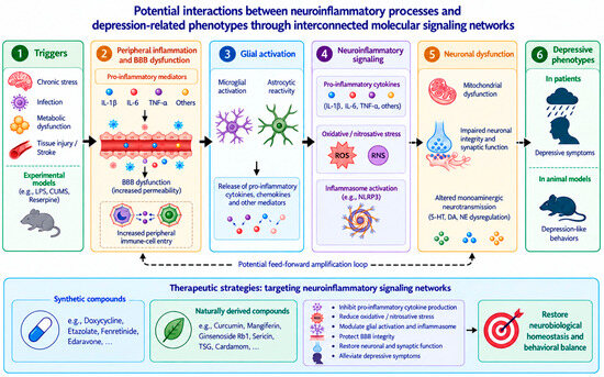
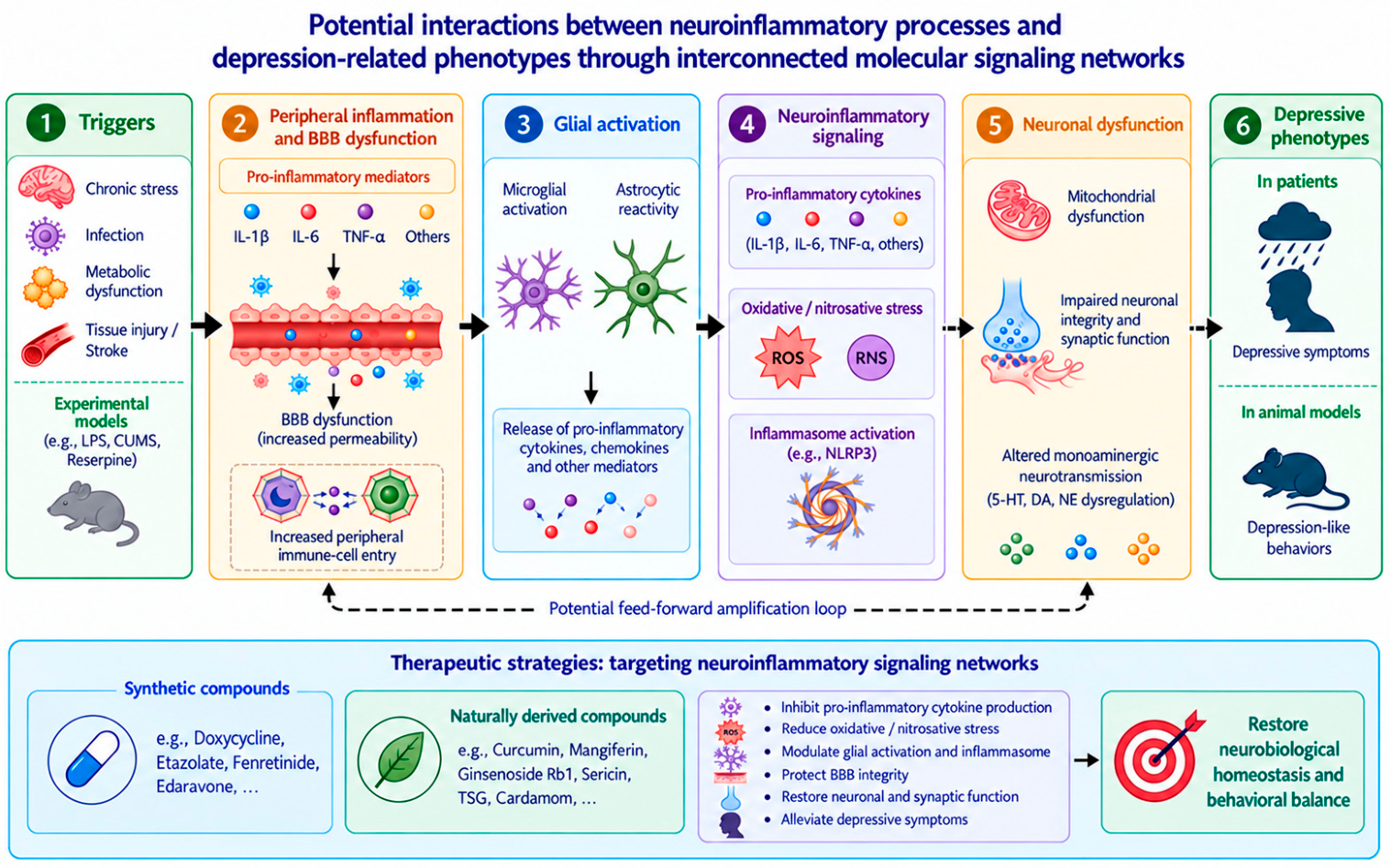
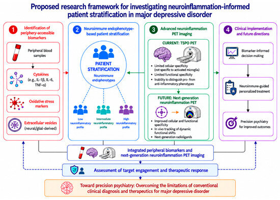
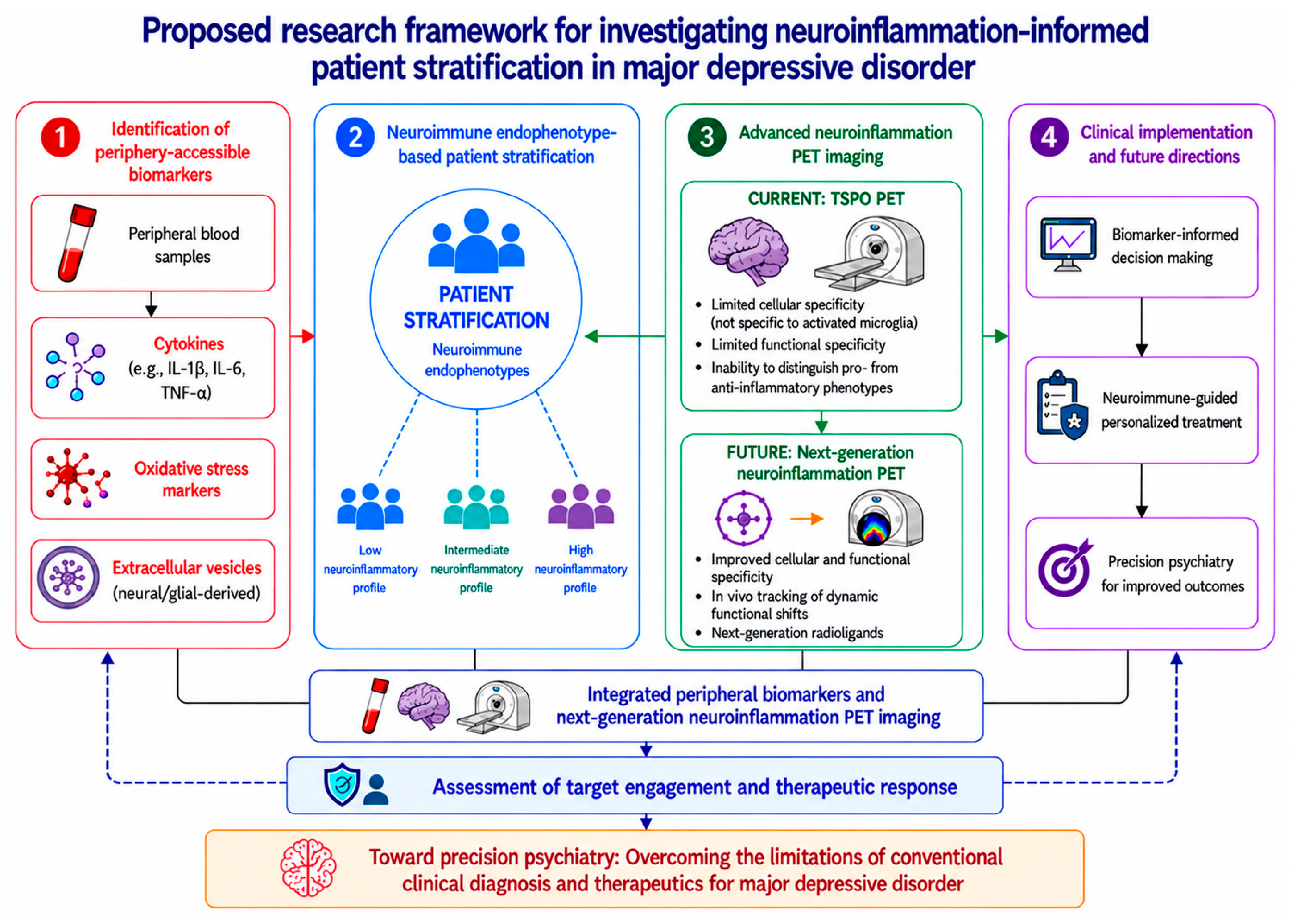
Disclaimer/Publisher’s Note: The statements, opinions and data contained in all publications are solely those of the individual author(s) and contributor(s) and not of MDPI and/or the editor(s). MDPI and/or the editor(s) disclaim responsibility for any injury to people or property resulting from any ideas, methods, instructions or products referred to in the content. |
© 2026 by the authors. Licensee MDPI, Basel, Switzerland. This article is an open access article distributed under the terms and conditions of the Creative Commons Attribution (CC BY) license.