


Abstract
Aim: The aim of this study was to investigate the antioxidant and anti-inflammatory potential of hederagenin (HD), a natural bioactive compound from the Sparganii Rhizoma–Curcumae Rhizoma herb pair, in deep vein thrombosis (DVT) and to elucidate its mechanism via the MDM2–p53 axis. Methods: A rat DVT model induced by inferior vena cava stenosis and an oxygen–glucose deprivation (OGD)-stimulated EA.hy926 endothelial cell model were established. Rats received HD (12.5, 25, 50 mg/kg) or heparin. Thrombus weight, coagulation, histopathology, immunofluorescence, immunohistochemistry, and Western blot were assessed. Inflammatory cytokines (TNF-α, IL-1β, CRP) and oxidative stress markers (SOD, MDA) were measured. Platelet aggregation was induced by ADP, arachidonic acid, and thrombin. Network pharmacology, molecular docking, and molecular dynamics simulations were performed. Results: HD dose-dependently reduced thrombus weight, improved coagulation, and downregulated tissue factor and von Willebrand factor. HD decreased TNF-α, IL-1β, CRP, and MDA levels and restored SOD activity, demonstrating antioxidant and anti-inflammatory effects. Network pharmacology identified MDM2 as a core target. Molecular docking showed favorable HD–MDM2 binding (−8.2 kcal/mol), and 100 ns molecular dynamics confirmed complex stability. HD downregulated MDM2, upregulated p53/p-p53, and suppressed NF-κB p65 phosphorylation and acetylation in IVC tissues and OGD-stimulated cells. HD also inhibited platelet aggregation, reduced vWF release, and downregulated CD41/CD61. Conclusions: HD, a natural bioactive compound, attenuates DVT through antioxidant and anti-inflammatory mechanisms by modulating the MDM2–p53 axis, concurrently suppressing endothelial inflammation, oxidative stress, and platelet activation. It is a promising dual-acting candidate for DVT therapy.
1. Introduction
Venous thromboembolism (VTE), which includes deep vein thrombosis (DVT) and pulmonary embolism (PE), is the third most common cardiovascular disease after myocardial infarction and stroke. The annual incidence of VTE is approximately 1–2 cases per 1000 adults [1], and DVT is its most common manifestation. DVT frequently leads to post-thrombotic syndrome (PTS) and fatal pulmonary embolism (PE), seriously threatening patients’ quality of life and survival. Current pharmacological treatments for DVT mainly include anticoagulants (heparin, warfarin, direct oral anticoagulants) and antiplatelet agents (aspirin, clopidogrel). However, these drugs have limitations in terms of bleeding risk, long-term adherence, and patient heterogeneity. Most agents act on a single pathway, either the coagulation cascade or platelet function, and fail to concurrently intervene in both the inflammatory response and platelet activation during thrombogenesis [2]. Therefore, the development of novel antithrombotic agents with both anti-inflammatory and antiplatelet activities is of considerable clinical significance.
The traditional pathogenesis of DVT is based on Virchow’s triad, encompassing venous stasis, endothelial injury, and hypercoagulability. However, recent studies have revealed that inflammation plays a critical role in the initiation and progression of DVT, and that the inflammatory and coagulation systems are interconnected through a complex network rather than functioning independently [3]. Under conditions of venous stasis or reduced blood flow, local tissue ischemia and hypoxia occur, activating vascular endothelial cells. Activated endothelial cells upregulate adhesion molecules such as P-selectin, E-selectin, and ICAM-1/VCAM-1, and release von Willebrand factor (vWF) [4,5]. These molecules mediate the recruitment, rolling, adhesion, and transendothelial migration of circulating neutrophils and monocytes to the injured endothelial surface, initiating a local inflammatory response [6,7].
Neutrophils recruited to the endothelial surface become activated upon inflammatory stimuli and release their nuclear contents to form neutrophil extracellular traps (NETs) [8]. NETs, composed of decondensed chromatin DNA and histones, provide a scaffold for platelet adhesion and aggregation, while simultaneously activating platelets and promoting thrombin generation [9]. Activated platelets undergo conformational changes in the surface integrin αIIbβ3 (composed of CD41 and CD61), shifting from a low-affinity to a high-affinity state, which enhances their binding to fibrinogen and vWF and mediates firm platelet adhesion and aggregation [10]. Concurrently, recruited monocytes differentiate into macrophage-like cells within the local microenvironment and become a major source of tissue factor (TF) [11]. As a key initiator of the extrinsic coagulation pathway, TF binds to FVIIa and activates FX, thereby triggering the coagulation cascade and promoting fibrin deposition and thrombus formation.
Within the molecular network of inflammation-driven thrombogenesis, nuclear factor-κB (NF-κB) occupies a central regulatory position. Activated endothelial cells initiate the transcription of multiple pro-inflammatory genes through NF-κB signaling, including those encoding inflammatory cytokines such as TNF-α, IL-1β, and IL-6, as well as adhesion molecules and vWF [12]. Once released into the circulation, these inflammatory cytokines stimulate the hepatic synthesis and release of C-reactive protein (CRP), a clinically established biomarker of inflammation. CRP, in turn, can feedback-activate endothelial cells and promote inflammatory cell infiltration, establishing a positive feedback amplification loop [13]. Meanwhile, vWF and P-selectin directly induce platelet adhesion and activation, whereas TNF-α and IL-1β further activate endothelial NF-κB, constituting a vicious cycle that propagates from endothelial inflammation to platelet activation and subsequently exacerbates endothelial inflammation.
It is noteworthy that the persistent activation of NF-κB during the above pathological process represents a core driver of the inflammatory response in DVT. Nevertheless, the activity of NF-κB is constrained by multiple endogenous negative regulatory mechanisms. Among these, p53 serves as a critical endogenous inhibitor of NF-κB and can suppress NF-κB transcriptional activity through various mechanisms, including competing for transcriptional coactivators, inducing IκBα expression, and directly interfering with the binding of p65 to DNA [14]. However, p53 itself is tightly negatively regulated by MDM2, which is the primary E3 ubiquitin ligase for p53 and maintains low p53 expression under resting conditions by promoting its ubiquitination and degradation [14]. Aberrant upregulation of MDM2 can suppress p53 activity, thereby relieving the braking effect of p53 on NF-κB and allowing inflammatory responses to become uncontrolled. In addition, MDM2 has been reported to modulate NF-κB signaling through p53-independent mechanisms; however, the direction of this regulation appears to be context-dependent, with both activating and inhibitory effects on NF-κB p65/RelA having been reported in different experimental settings [15]. Therefore, the MDM2–NF-κB relationship should be interpreted as a context-dependent interaction rather than a fixed positive regulatory axis. Accordingly, although the MDM2–p53 axis may constitute a candidate regulatory node in inflammation, inhibiting MDM2 may promote p53 activation (thereby restoring p53-mediated suppression of NF-κB) and, depending on context, may also modulate p53-independent MDM2–NF-κB crosstalk. Whether MDM2 exerts a net pro-inflammatory effect in this model remains to be determined.
Concurrently, p53 also plays an important role in the regulation of platelet function: p53-deficient mice exhibit significantly lower platelet counts than wild-type mice, and p53 regulates platelet production through the PI3K signaling pathway. In the diabetic state, sulforaphane can alleviate high glucose-induced platelet dysfunction by inhibiting the Src/ROS/p53 signaling pathway [16]. These lines of evidence suggest that p53 not only participates in the regulation of inflammation but may also directly affect platelet function. Thus, the MDM2-p53 axis may serve as a shared upstream target for simultaneously modulating the two pathological pathways of endothelial inflammation and platelet activation in DVT. By inhibiting MDM2 to activate p53, this axis can, on one hand, restore the inhibitory effect of p53 on NF-κB (suppressing inflammation) and, on the other hand, directly regulate platelet function (inhibiting platelet activation), thereby breaking the vicious cycle between inflammation and platelet activation and synergistically alleviating DVT.
In traditional Chinese medicine (TCM), blood stasis is a pivotal pathological basis of thrombotic diseases. The Sparganii Rhizoma–Curcuma Rhizoma herb pair is a classic combination used to activate blood circulation and resolve stasis. While its traditional use for blood stasis syndromes is well documented, modern pharmacological studies have validated that extracts of this herb pair possess anti-inflammatory, anticoagulant, and vasodilatory activities [17,18]. Network pharmacological analysis indicates that hederagenin (HD) is a core shared bioactive constituent, with targets enriched in inflammation- and coagulation-related pathways [19]. As a natural pentacyclic triterpenoid aglycone derived from Chinese herbal medicines, HD exhibits extensive pharmacological properties, including anti-inflammatory, antioxidant, and antitumor activities [20]. Nevertheless, it remains unclear whether HD alleviates deep vein thrombosis via simultaneous regulation of NF-κB-mediated endothelial inflammation and platelet activation through the MDM2-p53 axis.
Based on the above, the present study proposes the following scientific hypothesis: HD may attenuate IVC stenosis-induced DVT by inhibiting or modulating MDM2, restoring p53 stability/activity, thereby suppressing NF-κB-mediated endothelial inflammation and affecting platelet activation. To test this hypothesis, we employed network pharmacology to predict the potential targets of HD, and subsequently utilized a rat model of DVT induced by IVC stenosis and an oxygen-glucose deprivation (OGD)-stimulated EA.hy926 endothelial cell model to systematically verify the regulatory effects of HD on the MDM2-p53-NF-κB pathway and its anti-inflammatory and antiplatelet effects at the in vivo, cellular, and molecular levels. The proposed mechanism by which HD alleviates IVC thrombosis through the MDM2–p53 axis is summarized in Figure 1.
Figure 1.
HD alleviates IVC thrombosis by modulating the MDM2-p53 axis to concurrently suppress NF-κB-driven endothelial inflammation and platelet activation.(A) MDM2-p53 axis regulation. HD downregulates MDM2 and upregulates p-p53, which subsequently inhibits p65 (NF-κB) phosphorylation/acetylation and reduces TF expression. (B) In vivo anti-thrombotic effects. Representative IVC thrombus images, H&E staining, and immunofluorescence of TF, Ly-6G, and vWF. (C) In vitro platelet aggregation inhibition. Curves induced by ADP/AA/thrombin demonstrate that HD inhibits platelet aggregation. (D) In vitro p53-dependent mechanism. In LPS/TNF-α-stimulated EA.hy926 cells, HD inhibits MDM2, upregulates p-p53, suppresses NF-κB, and reduces TF expression, involving NETs. (E) Overall hypothesis. HD alleviates DVT via a dual mechanism: targeting MDM2 to suppress endothelial inflammation and directly inhibiting platelet aggregation.
2. Materials and Methods
2.1. Experimental Reagents
The main drugs and reagents used in this study were as follows: hederagenin (HD, Aladdin, Shanghai, China, C1925051, purity ≥ 98%), heparin sodium (Wanbang Pharmaceutical, Hefei, China, 2 mL:12,500 units), urethane, DMSO, PEG300, and Tween 80 (Biosharp, Hefei, China), and ceftriaxone sodium (Shenzhen Lijian, Shenzhen, China). Cell culture reagents (DMEM high-glucose medium, glucose-free DMEM, FBS, and penicillin–streptomycin) were purchased from Gibco (Grand Island, NY, USA); EA.hy926 cells were obtained from Wuhan Pricella; and the CCK-8 kit was purchased from Shanghai Panchao.
The sources and dilution ratios of antibodies were as follows: rabbit anti-MDM2, anti-p53, anti-p-p53(Ser15), and anti-p65 (Cell Signaling Technology, Danvers, MA, USA; WB 1:800); rabbit anti-p-p65(Ser536), anti-Ace-p65(Lys310), anti-CD41, anti-CD61, anti-Histone 3, and anti-β-actin (Cell Signaling Technology, Danvers, MA, USA; WB 1:1000); mouse anti-TF monoclonal antibody (Yaenzymes, Shanghai, China; IF 1:50); rabbit anti-vWF (Servicebio, Wuhan, China; IHC 1:300); FITC-conjugated rabbit anti-rat Ly6G (Servicebio, IF 1:1000); HRP-conjugated goat anti-rabbit and anti-mouse IgG secondary antibodies (Servicebio, Wuhan, China; WB 1:5000); and Alexa Fluor 549-conjugated goat anti-mouse IgG secondary antibody (Abbkine, IF 1:1000).
ELISA kits for TNF-α/IL-1β (Hangzhou Lianke, Hangzhou, China / Invitrogen, Carlsbad, CA, USA) and for CRP, SOD, and MDA (Nanjing Jiancheng, Nanjing, China); BCA protein assay kit, RIPA lysis buffer (containing protease and phosphatase inhibitors), and nuclear and cytoplasmic protein extraction kit (Beyotime, Shanghai, China); PVDF membranes and ECL chemiluminescent substrate (Shanghai Panchao, Shanghai, China). Platelet aggregation inducers, including ADP, arachidonic acid (AA), thrombin, and cilostazol, were obtained from Shanghai Xinyu. Anticoagulant blood collection tubes for coagulation function tests (Xihong, Shanghai, China) and PT, APTT, TT, and FIB assay kits (Rayto, Shenzhen, China). Histopathological and immunofluorescence staining reagents, including 4% paraformaldehyde, hematoxylin and eosin (H&E) staining kit, citrate antigen retrieval buffer (pH 6.0), 30% H2O2, goat serum, DAB chromogen kit, DAPI, anti-fade mounting medium, Triton X-100, and PBS, were all obtained from Servicebio. All antibodies were used at working concentrations optimized through preliminary experiments.
2.2. Animals and Ethics Statement
Sprague-Dawley rats (200–220 g, 7–8 weeks old) were purchased from Shanghai Jiesijie Laboratory Animal Co., Ltd. All animal experiments were approved by the Research Ethics Committee of Shanghai University of Medicine and Health Sciences (approval No. YS-2026-07-30; approval date: 10 January 2026; study period: 1 January 2026–31 December 2026) and performed in accordance with the Guide for the Care and Use of Laboratory Animals (license No. SCXK (Shanghai) 2023-0004; quality inspection unit for SPF-grade rats: Suzhou Xishan Biotechnology Co., Ltd., Suzhou, China). Before the experiments, rats were acclimatized for one week with free access to food and water under controlled conditions at 24.0 ± 0.5 °C with a 12 h light/dark cycle.
2.3. IVC Stenosis-Induced DVT Rat Model
The inferior vena cava stenosis model was established as follows. Rats were anesthetized with 20% urethane, and the abdomen was shaved. A midline abdominal incision of approximately 2 cm was made, and the IVC was isolated just caudal to the left renal vein. A 30-gauge needle (approximately 0.3 mm outer diameter) was placed alongside the IVC, and a 5-0 silk suture was tied around both the IVC and the needle. The needle was then removed to create a standardized stenosis. Lateral and posterior IVC branches were carefully preserved and not ligated. The degree of stenosis was estimated from the outer diameter of the needle relative to the pre-stenosis IVC outer diameter; the reported 70–80% stenosis is an estimate based on this ratio and was not directly confirmed by venography or ultrasound. This partial ligation produces venous stenosis and subsequent thrombus formation. After surgery, ceftriaxone sodium was applied around the wound to prevent bacterial infection. The sham-operated group underwent the same laparotomy procedure with exposure of the IVC but without stenosis.
2.4. Animal Grouping and Drug Administration
Forty-eight male Sprague-Dawley rats were randomly divided into 8 groups (n = 6 per group): normal group (saline, no surgery, p.o.); sham-operated group (saline, surgery without stenosis, p.o.); model group (saline, IVC stenosis, p.o.); vehicle group (vehicle consisting of 40% PEG300 + 5% Tween 80 + 55% saline containing an equivalent concentration of DMSO, IVC stenosis); heparin group (heparin 200 U/kg, intravenous injection via tail vein, IVC stenosis); HD low-dose group (HD 12.5 mg/kg, intragastric administration, IVC stenosis); HD medium-dose group (HD 25 mg/kg, intragastric administration, IVC stenosis); and HD high-dose group (HD 50 mg/kg, intragastric administration, IVC stenosis).
HD was dissolved in DMSO as a stock solution and diluted with 40% PEG300 + 5% Tween 80 + 55% saline to the desired concentrations immediately before use. The drug was administered intragastrically 3 h prior to surgery. Heparin was injected via the tail vein 1 h after surgery. At 24 h post-surgery, rats were re-anesthetized, and blood samples were collected from the abdominal aorta into tubes for serum, plasma, and whole blood. The thrombus and IVC wall tissues were harvested for subsequent analyses.
2.5. Blood Flow Monitoring
Laser speckle contrast imaging (Simopto Xunwei, SIM BFI HR Pro, Wuhan, China) was used to monitor IVC blood flow. Blood flow images were captured before and after ligation on day 1 (the day of modeling) and on day 2 (24 h post-surgery). Under anesthesia, rats were placed on an imaging pad with the IVC exposed. On day 1, the blood flow before and after stenosis was compared under identical exposure parameters, with flow velocity and volume analyzed. On day 2, the post-stenosis blood flow status was compared with the day 1 images to evaluate the inhibitory effect of drug treatment on thrombus formation.
2.6. Thrombus Weight Measurement
On day 2, rats were re-anesthetized and dissected. The IVC segments, with or without thrombus, were excised, and the wet weight of the thrombus was measured immediately after blotting dry on filter paper. Tissues intended for H&E staining or immunofluorescence were fixed in 4% paraformaldehyde, while the remaining tissues were rapidly frozen in liquid nitrogen and stored at −80 °C.
2.7. Coagulation Function and Complete Blood Count
Blood samples from the abdominal aorta were collected into sodium citrate anticoagulant tubes (9NC, Xiangyuan Medical, Shijiazhuang, China), EDTA-K2 tubes (KWS), and plain tubes without additives (Labtrip, Zhangjiakou, China). For coagulation assays, citrated whole blood was centrifuged at 3000 rpm for 10 min to obtain plasma. Prothrombin time (PT), activated partial thromboplastin time (APTT), thrombin time (TT), and fibrinogen (FIB) were measured using an automated coagulation analyzer (RAC-1830, Rayto Life Science) with kits.
EDTA-K2 anticoagulated whole blood was temporarily stored at 4 °C, and white blood cell (WBC), red blood cell (RBC), platelet (PLT), and hemoglobin (HGB) counts were determined using a hematology analyzer (Mindray veterinary automatic hematology analyzer, model BC-5000vet).
Blood collected in plain tubes was allowed to clot at room temperature for 30 min, then centrifuged at 2000× g for 10 min at 4 °C to obtain serum, which was aliquoted and stored at −80 °C for future use. All blood samples were centrifuged within 60 min of collection.
2.8. Histopathology and Immunohistochemistry
IVC tissues fixed in 4% paraformaldehyde for 48 h were embedded in paraffin and sectioned continuously at 4 μm thickness for H&E staining and immunohistochemistry.
For H&E staining, sections were dewaxed and rehydrated, stained with hematoxylin for 5 min, rinsed in running water, differentiated in acid-alcohol, and counterstained with eosin for 3 min. After dehydration through graded ethanol and clearing in xylene, sections were mounted. Neutrophil and monocyte infiltration within the thrombus and IVC wall were observed under a light microscope. For each section, five non-overlapping fields were randomly selected at 400× magnification, and the numbers of the two inflammatory cell types were counted and averaged for semi-quantitative analysis.
For immunohistochemistry, sections were dewaxed and rehydrated, then incubated in 3% H2O2 at room temperature for 20 min to block endogenous peroxidase activity. Antigen retrieval was performed by microwave heating in citrate buffer (pH 6.0) for 15 min, followed by blocking with 10% goat serum at 37 °C for 30 min. Sections were incubated with rabbit anti-vWF primary antibody overnight at 4 °C. After PBS washing, HRP-conjugated goat anti-rabbit IgG secondary antibody was applied and incubated at room temperature for 1 h. Immunoreactivity was visualized using DAB chromogen, and sections were counterstained with hematoxylin, mounted, and observed under a light microscope.
2.9. Immunofluorescence Staining
IVC tissues fixed in 4% paraformaldehyde for 48 h were embedded in paraffin and sectioned continuously at 4 μm thickness. After dewaxing and rehydration as described in Section 2.8, sections were permeabilized with 0.5% Triton X-100 at room temperature for 15 min and washed three times with PBS. After blocking with 5% goat serum at room temperature for 1 h, sections were incubated with the following primary antibodies: FITC-conjugated rabbit anti-rat Ly6G antibody at 37 °C for 1 h in the dark, or mouse anti-TF antibody overnight at 4 °C in a humidified chamber. Following PBS washing, sections stained for TF were incubated with Alexa Fluor 549-conjugated goat anti-mouse IgG secondary antibody (1:1000, A23310, Abbkine, Wuhan, China) at room temperature for 1 h in the dark. All sections were washed with PBS, counterstained with DAPI for 5 min, and mounted with anti-fade mounting medium. Images were acquired under a fluorescence microscope (BX53, Olympus, Tokyo, Japan).
2.10. Measurement of Inflammatory Cytokines, CRP, SOD Activity, and Lipid Peroxidation
Animal serum: Blood collected from the abdominal aorta in plain tubes without additives was allowed to clot at room temperature for 30 min, then centrifuged at 2000× g for 10 min at 4 °C. The upper serum layer was separated, aliquoted, and stored at −80 °C for subsequent analysis. Serum levels of tumor necrosis factor-α (TNF-α, Hangzhou Lianke, Hangzhou, China) and interleukin-1β (IL-1β, Invitrogen, Carlsbad, CA, USA) were measured using double-antibody sandwich enzyme-linked immunosorbent assay (ELISA) strictly according to the manufacturer’s instructions. Serum C-reactive protein (CRP) content was determined by immunoturbidimetry. Superoxide dismutase (SOD) activity was detected using the xanthine oxidase method (WST-1 method), with absorbance read at 450 nm.
Animal tissues: IVC tissues were weighed and homogenized in pre-cooled normal saline at a ratio of 1:9 (w/v) on ice. The homogenates were centrifuged at 12,000× g for 10 min at 4 °C, and the supernatants were collected. Malondialdehyde (MDA) content was determined by the thiobarbituric acid method, with absorbance measured at 532 nm. Protein concentration was quantified using the BCA method, and the results were expressed as nmol/mg protein for MDA.
Cell culture supernatants: Following OGD treatment and HD-containing medium intervention in EA.hy926 cells, culture supernatants were collected and centrifuged at 1000× g for 5 min at 4 °C to remove cell debris. TNF-α and IL-1β levels in the supernatants were detected using ELISA kits according to the manufacturer’s instructions. Cell lysates were quantified for protein content using the BCA method, and SOD activity and MDA content were determined using the WST-1 method and thiobarbituric acid method, respectively, with all results normalized to protein content.
2.11. Network Pharmacology, Molecular Docking, and Molecular Dynamics
To comprehensively screen the bioactive components of Sparganii Rhizoma (from Sparganium stoloniferum Buch.-Ham.) and Curcumae Rhizoma (from Curcuma phaeocaulis Valeton), a multi-source data integration approach was adopted. The botanical names were verified against The Plant List (www.theplantlist.org) on 20 June 2026. A multi-source data integration approach was adopted in this study. Chemical constituents of the two herbs were retrieved from databases including TCMSP and BATMAN-TCM, with an initial screening threshold of oral bioavailability ≥30% and drug-likeness ≥0.18. This was supplemented by manual literature mining from PubMed, CNKI, and other Chinese and English databases to collect compounds with reported pharmacological activities. After intersecting the active components from the above sources, hederagenin was identified as the major shared bioactive constituent of the two herbs. Subsequently, its chemical structure (CID: 73201) was obtained from the PubChem database and imported into SwissTargetPrediction and PharmMapper platforms to predict potential targets (species limited to Homo sapiens). DVT- and blood stasis syndrome-related targets were retrieved from the GeneCards and OMIM databases using the keywords “deep vein thrombosis”, “venous thrombosis”, “DVT”, “blood stasis syndrome”, and “blood stasis”. The two target sets were merged (union), and duplicates were removed to generate the disease target set. The intersection of HD targets and disease targets was then used to construct a protein–protein interaction (PPI) network using the STRING database (confidence score ≥ 0.4), with visualization and core target screening performed using Cytoscape software (MCODE and CytoHubba). GO functional annotation and KEGG pathway enrichment analysis of the core targets were performed using the DAVID database (with p < 0.05 as the threshold). For molecular docking, the crystal structure of MDM2 protein (PDB ID: 4JGR) was obtained from the RCSB PDB database. The structure of hederagenin was optimized using Chem3D, and the binding site was defined by the co-crystallized ligand. Semi-flexible molecular docking was performed using AutoDock Vina (version 1.5.7), and the conformation with the lowest binding energy was selected, with three-dimensional structures visualized using PyMOL (version 2.5).
For molecular dynamics simulations, the best docking conformation obtained from molecular docking was used as the initial structure. MD simulations were performed using GROMACS software (version 2022.3) for 100 ns. The AMBER99SB-ILDN force field was used for the protein, while ligand force field parameters were assigned through the GAFF force field. A solvent environment was constructed using the TIP3P water model, and the total system charge was neutralized by adding Na+ ions. The simulation temperature was set to 300 K and pressure to 1 bar, with temperature and pressure coupling achieved using the V-rescale thermostat and Parrinello-Rahman barostat, respectively. All bond lengths were constrained using the LINCS algorithm, electrostatic interactions were calculated using the PME method, and the cutoff distance was set to 1.2 nm. After simulation, the structural stability of the HD-MDM2 complex was evaluated by calculating root-mean-square deviation (RMSD), root-mean-square fluctuation (RMSF), and the number of hydrogen bonds.
2.12. Cell Culture and Treatment
EA.hy926 human umbilical vein endothelial cell line was purchased from Wuhan Procell Life Science & Technology Co., Ltd. (Wuhan, China). The cell line is deposited at ATCC under accession number CRL-2922. Cells were cultured in DMEM (Gibco) supplemented with 10% fetal bovine serum, 100 U/mL penicillin, and 100 μg/mL streptomycin at 37 °C in a 5% CO2 incubator. The EA.hy926 cells used in all experiments were between passages 4 and 10. For Western blot analysis, cells were seeded in 6-well plates at a density of 2 × 10⁵ cells per well.
2.13. Establishment of OGD Cell Model and Cell Treatment
To establish the oxygen-glucose deprivation (OGD) cell injury model, EA.hy926 cells were cultured in glucose-free DMEM and placed in a sealed hypoxic chamber containing 95% N2 and 5% CO2 for 12 h. The 12 h OGD duration and the HD concentration range (10, 20, and 30 μM) were selected based on preliminary concentration- and time-response CCK-8 screening assays and previously published protocols using EA.hy926 cells. HD was dissolved in DMSO and diluted with complete culture medium to final concentrations of 10, 20, and 30 μM (final DMSO concentration < 0.1% (v/v)). Cells were divided into the following groups: normal group (normal culture with an equal volume of DMSO-containing vehicle), OGD group (OGD injury with an equal volume of DMSO-containing vehicle without drug), OGD + HD low-dose group, OGD + HD medium-dose group, and OGD + HD high-dose group. Drug intervention groups were pretreated with media containing corresponding concentrations of HD for 24 h, followed by OGD exposure and continued culture in HD-containing media for 12 h. The normal and OGD groups were treated simultaneously with an equal volume of DMSO-containing medium.
2.14. Cell Viability Assay
EA.hy926 cells were seeded in 96-well plates at a density of 5 × 10³ cells per well. After overnight adherence, cells were pretreated with complete medium containing different concentrations of HD (10, 20, 30 μM) for 24 h, then switched to glucose-free DMEM and placed in a hypoxic chamber (95% N2, 5% CO2) for 12 h to induce OGD injury. Normal group cells were cultured in normal medium under normoxic conditions in parallel, and the model group (OGD group) was subjected to OGD injury after pretreatment with drug-free complete medium. After OGD injury, 10 μL of CCK-8 reagent was added to each well and incubated at 37 °C in the dark for 1–2 h. Absorbance at 450 nm was measured using a microplate reader. Cell viability of each group was calculated relative to the normal group, which was set as 100%. The experiment was independently repeated three times, with three replicate wells per group each time.
2.15. Platelet Isolation
Six normal SD rats were anesthetized by intraperitoneal injection of 20% urethane. After opening the abdominal cavity along the midline, the abdominal aorta was exposed, and blood was collected by puncture using a disposable blood collection needle, then injected into vacuum anticoagulant tubes containing 3.2% sodium citrate, with gentle inversion mixing for 10 times. The whole blood from all rats was pooled and centrifuged at 800× g for 10 min at 25 °C. The upper layer, which was platelet-rich plasma (PRP), was carefully transferred to a new centrifuge tube. Cilostazol was added to the PRP at a final concentration of 10 μM, mixed, and incubated at room temperature for 10 min to inhibit platelet activation. Subsequently, the PRP was centrifuged at 2000× g for 10 min at 25 °C, and the supernatant was discarded. The pellet was gently washed once with PBS. The washed platelet pellet was resuspended in pre-warmed buffer A (containing 130 mM NaCl, 10 mM sodium citrate, 9 mM NaHCO3, 6 mM glucose, 0.9 mM MgCl2, 0.81 mM KH2PO4, and 10 mM Tris, pH 7.4) and adjusted to a density of 2 × 10⁸ cells/mL for later use. The isolated platelets were maintained at 37 °C and gently pipetted to resuspend and pre-warmed to 37 °C before use. Three independent platelet preparations were performed. For each preparation, blood from six normal rats was collected and pooled, and platelets were isolated separately. Both platelet aggregation experiments and Western blot analyses for a given preparation were performed using the same batch of pooled platelets. Thus, three independent biological replicates were obtained, each derived from a separate pooled blood sample of six rats.
2.16. Platelet Aggregation Assay
HD was dissolved in DMSO as a stock solution and diluted with buffer A to the desired concentrations immediately before use, ensuring that the final DMSO concentration in each well was ≤0.1%. The control group received an equal volume of vehicle containing the same concentration of DMSO. Then, 90 μL of the PRP prepared above was pre-incubated with different concentrations of HD (5, 15, 50 μg/mL) or an equal volume of vehicle at 37 °C for 5 min, followed by the addition of ADP (final concentration 5 μM), thrombin (final concentration 0.5 U/mL), or arachidonic acid (AA, final concentration 0.5 mM) to initiate aggregation. An additional positive control group treated with aspirin (100 μM) was also included. The 96-well plate was immediately placed in a microplate reader, and absorbance was continuously monitored at 405 nm for 60 min, with readings recorded every 1 min. The initial absorbance of the PRP-containing wells was used as the baseline, and the maximum aggregation rate (%) was calculated using the following formula: (initial OD value—minimum OD value at each time point)/initial OD value × 100%. Each group had three replicate wells within a single experiment, and three independent experiments were performed, each using a separate pooled platelet preparation from six rats.
Given that thrombin induced the highest platelet aggregation rate and HD showed no significant difference in aggregation inhibition among the three inducers (ADP, AA, and thrombin), this suggested that the target of HD is located in the signaling pathway shared downstream of the three inducers. Therefore, thrombin was selected as the representative agonist for subsequent verification of the regulatory effect of HD on the MDM2-p53 pathway in platelets. A parallel reaction system (total volume 500 μL) was established for protein extraction from platelets for Western blot analysis, with group settings as detailed in Section 2.17. After treatment, the samples were centrifuged at 2000× g for 10 min at 4 °C, and the supernatant was discarded. The pellet was washed once with pre-chilled PBS, then lysed with RIPA lysis buffer (containing protease and phosphatase inhibitors) on ice for 30 min. After centrifugation at 12,000× g for 15 min, the supernatant was collected, and protein concentration was determined using the BCA method for subsequent Western blot analysis.
2.17. Western Blotting
IVC wall tissues, EA.hy926 cells, or platelets were fully lysed in RIPA lysis buffer containing protease and phosphatase inhibitors. After centrifugation at 12,000× g for 15 min at 4 °C, the supernatants were collected. For a portion of EA.hy926 cells, nuclear and cytoplasmic proteins were isolated using a nuclear and cytoplasmic protein extraction kit (Beyotime). Protein concentrations of all samples were determined using the BCA method. Equal amounts of protein (30 μg for tissue and total cell protein, 20 μg for nuclear protein) were separated by SDS-PAGE and wet-transferred onto PVDF membranes. The membranes were blocked with 5% non-fat milk at room temperature for 1 h, then incubated with corresponding primary antibodies overnight at 4 °C, followed by incubation with HRP-conjugated secondary antibodies at room temperature for 1 h. Protein bands were visualized using ECL chemiluminescent substrate, and densitometric analysis was performed using ImageJ software (version 1.54f; National Institutes of Health, Bethesda, MD, USA). Each Western blot experiment was independently repeated three times.
For tissue samples, MDM2, p53, p-p53 (Ser15), p65, p-p65 (Ser536), Ace-p65 (Lys310), CD41, and CD61 were detected, with β-actin as the loading control. For total cellular protein, MDM2, p53, p65, and TF were detected, with β-actin as the loading control. For nuclear protein, p53, p-p53 (Ser15), p65, p-p65 (Ser536), and Ace-p65 (Lys310) were detected, with Histone 3 as the loading control.
For platelet samples, groups were set as described in Section 2.16: blank control group (no treatment), thrombin model group (thrombin at a final concentration of 0.5 U/mL), and HD intervention group (HD 50 μg/mL pretreatment followed by thrombin 0.5 U/mL). MDM2, p53, and p-p53 (Ser15) were detected, with β-actin as the loading control. The relative expression level of each target protein was expressed as the ratio of the gray value of the target band to that of the loading control.
2.18. Statistical Analysis
Data are presented as mean ± standard deviation (SD). Normality was assessed using the Shapiro–Wilk test, and homogeneity of variances was evaluated using Levene’s test. Comparisons between two groups were performed using unpaired two-tailed Student’s t-test. Comparisons among multiple groups were performed using one-way analysis of variance (ANOVA) followed by Tukey’s HSD post hoc test. For repeated-measures data (e.g., platelet aggregation time curves), two-way repeated-measures ANOVA with Tukey’s correction was used. Animal experiments were conducted using randomization and blinding. For platelet experiments, n = 3 refers to three independent pooled platelet preparations, each derived from six rats; three replicate wells were averaged within each preparation before statistical comparison. Statistical significance was set at p < 0.05.
3. Results
3.1. HD Alleviates Thrombus Formation and Improves Blood Perfusion
Sparganii Rhizoma and Curcumae Rhizoma constitute a classic herb pair commonly used in traditional Chinese medicine for activating blood circulation and resolving stasis. Preliminary network pharmacology analysis combined with literature mining identified hederagenin (HD) as one of the shared bioactive constituents of the two herbs (Figure 2A,B). To verify its anti-DVT effects, a rat model of DVT induced by IVC stenosis was established in this study, with the experimental workflow illustrated in Figure 2C.
Figure 2.
Shared bioactive constituents of the Sparganii Rhizoma–Curcumae Rhizoma herb pair and their antithrombotic effects in rats with IVC stenosis-induced DVT. (A) Schematic diagram of the plant morphology of Sparganii Rhizoma and Curcumae Rhizoma; (B) Chemical structure of hederagenin (HD); (C) Schematic diagram of the experimental workflow; (D) Representative photographs of IVC thrombi from each group (n = 6); (E) Quantitative analysis of thrombus weight in each group (n = 6); (F) Laser speckle contrast imaging monitoring of IVC blood perfusion: before modeling (Day 1, pre-stenosis), immediately after stenosis (Day 1, post-stenosis), and 24 h after modeling (Day 2). Data are presented as mean ± SD. #### p < 0.0001, ### p < 0.001, ## p < 0.01, # p < 0.05 vs. NC group; aaaa p < 0.0001, aaa p < 0.001, aa p <0.01, a p < 0.05 vs. sham group; **** p < 0.0001, *** p < 0.001, ** p < 0.01, * p < 0.05 vs. MOD group; ns: not significant. Note: The laser speckle contrast image shown in panel (C) is a representative image reused from panel (F) for illustrative purposes to depict the experimental workflow.
At 24 h after IVC stenosis, a distinct red thrombus was observed in the IVC of rats in the model group, with an average thrombus weight of 66.21 mg (Figure 2D,E). Compared with the model group, the average thrombus weights in the HD medium- and high-dose groups (25 and 50 mg/kg) were reduced to 46.04 mg and 29.45 mg, with inhibition rates of 30.5% and 55.5%, respectively; the low-dose group (12.5 mg/kg) showed a reduction to 45.58 mg, with an inhibition rate of 31.2%. The thrombus weight in the positive control heparin group was 43.64 mg. HD at all doses reduced thrombus weight, with the most pronounced effect observed in the high-dose group.
Laser speckle contrast imaging was used to dynamically monitor changes in IVC blood perfusion in each group of rats. As shown in Figure 2F, before modeling (Day 1), the IVC blood flow signals in all groups were uniform, appearing red-yellow with good perfusion. Immediately after IVC stenosis, the blood flow signal rapidly weakened and the color turned deep blue, indicating acute blood flow disturbance. At 24 h after modeling (Day 2), the blood flow signal in the model group remained in a state of low perfusion (deep blue), suggesting that local microcirculation had not effectively recovered after thrombus formation. In contrast, the blood flow signals in the HD medium- and high-dose groups were markedly enhanced, with the color shifting from blue to red-yellow; the high-dose group showed the most significant improvement, indicating that HD effectively promoted IVC blood flow recanalization.
3.2. HD Regulates Coagulation Function and Complete Blood Count
To evaluate the effects of HD on systemic coagulation status and hematological parameters, blood samples were collected from the abdominal aorta at 24 h after modeling, and coagulation parameters and complete blood counts were measured.
Coagulation function: As shown in Figure 3A–D, compared with the normal group, the model group exhibited a trend of prolonged prothrombin time (PT) and activated partial thromboplastin time (APTT) (PT: 41.43 ± 9.84 s vs. 24.90 ± 1.35 s; APTT: 50.93 ± 5.29 s vs. 26.90 ± 2.22 s). These changes may reflect coagulation factor consumption following thrombus formation, postoperative stress, or model-related coagulation disturbances. Compared with the model group, the HD medium- and high-dose groups showed reductions in PT to 30.40 ± 6.60 s and 26.90 ± 2.98 s, respectively, and in APTT to 29.37 ± 6.56 s and 27.83 ± 2.16 s, respectively, demonstrating a dose-dependent trend of recovery, with the high-dose group approaching normal levels. No significant differences were observed among groups in thrombin time (TT) or fibrinogen (FIB) levels. The positive control heparin group showed marked prolongation of APTT and TT, consistent with the known anticoagulant profile of heparin. These results suggest that HD treatment is associated with the normalization of PT and APTT in this model; however, whether this reflects a direct effect on the coagulation cascade or is secondary to reduced thrombus burden and postoperative recovery cannot be determined from the current data.
Figure 3.
Effects of HD on coagulation function and complete blood count in DVT rats. (A) Prothrombin time (PT); (B) Activated partial thromboplastin time (APTT); (C) Thrombin time (TT); (D) Fibrinogen (FIB); (E) Hemoglobin (HGB); (F) Red blood cells (RBC); (G) Platelets (PLT); (H) Differential white blood cell counts (WBC, NEUT, LYMPH, MONO, EO, BASO). Data are presented as mean ± SD. #### p < 0.0001, ### p < 0.001, ## p < 0.01, # p < 0.05 vs. NC group; aaaa p < 0.0001, aaa p < 0.001, aa p < 0.01, a p < 0.05 vs. sham group; **** p < 0.0001, *** p < 0.001, ** p < 0.01, * p < 0.05 vs. MOD group; ns: not significant.
Complete blood count: As shown in Figure 3E–H, no significant dose-dependent differences or statistical significance were observed in hemoglobin (HGB) levels or red blood cell (RBC) counts among groups (p > 0.05). Total white blood cell (WBC) counts and their subtypes (neutrophils, lymphocytes, monocytes, eosinophils, and basophils) also showed no significant changes across groups. Notably, the platelet count (PLT) in the model group was 916.0 ± 167.3 × 10⁹/L, significantly lower than that in the normal group (1431.7 ± 33.5 × 10⁹/L, p < 0.001), representing a reduction of approximately 36%. This suggests that platelets were extensively recruited to the thrombus site and consumed during thrombus formation following IVC stenosis. Compared with the model group, PLT in the HD low-, medium-, and high-dose groups recovered to 1070.3 ± 137.0, 1220.7 ± 76.9, and 1391.7 ± 122.3 × 10⁹/L, respectively, showing a dose-dependent recovery trend with statistical significance. The high-dose group reached a level close to that of the normal group (1391.7 ± 122.3 vs. 1431.7 ± 33.5 × 10⁹/L). The PLT in the positive control heparin group was 1478.7 ± 106.5 × 10⁹/L. The recovery trend of PLT was highly consistent with the thrombus weight data (Figure 2E), with the high-dose group showing the lowest thrombus weight and the highest PLT, further suggesting that HD may protect peripheral platelet counts by inhibiting excessive platelet consumption at the thrombus site. Based on these findings, subsequent platelet aggregation experiments were performed to directly verify the inhibitory effect of HD on platelet function.
3.3. HD Reduces Inflammatory Cell Infiltration in the IVC Wall
To evaluate the effect of HD on local inflammatory responses associated with DVT, H&E staining was performed to observe inflammatory cell infiltration within the IVC wall and thrombus, and Ly6G immunofluorescence was used to verify neutrophil infiltration.
H&E staining: As shown in Figure 4A,B, the venous wall structure was intact in the sham-operated group, with no inflammatory cells observed at the thrombus site. In the model group, the venous wall was markedly thickened, and extensive neutrophil and monocyte infiltration was observed within the thrombus and vascular wall. In the HD medium- and high-dose groups, inflammatory cell infiltration was significantly reduced.
Figure 4.
Effects of HD on inflammatory cell infiltration in the IVC wall and thrombus in DVT rats. (A) Representative H&E staining images of thrombi (scale bar = 20 μm); (B) Representative H&E staining images of IVC walls (scale bar = 5 μm); (C) Quantitative analysis of neutrophils in thrombi and IVC walls (n = 3); (D) Quantitative analysis of monocytes in thrombi and IVC walls (n = 3); (E) Quantitative analysis of Ly6G immunofluorescence intensity (n = 3); (F,G) Representative immunofluorescence images of Ly6G-labeled neutrophils (green: Ly6G; blue: DAPI; scale bar = 100 μm). Data are presented as mean ± SD. #### p < 0.0001, ### p < 0.001, ## p < 0.01, # p < 0.05 vs. NC group; aaaa p < 0.0001, aaa p < 0.001, aa p < 0.01, a p < 0.05 vs. sham group; **** p < 0.0001, *** p < 0.001, ** p < 0.01, * p < 0.05 vs. MOD group; ns: not significant.
Inflammatory cell counts (Figure 4C,D): At the thrombus site, the neutrophil count in the model group was 1270.67 ± 342.24 cells/field, which decreased to 383.56 ± 155.70 cells/field in the HD high-dose group (p < 0.05); the monocyte count in the model group was 1116.00 ± 247.03 cells/field, which decreased to 582.22 ± 179.95 cells/field in the HD high-dose group (p < 0.05). In the venous wall, the neutrophil count in the model group was 392.22 ± 384.22 cells/field, which decreased to 50.44 ± 22.83 cells/field in the HD high-dose group; the monocyte count in the model group was 368.89 ± 105.21 cells/field, which decreased to 38.11 ± 20.50 cells/field in the HD high-dose group (p < 0.01).
Ly6G immunofluorescence: As shown in Figure 4E,F, Ly6G+ neutrophils were densely distributed around the thrombus and venous wall in the model group. In the HD medium- and high-dose groups, Ly6G+ positive cells were significantly reduced (p < 0.05, p < 0.001 vs. model group), consistent with the H&E staining results. These findings indicate that HD dose-dependently suppresses local inflammatory responses associated with DVT.
3.4. HD Reduces TF Expression in the IVC Wall and Modulates Systemic Inflammation and Oxidative Stress
To evaluate the effects of HD on systemic and local inflammation and oxidative stress, relevant indicators were measured in serum and IVC tissue homogenates, with further validation in OGD-stimulated EA.hy926 cells.
Serum and tissue inflammation/oxidative stress markers: As shown in Figure 5A–E, compared with the sham group, the model group exhibited significantly elevated serum levels of TNF-α, IL-1β, and CRP, and reduced serum SOD activity (p < 0.01), together with increased MDA content in IVC tissue homogenates (p < 0.05), indicating that DVT formation was accompanied by systemic inflammation and local oxidative stress. Compared with the model group, the HD medium- and high-dose groups showed dose-dependent reductions in serum TNF-α, IL-1β, and CRP levels and in IVC tissue MDA content (p < 0.05, p < 0.01), with serum SOD activity significantly restored, and the high-dose group showing the most pronounced effects.
Figure 5.
Effects of HD on serum and tissue inflammatory/oxidative stress markers and on TF expression in DVT rats and OGD-stimulated EA.hy926 cells. (A) Serum TNF-α; (B) serum IL-1β; (C) MDA content in IVC tissue homogenates; (D) serum SOD activity; (E) serum CRP (n = 6 per group). (F) Representative immunofluorescence images of TF in the IVC wall (scale bar = 100 μm); (G) Quantitative analysis of TF immunofluorescence intensity (n = 3). (H) SOD activity; (I) CRP; (J) TNF-α; (K) MDA; (L) IL-1β in OGD-stimulated EA.hy926 cells and/or their supernatants (n = 6). Data are presented as mean ± SD. **** p < 0.0001, *** p < 0.001, ** p < 0.01, * p < 0.05 vs. MOD group; ns: not significant.
TF immunofluorescence: As shown in Figure 5F,G, only weak TF fluorescence signals were observed in the IVC wall of the sham group. In the model group, TF fluorescence intensity was markedly enhanced, with positive staining mainly distributed in the endothelial layer of the venous wall and the perithrombotic region, suggesting local coagulation cascade activation. The HD medium- and high-dose groups showed significantly attenuated TF fluorescence intensity (p < 0.01, p < 0.001 vs. model group).
Cellular inflammation and oxidative stress markers: In OGD-stimulated EA.hy926 cells (Figure 5H–L), the model group showed significantly elevated levels of TNF-α, IL-1β, MDA, and CRP compared with the normal group (p < 0.05), along with significantly reduced SOD activity. Following intervention with HD-containing media (10, 20, 30 μM), the above indicators were reversed in a concentration-dependent manner, consistent with the findings from the animal experiments.
Collectively, these results demonstrate that HD effectively suppresses DVT-related systemic and local inflammatory responses, alleviates oxidative stress injury, and reduces TF expression in the IVC wall.
3.5. Network Pharmacology and Molecular Docking Predict Potential Targets and Related Pathways of HD Against DVT
To elucidate the potential molecular mechanisms underlying the anti-DVT effects of hederagenin (HD), network pharmacology was employed to predict its targets and related pathways. Active components of Sparganii Rhizoma and Curcumae Rhizoma were retrieved from the TCMSP and BATMAN-TCM databases, with oral bioavailability (OB) ≥ 30% and drug-likeness (DL) ≥ 0.18 as initial screening thresholds, supplemented by literature mining from PubMed, CNKI, and other databases to manually collect compounds with reported pharmacological activities. After intersecting the active components from the above sources, hederagenin was identified as the major shared bioactive constituent of the two herbs. Subsequently, 148 targets of HD were obtained from SwissTargetPrediction, SEA, and ETCM databases. A total of 663 disease targets were retrieved by combining DVT-related and blood stasis syndrome-related targets from GeneCards and OMIM. After intersecting drug targets and disease targets, a merged PPI network of 39 common targets was constructed using the STRING database (confidence score ≥ 0.4). Through MCODE and CytoHubba analysis in Cytoscape 3.10.3, the top 10 hub genes were identified as core targets (Figure 6A).
Figure 6.
Network pharmacology prediction of potential targets of HD against DVT and validation through multi-target molecular docking. (A) “Active components of Sparganii Rhizoma–Curcumae Rhizoma herb pair—shared targets—disease—pathway” network diagram, showing 10 core targets. (B) Three-dimensional and two-dimensional interaction diagrams of HD with SRC, with a binding energy of −8.5 kcal/mol. (C) Three-dimensional and two-dimensional interaction diagrams of HD with AR, with a binding energy of −7.9 kcal/mol. (D) Three-dimensional and two-dimensional interaction diagrams of HD with ESR2, with a binding energy of −8.1 kcal/mol. (E) Three-dimensional docking structures of HD with IGF1R, NR1H4, and PGR.
To further validate the direct interaction between HD and the core targets, molecular docking was performed using AutoDock Vina. Target protein crystal structures were obtained from the RCSB PDB database, with HD as the ligand and the co-crystallized ligand defining the binding site. The results showed that HD formed stable hydrogen bonds and hydrophobic interactions with SRC (Figure 6B), AR (Figure 6C), and ESR2 (Figure 6D), with binding energies of −8.5, −7.9, and −8.1 kcal/mol, respectively, all below the conventional threshold of −5.0 kcal/mol, indicating favorable binding affinities. Furthermore, docking results of HD with IGF1R, NR1H4, and PGR also demonstrated stable binding modes (Figure 6E). These results suggest that HD may exert anti-DVT effects through a multi-target regulatory network.
To further dissect the biological functions and signaling pathways of the above targets, GO functional annotation and KEGG pathway enrichment analyses were performed. GO enrichment analysis (Figure 7B) revealed that, in terms of biological process (BP), genes were significantly enriched in cellular response to lipopolysaccharide (LPS), response to hypoxia, and negative regulation of inflammatory response. Notably, LPS is a classic ligand of Toll-like receptor 4 (TLR4), and its downstream cascade directly activates the NF-κB signaling axis; hypoxia-inducible HIF-1α also frequently crosstalks with NF-κB at the transcriptional level. Meanwhile, the significant enrichment of positive regulation of PI3K/AKT signal transduction and platelet-derived growth factor receptor signaling pathway suggested possible crosstalk between NF-κB-mediated inflammatory microenvironment and platelet activation. In terms of cellular component (CC), in addition to localization at the plasma membrane and receptor complex, significant enrichment of blood microparticles further supported the association between platelet activation state and inflammatory granule release. In terms of molecular function (MF), enrichment of nuclear receptor activity and transcription factor binding activity was highly consistent with the characteristics of MDM2 as a transcriptional regulatory hub. KEGG pathway enrichment analysis (Figure 7A) showed that the relevant genes were significantly enriched in C-type lectin receptor signaling pathway, calcium signaling pathway, and platelet activation pathway, suggesting that the biological network constructed by MDM2 and its interacting genes centrally reflects the signal crosstalk between “NF-κB-driven inflammatory stress” and “platelet activation and aggregation.”
Figure 7.
GO and KEGG pathway enrichment analysis and molecular docking validation of HD with MDM2. (A) KEGG pathway enrichment analysis bubble chart; (B) GO functional annotation bar chart (BP, CC, MF); (C) Three-dimensional (left) and two-dimensional (right) interaction diagrams of HD with MDM2, with a binding energy of −8.2 kcal/mol (PDB ID: 4JGR).
Among the targets screened by network pharmacology, MDM2 ranked among the top (degree value = 14), suggesting that it may occupy a central node position in the HD regulatory network. To further verify the direct interaction between HD and MDM2, molecular docking was performed using AutoDock Vina. The crystal structure of MDM2 protein (PDB ID: 4JGR) was obtained from the RCSB PDB database, with the binding site defined by the co-crystallized ligand. The results showed that the binding energy between HD and MDM2 was −8.2 kcal/mol, below the conventional threshold of −5.0 kcal/mol, indicating a favorable theoretical binding affinity. Three-dimensional structural analysis revealed that HD was stably embedded in the active pocket of MDM2 through non-covalent bonds including hydrogen bonds and hydrophobic interactions (Figure 7C). Based on the above network pharmacology predictions and molecular docking results, MDM2 was identified as one of the core potential targets of HD against DVT, while the pathway enrichment results suggested that HD may directly intervene in platelet activation-related pathways. Accordingly, this study further verified the regulatory effects of HD on the MDM2-p53-NF-κB pathway in the DVT rat model, OGD-stimulated EA.hy926 endothelial cells, and platelets in vitro.
3.6. HD Inhibits Platelet Activation and Regulates the MDM2-p53 Pathway
Building upon the preceding platelet count results, which showed a significant reduction in peripheral platelets in the model group (indicating thrombotic consumption) and a dose-dependent restoration by HD, this section further verified the direct inhibitory effect of HD on platelet function. Platelet aggregation rate, von Willebrand factor (vWF) release, and expression of platelet activation markers CD41/CD61 were measured.
vWF immunohistochemistry: As shown in Figure 8A,B, only weak vWF-positive staining was observed in the IVC wall of the sham group, mainly distributed in the vascular endothelium. In the model group, vWF-positive signals were markedly enhanced with a dark brown diffuse distribution, consistent with increased vWF immunoreactivity in the vessel wall during thrombus formation. Compared with the model group, HD at all doses attenuated vWF-positive staining intensity in a dose-dependent manner, with the high-dose group showing the most significant effect (p < 0.001 vs. model group). However, vWF immunohistochemistry reflects vWF immunoreactivity in the vessel wall and cannot distinguish between reduced endothelial vWF expression/storage, reduced endothelial coverage, and reduced vWF secretion. Therefore, these data indicate that HD treatment was associated with reduced vWF immunoreactivity in the IVC wall, whereas the effect of HD on circulating vWF levels remains to be established.
Figure 8.
Effects of HD treatment on platelet aggregation, vWF expression, and platelet integrin markers in DVT rats. (A) Representative immunohistochemical images of vWF in the IVC wall (scale bar = 100 μm); (B) Quantitative analysis of mean optical density of vWF-positive staining (n = 3); (C) Time curve of ADP-induced platelet aggregation; (D) Time curve of AA-induced platelet aggregation; (E) Bar chart of maximum platelet aggregation rate induced by thrombin; (F) Bar chart of maximum platelet aggregation rate induced by AA; (G) Bar chart of maximum platelet aggregation rate induced by ADP; (H) Time curve of thrombin-induced platelet aggregation; (I) Quantitative analysis of CD41 protein expression (n = 3); (J) Representative Western blot bands of CD41; (K) Representative Western blot bands of CD61; (L–N) Representative Western blot bands of MDM2, p53, and p-p53 (Ser15) in platelets. Data are presented as mean ± SD. **** p < 0.0001, *** p < 0.001, ** p < 0.01, * p < 0.05 vs. MOD group; ns: not significant.
Platelet aggregation rate: Platelet aggregation induced by ADP (5 μM), arachidonic acid (AA, 0.5 mM), and thrombin (0.5 U/mL) was measured using a microplate reader kinetic method. As shown in Figure 8C–H, platelets from the model group rapidly aggregated upon stimulation with all three inducers, with maximum aggregation rates of 63.2 ± 0.2% (ADP), 61.8 ± 0.1% (AA), and 64.1 ± 0.6% (thrombin). Compared with the model group, HD at all doses (5, 15, 50 μg/mL) reduced platelet aggregation rates in a dose-dependent manner. Specifically, the high-dose group (50 μg/mL) reduced the maximum aggregation rates to 44.7 ± 0.3% (ADP), 41.8 ± 1.3% (AA), and 18.8 ± 0.8% (thrombin), corresponding to inhibition rates of 29.3%, 32.4%, and 70.7%, respectively. The medium-dose group also showed significant inhibition, while the low-dose group exhibited a decreasing trend.
CD41/CD61 protein levels: CD41 (αIIb) and CD61 (β3) are components of the platelet membrane integrin αIIbβ3 and serve as platelet integrin markers. As shown in Figure 8I–K, CD41 and CD61 protein levels in IVC tissues were significantly higher in the model group than in the normal group, consistent with increased platelet accumulation at the thrombus site. Compared with the model group, the HD high-dose group (50 mg/kg) showed significantly reduced CD41 and CD61 protein levels. However, because these measurements were performed in IVC tissue lysates containing variable amounts of thrombus, the reduction in CD41/CD61 may reflect reduced platelet content within a smaller thrombus and should not be interpreted as direct evidence of reduced platelet activation. Activation-specific markers such as P-selectin, PAC-1, CD40L, and PF4 were not measured in this study. Given that HD showed similar inhibitory trends against all three inducers (ADP, AA, and thrombin), consistent with the possibility that HD acts at a step shared downstream of these inducers, and that the high-dose group showed the most prominent effect in the aggregation assay, the high-dose group was selected for subsequent Western blot analysis of MDM2–p53 pathway proteins in platelets.
The above platelet-related indicators reflect the complete process of platelet activation from different dimensions, with a logical progression among them. vWF released by activated endothelial cells serves as a bridge for platelet adhesion to the injured vascular wall; CD41/CD61 (integrin αIIbβ3) are adhesion receptors on the platelet membrane whose upregulation marks the entry of platelets into an activated state; ultimately, activated platelets aggregate in response to agonists such as ADP, AA, or thrombin. These events constitute a continuous activation chain: endothelial vWF release → platelet adhesion → integrin upregulation → platelet aggregation. In this study, HD simultaneously inhibited the initiation (vWF release), intermediate (CD41/CD61 expression), and terminal (aggregation) steps of this chain, suggesting that its antiplatelet effects are multi-targeted in nature.
Collectively, these results demonstrate that HD concentration-dependently inhibits ADP-, AA-, and thrombin-induced platelet aggregation, downregulates vWF release in the IVC wall, and reduces CD41/CD61 protein expression, indicating that HD possesses antiplatelet activation potential.
To further explore the molecular basis of HD-mediated inhibition of platelet activation, Western blotting was performed to detect the protein expression of MDM2, p53, and p-p53 (Ser15) in platelet lysates from normal rats. According to the group settings described in Section 2.16, platelet samples were divided into three groups: blank control group (no treatment), thrombin model group (thrombin at a final concentration of 0.5 U/mL), and HD intervention group (HD 50 μg/mL pretreatment followed by thrombin 0.5 U/mL). As shown in Figure 8L–N, compared with the blank control group, the thrombin model group exhibited significantly increased MDM2 protein expression and markedly decreased p53 and p-p53 (Ser15) protein levels in platelets, indicating that thrombin-induced platelet activation was accompanied by changes in MDM2 and p53/p-p53 levels. Compared with the thrombin model group, the HD intervention group (HD 50 μg/mL pretreatment) showed significantly reduced MDM2 expression and restored p53 and p-p53 (Ser15) levels, indicating that HD treatment was associated with reversal of thrombin-induced MDM2 upregulation and p53/p-p53 downregulation. Because mature platelets are anucleate, these changes cannot be interpreted as transcription-dependent p53 activation. These results are therefore consistent with an association between HD treatment and modulation of MDM2–p53 signaling in platelets; whether this association reflects a direct, non-transcriptional regulatory mechanism requires further validation.
3.7. HD Modulates the MDM2-p53-NF-κB Pathway In Vivo and Molecular Dynamics Simulation of the HD-MDM2 Complex
Based on the network pharmacology prediction identifying MDM2 as a core target of HD against DVT, and the aforementioned phenotypes of HD in reducing inflammatory responses and platelet activation, this study further examined the expression changes of key proteins in the MDM2-p53-NF-κB pathway in IVC wall tissues by Western blotting to verify the in vivo regulatory effects of HD on this pathway. Given that the HD high-dose group (50 mg/kg) exhibited the most significant anti-DVT effects in phenotypic indicators including thrombus weight, inflammatory cell infiltration, and platelet aggregation, the high-dose group was selected as representative for Western blot analysis of the MDM2-p53-NF-κB pathway. The groups examined included the normal group, model group, and HD high-dose group (50 mg/kg).
As shown in Figure 9A,B, MDM2 protein expression in the IVC wall tissues was significantly elevated in the model group compared with the normal group (p < 0.05). Compared with the model group, the HD high-dose group showed significantly reduced MDM2 protein expression (p < 0.01), indicating that HD effectively suppresses DVT-induced MDM2 upregulation.
Figure 9.
Effects of HD on protein expression of the MDM2-p53-NF-κB pathway in IVC wall tissues of DVT rats. (A) Representative Western blot bands of MDM2; (B) Quantitative analysis of MDM2 protein expression (n = 3); (C) RMSD plots of MDM2 (black line), HD (red line), and the MDM2-HD complex (blue line); (D) SASA plot of the MDM2-HD complex; (E) RMSF plot of MDM2; (F) Rg plot of MDM2; (G) Number of hydrogen bonds in the MDM2-HD complex; (H) Representative Western blot bands of p53; (I) Quantitative analysis of p53 protein expression (n = 3); (J) Representative Western blot bands of p65, p-p65 (Ser536), and Ace-p65 (Lys310); (K) Representative Western blot bands of p-p53; (L) Quantitative analysis of p-p53 (Ser15) protein expression (n = 3); (M) Quantitative analysis of the Ace-p65/p65 ratio (n = 3); (N) Quantitative analysis of the p-p65/p65 ratio (n = 3). β-Actin served as the loading control. Data are presented as mean ± SD. **** p < 0.0001, *** p < 0.001, ** p < 0.01, * p < 0.05 vs. MOD group; ns: not significant.
As shown in Figure 9H,I,K,L, p53 and phosphorylated p53 (p-p53, Ser15) protein levels in the IVC wall tissues were significantly decreased in the model group compared with the normal group, suggesting that MDM2 upregulation during DVT formation promoted p53 degradation and suppressed p53 transcriptional activity. Compared with the model group, the HD high-dose group showed significantly restored p53 and p-p53 protein levels (p < 0.01, p < 0.05), indicating that HD restored p53 stability and activity through MDM2 inhibition.
As shown in Figure 9J,M,N, the phosphorylation (p-p65, Ser536) and acetylation (Ace-p65, Lys310) levels of the NF-κB p65 subunit were significantly elevated in the model group compared with the normal group (p < 0.01), indicating aberrant activation of the NF-κB signaling pathway, consistent with the exacerbated inflammatory state in DVT. Compared with the model group, the HD high-dose group showed significantly reduced p-p65 and Ace-p65 protein levels (p < 0.05, p < 0.01), while total p65 protein expression showed no significant changes among groups.
To further verify the binding stability between HD and MDM2, a 100 ns molecular dynamics simulation of the HD-MDM2 complex was performed. As shown in Figure 9C, the root-mean-square deviation (RMSD) of the HD-MDM2 complex remained below 0.2 nm throughout the simulation, indicating stable overall complex conformation. As shown in Figure 9D, the solvent-accessible surface area (SASA) remained relatively constant during the simulation, suggesting that the complex did not undergo significant conformational collapse or exposure. As shown in Figure 9E, root-mean-square fluctuation (RMSF) analysis revealed that amino acid residues in the MDM2 binding pocket exhibited small fluctuations (<0.25 nm), indicating that HD binding enhanced the rigidity of this region. As shown in Figure 9F, the radius of gyration (Rg) remained stable at approximately 1.32 nm throughout the simulation, further confirming the compactness of the complex structure. As shown in Figure 9G, HD and MDM2 maintained 0–1 hydrogen bonds throughout the simulation, suggesting relatively weak hydrogen bond interactions, with complex stability likely maintained primarily by hydrophobic interactions and van der Waals forces. These molecular dynamics simulation results corroborated the molecular docking results, collectively confirming the formation of a stable binding mode between HD and MDM2.
Collectively, these results demonstrate that HD effectively downregulates MDM2 expression, restores p53 and p-p53 levels, and suppresses NF-κB p65 phosphorylation and acetylation in IVC wall tissues in vivo, thereby confirming that HD exerts anti-inflammatory and anti-DVT effects through modulation of the MDM2-p53-NF-κB pathway. The molecular dynamics simulation further confirmed the binding stability of the HD-MDM2 complex at the molecular level.
3.8. HD Inhibits OGD-Induced EA.hy926 Cell Injury Through the MDM2-p53-NF-κB Pathway
To verify the regulatory effects of HD on the MDM2-p53-NF-κB pathway at the cellular level, OGD-stimulated EA.hy926 endothelial cells were used to mimic ischemic-hypoxic injury, with the experimental workflow illustrated in Figure 10A. HD was dissolved in DMSO and diluted with complete medium to final concentrations of 10, 20, and 30 μM (final DMSO concentration < 0.1%), with pretreatment for 24 h prior to OGD and continuous administration during OGD.
Figure 10.
HD inhibits OGD-induced EA.hy926 cell injury through the MDM2-p53-NF-κB pathway. (A) Schematic diagram of the OGD cell experiment workflow; (B) CCK-8 assay for cell viability in each group (n = 3); (C) Quantitative analysis of MDM2 protein expression (n = 3); (D) Quantitative analysis of total p53 protein expression (n = 3); (E) Representative Western blot bands of MDM2, total p53, and total p65; (F) Comparison of cell morphology between the normal and OGD groups (scale bar = 100 μm); (G) Quantitative analysis of nuclear Ace-p65 protein expression (n = 3); (H) Representative Western blot bands of nuclear p53 and nuclear p-p53 (Ser15); (I) Quantitative analysis of nuclear p53 protein expression (n = 3); (J) Quantitative analysis of nuclear p-p53 protein expression (n = 3); (K) Representative Western blot bands of nuclear p65, nuclear p-p65 (Ser536), and nuclear Ace-p65 (Lys310); (L) Quantitative analysis of the nuclear p65/total p65 ratio (n = 3); (M) Quantitative analysis of nuclear p-p65 protein expression (n = 3). β-Actin served as the loading control for total protein, and Histone 3 served as the loading control for nuclear protein. Data are presented as mean ± SD. **** p < 0.0001, *** p < 0.001, ** p < 0.01, * p < 0.05 vs. MOD group; ns: not significant.
The effect of HD on cell viability after OGD injury was first assessed by the CCK-8 assay (Figure 10B). Normalizing the cell viability of the normal group to 100%, the OGD group showed a significant reduction to 83.54 ± 4.21% (p < 0.01 vs. normal group). Compared with the OGD group, the HD low-, medium-, and high-concentration groups (10, 20, 30 μM) showed restored cell viability to 94.75 ± 3.86%, 93.94 ± 4.02%, and 88.25 ± 4.57%, respectively (p < 0.05 vs. OGD group). Although the high HD concentration (30 μM) resulted in slightly lower cell viability compared with the low and medium concentrations, all HD-treated groups showed significantly higher viability than the OGD group. Moreover, the low and medium concentrations, which exhibited the highest viability recovery, also significantly reversed the OGD-induced changes in inflammatory and oxidative stress markers, suggesting that these effects are not merely secondary to differences in cell viability. Consistent with these viability results, cell morphology exhibiting shrinkage, rounding, and detachment in the OGD group (Figure 10F) was markedly improved in the HD-treated groups.
Further Western blot analysis was performed to detect the expression changes of key pathway molecules in total protein and nuclear protein fractions. For total protein (Figure 10C–E): compared with the normal group, the OGD group showed significantly increased MDM2 expression (p < 0.05) and significantly decreased total p53 levels (p < 0.05), while total p65 showed no significant changes among groups. Compared with the OGD group, HD (30 μM) significantly reduced MDM2 expression (p < 0.0001) and restored total p53 levels (p < 0.01), indicating that HD also inhibits MDM2 and restores p53 expression at the cellular level.
For nuclear protein (Figure 10G–M): compared with the normal group, the OGD group showed significantly reduced nuclear p53 and p-p53 (Ser15) levels, while nuclear p65, p-p65 (Ser536), and Ace-p65 (Lys310) levels were significantly increased, suggesting that OGD injury reduced p53 nuclear translocation and enhanced NF-κB activation in the nucleus. Compared with the OGD group, HD (30 μM) significantly restored nuclear p53 and p-p53 levels, while reducing nuclear p65, p-p65, and Ace-p65 levels. The nuclear p65/total p65 ratio was also significantly reduced, indicating that HD inhibits OGD-induced p65 nuclear translocation.
Collectively, these results demonstrate that HD directly acts on EA.hy926 endothelial cells to inhibit MDM2 expression, restore p53 and its phosphorylation levels, and suppress NF-κB p65 nuclear translocation, phosphorylation, and acetylation, thereby confirming at the cellular level that HD protects endothelial cells from ischemic-hypoxic injury through regulation of the MDM2-p53-NF-κB pathway.
3.9. Mechanistic Summary
Collectively, these results are consistent with a model in which HD treatment attenuates IVC stenosis-induced DVT in association with MDM2 downregulation and the restoration of p53 and p-p53 (Ser15) levels. In this model, restored p53 activity may suppress NF-κB signaling in endothelial cells, as evidenced by reduced p-p65 (Ser536) and Ace-p65 (Lys310), thereby potentially decreasing TF expression and inflammatory cytokine release. In parallel, HD treatment was associated with inhibition of platelet aggregation, although the present data do not establish whether this effect is directly mediated by p53 or by p53-independent mechanisms. Together, these two effects may collectively contribute to the attenuation of DVT in the IVC stenosis model (Figure 11). However, the present study does not establish direct HD–MDM2 binding or a causal requirement of the MDM2–p53–NF-κB axis; these mechanistic links should be regarded as a working model requiring further validation.
Figure 11.
Schematic diagram illustrating the mechanism by which HD regulates the MDM2-p53-NF-κB axis and modulates endothelial inflammation and platelet activation.
4. Discussion
The present findings demonstrate that HD, a shared bioactive constituent of the Sparganii Rhizoma–Curcumae Rhizoma herb pair, exerts dual protective effects against DVT by concurrently suppressing endothelial inflammation and platelet activation through modulation of the MDM2-p53 axis. This mechanistic insight not only provides a modern pharmacological interpretation for the traditional “blood-activating and stasis-resolving” efficacy of this herb pair, but also extends the functional scope of the MDM2-p53 axis, a well-established tumor-related signaling pathway—into the field of thrombotic diseases, offering a new theoretical foundation for its therapeutic targeting in DVT [21].
The clinical significance of these findings lies in the fact that HD simultaneously addresses two key drivers of thrombogenesis—inflammation and platelet activation—through a single upstream regulatory node, a feature that distinguishes it from conventional anticoagulant or antiplatelet agents that act on downstream pathways [22]. Notably, while heparin, the positive control in this study, significantly prolonged APTT and TT, HD did not significantly alter PT, APTT, TT, or FIB in this model. However, this does not exclude a direct anticoagulant or antiplatelet effect, and bleeding risk was not assessed in this study. HD therefore remains a candidate compound requiring further efficacy and safety evaluation, rather than a confirmed safer antithrombotic strategy.
MDM2 is the major negative regulator of p53, promoting p53 ubiquitination and degradation through its E3 ubiquitin ligase activity, thereby suppressing p53-mediated transcriptional activity [23]. In the oncology field, the regulation of the MDM2-p53 axis has been extensively studied, and MDM2 inhibitors (such as Nutlin-3a) have entered clinical trials as anticancer agents [24]. However, the role of the MDM2-p53 axis in non-neoplastic diseases, particularly in thrombus formation, remains poorly understood. In recent years, a small number of studies have begun to focus on the role of this pathway in the cardiovascular system: MDM2 inhibitors have been shown to attenuate neointimal hyperplasia after vascular injury by upregulating p53 and suppressing NF-κB activation [25]; p53 has been demonstrated to regulate platelet production through the PI3K signaling pathway, with p53-deficient mice exhibiting significantly lower platelet counts than wild-type mice. In addition, in the diabetic state, sulforaphane has been reported to alleviate high glucose-induced platelet dysfunction by inhibiting the Src/ROS/p53 signaling pathway [16]. These pieces of evidence suggest that the MDM2-p53 axis may participate in the regulation of vascular inflammation and platelet function; however, its direct role in DVT has not yet been explored.
This study is among the first to examine the MDM2–p53 axis in the context of DVT. We found that MDM2 protein expression was significantly elevated in the IVC wall tissues of DVT model rats, accompanied by decreased p53 and p-p53 (Ser15) levels, while the phosphorylation (Ser536) and acetylation (Lys310) of NF-κB p65 were markedly increased. These findings are consistent with the possibility that aberrant upregulation of MDM2 may relieve the restraining effect of p53 on NF-κB by promoting p53 degradation [26], thereby potentially exacerbating local inflammatory responses and thrombus formation [27]. Notably, MDM2 is best known as the principal E3 ubiquitin ligase of p53, but its relationship with NF-κB is more complex than a simple positive regulatory axis. MDM2 has been reported to promote NF-κB activity in some contexts, whereas other studies have shown that MDM2 can directly bind and inhibit NF-κB p65/RelA in a p53-independent manner [15]. The direction of this crosstalk appears to depend on cell type, stimulus, subcellular localization, and the composition of MDM2–p65-containing complexes. In the present study, HD treatment was associated with decreased MDM2 expression and reduced p65 phosphorylation (Ser536) and acetylation (Lys310) in IVC tissues and OGD-stimulated EA.hy926 cells. These findings are consistent with the possibility that HD relieves the p53-dependent brake on NF-κB and/or alters MDM2–p65 crosstalk, but they do not establish whether MDM2 directly activates or inhibits p65 in this model. Future studies using MDM2 knockdown/overexpression, p65–MDM2 co-immunoprecipitation, and NF-κB reporter assays will be required to define the direction and causality of this regulation. Accordingly, MDM2 should be regarded as a candidate upstream node in the present study rather than a confirmed direct positive regulator of NF-κB. HD treatment was associated with MDM2 downregulation, p53 activation, and suppression of NF-κB p65 phosphorylation and acetylation; these changes are consistent with a model in which HD may modulate NF-κB through both p53-dependent and p53-independent mechanisms. Whether this dual-regulation mode represents the molecular basis of the anti-inflammatory effects of HD, and whether MDM2 constitutes a therapeutic target in DVT, requires further causal validation [28].
An important consideration in interpreting the IVC tissue data is cellular heterogeneity. Whole IVC wall lysates contain endothelial cells, smooth muscle cells, fibroblasts, infiltrating leukocytes, and thrombus components; therefore, changes in MDM2, p53/p-p53, NF-κB p65 phosphorylation/acetylation, TF, and oxidative stress markers cannot be attributed exclusively to endothelial cells. The EA.hy926 experiments provide endothelial-specific in vitro evidence but do not replace in vivo cell-type-specific validation. Future studies using endothelial-specific isolation, laser capture microdissection, or endothelial-specific genetic tools are required to determine which cell populations account for the observed changes.
Endothelial inflammation is an early critical event in the pathogenesis of DVT [11]. Under conditions of venous stasis or hypoxia, endothelial cells are activated, upregulating adhesion molecules and tissue factor (TF) and initiating the coagulation cascade [4]. NF-κB, as a central transcriptional factor in inflammatory responses, plays a pivotal role in the initiation and progression of DVT by inducing the expression of pro-inflammatory cytokines (TNF-α, IL-1β) and TF [29]. In this study, we systematically verified the regulation of the MDM2-p53-NF-κB pathway by HD at both the in vivo and in vitro levels. In the DVT rat model, the HD high-dose group (50 mg/kg) significantly downregulated MDM2 protein expression in IVC wall tissues, restored p53 and p-p53 levels, and concurrently inhibited p65 phosphorylation and acetylation. Consistent with the pathway regulation, HD intervention significantly reduced neutrophil and monocyte infiltration in the IVC wall and thrombus, decreased serum TNF-α, IL-1β, and CRP levels in a dose-dependent manner, markedly restored SOD activity, and reduced MDA content [30,31]. These results indicate that HD activates p53 by inhibiting MDM2, thereby suppressing NF-κB-mediated inflammatory responses and oxidative stress injury [32]. In OGD-stimulated EA.hy926 endothelial cells, HD similarly reduced MDM2 expression, restored nuclear p53 and p-p53 levels, and inhibited p65 nuclear translocation as well as its phosphorylation and acetylation. Of note, we observed that HD at the high concentration (30 μM) resulted in slightly lower cell viability compared with the low- and medium-concentration groups, suggesting that the protective effect of HD may have an optimal concentration window, with excessive concentrations potentially triggering other non-specific effects. This phenomenon is not uncommon in natural product research; xanthohumol has also been reported to exert antioxidant effects at low concentrations by inducing Sirt1 expression, whereas at high concentrations it promotes ROS generation through mitochondrial dysfunction [33]. Future studies should further define the safe concentration range of HD in vivo to provide a dosing basis for its pharmacological application.
Platelet activation represents another key driver of DVT formation [10]. During thrombus formation, activated endothelial cells release vWF, which mediates initial platelet adhesion to the injured site; subsequently, the upregulation of platelet membrane integrin αIIbβ3 (CD41/CD61) promotes firm adhesion and aggregation between platelets as well as between platelets and the endothelium [6,9]. One potentially innovative finding of this study is that HD directly inhibited platelet aggregation in vitro, and this effect was accompanied by changes in the MDM2–p53 pathway. Specifically, HD concentration-dependently inhibited platelet aggregation induced by three distinct agonists—ADP, arachidonic acid, and thrombin—with inhibition rates ranging from 29.3% to 70.7%, while also reducing vWF expression in the IVC wall and CD41/CD61 protein levels [34]. It should be emphasized that CD41/CD61 are platelet integrin markers and reflect platelet content rather than activation state per se. Because CD41/CD61 were measured in IVC tissue lysates containing variable amounts of thrombus, the reduced CD41/CD61 levels in HD-treated animals may at least partly reflect reduced platelet accumulation within a smaller thrombus, rather than a direct inhibitory effect on platelet activation. Activation-specific markers, such as P-selectin exposure, PAC-1 binding, PF4 release, and soluble CD40L release, were not measured in this study. Therefore, the present data support an association between HD treatment and reduced platelet aggregation and platelet accumulation, whereas the effect of HD on activation-specific platelet responses requires further confirmation by flow cytometry and ELISA-based assays. It should also be noted that vWF was assessed by immunohistochemistry in the IVC wall, which reflects vWF immunoreactivity rather than secretion. This method cannot distinguish between reduced endothelial vWF expression/storage, reduced endothelial cell coverage, and reduced vWF release. Plasma vWF and supernatant vWF were not measured in this study. Accordingly, the present data support an association between HD treatment and reduced vWF immunoreactivity in the vessel wall, whereas the effect of HD on circulating vWF levels and endothelial vWF secretion requires further confirmation by plasma/supernatant vWF ELISA. In parallel, HD-treated platelets exhibited significantly reduced MDM2 expression accompanied by elevated p53 and p-p53 (Ser15) levels, which is consistent with the molecular changes observed in endothelial cells. These findings raise the possibility that the MDM2–p53 axis may serve as a candidate shared upstream node linking endothelial inflammation and platelet activation [8]. However, an important caveat is that mature platelets are anucleate and lack canonical transcriptional machinery. Therefore, the changes in MDM2, p53, and p-p53 (Ser15) observed in platelets cannot be interpreted in the same transcription-dependent framework as in nucleated endothelial cells. p53 may exert transcription-independent cytoplasmic functions in other cell types [refs], and MDM2–p53 interactions in platelets may affect non-transcriptional processes such as platelet signaling, survival, or granule release; however, the precise non-transcriptional role of p53 in mature platelets remains poorly defined. Accordingly, the platelet data should be regarded as an association between HD treatment and MDM2–p53 pathway changes, rather than evidence of p53-mediated transcriptional regulation of platelet activation. Notably, previous studies on p53 and platelet function have primarily focused on the level of megakaryopoiesis, i.e., p53 regulates megakaryocyte differentiation and platelet production through the PI3K signaling pathway. However, whether p53 directly regulates the activation function of mature platelets and what its downstream effector molecules are remain largely unexplored. In this study, we observed at the protein level an association between the dynamic changes of the MDM2/p53/p-p53 pathway in mature platelets and platelet aggregation function, providing preliminary experimental evidence for a potential role of p53 in mature platelet function. Admittedly, this preliminary finding requires further validation: future studies should employ platelet-specific p53 knockout mice or MDM2 inhibitors/agonists to establish the causal role of the MDM2–p53 axis in platelet activation, while excluding the interference of systemic effects [35].
This study reveals that HD simultaneously inhibits both endothelial inflammation and platelet activation, exhibiting a “one-compound, dual-effect” feature that confers a unique advantage among anti-DVT agents. From a pathophysiological perspective, there exists a close reciprocal amplification relationship between endothelial inflammation and platelet activation in DVT: activated endothelial cells release vWF and inflammatory cytokines (TNF-α, IL-1β), which promote platelet adhesion and activation; activated platelets in turn release inflammatory mediators that feedback-activate endothelial cells, exacerbating the inflammatory response [36]. Conventional anticoagulants (e.g., heparin) primarily target the coagulation cascade with limited interference in inflammation and platelet activation, while antiplatelet agents (e.g., aspirin, clopidogrel) mainly target platelet activation but exert relatively weak direct effects on endothelial inflammation. The MDM2–p53 axis may serve as a candidate upstream node linking endothelial inflammation and platelet activation; whether HD interrupts both pathways through this axis requires causal validation, and whether it offers efficacy or safety advantages over single-target agents requires direct comparative studies [3]. Notably, heparin is a classic anticoagulant widely used in clinical DVT management, but its target and mechanism are fundamentally different from those of HD; heparin indirectly inhibits thrombin generation and activity through antithrombin III, without directly regulating platelet activation or endothelial inflammation. Regarding the positive control, heparin was selected primarily to exclude the possibility that a systemic anticoagulant mechanism predominates in the anti-DVT effect of HD. However, it should be noted that the different administration timings (HD at 3 h before surgery vs. heparin at 1 h after surgery) limit direct comparisons between the two compounds. In contrast to the significant prolongation of APTT and TT observed in the heparin group, HD at all doses did not alter PT, APTT, TT, or FIB beyond the normal physiological range. This suggests that the antithrombotic effect of HD in this model may not depend primarily on systemic coagulation interference; however, it does not exclude direct anticoagulant or antiplatelet effects, and the bleeding risk of HD was not assessed. However, given the clearly observed significant antiplatelet effect of HD and its close association with MDM2-p53 pathway regulation, the current positive control setting is insufficient to fully demonstrate the antiplatelet advantage of HD. In the study of Xuefu Zhuyu decoction, both clopidogrel and rivaroxaban were simultaneously used as positive controls, covering both antiplatelet and anticoagulant targets, which more comprehensively allowed for the evaluation of the therapeutic characteristics of the tested agent [37]. Therefore, we recommend the inclusion of clopidogrel or aspirin as an antiplatelet positive control group in future studies to more directly compare the efficacy differences between HD and first-line clinical antiplatelet agents [38].
From the perspective of traditional Chinese medicine (TCM), DVT falls within the category of “Vessel Bi” (vein obstruction), with the core pathogenesis being blood stasis obstructing the collateral vessels. The traditional efficacy of the Sparganii Rhizoma–Curcumae Rhizoma herb pair is described as “breaking blood stasis, promoting qi movement, eliminating accumulation, and relieving pain,” and it is primarily indicated for blood stasis syndromes. However, the modern scientific connotation of the TCM theory of “activating blood circulation and resolving stasis” has long lacked specific molecular interpretation. This study reveals that HD, through targeted inhibition of MDM2, on one hand suppresses endothelial inflammation via the MDM2-p53-NF-κB pathway (corresponding to “resolving stasis” in TCM), and on the other hand directly inhibits platelet activation through the MDM2-p53 axis (corresponding to “activating blood circulation” in TCM). These two pathways share MDM2-p53 as a common upstream regulatory node, unifying at the molecular level the two key pathological processes of “stasis” (thrombus formation)—inflammation and platelet activation—under a single signaling axis. This finding provides a modern scientific interpretation for the traditional “blood-activating and stasis-resolving” efficacy of the Sparganii Rhizoma–Curcumae Rhizoma herb pair, and also offers a specific molecular paradigm for the modern pharmacological research of TCM theory [39]. Furthermore, hederagenin is a naturally occurring pentacyclic triterpenoid compound that can be extracted from a variety of medicinal plants [40]. Previous studies have demonstrated that HD and its derivatives exert broad-spectrum anti-inflammatory activities by regulating multiple signaling pathways, including NF-κB, MAPK, JAK2/STAT3, and Keap1-Nrf2/HO-1, thereby inhibiting the production and release of pro-inflammatory cytokines. Building upon this foundation, the present study is the first to reveal that HD exerts anti-DVT effects in association with the MDM2-p53-NF-κB pathway, and links its anti-inflammatory activity with its antiplatelet activity along the same molecular axis, thereby expanding the pharmacological activity spectrum of HD [41] and laying a pharmacological basis for its development as an antithrombotic drug candidate.
In the context of previous literature, hederagenin and its derivatives have been reported to exert broad-spectrum anti-inflammatory and antioxidant effects by modulating multiple signaling pathways, including NF-κB, MAPK, JAK2/STAT3, and Keap1-Nrf2/HO-1 [20,41]. For instance, HD has been shown to alleviate ulcerative colitis by suppressing EGFR/p38 MAPK signaling and to improve Alzheimer’s disease through PPARα/TFEB-mediated autophagy [41]. Notably, the intersection of oxidative stress and NF-κB signaling is a well-established driver in vascular pathology. The MDM2–p53 axis adds another layer of complexity to this network, as p53 is a known modulator of cellular redox homeostasis and a negative regulator of NF-κB. While our study proposes that HD may simultaneously modulate these interconnected nodes, we acknowledge that HD might also exert its protective effects through other molecular targets independent of MDM2–p53. Therefore, the MDM2–p53–NF-κB pathway is presented here as a working model, providing a possible explanation for the multi-targeted effects of HD, rather than a confirmed exclusive mechanism.
Several limitations of this study should be acknowledged, which also provide clear directions for further in-depth investigation. First, there is an important limitation regarding the experimental design and administration timing. In this study, HD was administered intragastrically 3 h before IVC stenosis, whereas the positive control heparin was injected intravenously 1 h after surgery. This difference in administration timing limits a direct comparison of their efficacy. Furthermore, because HD was given prior to thrombus induction, the current model primarily evaluates its preventive (prophylactic) effect on thrombus formation rather than its therapeutic efficacy in treating an already established DVT. Future studies using a therapeutic administration regimen (e.g., administering HD after thrombus establishment) are required to determine whether HD can resolve pre-existing thrombi. Second, although network pharmacology predictions and molecular docking suggested favorable binding affinity between HD and MDM2, we have not yet employed direct binding assays such as cellular thermal shift assay (CETSA) or surface plasmon resonance (SPR) to verify the physical interaction between HD and the MDM2 protein. In published studies on target identification of natural products, CETSA and SPR have been widely used to confirm the direct binding of small molecules to target proteins [42]. Therefore, the current conclusion that “HD targets MDM2” is primarily based on correlative evidence of protein expression changes, and cannot rigorously demonstrate a direct binding relationship. Moreover, as a natural pentacyclic triterpenoid compound, HD contains multiple hydrophobic rings and polar groups in its chemical structure, which may simultaneously engage in non-specific interactions with multiple proteins. Relying solely on molecular docking and protein expression changes is insufficient to exclude the possibility of off-target effects. Future studies should employ SPR (quantitative binding affinity) and CETSA (intracellular target occupancy) for verification; SPR can provide the kinetic parameters (KD value) of HD-MDM2 binding, while CETSA can reflect the binding stability of HD with MDM2 under near-physiological conditions. The combined use of these two approaches can confirm the direct targeting of MDM2 by HD from different dimensions [43].
Third, this study did not employ genetic approaches such as MDM2 overexpression or knockdown for causal validation. In demonstrating that a small-molecule drug exerts its efficacy through a specific target, genetic intervention represents the gold standard for establishing causality [44]. An ideal experimental design should include: in vitro, knocking down or overexpressing MDM2 by siRNA to observe whether the protective effect of HD is attenuated or enhanced; in vivo, using conditional MDM2 knockout mice or AAV-mediated MDM2 overexpression to verify the MDM2 dependence of the anti-DVT effect of HD [45]. Of course, global MDM2 knockout mice are embryonically lethal; therefore, a more feasible strategy is to achieve tissue-specific (endothelial or platelet) knockout in adult mice through AAV-9 or LNP delivery of Cre recombinase. These experiments would provide more robust genetic evidence that HD exerts its anti-DVT effects through the MDM2-p53 axis, thereby addressing the current limitation of relying solely on correlative protein expression changes [46].
Fourth, whether HD exerts its inhibitory effects in platelets entirely through the MDM2-p53 pathway remains to be determined, and functional rescue experiments using MDM2-specific inhibitors (e.g., Nutlin-3a) or p53 inhibitors (e.g., PFT-α) are warranted [47]. These experiments would not only validate the causal role of the MDM2-p53 axis in platelet activation but also assess whether HD has additional targets beyond MDM2-p53. Concurrently, whether the regulation of mature platelet function by HD shares the same mechanism as p53-mediated megakaryocyte differentiation requires further distinction [48].
Fifth, the positive control setting in this study has limitations. The use of heparin alone as a positive control restricts the demonstration of the unique antiplatelet advantages of HD. As an indirect thrombin inhibitor, heparin has targets and mechanisms fundamentally different from those of HD and does not reflect the efficacy profile of antiplatelet agents. Referring to the design of the Xuefu Zhuyu decoction study, which simultaneously employed clopidogrel and rivaroxaban as positive controls covering both antiplatelet and anticoagulant targets, provides a more comprehensive evaluation of the therapeutic characteristics of the tested agent [37]. Therefore, future studies should include clopidogrel (a P2Y12 receptor inhibitor) or aspirin (a COX-1 inhibitor) as antiplatelet positive controls to more comprehensively compare the efficacy differences between HD and first-line clinical antiplatelet agents [49], and to better define the positioning of HD in antiplatelet therapy.
Sixth, this study did not systematically evaluate the long-term toxicity or bleeding risk of HD. Although previous literature has reported favorable safety profiles of HD and its derivatives in animal experiments, systematic toxicological studies on chronic administration of HD remain scarce. For antithrombotic agents, the assessment of bleeding risk is particularly critical [50]. In this study, HD did not significantly alter PT, APTT, TT, or FIB; however, normal PT and APTT do not exclude an increased bleeding tendency caused by platelet inhibition, and bleeding risk was not assessed. Future studies should include standardized hemostatic assays, such as tail bleeding time and liver bleeding volume, to evaluate the safety profile of HD.
Seventh, HD has extremely poor water solubility, with very low solubility in water and normal saline. Although the DMSO-PEG-Tween80-normal saline dissolution system employed in this study achieved dissolution of HD, it carries potential toxicity and uncertain in vivo bioavailability. The actual absorption amount and plasma concentration of HD after oral gavage cannot be accurately determined, which may result in actual effective concentrations in vivo being lower than expected, thereby underestimating its efficacy or affecting the reliability of mechanistic validation. Previous literature has reported that for poorly soluble natural products, the use of nanoformulations, liposomes, or polymeric micelles as delivery systems can significantly improve their solubility and targeted delivery efficiency [51,52]. For instance, hederagenin derivatives have been successfully encapsulated in co-assembled nanoparticles, achieving targeted delivery to the liver and significantly enhancing anti-hepatic fibrosis activity [53]. Future studies could prepare HD as vascular endothelium-targeted or platelet-targeted nanoformulations to enhance its in vivo exposure and target tissue distribution, thereby more accurately assessing its anti-DVT pharmacodynamic characteristics and reducing solvent-related toxicity interference [54].
In summary, this study suggests that hederagenin (HD) may be one of the important shared bioactive constituents through which the Sparganii Rhizoma–Curcumae Rhizoma herb pair exerts its anti-DVT effects. HD exerts dual anti-DVT actions by inhibiting MDM2 and activating p53, thereby suppressing NF-κB-mediated endothelial inflammation and platelet activation. This finding extends the MDM2-p53 axis—a classic tumor-related signaling pathway—into the field of thrombotic diseases, provides a modern scientific interpretation for the traditional clinical application of the Sparganii Rhizoma–Curcumae Rhizoma herb pair, and offers a novel candidate compound with “anti-inflammatory plus antiplatelet” dual effects for the treatment of DVT. By modulating the MDM2-p53 axis as an upstream node to simultaneously intervene in the two major drivers of thrombogenesis, HD may represent an antithrombotic strategy superior to single-target agents. Future studies should be further advanced in areas including direct target validation, genetic causality verification, improvement of positive controls, nanoformulation development, and long-term safety evaluation to promote the clinical translation of HD.
5. Conclusions
In conclusion, this study suggests that hederagenin (HD) is one of the candidate material bases underlying the “blood-activating and stasis-resolving” effects traditionally attributed to the Sparganii Rhizoma–Curcumae Rhizoma herb pair. In a rat model of IVC stenosis-induced DVT, HD administered before stenosis attenuated thrombus formation in association with MDM2 downregulation, p53/p-p53 restoration, and reduced NF-κB p65 phosphorylation and acetylation, as well as with reduced endothelial inflammation, oxidative stress, and platelet aggregation. These findings provide a possible modern pharmacological interpretation for the traditional use of this herb pair and identify HD as a candidate compound with combined anti-inflammatory and antiplatelet activities for further investigation in DVT. The MDM2–p53 axis may serve as a candidate upstream node linking inflammation and platelet activation; however, direct HD–MDM2 binding, the causal requirement of this axis, and the therapeutic efficacy of HD in established DVT remain to be established. Whether HD offers advantages over single-target antithrombotic agents requires direct comparative studies with antiplatelet and anticoagulant positive controls.
Author Contributions
T.W. and J.H. contributed equally to this work as co-first authors, and were primarily responsible for conceptualization, investigation, formal analysis, writing the original draft, and data curation. X.S., Y.Z., W.S., Z.C., L.S. and Y.C. participated in investigation, methodology, resources, validation, and formal analysis. X.Z., W.X. and G.W. contributed equally as corresponding authors, overseeing the project, securing funding, and critically revising the manuscript. All authors have read and agreed to the published version of the manuscript.
Funding
This study was supported by the Construction Project of Shanghai Key Laboratory of Molecular Imaging from the Science and Technology Commission of Shanghai Municipality (Grant no. 18DZ2260400).
Institutional Review Board Statement
The animal study protocol was approved by the Research Ethics Committee of Shanghai University of Medicine and Health Sciences (approval No. 20260110; approval date: 30 July 2026; study period: 1 January 2026–31 December 2026). This study did not involve human participants.
Informed Consent Statement
Not applicable. This study did not involve human participants.
Data Availability Statement
The data presented in this study are available on request from the corresponding author. No new datasets were generated or analyzed that require public archiving. The data supporting the findings of this study are included within the article.
Acknowledgments
The authors would like to thank all members of the research group for their technical assistance and valuable discussions during this study. The authors also appreciate the support from the Animal Experiment Center of Shanghai University of Medicine and Health Sciences for animal care and experimental assistance. During the preparation of this manuscript, the authors used DeepSeek-V4 for language polishing. All figures were manually drawn by the authors without AI assistance.
Conflicts of Interest
The authors declare no conflicts of interest.
Abbreviations
The abbreviations used in this study are as follows: DVT, deep vein thrombosis; IVC, inferior vena cava; HD, hederagenin; OGD, oxygen-glucose deprivation; MDM2, murine double minute 2; p53, tumor protein 53; NF-κB, nuclear factor-kappa B; TF, tissue factor; vWF, von Willebrand factor; NETs, neutrophil extracellular traps; ADP, adenosine diphosphate; AA, arachidonic acid; PT, prothrombin time; APTT, activated partial thromboplastin time; TT, thrombin time; FIB, fibrinogen; WB, Western blot; IHC, immunohistochemistry; IF, immunofluorescence; ELISA, enzyme-linked immunosorbent assay; TNF-α, tumor necrosis factor-alpha; IL-1β, interleukin-1 beta; CRP, C-reactive protein; SOD, superoxide dismutase; MDA, malondialdehyde; PPI, protein–protein interaction; SPR, surface plasmon resonance; CETSA, cellular thermal shift assay; RMSD, root-mean-square deviation; RMSF, root-mean-square fluctuation; FBS, fetal bovine serum; PBS, phosphate-buffered saline; DMSO, dimethyl sulfoxide; and H&E, hematoxylin and eosin.
References
- Lutsey, P.L.; Zakai, N.A. Epidemiology and prevention of venous thromboembolism. Nat. Rev. Cardiol. 2023, 20, 248–262. [Google Scholar] [CrossRef] [Scilit]
- May, J.E.; Moll, S. How I treat the co-occurrence of venous and arterial thromboembolism: Anticoagulation, antiplatelet therapy, or both? Blood 2024, 143, 2351–2362. [Google Scholar] [CrossRef] [Scilit]
- Zhang, Z.; Hu, J.; Bai, Y.; Liang, W.; Jin, Y. Emerging molecular targets in deep vein thrombosis: From inflammation to coagulation. Hematology 2025, 30, 2548735. [Google Scholar] [CrossRef] [Scilit]
- Lancellotti, S.; Sacco, M.; Basso, M.; De Cristofaro, R. Mechanochemistry of von Willebrand factor. Biomol. Concepts 2019, 10, 194–208. [Google Scholar] [CrossRef] [Scilit]
- Seliga, A.K.; Zabłocki, K.; Bandorowicz-Pikuła, J. Palmitate Stimulates Expression of the von Willebrand Factor and Modulates Toll-like Receptors Level and Activity in Human Umbilical Vein Endothelial Cells (HUVECs). Int. J. Mol. Sci. 2023, 25, 254. [Google Scholar] [CrossRef] [Scilit]
- Hu, Q.; Li, H.; Archibong, E.; Chen, Q.; Ruan, H.; Ahn, S.; Dukhovlinova, E.; Kang, Y.; Wen, D.; Dotti, G.; et al. Inhibition of post-surgery tumour recurrence via a hydrogel releasing CAR-T cells and anti-PDL1-conjugated platelets. Nat. Biomed. Eng. 2021, 5, 1038–1047. [Google Scholar] [CrossRef] [Scilit]
- Wei, X.; Zhang, B.; Wei, F.; Ding, M.; Luo, Z.; Han, X.; Tan, X. Gegen Qinlian pills alleviate carrageenan-induced thrombosis in mice model by regulating the HMGB1/NF-κB/NLRP3 signaling. Phytomedicine 2022, 100, 154083. [Google Scholar] [CrossRef] [Scilit]
- Mutua, V.; Gershwin, L.J. A Review of Neutrophil Extracellular Traps (NETs) in Disease: Potential Anti-NETs Therapeutics. Clin. Rev. Allergy Immunol. 2021, 61, 194–211. [Google Scholar] [CrossRef] [Scilit]
- von Brühl, M.L.; Stark, K.; Steinhart, A.; Chandraratne, S.; Konrad, I.; Lorenz, M.; Khandoga, A.; Tirniceriu, A.; Coletti, R.; Köllnberger, M.; et al. Monocytes, neutrophils, and platelets cooperate to initiate and propagate venous thrombosis in mice in vivo. J. Exp. Med. 2012, 209, 819–835. [Google Scholar] [CrossRef] [Scilit]
- Huang, J.; Li, X.; Shi, X.; Zhu, M.; Wang, J.; Huang, S.; Huang, X.; Wang, H.; Li, L.; Deng, H.; et al. Platelet integrin αIIbβ3: Signal transduction, regulation, and its therapeutic targeting. J. Hematol. Oncol. 2019, 12, 26. [Google Scholar] [CrossRef] [Scilit]
- Saghazadeh, A.; Hafizi, S.; Rezaei, N. Inflammation in venous thromboembolism: Cause or consequence? Int. Immunopharmacol. 2015, 28, 655–665. [Google Scholar] [CrossRef] [Scilit]
- Wang, Z.; Fang, C.; Yao, M.; Wu, D.; Chen, M.; Guo, T.; Mo, J. Research progress of NF-κB signaling pathway and thrombosis. Front. Immunol. 2023, 14, 1257988. [Google Scholar] [CrossRef] [Scilit]
- Chi, J.; Wang, Q.; Wang, Z.; Zeng, W.; Gao, Y.; Li, X.; Wang, W.; Wang, J.; Qu, M. S100 calcium-binding protein A8 exacerbates deep vein thrombosis in vascular endothelial cells. Sci. Rep. 2025, 15, 831. [Google Scholar] [CrossRef] [Scilit]
- Liu, X.; Wang, K.; Zheng, Q.; Liu, X.; Yang, Y.; Xie, C.; Yao, D.; Jiang, C.; Liu, Z.; Li, H.; et al. Schisandrol B inhibits calcification of aortic valve by targeting p53 related inflammatory and senescence. Biomed. Pharmacother. 2024, 178, 117241. [Google Scholar] [CrossRef] [Scilit]
- Luo, K.W.; Zhu, X.H.; Zhao, T.; Zhong, J.; Gao, H.C.; Luo, X.L.; Huang, W.R. EGCG Enhanced the Anti-tumor Effect of Doxorubicine in Bladder Cancer via NF-κB/MDM2/p53 Pathway. Front. Cell Dev. Biol. 2020, 8, 606123. [Google Scholar] [CrossRef] [Scilit]
- Bi, X.; Huang, X.; Zhang, C.; Zhao, X.; Ma, J.; Li, M.; Li, X.; Zeng, B.; Li, R.; Zhang, X.; et al. Sulforaphane attenuates aldose reductase-mediated platelet dysfunction in high glucose-stimulated human platelets via downregulation of the Src/ROS/p53 signaling pathway. Front. Nutr. 2025, 12, 1663245. [Google Scholar] [CrossRef] [Scilit]
- Lin, L.; Ge-Er, L.; Fang-Zhou, Y.; Bao-Chang, C.; Chunqin, M.; Tu-Lin, L.; De, J. Metabolomic profiling of the effects of Curcumae rhizoma and Sparganii rhizome on stress-led blood stasis. Pak. J. Pharm. Sci. 2018, 31, 333–339. [Google Scholar] [CrossRef] [Scilit]
- Jia, J.; Li, X.; Ren, X.; Liu, X.; Wang, Y.; Dong, Y.; Wang, X.; Sun, S.; Xu, X.; Li, X.; et al. Sparganii Rhizoma: A review of traditional clinical application, processing, phytochemistry, pharmacology, and toxicity. J. Ethnopharmacol. 2021, 268, 113571. [Google Scholar] [CrossRef] [Scilit]
- Hao, M.; Yao, Z.; Zhao, M.; Chen, Z.; Wang, P.; Sang, X.; Yang, Q.; Wang, K.; Han, X.; Cao, G. Active ingredients screening and pharmacological mechanism research of curcumae rhizoma-sparganii rhizoma herb pair ameliorates liver fibrosis based on network pharmacology. J. Ethnopharmacol. 2023, 305, 116111. [Google Scholar] [CrossRef] [Scilit]
- Zhang, H.; Li, Y.; Liu, Y. An updated review of the pharmacological effects and potential mechanisms of hederagenin and its derivatives. Front. Pharmacol. 2024, 15, 1374264. [Google Scholar] [CrossRef] [Scilit]
- Chang, C.Y.; Wang, J.; Zhao, Y.; Liu, J.; Yang, X.; Yue, X.; Wang, H.; Zhou, F.; Inclan-Rico, J.M.; Ponessa, J.J.; et al. Tumor suppressor p53 regulates intestinal type 2 immunity. Nat. Commun. 2021, 12, 3371. [Google Scholar] [CrossRef] [Scilit]
- Budnik, I.; Brill, A. Immune Factors in Deep Vein Thrombosis Initiation. Trends Immunol. 2018, 39, 610–623. [Google Scholar] [CrossRef] [Scilit]
- Klein, A.M.; de Queiroz, R.M.; Venkatesh, D.; Prives, C. The roles and regulation of MDM2 and MDMX: It is not just about p53. Genes Dev. 2021, 35, 575–601. [Google Scholar] [CrossRef] [Scilit]
- Zhao, S.; Liu, X.; Luo, R.; Jian, Z.; Xu, C.; Hou, Y.; Liu, X.; Zhang, P. USP38 functions as an oncoprotein by downregulating the p53 pathway through deubiquitination and stabilization of MDM2. Cell Death Differ. 2025, 32, 1128–1141. [Google Scholar] [CrossRef] [Scilit]
- Hashimoto, T.; Ichiki, T.; Ikeda, J.; Narabayashi, E.; Matsuura, H.; Miyazaki, R.; Inanaga, K.; Takeda, K.; Sunagawa, K. Inhibition of MDM2 attenuates neointimal hyperplasia via suppression of vascular proliferation and inflammation. Cardiovasc. Res. 2011, 91, 711–719. [Google Scholar] [CrossRef] [Scilit]
- Zhang, K.; Zhao, Y.; Chen, X.; Li, Y.; Lan, T.; Chang, M.; Wang, W.; Wang, C.; Zhuang, X.; Zhang, B.; et al. p53 promote oxidative stress, neuroinflammation and behavioral disorders via DDIT4-NF-κB signaling pathway. Redox Biol. 2025, 86, 103836. [Google Scholar] [CrossRef] [Scilit]
- Yao, X.; Chen, W.; Liu, J.; Liu, H.; Zhan, J.Y.; Guan, S.; Lu, Z.; Tang, P.; Li, P.; Lin, B. Deep Vein Thrombosis is Modulated by Inflammation Regulated via Sirtuin 1/NF-κB Signalling Pathway in a Rat Model. Thromb. Haemost. 2019, 119, 421–430. [Google Scholar] [CrossRef] [Scilit]
- Breitenstein, A.; Stein, S.; Holy, E.W.; Camici, G.G.; Lohmann, C.; Akhmedov, A.; Spescha, R.; Elliott, P.J.; Westphal, C.H.; Matter, C.M.; et al. Sirt1 inhibition promotes in vivo arterial thrombosis and tissue factor expression in stimulated cells. Cardiovasc. Res. 2011, 89, 464–472. [Google Scholar] [CrossRef] [Scilit]
- Mendes, K.L.; Lelis, D.F.; Santos, S.H.S. Nuclear sirtuins and inflammatory signaling pathways. Cytokine Growth Factor Rev. 2017, 38, 98–105. [Google Scholar] [CrossRef] [Scilit]
- Raaz-Schrauder, D.; Ekici, A.B.; Klinghammer, L.; Stumpf, C.; Achenbach, S.; Herrmann, M.; Reis, A.; Garlichs, C.D. The proinflammatory effect of C-reactive protein on human endothelial cells depends on the FcγRIIa genotype. Thromb. Res. 2014, 133, 426–432. [Google Scholar] [CrossRef] [Scilit]
- Yang, P.M.; Chen, H.Z.; Huang, Y.T.; Hsieh, C.W.; Wung, B.S. Lycopene inhibits NF-κB activation and adhesion molecule expression through Nrf2-mediated heme oxygenase-1 in endothelial cells. Int. J. Mol. Med. 2017, 39, 1533–1540. [Google Scholar] [CrossRef] [Scilit]
- Yamada, M.; Iwata, M.; Warabi, E.; Oishi, H.; Lira, V.A.; Okutsu, M. p62/SQSTM1 and Nrf2 are essential for exercise-mediated enhancement of antioxidant protein expression in oxidative muscle. FASEB J. 2019, 33, 8022–8032. [Google Scholar] [CrossRef] [Scilit]
- Blanquer-Rosselló, M.M.; Oliver, J.; Valle, A.; Roca, P. Effect of xanthohumol and 8-prenylnaringenin on MCF-7 breast cancer cells oxidative stress and mitochondrial complexes expression. J. Cell. Biochem. 2013, 114, 2785–2794. [Google Scholar] [CrossRef] [Scilit]
- Liu, H.; Li, P.; Lin, J.; Chen, W.; Guo, H.; Lin, J.; Liu, J.; Lu, Z.; Yao, X.; Chen, Y.; et al. Danhong Huayu Koufuye prevents venous thrombosis through antiinflammation via Sirtuin 1/NF-κB signaling pathway. J. Ethnopharmacol. 2019, 241, 111975. [Google Scholar] [CrossRef] [Scilit]
- Shi, R.; Sun, W.; Yin, S.; Sun, T.; Yang, N.; Zhang, H.; Yao, Y.; Lai, K.; Chen, X.; Yu, P. Qihuang Zhuyu formula modulates mitochondrial function to inhibit platelet activation via endocannabinoid signaling mediated by the SIRT1/PPARα pathway. J. Ethnopharmacol. 2025, 338, 119006. [Google Scholar] [CrossRef] [Scilit]
- Ding, P.; Zhang, S.; Yu, M.; Feng, Y.; Long, Q.; Yang, H.; Li, J.; Wang, M. IL-17A promotes the formation of deep vein thrombosis in a mouse model. Int. Immunopharmacol. 2018, 57, 132–138. [Google Scholar] [CrossRef] [Scilit]
- Huang, B.; Tang, P.; Liu, Y.; Liu, F.; Zheng, Y.; Yang, X.; Zhang, X.; Xie, H.; Lin, L.; Lin, B.; et al. Xuefu Zhuyu decoction alleviates deep vein thrombosis through inhibiting the activation of platelets and neutrophils via sirtuin 1/nuclear factor kappa-B pathway. J. Ethnopharmacol. 2024, 333, 118485. [Google Scholar] [CrossRef] [Scilit]
- Tang, P.; Liu, H.; Lin, B.; Yang, W.; Chen, W.; Lu, Z.; Li, P.; Gui, S.; Zhan, Y.; Lin, B. Spatholobi Caulis dispensing granule reduces deep vein thrombus burden through antiinflammation via SIRT1 and Nrf2. Phytomedicine 2020, 77, 153285. [Google Scholar] [CrossRef] [Scilit]
- Yan, L.G.; Ruan, J.S.; Zhang, L.; Fan, F.T.; Zhang, F.; Wang, A.Y.; Zheng, S.Z.; Zeng, L.; Li, W.L.; Lu, Y. Effect of aqueous extracts of several kinds of herbs on human platelet aggregation and expression of P-selectin in vitro. Chin. J. Integr. Med. 2015, 21, 286–290. [Google Scholar] [CrossRef] [Scilit]
- Zhang, G.; Feng, Y.; Huang, L.; Ren, C.; Gao, M.; Zhang, J.; Guan, T. Recent Progress in Health Benefits of Hederagenin and Its Glycosides. Molecules 2025, 30, 3393. [Google Scholar] [CrossRef] [Scilit]
- Jiang, L.; Lin, W.; Zhao, Y.; Xue, T.; Chi, C.; Zhang, Q. Hederagenin Alleviates Ulcerative Colitis via Suppressing the Activation of EGFR/p38 MAPK Signaling. J. Gastroenterol. Hepatol. 2026, 41, 1811–1824. [Google Scholar] [CrossRef] [Scilit]
- Jafari, R.; Almqvist, H.; Axelsson, H.; Ignatushchenko, M.; Lundbäck, T.; Nordlund, P.; Martinez Molina, D. The cellular thermal shift assay for evaluating drug target interactions in cells. Nat. Protoc. 2014, 9, 2100–2122. [Google Scholar] [CrossRef] [Scilit] [PubMed]
- Wang, M.; Zhang, L.; Liu, W.; Sun, J.; Yu, H.; Han, X.; Wang, C.; Yan, S.; Cui, X.; Sun, H. Shenfu decoction attenuates radiation-induced heart disease by modulating the P53-MDM2 complex and the STAT1-BATF2 axis. Phytomedicine 2026, 156, 158275. [Google Scholar] [CrossRef] [Scilit]
- Zhang, S.S.; Liu, M.; Liu, D.N.; Shang, Y.F.; Du, G.H.; Wang, Y.H. Network Pharmacology Analysis and Experimental Validation of Kaempferol in the Treatment of Ischemic Stroke by Inhibiting Apoptosis and Regulating Neuroinflammation Involving Neutrophils. Int. J. Mol. Sci. 2022, 23, 12694. [Google Scholar] [CrossRef] [Scilit]
- Liu, M.; Liu, D.N.; Shang, Y.F.; Zhang, W.F.; Feng, W.D.; Fang, L.H.; Du, G.H.; Wang, Y.H. Mechanism and potential targets of kaempferol alleviating photothrombotic stroke via integrating experimental validation, network pharmacology, and molecular docking. Phytomedicine 2025, 148, 157354. [Google Scholar] [CrossRef] [Scilit]
- Xin, G.; Wei, Z.; Ji, C.; Zheng, H.; Gu, J.; Ma, L.; Huang, W.; Morris-Natschke, S.L.; Yeh, J.L.; Zhang, R.; et al. Xanthohumol isolated from Humulus lupulus prevents thrombosis without increased bleeding risk by inhibiting platelet activation and mtDNA release. Free Radic. Biol. Med. 2017, 108, 247–257. [Google Scholar] [CrossRef] [Scilit]
- Li, K.; Shen, C.; Xu, P.; Zhu, L.; Zhou, M.; Ye, B.; Shen, Y.; Ding, Z. Tetrastigma hemsleyanum polysaccharides as natural inhibitors of platelet thrombosis via GPIIb/IIIa and MAPK pathways. Phytomedicine 2025, 148, 157439. [Google Scholar] [CrossRef] [Scilit]
- Ding, Y.; Xiang, Q.; Zhu, P.; Fan, M.; Tong, H.; Wang, M.; Cheng, S.; Yu, P.; Shi, H.; Zhang, H.; et al. Qihuang Zhuyu formula alleviates coronary microthrombosis by inhibiting PI3K/Akt/αIIbβ3-mediated platelet activation. Phytomedicine 2024, 125, 155276. [Google Scholar] [CrossRef] [Scilit]
- Xie, Z.S.; Zhao, J.P.; Wu, L.M.; Chu, S.; Cui, Z.H.; Sun, Y.R.; Wang, H.; Ma, H.F.; Ma, D.R.; Wang, P.; et al. Hederagenin improves Alzheimer’s disease through PPARα/TFEB-mediated autophagy. Phytomedicine 2023, 112, 154711. [Google Scholar] [CrossRef] [Scilit]
- Piccini, J.P.; Garg, J.; Patel, M.R.; Lokhnygina, Y.; Goodman, S.G.; Becker, R.C.; Berkowitz, S.D.; Breithardt, G.; Hacke, W.; Halperin, J.L.; et al. Management of major bleeding events in patients treated with rivaroxaban vs. warfarin: Results from the ROCKET AF trial. Eur. Heart J. 2014, 35, 1873–1880. [Google Scholar] [CrossRef] [Scilit]
- Chen, X.; Li, Q.; Kan, X.X.; Wang, Y.J.; Li, Y.J.; Yang, Q.; Xiao, H.B.; Chen, Y.; Weng, X.G.; Cai, W.Y.; et al. Extract of Caulis Spatholobi, a novel blocker targeting tumor cell-induced platelet aggregation, inhibits breast cancer metastasis. Oncol. Rep. 2016, 36, 3215–3224. [Google Scholar] [CrossRef] [Scilit]
- El-Sonbaty, Y.A.; Suddek, G.M.; Megahed, N.; Gameil, N.M. Protocatechuic acid exhibits hepatoprotective, vasculoprotective, antioxidant and insulin-like effects in dexamethasone-induced insulin-resistant rats. Biochimie 2019, 167, 119–134. [Google Scholar] [CrossRef] [Scilit]
- Lu, M.; Dai, Z.; Lin, Y.; Sun, C.; Li, S.; Guo, Z.; Liu, Y.; Liu, X.; Li, S.; Liu, R.; et al. Co-Assembled Nanoparticles Comprising Sorafenib and Hederagenin Derivative for Enhanced Anti-Hepatic Fibrosis Activity. Int. J. Nanomed. 2025, 20, 9135–9153. [Google Scholar] [CrossRef] [Scilit]
- Watson, L.; Broderick, C.; Armon, M.P. Thrombolysis for acute deep vein thrombosis. Cochrane Database Syst. Rev. 2016, 11, Cd002783. [Google Scholar] [CrossRef] [Scilit]
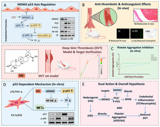
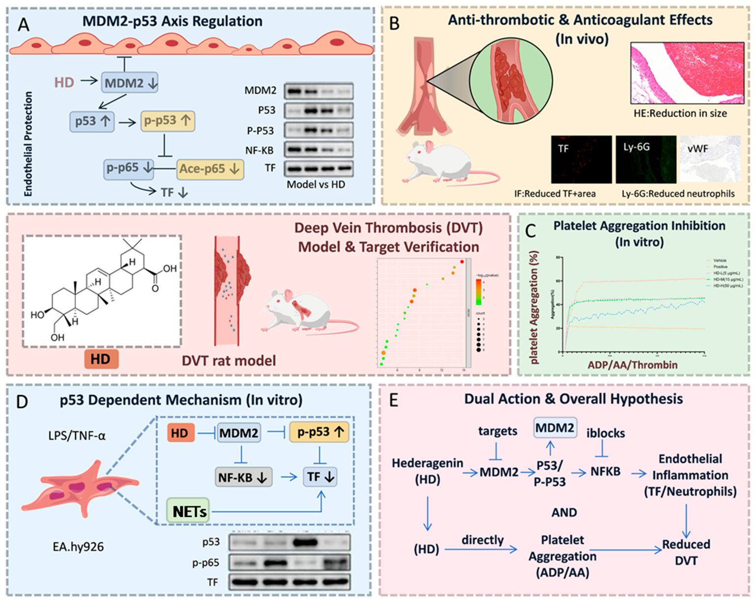
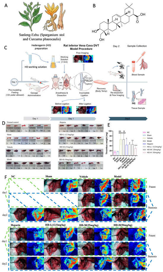
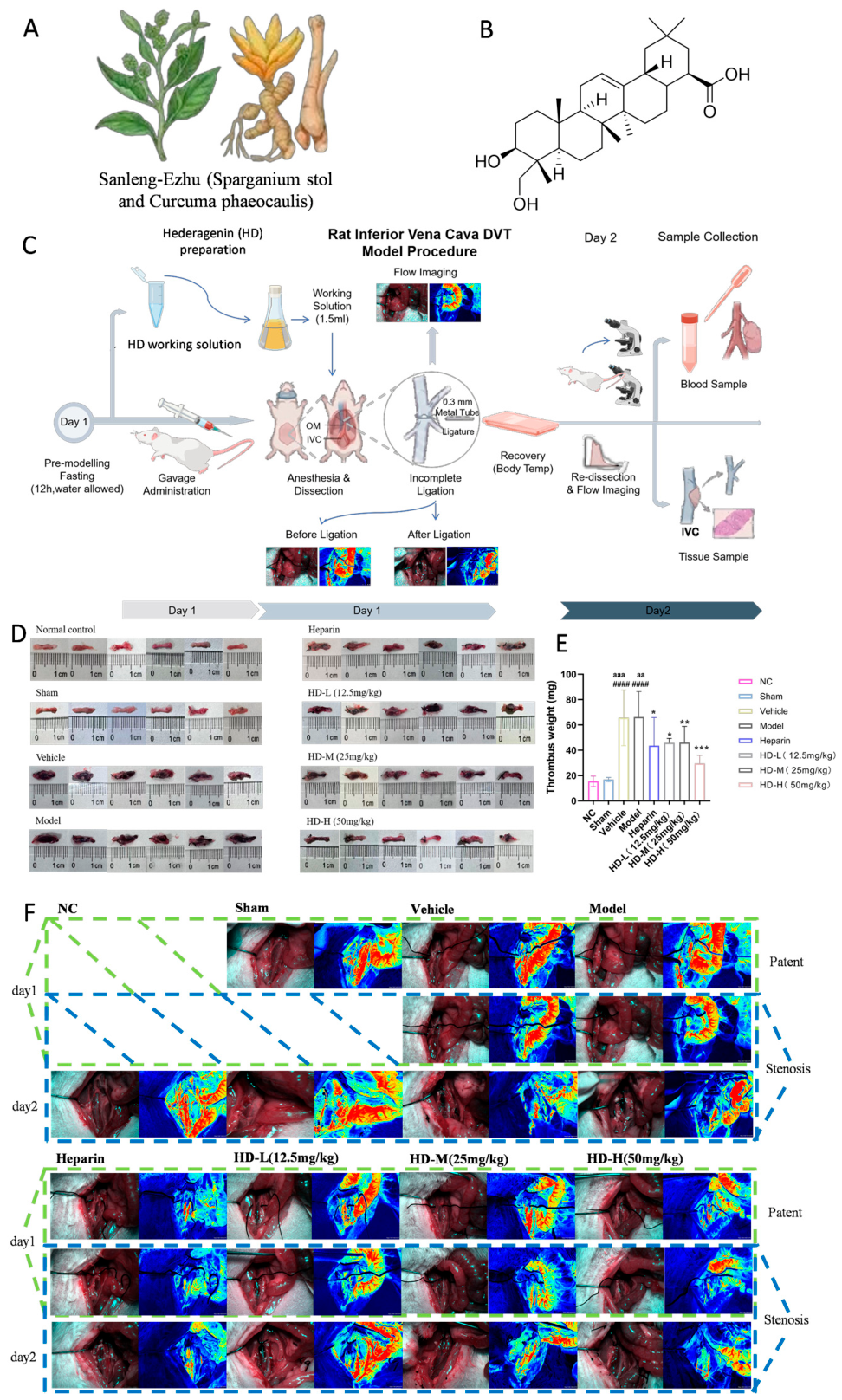
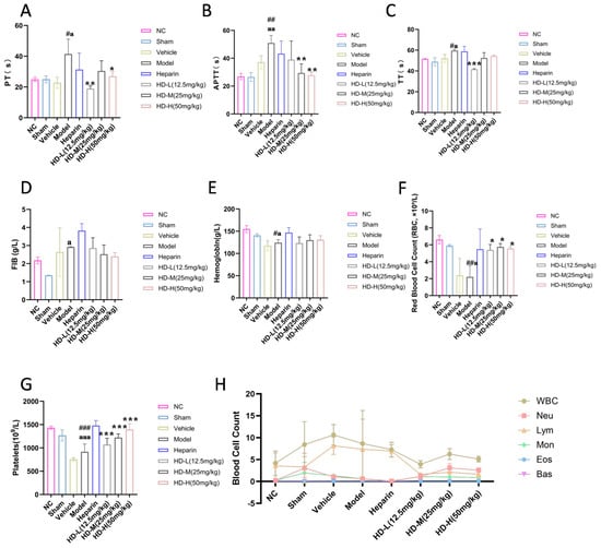
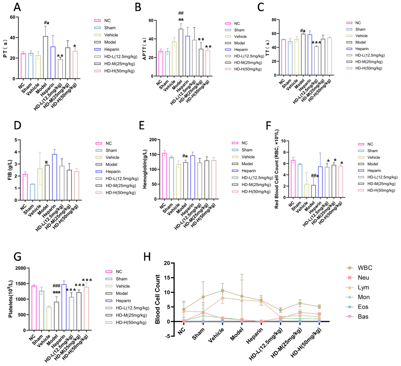
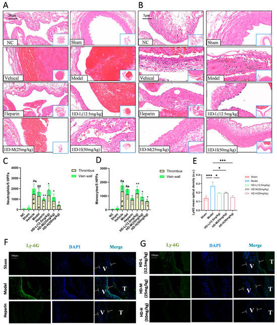
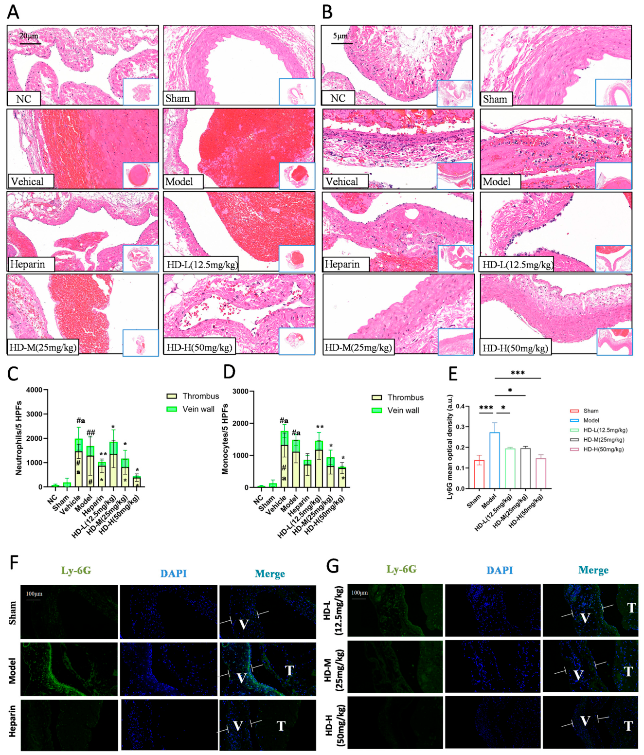
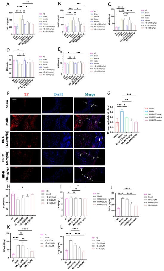
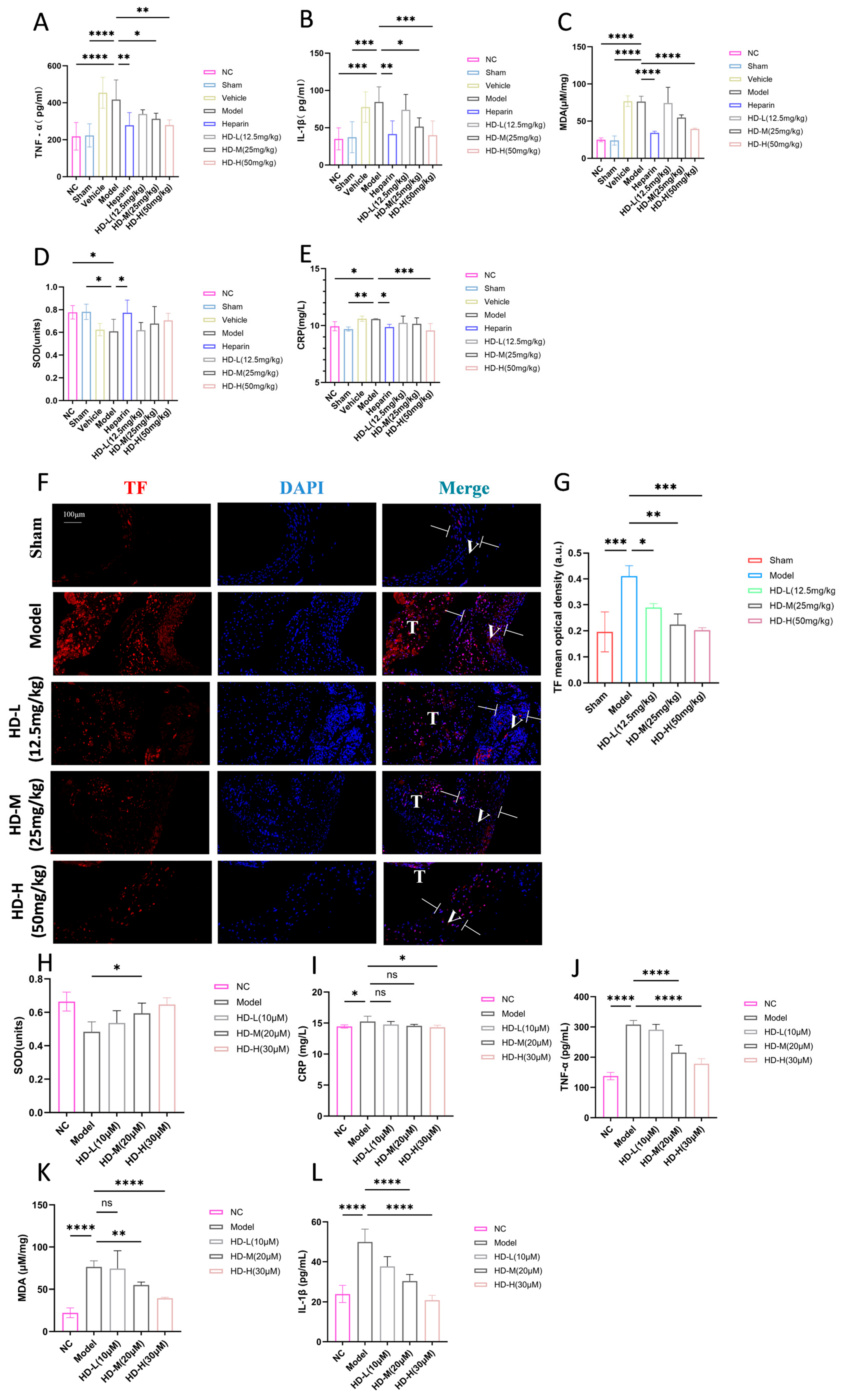
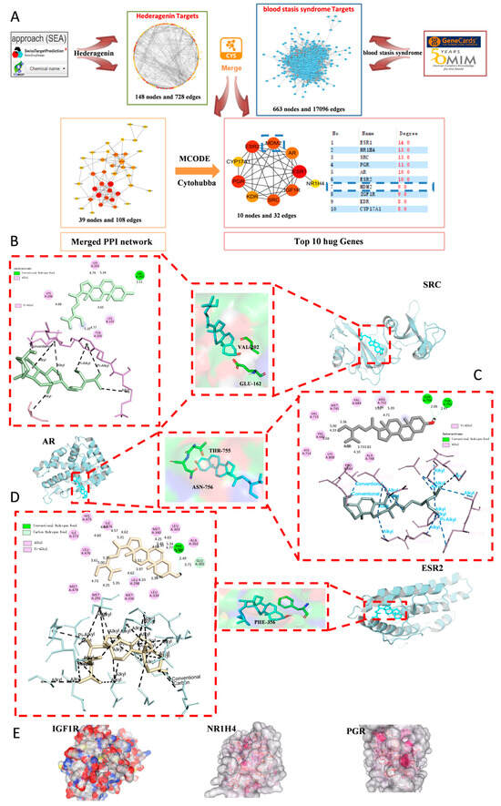
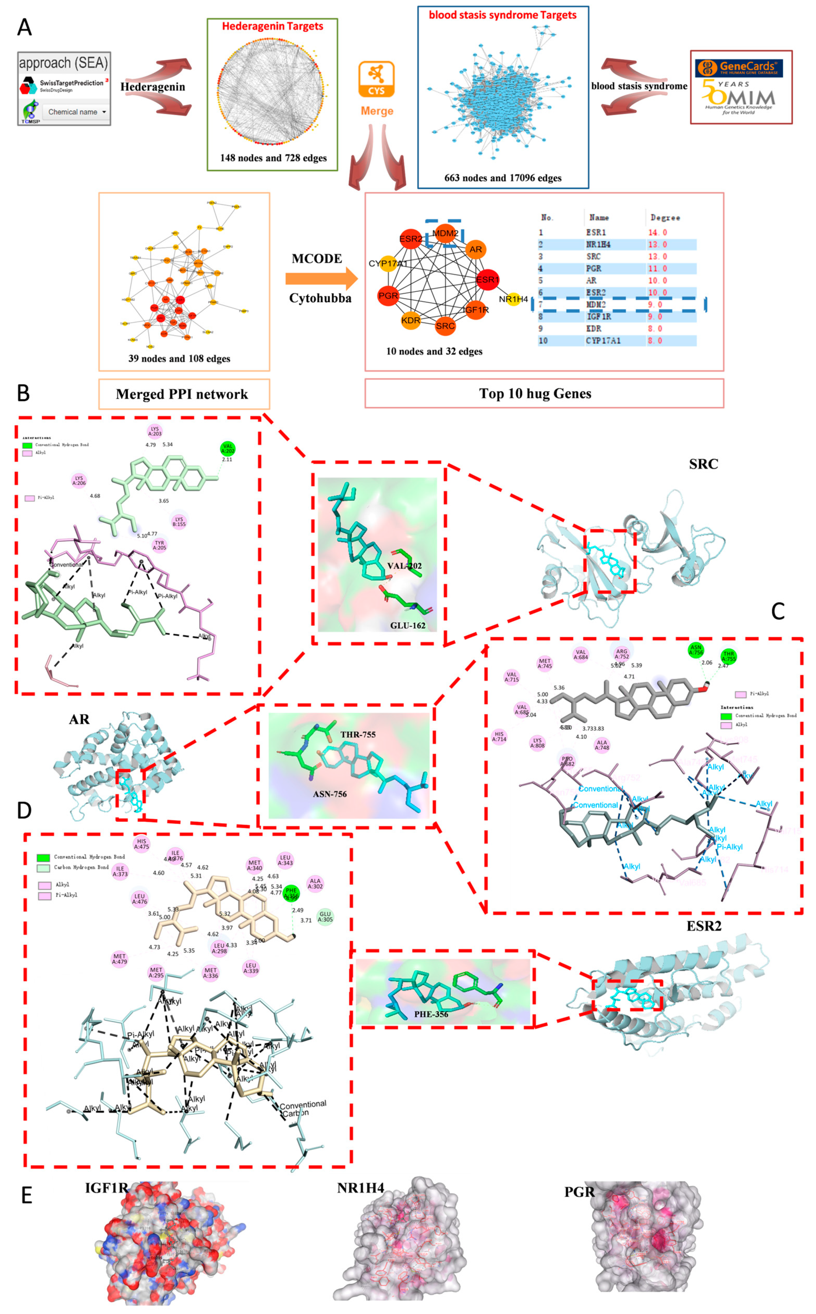
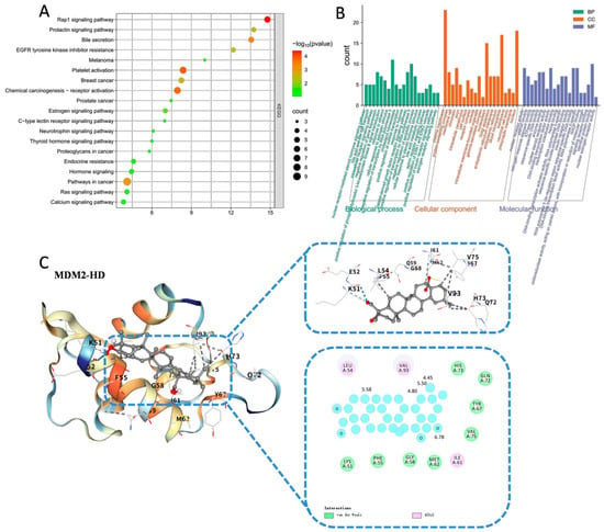
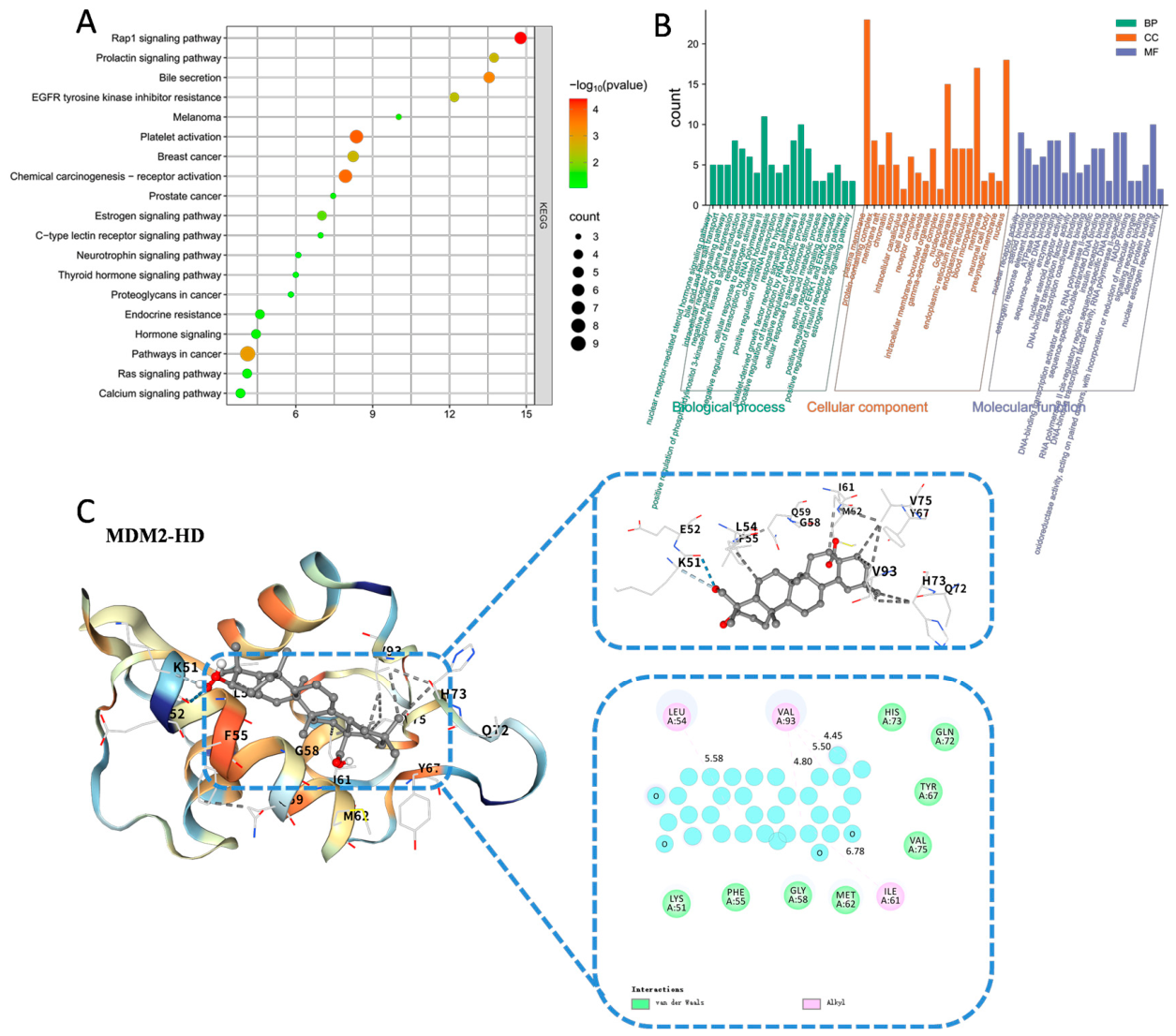
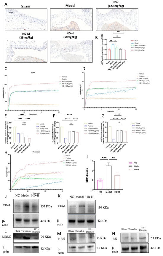
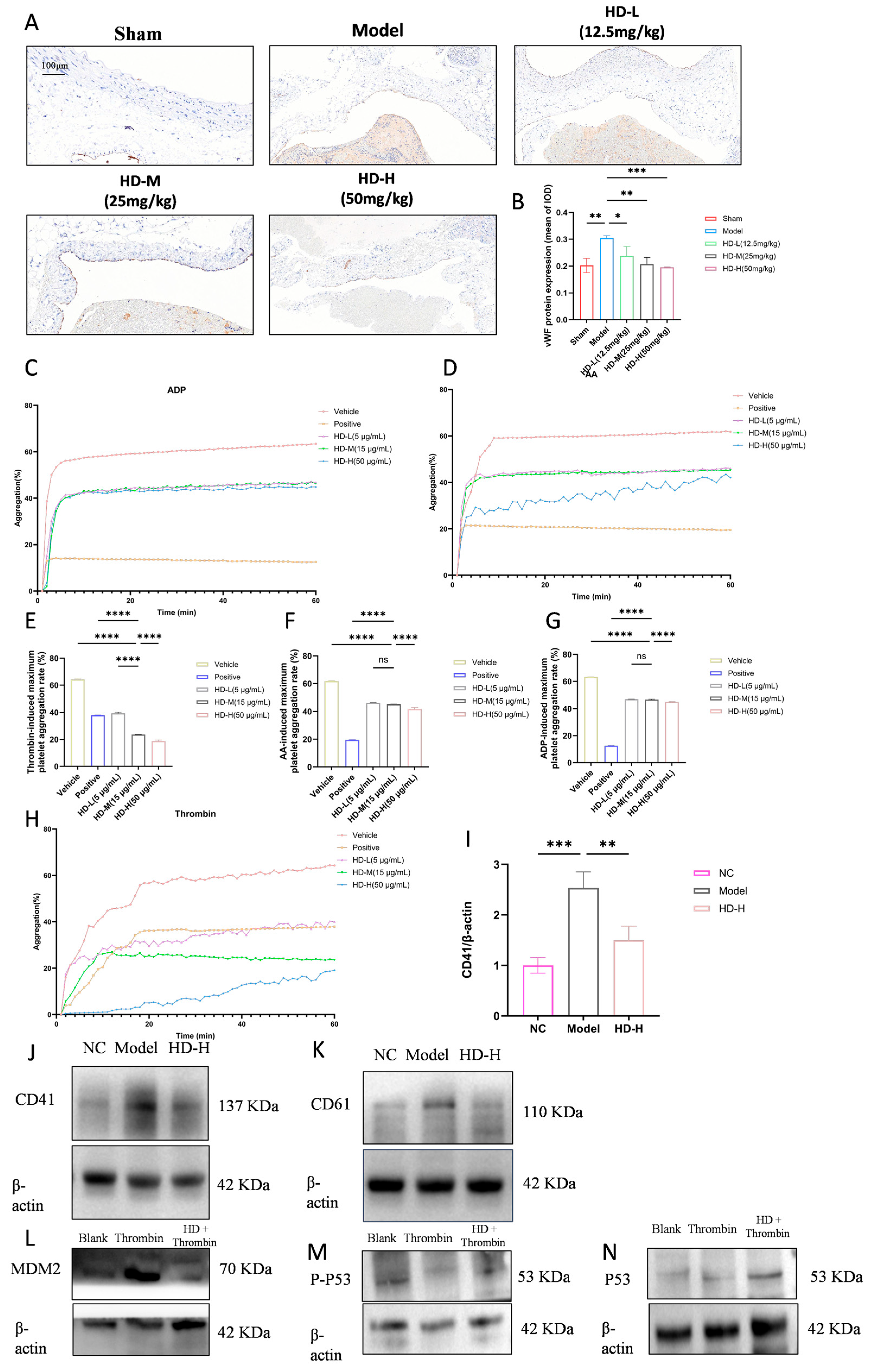
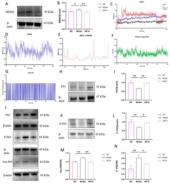
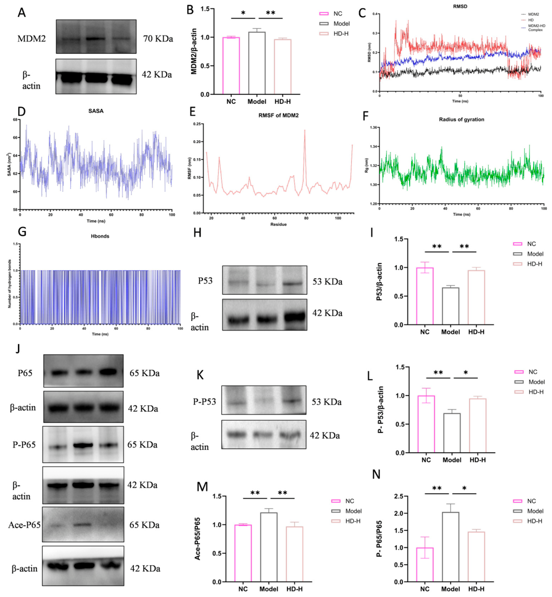
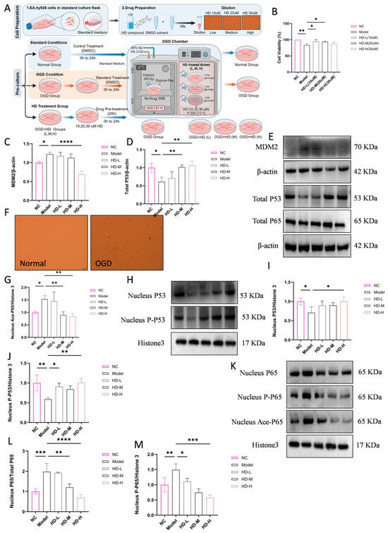
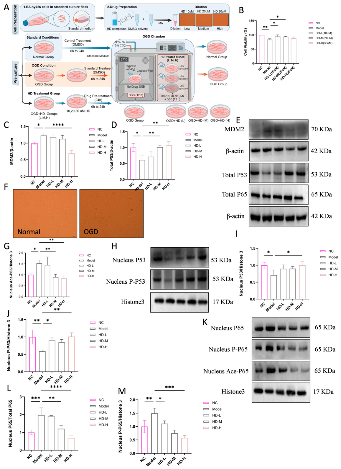
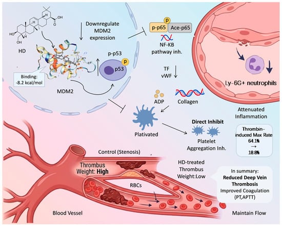
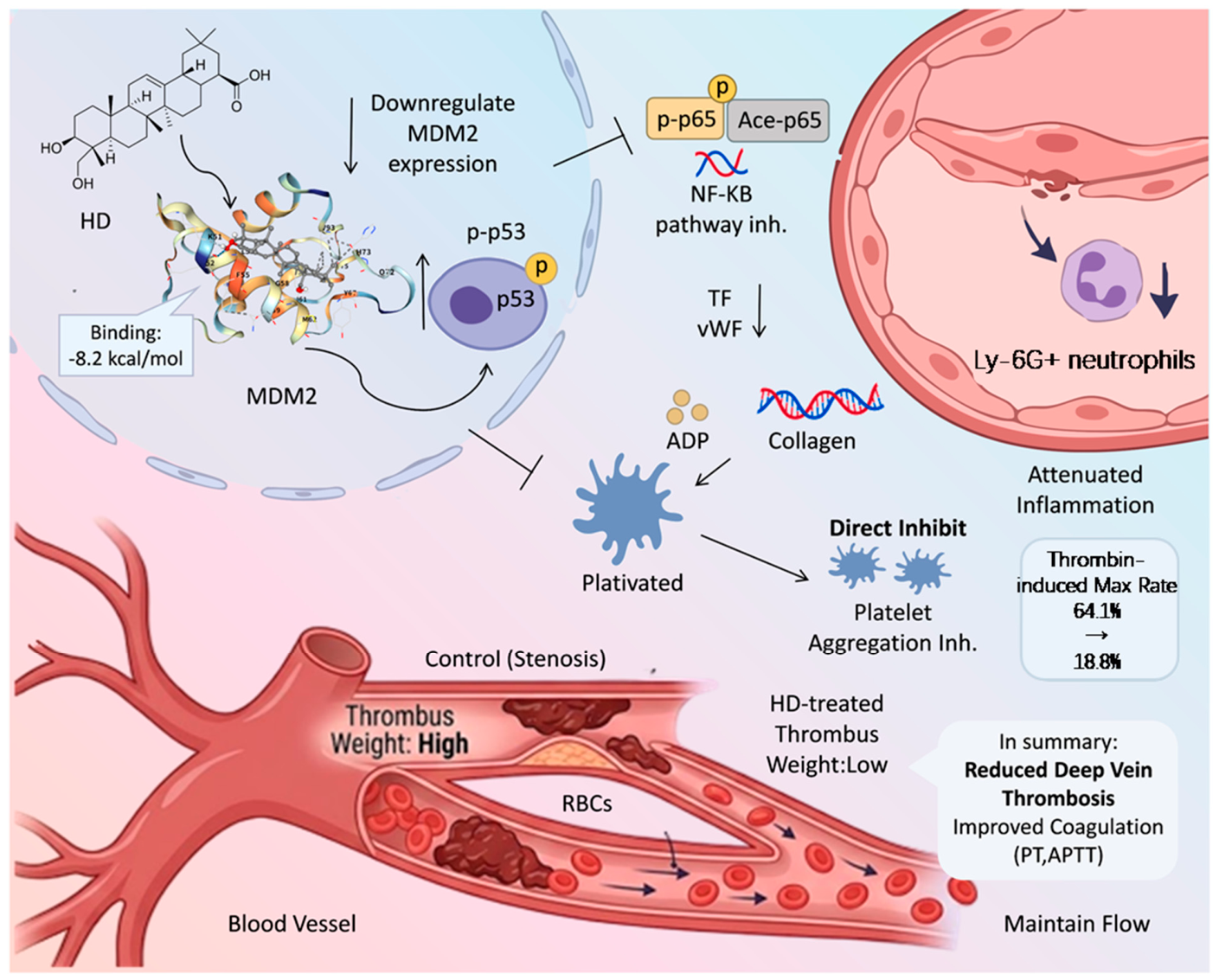
Disclaimer/Publisher’s Note: The statements, opinions and data contained in all publications are solely those of the individual author(s) and contributor(s) and not of MDPI and/or the editor(s). MDPI and/or the editor(s) disclaim responsibility for any injury to people or property resulting from any ideas, methods, instructions or products referred to in the content. |
© 2026 by the authors. Licensee MDPI, Basel, Switzerland. This article is an open access article distributed under the terms and conditions of the Creative Commons Attribution (CC BY) license.