


Abstract
Age-related macular degeneration (AMD) is a leading cause of irreversible vision loss worldwide, with dry AMD representing the predominant form of the disease, although effective treatments remain limited. The pathogenesis of dry AMD involves dysregulation of the complement system and impaired antioxidant defenses, contributing to retinal pigment epithelium (RPE) and photoreceptor degeneration. Given the multifactorial nature of the disease, targeting a single pathological pathway may not adequately address its complexity. Here, we developed a single recombinant adeno-associated virus serotype 8 (rAAV8) vector designed to overexpress nuclear factor erythroid 2-related factor 2 (NRF2) while knocking down complement component 3 (C3). In an in vitro screen using ARPE-19 cells, NRF2 overexpression provided the strongest protection against sodium iodate (NaIO3)-induced oxidative injury among the antioxidant candidates tested. We subsequently evaluated the dual-target vector in a mouse model of NaIO3-induced retinal degeneration. Compared with NaIO3-injured controls, the combination group showed significant improvements in electroretinographic responses and better preservation of retinal architecture. NRF2-containing treatment groups showed more favorable outcomes than shC3 monotherapy. Direct comparisons with NRF2 monotherapy revealed significant additional benefits of the combination for selected endpoints. These findings provide proof-of-concept evidence that dual targeting of oxidative stress and complement activation protects against NaIO3-induced retinal degeneration, supporting further evaluation of this approach in complementary chronic models.
1. Introduction
Age-related macular degeneration (AMD) is a progressive neurodegenerative disorder affecting the macula, the central retinal region responsible for high-acuity vision. Globally, the number of individuals affected by AMD is projected to rise from 196 million in 2020 to 288 million by 2040, posing a significant public health challenge [1,2]. AMD pathogenesis is complex and multifactorial, involving both genetic and environmental factors [3]. While neovascular “wet” AMD, characterized by choroidal neovascularization, can be managed with anti-angiogenic therapies [4], the “dry” form accounts for approximately 90% of all cases and remains clinically intractable. Dry AMD is characterized by drusen accumulation in Bruch’s membrane, progressive RPE cell death, and subsequent photoreceptor degeneration, eventually culminating in geographic atrophy (GA) [5]. Currently, treatments for dry AMD remain critically limited. Recently approved complement-target drugs for GA, such as Syfovre and Izervay [6], require frequent intravitreal injections and are associated with side effects including intraocular inflammation and a heightened risk of conversion to wet AMD [7,8]. Critically, neither drug has demonstrated the ability to restore lost vision. Consequently, there is an urgent need for long-lasting, prophylactic strategies. Recombinant adeno-associated virus (rAAV)-mediated gene therapy is ideally suited to meet this requirement for ophthalmic applications [9,10,11].
Genetic and epidemiological studies have identified more than 60 AMD risk loci, many of which implicate the complement system, highlighting the complement cascade, particularly complement component 3 (C3), as a key therapeutic target [12,13]. Another central driver of RPE dysfunction in dry AMD is oxidative stress [14,15]. The central retina is particularly susceptible to oxidative damage due to its high metabolic demand, continuous light exposure, and accumulation of lipid-rich debris [16]. Nuclear factor erythroid 2-related factor 2 (NRF2) serves as the master transcriptional regulator of the endogenous antioxidant response, governing the expression of key enzymes such as superoxide dismutases (SODs), heme oxygenase-1 (HO-1), catalase (CAT), glutathione peroxidases (GPXs) [17,18,19,20]. Under basal conditions, NRF2 is continuously targeted for ubiquitin-proteasomal degradation by kelch-like ECH-associated protein 1 (KEAP1), maintaining low steady-state NRF2 levels. However, oxidative stress triggers its nuclear translocation to activate a broad cytoprotective program via antioxidant response elements (AREs) [21]. Although antioxidative gene delivery strategies using viral vectors have been explored in several hereditary retinal disease models, no prior studies have directly compared the therapeutic efficacy of individual antioxidant enzymes with NRF2 overexpression in NaIO3-induced models [20,22,23].
Several mouse models have been developed through natural aging, genetic knockout, laser induction, or chemical induction to recapitulate features of dry AMD, each with inherent limitations [24,25]. Among these, the sodium iodate (NaIO3)-induced model is one of the most widely used. NaIO3 is an oxidizing agent that selectively induces RPE cell dysfunction and death across multiple mammalian species [26,27,28,29]. The resulting RPE loss occurs in a patchy pattern and progresses rapidly, initiating centrally near the optic nerve head (ONH), a distribution that closely mirrors the central RPE atrophy observed in dry AMD patients [30,31]. Furthermore, complement system activation, as evidenced by elevated C3 levels, has been reported in the NaIO3-induced model [32]. This is consistent with findings in patients with GA, in whom C3 immunoreactivity has been observed in the photoreceptor outer segment (POS) layer at the GA transition zone, and complement deposits have been detected underneath the RPE [33,34]. These characteristics make the NaIO3 model particularly suitable for studying oxidative damage and aberrant complement activation and for evaluating potential therapies for dry AMD.
The conventional “one-target-one-drug” paradigm has been instrumental in early-stage drug development, facilitating target identification through systematic loss-of-function studies [35,36]. However, this single-target strategy has shown limited clinical success in complex age-related diseases, where multiple pathological mechanisms converge to drive disease progression [37,38]. Dry AMD is a prototypical example of such complexity, in which oxidative damage and dysregulated complement activation collectively contribute to retinal degeneration, highlighting the necessity for the development of novel dual-target therapeutic drugs [39,40]. Notably, dual-target gene therapy strategies have recently been explored in wet AMD, providing a potential framework for addressing the multifactorial nature of retinal diseases [41,42]. However, whether a similar dual-target approach can be applied to dry AMD remains largely unexplored. Motivated by these limitations, we hypothesized that simultaneous modulation of both antioxidant and anti-inflammatory pathways would yield superior neuroprotection.
To test this hypothesis, we engineered a single rAAV8 vector designed to overexpress NRF2 while knocking down C3, thereby integrating the enhancement of endogenous antioxidant defenses with the modulation of complement activation. We first compared candidate antioxidant effectors in ARPE-19 cells and identified NRF2 as providing the strongest protection against NaIO3-induced oxidative injury among the candidates tested. We then evaluated the dual-target approach in a mouse model of NaIO3-induced retinal degeneration, assessing retinal function by electroretinography (ERG) and retinal structure by histological and morphometric analyses. Compared with NaIO3-injured controls, the dual-target treatment improved functional and structural outcomes. Notably, NRF2-containing treatment groups generally showed more favorable outcomes than shC3 monotherapy, whereas direct comparisons with NRF2 monotherapy revealed significant additional benefits for selected endpoints. Collectively, this study provides proof-of-concept evidence for integrating antioxidant and complement-targeting strategies within a single rAAV8 vector and supports further evaluation of this approach in chronic models of retinal degeneration.
2. Materials and Methods
2.1. Cell Lines and Culture Conditions
ARPE-19 cells (#CRL-2302, ATCC) were cultured in Dulbecco’s modified Eagle’s medium and F-12 culture medium (DMEM/F-12, SH30023.01, HyClone, Logan, UT, USA), HEK293T (#CRL-3216, ATCC) and Hepa1-6 cells (#CRL-1830, ATCC) were cultured in Dulbecco’s modified Eagle’s medium (C3122-0500, VivaCell, Shanghai, China), containing 10% fetal bovine serum (A5670201, Thermo Fisher Scientific, Waltham, MA, USA) and 1% penicillin/streptomycin (15140122, Thermo Fisher Scientific, USA) at 37 °C with 5% CO2. ARPE-19 cells were maintained in standard cell culture dishes and subcultured at a ratio of 1:4, with medium replacement or passaging performed every three days, as appropriate. Passage numbers were counted from the time the cells were first received from the supplier. ARPE-19 cells were cryopreserved at passage three (P3) and used for experiments within five passages after thawing. Each experiment was independently repeated at least three times.
2.2. Plasmid
SOD2/HO-1/CAT/GPX4/NRF2-FLAG sequences were PCR amplified from HEK293T cDNA and subcloned into a lentiviral transfer vector pLVX-CMVp-PuroR (632164, Takara Bio, Kusatsu, Japan) plasmid by XhoI (R0146, New England Biolabs, Ipswich, MA, USA) and BamHI (R0136, New England Biolabs, USA) restriction sites. For rAAV8 vectors, Nrf2-FLAG sequences were PCR amplified from C57BL/6J mice liver cDNA and were subcloned into a pAAV-CMVp-GFP plasmid (a gift from Prof. Chen Ling, Fudan University, China) through replacing the GFP coding sequence by EcoRI (R0101, New England Biolabs, USA) and XhoI restriction sites. shRNAs were designed and the interference sequences were listed in Figure S4, with non-targeting sequence as control (shCtrl). Synthesized oligonucleotides (Generay Biotechnology, Shanghai, China) were cloned to the recombinant adenoviral shuttle vector pAAV-hU6p-shCtrl-PuroR (a gift from Prof. Chen Ling, Fudan University, China) through replacing the non-targeting sequence using BamHI and HindIII (R0104, New England Biolabs, USA) restriction sites. The hU6p-shC3-4/shCtrl fragment was cloned from pAAV-hU6p-shC3-4/shCtrl to the recombinant adenoviral shuttle vector pAAV-CMVp-Nrf2-FLAG using MluI (R0198, New England Biolabs, USA) restriction sites. Primers were shown in Table S1.
2.3. Lentivirus Production and Transduction
Lentiviral vectors were produced by co-transfecting each pLVX transfer vector with the packaging plasmids psPAX2 (12260, Addgene, Watertown, MA, USA) and pCMV-VSV-G (8454, Addgene, USA) into HEK293T cells. Plasmids were mixed in a 4:3:1 (transfer vector:sPAX2:VSV-G) ratio by mass and mixed with an empirically optimized amount of branched polyethyleneimine (24765, Polysciences, Warrington, PA, USA) before adding to cells. Lentivirus-containing supernatants were collected at 48 and 72 h post-transfection, centrifuged at 500× g for 5 min, filtered through a 0.45 μm PES syringe filter (4614, Pall, Port Washington, NY, USA), aliquoted and stored at −80 °C. ARPE-19 cells seeding at 70% confluency in 6-well plate, 1.5 mL viral supernatants were added to ARPE-19 cells and culture for overnight, 8 μg/mL hexadimethrine bromide (HY-112735, MedChemExpress, Monmouth Junction, NJ, USA) was added during transduction. At 72 h post-transduction, the culture medium was replaced with selection medium containing 2 μg/mL puromycin (ST551, Beyotime, Shanghai, China). This medium was refreshed until all non-transduced cells were eliminated, after which the transduced cells were maintained in medium containing 1 μg/mL puromycin for expansion. Total RNA was collected after 7 days of selection.
2.4. NaIO3 Treatment
For cells, NaIO3 (S108351, Aladdin, Shanghai, China) were dissolved in DMEM/F12 culture medium with the stock concentration of 10 mg/mL. ARPE-19 cells were treated with various concentrations of NaIO3 according to the experimental design, while control groups received DMEM/F12 culture medium alone. The medium was replaced daily to maintain a constant level of NaIO3.
For in vivo NaIO3 modeling experiments, tail vein injections of 20 or 35 mg/kg NaIO3 or an equal volume of PBS were administered to 4- to 5-week-old male C57BL/6J mice. For the rAAV8 treatment experiments, subretinal injections of rAAV8 were performed at 4 to 5 weeks of age, followed by tail vein injection of NaIO3 four weeks later, at 8 to 9 weeks of age.
2.5. RNA Isolation and Reverse Transcription-Quantitative PCR (RT-qPCR)
Total RNA was extracted from cells or whole eyeballs using TRIzol reagent (15596018, Invitrogen, Carlsbad, CA, USA). cDNA was synthesized from 1 μg RNA using HiScript III RT SuperMix (R323-01, Vazyme, Nanjing, China). Gene expression was detected with ChamQ Blue Universal SYBR qPCR Master Mix (Q312-03, Vazyme, China) in a LightCycler 96 instrument (Roche, San Francisco, CA, USA). Relative mRNA expression was calculated using the ΔΔCT method and normalized against reference gene (GAPDH for human and Gapdh for mouse) in each sample. For genes with undetectable Ct values, a maximum cycle number of 40 was assigned to enable relative expression calculation. Gene-specific primer sets were shown in Table S1.
2.6. Western Blotting Analysis
Total proteins were extracted from cells using RIPA lysis buffer (P0013B, Beyotime, China) supplemented with proteases and phosphatase inhibitors cocktail (GRF103, Epizyme, Shanghai, China). Protein concentrations were quantified by BCA Protein Assay Kit (P0012, Beyotime, China). Samples were separated using 4–12% SDS-PAGE (LK408-20, Epizyme, China) electrophoresis and transferred to PVDF membrane (ISEQ85R, Merck Millipore, Darmstadt, Germany). The membrane was blocked with 5% skim milk (232100, BD biosciences, Heidelberg, Germany) in TBST for 1 h and incubated overnight at 4 °C with primary antibodies against FLAG (14793, 1:1000, Cell Signaling Technology, Danvers, MA, USA) or β-actin (ab8227, 1:2000, Abcam, Waltham, MA, USA) antibodies. After washing with TBST, the membrane was incubated with HRP-conjugated secondary antibodies at 1:10,000 for 1 h at room temperature (RT). Protein bands were detected with an enhanced chemiluminescence substrate (SQ203, Epizyme, China) and captured using an automatic chemiluminescence imager (Tanon, Shanghai, China).
2.7. Cell Counting Kit-8 (CCK-8) Assay
Cell viability was assessed using the CCK-8 assay. ARPE-19 cells were seeded in 96-well plates at a density of 7.5 × 10⁴ cells/mL (100 µL/well). After 24 h, cells were treated with NaIO3 (0, 5, 10, 15, or 20 mM) for 24 or 48 h. Separate 96-well plates were used for the two endpoint measurements, with both plates seeded and treated in parallel. After the respective treatment periods, cells were washed twice with PBS, followed by the addition of 100 µL of culture medium containing 10% CCK-8 reagent (CK04, Dojindo, Kumamoto, Japan) to each well. After incubation for an additional 2 h, absorbance was measured at 450 and 630 nm using a microplate reader (Spark, Tecan, Männedorf, Switzerland). Each treatment condition was assessed in five replicate wells at each time point. Wells containing 100 µL of culture medium supplemented with 10% CCK-8 reagent without cells served as blank controls. Relative cell viability was normalized to the corresponding untreated control at each time point.
2.8. Determination of the Reactive Oxygen Species (ROS)
Empty vector transduced ARPE-19 cells and antioxidant enzymes overexpression ARPE-19 cells were seeded in black 96-well plates at a density of 7.5 × 10⁴ cells/mL (100 μL/well). After 24 h, the cells were treated with varying concentrations of NaIO3 (0, 10, 15 mM) for an additional 48 h. ROS levels were measured using the DCFH-DA assay kit (R252, Dojindo, Japan) according to the manufacturer’s instructions. Briefly, following NaIO3 treatment, the cells were washed twice with HBSS (14175095, Gibco, Norristown, PA, USA) and incubated with freshly prepared DCFH-DA working solution at 37 °C for 30 min. After staining, the cells were washed twice again with HBSS. DCF fluorescence, resulting from intracellular hydrolysis of DCFH-DA, was visualized using a fluorescence microscope (IXplore IX73, Olympus, Tokyo, Japan) and quantified at excitation/emission wavelengths of 488/535 nm using a microplate reader (Spark, Tecan, Switzerland). ROS fluorescence values were normalized to cell viability as determined by CCK-8 assay.
2.9. Measurement of Malondialdehyde (MDA)
MDA levels were measured using an MDA Assay Kit (M496, Dojindo, Japan) according to the manufacturer’s instructions. Briefly, cells were seeded in 10 cm dishes and cultured at 37 °C with 5% CO2 until reaching approximately 80% confluency. The cells were then treated with or without 10 mM NaIO3 for 48 h. After treatment, cells were collected and lysed in PBS by ultrasonication. The lysate was centrifuged at 12,000× g for 10 min at 4 °C, and the supernatant was collected and divided equally into two aliquots for MDA quantification and protein concentration determination. MDA in the supernatant reacted with thiobarbituric acid (TBA) to form an MDA-TBA adduct, which was measured fluorometrically at excitation/emission wavelengths of 540/590 nm using a microplate reader (Spark, Tecan, Switzerland). MDA levels were calculated based on a standard curve and normalized to the protein concentration of each sample.
2.10. Live/Dead Cell Staining
Cell viability was assessed using a Live/Dead Cell Staining Kit (Calcein AM/PI) according to the manufacturer’s protocol (K2247, ApexBio, Houston, TX, USA). Briefly, NRF2-overexpressing ARPE-19 cells and control cells were seeded in 24-well plates at a density of 2 × 10⁵ cells/mL and allowed to attach for 24 h. The cells were then treated with 10 mM NaIO3 for 48 h. Following treatment, cells were washed twice with PBS and incubated with the staining solution at RT for 20 min in the dark. After incubation, cells were washed twice with PBS, and an appropriate volume of PBS was added to each well. Fluorescence images were acquired using a fluorescence microscope (IXplore IX73, Olympus, Japan). Live cells showed green fluorescence, whereas dead cells showed red fluorescence due to PI staining. The excitation/emission wavelengths were 494/517 nm for Calcein and 535/617 nm for PI.
2.11. Animals
Male C57BL/6J mice (4- to 5-week-old) (Institute of Laboratory Animal Sciences, JSJ, Beijing, China) were used for animal experiments. All mice were maintained in the animal facility of School of Life Sciences, Fudan University. Mice were housed under a 12 h light/dark cycle, with free access to water and food. The mouse procedures were approved by the Institutional Animal Care and Use Committee of Fudan University (No. 2022JS070 and No. 2024JS079). All procedures were performed in accordance with the Association for Research in Vision and Ophthalmology Statement for the Use of Animals in Ophthalmic and Vision Research.
2.12. rAAV Production and Subretinal Injection (SRI)
Subretinal injection was performed in 4- to 5-week-old C57BL/6J mice via transvitreal approach under a SZX10 dissecting stereomicroscope (Olympus, Japan). Briefly, mice were anesthetized with intraperitoneal (i.p.) injection of 200 μL per 10 g of mouse body weight of 1.25% Avertin (M2920, Aibei Biotechnology, Guangzhou, China) and trimmed the whiskers to avoid interference in the visual field of SRI procedures. The ocular surface was treated with 0.5% proparacaine hydrochloride (Alcon, Fort Worth, TX, USA) and tropicamide phenylephrine (Santen, Osaka, Japan). Corneal limbus at the nasal side was pierced with a 30-gauge BD insulin syringe needle (328438, BD biosciences, Germany). A 33-gauge blunt-tip needle (7803-05, Hamilton, Reno, NV, USA) was introduced into subretinal space through the corneal incision. Each eye received 1 μL rAAV8 solution at a titer of 1 × 10¹³ vg/mL, or equal volume of PBS. After injection, the needle was held in place for 20 s to allow full treatment delivery before being slowly withdrawn. Mice were treated with erythromycin ointment to prevent postoperative infection while recovering from anesthesia on a temperature therapy pad. rAAV8-EGFP, rAAV8-NRF2, rAAV8-shC3, rAAV8-NRF2/shC3 were produced by Lifespan Biotechnology (Shanghai, China) and used for all experiments performed in this work. The rAAV vectors were single-stranded rAAV8 vectors. The resulting rAAV8 preparations were assessed for protein purity, sterility, and fluorescent contamination. rAAV8 titers were determined by quantitative PCR targeting the rAAV genome. All rAAV8 preparations had titers exceeding 1 × 10¹³ vg/mL.
2.13. Electroretinography (ERG)
Full-field electroretinography was carried out to measure scotopic a- and b-wave and photopic a- and b-wave amplitudes. All experimental operations were performed under dim red light. Briefly, mice were dark-adapted overnight and anesthetized with 1.25% Avertin (M2920, Aibei Biotechnology, China) as previous described. Pupils were firstly dilated with 0.5% proparacaine hydrochloride (Alcon, USA) and tropicamide phenylephrine (Santen, Japan), and secondly lubricated with 2% hypromellose gel (HY-A0104, MedChemExpress, USA). Dark-adapted ERGs were elicited with 0.01 cd·s/m2 (rod-isolated) and 2 cd·s/m2 (rod-cone mixed). In addition, cone-isolated ERGs were elicited with 2 cd·s/m2 after a 25 cd·s/m2 background light for 10 min. ERG measurements were made using the APS-2000AER system (Kanghua, Chongqing, China). The a-wave amplitude was measured from the baseline to the negative peak, and the b-wave was measured from the a-wave trough to the maximum positive peak. Three to five traces were averaged per light intensity, with both eyes recorded.
2.14. RPE/Choroid Flat Mounts
After euthanasia, mouse eyeballs were enucleated and rinsed in PBS, then briefly fixed in 4% Paraformaldehyde (PFA) (J61899-AK, Thermo Fisher Scientific, USA) for 30 min at RT. After washing with PBS three times, the corneas and lens were removed, and the retinas were carefully separated from RPE/choroid complex. Afterwards, the RPE/choroid complexes were fixed in 4% PFA for additional 1 h at RT. After washing with PBS three times, the RPE/choroid complexes were treated with 0.1 M glycine (GB0235, Sangon Biotech, Shanghai, China) in PBS for 5 min at RT to quench excess formaldehyde. After washing with PBS three times, the RPE/choroid complexes were blocked and permeabilized at the same time by 1% BSA (A1933, Sigma, Darmstadt, German) and 0.2% Triton X-100 (TB0198, Sangon Biotech, China) in PBS for 1 h at RT. The blocking solution was aspirated, and the complex was washed three times in PBS, followed by incubation with phalloidin (ab176757, Abcam, USA) staining solution (prepared in 1% BSA/PBS) for 30 min at RT. After washing in PBS three times, the complexes were flattened onto a slide, and mounted with Mowiol 4-88 mounting medium (17591, Polyscienses, Warrington, PA, USA). Images were acquired using a FV3000 confocal microscope (Olympus, Japan).
2.15. Histological Analysis
Following euthanasia, the eyeballs were enucleated and a small incision was made at the corneoscleral junction using a 30-gauge needle to facilitate fixative penetration. The eyes were then fixed for 24 h in an ethanol-formalin-acetic acid fixative (prepared in a volumetric ratio of 10:7:2:1 of 75% ethanol, deionized water, formaldehyde, and acetic acid) at 4 °C. After fixation, the specimens were dehydrated and embedded in paraffin. Serial sections were cut at 4 μm thickness and stained with hematoxylin and eosin (H&E) (C0105S, Beyotime, China) for histological evaluation. Images were captured using a Nikon Eclipse Ni-U upright microscope (Nikon, Tokyo, Japan).
2.16. Immunofluorescence
Following euthanasia, the eyeballs were enucleated and a small incision was made at the corneoscleral junction using a 30-gauge needle to facilitate fixative penetration. The eyes were then fixed in 4% PFA for 30 min, followed by an additional 2 h fixation in fresh 4% PFA at 4 °C. After fixation, the eyes were washed with PBS three times, sequentially dehydrated in 10% and 20% sucrose (A100335, Sangon Biotech, China) in PBS for 2 h each, and then in 30% sucrose overnight at 4 °C. The next day, the eyecups were immersed in a 1:1 mixture of 30% sucrose and optimal cutting temperature (OCT) compound (4583, Sakura Finetek, Torrance, CA, USA) for 1 h prior to embedding in OCT medium. Cryosections of the retina were cut at 8 μm thickness, mounted onto slides, and subjected to a post-fixation step with 4% PFA for 15 min at RT. After three washes with PBS, the sections were stored at −20 °C until use.
For immunofluorescence staining, frozen sections were thawed and rewarmed to RT. The sections were blocked and permeabilized with PBS containing 0.5% Triton X-100 and 5% normal goat serum (C0265, Beyotime, China) for 1 h at RT. They were then incubated overnight at 4 °C with primary antibodies against RPE65 (ab231782, 1:250, Abcam, USA), Rhodopsin (MAB5356, 1:500, Sigma, German), Cone Arrestin (AB15282, 1:200, Sigma, German), and C3b/iC3b/C3c (HM1065, 1:1000, Hycult, Uden, The Netherlands). After three washes with PBS, the sections were incubated with appropriate Alexa Fluor-conjugated secondary antibodies: goat anti-mouse Alexa 488 (ab150113, 1:1000, Abcam, USA), goat anti-rabbit Alexa 647 (ab150079, 1:1000, Abcam, USA), and goat anti-rat Alexa 647 (ab150159, 1:1000, Abcam, USA) for 1 h at RT. Following three final washes with PBS, the sections were mounted with anti-fade mounting medium containing DAPI (P0131, Beyotime, China). Fluorescent images were acquired using an Olympus FV3000 confocal microscope (Olympus, Japan).
For cell immunofluorescence staining, ARPE-19 control cells and NRF2-overexpressing ARPE-19 cells were seeded in 24-well plates at a density of 2 × 10⁵ cells/mL. After 24 h, cells were treated with 10 mM NaIO3 for an additional 24 h. Cells were then washed twice with pre-cooled PBS and fixed with 4% PFA for 20 min at RT. After fixation, cells were blocked and permeabilized with 5% goat serum and 0.3% Triton X-100 for 1 h at RT. Cells were incubated overnight at 4 °C with primary antibodies against NRF2 (14596, 1:100, Cell Signaling Technology, USA) and FLAG (AE092, 1:300, ABclonal, Wuhan, China) diluted in 1% BSA containing 0.3% Triton X-100. After three washes with PBS, cells were incubated with the appropriate Alexa Fluor-conjugated secondary antibodies, including goat anti-rat Alexa Fluor 647 (ab150159, 1:1000, Abcam, USA) and goat anti-rabbit Alexa Fluor 488 (ab150077, 1:1000, Abcam, USA), for 1 h at RT. Following three washes with PBS, cells were mounted with anti-fade mounting medium containing DAPI. Fluorescence images were acquired using an Olympus FV3000 confocal microscope (Olympus, Japan).
For phalloidin staining, ARPE-19 cells were seeded on coverslips in 24-well plates at 80% confluency. After 24 h, cells were treated with 10 mM NaIO3 for an additional 48 h. The culture medium was carefully aspirated, and cells were washed once with PBS before fixation in 4% PFA for 30 min at RT. Cells were then permeabilized with 0.1% Triton X-100 in PBS for 5 min at RT, followed by three washes with PBS. Phalloidin conjugate working solution (1×) was added to each well and incubated for 30 min at RT in the dark. Cells were subsequently rinsed three times with PBS to remove excess phalloidin conjugate. Coverslips were mounted onto glass slides using anti-fade mounting medium containing DAPI. Fluorescent images were acquired using an Olympus FV3000 confocal microscope (Olympus, Japan).
2.17. Statistical Analysis
Data are presented as the mean ± SEM (Standard Error of the Mean). Unpaired Student’s t-test, or one-way ANOVA with Bonferroni’s post hoc test, was applied to determine statistical significance. Tests were performed using GraphPad Prism 9 (GraphPad Software, Boston, MA, USA). p < 0.05 was considered statistically significant.
3. Results
3.1. Establishment of an In Vitro Screening System for Antioxidant Gene Selection Using ARPE-19 Cells
ARPE-19 is a human RPE-derived cell line that has been used for preliminary in vitro studies of oxidative injury [43,44,45]. Consistent with previous reports, NaIO3 induced significant cytotoxicity in ARPE-19 cells in vitro (Figure 1A,B). Under physiological conditions, RPE cells and photoreceptors express high levels of antioxidant enzymes, including SOD2, HO-1, CAT, and GPX4, to counteract oxidative stress. RT-qPCR analysis following NaIO3 treatment revealed significant upregulation of these antioxidant genes in ARPE-19 cells after 24 h exposure to 10 mM NaIO3 (Figure 1C), suggesting an active cellular antioxidant response to oxidative insult. To identify the antioxidant candidate conferring the most robust protective effect, we constructed lentiviruses overexpressing each of these candidates and transduced ARPE-19 cells (Figure 1D). Following puromycin selection, stable overexpressing cell lines were successfully established, with overexpression levels ranging from approximately 7.10- to 1,108.33-fold relative to empty vector transduced controls, which were further confirmed at the protein level by Western blotting (Figure 1E,F). These stable cell lines provided a robust platform for the subsequent comparative evaluation of antioxidant efficacy.
Figure 1.
NaIO3-induced oxidative stress in ARPE-19 cells and screening of antioxidant gene candidates. (A) Cell morphology of ARPE-19 cells after 24 h of treatment with or without 10 mM NaIO3. Scale bar, 200 μm. (B) Cell viability assessed by CCK-8 assay (OD450) in wild-type (WT) ARPE-19 cells treated with increasing concentrations of NaIO3 for 24 or 48 h (n = 4). (C) mRNA expression levels of SOD2, HO-1, CAT, GPX4, and NRF2 in ARPE-19 cells following 24 h treatment with 10 mM NaIO3, determined by RT-qPCR. GAPDH was used as the reference gene. Data are normalized to WT ARPE-19 cells (n = 2 for HO-1 and n = 3 for all other genes). (D) Schematic diagram illustrating the construction of lentiviral vectors for stable overexpression of antioxidant genes in ARPE-19 cells. LTR, long terminal repeat; CMV, human cytomegalovirus promoter; PGK, phosphoglycerate kinase promoter; Puro(R), puromycin resistance (puromycin N-acetyltransferase). (E) RT-qPCR analysis of SOD2, HO-1, CAT, GPX4, and NRF2 mRNA expression levels in lentivirus-infected ARPE-19 stable cell lines. GAPDH was used as the reference gene. Data are presented relative to the Control ARPE-19 cells (n = 3). (F) Representative Western blot analysis of SOD2, HO-1, CAT, GPX4, and NRF2 protein expression in stable cell lines. FLAG indicates the FLAG tag, and molecular weights are indicated in kilodaltons (kDa). β-actin was used as a loading control. Data are representative of at least three independent experiments. All quantitative data are presented as mean ± SEM. Statistical significance was determined by unpaired Student’s t-test for panels B and C, and by one-way ANOVA followed by Bonferroni’s post hoc test for panel E. * p < 0.05, ** p < 0.01, *** p < 0.001, **** p < 0.0001; ns, not significant.
3.2. NRF2 Exhibits Highest Antioxidant Capacity Among the Candidates
As RPE dysfunction is a primary driver of dry AMD pathogenesis, we utilized ARPE-19 cells viability as the principal criterion for our initial screen. Using the Cell Counting Kit-8 (CCK-8) assay, we compared the relative viability of stable overexpressing cell lines across a range of NaIO3 concentrations. NRF2-overexpressing ARPE-19 cells consistently exhibited the highest viability under all tested conditions, with the protective effect becoming more pronounced as NaIO3 concentration and exposure duration increased (Figure 2A). Based on this screen, NRF2-overexpressing cell line was selected for further characterization using reactive oxygen species (ROS) and malondialdehyde (MDA) assays. Both assays confirmed superior antioxidant capacity of NRF2 relative to empty vector transduced controls. Specifically, ROS fluorescence intensity in NRF2-overexpressing cells was 0.60 ± 0.09-fold that of controls at 10 mM NaIO3, and further decreased to 0.40 ± 0.11-fold at 15 mM, representing approximately 40% and 60% reductions (Figure 2B). Similarly, NRF2-overexpressing cells maintained relatively stable MDA levels under NaIO3-induced oxidative stress, whereas controls exhibited a marked increase in lipid peroxidation. At 10 mM treatment, MDA content in NRF2 overexpressing cells was 0.70 ± 0.05-fold that of controls, reflecting a ~30% reduction in lipid peroxidation (Figure 2C). Notably, NRF2-overexpressing cells exhibited paradoxically elevated MDA levels compared to empty vector transduced controls at baseline (0 mM). We attribute this to constitutive, high-level NRF2 expression driven by the CMV promoter, which may chronically prime the antioxidant system and trigger compensatory stress responses or metabolic adaptations. To simultaneously assess cell survival and death, we performed live/dead staining, which further confirmed that NRF2 overexpression conferred enhanced resistance to NaIO3-induced cytotoxicity (Figure 2D).
Figure 2.
NRF2 demonstrates superior antioxidant capacity against NaIO3-induced oxidative injury, and lipid peroxidation. (A) Cell viability of antioxidant gene-overexpressing ARPE-19 stable cell lines assessed by CCK-8 assay following treatment with increasing concentrations of NaIO3 for 24 h (left) or 48 h (right). ARPE-19 cells transduced with empty lentiviral vectors served as the control group at each NaIO3 concentration (n = 4). Cell viability was normalized to that of the corresponding control group. Significance symbols indicate comparisons with the corresponding control group using one-way ANOVA followed by Bonferroni’s post hoc test. (B) Intracellular ROS levels in antioxidant gene-overexpressing ARPE-19 stable cell lines treated with 10 mM or 15 mM NaIO3 for 48 h, as determined by DCFH-DA fluorescence probe (green) (n = 4). Representative fluorescence images were shown. Scale bar, 200 μm. (C) MDA content quantified by thiobarbituric acid (TBA) assay after 48 h 10 mM NaIO3 treatment (n = 6). MDA levels are expressed as μmol/g protein. (D) Representative fluorescence images of live/dead cell staining (calcein-AM, green; propidium iodide/PI, red) in stable cell lines after 48 h 10 mM NaIO3 treatment. Scale bar, 200 μm. (E) Representative immunofluorescence images showing F-actin (phalloidin, red) in NRF2-overexpressing and control ARPE-19 cells following treatment with 10 mM NaIO3 for 48 h. Nuclei were counterstained with DAPI (blue). Scale bar, 100 μm. (F) Immunofluorescence staining of NRF2 (magenta) and FLAG (green) showing their co-localization after treatment with 10 mM NaIO3 for 24 h. Nuclei were counterstained with DAPI (blue). Scale bars: 40 μm (leftmost column) and 20 μm (columns 2−5 from the left). Data are normalized to the empty vector transduced control and presented as mean ± SEM. Statistical significance was determined by one-way ANOVA followed by Bonferroni’s post hoc test for panel A, and by unpaired Student’s t-test versus the control group for panels B and C. ** p < 0.01, **** p < 0.0001.
Furthermore, phalloidin staining revealed stress-fiber-like F-actin structures in NaIO3-treated, NRF2-overexpressing ARPE-19 cells, whereas F-actin staining was markedly reduced in the corresponding NaIO3-treated control cells (Figure 2E). Previous studies have reported NaIO3-induced F-actin remodeling in ARPE-19 cells, including increased F-actin polymerization and filopodia formation [46]. In the absence of NaIO3, no obvious differences in F-actin organization were observed between control and NRF2-overexpressing cells (Figure S1). Despite the stress-fiber-like structures observed, NRF2-overexpressing cells maintained higher viability than control cells following NaIO3 treatment. These findings suggest that the differences in F-actin staining were associated with NaIO3-induced stress, although the underlying mechanisms remain unclear. We also performed immunofluorescence staining of stable ARPE-19 cell lines using anti-NRF2 and anti-FLAG antibodies, with or without NaIO3 treatment. The results showed that C-terminal FLAG tagging did not affect the subcellular localization of NRF2. Following treatment with 10 mM NaIO3 for 24 h, NRF2 and FLAG signals co-localized in the nucleus (Figure 2F). Taken together, these in vitro findings support a protective effect of NRF2 overexpression against NaIO3-induced cellular injury in ARPE-19 cells and provide a rationale for further evaluation in in vivo therapeutic studies.
3.3. NaIO3-Induced Structural and Functional Disorders in the RPE and Retina
To validate our findings in vivo, we established a NaIO3-induced mouse model of retinal degeneration via tail vein injection. Comparison of two commonly used doses (20 and 35 mg/kg) revealed that the 20 mg/kg dose resulted in substantial inter-individual variability, with visual function remaining stable or partially preserved during long-term observation (Figure S2). However, the reduced sample size at D60 contributed to increased inter-individual variability and less consistent ERG measurements, limiting the reliability of functional assessment at this time point. Therefore, the 35 mg/kg dose, which produced more consistent and reproducible retinal injury, was selected for subsequent experiments. Eyeball samples were collected at 3-, 7-, 14-, and 28-day post-injection following functional assessment for histological analysis by hematoxylin and eosin (H&E) and phalloidin staining (Figure 3A−C).
Figure 3.
NaIO3 induces progressive retinal structural and functional degeneration in mice. (A) Representative H&E-stained retinal cross-sections from C57BL/6J mice at indicated time points after tail vein injection of 35 mg/kg NaIO3 (n = 5). Scale bar, 70 μm. RPE, retinal pigment epithelium; ONL, outer nuclear layer; INL, inner nuclear layer; GCL, ganglion cell layer. (B) Quantitative analysis of total retinal thickness at different distances from the ONH (left) and statistical comparison at 200 μm from the ONH (right). (C) Representative phalloidin-stained (red) RPE flat mount illustrating morphological disruption post-NaIO3. Scale bar, 20 μm. (D) Scotopic ERG a-wave (left) and b-wave (right) amplitudes at 2 cd·s/m2 flash intensity. PBS, n = 4. NaIO3, n = 5. (E) Representative scotopic (2 cd·s/m2) and photopic (2 cd·s/m2) ERG waveforms at each time point. Calibration: horizontal, 25 ms; vertical, 200 μV. Data are presented as mean ± SEM. Statistical significance was determined by one-way ANOVA followed by Bonferroni’s post hoc test for panel B, and by unpaired Student’s t-test for panel D. Comparisons were made with the age-matched PBS control group. ** p < 0.01, *** p < 0.001, **** p < 0.0001; ns, not significant.
In PBS-injected controls, the RPE monolayer displayed uniform pigmentation with preserved cellular polarity, and photoreceptor inner and outer segments (IS/OS) exhibited a highly organized architecture. At 3 days post-NaIO3 injection, RPE swelling and monolayer disruption were evident, accompanied by early reduction in photoreceptor segments. By 7 to 14 days, the RPE monolayer was largely absent, with aggregated pigment deposits dispersed throughout the subretinal space and depolarized RPE cells migrating into the neural retina. Progressive retinal thinning was observed, reflecting the loss of the IS/OS, outer nuclear layer (ONL) and outer plexiform layer (OPL). By day 28, RPE, IS/OS and ONL layer were virtually undetectable, with residual drusen-like pigmentary abnormalities remaining (Figure 3A).
To quantify neurosensory retinal survival, total neuroretina thickness was measured at fixed distances from the ONH. Significant thinning was first detected at 3 days post-injection, progressing continuously through day 28, by which point thickness had decreased to approximately 44.27% of PBS-injected controls (Figure 3B). RPE structural integrity was further assessed using RPE flat mounts stained with phalloidin to outline F-actin-based cell morphology. In NaIO3-injected animals, RPE injury was observed across both central and peripheral regions, with the central retina exhibiting more pronounced degeneration at a dose of 35 mg/kg (Figure 3C). Over time, RPE degeneration intensified, characterized by the complete loss of hexagonal morphology and a progressive reduction in phalloidin staining intensity, indicative of F-actin depletion and cytoskeletal disruption. Visual function analyses were also carried out by ERG. Following 35 mg/kg NaIO3 injection, mice exhibited progressive deterioration of both scotopic (rod-isolated and mixed) and photopic (cone-isolated) responses, with no functional recovery observed throughout the 60 days of observation and progressively reduced inter-individual variability over time (Figure 3D,E and Figure S3). Notably, the control group in the 35 mg/kg cohort showed a downward trend in ERG responses at D60. This observation may have been related to procedural effects associated with repeated ERG measurements.
Collectively, these results demonstrate that intravenous injection of 35 mg/kg NaIO3 induces time-dependent retinal degeneration in a dose-dependent manner as prior reported [47]. The 35 mg/kg dose produces continuous RPE and photoreceptor damage, establishing a robust and reproducible model suitable for therapeutic evaluation.
3.4. Enhanced Visual Function Preservation in RPE and Retina Treated with rAAV8-Mediated NRF2 Overexpression and C3 Knockdown
To develop a dual-target strategy targeting both oxidative stress and complement activation, we first screened short hairpin RNAs (shRNAs) targeting C3 in Hepa1-6 cells. Hepa1-6 cells are a murine hepatocellular carcinoma cell line that express complement system-related genes at relatively high levels and are readily transfected. Five shRNA sequences targeting different regions of the mouse C3 gene were designed and transfected into Hepa1-6 cells. Among the candidates, shC3-4 achieved the highest knockdown efficiency of 81.7 ± 4.9% and was subsequently incorporated into a U6-driven expression cassette for rAAV8 production (Figures S4 and S5). For in vivo evaluation, rAAV8 vectors were subretinal delivered to C57BL/6J mice, followed by NaIO3 challenge 4 weeks post-injection to induce RPE and retinal degeneration (Figure 4A and Figure S5). Six experimental groups were established: four treatment groups received subretinal injections of rAAV8 (NRF2, shC3, NRF2/shC3, or EGFP vector control), a PBS control group, and an untreated WT control. Robust marker gene expression in the RPE and retina was confirmed 4 weeks post-injection before the formal experiment (Figure S6).
Figure 4.
Dual-target rAAV8 treatment preserves visual function in NaIO3-induced mouse model of retinal degeneration. (A) Schematic diagram of the in vivo experimental design. Mice received subretinal injection of 1 μL rAAV8 (1 × 10¹³ vg/mL) or 1 μL of PBS. NaIO3 was administered 4 weeks post-subretinal injection. ERG and other assays were performed 7 days after NaIO3 administration (corresponding to 35 days post-rAAV injection). (B) Scotopic and photopic ERG recordings at 7 days post-NaIO3 injection. Average a-wave and b-wave amplitudes are shown for different flash intensity conditions. (C) Representative ERG waveforms of scotopic (2 cd·s/m2) and photopic (2 cd·s/m2) responses at 7 days post- NaIO3 injection. Calibration: horizontal, 25 ms; vertical, 200 μV. Data are presented as mean ± SEM (n = 12 per group). Statistical significance was determined by one-way ANOVA, and Bonferroni’s post hoc test was employed. p-values for the multiple comparisons are shown on the plots. WT, untreated controls; EGFP, rAAV8-EGFP + NaIO3; PBS, PBS + NaIO3; Therapeutic rAAV: rAAV8-NRF2, -shC3, or -NRF2/shC3).
ERG recordings performed 7 days after NaIO3 administration showed that the dual-target treatment group exhibited significantly higher ERG responses than the shC3 monotherapy group across all assessed scotopic (rod-dominated and mixed rod-cone) and photopic (cone-driven) conditions (Figure 4B,C). In contrast, NRF2 monotherapy did not show statistically significant differences from shC3 monotherapy in the corresponding comparisons. Direct comparisons between the dual-target treatment and NRF2 monotherapy revealed endpoint-specific benefits. At a scotopic flash intensity of 0.01 cd·s/m2, the combination group exhibited a significantly higher a-wave amplitude, whereas the b-wave amplitude showed only a numerical increase. At a scotopic flash intensity of 2 cd·s/m2, both a-wave and b-wave amplitudes were significantly higher in the combination group than in the NRF2 monotherapy group. Under photopic stimulation at 2 cd·s/m2, both a-wave and b-wave amplitudes showed numerical increases, but the differences were not statistically significant. Thus, the dual-target strategy consistently outperformed shC3 monotherapy across the assessed ERG conditions, whereas its additional benefits over NRF2 monotherapy were restricted to selected electrophysiological endpoints.
To assess the durability of this therapeutic effect, rAAV8-EGFP was mixed with each of the three therapeutic rAAV8 vectors and administered by SRI at a total dose of 4 × 10⁹ vg. ERG assessments were performed at 7, 14, 21, and 28 days after NaIO3 injection. Although ERG responses gradually declined over time across all treatment groups, the NRF2/shC3 group maintained higher mean ERG amplitudes under scotopic stimulation at 2 cd·s/m2 and showed significantly greater responses than the shC3 monotherapy group on day 28. In contrast, NRF2 monotherapy did not differ significantly from shC3 monotherapy (Figure S7A).
To confirm on-target therapeutic activity, transgene expression was quantified by RT-qPCR and immunofluorescence using whole eyeball lysates and retinal cryosections. Exogenous Nrf2 expression was verified using FLAG-tag specific primers and immunostaining, showing robust levels in both NRF2-treated groups (Figure 5). Notably, C3 expression was approximately 3-fold higher in both the EGFP and PBS groups than in untreated WT controls, with a numerically higher mean in the EGFP group than in the PBS group. Although this difference was not statistically significant, it raises the possibility that rAAV8 administration may contribute to elevated ocular C3 expression (Figure 5A). Despite this elevated baseline, C3 expression was significantly lower in both the NRF2/shC3 combination group (0.40 ± 0.07-fold relative to EGFP; 1.11 ± 0.20-fold relative to WT) and the shC3 monotherapy group (0.55 ± 0.08-fold relative to EGFP; 1.53 ± 0.22-fold relative to WT) than in the EGFP group. The rAAV8-NRF2 group also showed a numerical reduction in C3 expression (0.65 ± 0.06-fold relative to EGFP group). Consistent with this trend, the representative immunofluorescence images showed qualitatively weaker C3b/iC3b/C3c staining in the NRF2 group than in the EGFP group, with white asterisks indicating positive staining regions (Figure 5B). These observations raise the possibility that NRF2 overexpression may influence C3 expression and complement-associated responses through its potential anti-inflammatory effects, although this mechanism remains to be investigated. Collectively, these findings confirm successful transgene expression and support the intended molecular activity of the dual-target rAAV8 vector.
Figure 5.
Efficient rAAV8-mediated NRF2 overexpression and C3 knockdown via subretinal injection. (A) RT-qPCR analysis of mRNA expression levels in whole eyeballs at 7 days post-NaIO3 injection. Exogenous Nrf2-FLAG, endogenous Nrf2, and C3 transcript levels are shown relative to Gapdh. For Nrf2-FLAG, n = 3 (WT), n = 5 (others). For endogenous Nrf2 and C3, n = 5 per group. Data are presented as mean ± SEM. Statistical significance was determined by one-way ANOVA versus EGFP group, and Bonferroni’s post hoc test was employed. * p < 0.05, ** p < 0.01, *** p < 0.001, **** p < 0.0001. UD, undetected. (B) Immunofluorescence images of retinal cryosections showing C3 cleavage products (C3b/iC3b/C3c, red) and EGFP (green) in both panels. White represents FLAG-tagged exogenous NRF2 in the upper panel and transmitted light (TD) in the lower panel. NRF2-FLAG predominant localized at ONL layer and RPE. Images are representative of n = 5 mice per group. Nuclei were counterstained with DAPI (blue). Scale bar, 30 μm. RPE, retinal pigment epithelium; ONL, outer nuclear layer; INL, inner nuclear layer; GCL, ganglion cell layer.
3.5. NRF2-Containing Treatments Better Preserve RPE Morphology than C3 Knockdown Alone
GA represents a hallmark pathological feature of dry AMD [48]. To evaluate therapeutic efficacy with respect to RPE cytoskeletal integrity and morphological preservation, phalloidin staining was performed on RPE/choroid flat mounts. In healthy retinas, RPE cells form a tightly connected monolayer of pigmented, hexagonally or polygonal organized epithelial cells, an architecture that underlies outer blood–retinal barrier (oBRB) function and enables the directional transport essential for retinal homeostasis [49,50]. Morphological examination revealed that the RPE/choroid flat mounts from both the NRF2 monotherapy and NRF2/shC3 combination groups most closely resembled those of age- and sex-matched WT controls, retaining relatively regular polygonal cell organization (Figure 6). In contrast, the shC3 monotherapy group exhibited the most pronounced morphological disruption among the three treatment groups, with prominent pigment accumulation in lesioned areas. This observation is consistent with previous reports that C3 deletion attenuates NaIO3-induced photoreceptor degeneration but provides limited protection against RPE damage [51], suggesting that C3 suppression alone may be insufficient to preserve RPE morphology under oxidative stress. Consistent with this observation, combining C3 knockdown with NRF2 overexpression did not appear to provide an additional benefit in RPE morphological preservation beyond that observed with NRF2 monotherapy. Notably, the apparent protective effect of NRF2 overexpression on RPE morphology persisted for at least one month after NaIO3 injection (Figure S7B,C). Collectively, these observations suggest that NRF2-mediated enhancement of antioxidant defense provides more pronounced morphological preservation of the RPE than C3 suppression alone in this model.
3.6. The Dual-Target Treatment Better Protects the Mice Retina Against Oxidative Damage
Histological analysis of harvested eyes was performed to evaluate therapeutic efficacy at the tissue level. H&E staining seven days after NaIO3 injection revealed better preservation of retinal architecture in both the NRF2 monotherapy and NRF2/shC3 combination groups than in the EGFP control and shC3 monotherapy groups. Both NRF2-containing treatment groups exhibited greater overall retinal thickness and better preservation of multiple retinal layers, including the inner plexiform layer (IPL), inner nuclear layer (INL), OPL, ONL, and IS/OS (Figure 7A). The photoreceptor nuclei also appeared better aligned in both NRF2-containing groups, while RPE morphology was better preserved. In contrast, marked pigmentary disorganization and GA-like lesions were observed in the EGFP, PBS, and shC3 groups.
Figure 6.
NRF2-containing treatments preserve RPE morphology following NaIO3-induced retinal injury. Representative confocal images of phalloidin (red)-stained RPE/choroid flat mounts, with EGFP shown in green. WT controls exhibit regular hexagonal or polygonal morphology. PBS and EGFP group displayed severe disruptions, including enlargement, irregularity, and loss of hexagonal or polygonal organization. The shC3 group shows partial protection with evident pigment deposits (white asterisks). The NRF2 and NRF2/shC3 groups show significantly reduced morphological abnormalities. Scale bars, 500 μm (low magnification); 20 μm (high magnification). Images are representative of n = 5 per group.
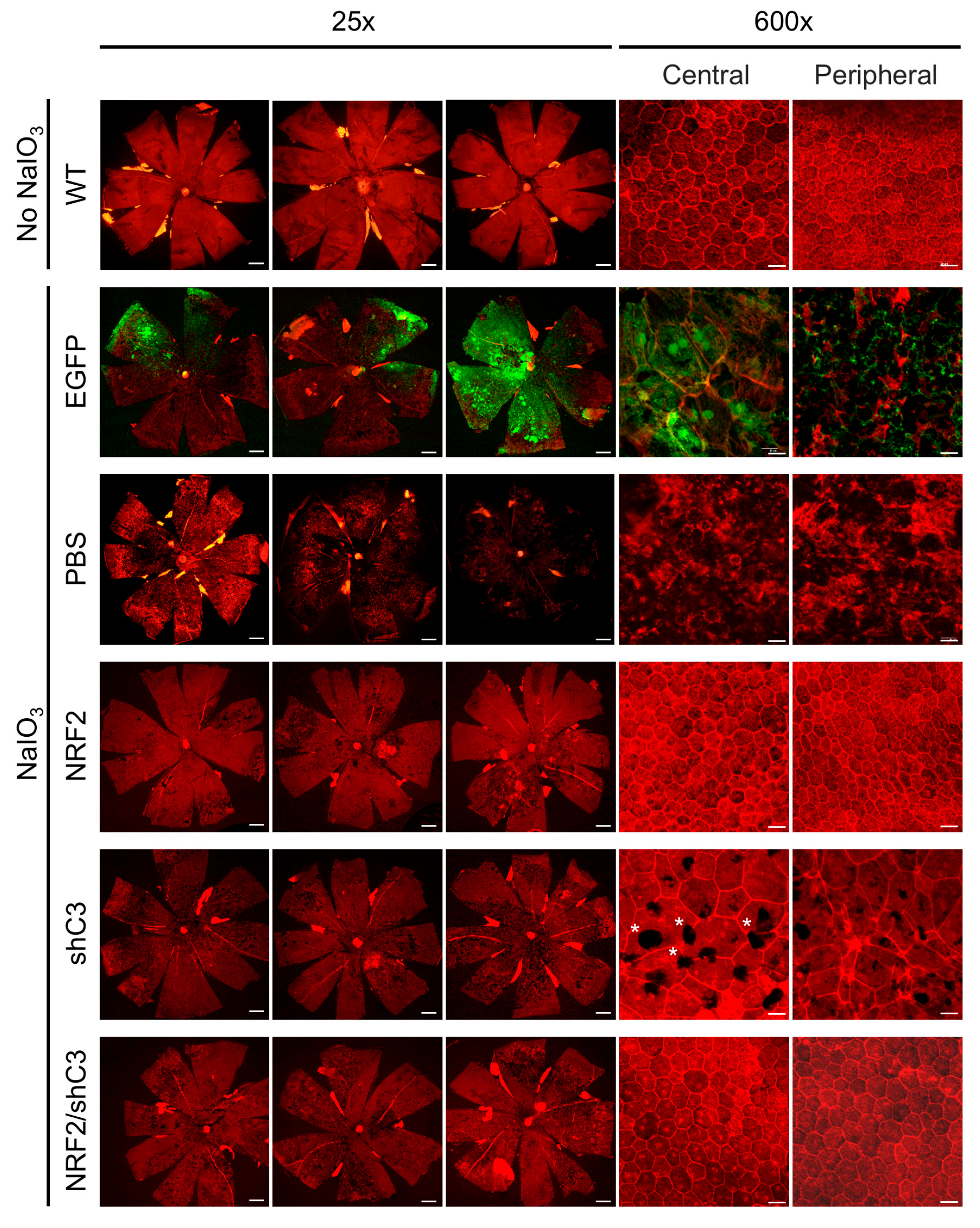
Figure 7.
Combinational rAAV8 therapy protects retinal architecture and cell-specific markers. (A) H&E staining of retinal cross-sections 7 days post-NaIO3 injection. Scale bars, 500 μm (low magnification, whole eyeball); 30 μm (high magnification). Images are representative of n = 5 per group. (B) ONL thickness profile across the retina (left) and statistical comparison at 200 μm from the ONH (n = 5). Statistical significance between NRF2/shC3 and shC3 is indicated on the plot. (C) Immunofluorescence RPE65 (magenta), Rhodopsin (green), and Cone Arrestin (yellow). Nuclei were stained with DAPI (blue). Scale bar, 30 μm. RPE, retinal pigment epithelium; IS/OS, inner segment/outer segment; ONL, outer nuclear layer; OPL, outer plexiform layer; INL, inner nuclear layer; IPL, inner plexiform layer; GCL, ganglion cell layer. Data are presented as mean ± SEM. Statistical significance was determined by one-way ANOVA, and Bonferroni’s post hoc test was employed. * p < 0.05, ** p < 0.01.
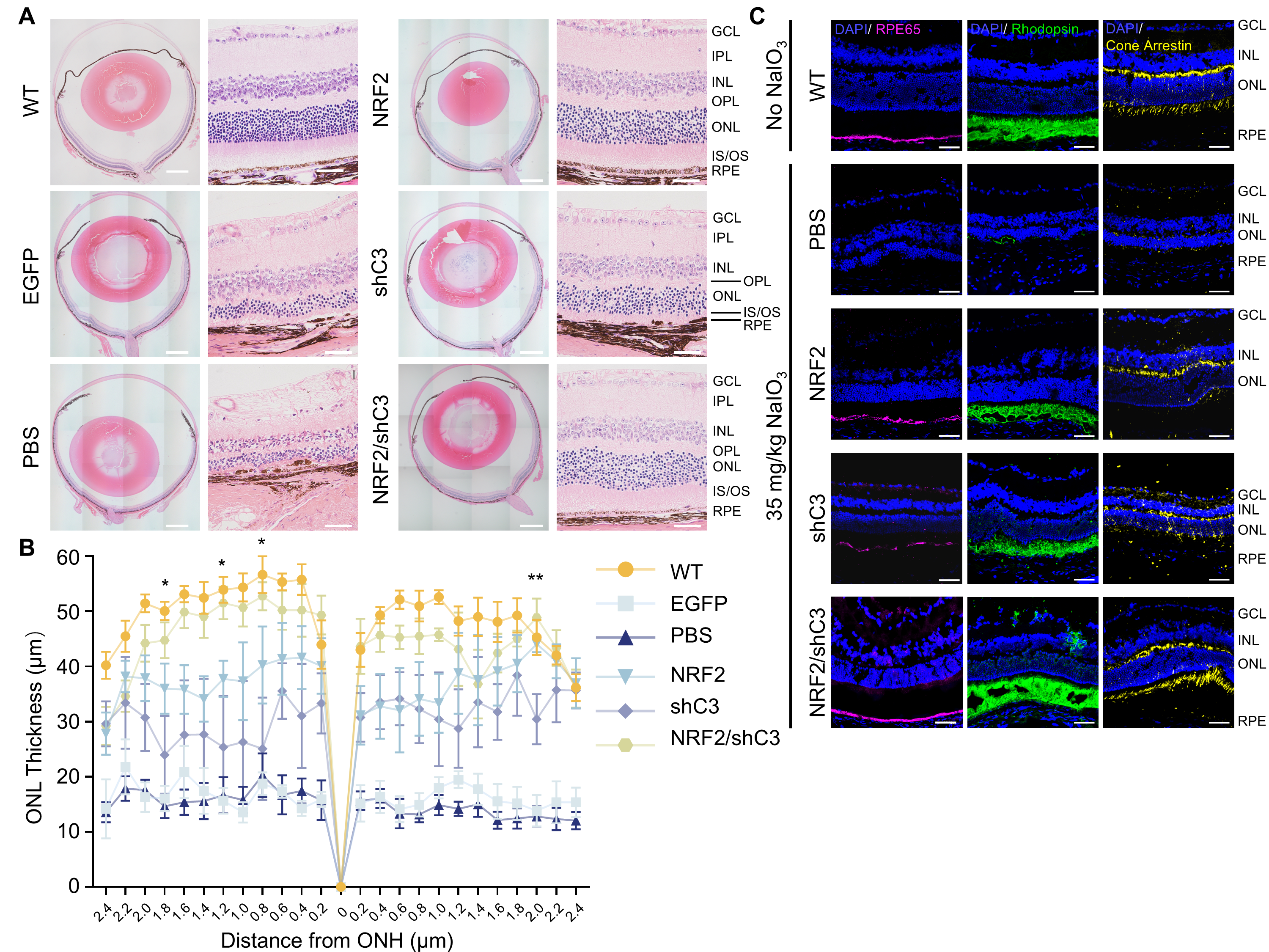
Quantitative measurements of ONL thickness at 200 μm intervals from the optic nerve head (ONH) showed that the NRF2/shC3 combination group generally exhibited the greatest preservation of ONL thickness, with values closest to those of WT controls, followed by NRF2 monotherapy and shC3 monotherapy (Figure 7B). However, the combination treatment showed significantly greater ONL thickness than shC3 monotherapy at specific measurement locations, as indicated by the asterisks in Figure 7B. NRF2 monotherapy did not differ significantly from shC3 monotherapy at these locations.
To further characterize these findings, immunofluorescence staining was performed using markers of RPE cells and photoreceptors (Figure 7C). Retinal pigment epithelium-specific 65 kDa protein (RPE65), an enzyme involved in the visual cycle, was used as an RPE-associated marker, whereas Rhodopsin and Cone Arrestin were used to assess rod and cone photoreceptors, respectively. Representative images showed more prominent staining for all three markers in the dual-target treatment group than in the other treatment groups, qualitatively supporting better preservation of RPE-associated and photoreceptor-associated markers.
Taken together, these histological findings indicate that NRF2-containing treatments provide greater retinal structural preservation than shC3 monotherapy. Compared with NRF2 monotherapy, the NRF2/shC3 combination showed a numerical trend toward greater ONL thickness preservation, while representative immunofluorescence images showed more prominent staining for RPE65, Rhodopsin, and Cone Arrestin. These findings suggest that the additional benefits of combination treatment vary across histological endpoints. The better retinal preservation observed in NRF2-containing treatment groups further suggests that oxidative stress is an important contributor to retinal degeneration at this stage of NaIO3-induced injury. However, the incomplete preservation of retinal structure relative to WT controls across all treatment groups suggests that additional pathological mechanisms beyond oxidative stress and complement activation may contribute to disease progression and warrant further investigation.
3.7. Dual-Target Strategy Maintains Intraocular Homeostasis with Visual Cycle Protection and Anti-Inflammatory Effects
To characterize the transcriptional changes associated with the protective effects of dual-target gene therapy, we assessed genes related to the visual cycle, antioxidant defense, inflammation, and lipid metabolism by RT-qPCR (Figure 8).
Figure 8.
Dual-target intervention provides selective additional benefits in visual-cycle and inflammatory gene expression compared with monotherapies. RT-qPCR analysis of eyeball mRNA levels 7 days post-NaIO3 injection for: (A) retina-related, (B) RPE-related, (C) NRF2-downstream, and (D) inflammation/lipid metabolism-related effects. Raw 2^(−ΔΔCt) values were log2-transformed for visualization. Gapdh served as the reference gene (n = 3–5). Data are presented as mean ± SEM. Statistical significance was determined by one-way ANOVA, and Bonferroni’s post hoc test was employed. * p < 0.05, ** p < 0.01, *** p < 0.001, **** p < 0.0001; ns, not significant.
The NRF2/shC3 combination showed endpoint-specific benefits across photoreceptor-associated and inflammatory markers (Figure 8A). Compared with NRF2 monotherapy, the combination significantly increased cone arrestin 3 (Arr3) and phosphodiesterase 6b (Pde6b) expression, while compared with shC3 monotherapy, it significantly increased Rhodopsin and retinol dehydrogenase 8 (Rdh8) expression and reduced retinal microglia marker, ionized calcium-binding adapter molecule 1 (Iba1) expression. Neither monotherapy showed significant differences from the other for these markers.
For RPE-associated markers, both NRF2-containing treatment groups showed significantly higher expression of Rpe65, lecithin retinol acyltransferase (Lrat), and retinol dehydrogenase 5 (Rdh5) than the shC3 monotherapy group (Figure 8B). No significant differences were observed between NRF2 monotherapy and the NRF2/shC3 combination group. These findings suggest that NRF2 overexpression contributes to the preservation of RPE-associated gene expression, whereas the additional effects of C3 knockdown on these markers were limited.
To assess the functional engagement of the NRF2 antioxidant pathway, we examined its canonical downstream targets (Figure 8C). NaIO3-induced injury markedly suppressed the expression of these genes, whereas both NRF2-containing treatment groups showed an overall increase in their expression. Notably, glutathione s-transferase mu 1 (Gstm1) and NAD(P)H quinone oxidoreductase 1 (Nqo1) expression was significantly elevated in both groups. An upward trend in Ho-1 expression was also observed across the rAAV8-treated groups, raising the possibility that vector administration itself may influence the NRF2/HO-1 pathway independently of the therapeutic transgene. This possibility, however, requires further investigation.
We further examined inflammatory responses following NaIO3-induced retinal injury. NaIO3 treatment markedly increased the expression of interleukin-1 beta (Il-1β), interleukin-6 (Il-6) and complement c1q b chain(C1qb) (Figure 8D). Although all treatment groups showed attenuation of this inflammatory response, the NRF2/shC3 combination group exhibited significantly lower expression of Il-1β, C1qb, and Il-6 than the shC3 monotherapy group. Moreover, Il-6 expression was significantly lower in the combination group than in the NRF2 monotherapy group, indicating an additional benefit of dual targeting for this inflammatory endpoint. Secreted phosphoprotein 1 (Spp1), a potential inflammatory biomarker of dry AMD [52], showed a downward trend in all three therapeutic groups compared with the EGFP and PBS control groups, with the lowest mean expression observed in the NRF2/shC3 group. Collectively, these findings suggest that the dual-target approach attenuates NaIO3-induced inflammatory responses, with effects varying across molecular endpoints.
Finally, we assessed apolipoprotein E (Apoe) expression as a marker of lipid metabolic dysregulation (Figure 8D). Lipid metabolism has been increasingly recognized as a key contributor to AMD pathogenesis, with APOE identified as a significant genetic risk factor in both epidemiological and experimental studies [53,54]. NaIO3 injection led to a significant increase in Apoe levels in control groups. Neither monotherapy effectively normalized Apoe expression; however, the combination treatment successfully restored Apoe to near-WT levels.
Collectively, these transcriptional findings suggest that the dual-target approach provides benefits across multiple biological processes associated with NaIO3-induced retinal injury, including visual cycle function, antioxidant defense, inflammatory regulation, and lipid metabolism. However, the magnitude of the additional benefits over single-target treatments varied across endpoints. These readouts reflect the complexity of the NaIO3-induced retinal injury model and the biological responses to dual-target therapy.
4. Discussion
In this study, we developed a combinatorial gene therapy strategy that integrates NRF2 overexpression and C3 knockdown within a single rAAV8 vector to target oxidative stress and complement activation in a NaIO3-induced oxidative damage model of dry AMD. Taking advantage of the ability of subretinally delivered rAAV8 to transduce both RPE cells and photoreceptors, this strategy was designed to target two major cellular populations affected in retinal degeneration. Compared with NaIO3-injured controls, the dual-target treatment significantly improved retinal function and preserved retinal structure. NRF2-containing treatment groups generally showed more favorable outcomes than shC3 monotherapy, highlighting the contribution of NRF2-mediated antioxidant defense to the observed protection in this model. Direct comparisons with NRF2 monotherapy revealed significant additional benefits of the dual-target approach for selected endpoints, including specific electrophysiological parameters and the expression of genes associated with the visual cycle, inflammation, and lipid metabolism.
AMD is a complex, multifactorial disease, yet current FDA-approved therapeutics exclusively target complement activation. Given the multifactorial etiology of AMD, the “one-drug-one-target” paradigm is fundamentally insufficient for achieving durable therapeutic benefit. Broad-spectrum gene therapy approaches, such as our dual-target strategy reported here, offer a cost-effective treatment option applicable across diverse genetic backgrounds, and may extend to other retinal and neurodegenerative diseases involving aging, oxidative stress and inflammation. Stable and directed expression of therapeutic genes via rAAV vectors is well suited to provide long-term protection for chronic conditions such as dry AMD [55,56]. In this study, we proposed that prophylactic subretinal delivery of this dual-cassette construct effectively preserves retinal integrity in the NaIO3 mouse model, providing a potential framework applicable across diverse genetic backgrounds.
An instructive aspect of this study is the systematic comparison of rescue efficacy among individual antioxidant enzymes (SOD2, HO-1, CAT, and GPX4), and the master redox regulator NRF2 in NaIO3-treated ARPE-19 cells. This in vitro screening platform allowed for the evaluation of multiple interventions, highlighting the therapeutic potential of these antioxidants across ocular degenerative conditions. Our systematic comparison in ARPE-19 cells demonstrated that NRF2 overexpression provided superior protection against NaIO3-induced injury compared to individual antioxidant enzymes. The therapeutic advantage of NRF2 over individual antioxidant enzymes suggests that restoring endogenous redox homeostasis at the transcriptional level may be more effective than augmenting individual antioxidant effectors. NRF2 coordinates the expression of a broad network of cytoprotective genes, including peroxiredoxins (PRX) and numerous glutathione- and NADPH-regenerating enzymes, beyond simple ROS scavenging [57,58,59]. However, the use of ARPE-19 cells is a limitation, as their epithelial characteristics and physiological functions depend on culture conditions. In this study, a fully differentiated RPE phenotype with mature polarity, barrier function, pigmentation, and marker expression was not established. Therefore, our in vitro findings may not fully represent native RPE responses. Further validation in differentiated RPE models or primary RPE cells is warranted.
NRF2 also mediates cytoprotective and anti-inflammatory effects through downstream targets such as NQO1 and HO-1 [60]. In our study, C3 mRNA levels showed a numerical decrease in the NRF2-treated group compared with the EGFP group, although this difference did not reach statistical significance. In parallel, representative immunofluorescence images showed weaker C3b/iC3b/C3c staining in the NRF2-treated group (Figure 5B). These observations may be consistent with a potential effect of NRF2 on complement-associated responses. However, C3 mRNA expression and local immunofluorescence signals for complement activation fragments represent distinct biological readouts, and the latter were not quantitatively assessed. Therefore, these findings should be interpreted as parallel observations rather than evidence of a confirmed reduction in C3 cleavage or complement activation. Whether NRF2 directly influences complement activation and the underlying mechanisms remain to be investigated.
In support of our findings, a recent study demonstrated that subretinal delivery of rAAV8/Best1-NRF2 protected the RPE and photoreceptors and preserved visual function in rodent models of NaIO3-induced retinal degeneration [61]. In a subsequent study, transcriptomic analysis identified growth differentiation factor 15 (GDF15) as a candidate secreted mediator of NRF2-mediated protection. Exogenous GDF15 reproduced key protective effects, including protection of the retinal and choroidal vasculature [62]. These studies provide complementary evidence for NRF2-mediated protection against oxidative retinal injury and highlight the importance of investigating downstream protective mediators. In contrast to these NRF2-centered approaches, our study integrates NRF2 overexpression and C3 knockdown within a single rAAV8 vector to simultaneously target oxidative stress and complement activation. Although our findings provide proof-of-concept evidence for this dual-target strategy, further studies incorporating transcriptomic analyses, vascular phenotyping, and mechanistic validation may help identify additional therapeutic targets and elucidate the pathways underlying the observed protection.
Although our combination therapy conferred effective protection, the incomplete restoration of retinal function highlights the multifaceted nature of NaIO3-induced pathology, which extends beyond oxidative stress and complement activation. We propose that persistent secondary injury responses, specifically extracellular matrix (ECM) remodeling and immune dysregulation, contribute to the residual disease burden. Following NaIO3 insult, the structural integrity of the RPE/Bruch’s membrane interface is compromised by acute adhesion loss and subsequent fibrotic changes [63,64]. This process is exacerbated by reactive Müller cell gliosis, which drives excessive ECM protein deposition via the Notch and transforming growth factor-β (TGF-β) signaling pathways [65]. Activated Müller cells breach the Bruch’s membrane into the choroid, accompanied by basal laminar deposit (BlamD) deposition and choroidal fibrosis [66]. Concurrently, the breakdown of the blood–retinal barrier facilitates the infiltration of peripheral IBA1+ and F4/80+ macrophages, which, alongside resident microglia, amplify inflammatory damage through Fas-mediated pro-apoptotic signaling and the release of cytokines such as IL-1β [30,67,68]. Furthermore, the induction of epithelial–mesenchymal transition (EMT) in RPE cells likely represents a terminal pathological axis that further compromises retinal homeostasis [46]. Consequently, achieving full therapeutic rescue may require a broader polypharmacology approach that simultaneously mitigates these sustained fibrotic and neuroinflammatory responses.
Notably, Nrf2 expression in the combination group 13.99 ± 3.31-fold was lower than in the NRF2 monotherapy group 24.38 ± 7.05-fold (Figure 5A), which may have partly limited the observed therapeutic benefits. This reduction in transgene expression within dual-cassette constructs is consistent with our previous findings [69]. However, in vitro transfection in Hepa1-6 cells showed no significant difference in shRNA knockdown efficiency or Nrf2 overexpression levels regardless of cassette orientation in our previous study. Within our vector, the U6 transcription termination signal (TTTTT) is located approximately 50 bp upstream of the CMV enhancer, this proximity may induce spatial interference or transcriptional competition between the two expression cassettes. Furthermore, secondary structures formed by the U6-shRNA transcript during transcription may interfere with downstream transcriptional activity. Following cellular entry, rAAV genomes form circular episomal monomers or concatemers [10,11], a process that inherently reduces the effective inter-promoter distance and may exacerbate interference. Previous studies have reported that U6-sgRNA orientation significantly influences expression levels in packaged rAAV, whereas no such effect is observed with unpackaged plasmid transfection [70]. This distinction suggests that transcriptional interference in this context is specific to the rAAV transduction state rather than an intrinsic property of the dual-cassette architecture. These observations underscore the importance of optimizing promoter orientation, inter-promoter spacing, and DNA secondary structure to ensure balanced therapeutic expression.
Although exogenous NRF2 transgene expression was confirmed at high levels, only modest transcriptional changes were observed in canonical downstream targets. This may reflect post-transcriptional regulation of NRF2 activity by KEAP1-mediated degradation, or the dilution of the transduced cell population within whole-eyeball lysates. Importantly, the restoration of Gstm1 and Nqo1 expression and significant structural and functional preservation confirm the functional engagement of the NRF2 antioxidant axis. The lack of statistically significant changes in some molecular endpoints in Figure 8 may also reflect the limited sensitivity of whole-eye tissue analysis for detecting changes in specific retinal cell populations. Future studies using isolated RPE and retinal tissues are therefore warranted to validate these findings and clarify their tissue-specific relevance.
Additionally, subretinal rAAV8 administration appeared to contribute to intraocular C3 upregulation, as indicated by numerically higher C3 expression in the rAAV8-EGFP group than in the PBS control group, although this difference was not statistically significant. This potential rAAV8-associated increase in C3 expression may have partially offset the therapeutic benefit of C3 knockdown, preventing C3 expression in the treated groups from being reduced below the WT baseline. Given that the vectors used in this study were administered at a relatively high titer and expressed their transgenes under the control of the ubiquitous CMV promoter, future studies should consider optimizing vector dosage and promoter selection, as well as improving AAV capsid tropism and targeting specificity for the intended retinal cell populations. These strategies may help refine transgene expression, minimize off-target transduction, and potentially reduce unwanted ocular immune responses, thereby improving therapeutic efficacy [71,72].
However, several limitations of the NaIO3 model should be considered. Although NaIO3-induced retinal injury partially recapitulates features of GA, its acute and oxidative stress-driven nature does not fully reproduce the chronic and multifactorial pathology of AMD, including sub-RPE age-related deposits and progression toward neovascularization. The absence of a macula and fovea in rodents further limits the direct extrapolation of findings to human disease, underscoring the need for validation in non-human primate models. In addition, the rapid and largely irreversible RPE injury induced by NaIO3 makes post-onset therapeutic intervention difficult, resulting in a predominantly prophylactic treatment paradigm. Therefore, while our findings support the potential of NRF2/shC3 targeting for early intervention, its therapeutic efficacy and translational potential should be further evaluated in complementary chronic animal models.
Advances in genomic and multi-omics profiling may enable the identification of individuals at elevated genetic risk of AMD before clinical onset, providing opportunities for early genetic risk stratification and prophylactic intervention. A key advantage of AAV-mediated gene therapy is its potential to achieve sustained, long-term transgene expression following a single administration, thereby reducing the need for repeated treatments and potentially alleviating the associated burden on long-term treatment adherence. Collectively, our findings support a dual-target strategy that simultaneously addresses oxidative stress and complement-driven inflammation within a single rAAV8 vector. Given that these pathological processes are also implicated in other age-related conditions, this combinatorial framework may provide a basis for developing similar therapeutic strategies beyond AMD, potentially extending to other neurodegenerative and age-related diseases. Further studies are warranted to evaluate its therapeutic potential in chronic disease models and across different genetic backgrounds.
Supplementary Materials
The following supporting information can be downloaded at https://www.mdpi.com/article/10.3390/antiox15111438/s1: Figure S1: F-actin staining in ARPE-19 cells using phalloidin; Figure S2: Time-course ERG assessment of visual function recovery following 20 mg/kg NaIO3 challenge; Figure S3: Time-course ERG assessment of visual function recovery following 35 mg/kg NaIO3 challenge; Figure S4: Screening and validation of shRNA candidates for efficient C3 knockdown in vitro; Figure S5: Design and construction of rAAV8 vectors for in vivo gene therapy; Figure S6: Transduction efficiency and localization of rAAV8-EGFP in the mouse retina; Figure S7: Long-term visual function and RPE protection conferred by rAAV8-NRF2 and NRF2/shC3 dual-target therapy. Table S1: List of oligonucleotides.
Author Contributions
Conceptualization, Investigation, Formal analysis, Validation and writing—original draft preparation, L.L.; Investigation, validation, writing—review and editing, Q.Z.; Software, visualization, writing—review and editing, B.C. Data Curation, writing—review and editing, R.Y., P.D. and Y.T.; Methodology, Y.Z. and M.L.; Software, J.L.; Writing—review and editing, supervision, project administration and funding acquisition, D.L. and H.C. All authors have read and agreed to the published version of the manuscript.
Funding
This work was funded by the Ministry of Science and Technology of China, grant number 2023YFC2705600 and 2023YFC2705602.
Institutional Review Board Statement
The mouse procedures were approved by the Institutional Animal Care and Use Committee of Fudan University (No. 2022JS070 and No. 2024JS079).
Informed Consent Statement
Not applicable.
Data Availability Statement
The primary data supporting the findings of this study are available within the article and its Supplementary Materials.
Acknowledgments
During the preparation of this manuscript, the authors used ChatGPT (GPT-5.6) for grammatical editing of the text. The authors have reviewed and edited the output and take full responsibility for the content of this publication.
Conflicts of Interest
The authors declare no conflicts of interest.
Abbreviations
The following abbreviations are used in this manuscript:
| AMD | Age-related macular degeneration |
| APOE/Aope | Apolipoprotein E |
| BlamD | Basal laminar deposit |
| C1qb | Complement c1q b chain |
| C3 | Complement component 3 |
| CAT/Cat | Catalase |
| Dry AMD | Dry age-related macular degeneration |
| ECM | Extracellular matrix |
| EMT | Epithelial–mesenchymal transition |
| ERG | Electroretinography |
| GA | Geographic atrophy |
| GCL | Ganglion Cell Layer |
| GPX4/Gpx4 | Glutathione peroxidase 4 |
| Gstm1 | Glutathione S-transferase Mu 1 |
| HO-1/Ho-1 | Heme oxygenase 1 |
| Iba-1 | Ionized calcium-binding adapter molecule 1 |
| IL-1β/Il-1β | Interleukin-1 beta |
| IL-6/Il-6 | Interleukin-6 |
| INL | Inner nuclear layer |
| IPL | Inner plexiform layer |
| Lrat | Lecithin retinol acyltransferase |
| MDA | Malondialdehyde |
| NaIO3 | Sodium iodate |
| Nqo1 | NAD(P)H quinone oxidoreductase 1 |
| NRF2 | Nuclear factor erythroid 2-related factor 2 |
| oBRB | Outer blood–retinal barrier |
| ONH | Optic nerve head |
| ONL | Outer nuclear layer |
| OPL | Outer plexiform layer |
| Pde6b | Phosphodiesterase 6b |
| POS | Photoreceptor outer segment |
| rAAV | Recombinant adeno-associated virus |
| Rdh5 | Retinol dehydrogenase 5 |
| Rdh8 | Retinol dehydrogenase 8 |
| ROS | Reactive oxygen species |
| RPE | Retinal pigment epithelium |
| RPE65 | Retinal pigment epithelium-specific 65 kDa protein |
| shRNA | Short hairpin RNA |
| SOD2/Sod2 | Superoxide dismutase 2 |
| SPP1/Spp1 | Secreted phosphoprotein 1 |
| TGF-β | Transforming growth factor-β |
| WT | Wild type |
References
- Wong, W.L.; Su, X.; Li, X.; Cheung, C.M.; Klein, R.; Cheng, C.Y.; Wong, T.Y. Global prevalence of age-related macular degeneration and disease burden projection for 2020 and 2040: A systematic review and meta-analysis. Lancet Glob. Health 2014, 2, e106–e116. [Google Scholar] [CrossRef] [Scilit] [PubMed]
- GBD 2021 Global AMD Collaborators. Global burden of vision impairment due to age-related macular degeneration, 1990-2021, with forecasts to 2050: A systematic analysis for the Global Burden of Disease Study 2021. Lancet Glob. Health 2025, 13, e1175–e1190. [Google Scholar] [CrossRef] [Scilit] [PubMed]
- Guymer, R.H.; Campbell, T.G. Age-related macular degeneration. Lancet 2023, 401, 1459–1472. [Google Scholar] [CrossRef] [Scilit] [PubMed]
- Hobbs, S.D.; Tripathy, K.; Pierce, K. Wet Age-Related Macular Degeneration (AMD). In StatPearls; StatPearls Publishing: Treasure Island, FL, USA, 2026. [Google Scholar]
- Somasundaran, S.; Constable, I.J.; Mellough, C.B.; Carvalho, L.S. Retinal pigment epithelium and age-related macular degeneration: A review of major disease mechanisms. Clin. Exp. Ophthalmol. 2020, 48, 1043–1056. [Google Scholar] [CrossRef] [Scilit] [PubMed]
- Csaky, K.G.; Miller, J.M.L.; Martin, D.F.; Johnson, M.W. Drug Approval for the Treatment of Geographic Atrophy: How We Got Here and Where We Need to Go. Am. J. Ophthalmol. 2024, 263, 231–239. [Google Scholar] [CrossRef] [Scilit] [PubMed]
- Bracha, P.; Suhail, S. Pegcetacoplan; American Academy of Ophthalmology: San Francisco, CA, USA, 2026; Available online: https://eyewiki.org/Pegcetacoplan#Injection:_Dosing,_Administration,_Preparation (accessed on 15 August 2026).
- Bracha, P.; Chu, S. Avacincaptad; American Academy of Ophthalmology: San Francisco, CA, USA, 2026; Available online: https://eyewiki.org/Avacincaptad#cite_note-:6-14 (accessed on 15 August 2026).
- Li, C.; Samulski, R.J. Engineering adeno-associated virus vectors for gene therapy. Nat. Rev. Genet. 2020, 21, 255–272. [Google Scholar] [CrossRef] [Scilit] [PubMed]
- Wang, D.; Tai, P.W.L.; Gao, G. Adeno-associated virus vector as a platform for gene therapy delivery. Nat. Rev. Drug Discov. 2019, 18, 358–378. [Google Scholar] [CrossRef] [Scilit] [PubMed]
- Wang, J.H.; Gessler, D.J.; Zhan, W.; Gallagher, T.L.; Gao, G. Adeno-associated virus as a delivery vector for gene therapy of human diseases. Signal Transduct. Target. Ther. 2024, 9, 78. [Google Scholar] [CrossRef] [Scilit] [PubMed]
- He, W.; Han, X.; Ong, J.S.; Wu, Y.; Hewitt, A.W.; Mackey, D.A.; Gharahkhani, P.; MacGregor, S. Genome-Wide Meta-analysis Identifies Risk Loci and Improves Disease Prediction of Age-Related Macular Degeneration. Ophthalmology 2024, 131, 16–29. [Google Scholar] [CrossRef] [Scilit] [PubMed]
- Fritsche, L.G.; Igl, W.; Bailey, J.N.; Grassmann, F.; Sengupta, S.; Bragg-Gresham, J.L.; Burdon, K.P.; Hebbring, S.J.; Wen, C.; Gorski, M.; et al. A large genome-wide association study of age-related macular degeneration highlights contributions of rare and common variants. Nat. Genet. 2016, 48, 134–143. [Google Scholar] [CrossRef] [Scilit] [PubMed]
- Ruan, Y.; Jiang, S.; Gericke, A. Age-Related Macular Degeneration: Role of Oxidative Stress and Blood Vessels. Int. J. Mol. Sci. 2021, 22, 1296. [Google Scholar] [CrossRef] [Scilit] [PubMed]
- Datta, S.; Cano, M.; Ebrahimi, K.; Wang, L.; Handa, J.T. The impact of oxidative stress and inflammation on RPE degeneration in non-neovascular AMD. Prog. Retin. Eye Res. 2017, 60, 201–218. [Google Scholar] [CrossRef] [Scilit] [PubMed]
- Eamegdool, S.S.; Sitiwin, E.I.; Cioanca, A.V.; Madigan, M.C. Extracellular matrix and oxidative stress regulate human retinal pigment epithelium growth. Free Radic. Biol. Med. 2020, 146, 357–371. [Google Scholar] [CrossRef] [Scilit] [PubMed]
- Liu, Y.; Yang, X.; Liu, Y.; Jiang, T.; Ren, S.; Chen, J.; Xiong, H.; Yuan, M.; Li, W.; Machens, H.G.; et al. NRF2 signalling pathway: New insights and progress in the field of wound healing. J. Cell. Mol. Med. 2021, 25, 5857–5868. [Google Scholar] [CrossRef] [Scilit] [PubMed]
- Xue, X.; Wang, M.; Cui, J.; Yang, M.; Ma, L.; Kang, R.; Tang, D.; Wang, J. Glutathione metabolism in ferroptosis and cancer therapy. Cancer Lett. 2025, 621, 217697. [Google Scholar] [CrossRef] [Scilit] [PubMed]
- Nishizawa, H.; Yamanaka, M.; Igarashi, K. Ferroptosis: Regulation by competition between NRF2 and BACH1 and propagation of the death signal. FEBS J. 2023, 290, 1688–1704. [Google Scholar] [CrossRef] [Scilit] [PubMed]
- Xiong, W.; MacColl Garfinkel, A.E.; Li, Y.; Benowitz, L.I.; Cepko, C.L. NRF2 promotes neuronal survival in neurodegeneration and acute nerve damage. J. Clin. Investig. 2015, 125, 1433–1445. [Google Scholar] [CrossRef] [Scilit] [PubMed]
- Baird, L.; Yamamoto, M. The Molecular Mechanisms Regulating the KEAP1-NRF2 Pathway. Mol. Cell. Biol. 2020, 40, e00099-20. [Google Scholar] [CrossRef] [Scilit] [PubMed]
- Liang, K.J.; Woodard, K.T.; Weaver, M.A.; Gaylor, J.P.; Weiss, E.R.; Samulski, R.J. AAV-Nrf2 Promotes Protection and Recovery in Animal Models of Oxidative Stress. Mol. Ther. 2017, 25, 765–779. [Google Scholar] [CrossRef] [Scilit] [PubMed]
- Wu, D.M.; Ji, X.; Ivanchenko, M.V.; Chung, M.; Piper, M.; Rana, P.; Wang, S.K.; Xue, Y.; West, E.; Zhao, S.R.; et al. Nrf2 overexpression rescues the RPE in mouse models of retinitis pigmentosa. JCI Insight 2021, 6, 145029. [Google Scholar] [CrossRef] [Scilit] [PubMed]
- Soundara Pandi, S.P.; Ratnayaka, J.A.; Lotery, A.J.; Teeling, J.L. Progress in developing rodent models of age-related macular degeneration (AMD). Exp. Eye Res. 2021, 203, 108404. [Google Scholar] [CrossRef] [Scilit] [PubMed]
- Pennesi, M.E.; Neuringer, M.; Courtney, R.J. Animal models of age related macular degeneration. Mol. Asp. Med. 2012, 33, 487–509. [Google Scholar] [CrossRef] [Scilit] [PubMed]
- Chowers, G.; Cohen, M.; Marks-Ohana, D.; Stika, S.; Eijzenberg, A.; Banin, E.; Obolensky, A. Course of Sodium Iodate-Induced Retinal Degeneration in Albino and Pigmented Mice. Investig. Ophthalmol. Vis. Sci. 2017, 58, 2239–2249. [Google Scholar] [CrossRef] [Scilit] [PubMed]
- Koh, A.E.; Alsaeedi, H.A.; Rashid, M.B.A.; Lam, C.; Harun, M.H.N.; Saleh, M.; Luu, C.D.; Kumar, S.S.; Ng, M.H.; Isa, H.M.; et al. Retinal degeneration rat model: A study on the structural and functional changes in the retina following injection of sodium iodate. J. Photochem. Photobiol. B 2019, 196, 111514. [Google Scholar] [CrossRef] [Scilit] [PubMed]
- Machalinska, A.; Lubinski, W.; Klos, P.; Kawa, M.; Baumert, B.; Penkala, K.; Grzegrzolka, R.; Karczewicz, D.; Wiszniewska, B.; Machalinski, B. Sodium iodate selectively injuries the posterior pole of the retina in a dose-dependent manner: Morphological and electrophysiological study. Neurochem. Res. 2010, 35, 1819–1827. [Google Scholar] [CrossRef] [Scilit] [PubMed]
- Hanus, J.; Anderson, C.; Sarraf, D.; Ma, J.; Wang, S. Retinal pigment epithelial cell necroptosis in response to sodium iodate. Cell Death Discov. 2016, 2, 16054. [Google Scholar] [CrossRef] [Scilit] [PubMed]
- Moriguchi, M.; Nakamura, S.; Inoue, Y.; Nishinaka, A.; Nakamura, M.; Shimazawa, M.; Hara, H. Irreversible Photoreceptors and RPE Cells Damage by Intravenous Sodium Iodate in Mice Is Related to Macrophage Accumulation. Investig. Ophthalmol. Vis. Sci. 2018, 59, 3476–3487. [Google Scholar] [CrossRef] [Scilit] [PubMed]
- Wang, J.; Iacovelli, J.; Spencer, C.; Saint-Geniez, M. Direct effect of sodium iodate on neurosensory retina. Investig. Ophthalmol. Vis. Sci. 2014, 55, 1941–1953. [Google Scholar] [CrossRef] [Scilit] [PubMed]
- Enzbrenner, A.; Zulliger, R.; Biber, J.; Pousa, A.M.Q.; Schafer, N.; Stucki, C.; Giroud, N.; Berrera, M.; Kortvely, E.; Schmucki, R.; et al. Sodium Iodate-Induced Degeneration Results in Local Complement Changes and Inflammatory Processes in Murine Retina. Int. J. Mol. Sci. 2021, 22, 9218. [Google Scholar] [CrossRef] [Scilit] [PubMed]
- Armento, A.; Ueffing, M.; Clark, S.J. The complement system in age-related macular degeneration. Cell. Mol. Life Sci. 2021, 78, 4487–4505. [Google Scholar] [CrossRef] [Scilit] [PubMed]
- Katschke, K.J., Jr.; Xi, H.; Cox, C.; Truong, T.; Malato, Y.; Lee, W.P.; McKenzie, B.; Arceo, R.; Tao, J.; Rangell, L.; et al. Classical and alternative complement activation on photoreceptor outer segments drives monocyte-dependent retinal atrophy. Sci. Rep. 2018, 8, 7348. [Google Scholar] [CrossRef] [Scilit]
- Abdelsayed, M. AI-Driven Polypharmacology in Small-Molecule Drug Discovery. Int. J. Mol. Sci. 2025, 26, 6996. [Google Scholar] [CrossRef] [Scilit] [PubMed]
- Kabir, A.; Muth, A. Polypharmacology: The science of multi-targeting molecules. Pharmacol. Res. 2022, 176, 106055. [Google Scholar] [CrossRef] [Scilit] [PubMed]
- Medina-Franco, J.L.; Giulianotti, M.A.; Welmaker, G.S.; Houghten, R.A. Shifting from the single to the multitarget paradigm in drug discovery. Drug Discov. Today 2013, 18, 495–501. [Google Scholar] [CrossRef] [Scilit] [PubMed]
- Kumar, A.; Tiwari, A.; Sharma, A. Changing Paradigm from one Target one Ligand Towards Multi-target Directed Ligand Design for Key Drug Targets of Alzheimer Disease: An Important Role of In Silico Methods in Multi-target Directed Ligands Design. Curr. Neuropharmacol. 2018, 16, 726–739. [Google Scholar] [CrossRef] [Scilit] [PubMed]
- Ambati, J.; Fowler, B.J. Mechanisms of age-related macular degeneration. Neuron 2012, 75, 26–39. [Google Scholar] [CrossRef] [Scilit] [PubMed]
- Bowes Rickman, C.; Farsiu, S.; Toth, C.A.; Klingeborn, M. Dry age-related macular degeneration: Mechanisms, therapeutic targets, and imaging. Investig. Ophthalmol. Vis. Sci. 2013, 54, ORSF68-80. [Google Scholar] [CrossRef] [Scilit] [PubMed]
- Corydon, T.J.; Bek, T. Multiple gene therapy as a tool for regulating the expression of molecules involved in neovascular age-related macular degeneration. Prog. Retin. Eye Res. 2025, 104, 101323. [Google Scholar] [CrossRef] [Scilit] [PubMed]
- Fabian-Jessing, B.K.; Askou, A.L.; Jakobsen, T.S.; Adsersen, R.L.; Lindholm, A.B.; Køllner Bjerre, A.K.; Alsing, S.; Bek, T.; Aagaard, L.; Corydon, T.J. AAV-mediated multiple gene therapy combining VEGFA-targeting miR-agshRNAs and PEDF for the suppression of choroidal neovascularization. Mol. Ther. Nucleic Acids 2026, 37, 102833. [Google Scholar] [CrossRef] [Scilit] [PubMed]
- Yang, I.H.; Lee, J.J.; Wu, P.C.; Kuo, H.K.; Kuo, Y.H.; Huang, H.M. Oxidative stress enhanced the transforming growth factor-β2-induced epithelial-mesenchymal transition through chemokine ligand 1 on ARPE-19 cell. Sci. Rep. 2020, 10, 4000. [Google Scholar] [CrossRef] [Scilit] [PubMed]
- Li, H.Y.; Yang, Y.H.; Dong, L.; Zhang, R.H.; Zhou, W.D.; Wu, H.T.; Dong, J.Y.; Yu, C.Y.; Wang, S.S.; Wei, W.B. Effects of HO-1 on the Integrity of Retinal Pigment Epithelial Cells in Regulating Axial Elongation in Experimental Myopia. Investig. Ophthalmol. Vis. Sci. 2026, 67, 38. [Google Scholar] [CrossRef] [Scilit] [PubMed]
- Pilat, A.; Herrnreiter, A.M.; Skumatz, C.M.; Sarna, T.; Burke, J.M. Oxidative stress increases HO-1 expression in ARPE-19 cells, but melanosomes suppress the increase when light is the stressor. Investig. Ophthalmol. Vis. Sci. 2013, 54, 47–56. [Google Scholar] [CrossRef] [Scilit] [PubMed]
- Yang, Y.C.; Chien, Y.; Yarmishyn, A.A.; Lim, L.Y.; Tsai, H.Y.; Kuo, W.C.; Tsai, P.H.; Yang, S.H.; Hong, S.I.; Chen, S.J.; et al. Inhibition of oxidative stress-induced epithelial-mesenchymal transition in retinal pigment epithelial cells of age-related macular degeneration model by suppressing ERK activation. J. Adv. Res. 2024, 60, 141–157. [Google Scholar] [CrossRef] [Scilit] [PubMed]
- Upadhyay, M.; Bonilha, V.L. Regulated cell death pathways in the sodium iodate model: Insights and implications for AMD. Exp. Eye Res. 2024, 238, 109728. [Google Scholar] [CrossRef] [Scilit] [PubMed]
- Danis, R.P.; Lavine, J.A.; Domalpally, A. Geographic atrophy in patients with advanced dry age-related macular degeneration: Current challenges and future prospects. Clin. Ophthalmol. 2015, 9, 2159–2174. [Google Scholar] [CrossRef] [Scilit] [PubMed]
- Lakkaraju, A.; Umapathy, A.; Tan, L.X.; Daniele, L.; Philp, N.J.; Boesze-Battaglia, K.; Williams, D.S. The cell biology of the retinal pigment epithelium. Prog. Retin. Eye Res. 2020, 78, 100846. [Google Scholar] [CrossRef] [Scilit] [PubMed]
- Simo, R.; Villarroel, M.; Corraliza, L.; Hernandez, C.; Garcia-Ramirez, M. The retinal pigment epithelium: Something more than a constituent of the blood-retinal barrier--implications for the pathogenesis of diabetic retinopathy. J. Biomed. Biotechnol. 2010, 2010, 190724. [Google Scholar] [CrossRef] [Scilit] [PubMed]
- Wang, T.; Song, Y.; Bell, B.A.; Anderson, B.D.; Lee, T.T.; Yu, W.; Dunaief, J.L. Complement C3 knockout protects photoreceptors in the sodium iodate model. Exp. Eye Res. 2025, 250, 110161. [Google Scholar] [CrossRef] [Scilit] [PubMed]
- Lekwuwa, M.; Choudhary, M.; Lad, E.M.; Malek, G. Osteopontin accumulates in basal deposits of human eyes with age-related macular degeneration and may serve as a biomarker of aging. Mod. Pathol. 2022, 35, 165–176. [Google Scholar] [CrossRef] [Scilit] [PubMed]
- Klaver, C.C.; Kliffen, M.; van Duijn, C.M.; Hofman, A.; Cruts, M.; Grobbee, D.E.; van Broeckhoven, C.; de Jong, P.T. Genetic association of apolipoprotein E with age-related macular degeneration. Am. J. Hum. Genet. 1998, 63, 200–206. [Google Scholar] [CrossRef] [Scilit] [PubMed]
- Rasmussen, K.L.; Tybjaerg-Hansen, A.; Nordestgaard, B.G.; Frikke-Schmidt, R. Associations of Alzheimer Disease-Protective APOE Variants With Age-Related Macular Degeneration. JAMA Ophthalmol. 2023, 141, 13–21. [Google Scholar] [CrossRef] [Scilit] [PubMed]
- Bainbridge, J.W.; Smith, A.J.; Barker, S.S.; Robbie, S.; Henderson, R.; Balaggan, K.; Viswanathan, A.; Holder, G.E.; Stockman, A.; Tyler, N.; et al. Effect of gene therapy on visual function in Leber’s congenital amaurosis. N. Engl. J. Med. 2008, 358, 2231–2239. [Google Scholar] [CrossRef] [Scilit] [PubMed]
- Maguire, A.M.; Simonelli, F.; Pierce, E.A.; Pugh, E.N., Jr.; Mingozzi, F.; Bennicelli, J.; Banfi, S.; Marshall, K.A.; Testa, F.; Surace, E.M.; et al. Safety and efficacy of gene transfer for Leber’s congenital amaurosis. N. Engl. J. Med. 2008, 358, 2240–2248. [Google Scholar] [CrossRef] [Scilit] [PubMed]
- Lee, J.M.; Calkins, M.J.; Chan, K.; Kan, Y.W.; Johnson, J.A. Identification of the NF-E2-related factor-2-dependent genes conferring protection against oxidative stress in primary cortical astrocytes using oligonucleotide microarray analysis. J. Biol. Chem. 2003, 278, 12029–12038. [Google Scholar] [CrossRef] [Scilit] [PubMed]
- Malhotra, D.; Portales-Casamar, E.; Singh, A.; Srivastava, S.; Arenillas, D.; Happel, C.; Shyr, C.; Wakabayashi, N.; Kensler, T.W.; Wasserman, W.W.; et al. Global mapping of binding sites for Nrf2 identifies novel targets in cell survival response through ChIP-Seq profiling and network analysis. Nucleic Acids Res. 2010, 38, 5718–5734. [Google Scholar] [CrossRef] [Scilit] [PubMed]
- Thimmulappa, R.K.; Mai, K.H.; Srisuma, S.; Kensler, T.W.; Yamamoto, M.; Biswal, S. Identification of Nrf2-regulated genes induced by the chemopreventive agent sulforaphane by oligonucleotide microarray. Cancer Res. 2002, 62, 5196–5203. [Google Scholar] [PubMed]
- Guo, W.Y.; Wu, Q.M.; Zeng, H.F.; Chen, Y.L.; Xu, J.; Yu, Z.Y.; Shu, Y.K.; Yang, X.N.; Zhang, C.H.; He, X.Z.; et al. A sinomenine derivative alleviates bone destruction in collagen-induced arthritis mice by suppressing mitochondrial dysfunction and oxidative stress via the NRF2/HO-1/NQO1 signaling pathway. Pharmacol. Res. 2025, 215, 107686. [Google Scholar] [CrossRef] [Scilit] [PubMed]
- Gardner, A.; Wang, S.; Daniels, A.; Li, D.; Wu, C.; Lin, L.; Hong, C.; Zhao, S.R.; Born, R.T.; Kruczek, K.; et al. AAV NRF2 gene therapy preserves retinal structure and function in rodent models of oxidative damage. Mol. Ther. 2026, 34, 3245–3269. [Google Scholar] [CrossRef] [Scilit] [PubMed]
- Wang, S.; Zhao, S.; Daniels, A.; Naaman, E.; Gardner, A.; Smith, L.E.H.; Cepko, C.L. AAV-NRF2 protects retinal and choroidal vasculature in a GDF15-dependent manner in an oxidative damage model of AMD. Proc. Natl. Acad. Sci. USA 2026, 123, e2616985123. [Google Scholar] [CrossRef] [Scilit] [PubMed]
- Ashburn, F.S., Jr.; Pilkerton, A.R.; Rao, N.A.; Marak, G.E. The effects of iodate and iodoacetate on the retinal adhesion. Investig. Ophthalmol. Vis. Sci. 1980, 19, 1427–1432. [Google Scholar]
- Bhutto, I.A.; Ogura, S.; Baldeosingh, R.; McLeod, D.S.; Lutty, G.A.; Edwards, M.M. An Acute Injury Model for the Phenotypic Characteristics of Geographic Atrophy. Investig. Ophthalmol. Vis. Sci. 2018, 59, AMD143–AMD151. [Google Scholar] [CrossRef] [Scilit] [PubMed]
- Fan, J.; Shen, W.; Lee, S.R.; Mathai, A.E.; Zhang, R.; Xu, G.; Gillies, M.C. Targeting the Notch and TGF-β signaling pathways to prevent retinal fibrosis in vitro and in vivo. Theranostics 2020, 10, 7956–7973. [Google Scholar] [CrossRef] [Scilit] [PubMed]
- Naik, P.; Grebe, R.; Bhutto, I.A.; McLeod, D.S.; Edwards, M.M. Histologic and Immunohistochemical Characterization of GA-Like Pathology in the Rat Subretinal Sodium Iodate Model. Transl. Vis. Sci. Technol. 2024, 13, 10. [Google Scholar] [CrossRef] [Scilit] [PubMed]
- Xiao, J.; Yao, J.; Jia, L.; Lin, C.; Zacks, D.N. Protective Effect of Met12, a Small Peptide Inhibitor of Fas, on the Retinal Pigment Epithelium and Photoreceptor After Sodium Iodate Injury. Investig. Ophthalmol. Vis. Sci. 2017, 58, 1801–1810. [Google Scholar] [CrossRef] [Scilit]
- Silverman, S.M.; Wong, W.T. Microglia in the Retina: Roles in Development, Maturity, and Disease. Annu. Rev. Vis. Sci. 2018, 4, 45–77. [Google Scholar] [CrossRef] [Scilit] [PubMed]
- Tian, X.; Zheng, Q.; Xie, J.; Zhou, Q.; Liang, L.; Xu, G.; Chen, H.; Ling, C.; Lu, D. Improved gene therapy for MFRP deficiency-mediated retinal degeneration by knocking down endogenous bicistronic Mfrp and Ctrp5 transcript. Mol. Ther. Nucleic Acids 2023, 32, 843–856. [Google Scholar] [CrossRef] [Scilit] [PubMed]
- Fry, L.E.; Peddle, C.F.; Stevanovic, M.; Barnard, A.R.; McClements, M.E.; MacLaren, R.E. Promoter Orientation within an AAV-CRISPR Vector Affects Cas9 Expression and Gene Editing Efficiency. CRISPR J. 2020, 3, 276–283. [Google Scholar] [CrossRef] [Scilit] [PubMed]
- Xiong, W.; Wu, D.M.; Xue, Y.; Wang, S.K.; Chung, M.J.; Ji, X.; Rana, P.; Zhao, S.R.; Mai, S.; Cepko, C.L. AAV cis-regulatory sequences are correlated with ocular toxicity. Proc. Natl. Acad. Sci. USA 2019, 116, 5785–5794. [Google Scholar] [CrossRef] [Scilit] [PubMed]
- Esumi, N.; Oshima, Y.; Li, Y.; Campochiaro, P.A.; Zack, D.J. Analysis of the VMD2 promoter and implication of E-box binding factors in its regulation. J. Biol. Chem. 2004, 279, 19064–19073. [Google Scholar] [CrossRef] [Scilit] [PubMed]
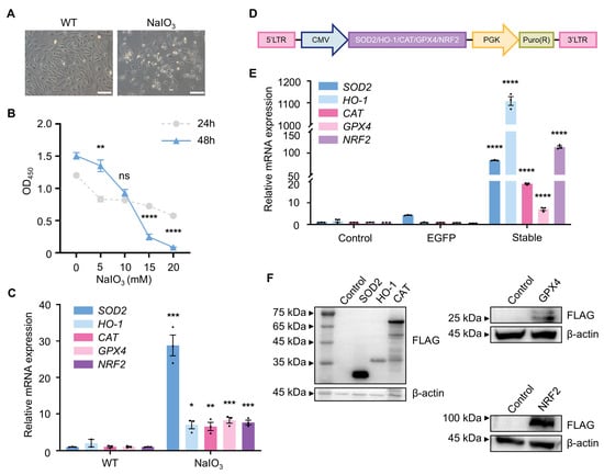
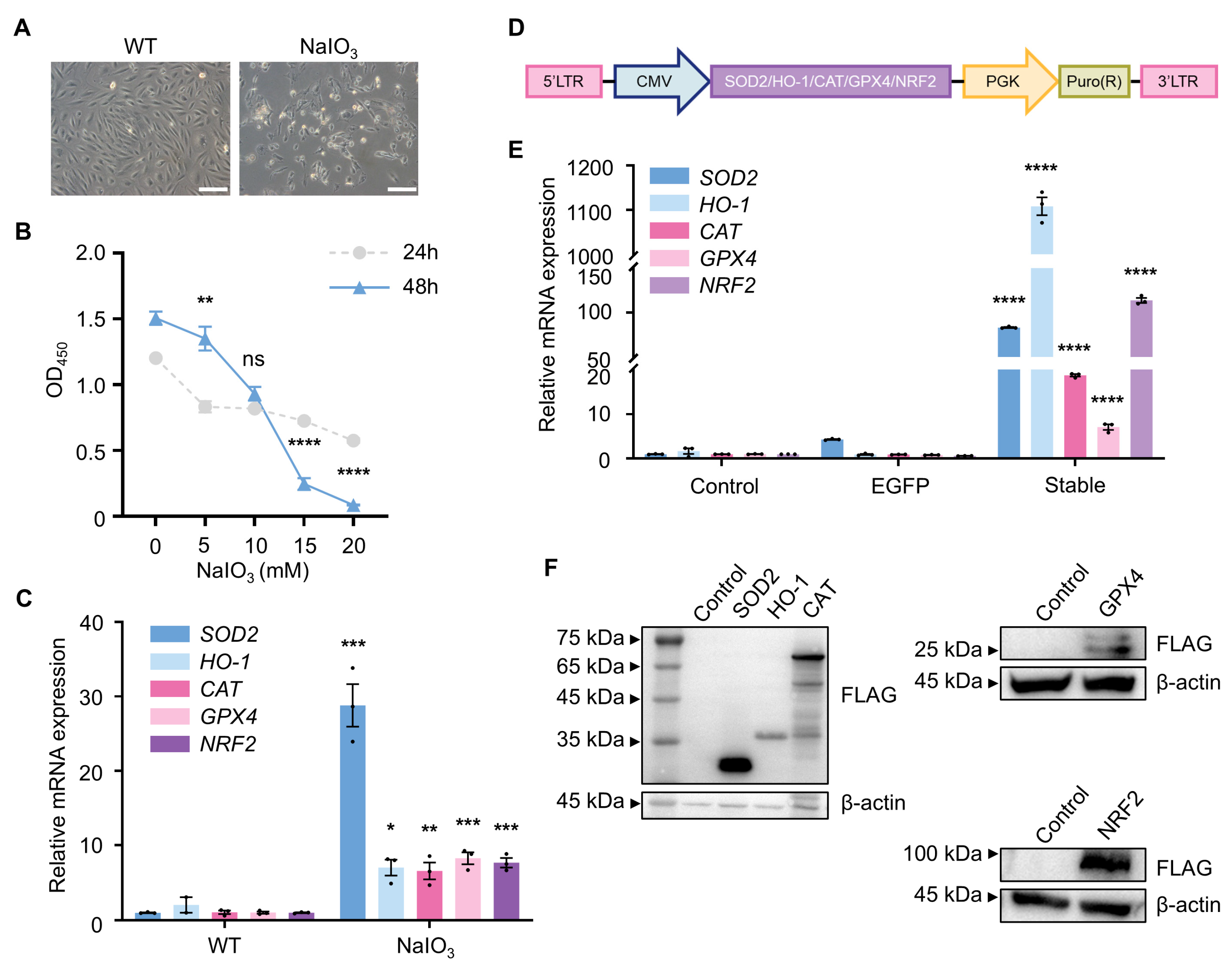
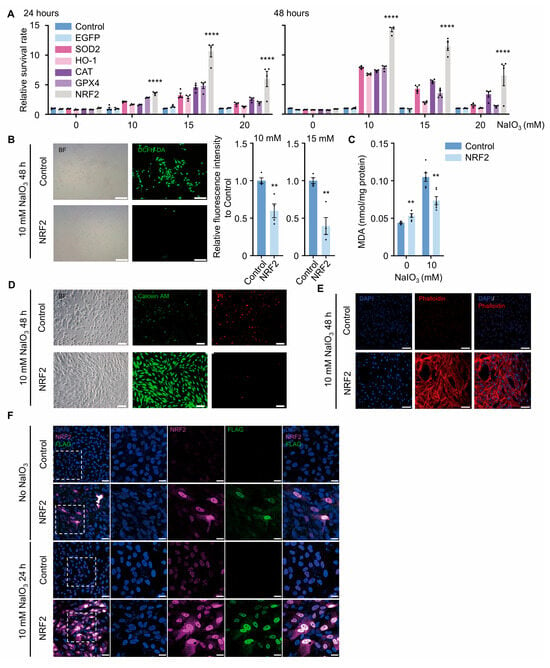
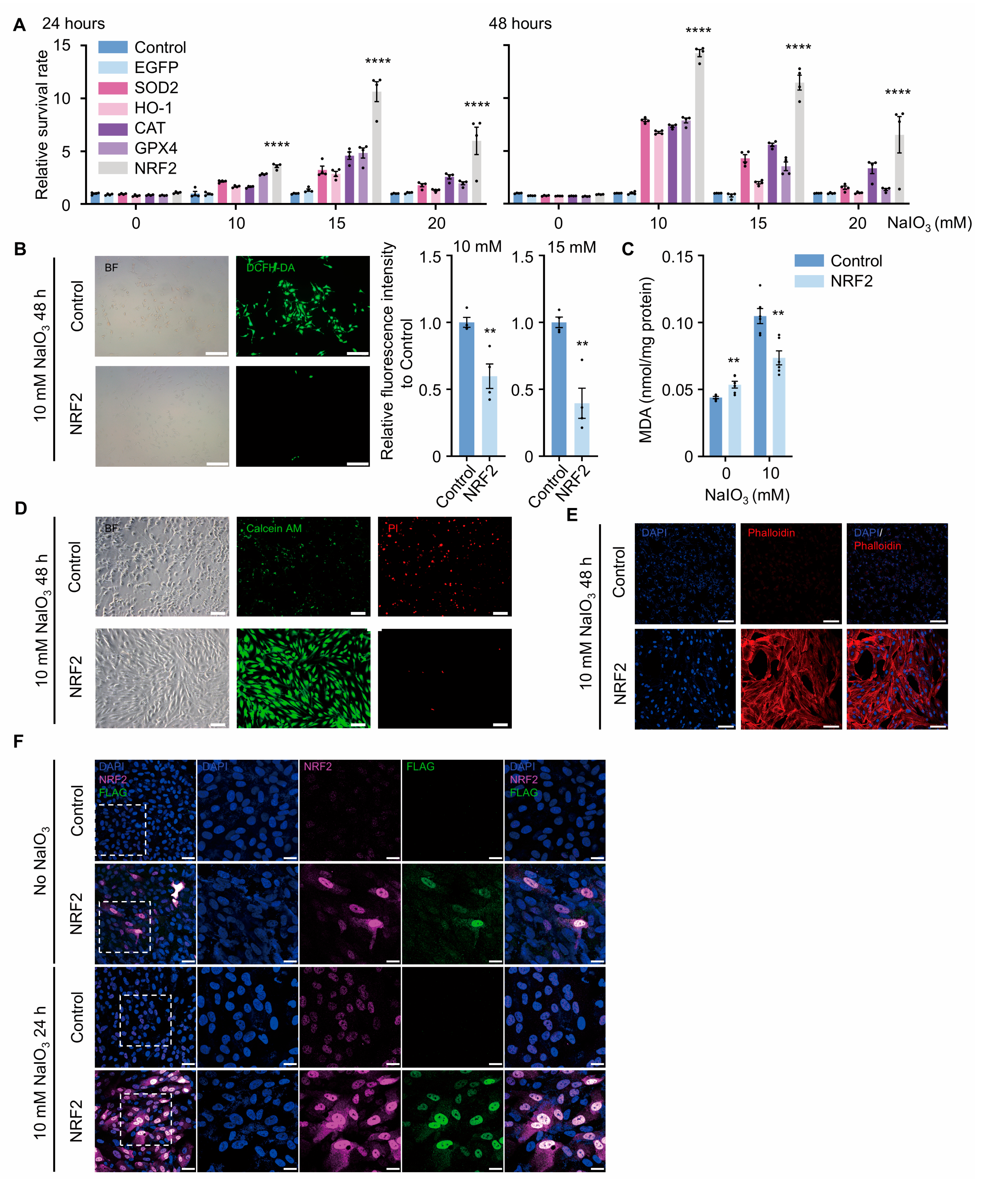
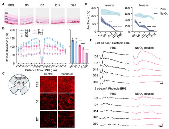
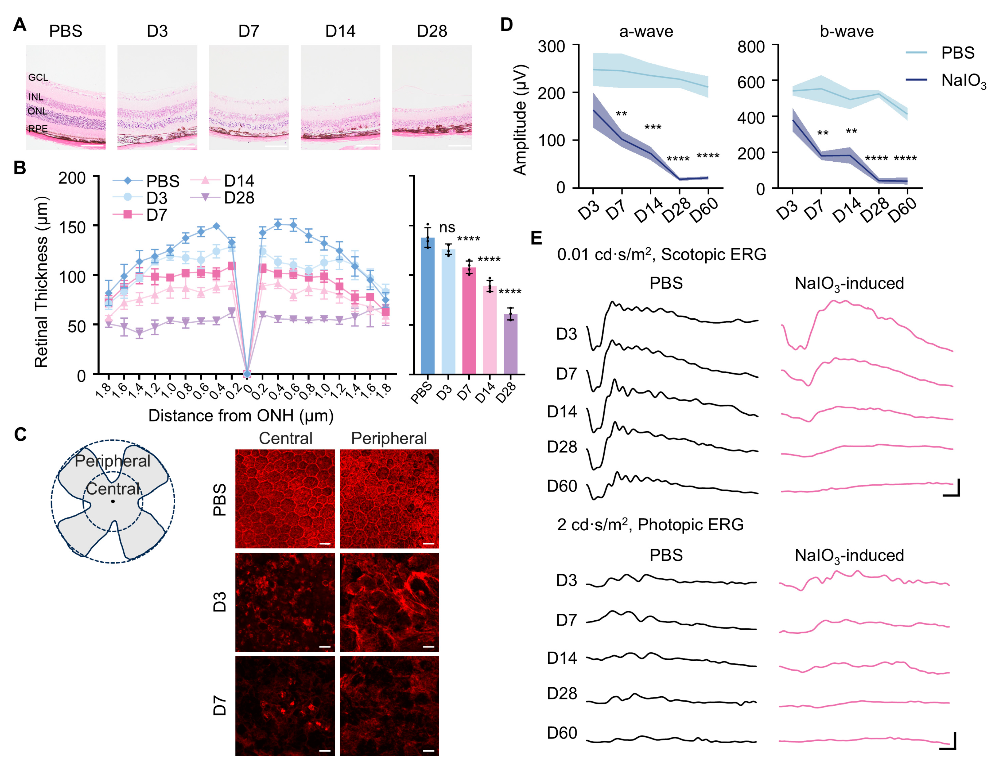
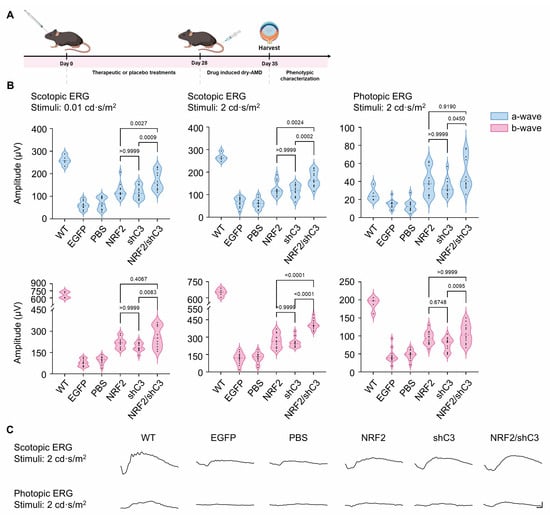
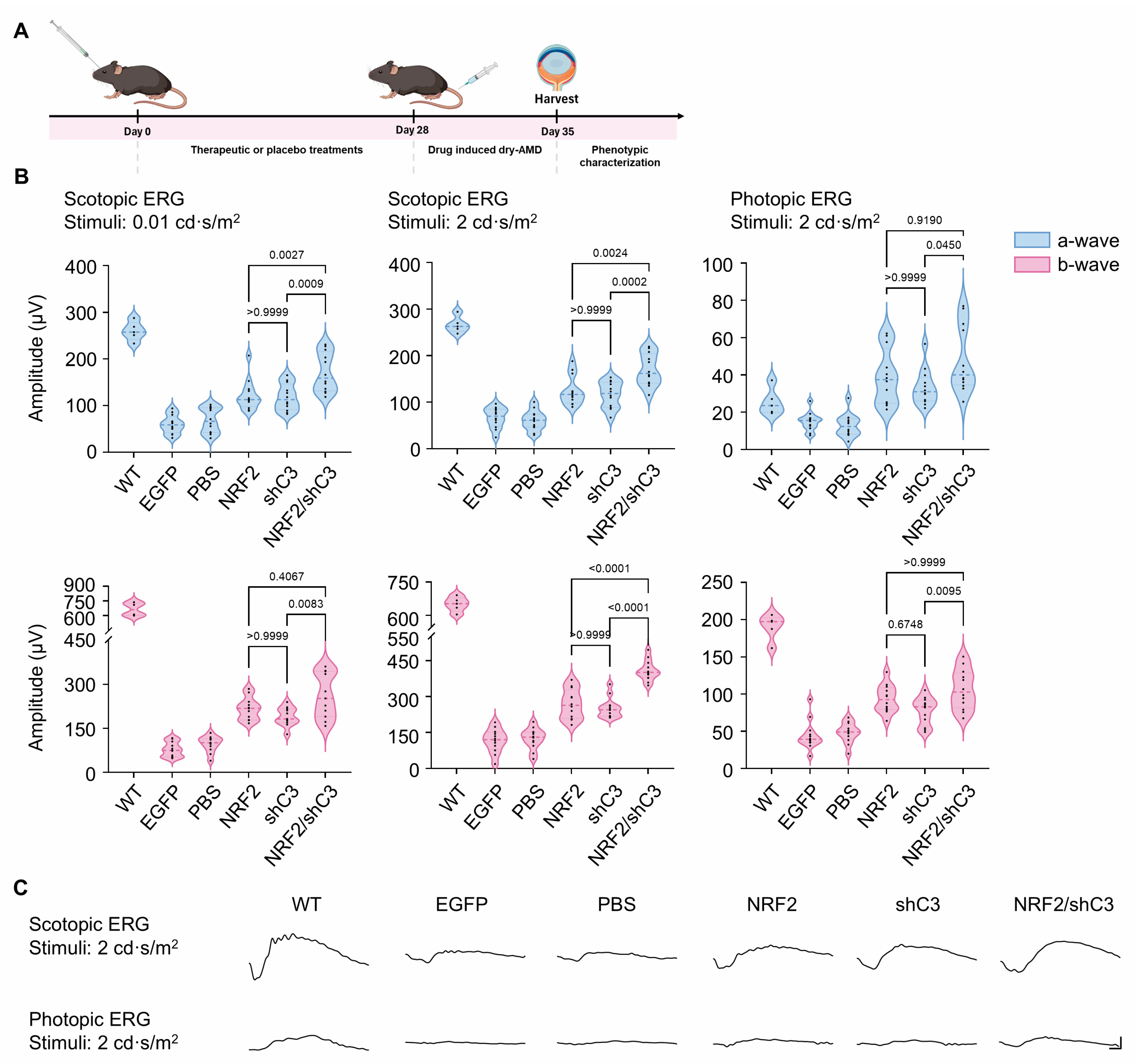
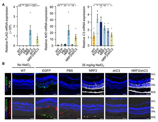
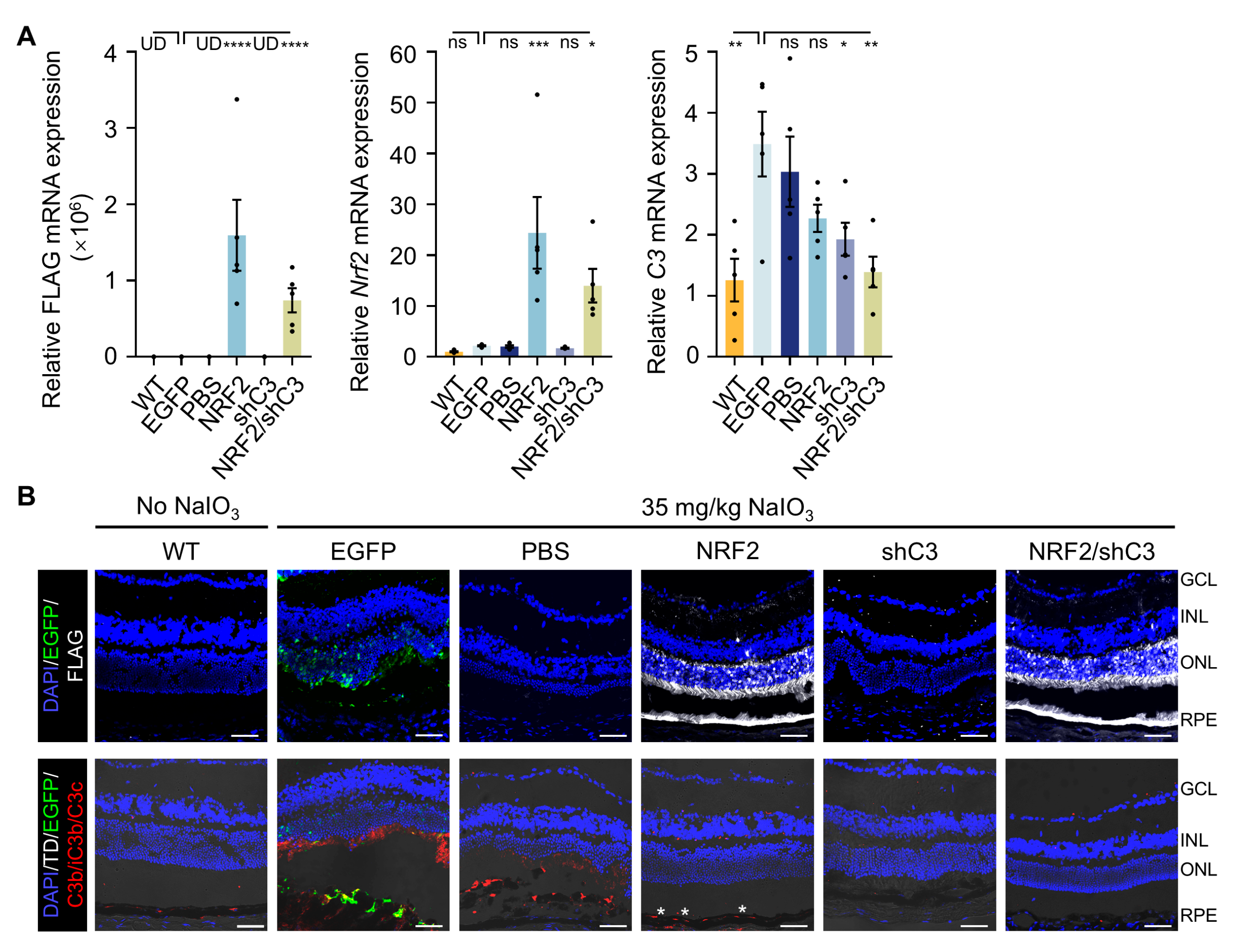
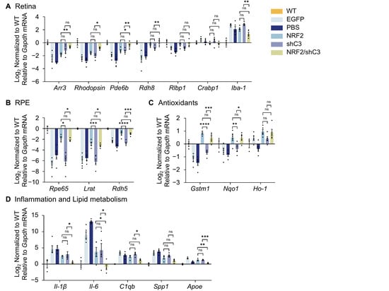
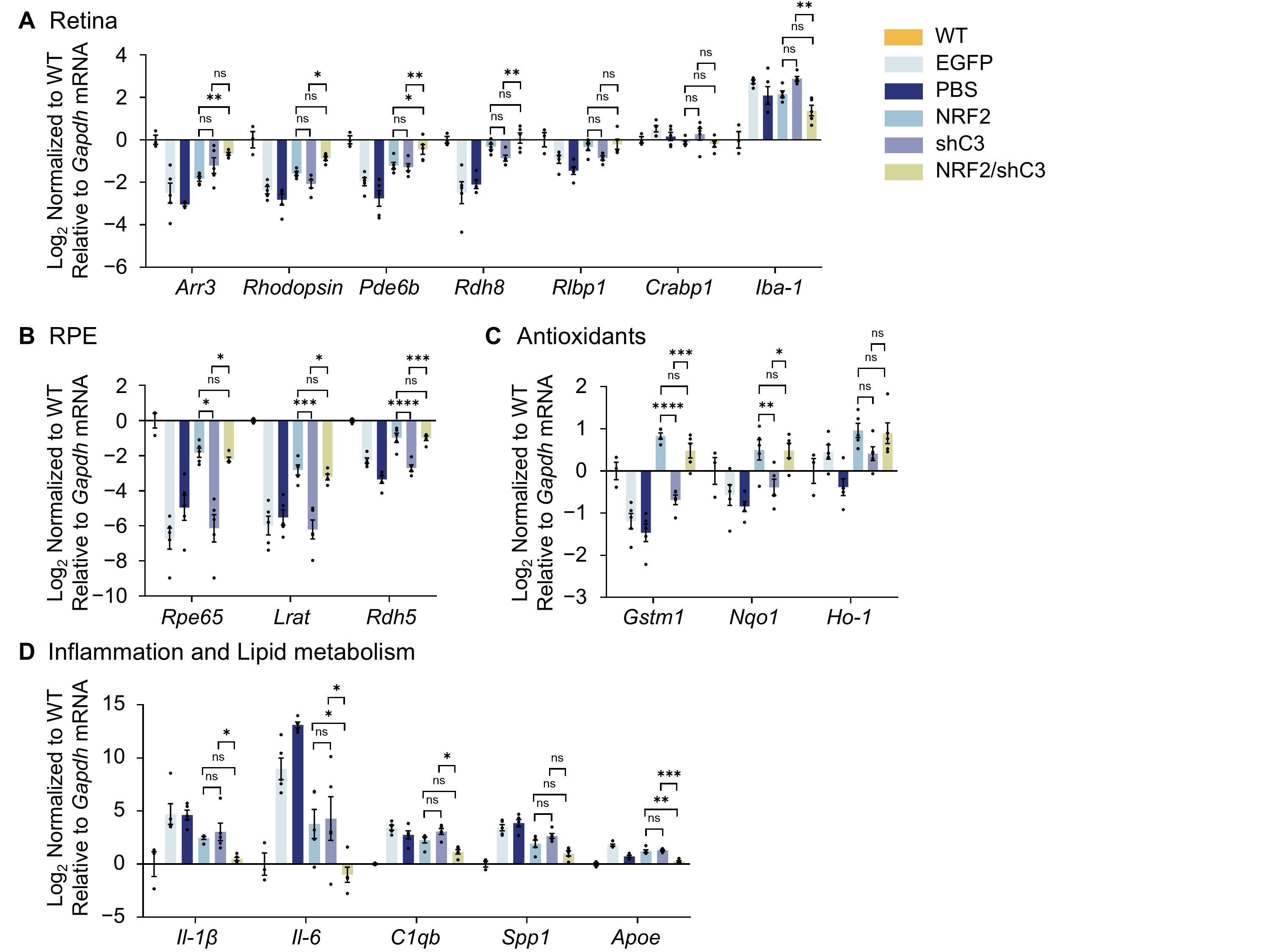
Disclaimer/Publisher’s Note: The statements, opinions and data contained in all publications are solely those of the individual author(s) and contributor(s) and not of MDPI and/or the editor(s). MDPI and/or the editor(s) disclaim responsibility for any injury to people or property resulting from any ideas, methods, instructions or products referred to in the content. |
© 2026 by the authors. Licensee MDPI, Basel, Switzerland. This article is an open access article distributed under the terms and conditions of the Creative Commons Attribution (CC BY) license.