


Abstract
Oxidative stress (OS) is a major cause of defective sperm function. During laboratory handling, gametes are exposed to OS, potentially mitigated by in vitro antioxidant supplementation. This study evaluates the protective role of caffeic acid (CAF) on basal human semen and under induced OS. First, six semen samples from normozoospermic donors were incubated with CAF concentrations ranging from 50 to 500 µM at 37 °C for 2 h. Sperm motility and DNA integrity (acridine orange) were evaluated. Then, ten semen samples were divided into four aliquots and incubated, respectively, with CAF at 100 µM, H2O2 at 2 mM, or H2O2 at 2 mM + CAF at 100 µM, or untreated. Motility, DNA integrity, acrosome status (Pisum sativum agglutinin), OS quantified by F2-isoprostanes (ELISA), and expression of Nrf2, Keap1, and HO-1 (qRT-PCR) were assessed. CAF at 100 µM improved progressive motility without damaging DNA and was selected for subsequent experiments. CAF showed protective effects on sperm damage induced by H2O2 treatment, restoring motility, DNA integrity, and acrosome status and reducing F2-isoprostane levels. Nrf2 and HO-1 expression were upregulated by CAF, downregulated by H2O2, and restored by the co-treatment. CAF supplementation may protect human spermatozoa during in vitro handling by reducing OS, improving several sperm parameters, with a possible mechanism of action involving the Nrf2 pathway.
1. Introduction
In recent years, increasing attention has been devoted to the evidence that oxidative stress (OS) plays a pivotal role in the etiology of male infertility [1]. OS refers to a condition arising from an imbalance between the levels of reactive oxygen species and the antioxidant defenses present in the organism [2,3]. As a result, substantial research has focused on identifying natural compounds with antioxidant properties that could be used to treat diseases primarily driven by OS [3,4].
The considered compound of this study is caffeic acid (CAF), a naturally occurring polyphenol found in various plant-based sources [5].
The chemical structure of CAF is thought to play a crucial role in its ability to scavenge free radicals, as it allows for the delocalization of unpaired electrons. The antioxidant activity, however, represents only one of its many beneficial biological effects. Indeed, CAF has shown antibacterial activity [6,7], antiproliferative effects against several cancer cells by acting on different receptors and signaling pathways [8,9,10,11,12], and anti-diabetic and anti-neuroinflammatory properties [13,14].
With regard to male reproductive health, most available studies on CAF have been conducted in animal models and demonstrated that CAF treatment was beneficial for sperm quality both in vivo [15] and in vitro during freezing–thawing procedures [16,17].
This study focused on investigating the in vitro effects of CAF on human semen, addressing a notable gap in the current scientific literature. Human spermatozoa represent an excellent in vitro model because they are easy to collect and allow straightforward assessment of parameters such as motility [18]. Spermatozoa possess minimal intracellular antioxidant defenses and exhibit limited transcriptional activity. Most antioxidant capacity resides in the seminal plasma, which modulates and mitigates the effects of in vitro-induced OS. This feature enables the development of controlled OS models using defined concentrations of oxidant stressors, thereby facilitating a more effective evaluation of antioxidant compounds and their biological effects. These characteristics make spermatozoa a particularly sensitive and suitable model for testing the effects of bioactive compounds such as CAF. Many routine procedures performed in reproductive laboratories cause OS in sperm cells. These include exposure to visible light, fluctuations in oxygen tension and temperature, pH variations, and cryopreservation [19,20]. The cell damage due to OS can be assessed by the quantification of biomarkers such as F2-isoprostanes (F2-IsoPs), identified as reliable and specific indicators of lipid peroxidation in biological tissues [21,22].
CAF has been demonstrated to influence the expression of various genes in different cell models [5,12], including nuclear factor-E2-related factor 2 [23] (NFE2L2, hereafter referred to by its transcription factor name, Nrf2), widely recognized as a key regulator of endogenous antioxidant responses [24]. Under physiological conditions, Keap1 directs Nrf2 for ubiquitin-dependent degradation, thereby repressing Nrf2-dependent gene expression [25,26]. In response to OS, the newly synthesized Nrf2 enters the nucleus, where it forms a heterodimer with small Maf proteins (musculoaponeurotic fibrosarcoma oncogene homologs). The resulting Nrf2-Maf complex selectively binds to antioxidant response elements (AREs) in the promoter regions of target genes such as heme oxygenase-1 (HO-1)—hereafter referred to by its protein name, HO-1—thereby activating Nrf2-dependent transcription [26]. Recently, Signorini and colleagues [27] reported an upregulation of Nrf2 expression following in vitro treatment of human spermatozoa with chlorogenic acid, a compound derived from the esterification of CAF and quinic acid, further supporting the involvement of the Nrf2 pathway in mediating the antioxidant effects of phenolic compounds.
This study aimed to evaluate the effects of CAF on human semen and to assess its potential biological activity. First, different concentrations of CAF were tested on semen samples from normozoospermic donors, evaluating sperm motility and DNA integrity, in order to identify the optimal concentration for subsequent experiments. Then, the effects of CAF were assessed in human semen samples exposed in vitro to OS induced by H2O2. The endpoints assessed included sperm motility, DNA integrity, acrosome status, levels of F2-IsoPs, and the gene expression of the Keap1-Nrf2 pathway.
2. Materials and Methods
2.1. Study Design
The study was conducted in two distinct steps:
- -
- Step 1: evaluation of the dose-dependent effects of CAF on human normal semen samples;
- -
- Step 2: investigation of the capacity of CAF to counteract the H2O2-induced OS in vitro in human normal semen samples.
2.2. Donors and Semen Sample Analysis
Semen samples were obtained from 16 donors aged 19–35 years who attended the Department of Molecular and Developmental Medicine at the University of Siena (Italy). The study protocol was approved by the Ethics Committee of the Siena University Hospital (ID CEAVSE 25612). All participants provided written informed consent prior to enrolment in this study. Specimens were collected following a period of 3 to 5 days of ejaculatory abstinence. In accordance with the World Health Organization (WHO) guidelines [28], semen parameters including sperm concentration, motility, vitality, pH, and morphology were evaluated.
2.3. Step 1: Evaluation of the Dose-Dependent Effects of CAF on Human Normal Semen Samples
Different concentrations of CAF were tested in 6 semen samples. Semen samples were divided into aliquots supplemented with 50 μM, 100 μM, 150 μM, 200 μM, and 500 μM CAF (Sigma-Aldrich, St. Louis, MO, USA) diluted in distilled water and incubated for two hours at 37 °C. An aliquot without compound supplementation served as an internal control. Following the incubation period, sperm motility was quantified in strict adherence to the WHO laboratory manual criteria [28]. DNA integrity was evaluated with the acridine orange (AO) assay.
AO Assay to Analyze the DNA Integrity
The AO test evaluates the susceptibility of sperm nuclear DNA to acid-induced denaturation in situ by quantifying the metachromatic shift of AO fluorescence from green, indicative of double-stranded (ds) DNA, to red, indicative of denatured DNA. Smeared sperm slides were processed according to Tejada and colleagues [29] and immediately examined with a Leitz Aristoplan fluorescence Microscope (Leica, Wetzlar, Germany) equipped with a 490 nm excitation source and 530 nm barrier filter, at 1000× magnification. A minimum of 300 sperm were evaluated, and the results are reported as the percentage of sperm with dsDNA.
2.4. Step 2: Investigation of the Capacity of CAF to Counteract the H2O2-Induced OS In Vitro in Normal Human Semen Samples
Based on the results of the dose–response analysis (step 1), semen samples from 10 donors were divided into four aliquots and treated as follows:
- -
- CTR, control aliquot with no supplementation;
- -
- CAF, aliquot supplemented with CAF at 100 μM;
- -
- H2O2, aliquot supplemented with H2O2 at 2 mM to induce OS;
- -
- H2O2 + CAF, aliquot supplemented with both CAF at 100 μM and H2O2 at 2 mM.
Aliquots were incubated at 37 °C for 2 h. At the end of the incubation, motility was evaluated according to the WHO guidelines [28]. A portion of each aliquot was mixed with an equal volume of phosphate-buffered saline (PBS) and centrifuged at 400× g for 10 min. The resulting pellets were resuspended in PBS, smeared onto glass slides, and fixed using two different protocols: in the first one, Carnoy’s solution was used for the evaluation of DNA integrity by the AO test, and in the second one, cold methanol (20 min at −20 °C) followed by cold acetone (5 min at −20 °C) was used for acrosome assessment. The remaining fraction of each aliquot was centrifuged at 400× g for 10 min to separate seminal plasma from the spermatozoa; then, both fractions were stored at −80 °C for further analyses. Seminal plasma was used for the quantification of F2-IsoPs using an ELISA kit, while the cell pellets were processed for the gene expression analysis of Nrf2, Keap1, and HO-1.
2.4.1. Evaluation of Acrosome with Pisum sativum Agglutinin
TRITC-conjugated Pisum sativum agglutinin (PSA, Vector Laboratories Inc., Burlingame, CA, USA) is a lectin that recognizes the carbohydrates of glycoproteins and is used to differentiate acrosome-intact from acrosome-reacted (or acrosome-damaged) human spermatozoa. The smeared slides fixed in methanol and acetone, as previously described, were rinsed in PBS for 10 min, incubated for 30 min (in the dark at room temperature) with TRITC-PSA solution diluted 1:1000 in PBS, and rinsed again in PBS for 15 min. The nuclei of the spermatozoa were stained with 4,6-diamidino-2-phenylindole (DAPI, 1:20,000 in methanol; Sigma-Aldrich, St. Louis, MO, USA), for 2 min, in the dark at room temperature. The slides were mounted with DABCO and observed with a Leica DMI 6000 fluorescence microscope (Leica Microsystems, Wetzlar, Germany). A Leica AF6500 Integrated System for Imaging and Analysis (Leica Microsystem, Wetzlar, Germany) enables image acquisition. Spermatozoa displaying a uniformly stained red acrosomal cap were classified as acrosome-intact. Those exhibiting a shrunken, shortened, or misshapen acrosome were categorized as acrosome-altered spermatozoa, while spermatozoa lacking any visible acrosomal staining were classified as acrosome-absent. A minimum of 300 spermatozoa were evaluated per sample, and the results are reported as percentages.
2.4.2. F2-IsoP Quantification
F2-isoprostanes were quantified by measuring 8-iso prostaglandin F2α (8-iso PGF2α; hereafter referred to as 8-isoprostane) in seminal plasma using a commercially available ELISA kit (8-Isoprostane ELISA Kit, Cayman Chemical, Ann Arbor, MI, USA). The assay is based on a competitive enzyme immunoassay employing a specific anti-8-isoprostane antibody, an 8-isoprostane–AChE tracer, and Ellman’s reagent. Ninety-six-well plates were pre-coated with the capture antibody (mouse anti-rabbit IgG). Absorbance was measured spectrophotometrically at 405 nm, and sample concentrations were calculated from a standard curve generated using 8-isoprostane standards ranging from 0.8 to 500 pg/mL. Each sample was analyzed in triplicate, and results are expressed as pg/mL.
2.4.3. Gene Expression Analysis
Total RNA was isolated from ejaculated spermatozoa using the PureLink® RNA Mini Kit (Thermo Fisher Scientific, Waltham, MA, USA) according to the manufacturer’s protocol. RNA purity and concentration were assessed by measuring A260/A280 absorbance ratios with a Thermo Scientific™ NanoDrop™ One/OneC Microvolume UV–Vis spectrophotometer (Thermo Fisher Scientific, Waltham, MA, USA). Complementary DNA (cDNA) was synthesized from 500 ng of total RNA per sample using the High-Capacity cDNA Reverse Transcription Kit (Applied Biosystems, Waltham, MA, USA).
Quantitative reverse transcription polymerase chain reaction (qRT-PCR) was then performed to evaluate the mRNA expression levels of Nrf2, Keap1, and HO-1 in sperm cells. Gene expression analysis was carried out using specific PrimePCR™ SYBR™ Green assays (Keap1 qHsaCID0017511; NFE2L2 qHsaCED0038543; HO-1 qHsaCID0022141; Bio-Rad Laboratories, Inc., Hercules, CA, USA) on a QuantStudio™ 5 Real-Time PCR System (Thermo Fisher Scientific, Waltham, MA, USA). To normalize target gene expression, mRNA levels of the housekeeping gene glyceraldehyde-3-phosphate dehydrogenase (GAPDH; PrimePCR™ SYBR™ Green Assay GAPDH qHsaCED0038674, Bio-Rad Laboratories, Inc., Hercules, CA, USA) were simultaneously measured in all samples.
Non-template controls and reverse-transcription negative samples were included and run in triplicate as negative controls to ensure the absence of contamination and to verify the specificity of the amplification reaction. The thermal cycling protocol was performed as follows: initial activation of uracil-DNA glycosylase (UDG) at 50 °C for 2 min, followed by enzyme activation at 95 °C for 2 min and by 40 amplification cycles, each consisting of denaturation at 95 °C for 15 s and annealing at 60 °C for 1 min. Fluorescent quantitative analysis was performed by calculating the difference in cycle threshold (∆Ct) between the target genes and the reference gene GAPDH. Relative changes in Keap1, Nrf2, and HO-1 mRNA expression were determined using the 2−∆∆Ct method. All qRT-PCRs were conducted in triplicate.
2.4.4. Statistical Analysis
Distribution normality was examined using the Kolmogorov–Smirnov test. Comparisons between two groups (step 1) were performed with the Mann–Whitney U test. For analyses involving more than two groups (step 2), differences among groups were initially evaluated using the Kruskal–Wallis test, followed by Dunnett’s post hoc test for multiple pairwise comparisons. Results are expressed as median (25th–75th percentile, interquartile range: IQR). A p value below 0.05 was considered statistically significant. All statistical analyses were conducted using GraphPad Prism version 8.4.2.
3. Results
3.1. Step 1: Evaluation of the Dose-Dependent Effects of CAF on Human Normal Semen Samples
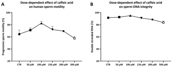
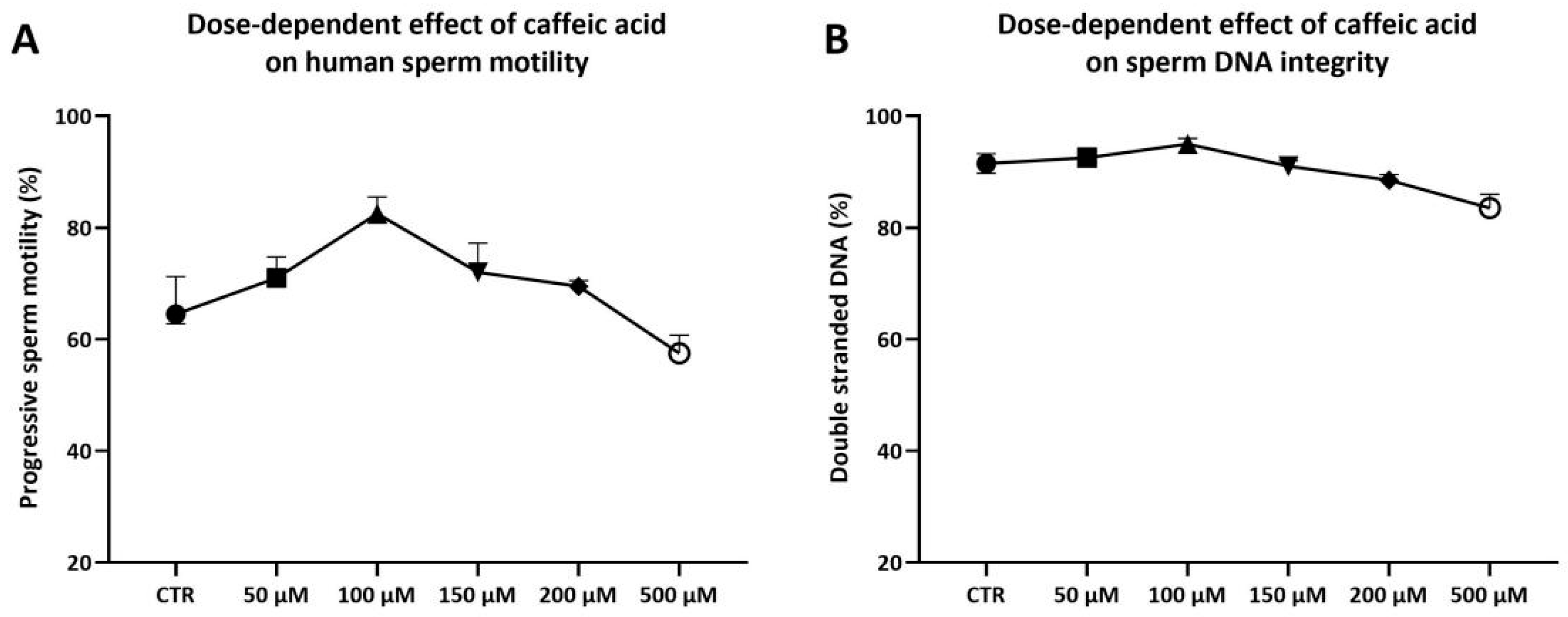
The six semen samples used to study the CAF dose-dependent effects showed sperm parameters above the 25th centile [28]. Each sample was divided into six aliquots: five aliquots were treated with increasing concentrations of CAF (50 μM, 100 μM, 150 μM, 200 μM, and 500 μM), while the sixth aliquot was incubated under the same conditions without treatment and served as a control (CTR). Following incubation, progressive sperm motility and DNA integrity were assessed. Treatment with CAF at 100 μM resulted in an increase in progressive motility (82.50% [80.75–85.50%], Figure 1A), calculated as the sum of the percentages of rapid and slow progressive sperm motility. In contrast, sperm treated with CAF at 50 μM (71.00% [67.25–74.75%]), 150 μM (72.00% [67.25–77.25%]), or 200 μM (69.50% [67.25–70.50%], Figure 1A) exhibited motility values similar to CTR (64.50% [62.75–71.25%], Figure 1A); a reduction in motility was observed in spermatozoa exposed to CAF at 500 μM (57.50% [54.75–60.75%], Figure 1A).
Figure 1.
Medians (IQRs) of percentages of progressive sperm motility (A) and DNA integrity (B) of human sperm after treatments for two hours at 37 °C with increasing concentrations of caffeic acid (range 0–500 μM). CTR indicates untreated aliquot.
None of the tested CAF concentrations exhibited any detrimental effects on DNA integrity (CTR: 91.50% [89.75–93.25%]; 50 μM: 92.50% [89.50–94.25%]; 100 μM: 95.00% [93.25–96.00%]; 150 μM: 91.00% [89.50–92.00%]; 200 μM: 88.50% [87.75–89.50%]; 500 μM: 83.50% [82.75–86.00%], Figure 1B).
3.2. Step 2: Investigation of the Capacity of CAF to Counteract the H2O2-Induced OS In Vitro in Normal Human Semen Samples
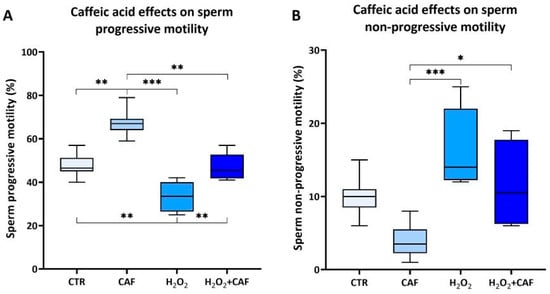
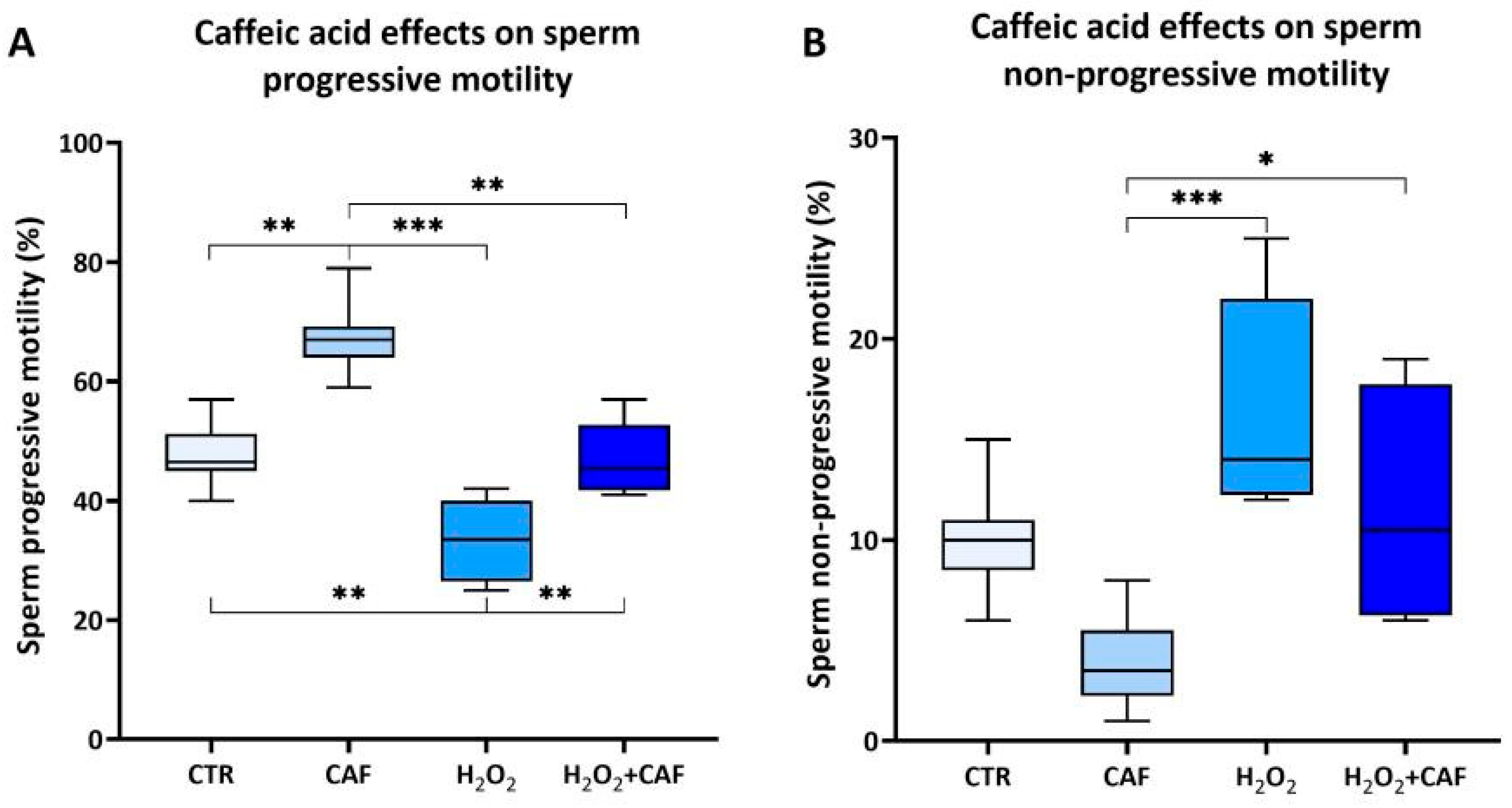
In the second step of the study, ten semen samples with sperm parameters above the 25th percentile [28] were divided into four aliquots, as detailed in the Section 2: CTR, CAF, H2O2, and H2O2 + CAF. Sperm motility was assessed [28] as the primary functional parameter. Exposure to CAF resulted in a statistically significant increase in the proportion of progressively motile spermatozoa (67.00% [64.00–69.25%]) compared to the CTR (46.50% [45.00–51.25%], p < 0.01), H2O2 (33.50% [26.50–40.00%], p < 0.001), and H2O2 + CAF (45.50% [41.75–52.75%], p < 0.01) aliquots (Figure 2A). A significant reduction in non-progressive sperm motility was observed in the CAF aliquot (3.50% [2.25–5.50%]) with respect to that of both the H2O2 (14.00% [12.25–22.00%], p < 0.001) and H2O2 + CAF (10.50% [6.25–17.75%], p < 0.05) aliquots (Figure 2B). No statistically significant differences in non-progressive sperm motility were detected between the CAF and CTR (10.00% [8.5–11.00%]) groups (Figure 2B).
Figure 2.
Medians (IQRs) of percentages of progressive sperm motility (A) and non-progressive sperm motility (B) after treatment for two hours at 37 °C with 100 μM caffeic acid (CAF), 2 mM H2O2, and combined treatment (H2O2 + CAF). CTR: untreated aliquot. *: p < 0.05; **: p < 0.01; ***: p < 0.001.
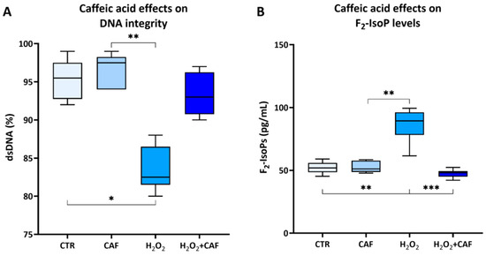
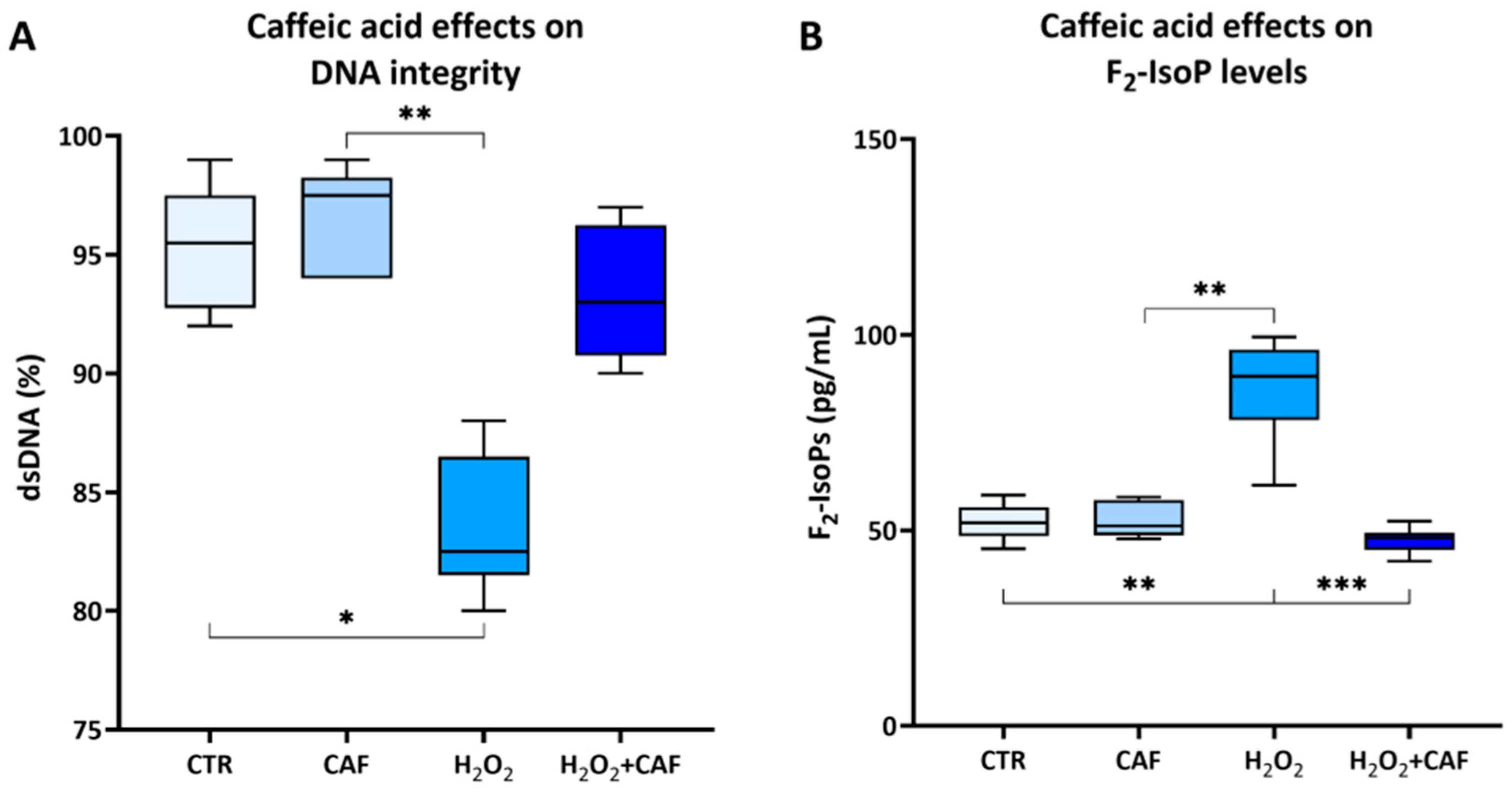
The in vitro exposure to H2O2 to induce OS led to a significant decrease in the percentage of spermatozoa with dsDNA (82.50% [81.50–86.50%]) compared to both the untreated spermatozoa (95.50% [92.75–97.50%], p < 0.05), and the spermatozoa treated with CAF at 100 µM (97.50% [94.00–98.25%), p < 0.01, Figure 3A). Concurrently, H2O2-treated samples exhibited a significant increase in F2-IsoP levels (89.38% [78.22–96.15%]), a well-established biomarker of lipid peroxidation (free radical-induced oxidative degradation of lipids), compared to all the other samples (CTR: 51.92% [48.46–55.94%], p < 0.01; CAF: (51.14% [48.75–57.78%], p < 0.01; H2O2 + CAF: 48.07% [45.03–49.39%], p < 0.001), which were similar to each other (Figure 3B).
Figure 3.
Medians (IQRs) of the percentages of double-stranded (ds) DNA (A) and seminal F2-isoprostane (F2-IsoP, pg/mL) levels (B) after treatment for two hours at 37 °C with 100 μM caffeic acid (CAF), 2 mM H2O2, and combined treatment (H2O2 + CAF). CTR indicates untreated aliquot. DNA integrity was evaluated using an acridine orange test, scoring a minimum of 300 spermatozoa; the F2-IsoP levels were assessed by ELISA. *: p < 0.05; **: p < 0.01; ***: p < 0.001.
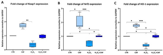
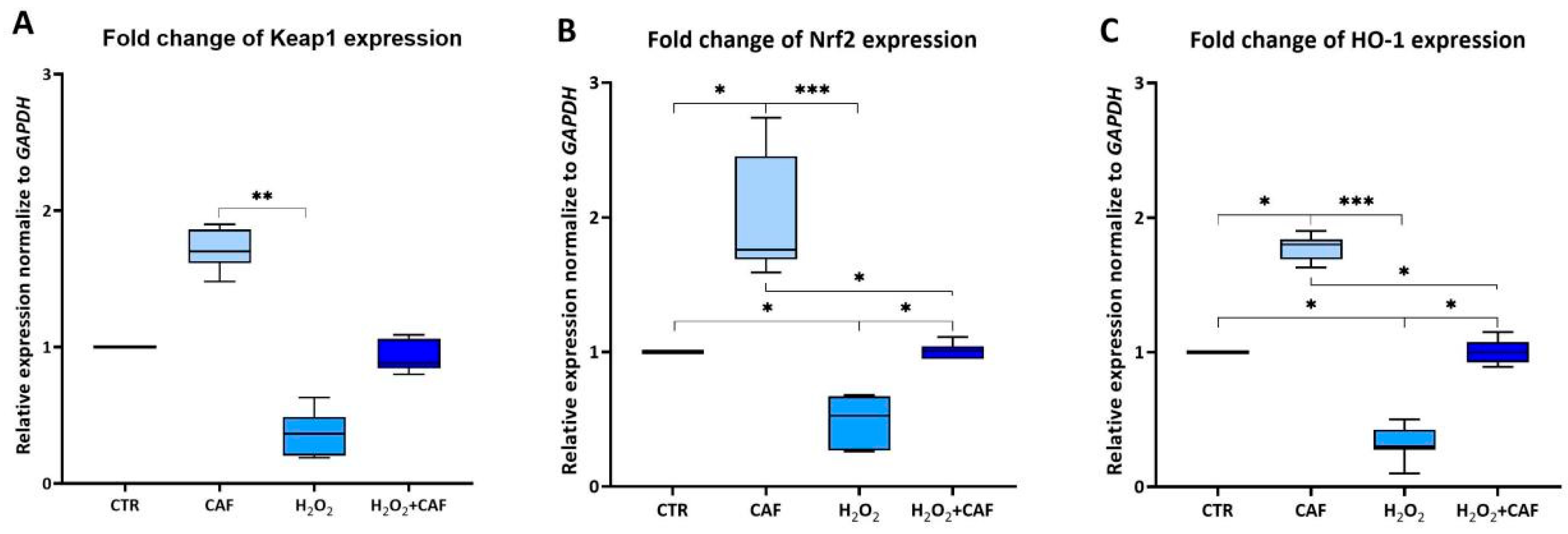
The expression levels of Keap1, Nrf2, and HO-1 were quantified by qRT-PCR. The expression patterns of the analyzed genes showed a similar trend across all the considered samples (Figure 4A–C). The treatment with CAF led to a significant increase in Nrf2 and HO-1 expression in spermatozoa compared to the CTR (p < 0.05; Figure 4B,C); keap-1 expression also increased, although nonsignificantly. These observations suggest a potential role of CAF in the activation of the Nrf2 pathway. The treatment of spermatozoa with H2O2 caused in a downregulation in the expression of all three analyzed genes (Figure 4A–C). While the H2O2 + CAF combination resulted in a significant recovery of sperm Nrf2 and HO-1 expression with respect to H2O2-treated samples (p < 0.05), Keap1 expression exhibited a similar trend, although the change did not reach statistical significance. In any case, the expression of all three considered genes in the samples treated with H2O2 + CAF reached similar values to CTR.
Figure 4.
Medians (IQRs) of relative Keap1 (A), Nrf2 (B), and HO-1 (C) expression normalized to GAPDH after treatment for two hours at 37 °C with 100 μM caffeic acid (CAF), 2 mM H2O2, and combined treatment (H2O2 + CAF). CTR indicates untreated aliquot. *: p < 0.05; **: p < 0.01; ***: p < 0.001.
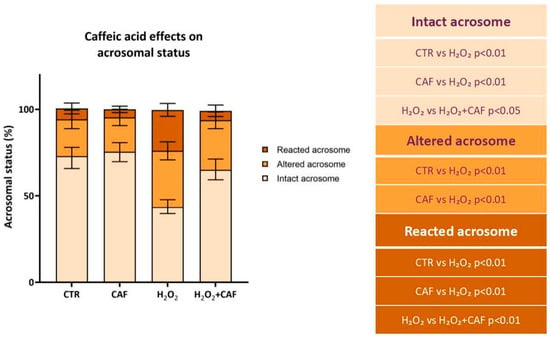
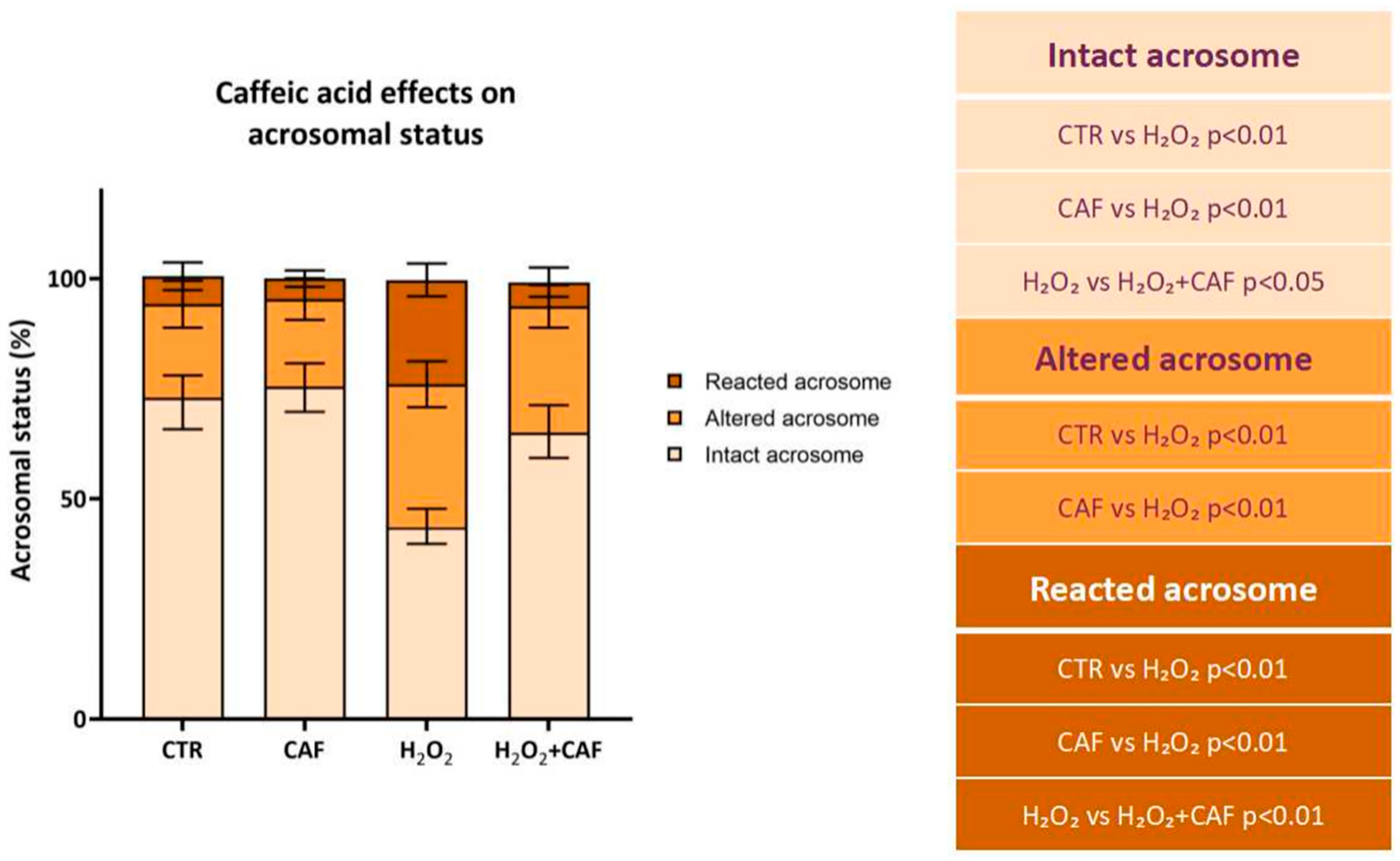
Finally, the acrosome was assessed through PSA staining. Exposure to H2O2 caused a significant decrease in the percentage of intact acrosomes (43.50% [40.50–46.5%]) compared to the CTR (73.00% [67.25–76.5%], p < 0.01), CAF (75.50% [71.00–79.25%], p < 0.01), and H2O2 + CAF (65.00% [61.00–68.25%], p < 0.05) (Figure 5). At the same time, H2O2 treatment led to a significant increase in the percentage of altered acrosomes (32.50% [28.50–37.25%]) compared to that of CTR (20.50% [17.75–23.25%], p < 0.01) and CAF (18.50% [17.25–23.50%], p < 0.01) (Figure 5). The percentage of reacted acrosomes was significantly increased in the H2O2-treated spermatozoa (23.50% [20.75–24.75%]) compared to all other samples (CTR: 6.50% [4.25–8.75%]; CAF: 5.00% [3.50–5.75%]; H2O2 + CAF: 5.50% [2.50–7.75%], p < 0.01), while in the H2O2 + CAF treatment, the values were comparable to those observed in the CTR and CAF groups (Figure 5).
Figure 5.
Median (IQR) of percentages of intact, altered, and reacted acrosomes after treatment for two hours at 37 °C with 100 μM caffeic acid (CAF), 2 mM H2O2, and combined treatment (H2O2 + CAF). CTR indicates untreated aliquot. The acrosomes were evaluated with Pisum sativum agglutinin conjugated with TRITC, scoring a minimum of 300 spermatozoa per sample. The table reports the significant comparisons between pairs.
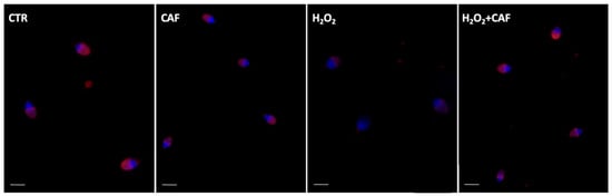
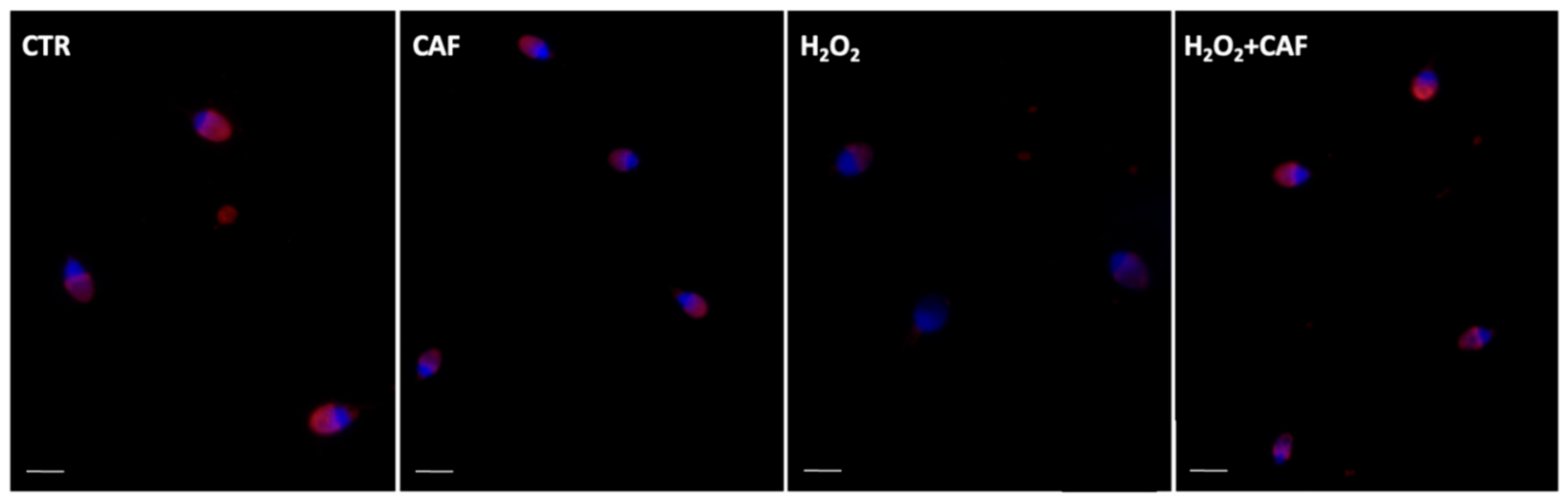
In Figure 6, the PSA staining of the spermatozoa from the four treatments is represented.
Figure 6.
UV micrographs of human spermatozoa treated with Pisum sativum agglutinin (PSA) conjugated with TRITC after incubation for two hours at 37 °C with 100 μM caffeic acid (CAF), 2 mM H2O2, and combined treatment (H2O2 + CAF). CTR indicates untreated aliquot. A minimum of 300 spermatozoa were scored per sample. The acrosomes were mostly intact in the CTR and CAF aliquots; they were altered and absent in spermatozoa treated with H2O2. In the sample treated with H2O2 + CAF, the acrosomes were present, sometimes altered. Bar (CTR, H2O2): 4 µm. Bar (CAF, H2O2 + CAF): 6 µm.
4. Discussion
This study investigated the protective and antioxidant effects of CAF on human spermatozoa under both physiological conditions and OS induced in vitro by H2O2. Overall, the findings demonstrate that CAF exerts beneficial effects on key sperm quality parameters—namely progressive motility, and DNA and acrosomal integrity—and modulates the F2-IsoP concentration and the expression of genes involved in the antioxidant response. CAF, a natural occurring hydroxycinnamic acid, is contained in coffee (also decaffeinated varieties), red wine, and other plant products such as chokeberries, apples, and plums, as well as herbs like sage and oregano—members of the mint family [5]. CAF has been extensively used as a potent antioxidant in animal semen preservation [30], improving post-thaw motility and membrane integrity in boar semen [16] and enhancing semen quality in buffalo bull following freezing–thawing procedures [17]. In addition, in vivo administration of CAF in mice improved spermatogenesis by reducing OS and enhancing endogenous antioxidant defenses, thereby mitigating the adverse effects of aging—an issue of growing relevance given its documented association with declining sperm quality [15].
Despite the great number of studies for CAF’s effects on animal male fertility, only a few studies have reported its effects in human semen. Human spermatozoa represent an ideal in vitro model for evaluating antioxidant efficacy due to their availability; their limited transcriptional activity, which makes them highly sensitive to the external environment; and the ease of the assessment of functional parameters, such as motility, DNA integrity, and acrosomal status.
Various antioxidants, especially phenolic compounds found in fruits and vegetables, have been tested using this model [18,27,31]. Our research group demonstrated that 100 µM chlorogenic acid, an ester of CAF and quinic acid, exerted significant in vitro antioxidant and protective effects on swim-up selected human spermatozoa when administered either as a free molecule [22] or encapsulated in liposomes [32], as well as on whole semen samples [27].
The use of antioxidants during semen handling is particularly relevant given that procedures commonly employed during assisted reproductive technologies (ARTs) can promote reactive oxygen species overproduction and OS, if not adequately balanced by antioxidant systems [20,33]. To address this issue, several strategies have been explored. Despite considerable interest in oral antioxidant supplementation as a therapeutic approach, robust clinical evidence confirming its efficacy is still controversial [34,35]. In contrast, the direct supplementation of semen with antioxidants in vitro offers a more targeted approach to limiting reactive oxygen species-induced damage during laboratory handling.
To the best of our knowledge, the only paper on the in vitro effect of CAF (in the form of CAF phenethyl ester) on human spermatozoa reported a protective effect of this compound against DNA oxidative damage in human spermatozoa [36].
In this study, human semen samples with normal parameters were supplemented in vitro with CAF to evaluate its antioxidant and protective effects in a physiological environment—that is, the seminal plasma. Many previous studies have focused on spermatozoa isolated by swim-up or similar selection techniques that, while providing a homogeneous sperm population, simultaneously remove the seminal plasma, which contains most antioxidant defenses. By using whole semen under standardized conditions and selecting normozoospermic donors, our approach aimed to preserve an environment as close to the physiological one as possible.
A preliminary dose–response analysis using a CAF concentration range from 50 µM to 500 µM was performed based on previous literature [16,30,36]. The concentration of 100 µM was selected for other experiments, as it elicited a moderate enhancement in progressive motility without inducing detectable DNA damage. Progressive motility is essential for natural fertilization and effective sperm selection in ARTs [37]. Likewise, DNA integrity is critical for embryo development, and its impairment is associated with miscarriage and abnormal fertilization outcomes [38]. Other studies dealing with semen and spermatozoa have identified the 100 µM CAF concentration as optimal. For instance, Namula and colleagues [16] supplemented the extender used during boar semen freezing with CAF at different concentrations and found that CAF at 100 µM yielded the most pronounced improvements in post-thaw sperm quality in terms of motility, vitality, and membrane integrity. Similar results were obtained supplementing buffalo semen extenders with CAF at 100 µM [17].
The concentration of polyphenols to be used in this kind of experiment is a nuanced topic since many of these compounds can exert dose-dependent toxic effects, reducing motility and viability and increasing reactive oxygen species production at high concentrations [39]. It is not a coincidence that high concentrations of CAF (1 mM) have been used to induce apoptosis in tumor cells [40]. This suggests how moderate polyphenol levels can exert antioxidant and cytoprotective effects, whereas elevated doses may shift CAF’s action toward pro-oxidant or cytotoxic activity, underscoring the compound’s dual redox nature.
In the second step of this study, we examined the influence of CAF at 100 µM on spermatozoa exposed to in vitro-induced OS. Treatment with CAF increased the percentage of progressively motile sperm while reducing non-progressively motile sperm, although its effect on non-progressive motility remained limited when CAF was combined with H2O2. This may be due to the irreversible oxidation of thiol groups on flagellar proteins such as outer dense fiber protein 1 [41], which antioxidants cannot fully repair.
To investigate the antioxidant potential of CAF in human semen, lipid peroxidation was assessed by measuring seminal F2-IsoPs. These molecules, formed from arachidonic acid via reactive oxygen species-mediated peroxidation, are reliable markers of oxidative damage in spermatozoa and seminal plasma [42]. Elevated seminal F2-IsoP levels have been consistently associated with decreased sperm motility, vitality, and normal morphology, as well as with pathological conditions such as varicocele and urogenital infections [43,44,45]. In this study, treatment of human semen with H2O2 led an increase in F2-IsoP levels and co-treatment with CAF significantly decreased their concentration. This decline in seminal F2-IsoP level following antioxidant treatment can be considered an objective biochemical marker of reduced OS and improved sperm function. The results of this study confirm the observations obtained with a similar protocol and reported in a recent paper [27] in which chlorogenic acid, an antioxidant compound derived from CAF, restored the functional competence of spermatozoa by mitigating lipid-oxidative damage.
Regarding DNA integrity, CAF displayed protective effects by reducing H2O2-induced damage. Such oxidative damage in semen and sperm has been documented since early investigations in the field [46]. Recent evidence further confirmed that H2O2 plays a negative role in mitochondrial function and motility but also induces oxidative DNA fragmentations that compromise chromatin stability and can reduce the sperm’s ability to support normal embryo development [47]. In this context, the ability of antioxidant compounds to mitigate H2O2-induced DNA damage represents a crucial protective mechanism. By scavenging reactive oxygen species and stabilizing redox homeostasis, these agents can preserve both the structural integrity of sperm chromatin and the functional parameters essential for fertilization.
Similarly, CAF preserved acrosomal integrity. It is known that H2O2 exposure increases the proportion of spermatozoa with reacted or damaged acrosomes in both humans [46,47,48,49,50] and rats [51]. Co-treatment with CAF protected the acrosome, and this protection is particularly important, as the acrosome reaction must occur only near the oocyte, rather than prematurely within the semen. Similarly, the polyphenol quercetin has also been shown to exert protective effects on sperm structure, including the acrosome, under oxidative or inflammatory stress [52]. Together, these findings underscore that several polyphenols can contribute to defending the acrosome during sperm exposure to physiological or experimental stressors.
The mechanism of action of CAF includes the scavenging properties arising from its catechol structure, which acts as a potent antioxidant by donating hydrogen atoms to neutralize free radicals [53].
In addition, we explored the cell-signaling pathway involving Nrf2, because recently, some of our group demonstrated the effect of a similar antioxidant, chlorogenic acid, on Nrf2 gene expression [27]. Nrf2 levels positively correlate with key sperm quality parameters, including concentration, motility, and vitality. Moreover, Cheng et al. [54] reported that, in cases of asthenozoospermia, low Nrf2 mRNA expression was associated with reduced seminal SOD activity and lower mRNA levels of CAT and SOD2 (although these decreases were not statistically significant).
The critical role of Nrf2 in protecting spermatogenesis from oxidative damage was also confirmed in Nrf2-knockout mice, generated by Nakamura and colleagues in 2010 [55]. In these models, deletion of Nrf2 resulted in significantly reduced sperm concentrations in both the testis and epididymis, along with a decrease in epididymal sperm motility, compared to wild-type mice.
In this study, we observed that CAF modulated the Keap1–Nrf2–HO-1 signaling pathway in vitro. Although spermatozoa have minimal transcriptional activity due to their highly condensed chromatin, increasing evidence suggests that a residual gene expression can occur in response to environmental stimuli [56]. Several studies demonstrated a modulation of sperm transcription in vitro using antioxidants, such as melatonin [57], curcumin [58], and chlorogenic acid [27]. The modulation of the Keap1-Nrf2 pathway suggests the activation of the ARE pathway, which leads to the transcription of cytoprotective genes such as HO-1 [59,60]. The observed upregulation of Nrf2 and HO-1 suggests the activation of endogenous antioxidant defenses. Despite the trend of Keap1 expression being similar to those of the other analyzed genes, the lack of all of those statistically significant changes in Keap1 is not unexpected, as Keap1’s activity is primarily regulated through conformational and post-translational modifications rather than through alterations in its expression levels. Under OS, Keap1 undergoes conformational changes via modification of key cysteine residues (e.g., Cys151, Cys273, Cys288), which functionally inactivate its ability to ubiquitinate Nrf2, stabilizing it and promoting its nuclear translocation [61,62,63]. As a result, newly synthesized Nrf2 escapes Keap1-mediated degradation, accumulates in the nucleus, and upregulates downstream antioxidant genes such as HO-1 [64].
In addition to the Nrf2 pathway, CAF acts as a regulatory compound modulating numerous biochemical pathways such as NF-κB, STAT3, and ERK1/2; the FOXO1/FIS pathway; tumor necrosis factor-α; interleukin-6; and cyclooxy-genase-2, thereby reducing inflammatory responses [6,8,11,13], which would also be interesting to investigate in spermatozoa.
We acknowledge that the study has several limitations, which include the relatively limited sample size. Increasing the number of cases would strengthen the statistical power and robustness of the results. Moreover, the assessment of DNA integrity could be further developed by applying more sensitive and advanced analytical methods. The investigation of additional molecular pathways potentially involved in the observed effects would provide a more comprehensive understanding of the underlying mechanisms. Finally, the effect of CAF on sperm capacitation could be an interesting issue to explore.
Based on the data currently available to us, treatment with CAF does not appear to influence the capacitation process; however, it would also be interesting to explore whether CAF could have a de-capacitating effect.
Further research is needed to confirm the potential of CAF as a supplement during in vitro semen handling and to evaluate whether in vitro improvements in sperm parameters translate into enhanced fertilization and pregnancy outcomes. In addition, studies should also include samples beyond normozoospermic donors, as these may not fully reflect the responses of the spermatozoa of infertile patients or individuals with altered semen parameters. Finally, due to its potent antioxidant properties, CAF should be tested in semen cryopreservation protocols.
In conclusion, CAF emerges as a potent in vitro antioxidant capable of preserving key functional and structural characteristics of human spermatozoa exposed to OS in vitro. Its ability to modulate the Keap1–Nrf2–HO-1 pathway provides support for its protective role. The supplementation of CAF into semen-processing protocols may represent a promising strategy for enhancing sperm quality in research and clinical settings.
Author Contributions
Conceptualization, E.M., G.C., and C.S.; Methodology, L.L. and C.M.; Investigation, L.L. and C.M.; Resources, G.C.; Writing—Original Draft Preparation, L.L. and E.M.; Writing—Review and Editing, E.M., G.C., C.S., L.L., and C.M.; Supervision, E.M. and C.S. All authors have read and agreed to the published version of the manuscript.
Funding
This research received no external funding.
Institutional Review Board Statement
This study was conducted according to the guidelines of the Declaration of Helsinki and approved by the Ethics Committee of the Federal Institute for Drugs and Medical Devices, ID German Clinical Trials Register DKRS00035009 (26 October 2023), and the Ethics Committee of Siena University Hospital, ID CEAVSE 25612 (22 November 2023).
Informed Consent Statement
Informed consent was obtained from all subjects involved in this study. Written informed consent has been obtained from the patients to publish this paper.
Data Availability Statement
The data presented in this study are available on request from the corresponding author. The data are not publicly available due to the privacy of the patients.
Conflicts of Interest
The authors declare that this research was conducted in the absence of any commercial or financial relationships that could be construed as a potential conflict of interest.
Abbreviations
The following abbreviations are used in this manuscript:
| OS | Oxidative Stress |
| CAF | Caffeic Acid |
| F2-IsoPs | F2-IsoProstanes |
| PBS | Phosphate-Buffered Saline |
| PSA | Pisum sativum Agglutinin |
| ARTs | Assisted Reproductive Technologies |
References
- Aitken, R.J.; Drevet, J.R.; Moazamian, A.; Gharagozloo, P. Male Infertility and Oxidative Stress: A Focus on the Underlying Mechanisms. Antioxidants 2022, 11, 306. [Google Scholar] [CrossRef] [Scilit]
- Agarwal, A.; Sharma, R.K.; Nallella, K.P.; Thomas, A.J.; Alvarez, J.G.; Sikka, S.C. Reactive oxygen species as an independent marker of male factor infertility. Fertil. Steril. 2006, 86, 878–885. [Google Scholar] [CrossRef] [Scilit]
- Aitken, R.J.; De Iuliis, G.N.; Drevet, J.R. Role of oxidative stress in the etiology of male infertility and the potential therapeutic value of antioxidants. In Oxidants, Antioxidants, and Impact of the Oxidative Status in Male Reproduction; Henkel, R., Samanta, L., Agarwal, A., Eds.; Elsevier/Academic Press: London, UK, 2018; pp. 91–100. [Google Scholar]
- Wagner, H.; Cheng, J.W.; Ko, E.Y. Role of reactive oxygen species in male infertility: An updated review of literature. Arab J. Urol. 2017, 16, 35–43. [Google Scholar] [CrossRef] [Scilit]
- Pavlíková, N. Caffeic Acid and Diseases-Mechanisms of Action. Int. J. Mol. Sci. 2022, 24, 588. [Google Scholar] [CrossRef] [Scilit] [PubMed]
- Cao, L.; Liu, J.; Ye, C.; Hu, Y.; Qin, R. Caffeic acid inhibits Staphylococcus aureus-induced endometritis through regulating the AMPKα/mTOR/HIF-1α signalling pathway. J. Cell. Mol. Med. 2024, 28, e70175. [Google Scholar] [CrossRef] [Scilit]
- Chen, M.L.; Jiang, H.Y.; Zeng, J.; Huang, L.; Lv, C.Z. Caffeic acid attenuates tissue damage and inflammatory response in Klebsiella pneumoniae by modulating the AhR-Src-STAT3-IL-10 signaling pathway. Environ. Toxicol. 2024, 39, 2254–2264. [Google Scholar] [CrossRef] [Scilit] [PubMed]
- Mirzaei, S.; Gholami, M.H.; Zabolian, A.; Saleki, H.; Farahani, M.V.; Hamzehlou, S.; Far, F.B.; Sharifzadeh, S.O.; Samarghandian, S.; Khan, H.; et al. Caffeic acid and its derivatives as potential modulators of oncogenic molecular pathways: New hope in the fight against cancer. Pharmacol. Res. 2021, 171, 105759. [Google Scholar] [CrossRef] [Scilit] [PubMed]
- Pandey, P.; Khan, F.; Upadhyay, T.K.; Giri, P.P. Therapeutic efficacy of caffeic acid phenethyl ester in cancer therapy: An updated review. Chem. Biol. Drug Des. 2023, 102, 201–216. [Google Scholar] [CrossRef] [Scilit]
- Villegas, N.; Burgos, V.; Cabrera-Pardo, J.R.; Ortiz, L.; González-Chavarría, I.; Nchiozem-Ngnitedem, V.A.; Paz, C. Adjuvant properties of caffeic acid in cancer treatment. Int. J. Mol. Sci. 2024, 25, 7631. [Google Scholar] [CrossRef] [Scilit]
- Xie, C.; Chan, L.; Pang, Y.; Shang, Y.; Cao, W.; Tuohan, M.; Deng, Q.; Wang, Y.; Zhao, L.; Wang, W. Caffeic acid inhibits the tumorigenicity of triple-negative breast cancer cells through the FOXO1/FIS pathway. Biomed. Pharmacother. 2024, 178, 117158. [Google Scholar] [CrossRef] [Scilit]
- Bai, X.; Li, S.T.; Liu, X.Y.; An, H.L.; Kang, X.J.; Guo, S. Caffeic acid, an active ingredient in coffee, combines with DOX for multitarget combination therapy of lung cancer. J. Agric. Food Chem. 2022, 70, 8326–8337. [Google Scholar] [CrossRef] [Scilit]
- Ganguly, R.; Singh, S.V.; Jaiswal, K.; Kumar, R.; Pandey, A.K. Modulatory effect of caffeic acid in alleviating diabetes and associated complications. World J. Diabetes 2023, 14, 62–75. [Google Scholar] [CrossRef] [Scilit]
- Zhang, Y.; Deng, Q.; Hong, H.; Qian, Z.; Wan, B.; Xia, M. Caffeic acid phenethyl ester inhibits neuroinflammation and oxidative stress following spinal cord injury by mitigating mitochondrial dysfunction via the SIRT1/PGC1α/DRP1 signaling pathway. J. Transl. Med. 2024, 22, 304. [Google Scholar] [CrossRef] [Scilit]
- Khoshdel, F.; Golmohammadi, M.G.; Jannat Dost, M.; Najafzade, N.; Salimnejad, R. Impact of Caffeic Acid on the Testicular Damages in D-Galactose-Induced Aging Model in Mice. Iran J. Basic Med. Sci. 2022, 25, 1190–1195. [Google Scholar]
- Namula, Z.; Hirata, M.; Wittayarat, M.; Tanihara, F.; Thi Nguyen, N.; Hirano, T.; Nii, M.; Otoi, T. Effects of Chlorogenic Acid and Caffeic Acid on the Quality of Frozen-Thawed Boar Sperm. Reprod. Domest. Anim. 2018, 53, 1600–1604. [Google Scholar] [CrossRef] [Scilit] [PubMed]
- Soleimanzadeh, A.; Talavi, N.; Shafiepour Yourdshahi, V.; Bucak, M.N. Caffeic Acid Improves Microscopic Sperm Parameters and Antioxidant Status of Buffalo Bull Semen following Freeze-Thawing Process. Cryobiology 2020, 95, 29–35. [Google Scholar] [CrossRef] [Scilit] [PubMed]
- Moretti, E.; Signorini, C.; Corsaro, R.; Giamalidi, M.; Collodel, G. Human Sperm as an In Vitro Model to Assess the Efficacy of Antioxidant Supplements during Sperm Handling: A Narrative Review. Antioxidants 2023, 12, 1098. [Google Scholar] [CrossRef] [Scilit]
- Agarwal, A.; Said, T.M.; Bedaiwy, M.A.; Banerjee, J.; Alvarez, J.G. Oxidative Stress in an Assisted Reproductive Technique Setting. Fertil. Steril. 2006, 86, 503–512. [Google Scholar] [CrossRef] [Scilit]
- Gualtieri, R.; Kalthur, G.; Barbato, V.; Longobardi, S.; Di Rella, F.; Adiga, S.K.; Talevi, R. Sperm Oxidative Stress during In Vitro Manipulation and Its Effects on Sperm Function and Embryo Development. Antioxidants 2021, 10, 1025. [Google Scholar] [CrossRef] [Scilit]
- Signorini, C.; Moretti, E.; Collodel, G. Role of Isoprostanes in Human Male Infertility. Syst. Biol. Reprod. Med. 2020, 66, 291–299. [Google Scholar] [CrossRef] [Scilit] [PubMed]
- Noto, D.; Collodel, G.; Cerretani, D.; Signorini, C.; Gambera, L.; Menchiari, A.; Moretti, E. Protective Effect of Chlorogenic Acid on Human Sperm: In Vitro Studies and Frozen-Thawed Protocol. Antioxidants 2021, 10, 744. [Google Scholar] [CrossRef] [Scilit]
- Zhu, X.; Qiao, T.; Huang, Z.; Jia, G.; Zhao, H.; Chen, X. Caffeic Acid Improves Intestinal Barrier Function Integrity through Activation of Nrf2 Signaling Pathway in Weaned Piglets and H2O2-Induced IPEC-J2 Cells. J. Nutr. Biochem. 2025, 143, 109952. [Google Scholar] [CrossRef] [Scilit]
- Rotimi, D.E.; Ojo, O.A.; Olaolu, T.D.; Adeyemi, O.S. Exploring Nrf2 as a Therapeutic Target in Testicular Dysfunction. Cell Tissue Res. 2022, 390, 23–33. [Google Scholar] [CrossRef] [Scilit]
- Li, Y.; Cao, Y.; Wang, F.; Li, C. Scrotal Heat Induced the Nrf2-Driven Antioxidant Response during Oxidative Stress and Apoptosis in the Mouse Testis. Acta Histochem. 2014, 116, 883–890. [Google Scholar] [CrossRef] [Scilit]
- Signorini, C.; Saso, L.; Ghareghomi, S.; Telkoparan-Akillilar, P.; Collodel, G.; Moretti, E. Redox Homeostasis and Nrf2-Regulated Mechanisms Are Relevant to Male Infertility. Antioxidants 2024, 13, 193. [Google Scholar] [CrossRef] [Scilit]
- Signorini, C.; Corsaro, R.; Collodel, G.; Maettner, R.; Sterzik, K.; Strehler, E.; Liguori, L.; Moretti, E. Addition of Chlorogenic Acid to Human Semen: Effects on Sperm Motility, DNA Integrity, Oxidative Stress, and Nrf2 Expression. Antioxidants 2025, 14, 382. [Google Scholar] [CrossRef] [Scilit] [PubMed]
- World Health Organization. WHO Laboratory Manual for the Examination and Processing of Human Semen, 6th ed.; WHO: Geneva, Switzerland, 2021. [Google Scholar]
- Tejada, R.I.; Mitchell, J.C.; Norman, A.; Marik, J.J.; Friedman, S. A Test for the Practical Evaluation of Male Fertility by Acridine Orange Fluorescence. Fertil. Steril. 1984, 42, 87–91. [Google Scholar] [CrossRef] [Scilit]
- Yılmaz, M.M.; Ardicli, S.; Cimen, T.; Ustuner, B. Effects of Mitoquinone and Caffeic Acid on Spermatological Parameters and OGG1/ROMO1 Gene Expression of Post-Thawed Ram Semen. Anim. Reprod. Sci. 2025, 278, 107858. [Google Scholar] [CrossRef] [Scilit] [PubMed]
- Iovine, C.; Mottola, F.; Santonastaso, M.; Finelli, R.; Agarwal, A.; Rocco, L. In Vitro Ameliorative Effects of Ellagic Acid on Vitality, Motility and DNA Quality in Human Spermatozoa. Mol. Reprod. Dev. 2021, 88, 167–174. [Google Scholar] [CrossRef] [Scilit] [PubMed]
- Moretti, E.; Bonechi, C.; Signorini, C.; Corsaro, R.; Micheli, L.; Liguori, L.; Centini, G.; Collodel, G. In Vitro Effects of Charged and Zwitterionic Liposomes on Human Spermatozoa and Supplementation with Liposomes and Chlorogenic Acid during Sperm Freezing. Cells 2024, 13, 542. [Google Scholar] [CrossRef] [Scilit]
- Baldi, E.; Tamburrino, L.; Muratori, M.; Degl’Innocenti, S.; Marchiani, S. Adverse Effects of In Vitro Manipulation of Spermatozoa. Anim. Reprod. Sci. 2020, 220, 106314. [Google Scholar] [CrossRef] [Scilit]
- Ahmadi, S.; Bashiri, R.; Ghadiri-Anari, A.; Nadjarzadeh, A. Antioxidant Supplements and Semen Parameters: An Evidence-Based Review. Int. J. Reprod. Biomed. 2016, 14, 729–736. [Google Scholar]
- Kallinikas, G.; Tsoporis, J.N.; Haronis, G.; Zarkadas, A.; Bozios, D.; Konstantinopoulos, V.; Kozyrakis, D.; Mitiliniou, D.; Rodinos, E.; Filios, A.; et al. The Role of Oral Antioxidants in the Improvement of Sperm Parameters in Infertile Men. World J. Urol. 2024, 42, 71. [Google Scholar] [CrossRef] [Scilit]
- Ayla, Ş.; Tunalı, G.; Bilgiç, B.E.; Sofuoğlu, K.; Özdemir, A.A.; Tanrıverdi, G.; Özdemir, S.; Soner, B.C.; Öztürk, B.; Karahüseyinoğlu, S.; et al. Antioxidant Activity of CAPE in Vitro Can Protect Human Sperm DNA from Oxidative Damage. Acta Histochem. 2018, 120, 117–121. [Google Scholar] [CrossRef] [Scilit]
- Kumari, S.; Salian, S.R.; Rao, A.; Somagond, S.M.; Kamble, R.R.; Nesaragi, A.; Das, J.; Rajanikant, G.K.; Mutalik, S.; Raghu, S.V.; et al. Quinoline Derivative Enhances Human Sperm Motility and Improves Functional Competence. Reprod. Sci. 2021, 28, 1316–1332. [Google Scholar] [CrossRef] [Scilit]
- Marinaro, J.A. Sperm DNA Fragmentation and Its Interaction with Female Factors. Fertil. Steril. 2023, 120, 715–719. [Google Scholar] [CrossRef] [Scilit]
- Aitken, R.J.; Muscio, L.; Whiting, S.; Connaughton, H.S.; Fraser, B.A.; Nixon, B.; Smith, N.D.; De Iuliis, G.N. Effects of Polyphenols on Human Spermatozoa: Impacts on Mitochondrial Potential, Oxidative Stress and DNA Integrity. Biochem. Pharmacol. 2016, 121, 78–96. [Google Scholar] [CrossRef] [Scilit] [PubMed]
- Kimsa-Dudek, M.; Synowiec-Wojtarowicz, A.; Krawczyk, A.; Kosowska, A.; Kimsa-Furdzik, M.; Francuz, T. The Apoptotic Effect of Caffeic or Chlorogenic Acid on C32 Cells Exposed to a Static Magnetic Field. Int. J. Mol. Sci. 2022, 23, 3859. [Google Scholar] [PubMed]
- Cabrillana, M.E.; Monclus, M.L.Á.; Lancellotti, T.E.S.; Boarelli, P.V.; Vincenti, A.E.; Fornés, M.M.; Sanabria, E.A.; Fornés, M.W. Thiols of Flagellar Proteins Are Essential for Progressive Motility in Human Spermatozoa. Reprod. Fertil. Dev. 2017, 29, 1435–1446. [Google Scholar] [CrossRef] [Scilit] [PubMed]
- Milne, G.L. Classifying Oxidative Stress by F2-Isoprostane Levels in Human Disease. Redox Biol. 2017, 12, 897–898. [Google Scholar] [CrossRef] [Scilit]
- Collodel, G.; Moretti, E.; Longini, M.; Pascarelli, N.A.; Signorini, C. Increased F2-Isoprostane Levels in Semen and Immunolocalization of 8-Iso-PGF2α in Spermatozoa of Infertile Patients with Varicocele. Oxid. Med. Cell. Longev. 2018, 2018, 7508014. [Google Scholar] [CrossRef] [Scilit]
- Moretti, E.; Signorini, C.; Ferretti, F.; Noto, D.; Collodel, G. Relevance of Semen F2-Isoprostanes in Human Male Infertility. Int. J. Environ. Res. Public Health 2022, 19, 1642. [Google Scholar] [CrossRef] [Scilit] [PubMed]
- Moretti, E.; Signorini, C.; Menchiari, S.; Liguori, L.; Corsaro, R.; Gambera, L.; Collodel, G. Are F2-Isoprostanes a Better Marker of Semen Lipid Peroxidation than MDA? Cytokine 2025, 188, 156889. [Google Scholar] [CrossRef] [Scilit]
- Oehninger, S.; Blackmore, P.; Mahony, M.; Hodgen, G. Effects of Hydrogen Peroxide on Human Spermatozoa. J. Assist. Reprod. Genet. 1995, 12, 41–47. [Google Scholar] [CrossRef] [Scilit] [PubMed]
- Pujianto, D.A.; Oktarina, M.; Sharma Sharaswati, I.A.; Yulhasri. Hydrogen Peroxide Has Adverse Effects on Human Sperm Quality Parameters, Induces Apoptosis, and Reduces Survival. J. Hum. Reprod. Sci. 2021, 14, 121–128. [Google Scholar] [CrossRef] [Scilit] [PubMed]
- Rivlin, J.; Mendel, J.; Rubinstein, S.; Etkovitz, N.; Breitbart, H. Role of Hydrogen Peroxide in Sperm Capacitation and Acrosome Reaction. Biol. Reprod. 2004, 70, 518–522. [Google Scholar] [CrossRef] [Scilit]
- Dutta, S.; Majzoub, A.; Agarwal, A. Oxidative Stress and Sperm Function: Systematic Review. Arab J. Urol. 2019, 17, 87–97. [Google Scholar] [CrossRef] [Scilit]
- Barati, E.; Nikzad, H.; Karimian, M. Oxidative Stress and Male Infertility: Pathophysiology and Antioxidant Therapy. Cell Mol. Life Sci. 2020, 77, 93–113. [Google Scholar] [CrossRef] [Scilit]
- Hsu, P.C.; Hsu, C.C.; Guo, Y.L. Hydrogen Peroxide Induces Premature Acrosome Reaction and Reduces Zona Pellucida Penetration in Rat Sperm. Toxicology 1999, 139, 93–101. [Google Scholar] [CrossRef] [Scilit]
- Wei, Z.; Hong, H.; Liu, W.; He, K.; Wang, J.; Guo, X.; Zhang, D.; Li, Q.; Yang, Z. Quercetin Protects Goat Sperm Motility by Inhibiting Neutrophil Extracellular Traps. Vet. Sci. 2024, 11, 553. [Google Scholar]
- Genaro-Mattos, T.C.; Maurício, Á.Q.; Rettori, D.; Alonso, A.; Hermes-Lima, M. Antioxidant Activity of Caffeic Acid against Iron-Induced Free Radical Generation. PLoS ONE 2015, 10, e0129963. [Google Scholar] [CrossRef] [Scilit] [PubMed]
- Chen, K.; Mai, Z.; Zhou, Y.; Gao, X.; Yu, B. Low NRF2 mRNA Expression in Spermatozoa from Men with Low Sperm Motility. Tohoku J. Exp. Med. 2012, 228, 259–266. [Google Scholar] [CrossRef] [Scilit]
- Nakamura, B.N.; Lawson, G.; Chan, J.Y.; Banuelos, J.; Cortés, M.M.; Hoang, Y.D.; Ortiz, L.; Rau, B.A.; Luderer, U. Knockout of NRF2 Disrupts Spermatogenesis in an Age-Dependent Manner. Free Radic. Biol. Med. 2010, 49, 1368–1379. [Google Scholar] [CrossRef] [Scilit]
- Ren, X.; Chen, X.; Wang, Z.; Wang, D. Is Transcription in Sperm Stationary or Dynamic? J. Reprod. Dev. 2017, 63, 439–443. [Google Scholar] [CrossRef] [Scilit]
- Deng, S.L.; Sun, T.C.; Yu, K.; Wang, Z.P.; Zhang, B.L.; Zhang, Y.; Wang, X.X.; Lian, Z.X.; Liu, Y.X. Melatonin Reduces Oxidative Damage and Upregulates Hsp90 in Cryopreserved Human Semen. Free Radic. Biol. Med. 2017, 113, 347–354. [Google Scholar] [CrossRef] [Scilit]
- Santonastaso, M.; Mottola, F.; Iovine, C.; Colacurci, N.; Rocco, L. Protective Effects of Curcumin on Cryopreserved Human Sperm. Reprod. Sci. 2021, 28, 2895–2905. [Google Scholar] [CrossRef] [Scilit]
- Balogun, E.; Hoque, M.; Gong, P.; Killeen, E.; Green, C.J.; Foresti, R.; Alam, J.; Motterlini, R. Curcumin Activates HO-1 Gene via Nrf2/ARE Pathway. Biochem. J. 2003, 371, 887–895. [Google Scholar] [CrossRef] [Scilit]
- Wang, C.; Chen, L.; Yang, Y.; Zhang, M.; Wong, G.; Zhang, H. CAPE Protects HepG2 Cells against Oxidative Stress via Nrf2/HO-1 through ERK Signaling. Food Chem. Toxicol. 2014, 65, 232–240. [Google Scholar]
- Zhang, D.D.; Hannink, M. Distinct Cysteine Residues in Keap1 Required for Nrf2 Regulation. Mol. Cell. Biol. 2003, 23, 8137–8151. [Google Scholar] [CrossRef] [Scilit]
- Kansanen, E.; Kuosmanen, S.M.; Leinonen, H.; Levonen, A.L. The Keap1–Nrf2 Pathway: Activation and Dysregulation in Cancer. Redox Biol. 2013, 1, 45–49. [Google Scholar] [CrossRef] [Scilit] [PubMed]
- Suzuki, T.; Takahashi, J.; Yamamoto, M. Molecular Basis of the KEAP1–NRF2 Pathway. Mol. Cells 2023, 46, 133–141. [Google Scholar] [CrossRef] [Scilit] [PubMed]
- Ngo, V.; Duennwald, M.L. Nrf2 and Oxidative Stress: General Overview. Antioxidants 2022, 11, 2345. [Google Scholar] [CrossRef] [Scilit] [PubMed]
Disclaimer/Publisher’s Note: The statements, opinions and data contained in all publications are solely those of the individual author(s) and contributor(s) and not of MDPI and/or the editor(s). MDPI and/or the editor(s) disclaim responsibility for any injury to people or property resulting from any ideas, methods, instructions or products referred to in the content. |
© 2026 by the authors. Licensee MDPI, Basel, Switzerland. This article is an open access article distributed under the terms and conditions of the Creative Commons Attribution (CC BY) license.