


Highlights
What are the main findings?
- Neurons and glia with different PARK2 mutations express receptors for RvE1 and RvD1 at both the mRNA and protein levels and have inflammatory gene activity distinct from HD cells.
- Compared with HD neurons, PARK2-mutant neurons showed similar CHEMR23 mRNA levels but significantly higher CHEMR23 protein expression.
- In a pilot experiment, RvD1 affected IL-6 protein secretion and cellular metabolic activity in the examined glial cell lines.
What are the implications of the main findings?
- Future studies aimed at developing resolvin-based therapeutic strategies for genetic forms of Parkinson’s disease should take into account the mutation-specific inflammatory status and resolvin receptor expression profiles in different cell types.
- Resolvin D1 warrants further investigation for its potential therapeutic effects in PD associated with PARK2 mutations.
Abstract
Background: Neuroinflammation and neurodegeneration are closely linked in Parkinson’s disease (PD). Specialized pro-resolving mediators (SPMs) are of interest as potential therapeutic agents for PD because they promote the resolution of inflammation. Methods: We investigated whether PARK2 mutations are associated with cell-type-specific patterns of SPM receptor expression by comparing neuronal and glial cultures derived from induced pluripotent stem cells (iPSCs) of PD patients carrying different PARK2 mutations with corresponding cultures from healthy donors (HDs), using real-time PCR and Western blotting. Results: The resolvin E1 receptors, BLT1 and CHEMR23, and the resolvin D1 (RvD1) receptors, GPR32 and FPR2, were detected at both the mRNA and protein levels in PARK2-mutant neurons and glia. Compared with HD cultures, mutant neurons had significantly higher CHEMR23 protein levels despite similar CHEMR23 mRNA levels, whereas no statistically detectable differences in the expression of any of the four receptors were found in glia. Receptor profiles also appeared to differ between cells carrying PARK2 mutations and those carrying mutations in other genes (GBA1, LRRK2). PARK2-mutant neurons showed lower levels of several inflammatory markers than HD neurons, in contrast to the pro-inflammatory profile previously reported in PARK2-mutant glia. A pilot experiment with glial cultures derived from one cell line per group suggested that in the presence of TNFα, RvD1 may reduce IL-6 secretion and increase cellular metabolic activity in PARK2-mutant glia, with responses that appeared to differ from those observed in HD glia. Discussion: The presence of resolvin receptors in PARK2-mutant neurons and glia, together with elevated CHEMR23 protein levels in mutant neurons compared with HD neurons, provides a basis for further investigation of resolvin signaling in PARK2-associated PD. However, the relationship between receptor expression and functional responses to resolvins remains to be established. The contrasting inflammatory profiles of neurons and glia suggest that PARK2-mutant glial cultures may be particularly relevant for investigating the anti-inflammatory effects of resolvins. Conclusions: Cell-type- and genetic-background-associated differences in receptor expression should be considered when developing resolvin-based therapeutic approaches for PD. Further studies are needed to assess the potential anti-inflammatory effects of RvD1 on glial cells in genetic forms of the disease.
Keywords:
Parkinson’s disease; PARK2 mutations; neuroinflammation; neuron; glia; resolvin E1; resolvin D1; receptors 1. Introduction
From a biological perspective, inflammation is an adaptive response to damaging agents aimed at restoring tissue homeostasis. Acute inflammation is self-limited and serves as a protective host reaction. In contrast, long-lasting chronic inflammation can lead to detrimental consequences and is involved in the pathogenesis of several complex diseases, such as atherosclerosis, diabetes mellitus, chronic obstructive pulmonary disease, multiple sclerosis, Crohn’s disease, ulcerative colitis, chronic periodontitis, asthma, renal fibrosis, and neurodegeneration, including Parkinson’s disease (PD) [1,2,3,4,5,6].
It has been proposed that specialized mechanisms exist to minimize inflammatory tissue damage while preserving the protective aspects of inflammation [7]. Such mechanisms were discovered by Serhan et al., which changed the paradigm of inflammation by highlighting an active, well-coordinated program of resolution [2,8,9]. The resolution phase is characterized by a shift in the arachidonic acid cascade from synthesis of inflammatory prostaglandins and leukotrienes to anti-inflammatory lipoxins, as well as the conversion of omega-3 polyunsaturated fatty acids (PUFAs) into resolvins (Rv) and protectins. These molecules, members of the superfamily of specialized pro-resolving lipid mediators (SPMs), are key mediators in the resolution of inflammation and exhibit anti-inflammatory activity without inducing immunosuppression.
Patients with PD have an increased inflammatory status. Concentrations of pro-inflammatory cytokines and chemokines have been found to be upregulated in serum, cerebrospinal fluid, and postmortem brain tissue samples from PD patients [10,11]. The death of dopaminergic neurons in the substantia nigra, along with the accumulation of modified α-synuclein, perpetuates chronic inflammation in brain tissue. The main source of inflammatory mediators is the microglial and astrocytic inflammatory response [12]. On one hand, excessive amounts of inflammatory mediators disrupt neuronal function, contributing to the progression of neurodegeneration. On the other hand, insufficient effectiveness of the resolution program and altered SPM function are also characteristic to neurodegeneration, since SPM levels in striatum and SN of PD patients are downregulated [12,13]. Thus, an imbalance between neuroinflammatory and pro-resolving processes is established in PD [14].
The functions of SPMs in the central nervous system have been investigated in cellular and animal experimental models of PD [15,16] primarily focusing on microglia and astrocytes [13]. Data on the effects of these molecules on neurons, traditionally considered inert victims of immune response, are scarce. However, evidence is gradually accumulating regarding the ability of neurons to express key immune molecules, including Toll-like and cytokine receptors, complement molecules, and components of main inflammatory signaling pathways [17,18,19]. This indicates that neurons have the capacity to respond to the inflammatory environment depending on the profile of surrounding mediators, including resolvins. Additionally, astrocytes can release PUFAs, including docosahexaenoic acid (DHA), which serves as a precursor for SPM biosynthesis [20]. Immunohistochemical studies of human brain tissue have shown that neurons and glial cells express 5-LOX and 12/15-LOX, enzymes involved in lipid mediator biosynthesis [21,22]. Together with the evidence for SPM receptor expression, these findings suggest that both cell types may participate in SPM biosynthesis and signaling.
Epidemiological studies have also reported an association between the use of some anti-inflammatory drugs and a reduced risk of developing PD [23]. PD-associated genes, including the PARK2 gene encoding the E3 ubiquitin-protein ligase Parkin, may also play an important role in neuroinflammation [18]. Recent studies have demonstrated a link between PARK2 mutations with “mito-inflammation” resulting from mitochondrial dysfunction [4]. Another report points to the direct involvement of Parkin in intracellular inflammatory signaling pathways [24].
We conducted the present study to determine the expression of specific receptors for SPMs in iPSC-derived neurons and glial cells from PD patients with different mutations in the PARK2 gene, in comparison to healthy donors (HDs). Given the involvement of Parkin in the immune response and the clear association of the PARK2 gene with PD, we propose that the expression of receptors for certain SPMs in cells with PARK2 mutations may exhibit specific characteristics.
2. Materials and Methods
2.1. iPSC Differentiation in Neuronal and Glial Direction
The characteristics of all of the cell lines used in this study are given in Table 1.
Table 1.
Cell lines description. HD, healthy donors; PD, patients with Parkinson’s disease.
The differentiation of iPSCs (in glial and neuronal direction) was performed as described earlier [25,26]. Once the iPSC cultures reached 60–80% confluence, the iPSC maintenance medium, StemMACS XF (Miltenyi Biotec, Auburn, CA, USA), was replaced with Neurobasal Medium (Thermo Fisher Scientific, Grand Island, NY, USA) supplemented with a cocktail of factors promoting neural differentiation: Noggin, 80 ng/mL (Gibco, Grand Island, NY, USA); 1% B-27 and 1% N-2 (both Gibco, Grand Island, NY, USA); and SB431542, 10 μM (Selleck Chemicals, Houston, TX, USA). After 10–14 days of culture, the resulting neural rosettes and ridges were mechanically transferred to 24-well ultra-low-attachment plates (Corning Costar, Corning, NY, USA) to induce neurosphere formation. After 5–7 days, the neurospheres were trypsinized to obtain a single-cell suspension and transferred to Petri dishes pre-coated with Matrigel (Corning, Corning, NY, USA). The cells were then cultured in neural progenitor (NP) medium for 14 days. Differentiation of the resulting NPs toward the neuronal lineage was performed using a two-stage protocol. (i) Regional specification. Uncommitted NPs at passages 2–4 were transferred to Neurobasal Medium supplemented with Shh, 100 ng/mL (PeproTech, Rocky Hill, NJ, USA); FGF8, 20 ng/mL (PeproTech, Rocky Hill, NJ, USA); and Purmorphamine, 10 μM (Stemgent, Cambridge, MA, USA) for 10 days. (ii) Generation of differentiated neurons: Shh, FGF8, and Pur were replaced in the culture medium with GDNF, 20 ng/mL and BDNF, 20 ng/mL (both PeproTech, Cranbury, NJ, USA); Forskolin, 4 μM (Stemgent, Cambridge, MA, USA); and ascorbic acid, 200 μM (PanEco, Moscow, RF). For differentiation toward the glial lineage, the same NPs at passages 2–4 were transferred to Neurobasal Medium supplemented with FGF2, 10 ng/mL (Gibco, Grand Island, NY, USA); heregulin, 200 ng/mL (Thermo Fisher Scientific, Waltham, MA, USA); IGF-1, 10 ng/mL (ProSpec, East Brunswick, NJ, USA); activin A, 10 ng/mL (Gibco, Grand Island, NY, USA), N-2 (1%) and B-27 (1%) for 28 days. For the pilot experiment with resolvin D1, IPSHD1.1S and IPSPDS14 glial cells were seeded at a density of 7000 cells per well in a 96-well plate and cultured in the presence or absence of RvD1, 20 ng/mL (MedChemExpress, Monmouth Junction, NJ, USA) for 24 h, both under unstimulated conditions and following the induction of inflammation with TNF-α, 10 ng/mL (#PSG250-10, SCI-store, Moscow, RF).
2.2. Immunocytochemistry
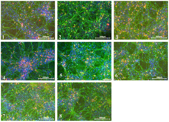
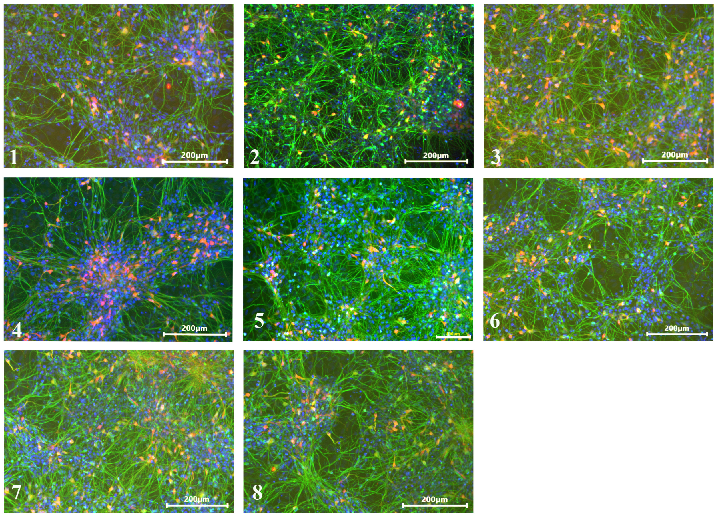
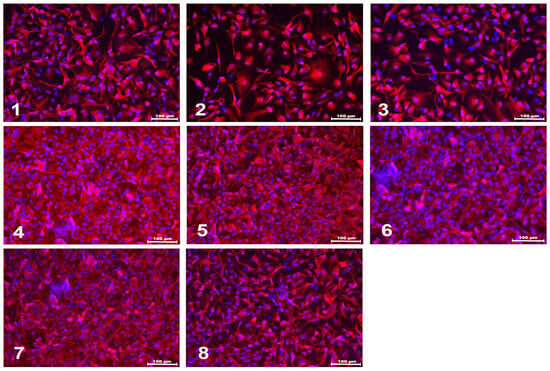
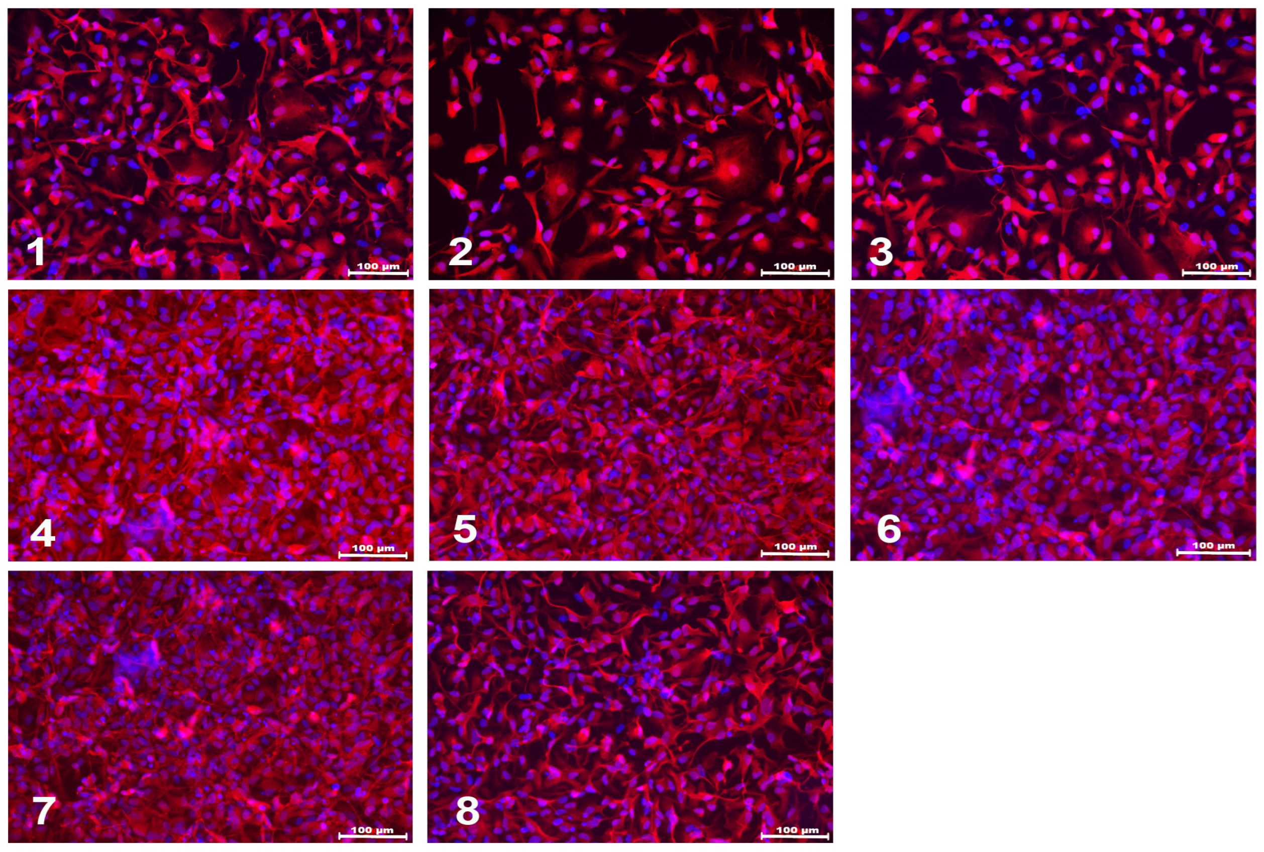
Following completion of differentiation, iPSC-derived neuronal and glial cultures were characterized by immunofluorescence staining for the cell-type-specific markers TUBB3/TH and S100, respectively (Figure 1 and Figure 2). Cells grown adherently in Petri dishes were rinsed with PBS and fixed in 4% paraformaldehyde prepared in PBS (pH 6.8) for 30 min at room temperature (RT). Following fixation, the cultures were washed three times for 5 min with PBS containing 0.1% Tween 20 (Sigma-Aldrich, Saint Louis, MO, USA). To reduce nonspecific antibody binding, the cells were incubated for 30 min at RT in blocking buffer consisting of PBS supplemented with 0.1% Triton X-100 and 5% fetal bovine serum (HyClone, Logan, UT, USA). The cultures were then incubated overnight at 4 °C with either mouse anti-TUBB3 (1:2000, AB7751, Abcam, Waltham, MA, USA) and rabbit anti-TH (1:1000, AB112, Abcam, Waltham, MA, USA), or rabbit anti-S100 (1:2, GA50461-2, Agilent Dako, Santa Clara, CA, USA) primary antibodies. After three 5 min washes with PBS containing 0.1% Tween 20, the corresponding secondary antibodies, goat anti-mouse (1:1000, A11008, Thermo Fisher, Waltham, MA, USA) or goat anti-rabbit (1:1000, A11010, Thermo Fisher, Waltham, MA, USA), were applied for 60 min at RT. The cultures were subsequently washed three times for 5 min with PBS containing 0.1% Tween 20. Cell nuclei were counterstained with 0.1 μg/mL DAPI (Sigma-Aldrich, Saint Louis, MO, USA) in PBS for 10 min, followed by two washes with PBS. Fluorescence imaging was performed using an AxioImager Z1 fluorescence microscope (Carl Zeiss, Oberkochen, Germany), and images were acquired using AxioVision release 4.8.1 (11-2009) software (Carl Zeiss, Oberkochen, Germany).
Figure 1.
Immunocytochemical staining of neurons with antibodies against the pan-neuronal marker TUBB3 (green), the dopaminergic neuron marker tyrosine hydroxylase (red), and the nuclear dye DAPI (blue). Cell lines: 1—IPSRG2L, 2—IPSHD1.1S, 3—IPSFD3.9L, 4—IPSPDS12, 5—IPSPDS13, 6—IPSPDS14, 7—IPSPDSG1.1, 8—IPSPDL2.15L. Scale bar: 100 µm for panel 5 and 200 µm for all other panels.
Figure 2.
Immunocytochemical staining of glial cells with antibodies against S100 (red) and the nuclear dye DAPI (blue). Cell lines: 1—IPSRG2L, 2—IPSHD1.1S, 3—IPSFD3.9L, 4—IPSPDS12, 5—IPSPDS13, 6—IPSPDS14, 7—IPSPDSG1.1, 8—IPSPDL2.15L. Scale bar: 100 µm.
2.3. Quantitative Real-Time PCR (qPCR)
Total RNA was isolated from cell cultures using TriReagent (MRC, Cincinnati, OH, USA) according to the manufacturer’s protocol. Residual genomic DNA was subsequently removed using the DNA-free DNA Removal Kit (Invitrogen, Carlsbad, CA, USA). For cDNA synthesis, 0.5–2 μg of total RNA was reverse-transcribed with M-MLV Reverse Transcriptase (Evrogen, Moscow, RF, Russia) using random primers. Primer sequences are provided in Table 2 and have been reported previously [27]. Quantitative PCR was performed using qPCRmix-HS SYBR (Evrogen, Moscow, RF, Russia) on a LightCycler 96 instrument (Roche, Basel, Switzerland). The amplification protocol consisted of an initial denaturation (preincubation) at 95 °C for 5 min, followed by 45 amplification cycles at 94 °C for 20 s, 55–65 °C for 20 s, and 72 °C for 20 s. Melting curve analysis was performed at 95 °C for 10 s, 65 °C for 60 s, and 97 °C for 1 s, followed by cooling at 37 °C for 30 s. 18S rRNA was used as the reference gene for glial cultures, whereas HMBS mRNA was used for neuronal cultures based on previously published recommendations [28]. The use of a single reference gene for normalization should be taken into consideration when interpreting the qPCR results. Relative changes in target gene expression were determined using the 2−ΔΔCt method and expressed as fold change (FC).
Table 2.
The sequences of primers used in the study.
2.4. Western Blot Hybridization
Cell and tissue samples were lysed in RIPA buffer (Thermo Fisher Scientific, Waltham, MA, USA) supplemented with a protease inhibitor mixture (Sigma-Aldrich, St. Louis, MO, USA). Total protein concentration was measured using the BCA assay. For each sample, 20 μg of total protein was subjected to SDS-polyacrylamide gel electrophoresis (SDS-PAGE), followed by transfer onto a PVDF membrane (Bio-Rad, Hercules, CA, USA). Membranes were blocked with 5% milk and subsequently incubated overnight at 4 °C with primary antibodies diluted in 1% milk: rabbit anti-BLT1 (1:500, #DF10223), rabbit anti-CHEMR23 (1:1000, #DF2712), rabbit anti-GPR32 (1:500, #DF2719), rabbit anti-FPRL1 (1:750, #DF4971) (all from Affinity Biosciences, Cincinnati, OH, USA), or mouse anti-β-actin (1:6000, #A5441, Sigma-Aldrich, Burlington, MA, USA). After primary antibody incubation, the membranes were washed five times for 5 min with Tris–NaCl–Tween (TNT) buffer and incubated for 2 h at RT with the appropriate horseradish peroxidase (HRP)-conjugated secondary antibody diluted in 1% milk (1:10,000, #31466, Thermo Fisher Scientific, Waltham, MA, USA; or 1:8000, #140777, Jackson ImmunoResearch, Cambridgeshire, UK). After washing five times for 5 min with TNT, protein signals were detected using an enhanced chemiluminescence reagent (#170-5061, Bio-Rad, Hercules, CA, USA) and visualized with the ChemiDoc MP Imaging System (Bio-Rad, Hercules, CA, USA).
2.5. ELISA
IL-6 protein levels in cell culture supernatants were quantified using a human IL-6 ELISA kit (cat. #A-8762, Vector-Best, Moscow, RF) in accordance with the manufacturer’s instructions. Absorbance was measured at 450 nm using a Metertech microplate reader (Nankang, Taipei, Taiwan). The measured IL-6 concentrations were normalized to the number of cells in each well and expressed as the amount per 1 × 10³ cells.
2.6. Statistical Analysis
Statistical analysis of the data was performed using GraphPad Prism version 10.4.0. (build 621; GraphPad Software, Boston, MA, USA) software. The data from 1 to 3 independent differentiation experiments for each cell line were obtained, and the mean value for each cell line was included in pairwise comparison, to avoid disproportionate contribution of any single cell line. All data was normalized to the mean value of the healthy donors (HD) group; differences between groups were expressed as fold changes. Results were presented as mean ± SD. Data distribution was assessed for normality using the Shapiro–Wilk test. For normally distributed data, an unpaired t-test was used for pairwise comparisons. The differences were considered statistically significant at * p < 0.05, ** p < 0.01. The pilot experiment on the effects of RvD1 on glial cells was analyzed separately, and since only one HD and one PARK2-mutant cell line were used, no statistical comparisons between the HD and PARK2-mutant cell lines were performed.
3. Results
According to the literature data, several SPM receptors are expressed in neurons: CHEMR23 [29], GPR32 [30], FPR2 [31], and in astrocytes: CHEMR23, BLT1, and GPR37 [13]. The protein product of the latter is a Parkin substrate for ubiquitination. The evidence exists that in the case of Parkin loss, GPR37 forms insoluble aggregates and triggers the unfolded protein response [32]. Similarly to α-synuclein, the protein has been found in the Lewy bodies of PD patients [33,34,35]. We hypothesized that in cell cultures with PARK2 mutations GPR37 might function incorrectly regardless of the expression level and, therefore, excluded the receptor from further analysis and focused on RvE1 and RvD1 receptors.
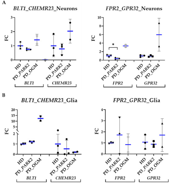
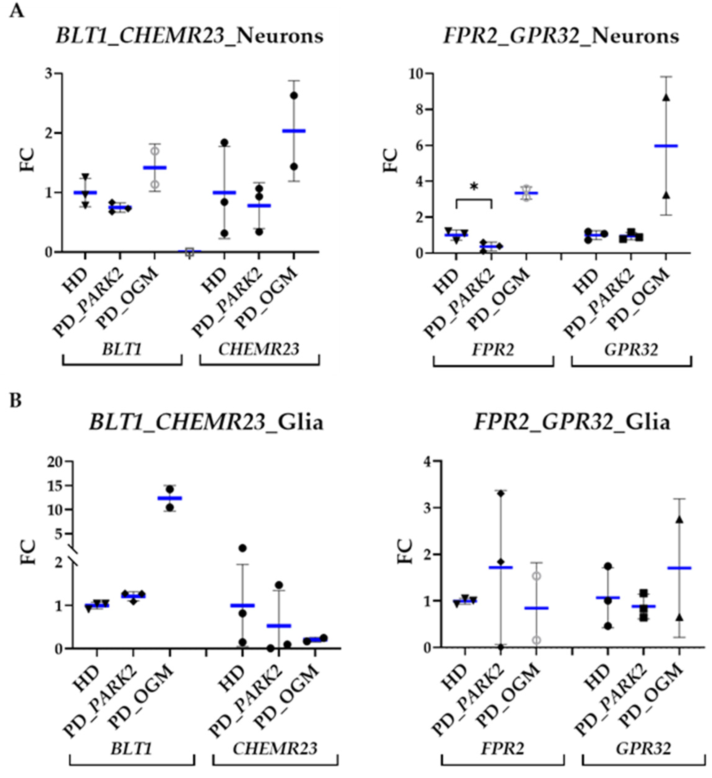
qPCR analysis showed that all four resolvin receptor genes were expressed in both neuronal and glial cells. Overall, the expression levels of all four receptors in PARK2-mutant neurons (PD_PARK2) did not exceed those observed in HD neurons. The only statistically significant difference was a decrease in FPR2 expression. To explore whether this expression pattern might be specifically associated with the PARK2 genetic background, we additionally compared PARK2-mutant cells with a group carrying PD-associated mutations in other genes (PD_OGM), which included neuronal and glial cell lines with mutations in LRRK2 (PARK8) and GBA1. In contrast to PD_PARK2 neurons, PD_OGM neurons showed a general tendency toward higher expression of all four receptor genes compared with HD cells. In PARK2-mutant glial cells, no significant changes in resolvin receptor gene expression were observed compared with HD cells, whereas PD_OGM glia showed a tendency toward higher BLT1 and lower CHEMR23 expression. Although these changes did not reach statistical significance, the observed trends suggest that resolvin receptor expression may vary depending on the underlying PD-associated mutation (Figure 3).
Figure 3.
Expression of genes encoding receptors for RvE1 (BLT1, CHEMR23) and RvD1 (GPR32, FPR2) in the cell cultures of patients with PARK2-associated PD (PD_PARK2) compared with HD cells and the group carrying PD-associated mutations in other genes (PD_OGM). Cell lines: 1—IPSRG2L, 2—IPSHD1.1S, 3—IPSFD3.9L, 4—IPSPDS12, 5—IPSPDS13, 6—IPSPDS14, 7—IPSPDSG1.1, 8—IPSPDL2.15L. qPCR. (A) neurons, ref. HMBS mRNA, FC (fold change) = gene expression/mean of gene expression in HD cells; (B) glial cells, ref 18S rRNA, FC. Each data point represents the mean value obtained for an individual cell line across independent differentiations. The results are presented as mean ± SD, with the mean value for the group indicated in blue. For normally distributed data, statistical analysis was performed using an unpaired t-test (GraphPad Prism 10.4.0. software). The differences were considered statistically significant at * p < 0.05.
Next, we investigated the pattern of the expression of RvE1 receptors (BLT1, CHEMR23) and RvD1 receptors (GPR32, FPR2) in PARK2-mutant and HD neurons and glial cells at the protein level.
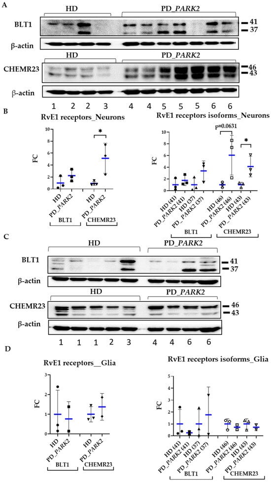
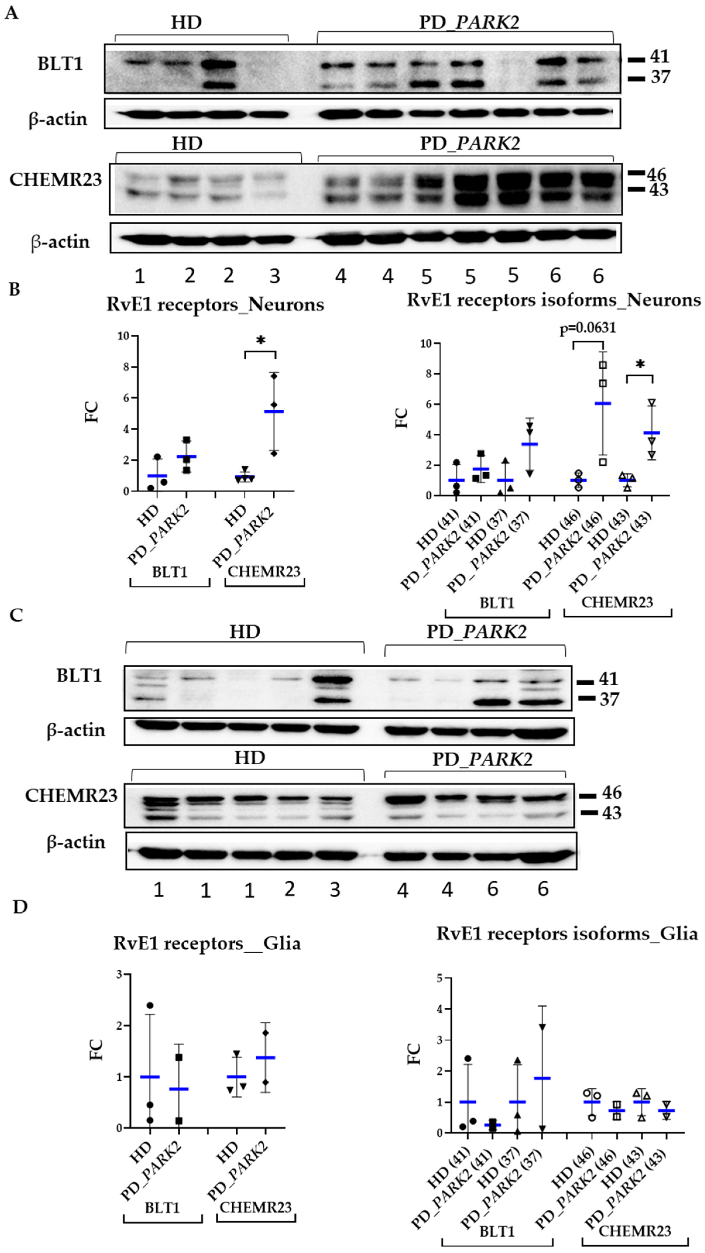
RvE1 receptors were detected in both cell types as two closely migrating protein bands: one at approximately 37 kDa for BLT1 and 43 kDa for CHEMR23, and another approximately 3–4 kDa higher, which may represent a modified form of the protein. Evidence exists that phosphorylation of BLT1 promotes receptor desensitization [36]. The main finding was that in PARK2-mutant neurons, CHEMR23 protein was present at a significantly higher level, compared to HD cells (Figure 4).
Figure 4.
Expression of receptors for RvE1 (BLT1, CHEMR23) at the protein level in the cell cultures of patients with PARK2-associated PD compared with those of healthy donors. Cell lines: 1—IPSRG2L, 2—IPSHD1.1S, 3—IPSFD3.9L, 4—IPSPDS12, 5—IPSPDS13, 6—IPSPDS14. Western blot hybridization in neurons (A) and glia (C), reference protein β-actin. Relative expression of the detected receptor bands in neurons (B) and glial cells (D). FC (fold change) = protein expression in cell culture/mean of protein expression in HD cells. Each data point represents the mean value obtained for an individual cell line across independent differentiations. The results are presented as mean ± SD, with the mean value for the group indicated in blue. For normally distributed data, statistical analysis was performed using an unpaired t-test (GraphPad Prism 10.4.0. software). The differences were considered statistically significant at * p < 0.05.
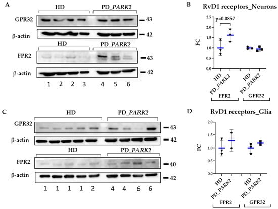
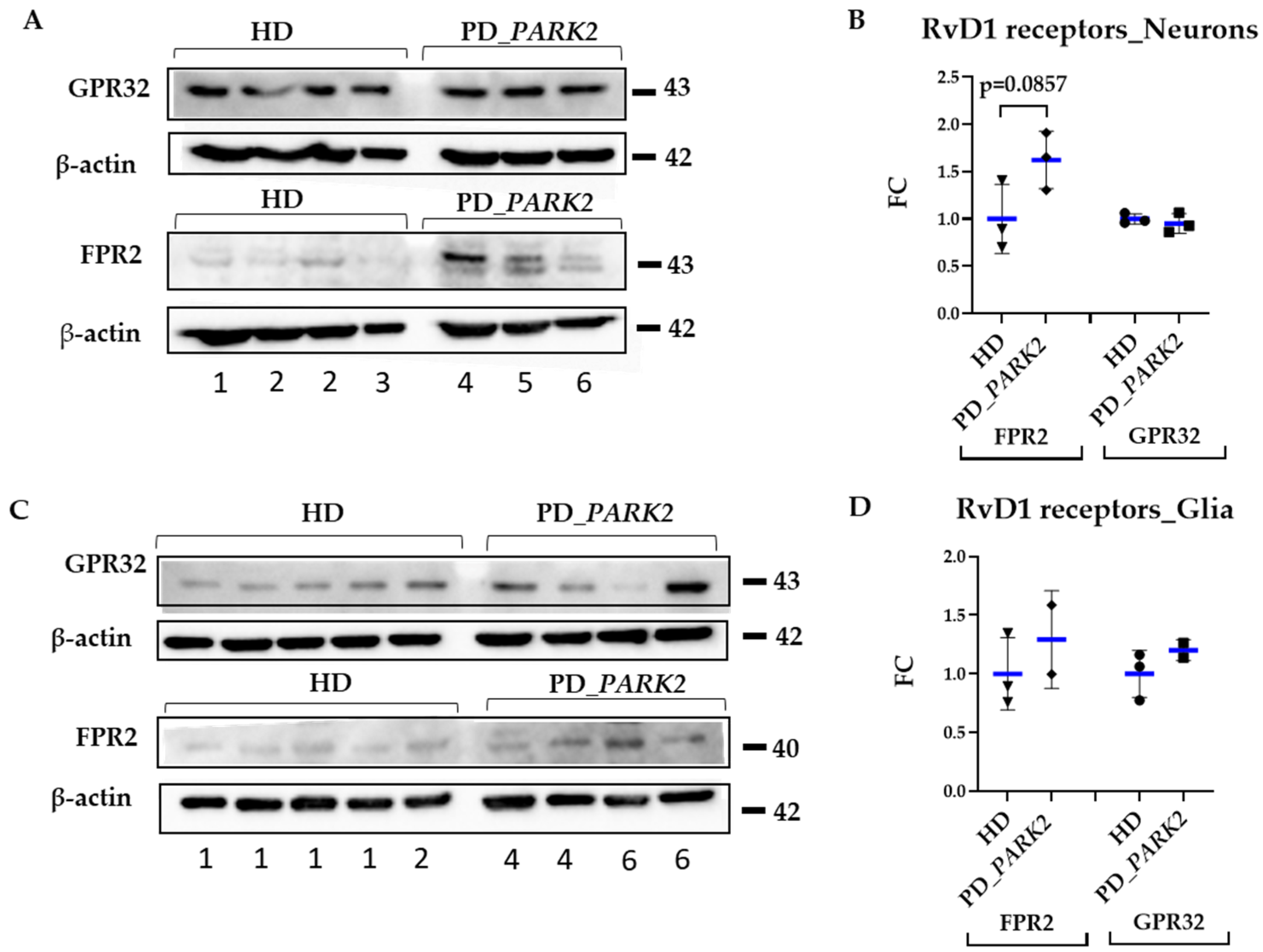
Expression of receptors for RvD1 (GPR32 and FPR2 proteins) was also detected both in neurons and glial cultures. Compared with the corresponding HD cells, FPR2 protein levels showed a tendency to be higher in PARK2-mutant neurons (Figure 5).
Figure 5.
Expression of receptors for RvD1 (GPR32, FPR2) at the protein level in the cell cultures of patients with PARK2-associated PD compared to those of healthy donors. Cell lines: 1—IPSRG2L, 2—IPSHD1.1S, 3—IPSFD3.9L, 4—IPSPDS12, 5—IPSPDS13, 6—IPSPDS14. Western blot hybridization in neurons (A,B) and glia (C,D), reference protein β-actin. FC (fold change) = protein expression in cell culture/mean of protein expression in HD cells. Each data point represents the mean value obtained for an individual cell line across independent differentiations. The results are presented as mean ± SD, with the mean value for the group indicated in blue. For normally distributed data, statistical analysis was performed using an unpaired t-test (GraphPad Prism 10.4.0. software).
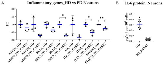
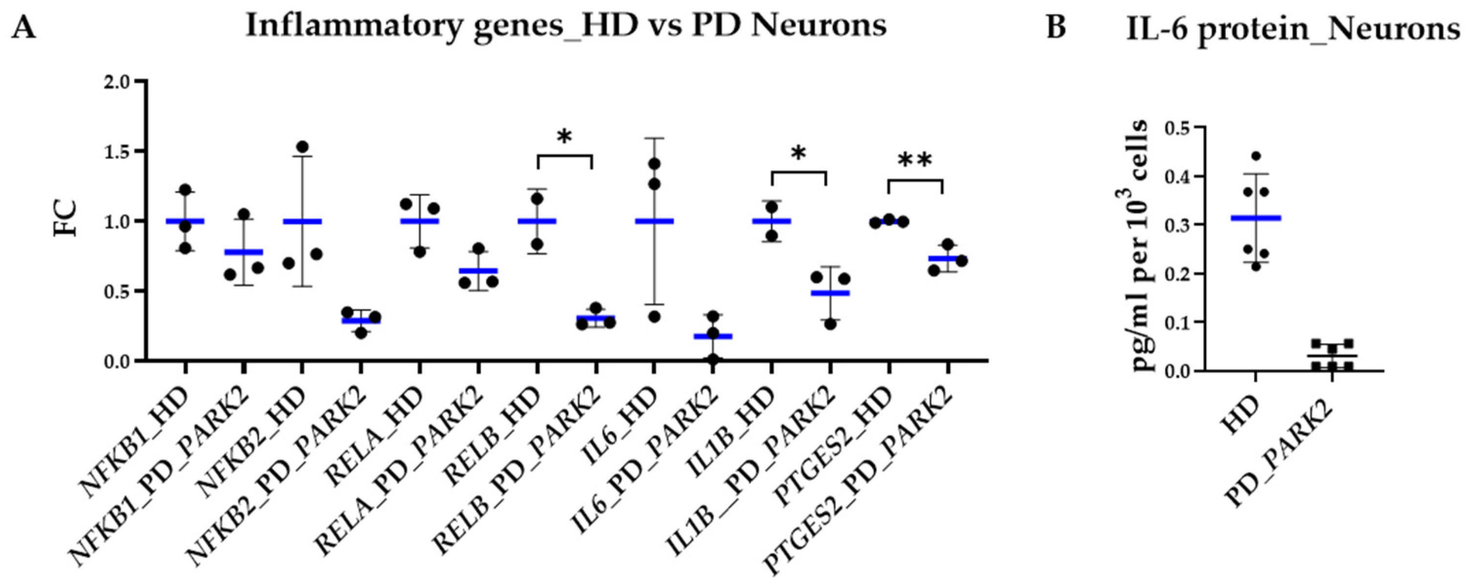
To assess how these findings corresponded to the overall inflammatory status of both cell types, we determined the expression of genes encoding components of NF-κB and the main pro-inflammatory cytokines IL1B and IL6, since their activity is a result of the network of various inflammatory signaling pathways. We also estimated the level of PTGES2 encoding microsomal prostaglandin E synthase-2 (mPGES-2), one of the enzymes that convert PGH2 into strong pro-inflammatory PGE2. We found that several of the studied genes (RELB, IL1B, PTGES2) were downregulated in PD_PARK2 neurons compared with HD neurons (Figure 6A). ELISA measurements of IL-6 protein levels revealed trends consistent with the qPCR data (Figure 6B). In a recent study we reported the opposite pattern characteristic for PARK2-mutant glial cultures as they exhibited a more pro-inflammatory status compared to HD [27].
Figure 6.
Expression of the inflammatory genes in neuronal cultures of patients with PARK2-associated PD compared to those of HD. Cell lines: 1—IPSRG2L, 2—IPSHD1.1S, 3—IPSFD3.9L, 4—IPSPDS12, 5—IPSPDS13, 6—IPSPDS14. (A) expression of the inflammatory genes, qPCR, ref. HMBS mRNA, FC (fold change) = gene expression/mean of gene expression in HD. The results are presented as mean ± SD. For normally distributed data, statistical analysis was performed using an unpaired t-test (GraphPad Prism 10.4.0. software). Differences were considered statistically significant at * p < 0.05, ** p < 0.01. (B) IL-6 protein level in two HD (IPSRG2L, IPSHD1.1S) vs. two PARK2-mutant (IPSPDS12, IPSPDS14) neuronal cell culture supernatants, ELISA. Data are from three independent experiments performed with each cell line and are presented as mean ± SD, with the mean value for the group indicated in blue. Statistical analysis was not performed because of the limited number of cell lines in both groups.
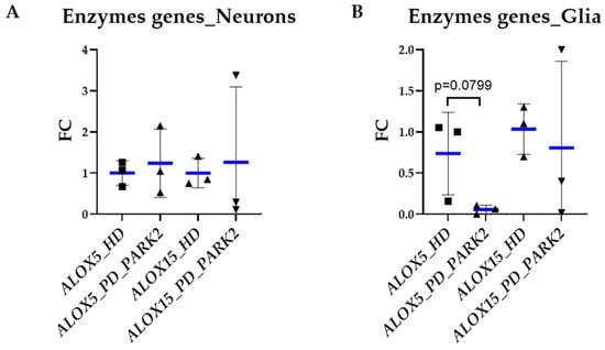
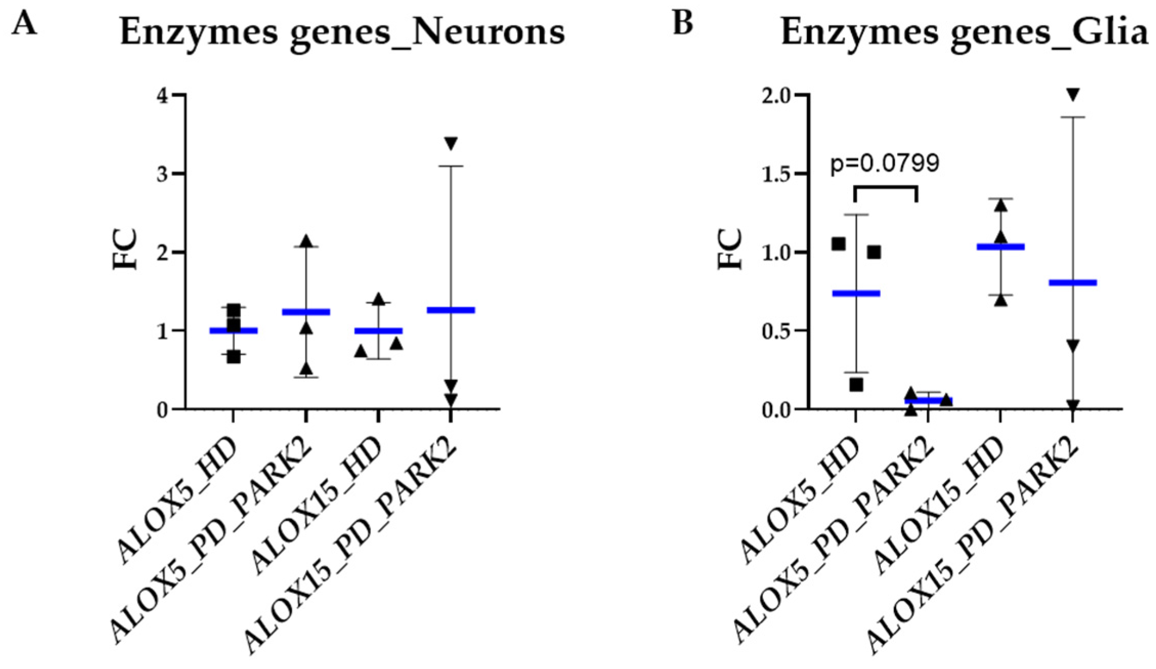
In order to assess the expression of genes involved in endogenous SPM biosynthesis in PARK2-mutant neurons and glial cells, we analyzed ALOX5 (encoding 5-lipoxygenase) and ALOX15 (encoding 15-lipoxygenase) [37]. Both genes were expressed in neuronal and glial cultures, with no significant differences between PARK2-mutant and HD cells, although ALOX5 showed a tendency toward lower expression in PARK2-mutant glial cultures compared with HD (Figure 7).
Figure 7.
Expression of genes encoding enzymes involved in resolvin synthesis in cell cultures from patients with PARK2-associated PD compared with healthy donors. Cell lines: 1—IPSRG2L, 2—IPSHD1.1S, 3—IPSFD3.9L, 4—IPSPDS12, 5—IPSPDS13, 6—IPSPDS14. (A) neurons, qPCR, ref. HMBS mRNA, FC (fold change) = gene expression/mean of gene expression in HD; (B) glial cell cultures, qPCR, ref 18S rRNA, FC. Each data point represents the mean value obtained for an individual cell line across independent differentiations. The results are presented as mean ± SD, with the mean value for the group indicated in blue. For normally distributed data, statistical analysis was performed using an unpaired t-test (GraphPad Prism 10.4.0. software).
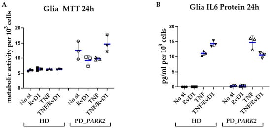
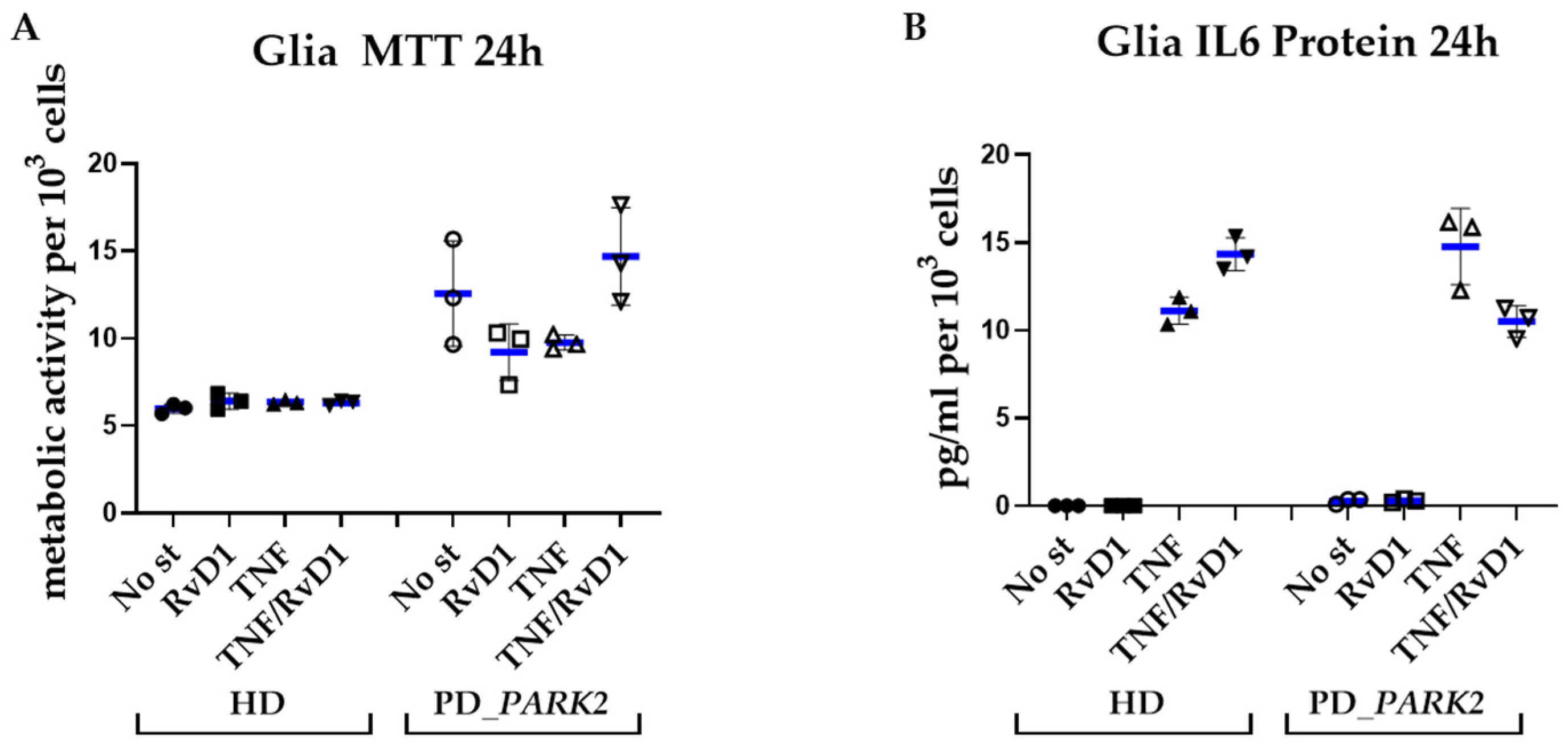
Next, we conducted a pilot experiment to evaluate the effects of RvD1 using one HD and one PARK2-mutant glial cell line. Glial cells were cultivated in the presence of RvD1 (20 ng/mL) for 24 h, either under basal conditions or following induction of inflammation by TNF-α (10 ng/mL). RvD1 did not induce any apparent cytotoxic effects in glial cells. According to the MTT assay, increased MTT reduction was observed in stimulated PARK2-mutant glial cells cultured in the presence of RvD1, suggesting an RvD1-associated increase in cellular metabolic activity. Different responses to RvD1 were also observed for IL-6 protein secretion in TNF-α-stimulated cell cultures: IL-6 secretion increased in the HD cell line, whereas it decreased in the PARK2-mutant cell line (Figure 8). Given that only one cell line of each genotype was used, these observations cannot be generalized to HD and PARK2-mutant glia. However, these pilot data support further investigation of the effects of RvD1 on glial cells using additional independent cell lines.
Figure 8.
The effect of resolvin D1 on glial cell cultures: Pilot experiment included 2 cell lines—HD (IPSHD1.1S) and PD with mutation in the PARK2 gene (IPSPDS14). Glial cells were cultured in the presence of RvD1 (20 ng/mL) for 24 h, either under basal conditions or following induction of inflammation by TNF-α (10 ng/mL). (A) MTT assay results. (B) IL-6 protein concentrations in glial supernatants, ELISA. Data are from three independent experiments performed with each cell line and are presented as mean ± SD, with the mean value for the group indicated in blue. Statistical analysis was not performed because of the limited number of cell lines in both groups.
4. Discussion
Nonresolving long-lasting inflammation is not a direct initiator of PD, but it contributes significantly to the disease pathogenesis [15,38,39]. Neuronal death induces an “eicosanoid storm,” including the production of prostaglandins and leukotrienes, which in turn initiates a robust inflammatory response. At a certain stage of the inflammatory response, the cascade of arachidonic acid (AA) and PUFA switches to the resolution phase, when specialized pro-resolving mediators (SPMs) are produced and stimulate macrophage-mediated clearance of debris, downregulate NF-κB, and attenuate pro-inflammatory cytokine production [9,39,40,41]. Resolution may fail if the secretion of resolution-driving factors or the expression of receptors for these molecules is reduced. Long-lasting neuroinflammation is characterized by deficiencies in resolution mechanisms, on the one hand, and by excessive activation of pro-inflammatory pathways, on the other hand [42]. Nonsteroidal anti-inflammatory drugs (NSAIDs) have therefore been investigated for their potential role in PD prevention and treatment. However, despite a recently published meta-analysis indicating a correlation between the use of several groups of NSAIDs and a reduced risk of developing PD [23], there is still no consensus regarding the effect of these drugs on neurodegeneration [23,43,44]. Multiple studies have shown that cyclooxygenase-2 (COX-2) is upregulated in DA neurons of the substantia nigra in PD patients and in experimental models and is a well-known therapeutic target for NSAIDs [45,46]. High activity of COX-2 leads to an excessive production of a potent pro-inflammatory prostaglandin E2 (PGE2) that might result in the death of DA neurons [43]. At the same time, PGE2 is necessary for the switch to SPM synthesis and timely resolution; for this reason, COX-2 inhibitors, despite their anti-inflammatory effect, disrupt endogenous resolution mechanisms [47,48]. In this regard, the issue of finding anti-inflammatory molecules that can neutralize the side effects of NSAIDs or act as independent anti-inflammatory agents in PD is highly relevant. SPMs may represent promising therapeutic agents, since they exert potent biological effects at low concentrations in experimental models of inflammation-associated diseases [40,49]. Nevertheless, it is not known which pro-resolving lipid mediator would be most effective in specific models of PD [39].
Data from the Human Protein Atlas (HPA) also provide information on the expression of the receptors for resolvin E1, BLT1 (LTB4R) and CHEMR23 (CMKLR1), and resolvin D1, GPR32 (GPR32) and FPR2/ALX (FPR2), in the human CNS. LTB4R, CMKLR1, and FPR2 transcripts are detected in different human brain regions, with relatively low regional specificity. Brain single-nucleus data also show CMKLR1 and FPR2 expression in CNS macrophage populations, although their cell-type expression patterns differ [50]. Although GPR32 is currently classified as not detected in the human brain in the HPA, a recent study demonstrated its expression in human cortical neurons and microglia, while it was nearly absent in astrocytes [30] and, in an earlier study, GPR32 mRNA was also detected in primary human astrocyte cultures [51]. Overall, the available HPA data support the presence of several resolvin receptors in the human CNS and also indicate cell-type-specific differences in their expression. Multiple studies have confirmed the expression of SPM receptors in neurons and astrocytes, which determines the responsiveness of these cells to the anti-inflammatory effect of resolvins [13,52]. However, there are no data specifically on cells from PD patients with mutations in the PARK2 gene. Meanwhile, it is well-established that the protein product of this gene, Parkin, significantly contributes to the development of neuroinflammation. The dysregulated process of mitochondrial clearance in Parkin deficiency causes the release of mitoDAMPs that trigger inflammation [53]. Due to its ubiquitin ligase activity, Parkin is directly related to NF-κB signaling and the function of SPM receptors as well. Wang et al. found that Parkin promotes NF-κB activation through K63 ubiquitination of RIPK1, resulting in increased p65 nuclear translocation [24]. Another substrate for ubiquitination by Parkin, GPR37, is the receptor for one of the SPM members, neuroprotectin D1 [34,54]. GPR37 deficiency was associated with increased IL-1β expression and impaired anti-inflammatory cytokine responses [34].
Considering the direct influence of Parkin dysfunction on inflammatory signaling, we carefully examined the expression of RvE1 and RvD1 receptors in neuronal and glial cell cultures of PARK2-associated PD patients compared to HDs, since specific studies addressing this issue are currently lacking. We also compared these data with the inflammatory status of each cell type, neurons and glia. Since both neurons and glial cells express enzymes involved in SPM biosynthesis, we also assessed the expression of the genes encoding 5- and 15-lipoxygenases, ALOX5 and ALOX15 [13]. Several published studies emphasize the dual role of these enzymes which, in conjunction with each other, generate SPMs [47,49,55].
Importantly, a dual nature is characteristic not only of inflammation in general but also of its particular mechanisms at different levels. Several studies classify 5-LOX as a key pro-inflammatory enzyme and 15-LOX as being responsible for the formation of pro-resolving mediators [56], whereas other work has demonstrated that 5-LOX deficiency leads to reduced SPM levels and impaired resolution of inflammation after myocardial infarction in mice [57]. Interestingly, 15-LOX is also capable of catalyzing all enzymatic steps without the need for 5-LOX [58]. It is known that the interaction of CHEMR23 with chemerin results in the increase in pro-inflammatory gene expression; the same effect is observed with the binding of leukotriene (LT) B4 to the BLT1 receptor. RvE1 interacts with both receptors in different ways. It acts as an agonist of CHEMR23, reducing TNF-α-induced NF-κB signaling, while at BLT1 it acts as a partial agonist and attenuates LTB4-induced signaling, including calcium mobilization and NF-κB activation [15,59,60,61]. Therefore, these two receptors modulate neuroinflammation depending on the ratio of pro-inflammatory and pro-resolving mediators in the intercellular environment. In contrast, RvD1 signaling via its receptors GPR32 and FPR2 mainly mediates anti-inflammatory and pro-resolving actions in neuroinflammation [15,62].
According to our results, in PARK2-mutant neurons, mRNA expression levels of resolvin receptors were generally comparable to those in HD neurons, with only FPR2 expression showing a slight but statistically significant decrease. Interestingly, the pattern appeared to differ between PARK2-mutant and PD_OGM cells. In particular, PD_OGM neurons tended to exhibit higher expression of all four receptor genes. In PARK2-mutant glial cells, no significant changes in resolvin receptor gene expression were observed compared with HD cells, whereas PD_OGM glia showed a tendency toward higher BLT1 and lower CHEMR23 expression. Although these differences were not statistically significant, they may indicate that resolvin receptor expression is influenced by mutations in different genes. At the protein level, the most prominent finding was a significantly higher level of CHEMR23 in PARK2-mutant neurons compared with HD neurons. This was accompanied by decreased expression of genes encoding mPGES-2 (PTGES2), NF-κB component RELB, and the main pro-inflammatory cytokine IL1B, i.e., PARK2-mutant neurons demonstrated signs of less activated inflammatory profile compared to HD. These findings corresponded to a recent report on the anti-inflammatory function of RvE1/CHEMR23: in blood and adipose tissue of individuals with gain-of-function CHEMR23, IL-6 level was downregulated; whereas, mice lacking ChemR23 expression showed increased IL-6 levels [63].
To date, relatively little is known about the inflammatory intracellular signaling of neurons in neurodegeneration. Our recently published data also indicated the presence of a specific (hypo-activated) inflammatory status of PARK2-mutant neurons, when affected by glial soluble factors [19], which corresponded to the present results obtained on neurons in the absence of glial influence. Importantly, it was shown that in the initial phase of plaque formation in AD, Aβ-burdened neurons, but not glia, acted as inflammatory agents [64]. Activated glia, in turn, release pro-inflammatory factors and can induce the death of DA-neurons [65]. Moreover, PARK2-mutant glia exhibit pro-inflammatory shift compared to HD cells [27]. Therefore, these observations support targeting dysregulated inflammatory status of glial cells in PARK2-associated PD. Importantly, in neurodegenerative disorders, the coexistence and interplay of multiple pathogenic mechanisms, including distinct proteinopathies, disrupted mitochondrial homeostasis, and dysregulated neuroinflammation, underscore the need for complementary therapeutic strategies aimed at slowing disease progression [66], including approaches that promote the resolution of inflammation.
In our pilot experiment, RvD1 produced different responses in the two glial cell lines under TNF-α stimulation. In the PARK2-mutant cell line, RvD1 reduced IL-6 protein secretion and increased MTT reduction, the latter indicating increased cellular metabolic activity, whereas neither of these effects was observed in the HD cell line. Importantly, because the differentiation protocol used here yielded microglia-free glial cultures, the observed inflammatory changes reflect astroglia responses in the absence of a microglial contribution. However, since only one HD and one PARK2-mutant cell line were examined, these observations cannot be generalized to PARK2-associated PD. These pilot data support further investigation of the effects of RvD1 on glial cells using additional cell lines with different PARK2 mutations.
5. Study Limitations
For this study, we used donor-matched neuronal and glial cultures derived through a common neural progenitor stage from iPSCs of healthy donors and PD patients carrying different mutations in the PARK2 gene. Generation of patient-specific iPSC-derived neuronal and glial cell lines is technically challenging, and individual pathogenic PARK2 variants are relatively rare, which limits the number of cell lines available for analysis. Therefore, the effects of individual PARK2 variants were not analyzed separately, and we focused mainly on common features associated with PARK2 mutations. This approach may be more relevant for the search for potential therapeutic strategies, since division into very small variant-specific groups would be less practical for this purpose. The number of available cell lines also limits the statistical power of between-group comparisons and the reliable assessment of data distribution. In addition to statistically significant differences, we also considered trends in gene expression, which should be interpreted with caution. Finally, an in vitro system cannot fully reproduce the complex cellular and molecular environment of the human brain. At the same time, this model allows us to examine cell-type-specific features associated with mutations in PD-related genes, which provides a basis for further studies using more complex experimental models. The glial cultures generated through neural progenitor-based differentiation are not expected to contain microglia, allowing the astroglia component of the neuroinflammatory response to be examined independently of microglial contribution, which represents an advantage of the model.
6. Conclusions
Parkinson’s disease involves multiple pathogenic mechanisms, which makes it important to explore different therapeutic strategies, including those that aim to restore resolution of inflammation. To our knowledge, this is the first study to characterize RvE1 (BLT1 and CHEMR23) and RvD1 (GPR32 and FPR2) receptor expression at both the mRNA and protein levels in iPSC-derived neuronal and glial cultures carrying PARK2 mutations. The presence of these receptors indicates that they may represent targets for resolvin-based therapeutic approaches in PARK2-associated PD. We identified cell-type-specific receptor expression in PARK2-mutant cells, and also observed trends toward distinct expression patterns in PD cells carrying mutations in other genes, including GBA1 and LRRK2, which may suggest an influence of genetic background on resolvin receptor expression. PARK2-mutant neurons exhibited a less inflammatory profile compared with HD cells, in contrast to the pro-inflammatory shift we previously reported in PARK2-mutant glial cultures. These findings highlight the importance of taking into account both cell-type- and PD gene-associated differences in the development of resolvin-based therapeutic approaches for genetic forms of PD. Our pilot data further support the need for further studies of the potential anti-inflammatory effects of RvD1 in PARK2-mutant glial cells using additional PARK2-mutant glial cell lines.
Author Contributions
T.G.: investigation, data curation, visualization, writing—original draft. E.N.: supervision, writing—review and editing. E.S.: investigation, L.N.—investigation. E.A.—investigation, visualization. A.E.: supervision. V.N.: data curation, writing—review and editing. All authors have read and agreed to the published version of the manuscript.
Funding
This work was supported by the Ministry of Science and Higher Education of the Russian Federation (the Federal Scientific-technical program for genetic technologies development for 2019–2030, agreement N 075-15-2025-518).
Institutional Review Board Statement
The present study was conducted according to the guidelines of the Declaration of Helsinki and met all the requirements and instructions of the Ethics Committee of the Lopukhin FRCC PCM (approval No. 1; approved on 1 June 2021).
Informed Consent Statement
All iPSC lines have been previously published; the consent form for participation was distributed to all participants and signed.
Data Availability Statement
The original contributions presented in this study are included in the article. Further inquiries can be directed to the corresponding authors.
Conflicts of Interest
The authors declare no conflicts of interest.
Abbreviations
| PD | Parkinson’s disease |
| SPMs | specialized pro-resolving mediators |
| HD | healthy donor |
| Rv | resolvin |
| PUFAs | polyunsaturated fatty acids |
| DHA | docosahexaenoic acid |
| EPA | eicosapentaenoic acid |
| iPSCs | induced pluripotent stem cells |
| NSAIDs | nonsteroidal anti-inflammatory drugs |
| NPs | neural progenitors |
References
- Nathan, C.; Ding, A. Nonresolving inflammation. Cell 2010, 140, 871–882. [Google Scholar] [CrossRef] [Scilit] [PubMed]
- Serhan, C.N. Discovery of specialized pro-resolving mediators marks the dawn of resolution physiology and pharmacology. Mol. Asp. Med. 2017, 58, 1–11. [Google Scholar] [CrossRef] [Scilit] [PubMed]
- Yao, C.; Narumiya, S. Prostaglandin–cytokine crosstalk in chronic inflammation. Br. J. Pharmacol. 2019, 176, 337–354. [Google Scholar] [CrossRef] [Scilit] [PubMed]
- Tansey, M.G.; Wallings, R.L.; Houser, M.C.; Herrick, M.K.; Keating, C.E.; Joers, V. Inflammation and immune dysfunction in Parkinson disease. Nat. Rev. Immunol. 2022, 22, 657–673. [Google Scholar] [CrossRef] [Scilit] [PubMed]
- Merlin, J.; Park, J.; Vandekolk, T.H.; Fabb, S.A.; Allinne, J.; Summers, R.J.; Langmead, C.J.; Riddy, D.M. Multipathway in vitro pharmacological characterization of specialized proresolving G protein-coupled receptors. Mol. Pharmacol. 2022, 101, 246–256. [Google Scholar] [CrossRef] [Scilit] [PubMed]
- Xue, J.; Tao, K.; Wang, W.; Wang, X. What Can Inflammation Tell Us about Therapeutic Strategies for Parkinson’s Disease? Int. J. Mol. Sci. 2024, 25, 1641. [Google Scholar] [CrossRef] [Scilit] [PubMed]
- Medzhitov, R. Inflammation 2010: New adventures of an old flame. Cell 2010, 140, 771–776. [Google Scholar] [CrossRef] [Scilit] [PubMed]
- Serhan, C.N.; Savill, J. Resolution of inflammation: The beginning programs the end. Nat. Immunol. 2005, 6, 1191–1197. [Google Scholar] [CrossRef] [Scilit] [PubMed]
- Serhan, C.N.; Brain, S.D.; Buckley, C.D.; Gilroy, D.W.; Haslett, C.; O’Neill, L.A.; Perretti, M.; Rossi, A.G.; Wallace, J.L. Resolution of inflammation: State of the art, definitions and terms. FASEB J. 2007, 21, 325–332. [Google Scholar] [CrossRef] [Scilit] [PubMed]
- Nagatsu, T.; Mogi, M.; Ichinose, H.; Togari, A. Changes in cytokines and neurotrophins in Parkinson’s disease. J. Neural Transm. Suppl. 2000, 60, 277–290. [Google Scholar] [CrossRef] [Scilit] [PubMed]
- Williams-Gray, C.H.; Wijeyekoon, R.; Yarnall, A.J.; Lawson, R.A.; Breen, D.P.; Evans, J.R.; Cummins, G.A.; Duncan, G.W.; Khoo, T.K.; Burn, D.J.; et al. Serum immune markers and disease progression in an incident Parkinson’s disease cohort (ICICLE-PD). Mov. Disord. 2016, 31, 995–1003. [Google Scholar] [CrossRef] [Scilit] [PubMed]
- Strempfl, K.; Unger, M.S.; Flunkert, S.; Trost, A.; Reitsamer, H.A.; Hutter-Paier, B.; Aigner, L. Leukotriene signaling as a target in α-synucleinopathies. Biomolecules 2022, 12, 346. [Google Scholar] [CrossRef] [Scilit] [PubMed]
- Tiberi, M.; Chiurchiù, V. Specialized pro-resolving lipid mediators and glial cells: Emerging candidates for brain homeostasis and repair. Front. Cell. Neurosci. 2021, 15, 673549. [Google Scholar] [CrossRef] [Scilit] [PubMed]
- Krashia, P.; Cordella, A.; Nobili, A.; La Barbera, L.; Federici, M.; Leuti, A.; Campanelli, F.; Natale, G.; Marino, G.; Calabrese, V.; et al. Blunting neuroinflammation with resolvin D1 prevents early pathology in a rat model of Parkinson’s disease. Nat. Commun. 2019, 10, 3945, Correction in Nat. Commun. 2019, 10, 4725. https://doi.org/10.1038/s41467-019-12538-2. [Google Scholar] [CrossRef] [Scilit] [PubMed]
- Chamani, S.; Bianconi, V.; Tasbandi, A.; Pirro, M.; Barreto, G.E.; Jamialahmadi, T.; Sahebkar, A. Resolution of inflammation in neurodegenerative diseases: The role of resolvins. Mediat. Inflamm. 2020, 2020, 3267172. [Google Scholar] [CrossRef] [Scilit] [PubMed]
- Xu, J.; Gao, X.; Yang, C.; Chen, L.; Chen, Z. Resolvin D1 Attenuates Mpp+-Induced Parkinson Disease via Inhibiting Inflammation in PC12 Cells. Med. Sci. Monit. 2017, 23, 2684–2691. [Google Scholar] [CrossRef] [Scilit] [PubMed]
- Chen, Y.; Chu, J.M.T.; Chang, R.C.C.; Wong, G.T.C. The complement system in the central nervous system: From neurodevelopment to neurodegeneration. Biomolecules 2022, 12, 337. [Google Scholar] [CrossRef] [Scilit] [PubMed]
- Deleidi, M.; Gasser, T. The role of inflammation in sporadic and familial Parkinson’s disease. Cell. Mol. Life Sci. 2013, 70, 4259–4273. [Google Scholar] [CrossRef] [Scilit] [PubMed]
- Gerasimova, T.; Poberezhniy, D.; Nenasheva, V.; Stepanenko, E.; Arsenyeva, E.; Novosadova, L.; Grivennikov, I.; Illarioshkin, S.; Lagarkova, M.; Tarantul, V.; et al. Inflammatory intracellular signaling in neurons is influenced by glial soluble factors in iPSC-based cell model of PARK2-associated Parkinson’s disease. Int. J. Mol. Sci. 2024, 25, 9621. [Google Scholar] [CrossRef] [Scilit] [PubMed]
- Aizawa, F.; Nishinaka, T.; Yamashita, T.; Nakamoto, K.; Koyama, Y.; Kasuya, F.; Tokuyama, S. Astrocytes Release Polyunsaturated Fatty Acids by Lipopolysaccharide Stimuli. Biol. Pharm. Bull. 2016, 39, 1100–1106. [Google Scholar] [CrossRef] [Scilit] [PubMed]
- Ikonomovic, M.D.; Abrahamson, E.E.; Uz, T.; Manev, H.; Dekosky, S.T. Increased 5-lipoxygenase immunoreactivity in the hippocampus of patients with Alzheimer’s disease. J. Histochem. Cytochem. 2008, 56, 1065–1073. [Google Scholar] [CrossRef] [Scilit] [PubMed]
- Praticò, D.; Zhukareva, V.; Yao, Y.; Uryu, K.; Funk, C.D.; Lawson, J.A.; Trojanowski, J.Q.; Lee, V.M. 12/15-lipoxygenase is increased in Alzheimer’s disease: Possible involvement in brain oxidative stress. Am. J. Pathol. 2004, 164, 1655–1662. [Google Scholar] [CrossRef] [Scilit] [PubMed]
- Badawoud, A.M.; Ali, L.S.; Abdallah, M.S.; El Sabaa, R.M.; Bahaa, M.M.; Elmasry, T.A.; Wahsh, E.; Yasser, M.; Eltantawy, N.; Eldesoqui, M.; et al. The relation between Parkinson’s disease and non-steroidal anti-inflammatories; a systematic review and meta-analysis. Front. Pharmacol. 2024, 15, 1434512. [Google Scholar] [CrossRef] [Scilit] [PubMed]
- Wang, Y.; Shan, B.; Liang, Y.; Wei, H.; Yuan, J. Parkin regulates NF-κB by mediating site-specific ubiquitination of RIPK1. Cell Death Dis. 2018, 9, 732. [Google Scholar] [CrossRef] [Scilit] [PubMed]
- Novosadova, E.V.; Nekrasov, E.D.; Chestkov, I.V.; Surdina, A.V.; Vasina, E.M.; Bogomazova, A.N.; Manuilova, E.S.; Arsenyeva, E.L.; Simonova, V.V.; Konovalova, E.V.; et al. A platform for studying molecular and cellular mechanisms of Parkinson’s disease based on human induced pluripotent stem cells. Sovrem. Tehnol. Med. 2016, 8, 157–166. [Google Scholar] [CrossRef] [Scilit]
- Novosadova, E.V.; Arsenyeva, E.L.; Antonov, S.A.; Kazantseva, E.A.; Novosadova, L.V.; Kurko, O.D.; Illarioshkin, S.N.; Ta-rantul, V.Z.; Grivennikov, I.A. Generation and characteristics of glial cells from induced human pluripotent stem cells. Neuro-chem. J. 2020, 14, 415–423. [Google Scholar] [CrossRef] [Scilit]
- Gerasimova, T.; Stepanenko, E.; Novosadova, L.; Arsenyeva, E.; Shimchenko, D.; Tarantul, V.; Grivennikov, I.; Nenasheva, V.; Novosadova, E. Glial cultures differentiated from iPSCs of patients with PARK2-associated Parkinson’s disease demonstrate a pro-inflammatory shift and reduced response to TNFα stimulation. Int. J. Mol. Sci. 2023, 24, 2000. [Google Scholar] [CrossRef] [Scilit] [PubMed]
- Artyukhov, A.S.; Dashinimaev, E.B.; Tsvetkov, V.O.; Bolshakov, A.P.; Konovalova, E.V.; Kolbaev, S.N.; Vorotelyak, E.A.; Vasiliev, A.V. New genes for accurate normalization of qRT-PCR results in study of iPS and iPS-derived cells. Gene 2017, 626, 234–240. [Google Scholar] [CrossRef] [Scilit] [PubMed]
- Zhang, J.; Liu, L.; Zhang, Y.; Yuan, Y.; Miao, Z.; Lu, K.; Zhang, X.; Ni, R.; Zhang, H.; Zhao, Y.; et al. ChemR23 signaling ameliorates cognitive impairments in diabetic mice via dampening oxidative stress and NLRP3 inflammasome activation. Redox Biol. 2022, 58, 102554. [Google Scholar] [CrossRef] [Scilit] [PubMed]
- Huang, K.; Li, J.; He, Z.; Li, Y.; Liu, X.; Wan, Y.; Yang, X.; Sang, D.; Li, S.; Wang, Z.; et al. G Protein-Coupled Receptor 32 Contributes to Inflammation Resolution and Neuronal Excitability Dysfunction in Patients With Focal Cortical Dysplasia IIb and Tuberous Sclerosis Complex. Neuropathol. Appl. Neurobiol. 2026, 52, e70062. [Google Scholar] [CrossRef] [Scilit] [PubMed]
- Liu, G.J.; Tao, T.; Wang, H.; Zhou, Y.; Gao, X.; Gao, Y.Y.; Hang, C.H.; Li, W. Functions of resolvin D1-ALX/FPR2 receptor interaction in the hemoglobin-induced microglial inflammatory response and neuronal injury. J. Neuroinflammation 2020, 17, 239. [Google Scholar] [CrossRef] [Scilit] [PubMed]
- Smith, N.J. Drug discovery opportunities at the endothelin B receptor-related orphan G protein-coupled receptors, GPR37 and GPR37L1. Front. Pharmacol. 2015, 6, 275. [Google Scholar] [CrossRef] [Scilit] [PubMed]
- Murakami, T.; Shoji, M.; Imai, Y.; Inoue, H.; Kawarabayashi, T.; Matsubara, E.; Harigaya, Y.; Sasaki, A.; Takahashi, R.; Abe, K. Pael-R is accumulated in Lewy bodies of Parkinson’s disease. Ann. Neurol. 2004, 55, 439–442. [Google Scholar] [CrossRef] [Scilit] [PubMed]
- Bang, S.; Xie, Y.K.; Zhang, Z.J.; Wang, Z.; Xu, Z.Z.; Ji, R.R. GPR37 regulates macrophage phagocytosis and resolution of inflammatory pain. J. Clin. Investig. 2018, 128, 3568–3582. [Google Scholar] [CrossRef] [Scilit] [PubMed]
- Li, T.; Oasa, S.; Ciruela, F.; Terenius, L.; Vukojevic, V.; Svenningsson, P. Cytosolic GPR37, but not GPR37L1, multimerization and its reversal by Parkin: A live cell imaging study. FASEB J. 2021, 35, e22055. [Google Scholar] [CrossRef] [Scilit] [PubMed]
- Gaudreau, R.; Le Gouill, C.; Venne, M.H.; Stankova, J.; Rola-Pleszczynski, M. Threonine 308 within a putative casein kinase 2 site of the cytoplasmic tail of leukotriene B4 receptor (BLT1) is crucial for ligand-induced, G protein-coupled receptor-specific kinase 6-mediated desensitization. J. Biol. Chem. 2002, 277, 31567–31576. [Google Scholar] [CrossRef] [Scilit] [PubMed]
- Lehmann, C.; Homann, J.; Ball, A.K.; Blöcher, R.; Kleinschmidt, T.K.; Basavarajappa, D.; Angioni, C.; Ferreirós, N.; Häfner, A.K.; Rådmark, O.; et al. Lipoxin and resolvin biosynthesis is dependent on 5-lipoxygenase activating protein. FASEB J. 2015, 29, 5029–5043. [Google Scholar] [CrossRef] [Scilit] [PubMed]
- Yan, M.; Zhang, S.; Li, C.; Liu, Y.; Zhao, J.; Wang, Y.; Yang, Y.; Zhang, L. 5-lipoxygenase as an emerging target against age-related brain disorders. Ageing Res. Rev. 2021, 69, 101359. [Google Scholar] [CrossRef] [Scilit] [PubMed]
- Ponce, J.; Ulu, A.; Hanson, C.; Cameron-Smith, E.; Bertoni, J.; Wuebker, J.; Fisher, A.; Siu, K.C.; Marmelat, V.; Adamec, J.; et al. Role of Specialized Pro-resolving Mediators in Reducing Neuroinflammation in Neurodegenerative Disorders. Front. Aging Neurosci. 2022, 14, 780811. [Google Scholar] [CrossRef] [Scilit] [PubMed]
- Fishbein, A.; Hammock, B.D.; Serhan, C.N.; Panigrahy, D. Carcinogenesis: Failure of resolution of inflammation? Pharmacol. Ther. 2021, 218, 107670. [Google Scholar] [CrossRef] [Scilit] [PubMed]
- de Wit, N.M.; Mol, K.; Rodríguez-Lorenzo, S.; de Vries, H.E.; Kooij, G. The Role of Sphingolipids and Specialized Pro-Resolving Mediators in Alzheimer’s Disease. Front. Immunol. 2021, 11, 620348. [Google Scholar] [CrossRef] [Scilit] [PubMed]
- Zhang, W.; Xiao, D.; Mao, Q.; Xia, H. Role of neuroinflammation in neurodegeneration development. Signal Transduct. Target. Ther. 2023, 8, 267. [Google Scholar] [CrossRef] [Scilit] [PubMed]
- Famitafreshi, H.; Karimian, M. Prostaglandins as the agents that modulate the course of brain disorders. Degener. Neurol. Neuromuscul. Dis. 2020, 10, 1–13. [Google Scholar] [CrossRef] [Scilit] [PubMed]
- Alrouji, M.; Al-Kuraishy, H.M.; Al-Gareeb, A.I.; Saad, H.M.; Batiha, G.E. A story of the potential effect of non-steroidal anti-inflammatory drugs (NSAIDs) in Parkinson’s disease: Beneficial or detrimental effects. Inflammopharmacology 2023, 31, 673–688. [Google Scholar] [CrossRef] [Scilit] [PubMed]
- Teismann, P.; Tieu, K.; Choi, D.K.; Wu, D.C.; Naini, A.; Hunot, S.; Vila, M.; Jackson-Lewis, V.; Przedborski, S. Cyclooxygenase-2 is instrumental in Parkinson’s disease neurodegeneration. Proc. Natl. Acad. Sci. USA 2003, 100, 5473–5478. [Google Scholar] [CrossRef] [Scilit] [PubMed]
- Bartels, A.L.; Leenders, K.L. Cyclooxygenase and neuroinflammation in Parkinson’s disease neurodegeneration. Curr. Neuropharmacol. 2010, 8, 62–68. [Google Scholar] [CrossRef] [Scilit] [PubMed]
- Serhan, C.N.; Chiang, N.; Dalli, J. The resolution code of acute inflammation: Novel pro-resolving lipid mediators in resolution. Semin. Immunol. 2015, 27, 200–215. [Google Scholar] [CrossRef] [Scilit] [PubMed]
- Lim, J.Y.; Park, C.K.; Hwang, S.W. Biological roles of resolvins and related substances in the resolution of pain. Biomed. Res. Int. 2015, 2015, 830930. [Google Scholar] [CrossRef] [Scilit] [PubMed]
- Tian, R.; Zuo, X.; Jaoude, J.; Mao, F.; Colby, J.; Shureiqi, I. ALOX15 as a suppressor of inflammation and cancer: Lost in the link. Prostaglandins Other Lipid Mediat. 2017, 132, 77–83. [Google Scholar] [CrossRef] [Scilit] [PubMed]
- Sjöstedt, E.; Zhong, W.; Fagerberg, L.; Karlsson, M.; Mitsios, N.; Adori, C.; Oksvold, P.; Edfors, F.; Limiszewska, A.; Hikmet, F.; et al. An atlas of the protein-coding genes in the human, pig, and mouse brain. Science 2020, 367, eaay5947. [Google Scholar] [CrossRef] [Scilit] [PubMed]
- Abdelmoaty, S.; Wigerblad, G.; Bas, D.B.; Codeluppi, S.; Fernandez-Zafra, T.; El-Awady, E.S.; Moustafa, Y.; Abdelhamid, A.E.S.; Brodin, E.; Svensson, C.I. Spinal Actions of Lipoxin A4 and 17(R)-Resolvin D1 Attenuate Inflammation-Induced Mechanical Hypersensitivity and Spinal TNF Release. PLoS ONE 2013, 8, e75543. [Google Scholar] [CrossRef] [Scilit]
- Wang, X.; Zhu, M.; Hjorth, E.; Cortés-Toro, V.; Eyjolfsdottir, H.; Graff, C.; Nennesmo, I.; Palmblad, J.; Eriksdotter, M.; Sambamurti, K.; et al. Resolution of inflammation is altered in Alzheimer’s disease. Alzheimers Dement. 2015, 11, 40–50.e5. [Google Scholar] [CrossRef] [Scilit] [PubMed]
- Pereira, S.L.; Grossmann, D.; Delcambre, S.; Hermann, A.; Grünewald, A. Novel insights into Parkin-mediated mitochondrial dysfunction and neuroinflammation in Parkinson’s disease. Curr. Opin. Neurobiol. 2023, 80, 102720. [Google Scholar] [CrossRef] [Scilit] [PubMed]
- Morató, X.; Garcia-Esparcia, P.; Argerich, J.; Llorens, F.; Zerr, I.; Paslawski, W.; Borràs, E.; Sabidó, E.; Petäjä-Repo, U.E.; Fernández-Dueñas, V.; et al. Ecto-GPR37: A potential biomarker for Parkinson’s disease. Transl. Neurodegener. 2021, 10, 8. [Google Scholar] [CrossRef] [Scilit] [PubMed]
- Li, K.; Wang, M.; Huang, Z.H.; Wang, M.; Sun, W.Y.; Kurihara, H.; Huang, R.T.; Wang, R.; Huang, F.; Liang, L.; et al. ALOX5 inhibition protects against dopaminergic neurons undergoing ferroptosis. Pharmacol. Res. 2023, 193, 106779. [Google Scholar] [CrossRef] [Scilit]
- Hofheinz, K.; Kakularam, K.R.; Adel, S.; Anton, M.; Polymarasetty, A.; Reddanna, P.; Kuhn, H.; Horn, T. Conversion of pro-inflammatory murine Alox5 into an anti-inflammatory 15S-lipoxygenating enzyme by multiple mutations of sequence determinants. Arch. Biochem. Biophys. 2013, 530, 40–47. [Google Scholar] [CrossRef] [Scilit]
- Halade, G.V.; Kain, V.; Hossain, S.; Parcha, V.; Limdi, N.A.; Arora, P. Arachidonate 5-lipoxygenase is essential for biosynthesis of specialized pro-resolving mediators and cardiac repair in heart failure. Am. J. Physiol. Heart Circ. Physiol. 2022, 323, H721–H737. [Google Scholar] [CrossRef] [Scilit] [PubMed]
- Gilbert, N.C.; Newcomer, M.E.; Werz, O. Untangling the web of 5-lipoxygenase-derived products from a molecular and structural perspective: The battle between pro- and anti-inflammatory lipid mediators. Biochem. Pharmacol. 2021, 193, 114759. [Google Scholar] [CrossRef] [Scilit] [PubMed]
- Emre, C.; Hjorth, E.; Bharani, K.; Carroll, S.; Granholm, A.C.; Schultzberg, M. Receptors for pro-resolving mediators are increased in Alzheimer’s disease brain. Brain Pathol. 2020, 30, 614–640. [Google Scholar] [CrossRef] [Scilit]
- Julliard, W.A.; Myo, Y.P.A.; Perelas, A.; Jackson, P.D.; Thatcher, T.H.; Sime, P.J. Specialized pro-resolving mediators as modulators of immune responses. Semin. Immunol. 2022, 59, 101605. [Google Scholar] [CrossRef] [Scilit] [PubMed]
- Arita, M.; Ohira, T.; Sun, Y.P.; Elangovan, S.; Chiang, N.; Serhan, C.N. Resolvin E1 selectively interacts with leukotriene B4 receptor BLT1 and ChemR23 to regulate inflammation. J. Immunol. 2007, 178, 3912–3917. [Google Scholar] [CrossRef] [Scilit] [PubMed]
- Bo, C.; Liu, X.; Liu, Y.; Xu, L.; Huang, Q. Resolvin D1 accelerates resolution of neuroinflammation by inhibiting microglia activation through the BDNF/TrkB signaling pathway. Eur. J. Med. Res. 2025, 30, 189. [Google Scholar] [CrossRef] [Scilit] [PubMed]
- López-Vicario, C.; Rius, B.; Alcaraz-Quiles, J.; González-Périz, A.; Martínez-Puchol, A.I.; Casulleras, M.; Duran-Güell, M.; Ibarzabal, A.; Corcelles, R.; Laguna-Fernández, A.; et al. Association of a variant in the gene encoding for ERV1/ChemR23 with reduced inflammation in visceral adipose tissue from morbidly obese individuals. Sci. Rep. 2017, 7, 15724. [Google Scholar] [CrossRef] [Scilit]
- Welikovitch, L.A.; Do Carmo, S.; Maglóczky, Z.; Malcolm, J.C.; Lőke, J.; Klein, W.L.; Freund, T.; Cuello, A.C. Early intraneuronal amyloid triggers neuron-derived inflammatory signaling in APP transgenic rats and human brain. Proc. Natl. Acad. Sci. USA 2020, 117, 6844–6854. [Google Scholar] [CrossRef] [Scilit] [PubMed]
- Wang, Z.L.; Yuan, L.; Li, W.; Li, J.Y. Ferroptosis in Parkinson’s disease: Glia–neuron crosstalk. Trends Mol. Med. 2022, 28, 258–269. [Google Scholar] [CrossRef] [Scilit] [PubMed]
- Huang, W.; Huang, J.; Kuang, N.; Wu, J.; Feng, F.; Luo, Y.; Huang, N. Mitochondrial homeostasis dysregulation: Potential mechanisms of Alzheimer’s disease mediated by TDP-43. Ageing Res. Rev. 2026, 121, 103290. [Google Scholar] [CrossRef] [Scilit]

Disclaimer/Publisher’s Note: The statements, opinions and data contained in all publications are solely those of the individual author(s) and contributor(s) and not of MDPI and/or the editor(s). MDPI and/or the editor(s) disclaim responsibility for any injury to people or property resulting from any ideas, methods, instructions or products referred to in the content. |
© 2026 by the authors. Licensee MDPI, Basel, Switzerland. This article is an open access article distributed under the terms and conditions of the Creative Commons Attribution (CC BY) license.