


Abstract
In this study, we investigated how the formation and properties of Pickering emulsions (PEs) stabilized by zein/tannic acid particles (ZTAPs) are impacted by the method used to generate the particles, before or during emulsification. ZTAPs were obtained through two antisolvent precipitation methods (sequential and co-precipitation) across zein/tannic acid molar ratio (R) range of 1/0 to 1/30. Emulsions were prepared using four methods, either with pre-formed ZTAPs or by mixing zein and tannic acid immediately before or during emulsification. The results indicated that pre-forming the particles prior to emulsification is crucial for controlling droplet size and homogeneity, while the quantity of tannic acid plays a supporting role. Tannic acid is not only critical for emulsion stabilization but also imparts its antioxidant activity to the emulsions. This activity is also controlled by the molar ratio R of the particles and the preparation protocol. ZTAPs are promising plant-based stabilizers for emulsions in the food and pharmaceutical industries. These findings provide a better understanding of the importance of their method of production in controlling the characteristics (droplet size, stability, and antioxidant properties) of the emulsions they stabilize.
1. Introduction
The use of small-molecule surfactants or amphiphilic polymers to stabilize emulsions in the food, pharmaceutical, and cosmetic industries is well-established. However, growing concerns regarding the health implications of these stabilizers, combined with a demand for environmentally friendly alternatives, have driven interest in bio-based stabilizers. Among these, protein–polyphenol complexes are emerging as promising candidates due to their biocompatibility coupled with antioxidant properties [1,2]. The assembly and packing behavior of colloidal particles or microgels at fluid interfaces is a key factor in stabilizing dispersed systems such as Pickering emulsions (PEs) and foams. PEs are unique in their stabilization mechanism, relying on solid material irreversibly adsorbed at the oil–water interface to form a steric barrier that prevents droplet coalescence [3,4,5]. Factors such as particle size and interfacial properties govern the stability of PEs, ensuring efficient adsorption at the oil–water interface and maximizing detachment energy, leading to highly stable emulsions [6,7]. Protein–polyphenol complexes have been shown to form stable interfacial layers that resist coalescence, thereby providing long-term stability. This behavior is analogous to synthetic microgels, which have been observed to form at fluid interfaces, enhancing adsorption energies and interfacial stability [1].
Among the diverse range of bio-based particles used in PEs, zein, a storage protein derived from corn, has emerged as a particularly promising candidate, notably due to its gluten-free nature and low allergenicity [8]. Zein has been widely used in the food industry as a food coating agent since the middle of the 20th century. Later in 1985, it obtained the GRAS (generally recognized as safe) status from the Food and Drug Administration (FDA) for film coating of pharmaceuticals [9]. Zein-based coatings are resistant to pH variations as well as the protease enzymes (pepsin and pancreatin) in the upper gastrointestinal tract, resulting in a prolonged release of the encapsulated drug with a tunable drug release profile for oral delivery [10]. Furthermore, films coated with zein exhibit lower oxygen and water vapor permeability, which in turn protects drugs against oxidation and hydrolytic degradation [11]. Additionally, zein’s biodegradability aligns with growing environmental concerns and sustainability initiatives [8,12,13]. The inherent hydrophobicity of zein, while advantageous for certain applications such as encapsulating hydrophobic drugs, poses a significant challenge in others, particularly for effective adsorption at the oil–water interface. To overcome this limitation, researchers have explored various modification strategies to improve the hydrophilicity and overall functionality of zein. One promising approach is the incorporation of polysaccharides or polyphenols, such as tannic acid [14]. These modifications serve a dual purpose: they improve the wettability of zein particles, facilitating their adsorption at the interface, while introducing additional beneficial properties such as the presence of numerous terminal hydroxyl groups, which enable multiple interactions with biological molecules such as proteins, facilitating complex formation and functional modification [8,14,15]. Tannic acid (TA) exhibits a wide range of bioactivities, including potent antimicrobial effects that can disrupt bacterial cell membranes, powerful antioxidant capacity to neutralize free radicals, antimutagenic properties that protect cellular DNA, and potential anticarcinogenic activities [16,17,18,19]. In addition, tannic acid was granted GRAS status in 1985 by the FDA as a direct food additive for various purposes, including coloring agent, flavor enhancer, pH regulator, and processing aid [20]. While the GRAS status of tannic acid is primarily associated with its use in food applications, there has been a growing interest in its therapeutic potential across a range of biomedical domains. It is noteworthy that tannic acid exhibits considerable ultraviolet (UV) protection properties, effectively shielding against both UV-A and UV-B radiation. This characteristic renders tannic acid particularly valuable in pharmaceutical and cosmetic formulations, where it can protect sensitive active ingredients or biological tissues from UV-induced degradation or inflammatory damage [21,22]. Beyond its role in photoprotection, tannic acid has been demonstrated to possess anti-inflammatory, antioxidant, and wound healing properties, thereby establishing it as a multifunctional bioactive agent [23]. Research has demonstrated that tannic acid can reduce UVB-induced DNA damage, suppress inflammatory cytokines such as IL-6 and TNF-α, and inhibit enzymes involved in photoaging, including MMP-1 and elastase [24]. The utilization of tannic acid-based hydrogels and nanoparticles in dermal applications is a subject of current research, with investigations focusing on their capacity to stabilize emulsions, as well as their potential to promote skin regeneration and alleviate symptoms associated with atopic dermatitis [23]. Furthermore, tannic acid’s antioxidant capacity is particularly useful in protecting emulsions rich in vegetable oils and encapsulated bio-sensitive drugs. These oils are susceptible to oxidative degradation, which results in rancidity and a concomitant loss of nutritional and pharmacological activity. It is therefore concluded that tannic acid contributes to the stability, bioactivity, and potential therapeutic efficacy of advanced drug delivery systems [25]. By combining the unique structural properties of zein with the antioxidant power of tannic acid, this could offer sophisticated solutions, addressing both physical stability (e.g., droplet structure and interfacial strength) and chemical preservation challenges (e.g., protection against oxidation) in a single bio-based system [6,26,27,28,29]. These interactions could also be influenced by many factors such as pH, zein-to-tannic acid ratios, and non-covalent or covalent bindings [30].
However, there is still a significant knowledge gap concerning the influence of preparation methods on the properties of PEs stabilized by zein–tannic acid particles (ZTAPs). This gap encompasses several critical parameters, including the impact of formulation techniques on the distribution of particle size, the stability of emulsions, interfacial tension, viscoelastic behavior, and the formation of microstructures at the oil–water interface. It is imperative to develop a deeper comprehension of these relationships in order to enhance the performance of ZTAP-stabilized systems. As demonstrated in previous studies, zein alone has limited capacity to stabilize emulsions, and this capacity can be enhanced by polyphenols such as tannic acid [31]. The mechanisms that govern particle assembly at the interface, particularly their structural organization, wettability, oil compatibility, and role in active compound protection, remain insufficiently explored and need further investigation.
The present study investigates the formation of ZTAPs via antisolvent precipitation under fixed pH conditions and varying zein/tannic acid molar ratios R ranging from 1/0 to 1/30. This study explores two preparation strategies, (1) sequential and (2) co-precipitation, in order to understand how processing conditions influence particle morphology, size distribution, surface charge, and interparticle interactions. Different emulsification strategies are also examined in order to determine whether this prior particle preparation step is necessary or if the zein/tannic acid association can be achieved directly in situ during emulsification in a one-step approach to efficiently stabilize concentrated oil-in-water (50% v/v) PEs.
2. Materials and Methods
Zein, tannic acid, Nile red, Nile blue, Trolox, and 2,2-diphenyl-1-picrylhydrazyl (DPPH) were purchased from Sigma-Aldrich (St. Louis, MO, USA). Milli-Q (18.2 MΩ·cm) ultrapure water was used in all experiments. Ethanol was used as a solvent for zein in the preparation of the particles, sourced from VWR BDH Chemicals. Medium-chain triglyceride Miglyol 812 N (MCT) oil was kindly provided by IOI Oleochemical.
2.1. Particle Synthesis
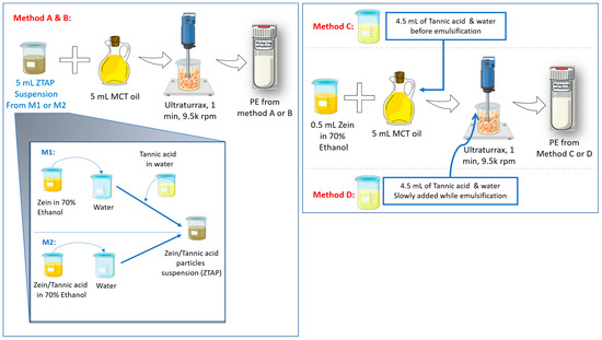
The zein/tannic acid nanoparticles were prepared using antisolvent precipitation methods (Figure 1, left panel), as previously described in the literature.
Figure 1.
Schematic illustrations of the two preparation methods for zein–tannic acid particles (ZTAPs). Sequential precipitation method 1 (M1) and co-precipitation method 2 (M2) and the four emulsification methods for oil-in-water Pickering emulsions (methods A–D).
Briefly, for the sequential precipitation method (method 1, ZTAPssp), stock solutions were prepared as follows: 0.30 g zein was dissolved in a 10 mL solution containing 70% ethanol and 30% water, with a concentration of 30 mg·mL−1 (1.25 mM), and 0.17 g tannic acid in 10 mL water at 17.5 mg·mL−1 (10 mM), under magnetic stirring (600 rpm) for 30 min. Next, 1 mL of zein stock solution was quickly added to 6 mL of water to form zein colloidal particles. The appropriate volumes of tannic acid stock solution were then successively added and completed with water to achieve a final volume of 10 mL of the ZTAPsp suspensions with final zein concentration (0.1 mM) and final tannic acid concentrations (0, 0.01, 0.1, 1 or 3 mM). In this way, particles with a zein/tannic acid ratio R of 1/0, 1/0.1, 1/1, 1/10 and 1/30 (M/M) were obtained.
For the co-precipitation method (method 2, ZTAPscp), zein and tannic acid were dissolved together in the same 70% ethanol/30% water solution, and then 1 mL of this composite solution was quickly added to 9 mL of water under magnetic stirring (600 rpm) to obtain the ZTAPscp suspension within the same range of concentration of Z/tannic acid R of 1/0, 1/0.1, 1/1, 1/10, 1/30.
Prior to selecting this range, we tested a variety of concentration conditions by either fixing the molar ratio and varying the concentrations or by exploring a broader concentration range. This preliminary optimization helped determine that the selected range R from 1/0 to 1/30 was the most suitable for observing reproducible and significant differences in nanoparticle characteristics and biological activity. The impact of pH on zein/tannic acid particles’ properties was previously studied and is therefore not within the scope of this work [26,32]. As pH values after particle preparation were slightly variable according to the concentrations of tannic acid, the final pH of particle suspensions was always adjusted using 0.1 M hydrochloric acid to 3.7, which exhibited the best particle stability.
ZTAPs were dried for further analysis (e.g., FTIR, NMR) by solvent evaporation using a rotary evaporator (The Rotavapor® R-300, BUCHI SARL, Villebon-sur-Yvette, France). The aqueous particle suspension was subjected to vacuum evaporation at 35 mbar at 30 °C for one hour to gently dry the particles without damaging zein.
2.2. Particle Characterization
2.2.1. Particle Size and Zeta Potential
The particle suspension was diluted with water by a factor of 1:10 (v/v). Subsequently, the diluted suspension was transferred to a 4 mL disposable cuvette for size measurement. A Dynamic Light Scattering (DLS) apparatus was employed to determine the particle size. For each sample, the instrument performed three successive measurements per run, and all measurements were conducted in triplicate using independently prepared samples. Reported values include the Z-average diameter (nm) and polydispersity index (PDI) with the Malvern Zeta Sizer PRO Blue label. A refractive index of 1.45 for colloidal particles (protein) and 1.33 for the dispersant (water) was applied. For the zeta potential measurement, the suspension was transferred to a zeta cuvette, and the zeta potential (mV) was evaluated with the same apparatus.
2.2.2. Transmission Electron Microscopy (TEM) Analysis and Confocal Laser Scanning Microscope (CLSM)
The morphology of ZTAPs was investigated using a transmission electron microscope; 5 µL of the sample was applied to copper grids for 1 min. After two washes with water, the coloration was performed with 8 µL of 2% phosphotungstic acid for 30 s, and then blotted. Imaging was performed at 200 kV using a Glacios 2 transmission cryo-electron microscope (Thermo Fisher Scientific, Waltham, MA, USA) equipped with a CETA-D CMOS detector. Data were acquired using the EPU software version 3.6.0.6389 (Thermo Fisher Scientific). ZTAPs were also observed using a Zeiss LSM 980 confocal laser scanning microscope (CLSM) (Carl Zeiss Microscopy GmbH, Jena, Germany) with an oil immersion objective Plan-Apochromat 63w/1.4 oil (scaling per pixel 5.6 nm × 5.6 nm × 0.2 µm). Then, 20 μL of (0.1%, m/v) Nile blue solution was added to 1 mL of the ZTAP suspension in order to stain zein. Nile blue was excited at 639 nm with 10% of power laser and detected with the Airyscan detector in mode 4Y. Images were processed using ImageJ 1.54p (NIH, Bethesda, MD, USA).
2.2.3. Fourier-Transform Infrared Spectroscopy Analysis
FTIR was used to investigate the interactions between zein and tannic acid in the ZTAP complexes using a PerkinElmer Spectrum IR (version 10.6.2). Spectra were recorded in the range of 4000–500 cm−1. Dried zein particles and ZTAPs (R 1/10 and 1/30), along with the powders of zein and tannic acid and their physical mixture, were analyzed in a solid state, and characteristic absorption bands were interpreted to assess hydrogen bonding and molecular interactions indicative of complex formation.
2.2.4. Solid-State 13C CP/MAS NMR Analysis
Solid-state 13C cross-polarization/magic angle spinning (CP/MAS) NMR spectroscopy was performed to investigate the molecular structure and interactions between zein and tannic acid within ZTAPs. A Bruker Avance III HD 300 MHz spectrometer equipped with a 4 mm triple-resonance probe was used. The cross-polarization pulse program was set with a CP contact time of 1 ms, a spinning frequency of 12,500 Hz, and a scan count of 4096. Heteronuclear decoupling was applied using SPINAL-64 at 80 kHz. Chemical shift referencing was carried out using an external standard (adamantane), and data were processed with Topspin 4.5.0 (Bruker, Karlsruhe, Germany).
2.2.5. Turbidity
The absorbance of the composite nanoparticle solution was measured at 600 nm using a Shimadzu UV-VIS spectrophotometer (UV-2600), and the light transmittance of the solution was used to represent the turbidity of the sample:
where T is light transmittance, and A is absorbance.
2.3. Evaluation of ZTAP Complexes as Potential Pickering Emulsion (PE) Stabilizer
2.3.1. Formulation of Emulsions
All emulsions were prepared at room temperature using an Ultra-Turrax® T25® homogenizer (Janke & Kunkel IKA-Labortechnik, Staufen, Germany) operating at 9500 rpm for one minute. During homogenization, the samples were kept in an ice bath to prevent thermal degradation. The emulsions were formulated with an MCT oil (oil phase) to be added to an aqueous phase containing ZTAPs or their precursor solutions, depending on the method used, at a ratio of 50:50. After preparation, the emulsions were stored at 4 °C in sealed containers for subsequent physicochemical characterization and stability assessment.
To investigate the influence of ZTAP formation and emulsification strategies on emulsion properties, four distinct formulation methods were employed. These methods are described schematically in the right part of Figure 1:
- Pre-formed ZTAPssp via sequential precipitation (method A):
In this approach, ZTAPssp were first prepared by sequential precipitation (method 1). These pre-formed ZTAPssp were then used as the aqueous phase for emulsification with MCT oil (5 mL each).
- ii.
- Pre-formed ZTAPscp via co-precipitation (method B):
Here, ZTAPscp were prepared via co-precipitation (method 2). These pre-formed ZTAPscp were then used as the aqueous phase for emulsification with MCT oil (5 mL each).
- iii.
- In situ complexation during emulsification (method C):
Rather than using pre-formed particles, in this method, 0.5 mL of zein alcoholic solution and 4.5 mL of tannic acid in water were employed as the aqueous phase. The two solutions were added at the same time to MCT oil, then emulsified into PEs.
- iv.
- Post-addition of tannic acid during emulsification (method D):
In this method, a 0.5 mL zein alcoholic solution was first mixed with 5 mL of MCT oil by gentle shaking. Then, the emulsification was started, and 4.5 mL of the aqueous tannic acid solution was added dropwise during homogenization at 9500 rpm for 1 min using the Ultra-Turrax® T25® homogenizer (Janke & Kunkel IKA-Labortechnik, Staufen, Germany). This setup was designed to assess the effect of delayed interaction between tannic acid and zein at the interface during dynamic emulsification.
The four methods were then systematically compared to assess the impact of the ZTAP formation pathway and timing on the droplet size, interfacial structure, stability, and antioxidant performance of the resulting Pickering emulsions.
2.3.2. Characterization of Emulsions
The formation of oil-in-water emulsions was systematically confirmed by mixing an oil phase containing Sudan red oily and by the ease of removal of the emulsions upon washing with water. A visual assessment was also carried out to detect creaming, modifications in color or viscosity, phase separation, or other macroscopic alterations (Figure S1, Supplementary File). Furthermore, the droplet size was measured by a laser diffraction analysis approach via a Master Sizer 2000 instrument (Malvern Panalytical, London, UK). These droplets were also visualized by optical microscopic evaluation (ECLIPSETi-S microscope, Nikon, Shinagawa, Japan).
2.3.3. Interfacial Structure
The microstructure of the emulsions was observed using a confocal laser scanning microscope (CLSM) Zeiss LSM 800 (Carl Zeiss Microscopy GmbH, Oberkochen, Germany) with an oil immersion objective (×25). 20 μL of a 0.1% (m/v) Nile blue solution was added to 1 mL of the emulsions to stain zein, and the mixture was homogenized gently. After staining, a small drop of the emulsion was placed on a glass slide and covered with a coverslip. Nile blue was excited at 633 nm. The scanning frequency was 100 Hz. Interfacial thickness was determined from CLSM micrographs by extracting fluorescence intensity profiles across the droplet interface using Zen Software version 3.8 (ZEISS). A straight, wide arrow was drawn perpendicular to the interfacial fluorescent ring at the diameter plane for each droplet, and the thickness was calculated as the full width at half maximum (FWHM) of the fluorescence peak. 3D structures were obtained with three stacks of images/droplets and processed using ImageJ 1.54p (NIH, USA). In preliminary experiments, 0.1% m/v of Nile red solution was also used to confirm that the type of emulsion formed was oil-in-water in all conditions (a representative example is shown in Figure S2, Supplementary File). Nile red was excited at 488 nm.
2.4. Antioxidant Activity
The antioxidant capacity of the samples was evaluated using the DPPH (2,2-diphenyl-1-picrylhydrazyl) radical scavenging assay. DPPH is a stable free radical that appears violet in solution and becomes yellow upon reduction by antioxidants. This change corresponds to a decrease in the degree of absorption, which was measured at a wavelength of 517 nm using an EL 800-microplate reader (Bio-TEK Instrument, Winooski, VT, USA).
A solution of 100 µM DPPH in absolute ethanol was freshly prepared and subsequently protected from light. 100 µL of test samples (after appropriate dilution with water) were mixed with 100 µL of the DPPH solution in a 96-well microplate. Trolox was used as a positive control and to build the standard curve (0 to 100 µM).
Following this, the samples were subjected to a 30 min incubation in the dark at room temperature. Thereafter, the absorbances of each well were recorded.
The percentage of DPPH scavenging was calculated using the following formula:
where (Ac) is the absorbance of the control, (As) is the absorbance of the sample, and (DF) is the dilution factor.
3. Results and Discussion
3.1. Particle Formation and Characterization
The employment of both the sequential (method 1) and co-precipitation (method 2) methods resulted in the production of ZTAPs with a size range of 100–250 nm (Table 1). The particle size and positive surface charge were found to be tunable depending on the zein/tannic acid ratio, a finding consistent with previous research on protein–polyphenol complexation. The zeta potential decreased slightly after tannic acid addition (~20 mV at the highest tannic acid concentrations, compared to ~35 mV initially), suggesting that tannic acid partially screened or masked the positive charges of zein and contributed to particle stabilization via non-electrostatic mechanisms, which likely play a dominant or overriding role in stabilization, compensating for the reduced electrostatic repulsion.
Table 1.
Dynamic light scattering (DLS) characterization of zein–tannic acid particles (ZTAPs) prepared by (A) sequential precipitation (method 1) and (B) co-precipitation (method 2) across a range of tannic acid concentrations (Z/TA ratios R = 1/0, 1/0.1, 1/1, 1/10, and 1/30). Measured parameters included hydrodynamic diameter (Z-average), polydispersity index (PDI), and zeta potential (ζ). Data are presented as mean ± standard deviation (n = 3). Statistical analysis was conducted using one-way ANOVA followed by Dunnett’s post hoc test; * = p < 0.05 versus the 1/0 group.
As tannic acid concentration increased, differences in particle behavior became more pronounced between the two methods. In method 1, ZTAPsp size remained constant across zein/tannic acid ratios (R), while the turbidity increased with R (Table S1, Supplementary File), suggesting an increase in density or scattering due to structural rearrangement or partial aggregation. In contrast, for method 2, ZTAPscp showed a clear increase in particle size and polydispersity beginning at R = 1/1, leading to the formation of larger, more clustered aggregates. This was accompanied by a more marked increase in turbidity. Notably, the zeta potential decreased significantly in both methods from R = 1/10, implying that this ratio may represent a critical point where the charge screening or interparticle interaction balance shifts.
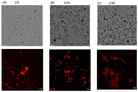
Further analysis using TEM confirmed that zein particles alone showed relatively uniform, discrete spherical morphology (Figure 2A, top row). ZTAPsp R = 1/10 (Figure 2B, top row) and ZTAPsp R = 1/30 (Figure 2C, top row) illustrated progressive clustering upon the addition of tannic acid, suggesting increased interparticle interactions mediated by tannic acid, presumably due to induced interparticle hydrogen bonding and hydrophobic interactions.
Figure 2.
Images of particles prepared with method 1. (A) Zein particles alone. (B) ZTAP 1/10 and (C) ZTAP 1/30. The top rows of each ratio show transmission electron microscopy (TEM) images; the bottom rows show corresponding confocal laser scanning microscopy (CLSM) images. Scale bars: 200 nm (upper panels) and 10 µm (lower panels).
These interactions are also highlighted in the CLSM images, where increased particle clustering was observed with increased tannic acid concentration. These morphological transitions likely arise from extensive intermolecular interactions, which are supported by turbidity measurements (Table S1, Supplementary File).
FTIR and solid-state NMR spectroscopy were employed to confirm intermolecular interactions, with a particular focus on zein/tannic acid R = 1/10 and 1/30.
FTIR analysis (Figure S3, Supplementary File) confirmed the formation of intermolecular interactions between zein and tannic acid in ZTAPs. These interactions were similar regardless of the method employed (1 or 2). Zein alone exhibited characteristic protein peaks, including a strong amide I band at 1643 cm−1 (C=O stretching), a clear amide II band at 1515 cm−1 (N–H bending/C–N stretching), and a broad absorption at 3287 cm−1 attributed to N–H and O–H stretching vibrations. Tannic acid displayed a broad O–H stretch around 3319 cm−1, multiple peaks in the 1600–1450 cm−1 range corresponding to aromatic C=C vibrations and ring deformations, and intense bands below 1300 cm−1, confirming its polyphenolic nature. In the ZTAP R = 1/10, the O–H/N–H stretching band shifted to ~3285 cm−1 with increased broadness, indicating the formation of new hydrogen bonds between tannic acid and zein. Concurrently, the amide I band slightly shifted to 1647 cm−1, and aromatic-associated peaks appeared at ~1530 and 1443 cm−1, supporting interactions between tannic acid’s aromatic rings and zein’s hydrophobic residues. Notably, in the ZTAP R = 1/30 spectrum, further broadening and a redshift of the O–H/N–H band to ~3280 cm−1 were observed, strongly suggesting intensified hydrogen bonding at higher tannic acid concentrations. New peaks at 1706 and ~1608 cm−1 emerged, which can be attributed to π–π stacking interactions between zein’s aromatic side chains and tannic acid’s phenolic structures. Additionally, a more pronounced shift and broadening in the amide I region (1649 cm−1) indicate enhanced cross-linking and stabilization of the complex network. Such binding induces conformational changes in zein, as reported in previous work, and tunes its surface wettability without significantly reducing net charge [33]. The findings of this study lend support to the hypothesis that tannic acid forms a hydrogen-bonded network, rendering the particles partially hydrophilic and structurally robust, as similarly observed in other tannic acid–biopolymer complexes [33,34].
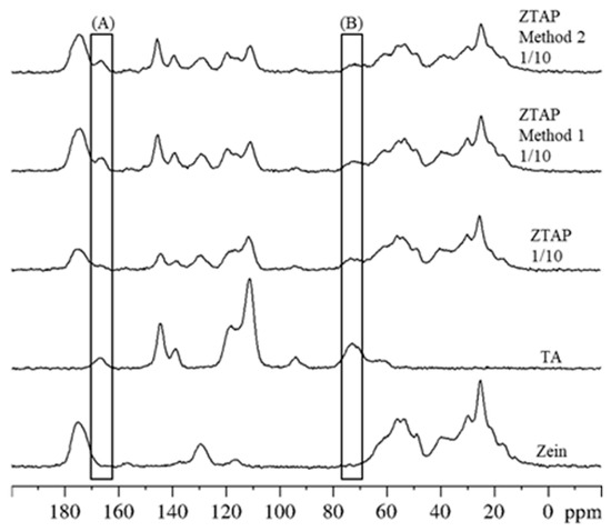
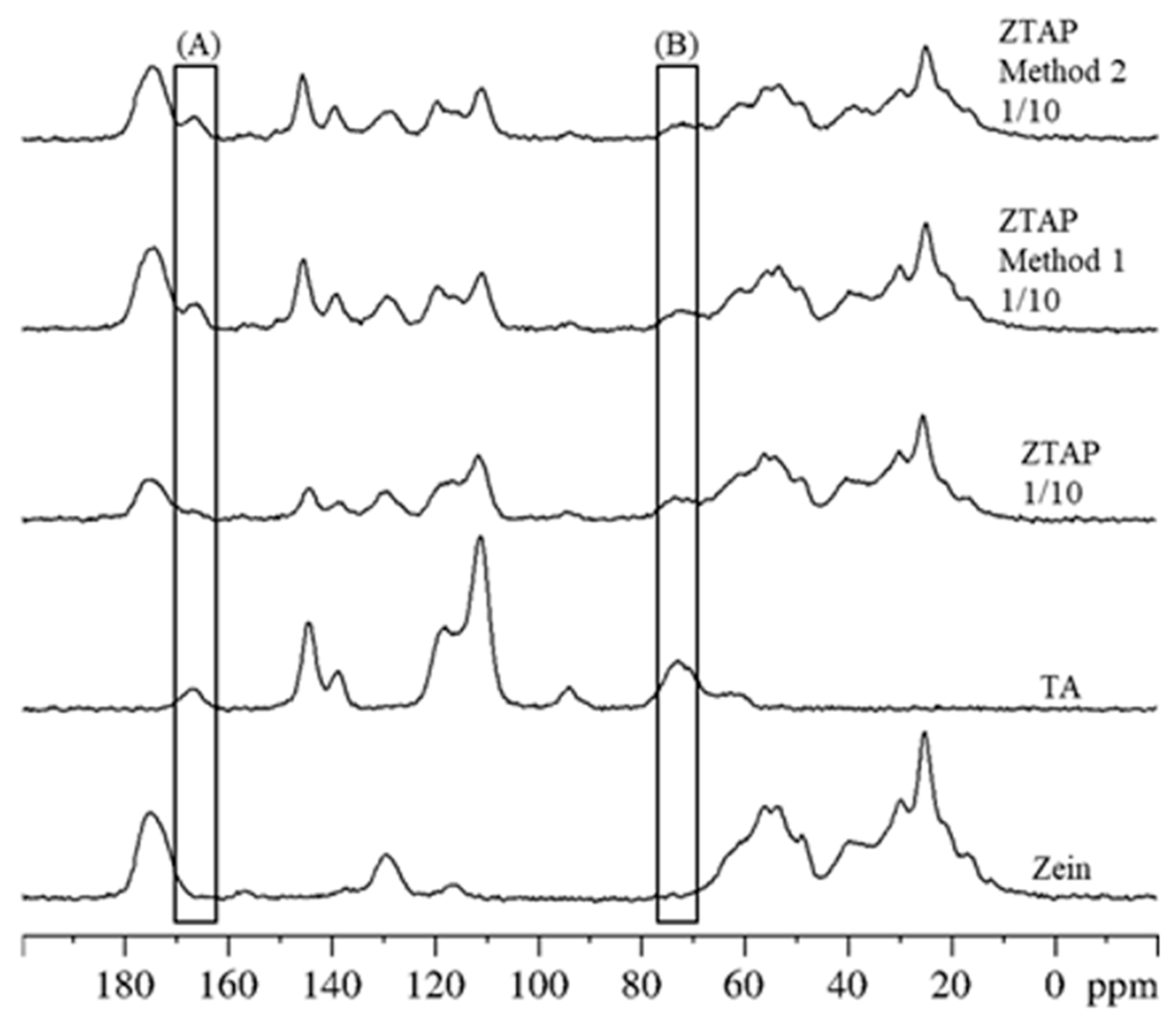
The 13C CP-MAS NMR spectra of tannic acid (TA) and zein showed the typical signals of each pure compound. For TA, the carbonyl (C=O) group appeared at 166 ppm (Peak A, Figure 3), while the central ring signal was at 73 ppm (Peak B). Zein’s carbonyl group signal was observable at 177 ppm. In the 1:10 zein/TA mixture (ZTAP), both components were clearly detected.
Figure 3.
13C CP-MAS NMR spectra of tannic acid (TA), zein, zein–tannic acid physical mixture (ZTAP 1/10), zein–tannic acid particles prepared by method 1 (ZTAP method 1 1/10) and method 2 (ZTAP method 2 1/10). The peak labeled A appears at 166 ppm and the one labeled B at 73 ppm.
The spectra of particles prepared by methods 1 and 2 were identical, with TA signals still visible. However, the intensity of the TA C=O signal (Peak A) was lower in the simple physical mixture than in particles from methods 1 and 2, while the Peak B intensity remained constant. This suggests an interaction, likely hydrogen bonding, that affects the carbonyl groups’ chemical environment for ZTAPs obtained by methods 1 and 2 but is absent in the simple physical mixture. Several 13C CP-MAS experiments with CP contact times varying from 5 µs to 1.4 ms showed the same variations in the intensity of the C=O groups (Figure S4, Supplementary File).
Ultimately, differences in particle structure, zein/tannic acid ratio, and even the method of preparation could translate into differences in emulsification performance. The particles are formed in an aqueous medium. Various tests we conducted (drying under different conditions, lyophilization, etc.) demonstrated that they cannot be isolated in sufficient quantities or without altering their properties in a form suitable for conventional contact angle measurements (which typically require planar solid surfaces or deposited/dried particles). Thus, it is experimentally infeasible to determine a triphasic contact angle in our system. Consequently, we relied on indirect approaches—such as droplet size, stability, and microscopic observations—which nonetheless effectively assess the particles’ stabilizing capacity.
3.2. Emulsion Stability and Interfacial Properties
Four emulsification methods were evaluated to produce concentrated oil-in-water emulsions (50% v/v), either by using zein/tannic acid particles pre-formed before emulsification (methods A and B) or by bringing them into contact immediately before emulsification (method C) or during emulsification (method D).
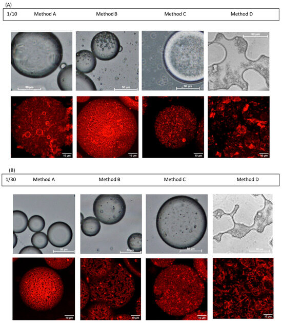
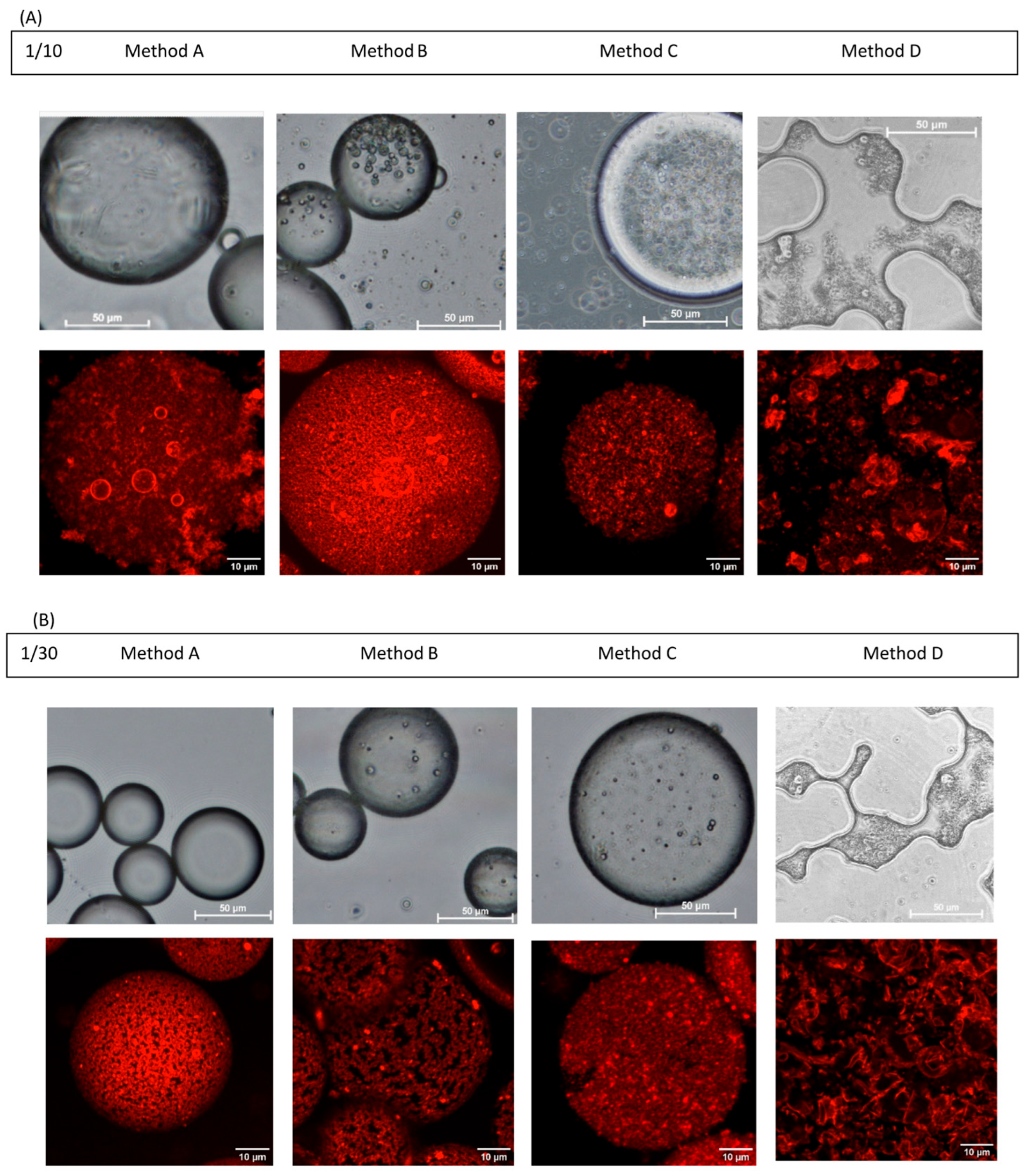
In method D, zein in alcohol was first brought into contact with the oil and emulsified before tannic acid was gradually added. Under these conditions, the zein, once wetted by the oil, was no longer readily accessible to tannic acid modification. Regardless of the proportion of tannic acid, this process failed to generate emulsions (Figure 4).
Figure 4.
Microscopic visualization of Pickering emulsions stabilized by ZTAP (methods A and B) or by zein/tannic acid aqueous solutions (methods C and D) at zein/tannic acid molar ratios of 1/10 (A) and 1/30 (B). The top rows of each ratio show optical microscopy images; the bottom rows show corresponding confocal laser scanning microscopy (CLSM) images. Zein adsorbed at the oil–water interface was stained with Nile blue, enabling fluorescence visualization (red signal) of particle distribution around the droplets. Scale bars: 50 µm (optical) and 10 µm (CLSM).
In the other methods, zein/tannic acid interactions in water were established before emulsification began, with outcomes depending on tannic acid inputs. For emulsions stabilized with zein alone (R = 1/0), regardless of the method used, destabilization and phase separation were consistently observed within two hours. Careful observation of the formulations with R = 1/0.1 revealed a thin upper layer of oil after preparation (qualitative observation, visible in Figure S1, Supplementary File), showing that the tannic acid amount added was not sufficient to perfectly stabilize an emulsion with such a high oil content. In contrast, formulations with higher tannic acid proportions creamed reversibly and displayed consistent appearances over time.
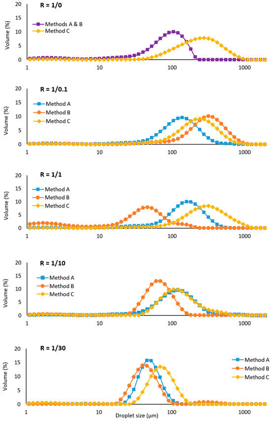
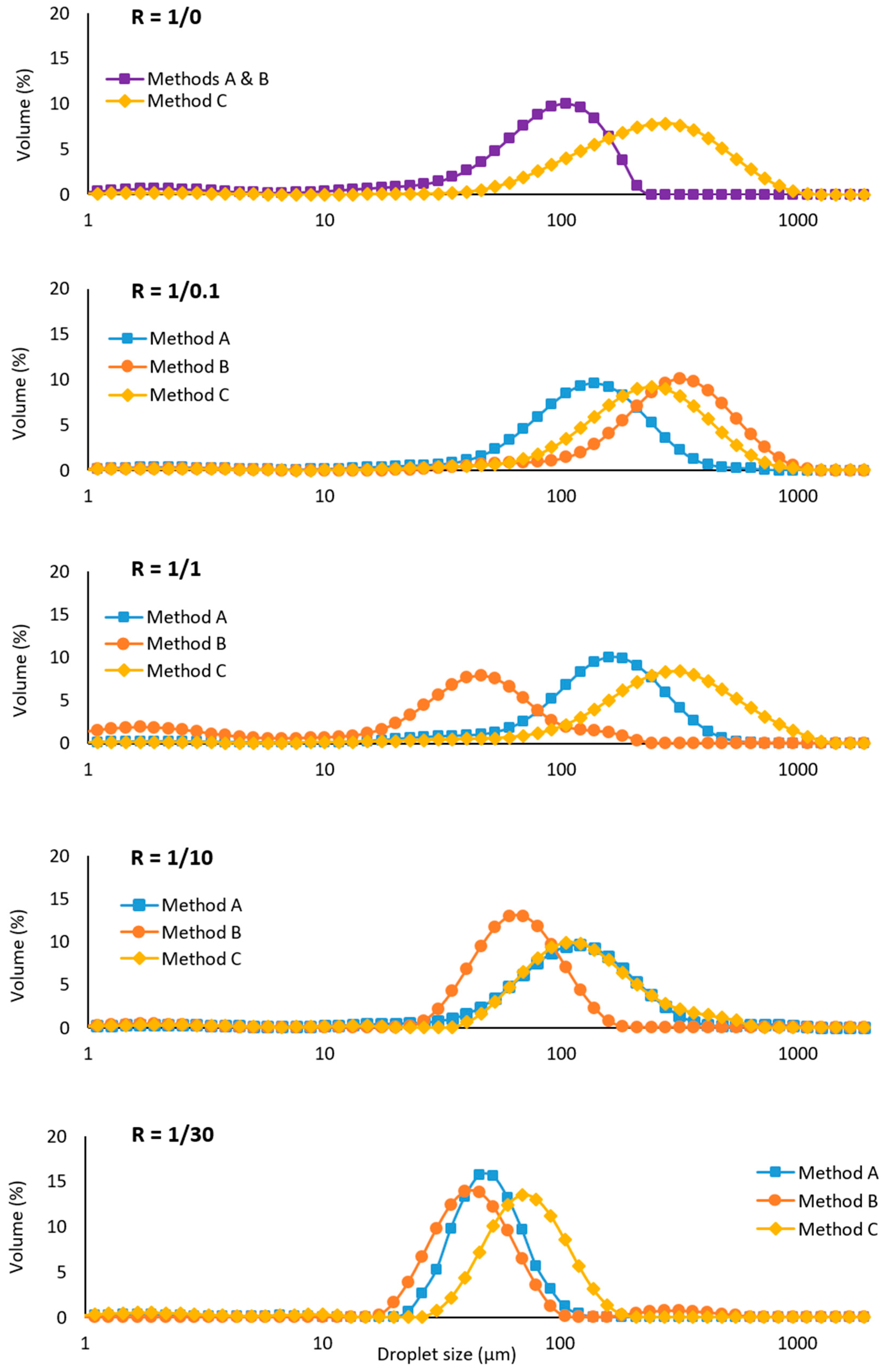
Emulsions prepared using pre-formed particles (methods A and B) had significantly smaller and more uniform droplet sizes, with method B generating the smallest and least dispersed droplets starting at R = 1 (Figure 5). Conversely, method C tended to produce bigger droplets with broader distributions. This finding suggests that the utilization of pre-formed ZTAPs facilitates more efficient PEs stabilization. At R = 1/30, the influence of the method on droplet size (~50 µm) became negligible, suggesting that a threshold had been reached beyond which the amount of tannic acid was the dominant parameter governing droplet size.
Figure 5.
Droplet size distribution (volume-based) of Pickering emulsions stabilized by zein/tannic acid particles or aqueous solutions prepared by the different methods (A–C), at various zein/tannic acid molar ratios R from 1/0 (zein only) to 1/30. Emulsions were analyzed by laser diffraction directly after preparation, and the droplet size is plotted on a logarithmic scale.
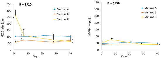
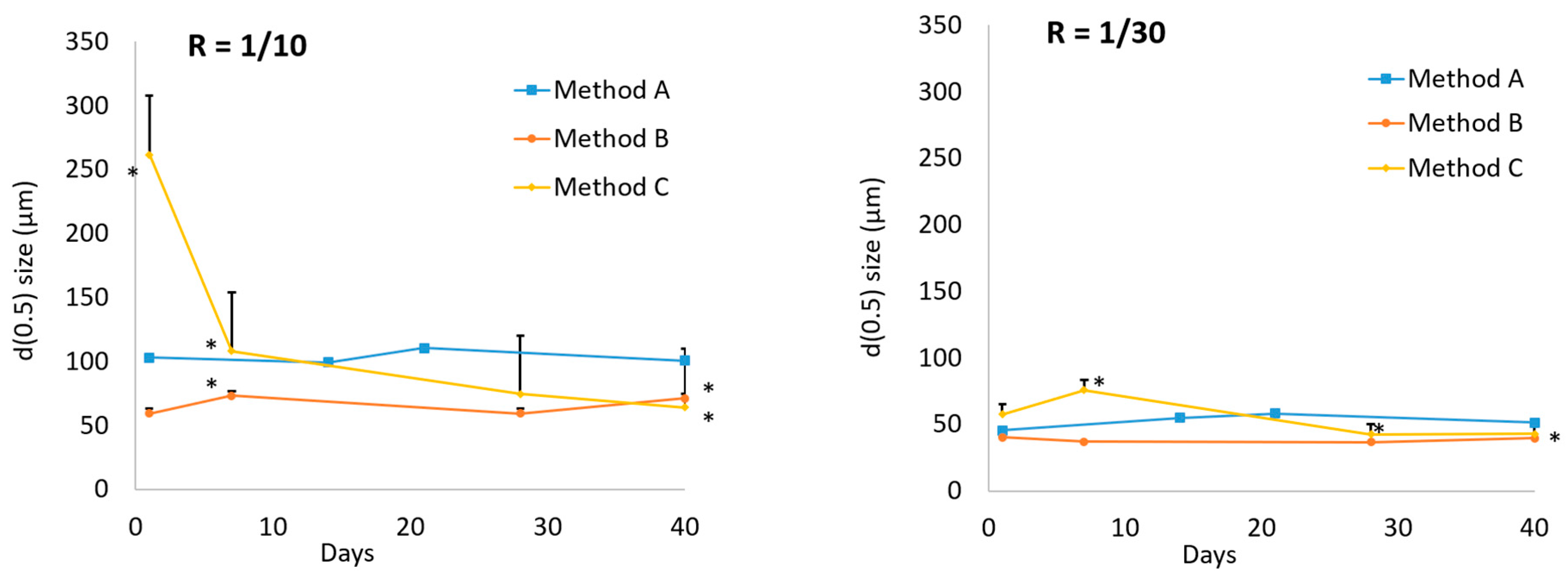
The observed variations in droplet size and homogeneity did not compromise the temporal stability of the emulsions, as shown by the monitoring of median droplet diameter (D(0.5)) over time in Figure 6. It is worth noting that these observations primarily reflect thermodynamic stability (i.e., resistance to coalescence), while gravitational stability (e.g., creaming) was also visually monitored. In this respect, formulations from method A showed minimal creaming over 40 days, supporting both structural and gravitational stabilization.
Figure 6.
Stability of Pickering emulsions over time, assessed by monitoring the median droplet diameter (D(0.5)) over a 40-day period. Emulsions were prepared using different methods at zein/TA molar ratios of 1/10 and 1/30. Data are presented as mean ± standard deviation (n = 3). Statistical comparisons were performed using one-way ANOVA followed by Dunnett’s post hoc test, * = p < 0.05 versus Day 1.
CLSM observations (Figure 4) gave a more in-depth understanding of the existence of method-dependent disparities in interfacial architecture. Methods A and B yielded droplets surrounded by nearly continuous ZTAP shells, while method C showed patchy or irregular coverage. Unlike in studies with other co-stabilizers [35,36], with tannic acid, the protein appeared to be completely located at the interface, as there was no visible signal in the continuous phase. It is also noteworthy that interfacial thickness varied across both emulsification methods and zein/tannic acid ratios. Method A emulsions exhibited a densely packed particle assembly as interfacial layers with thicknesses of approximately 1.7 ± 0.2 µm (R = 1:30) and 3 ± 0.5 µm (R = 1:10), a phenomenon that may be attributed to an increased particle demand at lower TA ratios. Method B resulted in irregular interphases (~2 ± 0.5 µm), while method C produced thinner, heterogeneous layers (~1.5 ± 1.2 µm). These disparities in interfacial structure are hypothesized to have contributed to the observed variations in emulsion characteristics. The interfacial thicknesses were much larger than the particle size (115–250 nm), suggesting that even when ZTAPs are pre-formed, larger complexes are formed during the emulsification to stabilize the interface.
It has already been demonstrated in the literature that tannic acid enhances the amphiphilicity of ZTAPs, thereby promoting rapid adsorption at the oil–water interface [33]. This, in turn, facilitates the formation of stable emulsions. This finding is consistent with the conclusions of previous studies, including those by Zhang et al., which reported that optimal zein/tannic acid ratios resulted in the formation of dense, viscoelastic interfacial films, thereby enhancing emulsion stability and regulating oil release [5]. The tannic acid present in the particles likely promotes cohesion between particles by enabling residual groups to bond neighboring particles. This trend was indeed observed by TEM and FTIR analysis. The formation of a rigid interfacial film has been demonstrated to enhance the mechanical strength of the material, thereby allowing it to withstand the deformation of droplets and to prevent coalescence. Furthermore, the viscoelastic nature of the interface has been demonstrated to inhibit destabilization mechanisms such as Ostwald ripening and flocculation [5,14,37]. In method C, where zein in alcohol and tannic acid were added to the oil just before the onset of emulsification, the organization of the system was not optimal. This led to droplets with a broad size distribution and relatively large diameters. However, increasing the proportion of tannic acid (from R = 1/10 to R = 1/30) made it possible to reduce the droplet size to around 100 µm. The interfacial thickness varied greatly, demonstrating irregular interactions between zein and tannic acid under these conditions. In methods A and B, particles were pre-formed prior to emulsification and acted as “seeds” for the interface. The interfaces observed were therefore more regular in appearance and thickness. Particles obtained by sequential precipitation (method 1, used for emulsions prepared by method A) likely exhibited a core–shell organization, with zein in the core and tannic acid at the surface. Particles obtained by co-precipitation (method 2, used for emulsions prepared by method B) displayed the same types of zein/TA interactions; however, these interactions appeared to be facilitated at higher tannic acid concentrations, as reflected by the increase in particle size and polydispersity, indicating a tendency of method 2 particles to aggregate. A greater involvement of tannic acid at the interface would explain why method B emulsions exhibited droplet sizes lower than 100 µm as early as R = 1/10, whereas R = 1/30 was required for method A emulsions.
In conclusion, it appears essential to pre-form the particles to achieve better control over the size and uniformity of emulsion droplets, with particles obtained by co-precipitation (method B) providing the best control of these characteristics. Tannic acid also plays a key role, as it is essential for emulsion formation, and increasing its proportion reduces droplet size and improves emulsion homogeneity. Unlike in previous work [14], in our study, no impact on the temporal stability of the emulsions was observed. Nevertheless, studies over longer durations and under a wider range of conditions, carefully tailored to the intended applications and encapsulated ingredients, should be conducted in the future.
3.3. Antioxidant Performance and Functional Potential
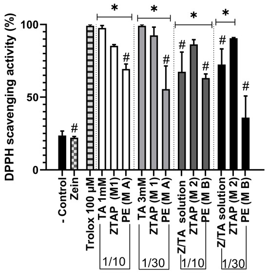
Tannic acid is a widely occurring polyphenol characterized by numerous phenolic hydroxyl groups, which give it distinct chemical properties and biological activities. When tannic acid interacts with proteins, it modifies the proteins’ secondary structure and markedly improves the antioxidant capacity and stability of the resulting particles [38]. Indeed, the low antioxidant activity of zein when used alone was overcome by the incorporation of tannic acid, which introduced strong radical scavenging properties [31]. The DPPH assay was conducted in order to ascertain the extent of scavenging efficiency of ZTAPs prepared by methods 1 and 2 at R = 1/30 and 1/10.
The results obtained demonstrated that these ZTAPs achieved approximately 70% scavenging efficiency, which was comparable to the Trolox (100 µM) positive control (Figure 7).
Figure 7.
DPPH radical-scavenging activity of ZTAPs prepared using method 1 (M1) and method 2 (M2), compared with free zein, tannic acid (TA), and Pickering emulsions (PEs) stabilized by ZTAPs from corresponding method A (M A) and method B (M B). Samples were tested at zein/TA molar ratios of 1/10 and 1/30. Bars represent mean ± standard deviation (n = 3). The negative control (−) corresponds to solvent only; Trolox (100 µM) served as the positive antioxidant reference. The values were normalized to the positive control, which was defined as 100%. Statistical analysis was performed using one-way ANOVA followed by Tukey’s post hoc test. * = p < 0.05 versus negative control; # = p < 0.05 versus Trolox.
It is noteworthy that the emulsions themselves exhibited significant antioxidant activity at R equal to 1/10; however, it was lower than the corresponding particle suspensions. At R equal to 1/30, the antioxidant effect is even lower, especially for method B. This might be explained by the implication of tannic acid in the interface, stabilizing the droplets (increasing with R, as shown by the NMR results), thus decreasing its availability for the antioxidant effect. These findings are consistent with previous reports indicating enhanced antioxidant properties in protein–polyphenol complexes in comparison to native proteins [6]. As a result, smaller droplets could be obtained using method B (compared to method A), particularly when increasing the ZTAP R (see Figure 4 and Figure 5). However, this comes at a cost, i.e., a reduction in antioxidant properties. The choice should therefore be guided by the most critical property required for the intended application.
In addition to its stability and oxidation resistance, the ZTAP interfacial layer exhibits the potential for controlled release. As Zhang et al. demonstrated, the incorporation of a zein shell that had been cross-linked with tannic acid resulted in a reduction in the rate of oil diffusion, thereby ensuring the sustained release of the oil [9]. Second, zein is known to be relatively resistant to gastric digestion, so tannic acid-crosslinked zein particles are likely to survive the stomach and release payloads later in the gut. Liang et al. observed that most encapsulated tannic acid in zein–pectin particles was released only during the intestinal phase of simulated digestion [34]. Consequently, ZTAP-stabilized emulsions have the potential to function as oral delivery vehicles, safeguarding actives during transit through the stomach and facilitating their release in the small intestine. Our confocal results showed that we can control the thickness of the ZTAP interface through the emulsification protocol and particle preparation method, therefore offering potential modulation of drug release. It is evident that the inherent antimicrobial and anti-inflammatory properties of tannic acid contribute to an enhancement in shelf life and health benefits [5]. The natural origin and biocompatibility of zein and tannic acid align well with consumer demand for safe, environmentally friendly ingredients. This renders them highly suitable for use in functional foods, nutraceuticals, cosmetics, and oral drug delivery systems for sensitive active biomolecules.
4. Conclusions
The present study demonstrates that zein/tannic acid complexes are highly effective stabilizers for oil-in-water Pickering emulsions, while tannic acid not only enhances the interfacial behavior of zein particles but also confers substantial antioxidant capacity, thereby contributing to both emulsion stability and functional performance. To finely control emulsion properties, it is essential to prepare the particles prior to emulsification. The choice of particle preparation method (sequential or co-precipitation) also influences emulsion characteristics without affecting their stability over a 40-day storage period.
From a functional perspective, ZTAP-stabilized emulsions offer a clean-label, plant-based solution for encapsulating and protecting bioactive oils, antioxidants, or drugs. Their resistance to gastric degradation and potential for controlled release in the intestinal tract further extends their potential application to oral drug delivery systems, functional foods, and cosmetics. While ZTAP-stabilized emulsions offer enhanced stability, their clinical and industrial translation faces challenges due to high-energy emulsification demands. Specifically, Pickering emulsions require high-shear processing to achieve optimal particle arrangement at the interface. Future research should thus prioritize three key areas: (1) optimizing critical process parameters to facilitate industrial upscaling, (2) implementing large-scale ZTAP production, and (3) enhancing encapsulation efficiency for targeted actives while evaluating performance in a simulated physiological environment.
The biocompatibility, biodegradability, and multifunctionality of ZTAPs render them a promising platform for the next generation of sustainable emulsification technologies.
Supplementary Materials
The following supporting information can be downloaded at https://www.mdpi.com/article/10.3390/app16094399/s1, Figure S1: Visual assessment of emulsions aspect (prepared with Method A) as a function of zein/tannic acid molar ratio (R), on the day of preparation.; Figure S2: Representative confocal laser scanning microscopy (CLSM) picture of the emulsions. Zein adsorbed at the oil–water interface was stained with Nile blue, enabling fluorescence visualization (red signal) of particle distribution around the droplets, and oil was stained with Nile red enabling fluorescence visualization (green signal) of particle distribution around the droplets, Scale bar: 10 µm. Table S1: Turbidity values of zein–tannic acid Particles (ZTAPs) prepared by two methods, sequential precipitation (method 1) and co-precipitation (method 2), were measured at varying zein/TA ratios from 1/0 to 1/30. Turbidity was recorded at 600 nm, where higher turbidity indicates increased particle concentration or aggregation, influenced by both the precipitation of zein and tannic acid complexes and tannic acid content. Data represent the mean ± standard deviation of three independent experiments (n = 3). Statistical analysis was performed using two-way ANOVA in order to compare the different concentrations as well as the two different methods; Figure S3: Fourier-Transform Infrared (FTIR) spectra of zein particles (ZP), pure zein (Z), tannic acid (TA), and their physical mixture (Z/TA). Spectra for zein–tannic acid particles (ZTAPs) prepared by sequential precipitation (method 1) and co-precipitation (method 2) are shown at zein/TA ratios of 1/10 and 1/30. Characteristic absorption bands reveal molecular interactions between zein and TA. Variations in peak positions and intensities indicate differences in interaction profiles depending on the preparation method and TA content; Figure S4: 13C CPMAS spectra intensity as a function of contact time (CP build-up performed with contact times ranging from 5 μs to 1.4 ms) for Peak A and Peak B exhibit distinct behaviors. Specifically, the intensity of the tannic acid C=O signal at 166 ppm (Peak A) is lower in the simple zein-tannic acid physical mixture (ZTAP 1/10) compared to particles obtained via methods 1 and 2, while Peak B (central ring signal is at 73 ppm) intensity remains constant. These observations imply an interaction—likely hydrogen bonding—that influences the chemical environment of the carbonyl groups in ZTAPs derived from methods 1 and 2, an interaction absent in the simple physical mixture. The CP build-up curves for ZTA particles reveal proton proximity effects that modify the 13C magnetization of these carbonyl groups (TA), in contrast to the mixed state of Z and TA alone.
Author Contributions
Investigation: R.A., Y.F.d.C., L.M.B., T.C., Y.B. and F.N.; Formal Analysis: R.A., Y.B. and L.B.; Writing—Original Draft: R.A., Y.F.d.C. and L.M.B.; Validation: R.A., Y.F.d.C. and L.M.B.; Writing—Review and Editing: R.A., Y.F.d.C., L.M.B., T.C., Z.B., Y.B., F.N., L.B., V.S., T.R.-C. and M.P.; Methodology: T.C. and F.N.; Conceptualization: Z.B., L.B., V.S., T.R.-C. and M.P.; Resources: Y.B., F.N. and L.B.; Supervision: M.P.; Funding Acquisition: M.P. All authors have read and agreed to the published version of the manuscript.
Funding
This work was supported by “Lorraine Université d’Excellence”, part of the France 2030 Program, reference ANR-15-IDEX-04-LUE. The authors would like to thank the ORION program for its contribution to the funding of YF’s research internship, and the Emergence program from Université de Lorraine for its contribution to the funding of LB’s research internship. This work has benefited from a government grant managed by the Agence Nationale de la Recherche with the reference ANR-20-SFRI-0009.
Data Availability Statement
The raw data supporting the conclusions of this article will be made available by the authors on request.
Acknowledgments
The authors would also like to thank the Cryo-TEM core facility of the Biophysique & Biologie Structurale (B2S) platform from the Service Mutualisé de Plateformes (SMP) of the Université de Lorraine.
Conflicts of Interest
The authors declare that they have no known competing financial interests or personal relationships that could have appeared to influence the work reported in this paper.
Abbreviations
The following abbreviations are used in this manuscript:
| ZTAPs | Zein/tannic acid particles |
| ZTAPssp | Zein/tannic acid particles obtained by sequential precipitation |
| ZTAPscp | Zein/tannic acid particles obtained by co-precipitation |
| TA | Tannic acid |
| R | Zein/tannic acid molar ratio |
| Pes | Pickering emulsions |
| O/W | Oil-in-water |
| DLS | Dynamic light scattering |
| TEM | Transmission electron microscopy |
| CLSM | Confocal laser scanning microscopy |
| FTIR | Fourier-transform infrared spectroscopy |
| NMR | Nuclear magnetic resonance |
| CP/MAS | Cross-polarization/magic angle spinning |
| PDI | Polydispersity index |
| DPPH | 2,2-diphenyl-1-picrylhydrazyl |
| D(0.5) | Median droplet diameter |
| ζ | Zeta potential |
| GRAS | Generally recognized as safe |
| FDA | Food and Drug Administration |
| UV | Ultraviolet |
References
- Wang, W.; Milani, A.H.; Cui, Z.; Zhu, M.; Saunders, B.R. Pickering Emulsions Stabilized by pH-Responsive Microgels and Their Scalable Transformation to Robust Submicrometer Colloidoisomes with Selective Permeability. Langmuir 2017, 33, 8192–8200. [Google Scholar] [CrossRef] [Scilit]
- Costa, C.; Medronho, B.; Filipe, A.; Mira, I.; Lindman, B.; Edlund, H.; Norgren, M. Emulsion Formation and Stabilization by Biomolecules: The Leading Role of Cellulose. Polymers 2019, 11, 1570. [Google Scholar] [CrossRef] [Scilit] [PubMed]
- Chen, L.; Ao, F.; Ge, X.; Shen, W. Food-Grade Pickering Emulsions: Preparation, Stabilization and Applications. Molecules 2020, 25, 3202. [Google Scholar] [CrossRef] [Scilit]
- Calero, N.; Muñoz, J.; Cox, P.W.; Heuer, A.; Guerrero, A. Influence of Chitosan Concentration on the Stability, Microstructure and Rheological Properties of O/W Emulsions Formulated with High-Oleic Sunflower Oil and Potato Protein. Food Hydrocoll. 2013, 30, 152–162. [Google Scholar] [CrossRef] [Scilit]
- Fan, S.; Yang, Q.; Wang, D.; Zhu, C.; Wen, X.; Li, X.; Richel, A.; Fauconnier, M.-L.; Yang, W.; Hou, C.; et al. Zein and Tannic Acid Hybrid Particles Improving Physical Stability, Controlled Release Properties, and Antimicrobial Activity of Cinnamon Essential Oil Loaded Pickering Emulsions. Food Chem. 2024, 446, 138512. [Google Scholar] [CrossRef] [Scilit]
- Li, M.; Ritzoulis, C.; Du, Q.; Liu, Y.; Ding, Y.; Liu, W.; Liu, J. Recent Progress on Protein-Polyphenol Complexes: Effect on Stability and Nutrients Delivery of Oil-in-Water Emulsion System. Front. Nutr. 2021, 8, 765589. [Google Scholar] [CrossRef] [Scilit] [PubMed]
- Zhao, Z.; Wang, W.; Xiao, J.; Chen, Y.; Cao, Y. Interfacial Engineering of Pickering Emulsion Co-Stabilized by Zein Nanoparticles and Tween 20: Effects of the Particle Size on the Interfacial Concentration of Gallic Acid and the Oxidative Stability. Nanomaterials 2020, 10, 1068. [Google Scholar] [CrossRef] [Scilit]
- Zou, Y.; van Baalen, C.; Yang, X.; Scholten, E. Tuning Hydrophobicity of Zein Nanoparticles to Control Rheological Behavior of Pickering Emulsions. Food Hydrocoll. 2018, 80, 130–140. [Google Scholar] [CrossRef] [Scilit]
- Zhang, Y.; Cui, L.; Che, X.; Zhang, H.; Shi, N.; Li, C.; Chen, Y.; Kong, W. Zein-Based Films and Their Usage for Controlled Delivery: Origin, Classes and Current Landscape. J. Control. Release 2015, 206, 206–219. [Google Scholar] [CrossRef] [Scilit]
- Bisharat, L.; Barker, S.A.; Narbad, A.; Craig, D.Q.M. In Vitro Drug Release from Acetylated High Amylose Starch-Zein Films for Oral Colon-Specific Drug Delivery. Int. J. Pharm. 2019, 556, 311–319. [Google Scholar] [CrossRef] [Scilit]
- Tihminlioglu, F.; Atik, İ.D.; Özen, B. Water Vapor and Oxygen-Barrier Performance of Corn–Zein Coated Polypropylene Films. J. Food Eng. 2010, 96, 342–347. [Google Scholar] [CrossRef] [Scilit]
- Pascoli, M.; de Lima, R.; Fraceto, L.F. Zein Nanoparticles and Strategies to Improve Colloidal Stability: A Mini-Review. Front. Chem. 2018, 6, 6. [Google Scholar] [CrossRef] [Scilit]
- Zhang, Y.; Huang, J.; Ren, F.; Li, Y.; Tong, Y.; Wen, P.; Wang, P. A Novel Method for Stabilizing Zein Gel Particles to Salt Ion-Induced Aggregation. Molecules 2021, 26, 1458. [Google Scholar] [CrossRef] [Scilit]
- Li, B.; Wang, X.; Gao, J.; Liang, H.; Wu, D.; Chu, S.; Zhu, X.; Zhou, B. Effect of Tannic Acid Modification on the Interface and Emulsification Properties of Zein Colloidal Particles. J. Sci. Food Agric. 2024, 104, 643–654. [Google Scholar] [CrossRef] [Scilit]
- Mallikarachchi, S.Y.; Rotich, N.C.; Gordon, E.; Hagerman, A.E. Different Chain Length Tannic Acid Preparations as Coating Agents for Zein Nanoparticles. Compounds 2024, 4, 401–414. [Google Scholar] [CrossRef] [Scilit]
- Rashidipour, M.; Abbaszadeh, S.; Birjandi, M.; Pajouhi, N.; Ahmadi Somaghian, S.; Goudarzi, G.; Shahryarhesami, S.; Moradi Sarabi, M.; Babaeenezhad, E. Antimicrobial Activity and Cytotoxic and Epigenetic Effects of Tannic Acid-Loaded Chitosan Nanoparticles. Sci. Rep. 2024, 14, 30405. [Google Scholar] [CrossRef] [Scilit] [PubMed]
- Youness, R.A.; Kamel, R.; Elkasabgy, N.A.; Shao, P.; Farag, M.A. Recent Advances in Tannic Acid (Gallotannin) Anticancer Activities and Drug Delivery Systems for Efficacy Improvement; A Comprehensive Review. Molecules 2021, 26, 1486. [Google Scholar] [CrossRef] [Scilit] [PubMed]
- Kaczmarek, B. Tannic Acid with Antiviral and Antibacterial Activity as A Promising Component of Biomaterials—A Minireview. Materials 2020, 13, 3224. [Google Scholar] [CrossRef] [Scilit]
- Baldwin, A.; Booth, B.W. Biomedical Applications of Tannic Acid. J. Biomater. Appl. 2022, 36, 1503–1523. [Google Scholar] [CrossRef] [Scilit]
- FDA. Code of Federal Regulations Title 21. GRAS Status of Tannic Acid. Federal Register; FDA: Silver Spring, MD, USA, 2022.
- Hassan, M.M. Enhanced Insect-Resistance, UV Protection, and Antibacterial and Antistatic Properties Exhibited by Wool Fabric Treated with Polyphenols Extracted from Mango Seed Kernel and Feijoa Peel. RSC Adv. 2021, 11, 1482–1492. [Google Scholar] [CrossRef] [Scilit]
- Chou, W.-W.; Wang, Y.-S.; Chen, K.-C.; Wu, J.-M.; Liang, C.-L.; Juo, S.-H.H. Tannic Acid Suppresses Ultraviolet B-Induced Inflammatory Signaling and Complement Factor B on Human Retinal Pigment Epithelial Cells. Cell. Immunol. 2012, 273, 79–84. [Google Scholar] [CrossRef] [Scilit]
- Jing, W.; Xiaolan, C.; Yu, C.; Feng, Q.; Haifeng, Y. Pharmacological Effects and Mechanisms of Tannic Acid. Biomed. Pharmacother. 2022, 154, 113561. [Google Scholar] [CrossRef] [Scilit]
- Wu, Y.; Zhong, L.; Yu, Z.; Qi, J. Anti-Neuroinflammatory Effects of Tannic Acid against Lipopolysaccharide-Induced BV2 Microglial Cells via Inhibition of NF-ΚB Activation. Drug Dev. Res. 2019, 80, 262–268. [Google Scholar] [CrossRef] [Scilit]
- Hennebelle, M.; Villeneuve, P.; Durand, E.; Lecomte, J.; van Duynhoven, J.; Meynier, A.; Yesiltas, B.; Jacobsen, C.; Berton-Carabin, C. Lipid Oxidation in Emulsions: New Insights from the Past Two Decades. Prog. Lipid Res. 2024, 94, 101275. [Google Scholar] [CrossRef] [Scilit]
- de Folter, J.W.J.; van Ruijven, M.W.M.; Velikov, K.P. Oil-in-Water Pickering Emulsions Stabilized by Colloidal Particles from the Water-Insoluble Protein Zein. Soft Matter 2012, 8, 6807–6815. [Google Scholar] [CrossRef] [Scilit]
- Liu, X.; Xie, F.; Zhou, J.; He, J.; Din, Z.; Cheng, S.; Cai, J. High internal phase Pickering emulsion stabilized by zein-tannic acid-sodium alginate complexes: β-Carotene loading and 3D printing. Food Hydrocoll. 2023, 142, 108762. [Google Scholar] [CrossRef] [Scilit]
- Fan, S.; Yang, Q.; Zhu, C.; Li, X.; Richel, A.; Fauconnier, M.-L.; Fang, F.; Zhang, D.; Hou, C. Zein/chitosan Janus film incorporated with tannic acid and cinnamon essential oil co-loaded Pickering emulsion for sustained controlled release and pork preservation. Int. J. Biol. Macromol. 2025, 286, 138429. [Google Scholar] [CrossRef] [Scilit]
- Jin, H.; Li, C.; Sun, Y.; Zhao, B.; Li, Y. Preparation and application of high internal phase pickering emulsion gels stabilized by starch nanocrystal/tannic acid complex particles. Gels 2024, 10, 335. [Google Scholar] [CrossRef] [Scilit]
- Yilmaz, H.; Gultekin Subasi, B.; Celebioglu, H.U.; Ozdal, T.; Capanoglu, E. Chemistry of Protein-Phenolic Interactions Toward the Microbiota and Microbial Infections. Front. Nutr. 2022, 9, 914118. [Google Scholar] [CrossRef] [Scilit]
- Zhou, Y.; Xu, Y.; Wang, P.; Zhang, J.; Chen, S.; Peng, D.; Wang, Z.; Yang, Y.; Luo, H.; Zhang, J. Modification of Pickering Emulsions Using Zein-Tannic Acid Particles: Enhancement of Antioxidant and Thermal Stability Properties of citral. LWT 2025, 223, 117761. [Google Scholar] [CrossRef] [Scilit]
- Zou, Y.; Yang, X.; Scholten, E. Tuning particle properties to control rheological behavior of high internal phase emulsion gels stabilized by zein/tannic acid complex particles. Food Hydrocoll. 2019, 89, 163–170. [Google Scholar] [CrossRef] [Scilit]
- Zou, Y.; Guo, J.; Yin, S.-W.; Wang, J.-M.; Yang, X.-Q. Pickering Emulsion Gels Prepared by Hydrogen-Bonded Zein/Tannic Acid Complex Colloidal Particles. J. Agric. Food Chem. 2015, 63, 7405–7414. [Google Scholar] [CrossRef] [Scilit]
- Liang, X.; Cao, K.; Li, W.; Li, X.; McClements, D.J.; Hu, K. Tannic Acid-Fortified Zein-Pectin Nanoparticles: Stability, Properties, Antioxidant Activity, and in Vitro Digestion. Food Res. Int. 2021, 145, 110425. [Google Scholar] [CrossRef] [Scilit]
- Cui, S.; McClements, D.J.; He, X.; Xu, X.; Tan, F.; Yang, D.; Sun, Q.; Dai, L. Interfacial properties and structure of Pickering emulsions co-stabilized by different charge emulsifiers and zein nanoparticles. Food Hydrocoll. 2024, 146B, 109285. [Google Scholar] [CrossRef] [Scilit]
- Wang, H.; Wei, Y.-L.; Liang, X.-Y.; Xu, M.-Z.; Chen, Q.-H.; Zeng, Q.-Z.; Yuan, Y. Novel bilayer Pickering emulsions stabilized by in situ modification of zein via selenium nanoparticles: Optimization, physicochemical properties and permeation. Food Hydrocoll. 2024, 156, 110323. [Google Scholar] [CrossRef] [Scilit]
- Dai, H.; Chen, Y.; Zhang, S.; Feng, X.; Cui, B.; Ma, L.; Zhang, Y. Enhanced Interface Properties and Stability of Lignocellulose Nanocrystals Stabilized Pickering Emulsions: The Leading Role of Tannic Acid. J. Agric. Food Chem. 2021, 69, 14650–14661. [Google Scholar] [CrossRef] [Scilit]
- Aelenei, N.; Popa, M.I.; Novac, O.; Lisa, G.; Balaita, L. Tannic Acid Incorporation in Chitosan-Based Microparticles and in Vitro Controlled Release. J. Mater. Sci. Mater. Med. 2009, 20, 1095–1102. [Google Scholar] [CrossRef] [Scilit]

Disclaimer/Publisher’s Note: The statements, opinions and data contained in all publications are solely those of the individual author(s) and contributor(s) and not of MDPI and/or the editor(s). MDPI and/or the editor(s) disclaim responsibility for any injury to people or property resulting from any ideas, methods, instructions or products referred to in the content. |
© 2026 by the authors. Licensee MDPI, Basel, Switzerland. This article is an open access article distributed under the terms and conditions of the Creative Commons Attribution (CC BY) license.