


Abstract
Cinnamomum zeylanicum (C. zeylanicum) is rich in bioactives, such as cinnamaldehyde and phenols, which are susceptible to thermal degradation, volatilisation, and oxidative deterioration during processing and storage, thereby reducing chemical stability and limiting bioavailability. Encapsulation using lecithin and chitosan-based systems mitigates these instabilities by forming a protective barrier against oxygen, light, and heat while enhancing structural stability. In this study, freeze-dried extracts of C. zeylanicum were encapsulated into lecithin-based primary liposomes (PL) and chitosan-coated secondary liposomes (CH/L). The coating of liposomes with chitosan improves the liposome stability, mucoadhesion, and provides protection in the gastric pH while facilitating electrostatic bonding with the biological membrane. The high compatibility and low toxicity of chitosan also make it a suitable carrier in food and nutraceutical applications. The formed liposomes were characterised for particle size, polydispersity index, zeta potential, encapsulation efficiency (EE), and storage stability over 8 weeks. CH/L showed superior EE (89.027%) compared to the PL (84.154%; p < 0.05). The particle size, polydispersity index, and zeta potential of the cinnamon-loaded lecithin-based primary liposome (CZ-PL) upon formation were 161.93 nm, 0.13, and −37.597 mV. In comparison, those of the cinnamon-loaded chitosan-coated liposomes (CZ-CH/L) were 591.7 nm, 0.27, and +28.17 mV. The particle size of CZ-PL and CZ-CH/L was 175.90 and 588.60 nm after 8 weeks of storage. The TEM confirmed the spherical morphology of the liposomes. The differential scanning calorimetry analysis demonstrated the disappearance of the characteristic cinnamon melting peak and shifts in liposomal transition temperatures, confirming successful encapsulation. FTIR analysis showed reduction or disappearance of characteristic cinnamon fingerprint peaks and slight band shifts, indicating successful encapsulation and non-covalent interactions, including hydrogen bonding and electrostatic effects, within the liposomal systems. These findings imply that lecithin-based and chitosan-coated liposomes could be employed to successfully carry C. zeylanicum bioactives.
1. Introduction
Ceylon cinnamon (Cinnamomum zeylanicum; C. zeylanicum) is a widely used spice and medicinal plant, with a long history of culinary and therapeutic applications. Its bioactive compounds, such as the phenols and cinnamaldehyde [1], exhibit diverse health benefits, such as antioxidant [2,3], anti-diabetic [4], anti-inflammatory [5,6] and potential anti-cancer effects [7], making them a potential functional food.
Both cinnamon essential oil (obtained by steam distillation of the bark and leaf) and aqueous extract (water extraction of the bark) are rich in bioactives. In this study, the aqueous extract of cinnamon was employed. Aqueous extract of cinnamon is highly compatible with a hydrophilic phase and most food matrices, facilitating easier incorporation into aqueous-based formulations, beverages, or functional foods without phase separation issues commonly seen with hydrophobic essential oils. Additionally, aqueous extraction methods minimise exposure to the more concentrated oil components, as they are not efficiently extracted into water [4]. Some studies have also shown that cinnamaldehyde in aqueous extracts has better cytotoxicity to cancerous cell lines than the purified cinnamaldehyde, as the effect is also due to the aqueous polyphenols in the water extract [8]. By using the aqueous extract, this study leverages the beneficial polyphenols while reducing risks associated with excessive volatile oil intake, aligning with safer profiles observed in many metabolic and functional food investigations.
However, these plant bioactives, particularly phenolic compounds, are highly unstable to light, heat, and alkaline conditions and can undergo oxidation and polymerisation [9,10]. During food processing and gastrointestinal digestion, they undergo structural transformations, which alter their stability, release, absorption, and metabolism. Factors such as poor aqueous stability [11], degradation in the gastrointestinal tract, and limited membrane permeability [12] can affect the physiological properties of food and bioavailability [13,14]. Phenols can also form reversible (non-covalent) and non-reversible (covalent) bonds with proteins and polysaccharides, causing changes in antioxidant ability, metabolic fate, and structure [15]. This results in bioavailability profiles that differ markedly from those of the native food compounds. Therefore, it is very important to develop delivery systems that can encapsulate phenols efficiently while addressing these challenges.
Encapsulation refers to the process of enclosing one substance within another to form particles that typically range in size from a few nanometres to several millimetres. The material being enclosed is commonly referred to as the core material, active compound, fill, internal phase, or payload, while the surrounding substance that provides protection is known as the coating, membrane, shell, carrier, wall material, external phase, or matrix [16]. In recent times, liposomal encapsulation has emerged as a highly promising tool in food applications. Liposomes can encapsulate sensitive vitamins, antioxidants, enzymes, and flavour compounds, shielding them from degradation triggered by light, oxygen, pH changes, or heat during food processing and storage. Their advantages include high encapsulation efficiency, improved solubility of bioactive compounds and food ingredients due to the amphiphilic bilayer, low toxicity derived from phospholipids, high drug loading capacity, prolonged circulation time, strong protection of sensitive ingredients from degradation factors such as pH, light, digestive enzymes, biocompatibility with the living system, and interactions at the cellular level [17,18,19]. Liposomes also offer surface modification by tuning their size and charge, which enables selective interaction with certain tissues [20].
However, conventional and unmodified liposomes are prone to aggregation and fusion, resulting in reduced physicochemical stability in aqueous suspensions during storage. They are also susceptible to leakage, oxidation, hydrolysis, rapid clearance from the bloodstream by the reticuloendothelial system, limited mucoadhesion for the mucosal drug delivery route, and degradation over time, leading to decreased storage stability and shorter shelf life [21,22,23,24].
These limitations can be overcome by modification of the liposome surface by coating with compatible polysaccharides such as chitosan, pectin, and sodium alginate [25]. Chitosan is a cationic polysaccharide that is highly biocompatible and biodegradable, and binds to liposomal surfaces by electrostatic forces and hydrogen bonding between the polar head groups of the phospholipids and the polysaccharides [26,27]. The free amino group of the chitosan can bind to the negatively charged surface of the liposome, making the surface charge positive. This causes greater repulsion between the liposomes, preventing aggregation and causing electrostatic stabilisation. Coating with chitosan also provides the system with enhanced mucoadhesive capacity by enabling electrostatic interaction with the negatively charged sialic acid and sulfate residue in the mucous layer. Chitosan also provides a higher circulation period by limiting immunogenic response and enabling controlled enzymatic reactions [24]. Though chitosan offers several benefits as a stabilising agent for liposomes [23,28,29], it also poses some challenges such as size expansion on coating, which may limit circulation half-life, difficulties with maintenance of the coating thickness during scaling up, and variation in physiochemical properties of chitosan, such as molecular weight and degree of deacetylation, which can influence zeta and particle size [24,30,31].
Several studies have explored the liposomal encapsulation of C. zeylanicum essential oil and Cinnamomum cassia, demonstrating the effects of chitosan coating on particle size, zeta potential, and encapsulation efficiency [32,33,34,35]. However, comparatively limited research has focused on the encapsulation of aqueous extracts of C. zeylanicum, which differ substantially from essential oils in their physicochemical composition, polarity, and distribution of bioactive compounds. Aqueous extracts are particularly relevant for food and nutraceutical applications due to their compatibility with hydrophilic systems.
Previous studies have reported encapsulation efficiency in terms of the essential oil [36] and the total phenolic encapsulated [37]. However, limited studies have simultaneously covered both the total phenolics and the cinnamaldehyde encapsulated, which are the important bioactives linked to the beneficial effects of cinnamon. Quantification of both these compounds will provide a more comprehensive assessment of encapsulation performance and bioactive retention for C. zeylanicum extracts. Therefore, in this study, we hypothesised that coating liposomes with chitosan could better encapsulate cinnamon bioactives, providing higher encapsulation efficiency (EE) than primary liposomes (PL) in terms of cinnamaldehyde content and total phenolic content (TPC). This study also aimed to evaluate and compare the physicochemical stability of PL and chitosan-coated liposome (CH/L) over the 8-week storage period by monitoring changes in particle size, polydispersity index (PDI), zeta potential, and overall structural integrity under defined storage conditions. It was hypothesised that chitosan coating would lead to better storage stability due to the electrostatic stabilisation. The outcomes provide preliminary insights into the potential suitability and compatibility of lecithin-based and CH/L systems as delivery carriers for bioactives from C. zeylanicum extracts, highlighting their possible application across diverse food systems such as dairy, beverages, meat, and baked products.
2. Materials and Methods
2.1. Materials and Chemicals
Cinnamomum zeylanicum (C. zeylanicum) barks were purchased from a local store (Palmerston North, New Zealand). Soya lecithin was purchased from Fearn Natural Foods (Mequon, WI, USA), low molecular weight chitosan (product no: 448869, Sigma-Aldrich Chemical Co., St. Louis, MO, USA) with a deacetylation ≥ 75% and viscosity 1% in 1% acetic acid, gallic acid, and Folin–Ciocâlteu were purchased from Sigma-Aldrich. Absolute ethanol was purchased from Supelco.
2.2. Preparation of C. zeylanicum Extracts
Dried bark of C. zeylanicum was first ground using an electric coffee grinder (Breville, Botany, Australia) to obtain a fine powder. The ground material was subsequently passed through a 250 µm stainless-steel sieve to ensure uniform particle size distribution. Hot water extraction was carried out by suspending the sieved bark powder in distilled water at a solid-to-liquid ratio of 1:40 (w/v). The mixture was incubated in a temperature-controlled hot water bath at 55 °C for a defined extraction period of 1 h to facilitate the release of bioactive compounds. Following extraction, the suspension was allowed to cool to room temperature, centrifuged for 10 min at 3000 rpm using a Beckman Coulter Avanti JXN-26 (Brea, CA, USA) centrifuge, and was then filtered by syringe filtration through a 0.45 µm membrane filter to remove insoluble particulates. The resulting filtrate was stored at −20 °C before freeze-drying.
2.3. Freeze-Drying Process
The pre-frozen aqueous C. zeylanicum bark extract solution was loaded at −20 °C and subsequently freeze-dried using a lyophiliser (Cuddon FD18, Blenheim, New Zealand) with the condenser setting at −40 °C. Drying was performed under a vacuum of approximately 1 mbar, and then the shelf temperature was adjusted to 20 °C to facilitate the sublimation process. Each freeze-drying cycle was conducted for approximately 72 h.
2.4. Liposomal Encapsulation of C. zeylanicum Extracts
2.4.1. Primary Liposome (PL)
The PL were prepared according to the method specified by Rashidinejad et al. [38] with some modifications. Briefly, soy lecithin was dispersed in 0.25 M acetate buffer at a concentration of 0.5% (w/v). The resulting system was stirred overnight at 100 rpm to enhance hydration of the lecithin. Subsequently, freeze-dried C. zeylanicum powder was introduced to the hydrated soy lecithin system at a concentration of 0.5% (w/v) and stirred for an additional 1 h to facilitate incorporation of the extract.
The resulting mixture was then homogenised using a high-shear homogeniser (T25 Ultra-Turrax, Janke and Kunkel, Staufen, Germany) at 24,000 rpm for 5 min. The homogenised suspension was further processed by microfluidisation (M-110P, Microfluidics, Newton, MA, USA) at a pressure of 200 MPa for four cycles to obtain uniformly sized C. zeylanicum loaded primary liposomes (CZ-PL) and empty liposomes (B-PL) without C. zeylanicum. The resulting microfluidised liposome suspension was used for subsequent characterisation and analysis.
2.4.2. Chitosan-Coated Liposome (CH/L)
A modified method from Mady et al. [39] was used to prepare CH/L. A chitosan solution (0.5% w/v in 0.25 M acetic acid) was added dropwise under continuous magnetic stirring to the prepared B-PL and CZ-PL suspensions, to obtain chitosan-coated liposomes without C. zeylanicum (B-CH/L) and with C. zeylanicum (CZ-CH/L). The mixture was maintained under stirring for 1 h. The resultant suspension was used in further analysis.
2.4.3. Determination of Total Phenol Content (TPC) of Free and Encapsulated Liposomes of C. zeylanicum Extract
The TPC of free extract and encapsulated liposomal formulations was determined using the Folin–Ciocâlteu colorimetric method as specified by Zhang et al. [40] with slight modification. Separate procedures were employed for PL and CH/L systems to enable effective separation of encapsulated and non-encapsulated phenolic compounds before analysis.
- Determination of TPC of Primary Liposomes
The encapsulated fraction, which is the liposomes containing the C. zeylanicum bioactives and phenols in PL, was separated from the unencapsulated fraction by gel filtration chromatography as specified by Rashidinejad, Birch, Sun-Waterhouse and Everett [38]. Briefly, a Sephadex gel column was prepared using Sephadex G-50 (5% w/w; Sigma–Aldrich) in deionised water. Around 5 mL of Sephadex suspension was placed in 6 mL syringes and was slightly swirled to expel excess water until a volume of approximately 3 mL of gel was obtained. Then, 1.5 mL of the acetate buffer was placed above the gel, and the syringe was introduced into a 50 mL centrifuge tube and centrifuged at 1207× g at 25 °C for 10 min using a bench-top centrifuge (HERAEUS, Multifuge X3R, Thermo Scientific, Illkirch, France) to equilibrate the gel. Following centrifugation, the 6 mL syringes were removed from the centrifuge tubes, and 1.5 mL of prepared PL was placed above the gel. Then the syringes were introduced to new centrifuge tubes, and the centrifugation step was repeated to obtain the gel-filtered liposomes containing the encapsulated fraction at the bottom of the tubes. The liposomes were stored at 4 °C in the dark in covered sample containers until further analysis.
To release the phenolic compounds present within the liposomes (encapsulated phenols), the collected liposomal fraction (1 mL) was disrupted by mixing with heated ethanol (2 mL) at 70 °C and vortexed for 1 min. The samples were then cooled to room temperature and centrifuged to remove lipid debris. The supernatant was used for TPC determination using the Folin-Ciocâlteu assay.
The TPC of the C. zeylanicum extract/disrupted PL/free unencapsulated CH/L was determined using the Folin-Ciocâlteu method. Briefly, 20 µL of the extract was mixed with 100 µL of 10% (v/v) Folin–Ciocâlteu reagent. Afterwards, 80 µL of 7.5% (w/v) sodium carbonate solution was added, and the reaction mixture was incubated at room temperature for 30 min to allow colour development. Absorbance was measured at 765 nm using a UV-Vis microplate reader (Synergy™ 2, BioTek® Instruments, Inc., Winooski, VT, USA). A gallic acid standard curve (7.8125–500 mg/mL) was used for quantification, and the results were expressed as milligrams of gallic acid equivalents (GAE).
- Determination of TPC of CH/L
For secondary liposome formulations, the free non-encapsulated fraction of the suspension were separated using centrifugal ultrafiltration according to the method specified by Azevedo et al. [41] with slight modification. The liposome mixture was transferred to an Amicon Ultra centrifugal filter unit (Millipore) with a molecular weight cut-off of 10 kDa and centrifuged at 14,000× g for 10 min. The collected filtrate, containing the unencapsulated phenolic compounds, was subsequently used for further analysis.
2.4.4. High-Performance Liquid Chromatography (HPLC)
A Dionex Ultimate 3000 high-performance liquid chromatography (HPLC) system fitted with a UV–Vis detector and a C18 reverse-phase column (5 μm particle size, 25 cm × 4.6 mm; Supelco Analytical, Bellefonte, PA, USA) was employed for the quantification of cinnamaldehyde at 287 nm. The method specified by Culas et al. [42] was followed. The mobile phase had formic acid/water (0.1%) as solvent A and acetonitrile as solvent B. The elution commenced with 5% B, which was increased to 100% over 20 min, followed by re-equilibration to 5% B at 24 min. The total analysis time was 29 min, with a flow rate of 1 mL/min, an injection volume of 10 µL, and the column temperature was controlled at 25 °C.
2.5. Determination of Encapsulation Efficiency
The encapsulation efficiency percentage for the PL and CH/L was calculated according to Equations (1) and (2), respectively.
where EE = encapsulation efficiency. TPCtot = total phenol content in the freeze-dried powder. TPClip = total phenol content in the primary liposome matrix (encapsulated). TPCfree = total free phenol content in the secondary liposome matrix.
2.6. Determination of Loading Capacity
The loading capacity of phenols was determined by Equation (3)
where, TPCtot = Total concentration of both encapsulated and free phenols. TPCfree = concentration of free phenols.
2.7. Characterisation of Liposomes
2.7.1. Zeta Potential, Particle Size, and Polydispersity Index (PDI)
The liposomal formulations were diluted 1:20 in 0.25 M acetate buffer, and their particle size, PDI, and zeta potential were monitored over 8 weeks (1st, 2nd, 3rd, 4th, and 8th). Measurements were performed using dynamic light scattering (DLS) on a Malvern Zetasizer Nano ZS (Malvern Instruments Ltd., Malvern, Worcestershire, UK).
2.7.2. FTIR
ATR–FTIR spectra of the C. zeylanicum and liposome samples were obtained using a Thermo Scientific™ Nicolet™ iS5 FTIR spectrometer (Waltham, MA, USA) equipped with an iD7 diamond attenuated total reflectance (ATR) accessory. Spectral data were collected over the range of 4000–600 cm−1 using the OMNIC™ series software (version 9), with 32 scans collected for each sample after background subtraction.
2.7.3. Differential Scanning Calorimetry (DSC)
Differential scanning calorimetry (DSC) analysis was performed using a Q2000 differential scanning calorimeter (TA Instruments, New Castle, DE, USA) and RSC40 cooling system (TA Instruments, New Castle, DE, USA). Approximately 10 mg of each sample was accurately weighed and preloaded into Tzero™ aluminium hermetic pans and sealed using a Tzero™ sample press (TA Instruments). All measurements were performed using Tzero™ aluminium hermetic pans and lids (Consumable Nos. 901683.901 and 901684.901, TA instruments, New Castle, DE, USA). Samples were heated at a constant rate of 5 °C/min over a temperature range of 40–240 °C under controlled conditions.
2.7.4. Morphology Analysis Using a Transmission Electron Microscope (TEM)
The morphology of the liposomes was examined using a TEM (FEI Tecnai G2 Spirit BioTWIN, Brno-Černovice, Czech Republic) operated at 100 kV. Briefly, a drop of the liposome suspension that was diluted 20 times was placed on parafilm. Subsequently, a 200 mesh formvar-coated copper grid was placed on the liposome and allowed to adsorb for 4 min. The grid was then stained with 2% (w/v) uranyl acetate for 4 min, blotted to remove excess stain, air-dried for 2 min, and loaded onto the TEM for imaging. Images were acquired using a Veleta CCD camera (Olympus Soft Imaging Solutions, Munster, Germany).
2.8. Statistical Analysis
All experiments were conducted in triplicate, and results are presented as mean ± standard deviation (SD). Statistical analysis was performed using one-way analysis of variance (ANOVA), followed by Tukey’s post hoc test for multiple comparisons. Differences were considered statistically significant at p < 0.05. Data analysis was carried out using Minitab 21 (Minitab Inc., State College, PA, USA).
3. Results and Discussion
3.1. Total Phenol Content and Cinnamaldehyde Content
The TPC and cinnamaldehyde concentration of the freeze-dried C. zeylanicum powder, as well as the corresponding liposomal formulations (CZ-PL and CZ-CH/L), are presented in Table 1. An aqueous extraction was employed to extract the bioactive compounds from C. zeylanicum, after which the extract was subjected to freeze-drying. The freeze-drying process resulted in a final extraction yield of 4.95% with respect to the weight of the initial raw dry powder extracted. A linear calibration curve with the equation y = 0.0048x − 0.0049 (R2 = 0.99), where y = absorbance at 765 nm and x = concentration of TPC, was used to determine the TPC of the samples. The freeze-dried C. zeylanicum powder demonstrated a high TPC, measuring 290.40 ± 4.16 mg gallic acid equivalents (GAE) per gram of freeze-dried powder, which is approximately 14.52 mg per gram of dry cinnamon powder used. In addition to TPC determination, cinnamaldehyde, a major bioactive component of C. zeylanicum, was also quantitatively analysed. The cinnamaldehyde content of both the freeze-dried powder and the liposomal formulations (CZ-PL and CZ-CH/L) was determined using HPLC (R2 = 0.99, LOD = 0.00968 mg/mL, and LOQ = 0.029 mg/mL) and is presented in Table 1.
Table 1.
TPC and cinnamaldehyde content of freeze-dried C. zeylanicum powder, PL and CH/L formulations.
An ethanolic extract of C. zeylanicum gave a TPC of 33.43 mg GAE per g of dry cinnamon bark in the study of Abeysekera et al. [43]. Water extract of Cinnamomum cassia reported a TPC of 5.48 mg GAE per g of dry matter at the end of a 60 min extraction [44]. The lyophilised water extract and ethanolic extract of Cinnamomum verum reported values of 153.5 and 205.5 GAE/g [45]. The TPC of cinnamon depends on a variety of factors, such as the type of species, method of extraction, solvent of extraction, and age of the cinnamon. These variables collectively contribute to the variation seen in the literature.
The cinnamaldehyde content of the freeze-dried extract was 101.75 ± 2.16. Since the freeze-dry yield is 4.95%, the cinnamaldehyde content of the raw material is approximately 5.0875 mg/g of dry cinnamon. The research of Al Tamim et al. [46] reported a cinnamaldehyde concentration of 7.195 to 35.236 mg/g after water extraction of cinnamon bark and powders. The methanol: water extract of Liu et al. [47] reported cinnamaldehyde concentrations between 0.5 and 25.8 mg/g and the methanolic extract of cinnamon in the study of Gursale et al. [48] reported a cinnamaldehyde concentration of 8.76 mg/g of bark powder. The reported cinnamaldehyde content is consistent with the literature.
3.2. Encapsulation Efficiency of C. zeylanicum Liposomal Systems
The EE of C. zeylanicum bioactives in the liposomal systems, calculated as the percentage of total phenolic or cinnamaldehyde content retained within the liposomal systems relative to the total phenolic content or cinnamaldehyde present in the freeze-dried cinnamon powder, at the 1st and 8th week of storage, is presented in Table 2. The CZ-PL exhibited a high EE in terms of total TPC, with an EE of 85.78 ± 1.44%, indicating effective incorporation of phenolic compounds within the lipid bilayer. Following chitosan coating, the CZ-CH/L showed a further increase in TPC EE to 88.82 ± 0.19%, suggesting that the chitosan layer contributed to improved retention of phenolic compounds, likely by reducing diffusion of encapsulated constituents and enhancing structural stability. Such an increase has been noted in many other studies [49,50,51].
Table 2.
Encapsulation efficiency of CZ-PL and CZ-CH/L formulations expressed in terms of total phenolic content (TPC) and cinnamaldehyde.
At Week 8, a reduction in EE in terms of TPC was observed for both formulations during storage. The EE of CZ-PL decreased to 75.64 ± 0.676%, indicating a gradual loss of retained phenolic compounds over time, which may be attributed to leakage of encapsulated constituents or lipid bilayer destabilisation during storage. In contrast, the CZ-CH/L maintained a significantly higher EE of 83.194 ± 0.311% in Week 8. This improved retention suggests that the chitosan coating provided an additional protective barrier around the liposomal vesicles, reducing permeability and enhancing structural integrity during an 8-week storage time. The comparatively higher stability of CZ-CH/L over the storage period supports the role of chitosan as a stabilising biopolymer capable of improving the long-term retention of phenolic compounds within liposomal delivery systems for C. zeylanicum extracts.
In contrast, the EE calculated based on cinnamaldehyde content was notably lower in the CZ-PL (50.50 ± 2.12%). However, a substantial increase in cinnamaldehyde EE was observed after chitosan coating, reaching 78.53 ± 3.10% in the CZ-CH/L. This improvement can be attributed to the formation of a protective polymeric compact layer around the liposomes, which may limit compound leakage and volatilisation by filling the gaps in the hydrophobic bilayer by various forces such as van der Waals, electrostatic interaction, and hydrogen bonding, while enhancing interfacial interactions between cinnamaldehyde, the lipid bilayer, and the chitosan coating [52]. This may also be attributed to the incorporation of free bioactive compounds into the polyelectrolyte shell during chitosan coating and cross-linking, particularly those that were not initially encapsulated within the liposomes [53]. At the end of the 8 weeks, the EE of cinnamaldehyde of CZ-PL and CZ-CH/L showed a slight non-significant (p > 0.05) reduction to 43.99 ± 6.68% and 72.503 ± 0.641%, respectively. Overall, the results demonstrate that chitosan coating significantly enhances the EE of both phenolic compounds and cinnamaldehyde, highlighting the effectiveness of CH/L systems for improving the stability and retention of C. zeylanicum bioactive compounds.
The loading capacity of CZ-PL and CZ-CH/L, expressed in terms of total phenolic content (TPC) and cinnamaldehyde, represents the proportion of bioactive compounds retained within the liposomal system relative to the mass of the co-precipitates (i.e., the dried mass of liposomes and encapsulated bioactives) [54]. The calculated values are presented in Table 3. A significant difference (p < 0.05) was observed between the two formulations for both measured parameters. CZ-CH/L exhibited a significantly higher LC in terms of TPC (11.245 ± 0.193%) compared to CZ-PL (6.198 ± 0.294%). A similar trend was observed for cinnamaldehyde loading, where CZ-CH/L demonstrated a markedly greater LC (3.484 ± 0.06%) than CZ-PL (1.276 ± 0.03%).
Table 3.
Loading capacity of CZ-PL and CZ-CH/L formulations expressed in terms of TPC and cinnamaldehyde.
3.3. Physicochemical Characterisation of C. zeylanicum-Loaded Liposomal Systems
The average size, PDI, and zeta potential of the PL, CZ-PL, CH/L, and CZ-CH/L over the 8 weeks are given in Table 4. To further evaluate the physicochemical stability of the liposomes, both in the presence and absence of C. zeylanicum extract, changes in particle size (Z-average), PDI, and zeta potential were analysed over 8 weeks. Differences among storage weeks and formulations were assessed using Tukey’s multiple comparison test at a 95% confidence level (p < 0.05), and the corresponding results are summarised in Table 5, Table 6 and Table 7. The particle size of B-PL and CZ-PL was ~212.4 and ~161.93 nm, respectively. On coating with chitosan, the particle size increased to ~713 and ~591 nm, respectively. At the end of the 8-week storage period, the particle sizes of B-PL and CZ-PL increased to 214.833 and 175.9 nm, respectively. A slight decrease in particle size was observed for B-CH/L and CZ-CH/L, reaching 657 nm and 589 nm, respectively. However, no significant difference was detected relative to week 1 (p > 0.05), suggesting stable particle sizes of the liposomal systems during the monitoring period. This decrease could be due to polymer compaction or reorganization, and such non-significant reductions have also been reported in the literature [55]. The chitosan-coated liposomes produced particles with diameters greater than 500 nm. The particle size of the liposome can affect absorption in the gastrointestinal tract and also biodistribution [56]. Liposomes that have a diameter between 150 and 200 nm have the highest circulation time, while the liposomes less than 70 nm and greater than 300 nm tend to accumulate in the liver and spleen and tend to be cleared out by the circulation system [57]. While the PL are in the expected range, the CH/L are greater than the optimum size. Nevertheless, larger-sized liposomes may offer certain functional advantages, including improved loading capacity and enhanced retention of encapsulated compounds, and previous studies have reported that chitosan-coated liposomes can demonstrate more controlled release kinetics compared to uncoated liposomes [58]. Although an increase in particle size is generally not favored, as it may indicate aggregation or agglomerate formation, the results of the present study showed no significant differences in particle size between the 1st and 8th weeks of storage, suggesting good colloidal stability of the formulations over time. Future studies investigating bioaccessibility and release kinetics under simulated gastrointestinal conditions would provide a more comprehensive understanding of the gastrointestinal stability and functional performance of the produced PL and CH/L systems.
Table 4.
The average particle size, polydispersity index, and zeta potential of the liposome over 8 weeks.
Table 5.
The average particle size (nm) of the primary and chitosan-coated liposomes over 8 weeks.
Table 6.
The average polydispersity index of the primary and chitosan-coated liposomes over 8 weeks.
Table 7.
The average zeta potential of the primary and chitosan-coated liposomes over 8 weeks.
There were no significant differences in PDI of all loaded and cinnamon-loaded formulations over the course of 8 weeks. In this study, except for B-CH/L, all other liposomes possessed a mean PDI of less than 0.3 over the 8-week storage period (p < 0.05), stating uniform particle size distribution during storage.
Zeta potential measurements are widely employed to assess the stability of colloidal systems. When suspended particles possess sufficiently high positive or negative zeta potential values, strong electrostatic repulsion occurs between them, thereby minimising particle aggregation and enhancing colloidal stability [59]. Systems that have a zeta potential greater than or equal to +30 mV or less than −30 mV are considered stable systems [60]. The zeta potentials of the PL were negative values and turned positive with the addition of chitosan. The negative values are due to the (PO2−) groups of the lecithin [61]. As chitosan possesses a strong positive charge, its attachment to the liposomal membrane enhanced the surface charge density, resulting in a shift of the zeta potential toward positive values in the chitosan-coated liposomes [39]. Both the liposome system (−34 to −42 mV) and chitosan-coated liposomal systems (+28 to +34 mV) possessed zeta values greater than or close to the stable zeta value, showing that the lecithin-based liposomes and chitosan-coated liposomes were stable particles over the 8-week storage.
3.4. Characterisation of C. zeylanicum Liposomal Systems
3.4.1. Transmission Electron Microscopy (TEM)
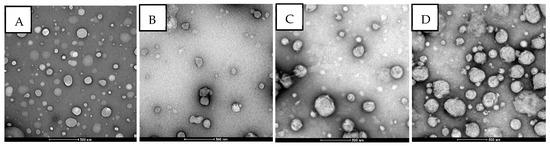
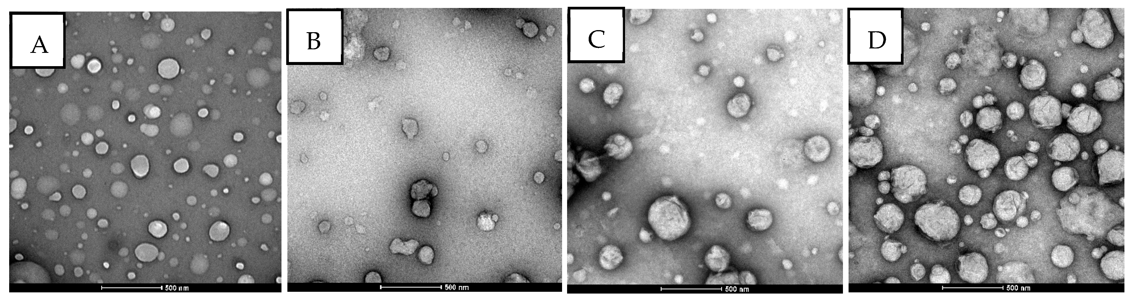
The TEM images of B-PL, CZ-PL, B-CH/L, and CZ-CH/L are presented in Figure 1. TEM analysis confirmed the morphology and particle size of the liposomal formulations. The PL and CH/L both exhibited a spherical shape, with the CH/L being slightly thicker, as reported in the literature [30], and the observed particle sizes were consistent with those determined by dynamic light scattering using the Zetasizer on the nanometer scale.
Figure 1.
Transmission electron microscope images of the formed primary and chitosan-coated liposomes. (A) B-PL: Empty primary liposomes, (B) CZ-PL: Primary liposomes loaded with aqueous cinnamon extract, (C) B-CH/L: Empty chitosan-coated liposomes, (D) CZ-CH/L: Chitosan-coated liposomes with aqueous cinnamon extract. Magnification: 43,000×.
3.4.2. Fourier Transform Infrared Spectroscopy (FTIR)
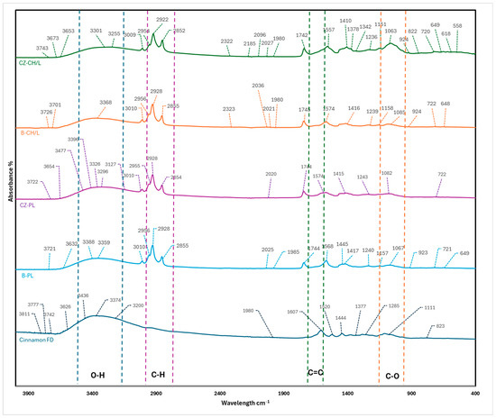
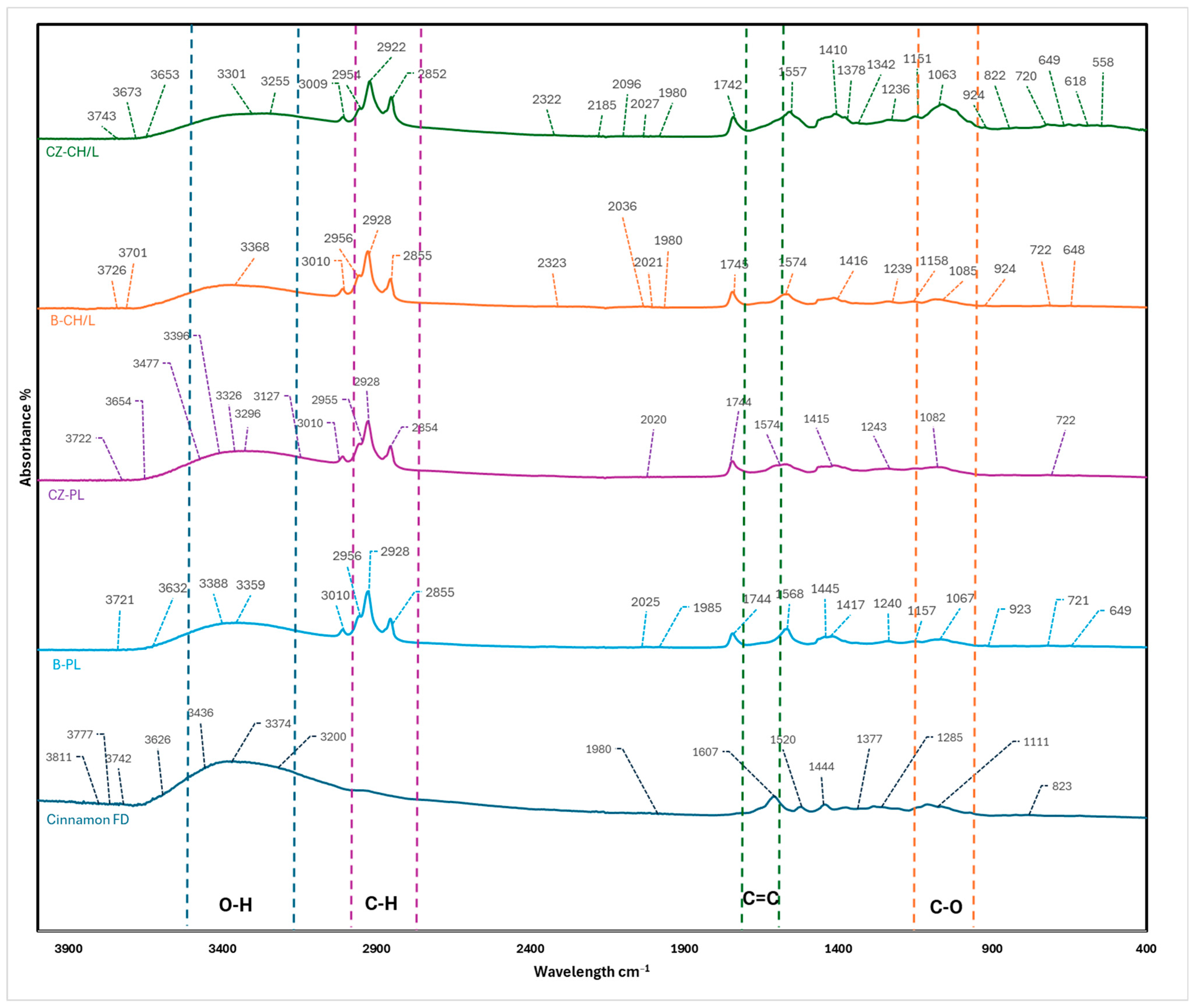
The identification of potential interactions between the encapsulating material and the core material is a critical factor in evaluating the success of the encapsulation process. Accordingly, FTIR spectroscopy provides a dependable approach for investigating molecular structure, conformation, and intermolecular interactions in encapsulation-based delivery systems [62]. Therefore, FTIR spectroscopic analysis was performed on C. zeylanicum freeze-dried powder, C. zeylanicum loaded PL, and CH/L, and the corresponding spectra are presented and interpreted in Figure 2 and Table 8.
Figure 2.
FTIR spectra of cinnamon freeze-dried powder, empty primary liposomes (B-PL), cinnamon liposomes (CZ-PL), empty chitosan-coated liposomes (B-CH/L), and chitosan-coated cinnamon liposomes (CZ-CH/L).
Table 8.
FTIR characteristic peaks and spectral changes in cinnamon extract, liposome-, and chitosan-coated liposome formulations, indicating encapsulation and molecular interactions.
The FTIR spectra confirmed characteristic peaks for C. zeylanicum. The broad absorption band observed in the 3200–3500 cm−1 region is attributed to O–H stretching vibrations of phenolic hydroxyl groups [63]. The peaks in the region of 2920–2850 cm−1 correspond to C–H stretching vibrations of aliphatic groups. The peaks around the 1600–1700 cm−1 can be attributed to the C=O carbonyl bond from aldehyde vibrations, such as cinnamic aldehyde, which are marker components of cinnamon, and the C=C stretching vibrations of alkenes [64]. The absorption band observed near 1400 cm−1 corresponds to C–H bending vibrations of alkanes. The band detected at approximately 1300 cm−1 is associated with vibrations of aromatic amines [65]. The spectral region contrast, bands observed in the 900–650 cm−1 region, are attributed to out-of-plane C–H bending vibrations of the aromatic ring [66].
In contrast, the spectrum of CZ-PL was dominated by characteristic phospholipid bands, including CH2 and CH3 stretching vibrations at around 2956, 2928, and 2854 cm−1, ester carbonyl (C=O) stretching at 1744 cm−1, and asymmetric and symmetric phosphate-related vibrations at 1243 and 1081 cm−1, respectively. This is consistent with the spectra obtained from various studies [39,67,68].
Following chitosan coating, additional absorption bands characteristic of chitosan appeared, including N–H bending vibrations around ~1557 cm−1 and polysaccharide C–O–C and C–O stretching bands in the 1150–1026 cm−1 region, further confirming successful surface modification. The band at 2854 cm−1 for PLs, attributed to the symmetric CH2 stretching modes of the acyl chains, exhibited a slight shift to lower frequency when coated with chitosan. This is consistent with the study of Refai et al. [69].
The characteristic cinnamon fingerprint bands at ~1607, 1520, 1444 cm−1 were significantly reduced or absent in the CZ-PL and CZ-CH/L spectrum, indicating effective encapsulation of cinnamon within the lipid bilayer rather than surface adsorption. The broadening and slight shifts observed in the O–H stretching region suggest the presence of intermolecular interactions, such as hydrogen bonding, between cinnamon polyphenols and phospholipid head groups.
Overall, the FTIR results confirm successful encapsulation of cinnamon within the liposomes and the formation of a chitosan coating, with interactions governed primarily by non-covalent intermolecular forces rather than the formation of new covalent bonds.
3.4.3. Thermal Analysis
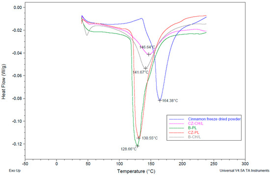
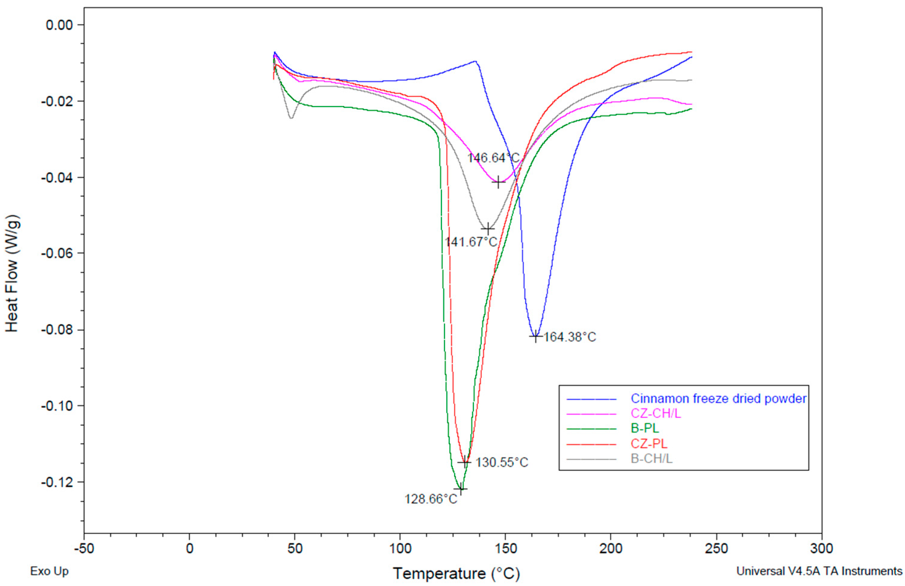
DSC analysis is a reliable method to confirm the encapsulation of a food ingredient within a wall material [70]. The DSC thermograms of freeze-dried C. zeylanicum powder, PL, and CH/L are presented in Figure 3, demonstrating clear differences in phase transition behaviour associated with encapsulation and polymer coating. The freeze-dried C. zeylanicum powder exhibited the highest melting temperature, with a prominent endothermic peak at 164.38 °C. This observation is consistent with previously reported DSC results for C. zeylanicum, where Aslan et al. [71] reported an endothermic transition at approximately 152.07 °C, indicating comparable thermal behavior. In contrast, both primary and chitosan-coated liposomes, empty liposomes displayed lower melting temperatures compared with their cinnamon-loaded counterparts, indicating that the incorporation of C. zeylanicum altered the thermal properties of the liposomal systems. This shift towards higher endothermic peak temperatures compared to empty liposomes suggests enhanced thermal stability of the liposomal system due to the incorporation of the cinnamon bioactives. The disappearance of the characteristic freeze-dried C. zeylanicum peak at ~164 °C further indicates successful encapsulation, as the phenolic compounds are no longer present in their free form but are molecularly dispersed within the liposomal bilayer or aqueous core. The observed increase in transition temperature and modification of the DSC profile in cinnamon-loaded liposomes can be attributed to interactions between phenolic compounds and lipid chains, as well as electrostatic interactions with the chitosan coating.
Figure 3.
DSC thermograms of Cinnamon freeze-dried powder, PL, CH/L systems. B-PL: Empty lecithin-based primary liposomes; CZ-PL: cinnamon-loaded primary liposomes; B-CH/L: Empty lecithin-based liposomes coated with chitosan; CZ-CH/L: cinnamon-loaded chitosan-coated liposomes.
4. Conclusions
Chitosan coating substantially enhanced encapsulation efficiency for C. zeylanicum extracts, with CZ-CH/L demonstrating markedly higher retention of cinnamon bioactives than CZ-PL. Both liposomal systems exhibited strong colloidal stability over the 8-week storage period, as reflected by only minor variations in zeta potential, particle size, and PDI. CZ-PL maintained a stable negative surface charge, while CZ-CH/L consistently retained its positive charge, confirming the robustness of the chitosan coating. The narrow and relatively unchanged PDI values further indicated uniform particle size distribution across storage. Structural analyses supported these observations: TEM imaging confirmed spherical vesicle morphology; FTIR spectra revealed characteristic band shifts associated with non-covalent interactions between cinnamon constituents and the lipid/chitosan matrices; and DSC thermograms indicated the disappearance of cinnamon’s characteristic melting peak, confirming successful encapsulation and altered thermal behaviour of the liposomal systems. Collectively, these findings demonstrate that both PL and CH/L are effective delivery systems that enhance the stability of C. zeylanicum bioactives, with CH/L offering superior retention and structural stability. The two systems, therefore, hold strong promise for application in functional foods that require the protection of sensitive bioactives during processing and storage. Future studies should investigate the release behaviour and bioaccessibility of the encapsulated compounds under simulated gastrointestinal conditions to determine whether chitosan coating provides a more controlled release of bioactives compared to uncoated liposomes. Additionally, further research is also needed to evaluate the performance and stability of these formulations within different food matrices and processing environments.
Author Contributions
Conceptualization, S.M.C. and A.R.; Methodology, S.M.C., L.K., D.G.P. and A.R.; Software, D.G.P.; Validation, L.K., D.G.P. and A.R.; Formal analysis, S.M.C.; Investigation, S.M.C. and A.R.; Resources, S.M.C. and A.R.; Data curation, S.M.C.; Writing—original draft, S.M.C.; Writing—review & editing, L.K., D.G.P. and A.R. All authors have read and agreed to the published version of the manuscript.
Funding
This research received no external funding.
Institutional Review Board Statement
Not applicable.
Informed Consent Statement
Not applicable.
Data Availability Statement
The original contributions presented in the study are included in the article, further inquiries can be directed to the corresponding author.
Acknowledgments
The first author acknowledges Massey University (Palmerston North, New Zealand) and the Riddet Institute for the Doctoral Scholarship received during this project. The authors gratefully acknowledge Maggie Zou from the Riddet Institute (Massey University, Palmerston North, New Zealand) for her laboratory assistance, Yanyu He for her technical support with the TEM at the Manawatū Microscopy and Imaging Centre, and Chris Hall for the support with the differential scanning calorimetry.
Conflicts of Interest
The authors declare that there are no conflicts of interest.
References
- Helal, A.; Tagliazucchi, D.; Verzelloni, E.; Conte, A. Bioaccessibility of polyphenols and cinnamaldehyde in cinnamon beverages subjected to in vitro gastro-pancreatic digestion. J. Funct. Foods 2014, 7, 506–516. [Google Scholar] [CrossRef] [Scilit]
- Ashfaq, M.H.; Siddique, A.; Shahid, S. Antioxidant activity of cinnamon zeylanicum: (A review). Asian J. Pharm. Res. 2021, 11, 106–116. [Google Scholar] [CrossRef] [Scilit]
- Mathew, S.; Abraham, T.E. Studies on the antioxidant activities of cinnamon (Cinnamomum verum) bark extracts, through various in vitro models. Food Chem. 2006, 94, 520–528. [Google Scholar] [CrossRef] [Scilit]
- Cheng, D.M.; Kuhn, P.; Poulev, A.; Rojo, L.E.; Lila, M.A.; Raskin, I. In vivo and in vitro antidiabetic effects of aqueous cinnamon extract and cinnamon polyphenol-enhanced food matrix. Food Chem. 2012, 135, 2994–3002. [Google Scholar] [CrossRef] [Scilit]
- Ranasinghe, P.; Pigera, S.; Premakumara, G.; Galappaththy, P.; Constantine, G.R.; Katulanda, P. Medicinal properties of ‘true’cinnamon (Cinnamomum zeylanicum): A systematic review. BMC Complement. Altern. Med. 2013, 13, 275. [Google Scholar] [CrossRef] [Scilit]
- Hong, J.-W.; Yang, G.-E.; Kim, Y.B.; Eom, S.H.; Lew, J.-H.; Kang, H. Anti-inflammatory activity of cinnamon water extract in vivo and in vitro LPS-induced models. BMC Complement. Altern. Med. 2012, 12, 237. [Google Scholar] [CrossRef] [Scilit]
- Kubatka, P.; Kello, M.; Kajo, K.; Samec, M.; Jasek, K.; Vybohova, D.; Uramova, S.; Liskova, A.; Sadlonova, V.; Koklesova, L. Chemopreventive and therapeutic efficacy of Cinnamomum zeylanicum L. bark in experimental breast carcinoma: Mechanistic in vivo and in vitro analyses. Molecules 2020, 25, 1399. [Google Scholar] [CrossRef] [Scilit]
- Khedkar, S.; Ahmad Khan, M. Aqueous extract of cinnamon (Cinnamomum spp.): Role in cancer and inflammation. Evid.-Based Complement. Altern. Med. 2023, 2023, 5467342. [Google Scholar] [CrossRef] [Scilit] [PubMed]
- Grgić, J.; Šelo, G.; Planinić, M.; Tišma, M.; Bucić-Kojić, A. Role of the encapsulation in bioavailability of phenolic compounds. Antioxidants 2020, 9, 923. [Google Scholar] [CrossRef] [Scilit] [PubMed]
- Zhang, L.; McClements, D.J.; Wei, Z.; Wang, G.; Liu, X.; Liu, F. Delivery of synergistic polyphenol combinations using biopolymer-based systems: Advances in physicochemical properties, stability and bioavailability. Crit. Rev. Food Sci. Nutr. 2020, 60, 2083–2097. [Google Scholar] [CrossRef] [Scilit]
- de Souza, V.B.; Thomazini, M.; Barrientos, M.A.E.; Nalin, C.M.; Ferro-Furtado, R.; Genovese, M.I.; Favaro-Trindade, C.S. Functional properties and encapsulation of a proanthocyanidin-rich cinnamon extract (Cinnamomum zeylanicum) by complex coacervation using gelatin and different polysaccharides. Food Hydrocoll. 2018, 77, 297–306. [Google Scholar] [CrossRef] [Scilit]
- Guan, H.; Zhang, W.; Sun-Waterhouse, D.; Jiang, Y.; Li, F.; Waterhouse, G.I.; Li, D. Phenolic-protein interactions in foods and post ingestion: Switches empowering health outcomes. Trends Food Sci. Technol. 2021, 118, 71–86. [Google Scholar] [CrossRef] [Scilit]
- Shahidi, F.; Athiyappan, K.D. Polyphenol-polysaccharide interactions: Molecular mechanisms and potential applications in food systems—A comprehensive review. Food Prod. Process. Nutr. 2025, 7, 42. [Google Scholar] [CrossRef] [Scilit] [PubMed]
- Conte, A.; Martini, S.; Tagliazucchi, D. Influence of Processing and Digestion on the Stability, Bioaccessibility and Bioactivity of Food Polyphenols. Foods 2023, 12, 851. [Google Scholar] [CrossRef] [Scilit]
- Nemli, E.; Ozkan, G.; Gultekin Subasi, B.; Cavdar, H.; Lorenzo, J.M.; Zhao, C.; Capanoglu, E. Interactions between proteins and phenolics: Effects of food processing on the content and digestibility of phenolic compounds. J. Sci. Food Agric. 2024, 104, 2535–2550. [Google Scholar] [CrossRef] [Scilit]
- Zuidam, N.J.; Nedovic, V. Encapsulation Technologies for Active Food Ingredients and Food Processing; Springer: New York, NY, USA, 2010; Volume 410. [Google Scholar]
- Subramani, T.; Ganapathyswamy, H. An overview of liposomal nano-encapsulation techniques and its applications in food and nutraceutical. J. Food Sci. Technol. 2020, 57, 3545–3555. [Google Scholar] [CrossRef] [Scilit]
- Liu, P.; Chen, G.; Zhang, J. A review of liposomes as a drug delivery system: Current status of approved products, regulatory environments, and future perspectives. Molecules 2022, 27, 1372. [Google Scholar] [CrossRef] [Scilit]
- Prakash, S. Nano-based drug delivery system for therapeutics: A comprehensive review. Biomed. Phys. Eng. Express 2023, 9, 052002. [Google Scholar] [CrossRef] [Scilit]
- Mankan, E.; Karakas, C.Y.; Saroglu, O.; Mzoughi, M.; Sagdic, O.; Karadag, A. Food-Grade Liposome-Loaded Delivery Systems: Current Trends and Future Perspectives. Foods 2025, 14, 2978. [Google Scholar] [CrossRef] [Scilit]
- Chen, X.-a.; Chuang, C.-c.; Chen, C.-c.; Lee, C.-y.; Chin, C.-y.; Young, J.-j.; Bai, M.-y.; Chuang, C.-c. Polyelectrolyte-coated liposomes microfluidically assembled in one-step for enhancing cell endocytosis and in-vivo immune responses. Colloids Surf. B Biointerfaces 2024, 241, 114030. [Google Scholar] [CrossRef] [Scilit]
- Zhang, X.; Wu, Z.; Zhang, W.; Wang, L.; Zhao, P.; Lv, X.; Guo, P.; Chen, J. Surface modification by chitosan for improving stability and antioxidative activity of astaxanthin-loaded liposomes. LWT 2024, 198, 116033. [Google Scholar] [CrossRef] [Scilit]
- Kang, B.R.; Park, J.S.; Ryu, G.R.; Jung, W.-J.; Choi, J.-S.; Shin, H.-M. Effect of chitosan coating for efficient encapsulation and improved stability under loading preparation and storage conditions of bacillus lipopeptides. Nanomaterials 2022, 12, 4189. [Google Scholar] [CrossRef] [Scilit]
- Alcantara, K.P.; Pajimna, R.M.B.; Aliga, P.J.S.; Malabanan, J.W.T.; Tangwongsiri, C.; Haworth, I.S.; Rojsitthisak, P.; Rojsitthisak, P. Review of chitosan-coated nanoscale liposomes for enhanced drug delivery. ACS Appl. Nano Mater. 2025, 8, 21125–21147. [Google Scholar] [CrossRef] [Scilit]
- Gil-Gonzalo, R.; Durante-Salmerón, D.A.; Pouri, S.; Doncel-Pérez, E.; Alcántara, A.R.; Aranaz, I.; Acosta, N. Chitosan-coated liposome formulations for encapsulation of ciprofloxacin and etoposide. Pharmaceutics 2024, 16, 1036. [Google Scholar] [CrossRef] [Scilit] [PubMed]
- Wang, X.; Cao, Z.; Su, J.; Ma, Y.; Zhang, S.; Shao, Z.; Ge, X.; Cheng, X.; Zhou, Z. Preparation of sodium alginate and chitosan modified curcumin liposomes and study on the formation of protein corona. Int. J. Biol. Macromol. 2025, 293, 139392. [Google Scholar] [CrossRef] [Scilit] [PubMed]
- Hasan, M.; Messaoud, G.B.; Michaux, F.; Tamayol, A.; Kahn, C.J.; Belhaj, N.; Linder, M.; Arab-Tehrany, E. Chitosan-coated liposomes encapsulating curcumin: Study of lipid–polysaccharide interactions and nanovesicle behavior. RSC Adv. 2016, 6, 45290–45304. [Google Scholar] [CrossRef] [Scilit]
- Shin, G.H.; Chung, S.K.; Kim, J.T.; Joung, H.J.; Park, H.J. Preparation of chitosan-coated nanoliposomes for improving the mucoadhesive property of curcumin using the ethanol injection method. J. Agric. Food Chem. 2013, 61, 11119–11126. [Google Scholar] [CrossRef] [Scilit]
- Sebaaly, C.; Haydar, S.; Greige-Gerges, H. Eugenol encapsulation into conventional liposomes and chitosan-coated liposomes: A comparative study. J. Drug Deliv. Sci. Technol. 2022, 67, 102942. [Google Scholar] [CrossRef] [Scilit]
- Maleki, G.; Woltering, E.J.; Mozafari, M. Applications of chitosan-based carrier as an encapsulating agent in food industry. Trends Food Sci. Technol. 2022, 120, 88–99. [Google Scholar] [CrossRef] [Scilit]
- Ren, Z.; Gao, W.; Li, X.; Jing, Y.; Gu, M.; Xing, P.; Jing, Z.; Ashrafizadeh, M.; Ren, J.; Orive, G. Engineering chitosan-functionalized liposomes for targeted drug delivery and biomedical applications. Int. J. Biol. Macromol. 2026, 345, 150563. [Google Scholar] [CrossRef] [Scilit]
- Cui, H.; Li, W.; Li, C.; Vittayapadung, S.; Lin, L. Liposome containing cinnamon oil with antibacterial activity against methicillin-resistant Staphylococcus aureus biofilm. Biofouling 2016, 32, 215–225. [Google Scholar] [CrossRef] [Scilit]
- Ellboudy, N.M.; Elwakil, B.H.; Shaaban, M.M.; Olama, Z.A. Cinnamon oil-loaded nanoliposomes with potent antibacterial and antibiofilm activities. Molecules 2023, 28, 4492. [Google Scholar] [CrossRef] [Scilit] [PubMed]
- Ma, Y.; Wang, W.; Cao, Y.; Song, Z.; Yu, Q. Development of multifunctional liposomes co-loaded with proanthocyanidins and cinnamon essential oil: Storage stability, antioxidant and antibacterial activities, and sustained release study. Food Biosci. 2025, 72, 107492. [Google Scholar] [CrossRef] [Scilit]
- Emami, S.; Ahmadi, M.; Nasiraie, L.R.; Shahidi, S.A.; Jafarizadeh-Malmiri, H. Cinnamon extract and its essential oil nanoliposomes–preparation, characterization and bactericidal activity assessment. Biologia 2022, 77, 3015–3025. [Google Scholar] [CrossRef] [Scilit]
- Wu, K.; Zhang, T.; Chai, X.; Duan, X.; He, D.; Yu, H.; Liu, X.; Tao, Z. Encapsulation efficiency and functional stability of cinnamon essential oil in modified β-cyclodextrins: In vitro and in silico evidence. Foods 2022, 12, 45. [Google Scholar] [CrossRef] [Scilit] [PubMed]
- Muhammad, D.R.A.; Doost, A.S.; Gupta, V.; bin Sintang, M.D.; Van de Walle, D.; Van der Meeren, P.; Dewettinck, K. Stability and functionality of xanthan gum–shellac nanoparticles for the encapsulation of cinnamon bark extract. Food Hydrocoll. 2020, 100, 105377. [Google Scholar] [CrossRef] [Scilit]
- Rashidinejad, A.; Birch, E.J.; Sun-Waterhouse, D.; Everett, D.W. Delivery of green tea catechin and epigallocatechin gallate in liposomes incorporated into low-fat hard cheese. Food Chem. 2014, 156, 176–183. [Google Scholar] [CrossRef] [Scilit]
- Mady, M.M.; Darwish, M.M.; Khalil, S.; Khalil, W.M. Biophysical studies on chitosan-coated liposomes. Eur. Biophys. J. 2009, 38, 1127–1133. [Google Scholar] [CrossRef] [Scilit]
- Zhang, Q.; Zhang, J.; Shen, J.; Silva, A.; Dennis, D.A.; Barrow, C.J. A simple 96-well microplate method for estimation of total polyphenol content in seaweeds. J. Appl. Phycol. 2006, 18, 445–450. [Google Scholar] [CrossRef] [Scilit]
- Azevedo, M.A.; Bourbon, A.I.; Vicente, A.A.; Cerqueira, M.A. Alginate/chitosan nanoparticles for encapsulation and controlled release of vitamin B2. Int. J. Biol. Macromol. 2014, 71, 141–146. [Google Scholar] [CrossRef] [Scilit]
- Culas, M.; Kaur, L.; Popovich, D.; Rashidinejad, A. Comparative efficiency of extraction techniques for bioactive compounds in Cinnamomum zeylanicum. Food Chem. 2025, 493, 145891. [Google Scholar] [CrossRef] [Scilit] [PubMed]
- Abeysekera, W.; Premakumara, G.; Ratnasooriya, W. In vitro antioxidant properties of leaf and bark extracts of ceylon cinnamon (Cinnamomum zeylanicum Blume). Trop. Agric. Res. 2013, 24, 128–138. [Google Scholar]
- Sana, S.; Arshad, M.U.; Farhan, S.; Ahmad, R.; Ali, I.; Tabussam, T. Nutritional characterization of cinnamon and turmeric with special reference to their antioxidant profile. Int. J. Biosci 2019, 15, 178–187. [Google Scholar]
- Gulcin, I.; Kaya, R.; Goren, A.C.; Akincioglu, H.; Topal, M.; Bingol, Z.; Cetin Çakmak, K.; Ozturk Sarikaya, S.B.; Durmaz, L.; Alwasel, S. Anticholinergic, antidiabetic and antioxidant activities of cinnamon (Cinnamomum verum) bark extracts: Polyphenol contents analysis by LC-MS/MS. Int. J. Food Prop. 2019, 22, 1511–1526. [Google Scholar] [CrossRef] [Scilit]
- Al Tamim, A.; Alharbi, H.; Hawsawi, E.; Aldosari, Z.; Alqahtani, A. Optimized HPLC-DAD Method for Quantifying Coumarin and Cinnamaldehyde in Local Cinnamon Sample. Appl. Food Res. 2025, 5, 101336. [Google Scholar] [CrossRef] [Scilit]
- Liu, C.; Long, H.; Wu, X.; Hou, J.; Gao, L.; Yao, S.; Lei, M.; Zhang, Z.; Guo, D.-a.; Wu, W. Quantitative and fingerprint analysis of proanthocyanidins and phenylpropanoids in Cinnamomum verum bark, Cinnamomum cassia bark, and Cassia twig by UPLC combined with chemometrics. Eur. Food Res. Technol. 2021, 247, 2687–2698. [Google Scholar] [CrossRef] [Scilit]
- Gursale, A.; Dighe, V.; Parekh, G. Simultaneous quantitative determination of cinnamaldehyde and methyl eugenol from stem bark of Cinnamomum zeylanicum Blume using RP-HPLC. J. Chromatogr. Sci. 2010, 48, 59–62. [Google Scholar] [CrossRef] [Scilit]
- Chen, W.-T.; Kuo, Y.-L.; Chen, C.-H.; Wu, H.-T.; Chen, H.-W.; Fang, W.-P. Improving the stability and bioactivity of curcumin using chitosan-coated liposomes through a combination mode of high-pressure processing. LWT 2022, 168, 113946. [Google Scholar] [CrossRef] [Scilit]
- Reyhani Poul, S.; Yeganeh, S. Physicochemical and antioxidant properties of chitosan-coated nanoliposome loaded with bioactive peptides produced from shrimp wastes hydrolysis. Iran. J. Fish. Sci. 2022, 21, 987–1003. [Google Scholar]
- Gibis, M.; Ruedt, C.; Weiss, J. In vitro release of grape-seed polyphenols encapsulated from uncoated and chitosan-coated liposomes. Food Res. Int. 2016, 88, 105–113. [Google Scholar] [CrossRef] [Scilit] [PubMed]
- Jiao, Z.; Wang, X.; Yin, Y.; Xia, J.; Mei, Y. Preparation and evaluation of a chitosan-coated antioxidant liposome containing vitamin C and folic acid. J. Microencapsul. 2018, 35, 272–280. [Google Scholar] [CrossRef] [Scilit] [PubMed]
- Caddeo, C.; Díez-Sales, O.; Pons, R.; Carbone, C.; Ennas, G.; Puglisi, G.; Fadda, A.M.; Manconi, M. Cross-linked chitosan/liposome hybrid system for the intestinal delivery of quercetin. J. Colloid Interface Sci. 2016, 461, 69–78. [Google Scholar] [CrossRef] [Scilit]
- Rashidinejad, A.; Jameson, G.B.; Singh, H. The effect of pH and sodium caseinate on the aqueous solubility, stability, and crystallinity of rutin towards concentrated colloidally stable particles for the incorporation into functional foods. Molecules 2022, 27, 534. [Google Scholar] [CrossRef] [Scilit] [PubMed]
- Hemmingsen, L.M.; Panchai, P.; Julin, K.; Basnet, P.; Nystad, M.; Johannessen, M.; Škalko-Basnet, N. Chitosan-based delivery system enhances antimicrobial activity of chlorhexidine. Front. Microbiol. 2022, 13, 1023083. [Google Scholar] [CrossRef] [Scilit]
- Ong, S.G.M.; Ming, L.C.; Lee, K.S.; Yuen, K.H. Influence of the encapsulation efficiency and size of liposome on the oral bioavailability of griseofulvin-loaded liposomes. Pharmaceutics 2016, 8, 25. [Google Scholar] [CrossRef] [Scilit]
- Fathi, S.; Oyelere, A.K. Liposomal drug delivery systems for targeted cancer therapy: Is active targeting the best choice? Future Med. Chem. 2016, 8, 2091–2112. [Google Scholar] [CrossRef] [Scilit] [PubMed]
- Esposto, B.S.; Jauregi, P.; Tapia-Blacido, D.R.; Martelli-Tosi, M. Liposomes vs. chitosomes: Encapsulating food bioactives. Trends Food Sci. Technol. 2021, 108, 40–48. [Google Scholar] [CrossRef] [Scilit]
- Laouini, A.; Jaafar-Maalej, C.; Limayem-Blouza, I.; Sfar, S.; Charcosset, C.; Fessi, H. Preparation, characterization and applications of liposomes: State of the art. J. Colloid Sci. Biotechnol. 2012, 1, 147–168. [Google Scholar] [CrossRef] [Scilit]
- Tripathy, S.; Srivastav, P.P. Encapsulation of Centella asiatica leaf extract in liposome: Study on structural stability, degradation kinetics and fate of bioactive compounds during storage. Food Chem. Adv. 2023, 2, 100202. [Google Scholar] [CrossRef] [Scilit]
- Huang, R.; Song, H.; Wang, X.; Shen, H.; Li, S.; Guan, X. Fatty acids-modified liposomes for encapsulation of bioactive peptides: Fabrication, characterization, storage stability and in vitro release. Food Chem. 2024, 440, 138139. [Google Scholar] [CrossRef] [Scilit]
- Jafari, S.M. Characterization of Nanoencapsulated Food Ingredients; Academic Press: Cambridge, MA, USA, 2020; Volume 4. [Google Scholar]
- Aqeel Salim, A.; Bidin, N.; Bakhtiar, H.; Krishna Ghoshal, S.; Al Azawi, M.; Krishnan, G. Optical and structure characterization of cinnamon nanoparticles synthesized by pulse laser ablation in liquid (PLAL). In Proceedings of the Journal of Physics: Conference Series; IOP Publishing: Bristol, UK, 2018; p. 012002. [Google Scholar]
- Popa, R.M.; Fetea, F.; Socaciu, C. Attenuated Total Reflectance—Fourier Transform Infrared Spectroscopy Applied for the Evaluation of Essential Oils’ Pattern Recognition and Thermo-Oxidative Stability: A Comparative Study. Stud. Univ. Babes-Bolyai Chem. 2021, 66, 33–50. [Google Scholar] [CrossRef] [Scilit]
- Maruthamuthu, R.; Ramanathan, K. Phytochemical analysis of bark extract of Cinnamomum verum: A medicinal herb used for the treatment of coronary heart disease in malayali tribes, Pachamalai Hills, Tamil Nadu, India. Int. J. Pharmacogn. Phytochem. Res. 2016, 8, 1218–1222. [Google Scholar]
- Ramos, V.; Bastos da Silva, E.; Bello, T. Comprehensive Characterization of Cinnamomum cassia Stem Extracts by NMR and Spectroscopic Techniques for Food and Medical Applications. Food Sci. Nutr. Res. 2025, 8, 1–9. [Google Scholar] [CrossRef] [Scilit]
- Chotphruethipong, L.; Hutamekalin, P.; Sukketsiri, W.; Benjakul, S. Effects of sonication and ultrasound on properties and bioactivities of liposomes loaded with hydrolyzed collagen from defatted sea bass skin conjugated with epigallocatechin gallate. J. Food Biochem. 2021, 45, e13809. [Google Scholar] [CrossRef] [Scilit]
- Tai, K.; Liu, F.; He, X.; Ma, P.; Mao, L.; Gao, Y.; Yuan, F. The effect of sterol derivatives on properties of soybean and egg yolk lecithin liposomes: Stability, structure and membrane characteristics. Food Res. Int. 2018, 109, 24–34. [Google Scholar] [CrossRef] [Scilit]
- Refai, H.; Hassan, D.; Abdelmonem, R. Development and characterization of polymer-coated liposomes for vaginal delivery of sildenafil citrate. Drug Deliv. 2017, 24, 278–288. [Google Scholar] [CrossRef] [Scilit] [PubMed]
- Gharanjig, H.; Gharanjig, K.; Hosseinnezhad, M.; Jafari, S.M. Differential scanning calorimetry (DSC) of nanoencapsulated food ingredients. In Characterization of Nanoencapsulated Food Ingredients; Elsevier: Amsterdam, The Netherlands, 2020; pp. 295–346. [Google Scholar]
- Aslan, M.; Ertaş, N.; Demir, M.K. Storage stability, heat stability, controlled release and antifungal activity of liposomes as alternative fungal preservation agents. Food Biosci. 2023, 51, 102281. [Google Scholar] [CrossRef] [Scilit]

Disclaimer/Publisher’s Note: The statements, opinions and data contained in all publications are solely those of the individual author(s) and contributor(s) and not of MDPI and/or the editor(s). MDPI and/or the editor(s) disclaim responsibility for any injury to people or property resulting from any ideas, methods, instructions or products referred to in the content. |
© 2026 by the authors. Licensee MDPI, Basel, Switzerland. This article is an open access article distributed under the terms and conditions of the Creative Commons Attribution (CC BY) license.