


Featured Application
The identified phenolic compounds and antioxidant-rich extracts from selected Rosaceae fruits may serve as natural ingredients for developing functional foods, nutraceutical formulations, and antioxidant-enhanced dietary products. Their characterized chemical profiles provide a foundation for selecting species and cultivars with the highest bioactive potential for use in food fortification, natural preservation systems, and health-oriented product design.
Abstract
Diet plays a fundamental role in maintaining human health, which has intensified scientific interest in bioactive food constituents and contributed to the development of functional foods. Polyphenols, one of the most important groups of plant secondary metabolites, are valued for their strong antioxidant properties and potential health benefits. Species belonging to the Rosaceae family, including Rosa, Crataegus, and Pyracantha, are recognized as promising sources of phenolic compounds, although their chemical profiles and antioxidant potential remain insufficiently characterized. The aim of this study was to quantitatively assess selected phenolic compounds in extracts obtained from ripe fruits of selected Rosaceae species and cultivars. The extracts were prepared using ultrasound-assisted solvent extraction, pressurized liquid extraction, and matrix solid-phase dispersion. The resulting samples were subsequently subjected to comprehensive analyses of their chemical composition and antioxidant capacity. These extraction techniques differ substantially in their operational principles and process parameters; notably, ultrasound-assisted solvent extraction and pressurized liquid extraction require more complex and tightly controlled conditions, whereas matrix solid-phase dispersion constitutes a comparatively simpler and less parameter-dependent approach. The results revealed distinct phenolic profiles among the examined species and confirmed the presence of compounds exhibiting strong antioxidant activity. Collectively, these findings broaden current knowledge of the phytochemical diversity present in Rosaceae fruits and underscore their potential as natural sources of bioactive constituents relevant to the development and formulation of functional food products.
1. Introduction
A well-balanced diet has long been recognized as a key factor influencing human health, appearance, and overall well-being. Ludwig Feuerbach’s famous statement, “Man is what he eats,” although formulated more than 150 years ago, remains highly relevant today, underscoring the close relationship between food quality and physiological function. In recent years, public interest in the health effects of nutrition has grown markedly, driven both by warnings from clinicians and researchers about the role of poor diet in the development of lifestyle diseases and by increasing consumer awareness of the nutritional value of food products [1]. This intensified focus on the diet–health relationship has stimulated research into the biological activity of food components and contributed to the emergence of the concept of functional foods—first introduced in Japan over two decades ago to describe products with documented health-promoting effects that extend beyond basic nutritional functions [2,3].
Functional foods encompass products naturally rich in bioactive constituents as well as foods enriched through the removal of undesirable components, the addition of beneficial ones, or the modification of their bioavailability. In response to growing interest in health-oriented lifestyles, the food industry increasingly incorporates functional additives to enhance flavour, texture, and nutritional value. Among the compounds of greatest scientific interest are plant secondary metabolites, particularly phenolic compounds, which support endogenous defence mechanisms and play an important role in the prevention of lifestyle-related disorders [4,5,6,7]. Polyphenols constitute the most abundant and structurally diverse group of these metabolites, with more than 8000 compounds described to date [8,9,10,11]. They are widely distributed in herbs, spices, tea, vegetables, and fruits, where they protect plant tissues from oxidative stress and UV radiation. Particularly high polyphenol levels have been reported in fruits such as Cornus mas, Ribes nigrum, Ginkgo biloba, Sambucus nigra, Juglans regia, and Punica granatum [12,13,14,15,16]. Numerous ornamental and economically important species of the Rosaceae family—including Rubus, Fragaria, Chaenomeles japonica, Cotoneaster, and Potentilla—are also recognized as valuable sources of polyphenols [17,18,19,20,21].
In recent years, considerable attention has been directed toward rose hips (Rosa spp.), hawthorn (Crataegus), and firethorn (Pyracantha), which have been identified as exceptionally rich sources in phenolic compounds associated with high antioxidant activity and potential health-promoting effects [22,23,24,25,26,27]. Despite the growing number of studies, the phenolic profiles and biological potential of many species and cultivars within these genera remain insufficiently characterized, highlighting the need for further systematic research.
In line with this research gap, the present study focused on quantifying selected phenolic compounds and vitamin C, as well as evaluating the relationship between their concentrations and the antioxidant capacity of the extracts obtained from ripe, edible fruits of selected species and cultivars of the Rosaceae family. The analyzed material included fruits of Rosa spinosissima, Rosa villosa, Rosa villosa ‘Karpatia’, Rosa sherardii, Rosa glauca, Rosa jundzillii, Crataegus coccinea, and Pyracantha coccinea cultivars ‘Soleil d’Or’, ‘Red Column’, and ‘Orange Charmer’. Fruits were harvested at full maturity, confirmed by characteristic coloration and easy detachment of peduncles from the stem. Extracts were prepared using ultrasound-assisted solvent extraction (UASE), pressurized liquid extraction (PLE), and matrix solid-phase dispersion. All extracts were analyzed for their chemical composition and antioxidant properties. The resulting data provide new insights into the chemical profiles and bioactive potential of these ornamental Rosaceae species, underscoring their relevance as promising as natural sources of valuable bioactive compounds suitable for incorporation into functional food products.
2. Materials and Methods
2.1. Preparation of Extracts
Extracts were obtained from 0.5 g of ground ripe fruit (unless otherwise stated) and 50 mL of solvent. Three solid–liquid extraction techniques were applied: PLE, UASE, and MSPD, a low-cost technique based on mechanical disruption of the sample with an inert abrasive material. All extracts were subsequently analysed chromatographically and evaluated for antioxidant activity.
2.1.1. PLE
PLE was carried out using a Dionex ASE200 extractor (Dionex Corp., Sunnyvale, CA, USA). Each portion of plant material was blended with sand to reduce solvent consumption, and the resulting mixture was placed in stainless-steel extraction cells (22 mL) equipped with cellulose filters, which constitute the pressure-resistant vessels used in the PLE system to hold the sample during solvent circulation under controlled temperature and pressure [28]. Extraction was performed at 60 °C, typically used in PLE conditions for the extraction of labile bioactive compounds, including phenolic compounds and anthocyanins, at 40 bar pressure, with a static extraction time of 10 min [29]. After each extraction, the system was flushed with solvent corresponding to 100% of the empty cell volume, followed by a 60 s nitrogen purge at 10 bar. Between runs, the instrument was rinsed with methanol to prevent cross-contamination. The obtained extracts were transferred quantitatively to volumetric flasks and adjusted to a final volume of 50 mL with the extraction solvent.
2.1.2. UASE
UASE was carried out in a thermostated Elmasonic P ultrasonic bath (Elma, Singen, Germany). Methanol, ethyl acetate, or n-hexane was used as extraction solvents. The conditions were as follows: temperature—25 °C; ultrasonic frequency—37 kHz; generator power—100% (720 W); extraction time—10 min. In each procedure, 0.5 g of plant material was mixed with 50 mL of the selected solvent. The obtained extracts were filtered through Whatman No. 4 filter paper, and the filtrate was then made up to a final volume of 50 mL with the corresponding solvent [28].
2.1.3. MSPD
For MSPD, 0.25 g of plant material was combined with 1 g of sand and transferred to a glass mortar [30]. The mixture was ground for 10 min, with 1 mL of methanol added as a dispersing liquid before the mixture dried. To ensure uniform homogenization, the mortar walls and pestle were periodically scraped with a spatula. The ground mixture was then transferred to a syringe barrel lined with filter paper, gently compressed, and all tools (mortar, pestle, and spatula) were rinsed with methanol. The rinsing solution was added to the syringe and the extract was eluted with methanol under slight vacuum into a 25 mL volumetric flask.
2.2. Chromatographic Analysis
All chromatographic analyses were carried out using a Varian ProStar 210/215 liquid chromatograph equipped with a ProStar 325 UV/VIS detector (Varian, Walnut Creek, CA, USA) and a 150 × 4.6 mm ODS column (Microsorb MV100 C18, Varian, Walnut Creek, CA, USA). Samples of 20 μL were injected using a Rheodyne 7725 injector (Sigma-Aldrich, Poznań, Poland).
2.2.1. Analysis of Phenolic Compounds
Phenolic compounds were analyzed under the following gradient conditions: an initial 6 min isocratic step (95% solvent A and 5% solvent B), followed by gradients of 5–50% B over 10 min, 50–70% B over 5 min, 70–80% B over 5 min, and 80–100% B over 5 min. The system was held at 100% B for an additional 5 min, then returned to the initial conditions (95% A, 5% B) for 5 min. Solvent A consisted of 0.5% acetic acid (v/v), and solvent B was methanol. Detection was performed at 280 nm for most analytes, and at 254 nm for gentisic acid and quercetin glycoside. Standard solutions of gallic acid, protocatechuic acid, catechin, epigallocatechin gallate, chlorogenic acid, epicatechin, gentisic acid, coumaric acid, rutin, quercetin glycoside, quercetin, and myricetin were prepared in methanol. Calibration curves were constructed for each compound in the concentration range of 0.01–0.1 mg/mL. Table 1 presents the calibration equations, coefficients of determination (R2), and the limits of detection (LOD) and quantification (LOQ), where LOD was calculated as 3.3 × SY (with SY representing the standard error of the regression), and LOQ was defined as three times the LOD.
Table 1.
Calibration curves and the corresponding limit of detection (LOD) and quantification (LOQ) values.
2.2.2. Vitamin C Determination
Vitamin C content was quantified under isocratic conditions using a mobile phase composed of 95% solvent A (20 mM monobasic phosphate solution adjusted to pH 3 with phosphoric acid) and 5% solvent B (methanol). Detection was carried out at 242 nm. A calibration curve was constructed in the concentration range of 0.0025–0.1 mg/mL (y = 716,512,844.202x + 587,003.422; R2 = 0.999). The LOD and LOQ were 0.001 mg/mL and 0.003 mg/mL, respectively.
2.3. Spectrophotometric Evaluation of Antioxidant Activity and Total Phenolic Content
The antioxidant properties of the extracts were evaluated using spectrophotometric assays, including the 2,2-diphenyl-1-picrylhydrazyl radical-scavenging (DPPH) assay, the 2,2′-azino-bis(3-ethylbenzothiazoline-6-sulfonic acid) cation radical decolorization (ABTS) assay, and the ferric-reducing antioxidant power (FRAP) assay. Total phenolic content (TPC) was also determined. All measurements were performed using a Shimadzu 2550 UV–VIS spectrophotometer (Shimadzu Corporation, Kyoto, Japan).
2.3.1. DPPH Method
To determine DPPH radical-scavenging activity, 100 µL of extract was mixed with 2900 µL of a methanolic DPPH• radical solution (initial absorbance 0.70 ± 0.05) [31]. After 30 s of vigorous mixing, samples were incubated for 60 min and transferred to 1 cm glass cuvettes. Methanol served as the blank. The percentage inhibition (I%) of the DPPH radical was calculated using the equation:
where (A0) is the initial absorbance and (A60) is the absorbance after 60 min. Each sample was analyzed in triplicate, and the results are expressed as Trolox equivalents (TE, mg/g).
(I%) = (1 − A60/A0)⋅100%,
2.3.2. ABTS Assay
ABTS•+ radical-scavenging activity was determined following the procedure described in [32]. A volume of 100 µL of extract was added to 2900 µL of ABTS•+ solution. The radical solution was prepared 16 h prior to analysis by mixing 5 mL of 7 mM ABTS with 88 µL of 140 mM potassium persulfate and storing the mixture in the dark at room temperature. Before measurement, the solution was diluted with methanol to obtain an absorbance of 0.70 ± 0.05 at 744 nm. After adding the extract, samples were incubated for 1 h, transferred to cuvettes, and absorbance was recorded at 744 nm. Percentage inhibition was calculated as described for the DPPH assay. The results represent the mean of three measurements and are expressed as Trolox equivalents (TE, mg/g).
2.3.3. FRAP Assay
The FRAP assay was used to assess the ability of extracts to reduce Fe3+ to Fe2+ in the Fe2+–TPTZ complex, producing a blue colour with maximum absorbance at 593 nm [33]. The FRAP reagent was freshly prepared by mixing 25 mL of 0.3 M acetate buffer (pH 3.6), 2.5 mL of 10 mM TPTZ in 40 mM HCl, and 2.5 mL of 20 mM FeCl3·6H2O (10:1:1, v/v/v). A total volume of 2900 µL of FRAP reagent was combined with 100 µL of extract, mixed for 30 s, and incubated for 1 h at 37 °C. Absorbance was measured at 593 nm. A blank solution containing FRAP reagent and extraction solvent was used to zero the spectrophotometer. Antioxidant activity was expressed as Trolox equivalents (TE, mg/g). All measurements were performed in triplicate.
2.3.4. Determination of Total Phenolic Content
TPC was determined using the Folin–Ciocalteu method [34]. A volume of 100 µL of extract was mixed with 1.58 mL of distilled water and 100 µL of Folin–Ciocalteu reagent. After 5 min, 300 µL of 20% (w/v) sodium carbonate solution was added to ensure alkaline conditions. Samples were mixed and incubated for 2 h, after which absorbance was measured at 765 nm. TPC was calculated from a gallic acid calibration curve and expressed as gallic acid equivalents (GAE, mg GAE/g). All measurements were performed in triplicate.
2.4. Statistical Analysis
Each result is presented as the mean value of three independent measurements ± standard deviation (SD). The collected data were subjected to statistical evaluation using Pearson’s correlation analysis. Six predefined variable pairs were examined: TPC–ABTS, TPC–DPPH, TPC–FRAP, and PC–ABTS, PC–DPPH, and PC–FRAP. In this context, TPC denotes the total phenolic content determined spectrophotometrically, whereas PC refers to the polyphenol fraction quantified chromatographically. For each pair of variables, the Pearson correlation coefficient (r) and the corresponding two-tailed p-value were calculated to assess both the strength and statistical significance of the linear relationships.
In addition to these analyses, Pearson’s correlation coefficients were also computed for the variable pairs: vitamin C vs. ABTS, DPPH, and FRAP, as well as polyphenols + vitamin C vs. ABTS, DPPH, and FRAP. For each of these combinations, as before, the coefficient value (r) and the two-sided p-value were obtained, which enabled a comprehensive assessment of the magnitude and significance of the relationships between antioxidant capacity and the concentrations of vitamin C and combined phenolic compounds.
3. Results and Discussion
In the present study, the antioxidant properties of thirteen extracts obtained from dried and milled fruits of various cultivars belonging to selected representatives of the Rosaceae family were evaluated. The names of the cultivars included in the study are listed in Table 2, and the sample identification numbers assigned to them are used consistently throughout all subsequent figures.
Table 2.
Names and codes of the examined Rosaceae cultivars.
Antioxidant activity was assessed using spectrophotometric assays, including the ABTS and DPPH radical-scavenging tests, as well as the FRAP assay, which measures the ferric-reducing potential of the extracts. These techniques are among the most widely applied screening methods in research on plant-derived materials, including those from the Rosaceae family [35,36,37]. The aim of the analysis was not only to compare antioxidant activity values obtained with different analytical approaches, but also to evaluate the effects of the extraction solvent and method on the antioxidant capacity of the individual extracts, and to identify potential correlations between polyphenol content and the observed biological activity.
3.1. Influence of Solvent Type on the Antioxidant Properties of Extracts
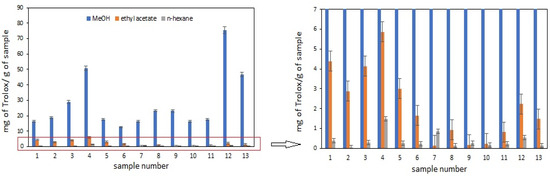
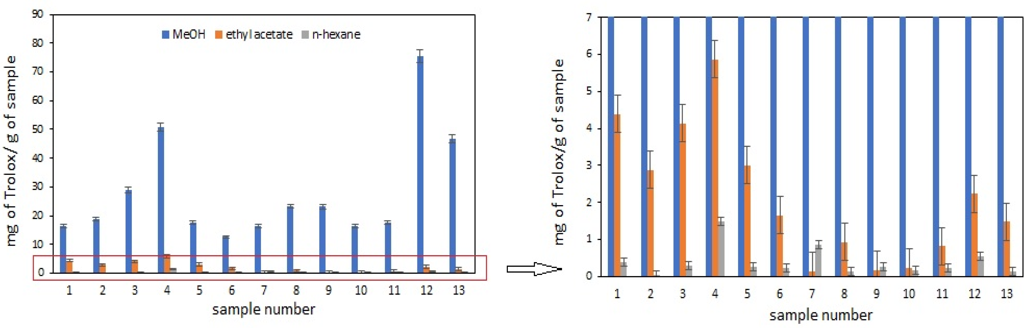
Figure 1 presents the antioxidant activity of extracts obtained with three solvents, methanol, ethyl acetate, and n-hexane, determined using the ABTS assay. A key advantage of the ABTS method is the high solubility of the ABTS•+ radical in both aqueous and aqueous–organic media, which makes this assay less sensitive to solvent polarity and allows for a more universal assessment of antioxidant capacity in chemically diverse extracts [38]. UASE was selected for sample preparation due to its rapidity, good reproducibility, and the possibility of processing multiple samples simultaneously.
Figure 1.
Effect of solvent type on antioxidant properties of Rosaceae plant extracts obtained by UASE and assessed by ABTS method; sample codes according to Table 2.
It should be emphasized, however, that the choice of solvent affects the evaluation of antioxidant properties on two levels. First, it determines the efficiency of isolating bioactive compounds from the plant matrix. Second, it influences the reaction environment in which free radicals are neutralized, thereby affecting the analytical outcome [39]. According to the literature, the antioxidant activity depends not only on the chemical structure of the compounds—related to their ability to donate hydrogen atoms and electrons, chelate metal ions, or stabilize unpaired electrons in aromatic structures—but also on the solvent in which antioxidant activity is determined [40]. Thus, choosing an appropriate solvent is crucial for both extraction efficiency and a reliable antioxidant assessment.
Antioxidant assays generally rely on two reaction mechanisms: single-electron transfer (SET) and hydrogen atom transfer (HAT) [39,40,41]. In practice, both mechanisms can occur simultaneously, and their relative contribution depends on the antioxidant’s properties, its partitioning behaviour, and the solvent used. In tests such as ABTS and DPPH, the reaction course is strongly dependent on the polarity of the reaction medium, which is closely related to the solvent’s dielectric constant (ε).
In the ABTS method, the dominant mechanism is SET, in which the neutralization of reactive molecules proceeds through ionization or dissociation of the antioxidant. Solvents with high dielectric constants favor this process because they stabilize the ionic species formed during the reaction. Consequently, a more polar environment favors a more efficient reaction and often leads to the observation of higher antioxidant activity. The results obtained in this study confirm this relationship. Methanol extracts exhibited the strongest antioxidant properties, which may be attributed to the high dielectric constant of methanol (ε = 32.6). Lower activity was observed for ethyl acetate extracts (ε = 6.02), while the weakest activity for n-hexane extracts (ε = 1.9) [42,43]. This indicates that more polar solvents not only extract phenolic compounds more efficiently but also enhance the analytical response in tests based on the SET mechanism.
Numerous comparative studies have demonstrated that the choice of solvent influences both extraction efficiency and the final antioxidant activity values. Differences arise from variations in the solubility of individual phenolic compounds as well as from the analytical characteristics of the assays used (e.g., ABTS, DPPH, or FRAP). For this reason, the literature frequently highlights the importance of standardizing assay conditions, including solvent composition and reaction times [41,44]. Considering both the present results and literature data, methanol extracts were selected for further analyses. Methanol proved to be the most effective solvent for isolating phenolic compounds and provided the most representative antioxidant profile of the studied material. Consequently, subsequent experiments—including additional extraction techniques and antioxidant assays—were conducted using methanol as the extraction solvent.
3.2. Effect of Extraction Technique on the Bioactive Properties of the Extracts
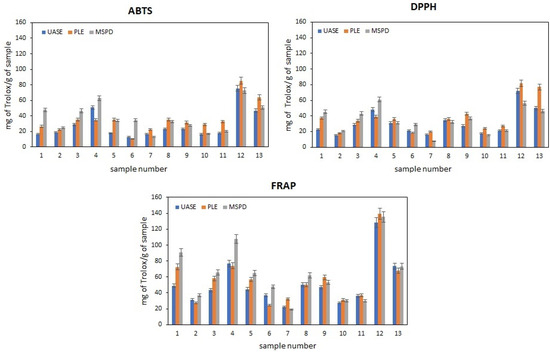
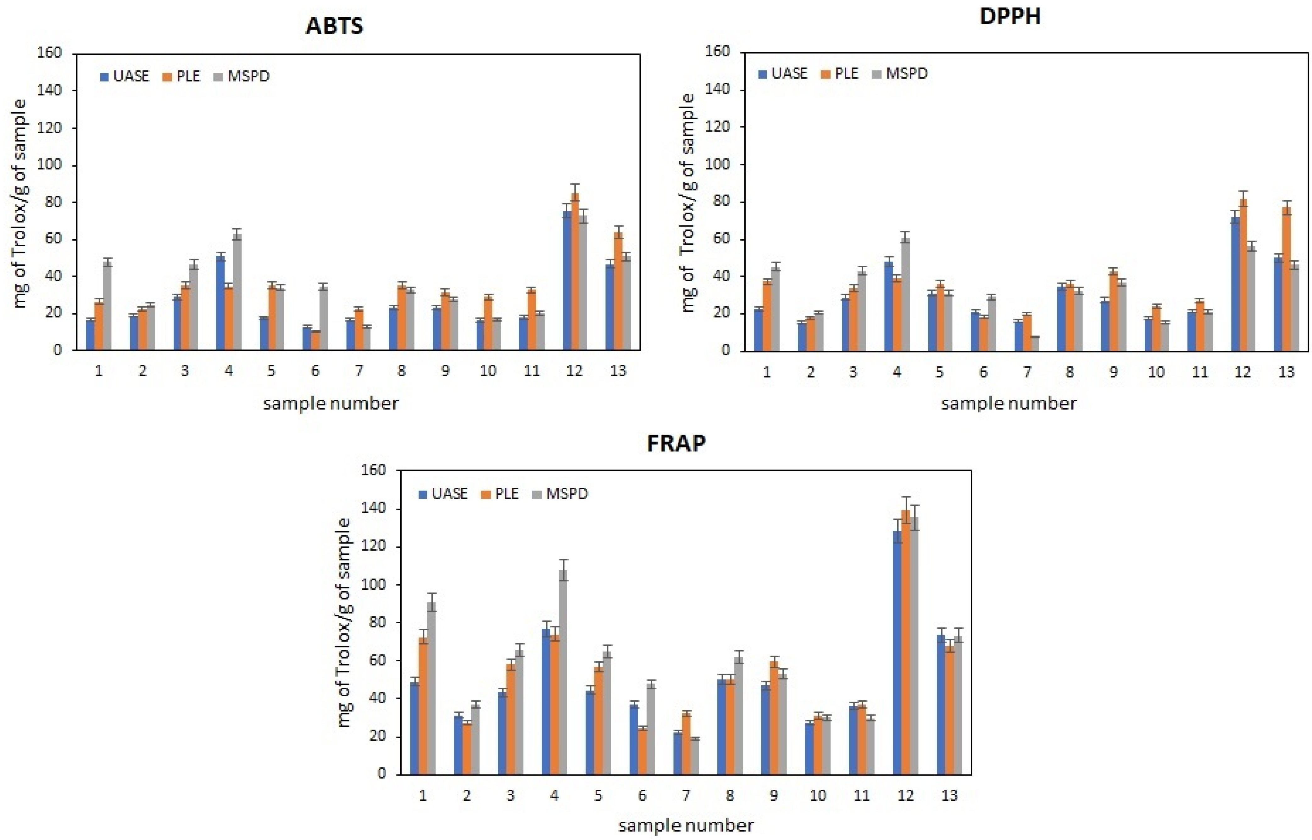
Figure 2 presents the relationship between the extraction technique and the antioxidant activity of methanolic extracts obtained from the studied Rosaceae cultivars. Antioxidant activities were evaluated using three complementary methods—ABTS, DPPH, and FRAP—which differ in their underlying reaction mechanisms. ABTS and DPPH measure the ability of compounds to neutralize free radicals, whereas FRAP assesses the reducing power of the extracts toward metal ions [40]. The combined use of these assays provides a more complete picture of antioxidant potential, enabling the detection of both radical-scavenging and strongly reducing constituents, which is particularly important when analysing complex phytochemical matrices.
Figure 2.
Antioxidant activity of methanolic extracts from thirteen Rosaceae cultivars, depending on the applied extraction technique (UASE, PLE, MSPD), as measured by the ABTS, DPPH, and FRAP assays.
As mentioned, the antioxidant assays applied in this study are based on different reaction mechanisms, which are strongly influenced by the measurement conditions, including solvent type and pH. In the ABTS and DPPH assays, radical neutralization proceeds primarily through the SET mechanism, although the HAT mechanism may also contribute. It is generally accepted that HAT plays only a minor role in the DPPH assay, whereas in the ABTS assay its contribution becomes more pronounced. In contrast, the FRAP assay relies exclusively on the SET mechanism. These mechanistic differences help explain the variability observed in the antioxidant properties of the extracts. The results therefore reflect not only the efficiency of the extraction techniques used to isolate active constituents but also the distinct reaction pathways underlying each analytical method. The higher values obtained in the FRAP assay can be attributed to the SET-driven reaction, which proceeds rapidly under the assay conditions. Moreover, FRAP specifically measures the reducing power of the sample, making it particularly suitable for extracts rich in compounds with low reduction potential. In turn, radical-based assays such as DPPH and ABTS provide a broader assessment of total radical-scavenging capacity, offering complementary insight into antioxidant behaviour [40].
Analyses were conducted on extracts obtained using three sample preparation methods, i.e., UASE, MSPD, and PLE. These methods differ in their extraction mechanisms, selectivity, and operating conditions, which ultimately shape the qualitative and quantitative profile of isolated phenolic compounds and other secondary metabolites [28]. While solvent choice affects both extraction efficiency and the analytical response, the extraction technique itself determines how effectively specific classes of bioactive compounds are released from the plant matrix. UASE utilizes the phenomenon of ultrasonic cavitation, which mechanically disrupts the structure of plant material, enabling the release of analytes at low temperatures and using relatively simple equipment [45]. PLE operates differently, relying on elevated pressure and temperature, which increases the solubility of compounds and accelerates their diffusion, but requires more advanced, expensive equipment and precise control of process parameters [46]. MSPD, on the other hand, combines mechanical sample fragmentation with solid-phase sorption, simplifying material preparation and allowing the process to be carried out at room temperature [30]. As repeatedly demonstrated in the literature, MSPD does not cause thermal degradation of unstable compounds, although the technique is less amenable to automation and more difficult to scale [47]. In terms of scalability and optimization, PLE offers the widest range of controllable conditions, while UASE stands out for its simplicity of process operation and potential for adaptation to semi-technical applications.
The data presented in Figure 2 shows that the ABTS and DPPH methods demonstrate similar activity trends across most analysed samples, with three highest values observed for samples 4, 12, and 13. This overall convergence is consistent with previous studies indicating that both assays respond strongly to extracts rich in phenolic compounds, although the degree of correlation may vary depending on the chemical composition of the extract, the pH of the reaction medium, and the solvent used [48]. The broader applicability of the ABTS method, resulting from the solubility of the ABTS•+ radical in both aqueous and aqueous–organic systems [38], contrasts with the more selective nature of DPPH, which has reduced sensitivity toward hydrophilic antioxidants and peptide-based molecules [40]. These assay-specific properties help explain both the general similarities and the occasional discrepancies observed between ABTS and DPPH results. The literature on raw materials from the Rosaceae family has repeatedly demonstrated that increased phenolic and flavonoid content translates into higher activity in ABTS and DPPH tests, which is consistent with the qualitative observations obtained in this study [49,50].
Beyond the general trends, interspecific comparisons revealed biologically meaningful differences among the studied taxa. Within the group of rose samples (1, 2, 4, 6, 7, and 12), the strongest antioxidant activity was recorded for samples 4 and 12. Similarly, sample 13 showed the highest activity among firethorn accessions (8–11 and 13). Particularly noteworthy were the hawthorn samples (3 and 5), which, despite originating from the same species, differed in phytosanitary status—one of the plants showed signs of fungal infection. Biotic stress is well known to induce metabolic reprogramming in plants, often leading to enhanced accumulation of phenolic compounds and other protective secondary metabolites. The higher antioxidant activity observed for sample 3 compared to sample 5 suggests stress-induced enhancement in the phenolic profile, a phenomenon extensively documented in studies on plant adaptive responses to pathogens [51]. This observation underscores the importance of environmental factors in shaping the antioxidant potential of fruits, even within the same species.
To further interpret these results, it is essential to consider the influence of extraction techniques. The extraction method had a significant but sample-dependent effect on antioxidant activity. In samples 1–4, the highest activity values were obtained using MSPD, whereas for samples 7–13, PLE proved more effective. These divergences likely reflect differences in extraction selectivity and the physicochemical characteristics of the techniques. MSPD promotes efficient release of phenolic compounds from compact or lignified matrices, while PLE increases extraction yield through the combined effects of elevated temperature and pressure, although such conditions may compromise the stability of thermolabile constituents. Therefore, the final antioxidant activity depends on a delicate balance between maximizing extraction efficiency and minimizing degradation of sensitive metabolites. From a practical perspective, combining extracts with complementary chemical profiles—such as vitamin C-rich UASE extracts and polyphenol-rich PLE extracts—may offer additional benefits. Such combinations could potentially help maintain or modulate antioxidant activity under different processing conditions, although confirmation of these effects would require further targeted studies.
Comparing our results to those published, it is difficult to find information in the literature regarding the plant material we selected and sample preparation methods. The most frequently cited methods for obtaining phenolic extracts from Rosacea fruits include conventional extraction methods, such as infusions, decoctions, maceration, and extraction in the Soxhlet apparatus, as well as assisted techniques, such as PLE, microwave-assisted extraction (MASE), or UASE [52,53]. Of the former, Soxhlet extraction is the most effective, while PLE and ultrasound dominate among the assisted methods. It should be noted that the latter (ultrasound-assisted solvent extraction) is the most frequently chosen method for preparing samples from Rosacea fruits [54]. Oargă et al. [53] reported that, when comparing the extraction efficiency of phytoactive compounds from rosehips, the highest polyphenol concentrations and antioxidant activity were observed with the UAE method, which provides better efficiency than methods using water baths, microwaves, and ultrasound. Since PLE also gives good results, as confirmed by our studies, effective extraction is achieved by combining both techniques. Examples of the use of ultrasound-assisted extraction combined with pressurized liquid extraction to increase the extraction efficiency of phenolic compounds from Rosacea plants can be found in the literature [55].
A broader perspective on assay behavior is provided by the FRAP results, which closely mirrored the activity patterns observed in the ABTS and DPPH assays, once again highlighting samples 4, 12, and 13 as the strongest antioxidants. Because FRAP measures the ability of antioxidants to reduce Fe3+ to Fe2+, it reflects not only radical-scavenging potential but also the overall reducing capacity of the extracts [40]. The strong concordance between FRAP and ABTS results suggests that electron transfer (SET) is the predominant antioxidant mechanism in the examined methanolic extracts. This interpretation is consistent with the chemical composition of the samples, which are rich in phenols with structures that allow resonance stabilization of radicals and thus favor electron donation. Moreover, the presence of vitamin C, anthocyanins, and reducing sugars may contribute to additive or synergistic interactions that further enhance the observed antioxidant activity.
Taken together, these observations underline that antioxidant capacity is not solely determined by the quantity of phenolic compounds but results from the interplay between extract composition, solvent effects, assay mechanisms, and extraction technique. In the methanol extracts analyzed, SET processes appear to dominate, as evidenced by the strong performance in ABTS and FRAP and the moderate but consistent activity seen in DPPH. This mechanistic pattern aligns with reports emphasizing that polyphenol-rich extracts, particularly those containing compounds with extensive electron delocalization, preferentially engage in SET-based reactions, while phenolics with low O–H bond dissociation energies contribute more prominently to HAT pathways.
Overall, the combined results highlight the complexity of antioxidant behavior in plant extracts and demonstrate that robust interpretation requires simultaneous consideration of chemical composition, biological context, and methodological parameters. Such insights are essential not only for evaluating the antioxidant potential of Rosaceae fruits but also for optimizing extraction protocols and selecting appropriate analytical assays in future studies.
3.3. The Complexity of the Relationship Between Chemical Composition and Antioxidant Activity
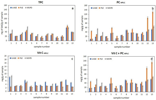
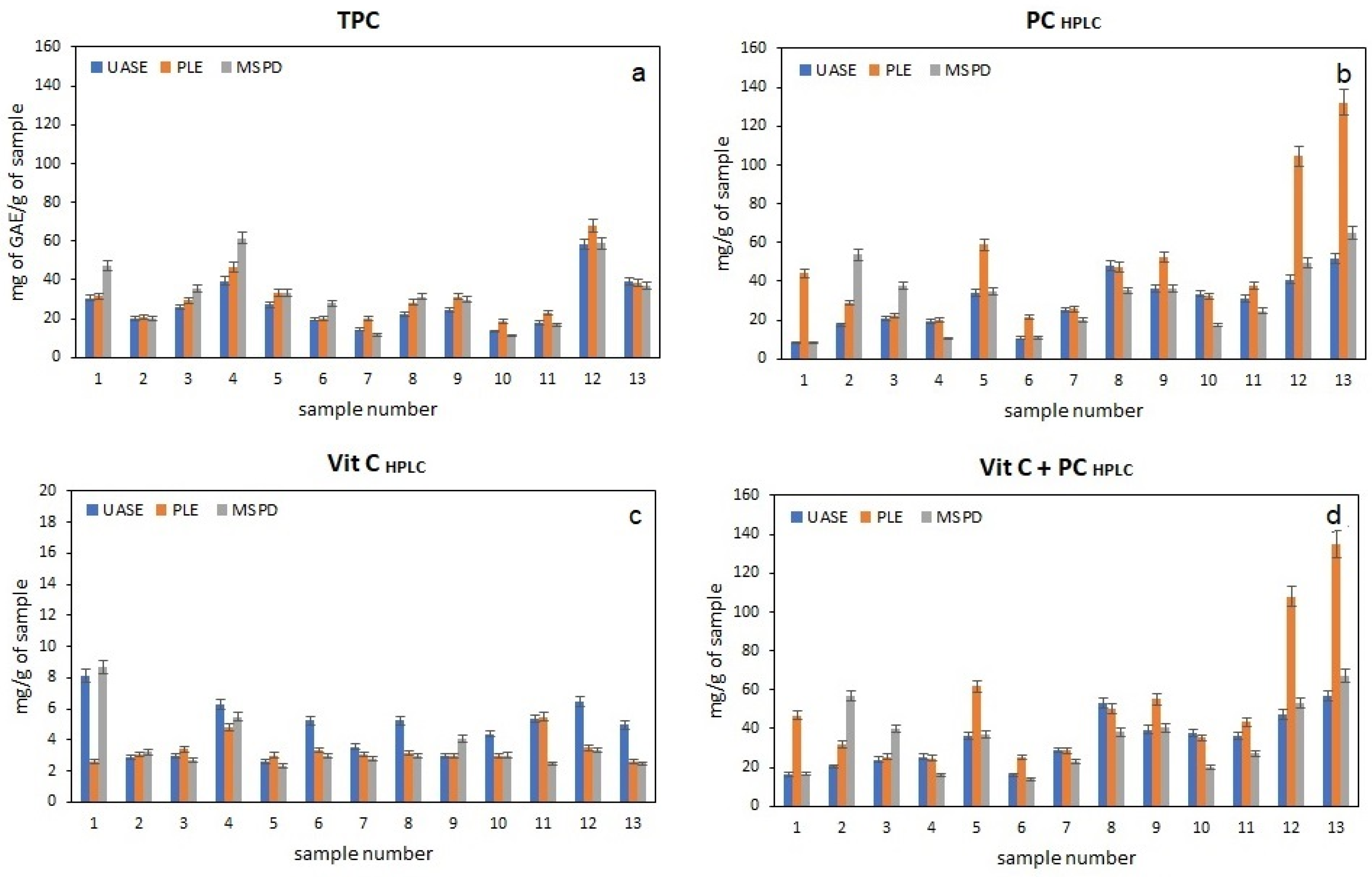
For all methanol extracts obtained using the three extraction techniques (UAE, MSPD, and PLE), the total phenolic content was determined using the Folin–Ciocalteu method (TPC). The results are summarized in Figure 3a. This method, commonly used in the analysis of plant materials, relies on the ability of reducing compounds to reduce the phosphomolybdic–phosphotungstic reagent. The obtained values allow for a preliminary assessment of the total reduction potential of the extracts; however, it should be emphasized that the Folin–Ciocalteu method is not selective only for polyphenols. The analytical response can also be generated by other reducing compounds, such as vitamin C, certain reducing sugars, or plant pigments [56,57].
Figure 3.
Content of active compounds in methanolic extracts from thirteen Rosaceae cultivars obtained using different extraction techniques (UASE, PLE, MSPD): (a) total phenolic content (TPC) determined by the Folin–Ciocalteu method, (b) polyphenolic content quantified by HPLC (PCHPLC), (c) vitamin C content determined by HPLC (Vit. C), and (d) the combined content of vitamin C and HPLC-determined phenolics (Vit. C + PCHPLC).
To characterize the phenolic composition in more detail, HPLC chromatographic analysis was performed to determine the content of selected compounds belonging to the main classes of polyphenols found in Rosaceae fruits, including gallic acid, protocatechuic acid, catechin, epicatechin gallate, epicatechin, gentisic acid, coumaric acid, and quercetin glycoside. The results are presented in Figure 3b. A sample chromatogram is shown in Figure S1. Data analysis indicates that in most cases, the highest content of chromatographically determined polyphenols was obtained in extracts obtained by the PLE method. However, this result is partially inconsistent with the results obtained in antioxidant activity tests, where the highest values were observed for samples 4, 12, and 13, which showed different trends depending on the extraction method used. This suggests that the amount of determined polyphenols is not the only factor determining the reducing properties of the extracts studied.
Therefore, we decided to perform additional chromatographic analysis of vitamin C content, which is a powerful antioxidant with pronounced reducing properties. This compound can affect both the results of spectrophotometric determinations using the Folin–Ciocalteu method, masking the actual polyphenol content, and the results of antioxidant tests based on reducing mechanisms such as ABTS, DPPH, and FRAP. The obtained data are summarized in Figure 3c. Chromatographic analysis showed that the highest vitamin C content was found in extracts obtained from rose hips (samples 1, 4, and 12), with the highest values observed in extracts obtained using the UASE method. This may indicate that the high temperature used in PLE does not favour the preservation of this compound and leads to its partial degradation. The MSPD method, on the other hand, proved less effective in recovering vitamin C from the plant material studied. Ultrasonic extraction (sonication) is considered one of the most effective methods for obtaining thermally sensitive compounds (vitamin C) from plant materials [58]. It utilizes high-frequency ultrasonic waves, which induce cavitation in the extraction solution. The formation and implosion of microbubbles lead to mechanical damage to cell walls, which facilitates the release of bioactive compounds into the solvent and increases the extraction efficiency of thermally sensitive compounds.
When the total polyphenol content determined by HPLC was compared with the vitamin C content, a picture was obtained that differed from the trends observed based on the results presented in the figures concerning antioxidant activity (Figure 1, Figure 2 and Figure 3). The obtained results suggest that a complex mixture of multiple bioactive compounds present in the fruits is responsible for the antioxidant activity. Comparison of the total polyphenol content determined by HPLC with the vitamin C content (see Figure 3d) did not clearly match the trends observed in the antioxidant activity test results shown in Figure 2. This indicates that the observed reducing properties of the extracts are not solely due to the presence of vitamin C or the chromatographically determined polyphenols. The obtained results suggest that a complex mixture of multiple bioactive compounds present in the fruit is responsible for the antioxidant activity.
It is also important to consider that fruits constitute a complex chemical matrix, rich in sugars, organic acids, and numerous plant pigments, such as anthocyanins. These compounds can influence both the reducing properties of the extracts and the analytical reactions used in spectrophotometric methods. Consequently, the observed antioxidant activity values are the result of the synergistic effects of multiple components. In some cases, high levels of specific bioactive compounds do not directly translate into the highest reducing activity, as the presence of other components can modulate or partially suppress their effects. Sample 13, for example, despite its high content of phenolic components, did not demonstrate the highest proportion of activity in the analyzed tests.
The obtained results confirm that interpreting the antioxidant properties of plant extracts requires a multifaceted approach, taking into account both the qualitative and quantitative composition of bioactive compounds, the influence of the extraction method used, and possible interactions between individual components present in the tested matrix.
3.4. Analysis of the Correlation Between the Content of Phenolic Compounds and Vitamin C and Antioxidant Activity
To quantitatively assess the relationship between phenolic compound content and antioxidant activity, a correlation analysis was performed between total polyphenol content (TPC) determined by the Folin–Ciocalteu spectroscopic method, polyphenol content determined by chromatography (PC HPLC), and the results of the ABTS, DPPH, and FRAP assays. These data are summarized in Table 3.
Table 3.
Correlative relationships between TPC, PC and vitamin C and antioxidant activity (ABTS, DPPH, FRAP), with r and p values (n = 13).
All correlations were statistically significant (p < 0.001), confirming the reliability of these relationships. The particularly high correlation between TPC and FRAP indicates that the total content of strongly reducing polyphenols is a good indicator of the reducing capacity of samples. Significant correlations were also observed between the content of chromatographically determined polyphenols and antioxidant activity. The strongest correlations were obtained for the PC–DPPH (r = 0.907) and PC–ABTS (r = 0.843) relationships, while the FRAP method showed a weaker correlation (r = 0.614). This may indicate that the reducing capacity of extracts depends not only on the determined polyphenols but also on other components present in the plant matrix. Statistical analysis showed that all tested measures of antioxidant activity were positively associated with both total polyphenol content (TPC) and chromatographically determined polyphenol content (PC HPLC), confirming the key role of phenolic compounds in shaping the antioxidant properties of the tested extracts. The strongest correlation was observed between TPC and activity determined by the FRAP method (r = 0.969), while slightly lower, though still high, correlations were obtained for the ABTS (r = 0.855) and DPPH (r = 0.848) methods. This indicates that increasing polyphenol content leads to increased free-radical-scavenging capacity, both hydrophilic (ABTS) and lipophilic (DPPH) [59,60]. However, the ABTS mechanism is more complex, and correlations with selected compounds may be weaker [61]. The differences in correlation strength likely result from the different mechanisms of the two assays—the DPPH radical is more selective and reacts more slowly with certain classes of polyphenols, which may slightly weaken the linear TPC–DPPH relationship compared to TPC–ABTS. All correlations were statistically significant (p < 0.001), confirming the reliability of the obtained relationships. The particularly high correlation between TPC and FRAP indicates that the total content of polyphenols with strong reducing properties is a good indicator of the reduction potential of the samples.
PC HPLC correlation analysis also revealed significant relationships between the content of identified phenolic compounds and antioxidant activity. The highest values were observed for the PC–DPPH (r = 0.907) and PC–ABTS (r = 0.843) relationships. These results support the interpretation that polyphenols constitute the main group of active ingredients responsible for the overall antioxidant potential of the extracts, and their contribution to the overall antioxidant potential is significant. A slightly weaker correlation was observed for PC–FRAP (r = 0.614). This result is still statistically significant, but indicates that the reducing activity in the FRAP method depends not only on the polyphenols determined chromatographically but also on additional compounds present in the matrix, such as organic acids, amino acids, or other non-phenolic reducing agents.
In summary, both TPC and PC HPLC demonstrate strong positive correlations with antioxidant activity determined by the ABTS, DPPH, and FRAP methods, with TPC better reflecting the overall redox potential of the extracts studied. This is due to the properties of the Folin–Ciocalteu reagent, which is not specific to polyphenols but reacts with any compound with reducing activity. Consequently, TPC is an indicator of a broader spectrum of compounds with antioxidant properties, which explains the stronger and more stable correlations of TPC with all antioxidant assays. Unlike TPC, the HPLC method allows for a more detailed assessment of the contribution of individual compounds to antioxidant activity, which may not necessarily encompass all the components responsible for reducing activity in the FRAP assay. Therefore, the PC–FRAP correlation is significantly weaker than the TPC–FRAP correlation. Nevertheless, the strong positive PC–DPPH and PC–ABTS correlations indicate that the identified polyphenols play a key role in free-radical-scavenging, constituting important active components of plant extracts. An additional element of the statistical analysis was to determine the effect of vitamin C on the antioxidant activity of the tested samples. As shown in Table 3, the correlations between vitamin C content and the results of the ABTS, DPPH, and FRAP methods were relatively weak to moderate (r = 0.353–0.482) and did not reach statistical significance at the α = 0.05 level, although in one case a statistical trend was noted (p ≈ 0.051). This indicates that vitamin C is one of the factors influencing antioxidant potential in the tested material, but it is not the dominant factor. At the same time, the analysis including the total content of polyphenols and vitamin C revealed slightly stronger correlations, which may suggest the presence of synergistic effects between different classes of bioactive compounds. This is consistent with literature reports emphasizing that vitamin C, although a strong reducing agent, does not always dominate antioxidant activity in complex plant matrices, and its effect can often be masked or enhanced by the presence of other compounds, especially polyphenols. Numerous studies have emphasized that polyphenols typically exhibit stronger and more stable activity than vitamin C, in part due to their aromatic structure and ability to readily donate electrons and hydrogen atoms [62].
In summary, the obtained results confirm the significant role of phenolic compounds in shaping the antioxidant activity of the tested extracts. Total polyphenol content determined by the Folin–Ciocalteu method proved to be a good indicator of overall antioxidant potential, while HPLC analysis provided more detailed information on the contribution of individual phenolic compounds to the mechanisms of free-radical neutralization. The results also indicate that the total antioxidant activity of the extracts results from the combined effects of multiple bioactive components present in the studied plant matrix. The observed differences in the strength of correlations between the analyzed variables and the results of individual antioxidant assays reflect different mechanisms of action. The DPPH method, whose results were most closely related to total polyphenols and vitamin C, is particularly sensitive to compounds exhibiting hydrogen atom donor properties, which is characteristic of many polyphenols. FRAP, based on the ability to reduce Fe3+ ions to Fe2+, showed a moderate correlation with both vitamin C and total polyphenols, consistent with its characteristics as a method that favours reducing compounds. In the case of ABTS, the correlations were weaker, which may be due to the greater diversity of antioxidant types reacting with the ABTS•+ radical and possible matrix interactions in the samples.
4. Conclusions
This study demonstrated that ripe fruits of selected Rosaceae species and cultivars constitute valuable natural sources of antioxidant-active compounds. Extracts obtained using UASE, PLE, and MSPD exhibited distinct phenolic profiles and varying antioxidant capacities, highlighting the influence of both phytochemical composition and extraction parameters.
Methanol proved to be the most effective solvent, yielding extracts with substantially higher antioxidant activity compared to solvents of lower polarity. Among the analyzed techniques, UASE was particularly effective in preserving vitamin C and other thermolabile constituents, whereas PLE provided the highest recovery of selected polyphenols. MSPD, in turn, demonstrated favorable selectivity toward specific metabolite groups, resulting in enhanced antioxidant performance in certain samples.
The highest antioxidant activity was observed in extracts of rosehip species—especially Rosa jundzillii and Rosa spinosissima—as well as in firethorn cultivars such as Pyracantha coccinea ‘Kuntayi’. Notably, discrepancies between chromatographically determined polyphenol content and antioxidant activity suggest that the reducing properties of the extracts stem from the combined action of multiple metabolites, including phenolics, vitamin C, anthocyanins, sugars, and organic acids. The lack of full correspondence across ABTS, DPPH, and FRAP results further reflects differences in the underlying SET/HAT mechanisms and method-specific analytical conditions.
In light of these findings, it can be concluded that:
- Rosaceae fruits represent promising sources of bioactive compounds with strong antioxidant potential and may be suitable for applications in functional foods and natural antioxidant formulations.
- The extraction technique plays a critical role in shaping both the chemical profile and biological activity of plant extracts; depending on the metabolite class, UASE or PLE provided the most favorable outcomes.
- Rosehip and firethorn fruits, owing to their particularly high antioxidant capacity, warrant further technological and nutraceutical investigation.
From a practical perspective, combining extracts with complementary chemical profiles—such as vitamin C-rich UASE extracts and polyphenol-rich PLE extracts—may provide added value. This approach may help maintain or modulate antioxidant activity under different processing conditions, but confirmation of such effects would require additional targeted studies
Supplementary Materials
The following supporting information can be downloaded at: https://www.mdpi.com/article/10.3390/app16083696/s1, Figure S1: The exemplary chromatogram containing peaks of the determined compounds.
Author Contributions
Conceptualization and methodology, M.O.-T., K.K. and D.W.; writing—original draft preparation, M.O.-T., K.K. and D.W.; writing—review and editing, D.W.; supervision, D.W. All authors have read and agreed to the published version of the manuscript.
Funding
This research received no external funding.
Institutional Review Board Statement
Not applicable.
Informed Consent Statement
Not applicable.
Data Availability Statement
The original contributions presented in this study are included in the article and Supplementary Materials. Further inquiries can be directed to the corresponding author.
Acknowledgments
The authors would like to thank the Institute of Chemical Sciences of the Maria Curie-Skłodowska University in Lublin for creating the research infrastructure, without which this research would not be possible.
Conflicts of Interest
The authors declare no conflicts of interest.
Abbreviations
The following abbreviations are used in this manuscript:
| ABTS | 2,2′-Azino-Bis(3-ethylbenzothiazoline-6-sulfonic acid) |
| DPPH | 2,2-diphenyl-1-picrylhydrazyl |
| FRAP | Ferric-Reducing Antioxidant Power |
| HAT | Hydrogen Atom Transfer |
| HPLC | High-Performance Liquid Chromatography |
| LOD | Limit of Detection |
| LOQ | Limit of Quantification |
| MSPD | Matrix Solid-Phase Dispersion |
| ODS | Octadecylsilyl |
| PC | Polyphenolic Content |
| PLE | Pressurized Liquid Extraction |
| SET | Single-Electron Transfer |
| SSDM | Sea Sand Disruption Method |
| TPC | Total Polyphenolic Content |
| UASE | Ultrasound-Assisted Solvent Extraction |
| UV | Ultraviolet |
| VIS | Visible |
References
- Ghasemi, S.; Khanavi, M.; Yousefbeyk, F. A review of edible fruits from the Rosaceae family: Rich sources of phytochemicals with potent anti-inflammatory properties. Res. J. Pharmacogn. 2025, 12, 77–96. [Google Scholar] [CrossRef]
- Mironiuk, K. Functional food—A challange for the consumer, opportunity for producer. Annals PAAAE 2018, XX, 112–118. [Google Scholar] [CrossRef] [Scilit]
- Banerjee, P.; Ray, D.P. Functional Food: A Brief Overview. Int. J. Bioresour. Sci. 2019, 6, 57–60. [Google Scholar] [CrossRef] [Scilit]
- Khan, H.Y.; Zubair, H.; Faisal, M.; Ullah, M.F.; Farhan, M.; Sarkar, F.H.; Ahmad, A.; Hadi, S.M. Plant polyphenol induced cell death in human cancer cells involves mobilization of intracellular copper ions and reactive oxygen species generation: A mechanism for cancer chemopreventive action. Mol. Nutr. Food Res. 2014, 58, 437–446. [Google Scholar] [CrossRef] [Scilit]
- Olędzki, R. Polyphenolic Compounds in the Prevention and Treatment of Hypertension: A Review. Int. J. Mol. Sci. 2025, 26, 10665. [Google Scholar] [CrossRef] [Scilit]
- Barik, S.K.; Sengupta, S.; Arya, R.; Kumar, S.; Kim, J.J.; Chaurasia, R. Dietary Polyphenols as Potential Therapeutic Agents in Type 2 Diabetes Management: Advances and Opportunities. Adv. Nutr. 2025, 16, 100346. [Google Scholar] [CrossRef] [Scilit]
- Rashidinejad, A.; Ahmmed, M.K. The influence of ripening on the nutrient composition and antioxidant properties of New Zealand damson plums. Food Sci. Nutr. 2024, 12, 4311–4320. [Google Scholar] [CrossRef] [Scilit]
- Scalbert, A.; Johnson, I.T.; Saltmarsh, M. Polyphenols: Antioxidants and beyond. Am. J. Clin. Nutr. 2005, 81, 215S–217S. [Google Scholar] [CrossRef] [Scilit]
- Cheynier, V. Polyphenols in foods are more complex than often thought. Am. J. Clin. Nutr. 2005, 81, 223S–229S. [Google Scholar] [CrossRef] [Scilit]
- Pandey, K.B.; Rizvi, S.I. Plant polyphenols as dietary antioxidants in human health and disease. Oxid. Med. Cell. Longev. 2009, 2, 270–278. [Google Scholar] [CrossRef] [Scilit]
- Zalega, J.; Szostak-Węgierek, D. Nutrition in cancer prevention. Part I. Plant polyphenols, carotenoids, dietary fiber. Probl. Hig. Epidemiol. 2013, 94, 41–49. [Google Scholar]
- Zawirska-Olszańska, A.; Kucharska, A.Z.; Sokół-Łętowska, A.; Biesiada, A. Quality assessment of pumpkin jams enriched with japanese quince, cornelian cherry and strawberries. Żywn. Nauka. Technol. Jakość 2010, 1, 40–48. [Google Scholar] [CrossRef] [Scilit]
- García-Villalba, R.; Espín, J.C.; Aaby, K.; Alasalvar, C.; Heinonen, M.; Jacobs, G.; Voorspoels, S.; Koivumaki, T.; Kroon, P.A.; Pelvan, E.; et al. Validated Method for the Characterization and Quantification of Extractable and Nonextractable Ellagitannins after Acid Hydrolysis in Pomegranate Fruits, Juices, and Extracts. J. Agric. Food Chem. 2015, 63, 6555–6566. [Google Scholar] [CrossRef] [Scilit]
- Derosa, G.; Maffioli, P.; Sahebkar, A.; Gupta, S.C.; Prasad, S.; Aggarwal, B.B. Ellagic acid and its role in chronic diseases. In Anti-Inflammatory Nutraceuticals and Chronic Diseases; Springer International Publishing: Cham, Switzerland, 2016; pp. 473–479. [Google Scholar] [CrossRef] [Scilit]
- Sánchez-Hernández, E.; Balduque-Gil, J.; González-García, V.; Barriuso-Vargas, J.J.; Casanova-Gascón, J.; Martín-Gil, J.; Martín-Ramos, P. Phytochemical Profiling of Sambucus nigra L. Flower and Leaf Extracts and Their Antimicrobial Potential against Almond Tree Pathogens. Int. J. Mol. Sci. 2023, 24, 1154. [Google Scholar] [CrossRef] [Scilit]
- Ghimeray, A.K.; Jung, U.S.; Lee, H.Y.; Kim, Y.H.; Ryu, E.K.; Chang, M.S. In vitro antioxidant, collagenase inhibition, and in vivo anti-wrinkle effects of combined formulation containing Punica granatum, Ginkgo biloba, Ficus carica, and Morus alba fruits extract. Clin. Cosmet. Investig. Dermatol. 2015, 8, 389–396. [Google Scholar] [CrossRef] [Scilit]
- Kazimierczak, R.; Kopczyńska, K.; Ponder, A.; Hallmann, E.; Żebrowska-Krasuska, M.; Średnicka-Tober, D. The Concentrations of Phenolic Compounds and Vitamin C in Japanese Quince (Chaenomeles japonica) Preserves. Foods 2025, 14, 1369. [Google Scholar] [CrossRef] [Scilit] [PubMed]
- Teleszko, M.; Nowicka, P.; Wojdyło, A. Effect of cultivar and storage temperature on identification and stability of polyphenols in strawberry cloudy juices. J. Food Compost. Anal. 2016, 54, 10–19. [Google Scholar] [CrossRef] [Scilit]
- Raal, A.; Vahtra, A.; Koshovyi, O.; Ilina, T.; Kovalyova, A.; Püssa, T. Polyphenolic Compounds in the Stems of Raspberry (Rubus idaeus) Growing Wild and Cultivated. Molecules 2024, 29, 5016. [Google Scholar] [CrossRef] [Scilit]
- Kicel, A.; Owczarek, A.; Gralak, P.; Ciszewski, P.; Olszewska, M.A. Polyphenolic profile, antioxidant activity, and pro-inflammatory enzymes inhibition of leaves, flowers, bark and fruits of Cotoneaster integerrimus: A comparative study. Phytochem. Lett. 2019, 30, 349–355. [Google Scholar] [CrossRef] [Scilit]
- Wang, S.S.; Wang, D.M.; Pu, W.J.; Li, D.W. Phytochemical profiles, antioxidant and antimicrobial activities of three Potentilla species. BMC Complement. Altern. Med. 2013, 13, 321. [Google Scholar] [CrossRef] [Scilit]
- Karczmarz, K.; Szmagara, A.; Stefaniak, E.A. Ellagic acid content in selected wild species of fruit roses. Acta Sci. Pol. Hortorum Cultus 2019, 18, 131–140. [Google Scholar] [CrossRef] [Scilit]
- Stoenescu, A.-M.; Trandafir, I.; Cosmulescu, S. Determination of Phenolic Compounds Using HPLC-UV Method in Wild Fruit Species. Horticulturae 2022, 8, 84. [Google Scholar] [CrossRef] [Scilit]
- Butkeviciute, A.; Urbstaite, R.; Liaudanskas, M.; Obelevicius, K.; Janulis, V. Phenolic Content and Antioxidant Activity in Fruit of the Genus Rosa L. Antioxidants 2022, 11, 912. [Google Scholar] [CrossRef] [Scilit]
- Szmagara, A.; Szmagara, M.; Krzyszczak, A.; Sadok, I. Morphological and phytochemical characterization of Rosa sweginzowii fruit from Poland. LWT 2023, 173, 114349. [Google Scholar] [CrossRef] [Scilit]
- Kunc, N.; Hudina, M.; Mikulic-Petkovsek, M.; Osterc, G. Breeding of Modern Rose Cultivars Decreases the Content of Important Biochemical Compounds in Rose Hips. Plants 2023, 12, 3734. [Google Scholar] [CrossRef] [Scilit]
- Song, S.; Li, J.; Liu, H.; Qi, Y.; Subbiah, V.; Sharifi-Rad, J.; Setzer, W.N.; Suleria, H.A.R. Pyracantha as a promising functional food: A comprehensive review on bioactive characteristics, pharmacological activity, and industrial applications. Food Front. 2023, 4, 1720–1736. [Google Scholar] [CrossRef] [Scilit]
- Olszowy-Tomczyk, M.; Wianowska, D. Evaluation of the antioxidant ability of Polygonum cuspidatum extracts with that of resveratrol itself. Processes 2025, 13, 9. [Google Scholar] [CrossRef] [Scilit]
- Antony, A.; Farid, M. Effect of temperatures on polyphenols during extraction. Appl. Sci. 2022, 12, 2107. [Google Scholar] [CrossRef] [Scilit]
- Wianowska, D. Application of Sea Sand Disruption Method for HPLC Determination of Quercetin in Plants. J. Liq. Chromatogr. Relat. Technol. 2015, 38, 1037–1043. [Google Scholar] [CrossRef] [Scilit]
- Wianowska, D.; Olszowy-Tomczyk, M.; Garbaczewska, S. A Central Composite Design in increasing the quercetin content in the aqueous onion waste isolates with antifungal and antioxidant properties. Eur. Food Res. Technol. 2022, 248, 497. [Google Scholar] [CrossRef] [Scilit]
- Re, R.; Pellegrini, N.; Proteggente, A.; Pannala, A.; Yang, M.; Rice-Evans, C. Antioxidant activity applying an improved ABTS radical cation decolorization assay. Free Radic. Biol. Med. 1999, 26, 1231–1237. [Google Scholar] [CrossRef] [Scilit]
- Spiegel, M.; Kapusta, K.; Kołodziejczyk, W.; Saloni, J.; Zbikowska, B.; Hill, G.A.; Sroka, Z. Antioxidant activity of selected phenolic acids–Ferric Reducing Antioxidant Power Assay and QSAR analysis of the structural features. Molecules 2020, 25, 3088. [Google Scholar] [CrossRef] [Scilit]
- Sobstyl, E.; Szopa, A.; Olszowy-Tomczyk, M.; Gnat, S.; Jafernik, K. Chromatographic and biological screening of chosen species of Schisandraceae family: Schisandra chinensis, S. rubriflora, S. sphenanthera, S. henryi and Kadsura japonica. Chem. Biodivers. 2023, 20, e202300741. [Google Scholar] [CrossRef] [Scilit]
- Kayahan, S.; Ozdemir, Y.; Gulbag, F. Functional compounds and antioxidant activity of Rosa species grown in Turkey. Erwerbs-Obstbau 2023, 65, 1079–1086. [Google Scholar] [CrossRef] [Scilit]
- Veličković, I.; Živković, J.; Stojković, D.; Soković, M.D.; Marin, P.D.; Grujić, S. Evaluation of antioxidant, antimicrobial and potential food preserving properties of Rubus discolor (Rosaceae) fruit extracts. Nat. Prod. Commun. 2021, 16, 1934578X211009692. [Google Scholar] [CrossRef] [Scilit]
- Hameed, A.; Liu, Z.; Wu, H.; Zhong, B.; Ciborowski, M.; Suleria, H.A.R. A Comparative and Comprehensive Characterization of Polyphenols of Selected Fruits from the Rosaceae Family. Metabolites 2022, 12, 271. [Google Scholar] [CrossRef] [Scilit] [PubMed]
- Martysiak-Żurowska, D.; Wenta, W. A comparison of ABTS and DPPH methods for assessing the total antioxidant capacity of human milk. Acta Sci. Pol. Technol. Aliment. 2012, 1, 83–89. [Google Scholar]
- Olszowy-Tomczyk, M.; Wianowska, D. Comparison of the Antioxidant Properties of Extracts Obtained from Walnut Husks as well as the Influence of Juglone on Their Evaluation. Appl. Sci. 2024, 14, 2972. [Google Scholar] [CrossRef] [Scilit]
- Christodoulou, M.C.; Orellana Palacios, J.C.; Hesami, G.; Jafarzadeh, S.; Lorenzo, J.M.; Domínguez, R.; Moreno, A.; Hadidi, M. Spectrophotometric Methods for Measurement of Antioxidant Activity in Food and Pharmaceuticals. Antioxidants 2022, 11, 2213. [Google Scholar] [CrossRef] [Scilit]
- Knez, E.; Kadac-Czapska, K.; Grembecka, M. Evaluation of Spectrophotometric Methods for Assessing Antioxidant Potential in Plant Food Samples—A Critical Approach. Appl. Sci. 2025, 15, 5925. [Google Scholar] [CrossRef] [Scilit]
- Shcherbakov, V.V.; Artemkina, Y.M.; Akimova, I.A.; Artemkina, I.M. Dielectric characteristics, electrical conductivity and solvation of ions in electrolyte solutions. Materials 2021, 14, 5617. [Google Scholar] [CrossRef] [Scilit] [PubMed]
- Ahmad, I.; Sabah, A.; Anwar, Z.; Arif, A.; Arslan, A.; Qadeer, K. Effect of solvent polarity on the extraction of components of pharmaceutical plastic containers. Pak. J. Pharm. Sci. 2017, 30, 247–252. [Google Scholar]
- Munteanu, I.G.; Apetrei, C. Analytical Methods Used in Determining Antioxidant Activity: A Review. Int. J. Mol. Sci. 2021, 22, 3380. [Google Scholar] [CrossRef] [Scilit]
- Olszowy-Tomczyk, M.; Wianowska, D. Impact of Extraction Conditions on Resveratrol Content and Antioxidant Properties of Japanese Knotweed Extracts. Appl. Sci. 2025, 15, 7726. [Google Scholar] [CrossRef] [Scilit]
- Barp, L.; Višnjevec, A.M.; Moret, S. Pressurized Liquid Extraction: A Powerful Tool to Implement Extraction and Purification of Food Contaminants. Foods 2023, 12, 2017. [Google Scholar] [CrossRef] [Scilit]
- Wianowska, D.; Olszowy-Tomczyk, M. Matrix Solid-Phase Dispersion. In Integrated Analytical Systems Microextraction Techniques; Rodríguez-Delgado, M.Á., Socas-Rodríguez, B., Herrera-Herrera, A.V., Eds.; Springer International Publishing: Cham, Switzerland, 2024; pp. 161–191. [Google Scholar] [CrossRef] [Scilit]
- Laloo, N.; Terblanche, U.; Ssemakalu, C.C.; Pillay, M. Effect of solvent, pH, extraction time and temperature on the extraction of phenolic compounds and antioxidant activity of Carpobrotus edulis. J. Phytol. 2024, 16, 1–7. [Google Scholar] [CrossRef] [Scilit]
- Džarić, T.; Petrović, D.; Božović, M. Antioxidant Activity and Total Phenolic Content of Different Extracts from Rosa canina L. Fruits. Nat. Prod. Commun. 2025, 20, 1934578X251369590. [Google Scholar] [CrossRef] [Scilit]
- Razola-Díaz, M.D.; Aznar-Ramos, M.J.; Benítez, G.; Gómez-Caravac, A.M.; Verardoa, V. Exploring the potential of phenolic andantioxidant compounds in new Rosaceae fruits. J. Sci. Food Agric. 2024, 104, 3705–3718. [Google Scholar] [CrossRef] [Scilit]
- Jahan, T.; Huda, M.N.; Zhang, K.; He, Y.; Lai, D.; Dhami, N.; Quinet, M.; Ali, M.A.; Kreft, I.; Woo, S.H.; et al. Plant secondary metabolites against biotic stresses for sustainable crop protection. Biotechnol. Adv. 2025, 79, 108520. [Google Scholar] [CrossRef] [Scilit]
- Stojiljković, D.; Arsić, I.; Tadić, V. Extracts of wild apple fruit (Malus sylvestris (L.) Mill., Rosaceae) as a source of antioxidant substances for use in production of nutraceuticals and cosmeceuticals. Ind. Crop. Prod. 2015, 80, 165–176. [Google Scholar] [CrossRef] [Scilit]
- Oargă Porumb, D.P.; Cornea-Cipcigan, M.; Cordea, M.I. Unveiling the mechanisms for the development of rosehip-based dermatological products: An updated review. Front. Pharmacol. 2024, 15, 1390419. [Google Scholar] [CrossRef] [Scilit]
- Ikhsan, M.H.; Putri, S.A.; Anggraeni, H.R.; Septiyanti, R.; Hardianto, A.; Latip, J.; Herlina, T. Anthocyanins from Rosaceae fruits: Diversity, bioactivity, and potential as natural colorants. Food Chem. 2026, 35, 103606. [Google Scholar] [CrossRef] [Scilit]
- Andrade, T.A.; Hamerski, F.; Lopez Fetzer, D.E.; Roda-Serrat, M.C.; Corazza, M.L.; Norddahl, B.; Errico, M. Ultrasound-assisted pressurized liquid extraction of anthocyanins from Aronia melanocarpa pomace. Sep. Purif. Technol. 2021, 276, 119290. [Google Scholar] [CrossRef] [Scilit]
- Pérez, M.; Dominguez-López, I.; Lamuela-Raventós, R.M. The Chemistry Behind the Folin-Ciocalteu Method for the Estimation of (Poly)phenol Content in Food: Total Phenolic Intake in a Mediterranean Dietary Pattern. J. Agric. Food Chem. 2023, 71, 17543–17553. [Google Scholar] [CrossRef] [Scilit] [PubMed]
- Olgun, F.A.O.; Ozyurt, D.; Berker, K.I.; Demirata, B.; Apak, R. Folin–Ciocalteu spectrophotometric assay of ascorbic acid in pharmaceutical tablets and orange juice with pH adjustment and pre-extraction of lanthanum(III)–flavonoid complexes. J. Sci. Food Agric. 2014, 94, 2401–2408. [Google Scholar] [CrossRef] [Scilit]
- Aboonajmi, M.; Kakelahi, A.; Arabhosseini, A.; Sadat-No, S.A. Efficacy of Ultrasonic Waves in Extraction Ascorbic acid and Bioactive Compounds from Ajowan seeds (Carum copticum L.) and Comparison with Conventional Soxhlet Extraction. J. Med. Plants By-Prod. 2024, 4, 1159–1165. [Google Scholar] [CrossRef]
- BenchChem Technical Support Team. A Researcher’s Guide to Cross Validation of DPPH, ABTS, and FRAP Antioxidant Assays for Phenolic Compounds; BenchChem: Austin, TX, USA, 2025; Available online: https://europepmc.org (accessed on 15 March 2026).
- Cacique, A.P.; Barbosa, É.S.; de Pinho, G.P.; Silvério, F.O. Miniaturized Methodologies for Determining the Total Phenol and Flavonoid Concentrations and the Antioxidant Activity. Food Anal. Methods 2021, 14, 1110–1120. [Google Scholar] [CrossRef] [Scilit]
- Shah, P.; Modi, H.A. Comparative Study of DPPH, ABTS and FRAP Assays for Determination of Antioxidant Activity. Int. J. Res. Appl. Sci. Eng. Technol. 2016, 3, 636–640. [Google Scholar]
- Rumpf, J.; Burger, R.; Schulze, M. Statistical evaluation of DPPH, ABTS, FRAP, and Folin-Ciocalteu assays to assess the antioxidant capacity of lignins. Int. J. Biol. Macromol. 2023, 233, 123470. [Google Scholar] [CrossRef] [Scilit]

Disclaimer/Publisher’s Note: The statements, opinions and data contained in all publications are solely those of the individual author(s) and contributor(s) and not of MDPI and/or the editor(s). MDPI and/or the editor(s) disclaim responsibility for any injury to people or property resulting from any ideas, methods, instructions or products referred to in the content. |
© 2026 by the authors. Licensee MDPI, Basel, Switzerland. This article is an open access article distributed under the terms and conditions of the Creative Commons Attribution (CC BY) license.