


Abstract
The growing need for remote patient monitoring, accelerated by the global pandemic and an aging population, necessitates the development of advanced non-contact technologies for measuring vital signs. In this study, an integrated, non-contact system for accurately measuring heart rate (HR) and body temperature (BT) is developed and validated. The proposed system combines a 60 GHz radar sensor and infrared (IR) sensor for HR and BT measurements, respectively, enhanced with advanced signal processing and an AI-based computer vision algorithm. A Window Filter and a Peak Uniformity algorithm were applied to the raw radar signal to mitigate noise and motion artifacts. For Temp measurement, an IR sensor with a narrow five-degree field of view (FOV) was integrated with a YOLO Pose-based tracking system using a camera and servo motors to automatically orient the sensor towards the user’s face. The system was validated with 30 healthy adult participants, benchmarked against a MAX30102 PPG sensor and Braun ThermoScan 7 for BT and BT measurements, respectively. The advanced signal processing reduced the HR Mean Absolute Error from 13.73 BPM to 5.28 BPM (p = 0.002), while the AI-guided IR sensor reduced the BT MAE from 4.10 °C to 1.64 °C (p < 0.001). These findings demonstrate that integrating 60 GHz radar with AI-driven tracking provides a promising approach for home-based trend monitoring.
1. Introduction
The COVID-19 pandemic and society’s accelerating progression towards aged populations have driven a paradigm shift in healthcare from hospital-centric models to remote patient monitoring (RPM), which enables continuous vital sign surveillance outside clinical settings, reducing medical costs, minimizing infection risks, and improving patient outcomes through the early detection of health deterioration [1]. Among the various vital signs, heart rate (HR) and body temperature (BT) are the most fundamental indicators for assessing a patient’s health status: HR is critical for cardiovascular disease detection and physiological assessment, while BT is essential for diagnosing infectious diseases and metabolic disorders [2,3].
Conventional contact-based sensors, such as ECG electrodes and skin-adhered thermistors for HR and BT, respectively, have been the standard in clinical settings [4]. The growth of the wearable device industry has further expanded RPM capabilities: wearable ECG patches have demonstrated the ability to detect previously undiagnosed atrial fibrillation [5], wearable temperature sensors have shown feasibility for continuous fever monitoring [6], and multi-parameter smartwatches now track HR, activity, and skin temperature simultaneously [7,8]. However, contact-based methods possess inherent limitations that hinder long-term deployment, including skin irritation and discomfort from prolonged wear, measurement variability due to perspiration and skin tone differences, and increased infection risk during disease outbreaks [9,10].
Non-contact technologies have emerged as a promising alternative. For HR measurement, video-based photoplethysmography (vPPG) estimates blood flow changes by detecting subtle skin color variations through a camera [11,12]; however, its heavy dependence on lighting conditions, user motion, and camera angle limits its practical applicability [13]. Radar-based methods offer a more robust approach by detecting minute chest wall displacements via the Doppler effect, enabling stable measurements even at night or in occluded environments [14,15]. Recent studies using millimeter-wave radar including 77 GHz FMCW systems have demonstrated high-resolution HR detection capabilities [16,17]. For BT measurement, non-contact infrared (IR) thermometers are widely available but are limited to short measurement distances and are susceptible to ambient temperature and humidity variations [18,19]. While conventional handheld IR thermometers achieve high accuracy at short distances, they require manual operation by a trained operator for each measurement and are therefore unsuitable for continuous, unattended remote monitoring scenarios such as isolation wards for infectious disease patients, home-based care for elderly individuals living alone, or large-scale screening in public facilities. Hybrid approaches combining radar and IR sensors have been proposed as an alternative to enable autonomous, continuous vital sign surveillance; however, most remain at the laboratory level without addressing measurement degradation caused by user motion [20]. Computer vision-based position recognition using pose estimation algorithms [21] has shown potential for automatically correcting sensor alignment by tracking a patient’s movements, yet its integration with non-contact vital sign sensors remains largely unexplored.
Despite these advances, several critical gaps remain in the current literature. First, non-contact HR monitoring using conventional radar methods remains challenged by respiratory harmonic interference and motion artifacts, resulting in limited accuracy that is insufficient for reliable clinical monitoring [14,15]. Second, non-contact IR thermometry at distances beyond tens of centimeters suffers from large measurement errors (typically 3–5 °C), yet few studies have systematically addressed this through optical or algorithmic enhancements. Third, while sensor fusion and computer vision techniques have been applied individually, integrated systems that combine radar-based HR, AI-guided IR thermometry, and real-time pose tracking within a single platform are rarely implemented as standalone, cost-effective RPM solutions. Fourth, existing studies rarely extend beyond isolated sensor validation to include end-to-end data processing and visualization for practical deployment.
To address these gaps, this study proposes and validates an integrated non-contact vital sign monitoring system that combines three key technologies. First, a 60 GHz radar sensor with advanced signal processing (a 4th-order Butterworth bandpass filter and a Peak Uniformity algorithm) is employed for accurate HR measurement. Second, a high-precision IR sensor with a narrow 5° field of view, enhanced by ambient temperature compensation and multi-sampling noise reduction, enables BT measurement at distances up to one meter. Third, a YOLOv8 Pose-based computer vision system automatically tracks the subject’s face and orients the sensors via servo motors, minimizing measurement errors caused by positional changes. These components are integrated into a complete RPM platform with real-time data processing on an ESP32 microcontroller and web-based visualization through a Flask interface. The system is validated with 30 healthy adult participants against commercially available reference devices to quantify the accuracy improvements achieved by each enhancement.
2. Materials and Methods
2.1. Overall System Architecture
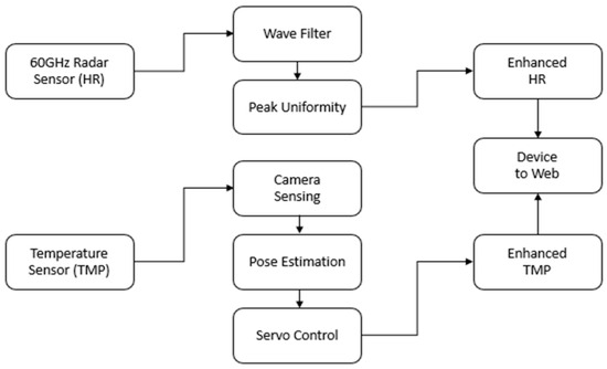
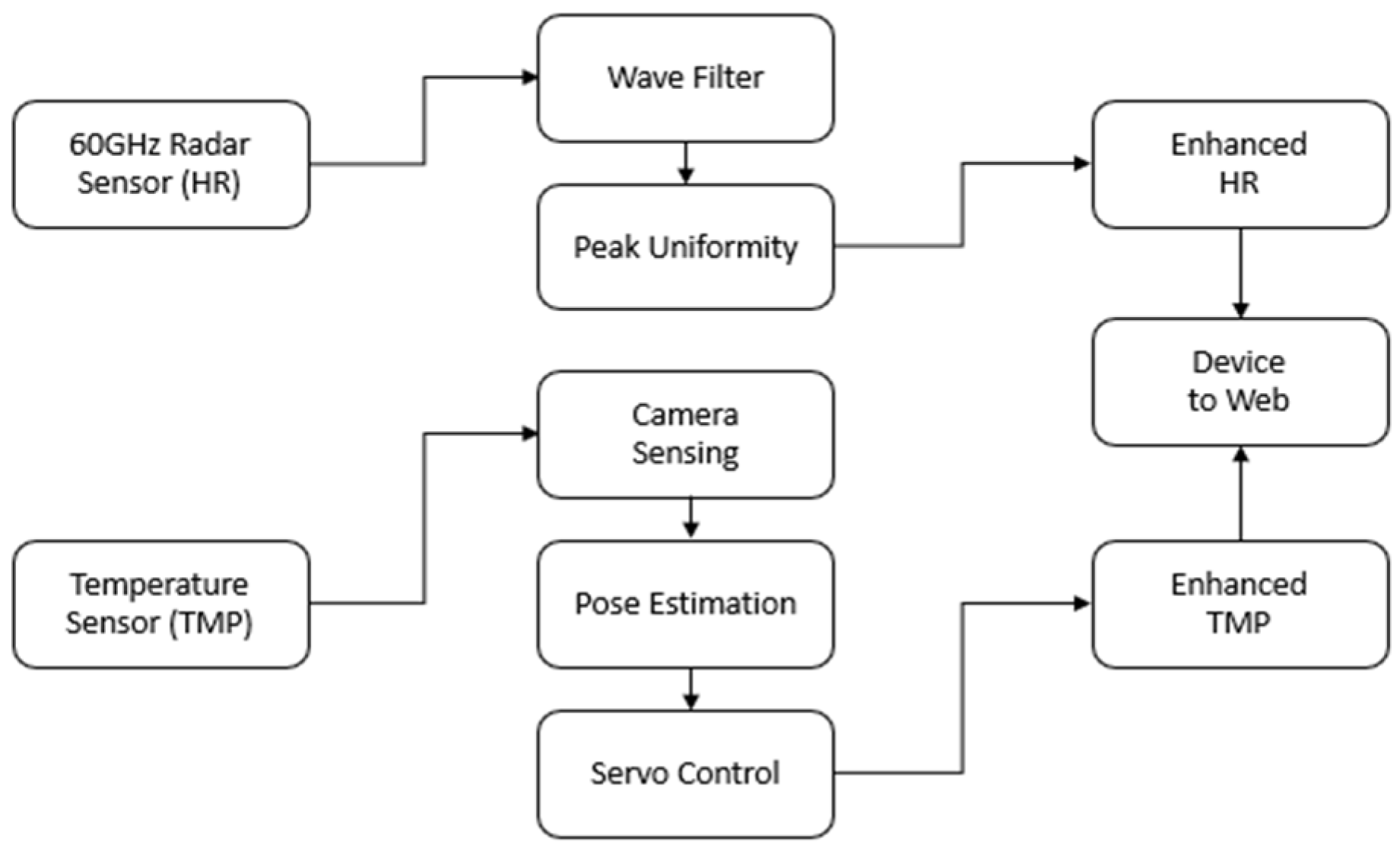
The objective of this system is the non-contact measurement of a user’s heart rate and body temperature, for which a 60 GHz radar sensor and an infrared temperature sensor are employed as key modules. An integrated camera first identifies the user’s facial coordinates using a Pose Detection algorithm, and these data are used to guide a servo motor that precisely orients the sensors, thereby actively establishing an optimal environment for data acquisition. This study aims to ascertain the level of signal collection accuracy achievable through this signal refinement process. Figure 1 illustrates the overall architecture of the proposed system.
Figure 1.
Overall architecture of the proposed system, including signal processing flow.
In the context of heart rate measurement, we sought to quantify the improvement in accuracy. This was achieved by first applying a Window Filter to the raw signal to effectively eliminate extraneous noise, followed by Peak Uniformity analysis to identify regular patterns within the cardiac waveform. The goal was to determine the quantifiable enhancement in accuracy resulting from these methods.
2.2. Heart Rate Measurement Using a 60 GHz RADAR with Signal Enhancement Techniques
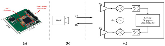
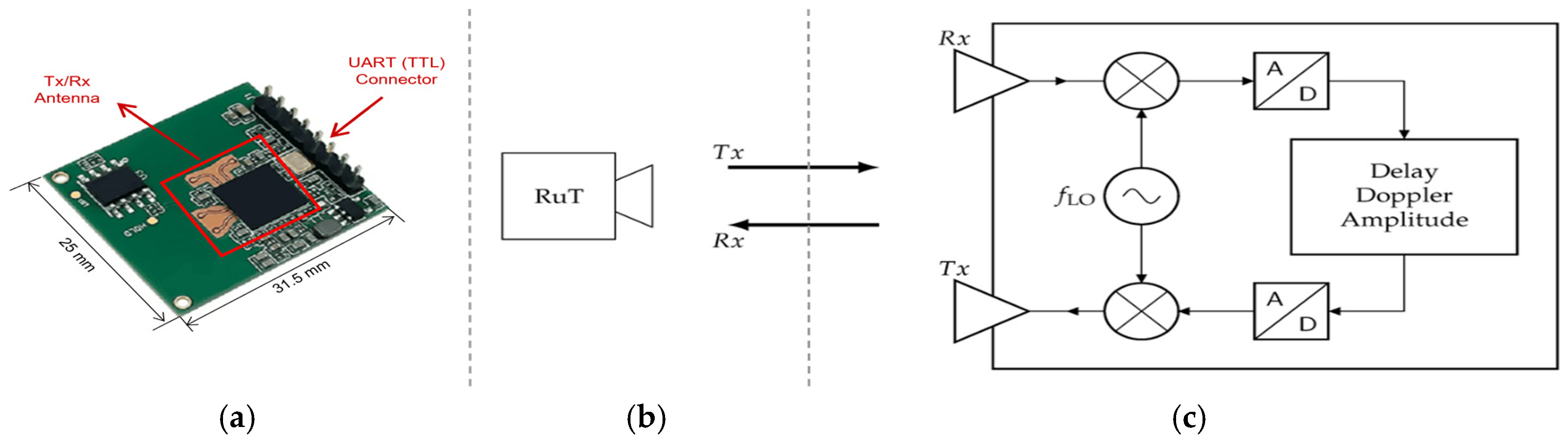
In this study, the HLK-LD6002 60 GHz radar sensor (Hi-Link Electronic Co., Ltd., Shenzhen, China), presented in Figure 2, was utilized for heart rate measurement. The 60 GHz band is optimized for detecting minute vibrations on the skin surface caused by heartbeats, as its high frequency provides excellent range resolution and it has low transmittance through the human body. Furthermore, its short wavelength enables antenna miniaturization, which is advantageous for reducing the overall system size. A key consideration was also that this frequency is within the ISM (Industrial, Scientific, and Medical) band, making it suitable for medical device applications. Based on these advantages, the HLK-LD6002 sensor was ultimately selected, as it achieves a range resolution of 1.7 cm and a velocity resolution of 0.1 m/s with a center frequency of 60.5 GHz and a bandwidth of 2 GHz.
Figure 2.
HLK-LD6002 60 GHz radar sensor: (a) physical module, (b) RuT configuration, (c) system block diagram.
An important architectural consideration of the HLK-LD6002 module is its internal signal processing pipeline. Although the manufacturer does not disclose the detailed firmware implementation, the physical constraints of 60 GHz FMCW radar operation necessitate specific signal processing stages within the module’s on-chip SoC. First, the detection of sub-millimeter cardiac displacement (0.1–0.5 mm) at a wavelength of approximately 5 mm is physically achievable only through phase-domain analysis, requiring I/Q (in-phase/quadrature) channel separation and arctangent-based phase extraction [φ = arctan(Q/I)]. Second, the large chest displacement due to respiration (~5–10 mm) causes phase wrapping that must be resolved through a phase unwrapping algorithm to produce a continuous displacement signal. Third, the limited UART bandwidth (256,000 bps) is insufficient for streaming raw I/Q baseband data sampled at 60 GHz IF rates, confirming that the module outputs internally demodulated data. These considerations indicate that the UART-output waveform used in this study represents a linearized displacement signal in which the cosine-nonlinearity-induced phase artifacts—specifically, the spurious respiratory harmonics that would arise from a simple single-channel phase detector—have been substantially resolved through the internal arctangent demodulation and phase unwrapping process. The term “raw signal” in this paper therefore refers to this module-output displacement waveform prior to the application of our external signal processing pipeline (BPF and Peak Uniformity), and not to the unprocessed I/Q baseband data.
To extract an accurate heart rate from the raw signal collected by the 60 GHz radar, two key signal processing stages are applied sequentially: an adaptive bandpass filter for signal preprocessing, followed by a Peak Uniformity algorithm for robust peak detection and validation. In the preprocessing stage, a 4th-order Butterworth bandpass filter [22] is employed to isolate the cardiac signal component from the raw radar data. The passband is defined between f_L = 0.8 Hz and f_H = 2.5 Hz, corresponding to physiologically valid heart rates of 48 to 150 BPM at rest. The filter’s transfer function is expressed as:
where and are the lower and upper angular frequencies, respectively, and is the filter order. The Butterworth design was selected for its maximally flat magnitude response in the passband, which minimizes distortion of the cardiac waveform. The radar sensor outputs data at a sampling rate of 20 Hz via UART communication to an ESP32 microcontroller (Espressif Systems Co., Ltd., Shanghai, China), which performs filtering in real-time. An adaptive mechanism adjusts the passband boundaries by ±0.2 Hz based on the running estimate of the dominant frequency in the previous 10-s window, allowing the filter to accommodate variations in the subject’s heart rate over time.
Following the bandpass filtering, the Peak Uniformity algorithm is applied to detect valid cardiac peaks and compute the heart rate. Peak candidates are first identified using a local maximum detection method with a minimum inter-peak distance constraint of 0.3 s (corresponding to 200 BPM). The algorithm then evaluates two uniformity criteria for the detected peaks. First, it computes the coefficient of variation (CV) of the inter-beat intervals (IBIs), defined as CV = SD(IBI)/mean(IBI). A CV value exceeding 0.30 triggers a re-evaluation cycle, as it indicates irregular peak spacing likely caused by noise or motion artifacts. Second, the algorithm examines peak height consistency by computing an adaptive threshold T_h = mean(A) − k × SD(A), where A represents the set of detected peak amplitudes and k is a sensitivity parameter set to 1.5. Peaks with amplitudes below T_h are classified as artifacts and excluded from the heart rate calculation. Additionally, the algorithm enforces a physiological constraint that discards any IBI shorter than 0.3 s (corresponding to a maximum of 200 BPM) or longer than 2.0 s (corresponding to a minimum of 30 BPM). The final heart rate is computed as HR = 60/mean (IBI_valid), where IBI_valid represents the set of validated inter-beat intervals. This two-stage approach ensures that only physiologically plausible and temporally consistent peaks contribute to the heart rate estimate.
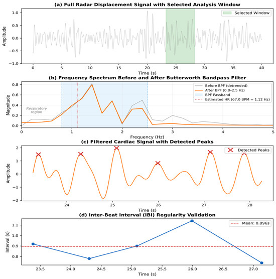
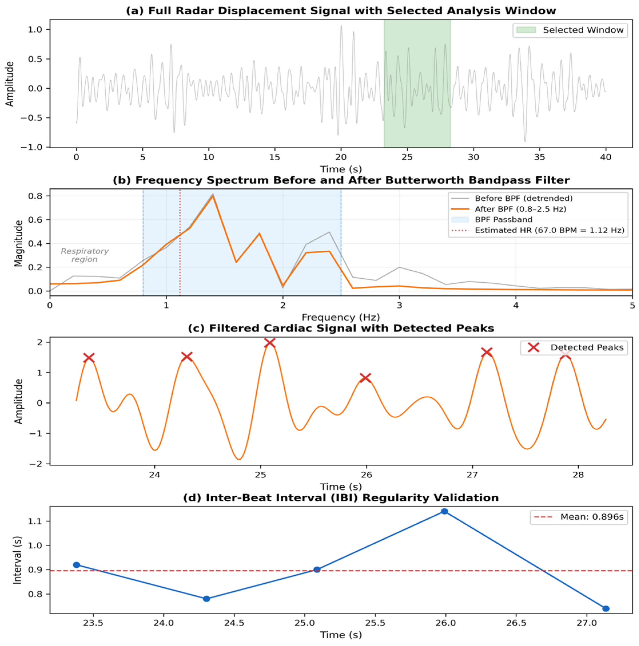
Figure 3 visually demonstrates the step-by-step efficacy of the proposed signal processing pipeline. Figure 3a shows the full raw radar displacement signal, where the green shaded region highlights the optimal analysis window automatically selected based on amplitude periodicity and signal-to-noise ratio (SNR). Figure 3b presents the frequency spectrum of the selected window before and after applying the 4th-order Butterworth bandpass filter (Equation (1), passband 0.8–2.5 Hz). The pre-filter spectrum (gray) exhibits energy distributed across a broad frequency range including the respiratory region below 0.8 Hz and high-frequency components above 2.5 Hz, while the post-filter spectrum (orange) shows that the BPF effectively isolates the cardiac frequency band, with the dominant peak corresponding to the estimated heart rate. Figure 3c presents the filtered time-domain waveform within the selected window, representing the isolated cardiac component with valid peaks detected by the Peak Uniformity algorithm explicitly marked. It should be noted that the change in waveform morphology between Figure 3a and Figure 3c is attributable to the bandpass filtering operation, not merely to a change in the observation time scale. Figure 3d plots the inter-beat intervals (IBI) derived from these peaks to verify the regularity of the heart rhythm; the consistency of the intervals around the mean IBI (indicated by the red dashed line) serves as a validation metric. This multi-stage verification process effectively excludes irregular noise and ensures that only physiologically meaningful data are incorporated into the final heart rate calculation, thereby maximizing measurement reliability.
Figure 3.
Signal processing pipeline: (a) raw signal with selected window, (b) frequency spectrum before and after BPF, (c) filtered signal with detected peaks, (d) inter-beat interval validation.
2.3. Infrared Temperature Sensor with YOLO-Based Tracking
In this study, the body temperature measurement module employs the MLX90614ESF-DCI infrared (IR) sensor (Melexis NV, Ieper, Belgium), characterized by a broad measurement range of −70 °C to +380 °C, a high degree of accuracy at ±0.5 °C within ambient temperatures (0 °C to +50 °C), and a fine resolution of 0.02 °C. It interfaces with the ESP32 microcontroller via I2C at a clock frequency of 100 kHz. The sensor’s native 90° field of view (FOV) has been narrowed to approximately 5° by integrating a germanium (Ge) Fresnel lens with a focal length of 25 mm and a transmittance of approximately 95% in the 8–14 µm wavelength range. This optical modification reduces the measurement spot diameter to approximately 8.7 cm at a distance of 1 m, enabling the sensor to accurately target the facial region while minimizing interference from the surrounding background. To improve measurement reliability, an ambient temperature compensation algorithm is applied using the sensor’s built-in ambient temperature register (T_a). The corrected object temperature is computed as T_corrected = T_object + α × (T_a − T_ref), where T_object is the raw measured temperature, T_a is the current ambient temperature, T_ref is the calibration reference ambient temperature (25 °C), and α is an empirically determined compensation coefficient (α = 0.12). This linear correction accounts for the systematic bias introduced by variations in ambient thermal conditions. Additionally, a multi-sampling noise reduction strategy is employed in which 10 consecutive temperature readings are acquired at 100 ms intervals (1-s acquisition window), and the median value is selected as the final measurement. The median filter was chosen over averaging to provide robustness against occasional outlier readings caused by transient obstructions or sensor noise spikes.
A camera and servo motors are integrated to function as a real-time user-tracking system, which ensures that the IR sensor maintains an optimal orientation toward the subject’s face during measurement. The process begins with the use of an HD camera (OV2640, OmniVision Technologies, Inc., Santa Clara, CA, USA) (1280 × 720 resolution, 30 FPS) to acquire video footage. Video frames are processed by a YOLOv8-pose model [23] pre-trained on the COCO keypoint dataset, which detects 17 body keypoints per person. Among these, the nose keypoint (keypoint index 0) is selected as the primary reference for face localization, as it provides the most stable and centered representation of the facial region. Although the inner canthus is the preferred clinical measurement site, the large measurement spot size (approx. 8.7 cm at 1 m) of the sensor necessitates a different approach to avoid the partial volume effect [18]. Aligning the sensor to the geometric center of the face (the nose) ensures that the sensor’s field of view is fully occupied by facial tissue (cheeks and periorbital regions), thereby preventing thermal attenuation caused by background or hair interference [19]. This strategy maximizes the fill factor within the measurement spot. The model operates at an input resolution of 640 × 640 pixels and achieves an inference speed of approximately 15 FPS on the ESP32-CAM module (Ai-Thinker Technology Co., Ltd., Shenzhen, China), providing sufficient temporal resolution for tracking a stationary or slowly moving subject at a distance of 1 m. The detected nose coordinates are converted into servo motor control angles using a proportional control scheme. The update laws for the pan () and tilt () angles are defined as:
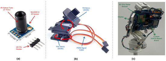
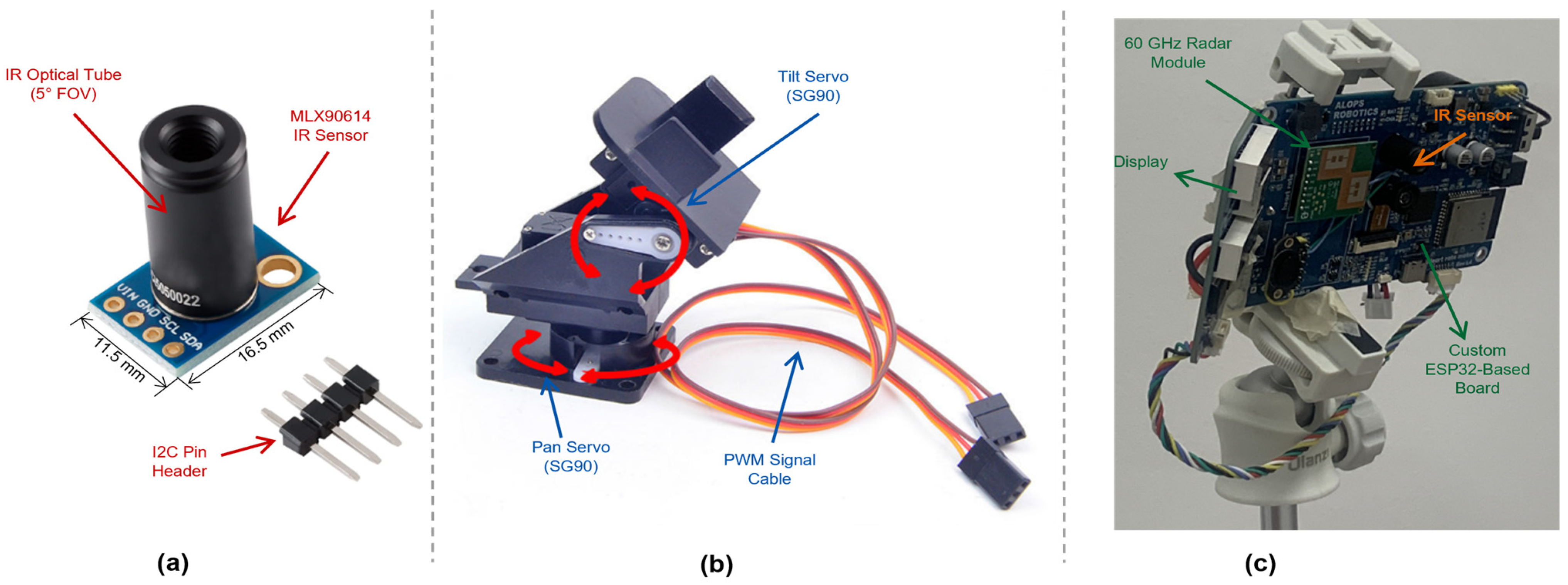
where denotes the current servo angle, represents the image center coordinates , and is the proportional gain set to 0.05 degrees/pixel. The 2-axis servo motor arrangement (SG90 servos (Tower Pro Pte Ltd., Taipei, Taiwan), 1.5 kg·cm torque, 180° operational range) is driven by PWM control signals from the ESP32 at a frequency of 50 Hz. To prevent mechanical jitter, a dead zone of ±10 pixels around the image center is implemented, within which no servo adjustment is made. The tracking system includes a fallback strategy for cases where the YOLO model fails to detect the subject for more than 5 consecutive frames: the servos hold their last known position for up to 3 s before returning to the default center position. The end-to-end tracking latency from frame capture to servo response is approximately 150 ms. Figure 4 shows the hardware components of the proposed system, including the IR sensor module, pan/tilt servo mechanism, and assembled monitoring unit.
Figure 4.
Hardware components of the proposed system: (a) IR sensor module, (b) pan/tilt servo mechanism, (c) assembled monitoring unit.
2.4. System Integration and Data Processing
The central processing unit of the proposed system is an ESP32-WROOM-32 microcontroller [24] (Espressif Systems (Co., Ltd., Shanghai, China)), which operates at a clock frequency of 240 MHz with 520 KB of SRAM. The ESP32 manages all sensor communications, signal processing, and servo motor control. The 60 GHz radar sensor (HLK-LD6002 (Hi-Link Electronic Co., Ltd., Shenzhen, China)) communicates with the ESP32 via UART at a baud rate of 256,000 bps, transmitting raw displacement data at 20 Hz. The IR temperature sensor (MLX90614ESF-DCI (Melexis NV, Ieper, Belgium)) is connected via I2C at 100 kHz. The camera module (OV2640 (OmniVision Technologies, Inc., Santa Clara, CA, USA) on ESP32-CAM (Ai-Thinker Technology Co., Ltd., Shenzhen, China)) captures frames at 30 FPS, which are processed by the YOLO Pose model running on a custom-designed MCU board connected via serial communication. This board performs the computationally intensive pose estimation and transmits the detected keypoint coordinates back to the ESP32 for servo motor control. The radar and IR measurements are synchronized through a shared timestamp protocol, with both sensors triggered within the same 1-s acquisition cycle. The processed vital sign data (heart rate and body temperature) are transmitted to a Flask-based web server running on the board, which provides a real-time dashboard accessible via a local Wi-Fi network. The web interface displays the current heart rate, body temperature, measurement confidence indicators, and historical trend graphs with an update interval of 2 s.
3. Results
The experiments for this study were conducted within a controlled laboratory environment to ensure consistent conditions. The ambient background noise was maintained at a level below 40 dB to minimize its potential impact on the measurements. Throughout the data acquisition process, both the heart rate (HR) and body temperature (TEMP) sensors were positioned at a fixed distance of 1.0 m from each participant. The study cohort comprised 30 healthy adult participants (designated PA1-PA30). The age demographics ranged from 27 to 68 years, with a mean age of 40.8. The gender distribution consisted of 21 male (70%) and 9 female (30%) participants. All participants exhibited a resting heart rate within the normal physiological range of 50–110 BPM. This study was approved by the Institutional Review Board (IRB) of Kangwon National University (KWNUIRB-2025-07-007-001).
To objectively assess the performance of the proposed system, its measurements were benchmarked against commercially available reference devices. For the comparison of heart rate accuracy, a MAX30102 PPG sensor (Analog Devices, Inc., Wilmington, MA, USA) [25] was employed, featuring an accuracy of ±2 BPM and a sampling rate of 100 Hz via I2C communication. While a three-lead electrocardiogram (ECG) serves as the clinical gold standard for HR measurement, the MAX30102 PPG sensor was selected as a practical reference device due to its validated accuracy under resting conditions. In this study, a Braun ThermoScan 7 ear thermometer (Braun GmbH, Kronberg, Germany) (accuracy ±0.2 °C) was used to establish reference values for body temperature, as it is a clinically validated tympanic thermometer widely accepted for clinical use. Prior to performing comparative statistical analyses, the normality of the error distributions was assessed using the Shapiro–Wilk test [26] for both HR and temperature measurements (n = 30). Where normality was confirmed (p > 0.05), paired t-tests were used for statistical comparison; otherwise, the Wilcoxon signed-rank test was applied as a non-parametric alternative. In addition to the paired comparisons, Bland–Altman analysis [27] was performed to evaluate the agreement between the proposed system and reference devices. For each comparison, the mean bias and 95% limits of agreement (LoA = mean bias ± 1.96 × SD) were calculated. Effect sizes were quantified using Cohen’s d [28] to assess the practical significance of the observed improvements.
All error values are reported in beats per minute (BPM). “Raw Radar Signal” refers to heart rate estimated directly from the unprocessed 60 GHz radar output using simple peak detection. “With BPF + Peak Uniformity” refers to heart rate estimated after applying the fourth-order Butterworth bandpass filter (0.8–2.5 Hz) and the Peak Uniformity validation algorithm described in Section 2.2. MAE: mean of |HR_measured − HR_reference| across all 30 participants; Std. Dev. of Error: standard deviation of the absolute errors; Max./Min. Absolute Error: largest/smallest individual absolute error observed. * Result of a paired t-test on the Mean Absolute Error.
The maximum absolute error was 57.0 BPM for the raw radar signal and 19.8 BPM for the processed signal (with BPF + Peak Uniformity), showing that the unprocessed radar produced very large errors in some measurements. The difference in accuracy between the two conditions is also statistically significant. A paired t-test on the Mean Absolute Error resulted in a p-value of 0.002 (p < 0.01), demonstrating that the accuracy improvement achieved by the proposed signal processing pipeline is statistically significant.
Regarding the range of errors, the maximum error for the IR sensor was 5.0 °C, while the maximum error for the face recognition AI was 2.2 °C. Notably, the maximum error of the face recognition AI (2.2 °C) is lower than the minimum error of the IR sensor (2.6 °C). The difference in the mean errors between the two measurement methods was confirmed to be statistically significant by a paired t-test (p < 0.001). It is important to interpret this Mean Absolute Error (MAE) of 1.64 °C in the context of remote monitoring: measurements were taken at a distance of 1.0 m from unconstrained participants. Under these challenging conditions, where fixed sensors typically exhibit errors exceeding 4 °C due to misalignment, the proposed tracking system’s ability to maintain a significantly reduced error margin represents a critical technical advancement. This validates the feasibility of using low-cost IR sensors for relative trend monitoring in home environments, overcoming the fundamental alignment limitations of conventional non-contact thermometry.
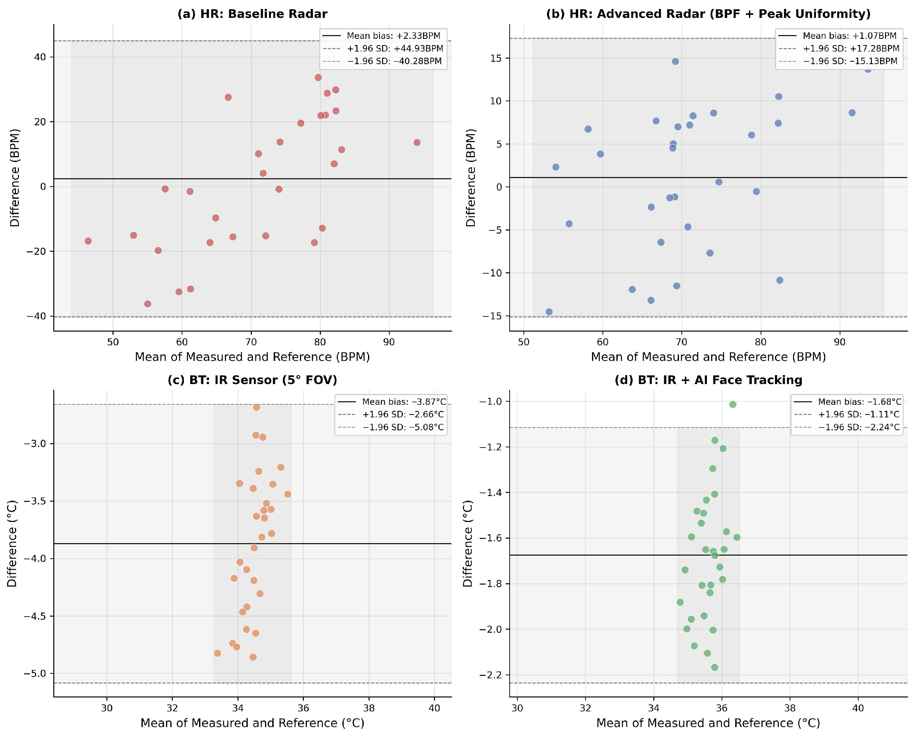
To further evaluate the agreement between the proposed system and the reference devices, Bland–Altman analysis was performed. Figure 5 presents the Bland–Altman plots for all four measurement conditions: (a) baseline radar HR, (b) advanced radar HR with BPF and Peak Uniformity, (c) IR sensor BT with 5° FOV, and (d) IR sensor BT with AI-based face tracking. While the reference device (MAX30102 (Analog Devices, Inc., Wilmington, MA, USA)) possesses inherent uncertainty compared to clinical-grade ECGs, the substantial narrowing of the 95% limits of agreement (LoA) from ±44.9 BPM (baseline) to ±17.3 BPM (advanced) indicates that the proposed signal processing pipeline significantly enhances the consistency and stability of the measurement against motion artifacts. This improvement demonstrates the system’s robustness in extracting reliable cardiac rhythms, independent of the reference device’s absolute precision limitations. Similarly, the AI-enhanced BT measurement showed a tighter LoA (±0.56 °C) compared to the IR-only configuration (±1.17 °C). Notably, all four configurations showed no proportional bias pattern, indicating stable performance across the physiological measurement range.
Figure 5.
Bland–Altman analysis of measurement agreement. Subfigures (a,b) show heart rate (HR) results, and subfigures (c,d) show body temperature (BT) results.
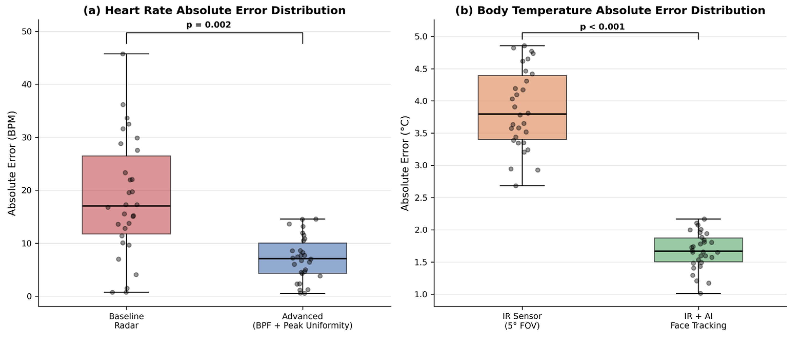
Figure 6 shows box plots comparing the absolute error distributions for both HR and BT measurements. The median absolute error for HR decreased from 17.8 BPM (baseline) to 5.8 BPM (advanced), with the interquartile range also narrowing substantially. For BT, the median absolute error decreased from 3.8 °C (IR only) to 1.6 °C (AI-enhanced). In both cases, the advanced methods produced fewer outliers and a more compact error distribution. The differences were statistically significant (HR: p = 0.002; BT: p < 0.001).
Figure 6.
Comparison of measurement error distribution (n = 30): (a) heart rate; (b) body temperature.
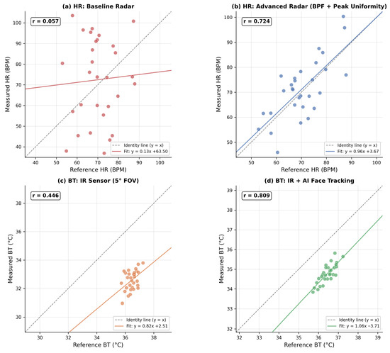
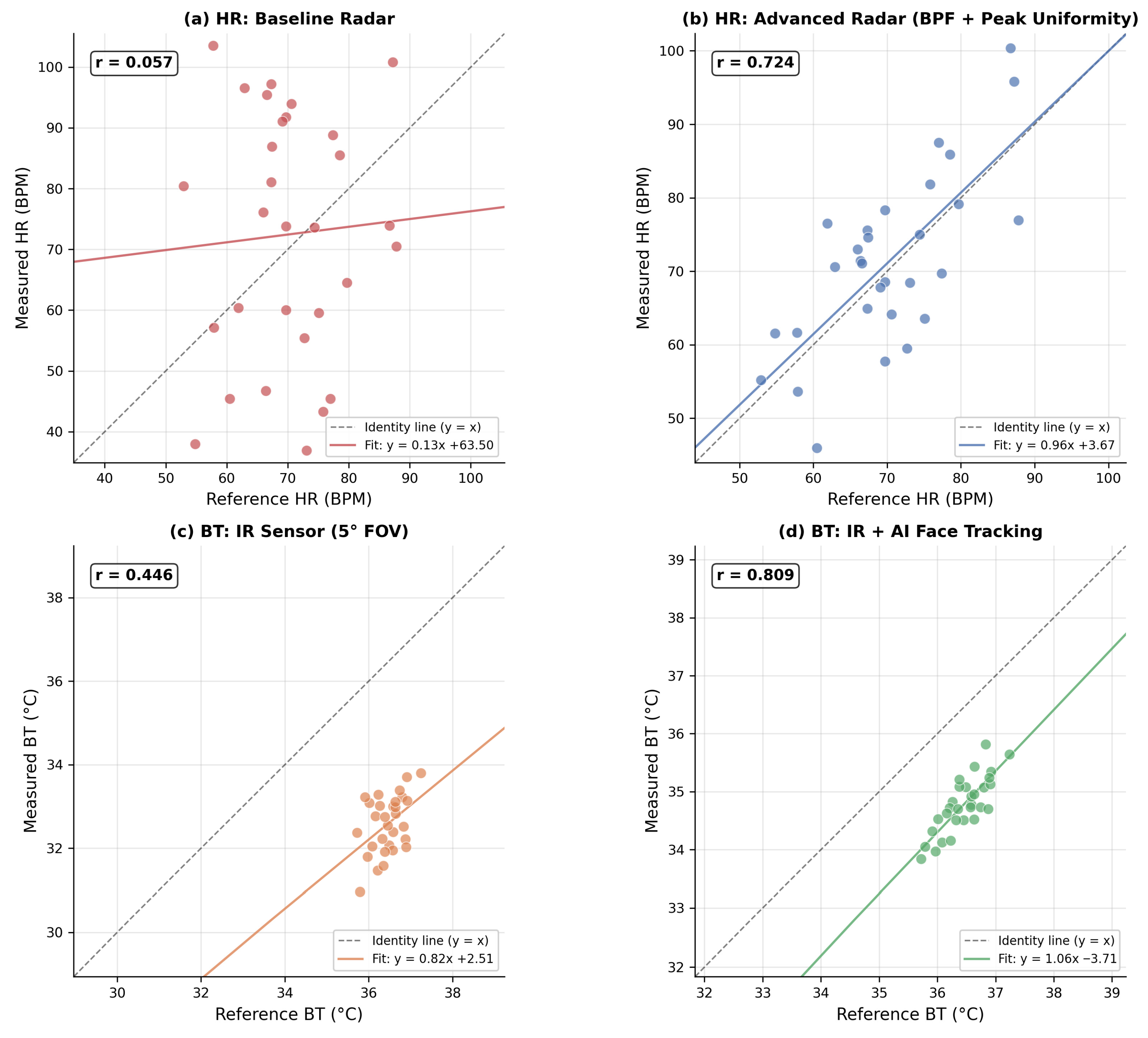
Figure 7 presents scatter plots of measured versus reference values with regression analysis. The advanced radar HR measurements showed a markedly improved correlation (r = 0.724) compared to the baseline (r = 0.057), with the regression line closely approximating the identity line (y = x). For BT, the AI-enhanced method achieved a correlation of r = 0.809 versus r = 0.446 for the IR-only method, demonstrating that face tracking substantially improved the sensor’s ability to capture true temperature variations across participants.
Figure 7.
Measured vs. reference values with regression analysis: (a,b) heart rate; (c,d) body temperature.
4. Discussion
This study sought to validate the performance advantages of a biosignal measurement system, which incorporates artificial intelligence (AI) and advanced signal processing techniques, in comparison to conventional methods via two independent experiments.
The results of the first experiment (Table 1) indicated that the proposed signal processing pipeline (BPF + Peak Uniformity) achieved a statistically significant enhancement in heart rate measurement accuracy over the raw radar signal (p = 0.002). Whereas the raw radar signal was prone to extreme outliers with errors as high as 57.0 BPM, the processed signal effectively mitigated such anomalies and maintained consistent performance. This demonstrates that the combination of Butterworth bandpass filtering for noise suppression and Peak Uniformity analysis for robust peak validation can be used to manage signal distortions that may arise in practical use-case scenarios.
Table 1.
Comparison of heart rate measurement error between raw radar signal and proposed signal processing pipeline (BPF + Peak Uniformity) (n = 30).
The second experiment (Table 2) revealed that the face recognition AI method exhibited markedly superior accuracy and consistency in non-contact thermometry compared to the conventional IR sensor. The AI system’s Mean Absolute Error (MAE) of 1.64 °C was less than half that of the IR sensor (4.10 °C), and its standard deviation was smaller by more than a factor of two, indicating high measurement reliability. This implies that the AI is capable of inferring a value more proximate to core body temperature by holistically considering environmental variables and individual physiological characteristics. The observation that the maximum error of the AI system was lower than the minimum error of the conventional sensor particularly underscores the system’s high stability and its reduced susceptibility to measurement failures.
Table 2.
Analysis of Body temperature measurement error and statistical significance by measurement method.
To contextualize these results, Table 3 presents a quantitative comparison of the proposed system with representative studies in non-contact vital sign monitoring.
Table 3.
Quantitative comparison of the proposed system with representative studies in non-contact vital sign monitoring.
For HR measurement, Droitcour et al. [14] achieved radar-based vital sign detection using a 2.4 GHz Doppler radar but reported limited accuracy due to low frequency resolution. Chen et al. [16] demonstrated improved HR estimation using 77 GHz FMCW mm Wave radar with a DR-MUSIC algorithm, reporting an error rate of approximately 1.69–2.61% across distances up to 1.5 m under controlled conditions. More recently, Hao et al. [17] validated mmWave radar-based vital sign detection with improved signal processing techniques, and Malešević et al. [29] demonstrated that deep learning approaches can enhance Doppler radar heartbeat detection accuracy. Our system achieves an MAE of 5.28 BPM at 1.0 m distance with 30 participants, which compares favorably given the longer measurement range. For BT measurement, Dell’Isola et al. [18] reported that non-contact IR thermometers exhibit typical uncertainties of 0.5–2.0 °C depending on ambient conditions [19]. Our AI-guided system reduces the MAE to 1.64 °C, representing a meaningful improvement. However, it should be noted that this temperature MAE, while substantially better than the baseline IR sensor (4.10 °C), does not yet meet the clinical requirements for fever screening (±0.3 °C per ISO 80601-2-56 [30]). Consequently, the current system is more suitable for relative temperature trend monitoring rather than absolute temperature determination for diagnostic purposes. Further refinement of the IR measurement pipeline, including improved ambient compensation and higher-precision optics, would be necessary to achieve clinically acceptable accuracy for fever detection.
The practical implications of this integrated system are substantial. Unlike conventional handheld IR thermometers that require a trained operator to manually aim the device at close range for each individual measurement, the proposed system enables fully autonomous, continuous temperature and heart rate monitoring at a distance of 1.0 m without human intervention. This distinction is critical for several practical deployment scenarios, for example, in infectious disease isolation wards where minimizing healthcare worker exposure is paramount, in home-based monitoring of elderly patients living alone who may be unable to perform self-measurements, and in long-term care facilities where continuous trend monitoring of multiple residents is needed. The automatic tracking capability further reduces the need for user training or operator expertise, making deployment feasible in diverse healthcare settings. For pandemic-response scenarios, this technology provides a critical tool for the rapid screening and monitoring of large populations without physical contact.
A key advantage of this work is the comprehensive integration of multiple technologies into a single coherent platform. Unlike prior studies that typically address heart rate or temperature in isolation, our system combines advanced signal processing for HR, high-precision IR thermography for BT, YOLO Pose-based tracking for sensor alignment, and real-time data visualization through a microcontroller and web interface. This holistic approach not only improves individual measurement accuracy but also enhances the robustness and reliability of the system across varying environmental conditions and user behaviors.
Several practical considerations merit discussion regarding the deployment of the AI-guided tracking system. First, in clinical settings or during pandemics, patients commonly wear face masks, which may occlude the nose keypoint used as the primary tracking reference. While the YOLOv8-pose model was trained on the COCO dataset, which includes partially occluded subjects, and could still infer the nose position with reduced confidence, systematic evaluation under mask-wearing conditions was not performed in this study. A potential fallback strategy would involve switching the tracking reference to the midpoint of the two eye keypoints (keypoints 1 and 2), which remain visible above the mask, and applying a fixed vertical offset to estimate the facial center. Second, if the forehead was selected as the temperature measurement target instead of the nose region, the measured temperature would be expected to differ, as forehead skin temperature is typically 0.5–1.0 °C lower than the periorbital or nasal region. However, given the current sensor’s measurement spot diameter of approximately 8.7 cm at 1.0 m, the field of view inherently encompasses multiple facial regions (forehead, cheeks, and periorbital area) when aligned to the nose, partially mitigating this concern. Dedicated calibration for forehead-targeted measurement would be necessary for precise absolute temperature estimation.
The current system is designed for single-person monitoring and operates at a fixed sensor-to-subject distance of 1.0 m. In multi-person scenarios where more than one individual is present within the sensor’s field of view, the YOLOv8-pose model can detect multiple persons simultaneously; however, the HLK-LD6002 radar sensor operates in a single-target mode and cannot spatially resolve multiple subjects. Consequently, radar-based HR measurements would be unreliable in the presence of multiple persons at similar distances. Extension to multi-person monitoring would require a MIMO radar architecture with beamforming capabilities to spatially separate individual targets, combined with a multi-object tracking algorithm to maintain consistent identity association across frames.
Regarding the measurement range, the system was validated at a fixed distance of 1.0 m, which represents a practical limit determined by three independent factors. First, the IR sensor’s measurement spot diameter scales linearly with distance (approximately 8.7 cm per meter with the 5° FOV lens); beyond 1.0 m, the spot increasingly extends beyond the facial boundary, introducing background thermal interference that degrades temperature accuracy. Second, the 60 GHz radar signal experiences atmospheric attenuation of approximately 15 dB/km combined with free-space path loss, which reduces the signal-to-noise ratio of the reflected cardiac displacement signal at greater distances. Third, the camera-based keypoint detection accuracy degrades with distance as the facial region occupies fewer pixels in the image frame, reducing the precision of servo motor alignment. Future work could address these limitations through higher-gain antenna designs for the radar, narrower FOV optics for the IR sensor, and higher-resolution cameras with optical zoom capabilities.
5. Conclusions
This study presents a comprehensive, non-contact vital sign monitoring system that successfully integrates 60 GHz radar sensor technology, infrared thermography, and AI-driven YOLO Pose-based tracking to achieve significant improvements in both heart rate and body temperature measurement accuracy. The advanced signal processing algorithms (Window Filter and Peak Uniformity) reduced HR measurement error by 61.5%, while the AI-guided IR sensor with automatic tracking reduced temperature measurement error by 60.0%, with both improvements being statistically significant.
The significance of these findings extends beyond the laboratory setting. By eliminating physical contact and automating sensor alignment, the system addresses critical limitations of existing vital sign monitoring technologies, which are particularly relevant in the context of infectious disease outbreaks and the growing demand for home-based healthcare solutions. While the HR measurement accuracy (MAE 5.28 BPM) shows promise for remote monitoring applications, the temperature measurement demonstrates a statistically significant technical improvement over conventional fixed-sensor methods, establishing a foundation for future high-precision remote thermography rather than immediate clinical diagnosis.
Several limitations of this study should be acknowledged. First, the experiments were conducted in a controlled laboratory environment with ambient noise below 40 dB and a fixed sensor-to-subject distance of 1.0 m, which may not fully represent the variability of real-world clinical or home settings. Second, the participant cohort (n = 30) was relatively homogeneous in terms of age distribution (mean 40.8 years) and was predominantly male (70%), which limits the generalizability of the results to pediatric, geriatric, or more diverse populations. Third, the MAX30102 PPG sensor used as the HR reference device, while validated for resting conditions (±2 BPM), is not equivalent to clinical gold standard equipment such as a three-lead ECG; this distinction should be considered when interpreting the reported accuracy metrics. Fourth, the body temperature MAE of 1.64 °C, although significantly improved from the baseline, exceeds the ±0.3 °C threshold required by ISO 80601-2-56 for clinical fever screening, indicating that the current system is more appropriate for trend monitoring than for absolute diagnostic temperature assessment. Fifth, the system is currently designed for single-person monitoring only; the radar sensor operates in a single-target mode and cannot spatially resolve multiple subjects, which limits deployment in multi-occupancy environments. Sixth, the AI-guided tracking system was not evaluated under conditions where subjects wear face masks, which may affect the reliability of nose keypoint detection in clinical settings. Seventh, although the HLK-LD6002 module’s internal arctangent demodulation substantially eliminates the spurious harmonic generation caused by phase detector nonlinearity, residual respiratory harmonics originating from the actual nonlinear biomechanics of chest wall motion may still persist at low amplitudes within the cardiac frequency band. Explicit respiratory signal extraction and adaptive interference cancelation were not implemented in this study and remain a direction for future work. Future research should evaluate the system’s performance across diverse age groups, in varied environmental conditions (e.g., different ambient temperatures and humidity levels), and at varying measurement distances to fully establish its clinical applicability.
Future directions for this work include expansion to multi-person monitoring scenarios, the integration of additional vital signs (such as respiratory rate and oxygen saturation), and validation in clinical settings with patient populations exhibiting various pathological conditions. A particularly promising direction is the extension of AI from its current role in sensor orientation to radar signal processing itself. By synchronizing heart rate data from a wearable reference device (e.g., PPG or ECG) with simultaneously recorded radar signals, a supervised training dataset could be constructed to train deep learning models for more accurate cardiac signal extraction. Such an approach could learn to implicitly separate respiratory harmonics and intermodulation products from the cardiac component, potentially achieving higher accuracy than traditional bandpass filtering alone. Recent work by Malešević et al. [29] has demonstrated the feasibility of neural network-based heartbeat detection from Doppler radar signals, and self-supervised or transfer learning strategies could further reduce the labeled data requirements. Additionally, the incorporation of machine learning models for predictive health analytics could enhance the system’s utility for early disease detection and intervention.
Author Contributions
Conceptualization, S.S. and C.K.; methodology, S.S.; software, S.S.; validation, S.S. and C.K.; formal analysis, S.S.; investigation, S.S.; resources, C.K.; data curation, S.S.; writing—original draft preparation, S.S.; writing—review and editing, C.K.; visualization, S.S.; supervision, C.K.; project administration, C.K. All authors have read and agreed to the published version of the manuscript.
Funding
This work was supported by the Technology Innovation Program (RS-2024-00507228, Development of process upgrade technology for AI self-manufacturing in the cement industry) funded by the Ministry of Trade, Industry & Energy (MOTIE, Korea). This study was supported by 2025 Research Grant from Kangwon National University.
Institutional Review Board Statement
The study was conducted in accordance with the Declaration of Helsinki, and approved by the Institutional Review Board of Kangwon National University (KWNUIRB-2025-07-007-001; 16 September 2025).
Informed Consent Statement
Informed consent was obtained from all subjects involved in the study.
Data Availability Statement
The data presented in this study are available on request from the corresponding author due to privacy restrictions.
Conflicts of Interest
The authors declare no conflicts of interest.
Abbreviations
| HR | Heart Rate |
| Temp | Body Temperature |
| RADAR | Radio Detection and Ranging |
| IR | Infrared |
| FOV | Field of View |
| RPM | Remote Patient Monitoring |
| BPM | Beats Per Minute |
| MAE | Mean Absolute Error |
| PPG | Photoplethysmography |
| vPPG | Video-based Photoplethysmography |
| ECG | Electrocardiogram |
| ISM | Industrial, Scientific, and Medical |
References
- Taylor, M.L.; Thomas, E.E.; Snoswell, C.L.; Smith, A.C.; Caffery, L.J. Does remote patient monitoring reduce acute care use? A systematic review. BMJ Open 2021, 11, e040232. [Google Scholar] [CrossRef] [Scilit]
- World Health Organization. Global Health Observatory Data Repository; WHO: Geneva, Switzerland, 2023. Available online: https://www.who.int/data/gho (accessed on 24 February 2025).
- Noah, B.; Keller, M.S.; Mosadeghi, S.; Stein, L.; Johl, S.; Delshad, S.; Tashjian, V.C.; Lew, D.; Kwan, J.T.; Jusufagic, A.; et al. Impact of remote patient monitoring on clinical outcomes: An updated meta-analysis of randomized controlled trials. NPJ Digit. Med. 2018, 1, 20172. [Google Scholar] [CrossRef] [Scilit]
- Geddes, L.A.; Wald, A. Retrospectroscope: Horatio B. Williams and the first electrocardiographs made in the United States. IEEE Eng. Med. Biol. Mag. 2000, 19, 117–119. [Google Scholar] [CrossRef] [Scilit]
- Steinhubl, S.R.; Waalen, J.; Edwards, A.M.; Ariniello, L.M.; Mehta, R.R.; Ebner, G.S.; Carter, C.; Baca-Motes, K.; Felber, E.; Topol, E.J. Effect of a home-based wearable continuous ECG monitoring patch on detection of undiagnosed atrial fibrillation: The mSToPS randomized clinical trial. JAMA 2018, 320, 146–155. [Google Scholar] [CrossRef] [Scilit]
- Smarr, B.L.; Aschbacher, K.; Fisher, S.M.; Chowdhary, A.; Dilchert, S.; Puldon, K.; Rao, A.; Hecht, F.M.; Mason, A.E. Feasibility of continuous fever monitoring using wearable devices. Sci. Rep. 2020, 10, 21640. [Google Scholar] [CrossRef] [Scilit]
- Hahnen, C.; Freeman, C.G.; Haldar, N.; Hamati, J.N.; Bard, D.M.; Murali, V.; Merli, G.J.; Joseph, J.I.; van Helmond, N. Accuracy of vital signs measurements by a smartwatch and a portable health device: Validation study. JMIR mHealth uHealth 2020, 8, e16811. [Google Scholar] [CrossRef] [Scilit]
- Dunn, J.; Runge, R.; Snyder, M. Wearables and the medical revolution. Per. Med. 2018, 15, 429–448. [Google Scholar] [CrossRef] [Scilit]
- Tarar, A.A.; Mohammad, U.; Srivastava, S.K. Wearable skin sensors and their challenges: A review of transdermal, optical, and mechanical sensors. Biosensors 2020, 10, 56. [Google Scholar] [CrossRef] [Scilit] [PubMed]
- Iqbal, S.M.A.; Mahgoub, I.; Du, E.; Leavitt, M.A.; Asghar, W. Advances in healthcare wearable devices. NPJ Flex. Electron. 2021, 5, 9. [Google Scholar] [CrossRef] [Scilit]
- Verkruysse, W.; Svaasand, L.O.; Nelson, J.S. Remote plethysmographic imaging using ambient light. Opt. Express 2008, 16, 21434–21445. [Google Scholar] [CrossRef] [Scilit] [PubMed]
- Wang, W.; den Brinker, A.C.; Stuijk, S.; de Haan, G. Algorithmic principles of remote PPG. IEEE Trans. Biomed. Eng. 2017, 64, 1479–1491. [Google Scholar] [CrossRef] [Scilit]
- Rouast, P.V.; Adam, M.T.; Chiong, R.; Cornforth, D.; Lux, E. Remote heart rate measurement using low-cost RGB face video: A technical literature review. Front. Comput. Sci. 2018, 12, 858–872. [Google Scholar] [CrossRef] [Scilit]
- Droitcour, A.D.; Boric-Lubecke, O.; Lubecke, V.M.; Lin, J.; Kovacs, G.T.A. Range correlation and I/Q performance benefits in single-chip silicon Doppler radars for noncontact cardiopulmonary monitoring. IEEE Trans. Microw. Theory Tech. 2004, 52, 838–848. [Google Scholar] [CrossRef] [Scilit]
- Dong, S.; Wen, L.; Ye, Y.; Zhang, Z.; Wang, Y.; Liu, Z.; Qing, C.; Xu, Y.; Li, C.; Gu, C. A review on recent advancements of biomedical radar for clinical applications. IEEE Open J. Eng. Med. Biol. 2024, 5, 707–724. [Google Scholar] [CrossRef] [Scilit] [PubMed]
- Chen, Y.; Yuan, J.; Tang, J. A high precision vital signs detection method based on millimeter wave radar. Sci. Rep. 2024, 14, 25535. [Google Scholar] [CrossRef] [Scilit] [PubMed]
- Hao, Z.; Wang, Y.; Li, F.; Ding, G.; Fan, K.; Gao, Y. Detection of vital signs based on millimeter wave radar. Sci. Rep. 2025, 15, 28112. [Google Scholar] [CrossRef] [Scilit]
- Dell’Isola, G.B.; Cosentini, E.; Canale, L.; Ficco, G.; Dell’Isola, M. Non-contact body temperature measurement: Uncertainty evaluation and screening decision rule to prevent the spread of COVID-19. Sensors 2021, 21, 346. [Google Scholar] [CrossRef] [Scilit] [PubMed]
- Wang, Q.; Zhou, Y.; Ghassemi, P.; McBride, D.; Casamento, J.P.; Pfefer, T.J. Infrared thermography for measuring elevated body temperature: Clinical accuracy, calibration, and evaluation. Sensors 2022, 22, 215. [Google Scholar] [CrossRef] [Scilit]
- Hassanpour, A.; Yang, B. Contactless vital sign monitoring: A review towards multi-modal multi-task approaches. Sensors 2025, 25, 4792. [Google Scholar] [CrossRef] [Scilit]
- Cao, Z.; Hidalgo, G.; Simon, T.; Wei, S.-E.; Sheikh, Y. OpenPose: Realtime multi-person 2D pose estimation using part affinity fields. IEEE Trans. Pattern Anal. Mach. Intell. 2019, 43, 172–186. [Google Scholar] [CrossRef] [Scilit]
- Challis, R.E.; Kitney, R.I. The design of digital filters for biomedical signal processing. Part 3: The design of Butterworth and Chebychev filters. J. Biomed. Eng. 1983, 5, 91–102. [Google Scholar] [CrossRef] [Scilit] [PubMed]
- Jocher, G.; Chaurasia, A.; Qiu, J. Ultralytics YOLOv8. Available online: https://github.com/ultralytics/ultralytics (accessed on 24 February 2025).
- Mujeeb Rahman, K.K.; Nasor, M.; Zidan, R.; Alsarraj, I.; Hasan, B. IoT-based wireless patient monitor using ESP32 microcontroller. In Proceedings of the 2023 24th International Arab Conference on Information Technology (ACIT); IEEE: New York, NY, USA, 2023; pp. 1–6. [Google Scholar] [CrossRef] [Scilit]
- Tjitra, M.A.; Anju, N.E.; Sudiana, D.; Rizkinia, M. A wireless noninvasive blood pressure measurement system using MAX30102 and random forest regressor for photoplethysmography signals. Computers 2024, 13, 125. [Google Scholar] [CrossRef] [Scilit]
- Shapiro, S.S.; Wilk, M.B. An analysis of variance test for normality (complete samples). Biometrika 1965, 52, 591–611. [Google Scholar] [CrossRef] [Scilit]
- Bland, J.M.; Altman, D.G. Statistical methods for assessing agreement between two methods of clinical measurement. Lancet 1986, 1, 307–310. [Google Scholar] [CrossRef] [Scilit] [PubMed]
- Cohen, J. Statistical Power Analysis for the Behavioral Sciences, 2nd ed.; Lawrence Erlbaum Associates: Hillsdale, NJ, USA, 1988. [Google Scholar] [CrossRef] [Scilit]
- Malešević, N.; Petrović, V.; Belić, M.; Antfolk, C.; Mihajlović, V.; Janković, M. Contactless real-time heartbeat detection via 24 GHz continuous-wave Doppler radar using artificial neural networks. Sensors 2020, 20, 2351. [Google Scholar] [CrossRef] [Scilit] [PubMed]
- ISO 80601-2-56:2017; Medical Electrical Equipment—Part 2-56: Particular Requirements for Basic Safety and Essential Performance of Clinical Thermometers for Body Temperature Measurement. ISO: Geneva, Switzerland, 2017. Available online: https://www.iso.org/standard/67348.html (accessed on 24 February 2025).

Disclaimer/Publisher’s Note: The statements, opinions and data contained in all publications are solely those of the individual author(s) and contributor(s) and not of MDPI and/or the editor(s). MDPI and/or the editor(s) disclaim responsibility for any injury to people or property resulting from any ideas, methods, instructions or products referred to in the content. |
© 2026 by the authors. Licensee MDPI, Basel, Switzerland. This article is an open access article distributed under the terms and conditions of the Creative Commons Attribution (CC BY) license.