


Featured Application
The integrated analysis of gene expression and cytokine concentrations, specifically TNF-α and IL-4, offers a dual-layer approach to monitoring training status. Practically, the observed rapid resolution of TNF-α protein and stable IL-4 expression 48 h post-exercise in athletes could serve as a “recovery benchmark.” Monitoring these markers during micro-cycles could help identify individuals with “delayed inflammatory resolution,” indicating a risk of cumulative fatigue or overreaching. Furthermore, the multi-point correlation between leukocyte indices and IL-4/TNF-α in trained individuals suggests that routine, low-cost hematological tests could be calibrated against molecular signatures to provide a more accessible, daily monitoring tool for assessing “immune readiness” and individualizing training loads in professional soccer.
Abstract
Background: Acute exercise induces measurable inflammatory and hematological responses, which may differ according to training status. Methods: Trained soccer players and sedentary controls completed a progressive exercise test until exhaustion. Blood samples were collected at six time points (up to 48 h post-exercise). Gene expression was assessed using real-time PCR, protein concentration via ELISA, and leukocyte, lymphocyte and neutrophil counts through routine hematology. Analyses included between-group comparisons, within-group time effects, and gene–protein correlations. Results: Sedentary individuals showed delayed increases in TNF-α mRNA (p < 0.05), whereas athletes maintained stable levels. Athletes exhibited consistently higher TNFR-1 expression (p < 0.05), while TNFR-2 showed no differences between groups. IL-4 expression was higher in athletes 30 min (p = 0.038) and 24 h after test (p = 0.002), whereas IL-4 protein decreased in sedentary subjects after 6 h (p = 0.009) and 24 h (p = 0.019). IL-4Rα was transiently elevated post-exercise in sedentary individuals and briefly suppressed in athletes (p = 0.041). Hematological responses showed stronger leukocytosis and neutrophilia in sedentary individuals. Conclusions: The temporal inflammatory patterns differed substantially between groups, indicating more efficient resolution dynamics in trained individuals. These findings highlight practical value for monitoring adaptation and recovery.
1. Introduction
As physical exercise gains popularity, the need for reliable biomarkers to evaluate the effectiveness of training programs becomes increasingly important. Biomarkers are measurable indicators of biological processes that offer valuable insights into the body’s response to exercise [1]. They are critical for designing training programs tailored to individuals or specific groups. By assessing biomarkers before and after exercise, it is possible to identify which types of activity are most effective in achieving targeted health outcomes. Furthermore, biomarkers enable individuals to track their progress over time, providing concrete evidence of the benefits derived from their efforts.
A range of biomarkers is currently used to measure the impact of exercise, including physiological markers such as heart rate, blood pressure, and VO2max, as well as biochemical markers like cytokines, hormones, and metabolic indicators [2,3,4]. While these markers provide valuable information, there is a need for more accurate and specific tools to better understand exercise-induced changes in the body. Cytokines, in particular, have emerged as a promising option for assessing training effectiveness [4]. These signaling molecules play a key role in immune and inflammatory responses and are integral to the body’s adaptation to physical activity [5]. By measuring cytokine levels pre- and post-exercise, researchers can evaluate how different types of exercise influence immune and inflammatory responses, offering deeper insights into the effectiveness of various training regimens.
Cytokines are a diverse group of signaling molecules essential for regulating immune and inflammatory responses, as well as cellular growth and differentiation. This broad category includes various types, each with specific functions and effects on the body, such as interleukins (ILs), tumor necrosis factor (TNF), interferons (IFNs), chemokines, colony-stimulating factors (CSFs), and growth factors. The distinct roles of these cytokines are vital for maintaining homeostasis, but their dysregulation can contribute to the onset and progression of numerous diseases [6].
Recent research has increasingly explored the connection between cytokines and physical activity, as exercise has been shown to influence their expression and secretion in multiple tissues and organs. These findings highlight the potential of cytokines not only as biomarkers for exercise effectiveness but also as mediators of the health benefits associated with regular physical activity.
Interleukins (ILs) are a group of proteins that facilitate communication with leukocytes by binding to specific receptors. They are primarily produced by monocytes, macrophages, CD4+ helper T cells, and endothelial cells [6]. The IL-1 family, a subgroup of pro-inflammatory cytokines, consists of 11 members and plays a pivotal role in innate immune system activation and the enhancement of T lymphocyte function [7].
Interleukin 4 (IL-4) is a cytokine mainly secreted by Th2 lymphocytes, as well as mast cells, eosinophils, and basophils [6]. Functioning as an anti-inflammatory and regulatory mediator within the type I cytokine family, IL-4 plays a central role in adaptive immunity by driving Th2 cell differentiation and inducing IgE production by B cells [8]. It contributes to immune modulation by suppressing classical (M1) macrophage activation and promoting alternative (M2) polarization, thereby facilitating tissue remodeling and repair.
During physical exertion, IL-4 exhibits a different response profile than acute-phase cytokines such as IL-6 or IL-1ra. Its concentration typically remains unchanged immediately following exercise, pointing to its limited role in acute inflammatory response. Studies involving endurance athletes—including distance runners and rowers—have consistently reported stable IL-4 levels both at the protein and mRNA level after intense activity. However, the cytokine’s response appears to be influenced by exercise duration and total workload. While shorter events like a 40 km cycling race produce no measurable change in IL-4 levels, longer endurance challenges (e.g., 171 km) have been associated with significant post-exercise increases in this level [9]. This suggests that IL-4 may become active during the recovery phase, contributing to resolution of inflammation and immune system recalibration with a potential role in the later phases of immune recovery or secondary inflammation resolution.
Rather than mediating immediate immune defense, IL-4 likely acts as a modulator of longer-term responses, particularly by favoring Th2 dominance and, thus, antagonizing Th1-associated pro-inflammatory pathways. This may help prevent excessive inflammation and support immune homeostasis during repeated or prolonged physical stress. Subtle or delayed changes in IL-4 may, therefore, reflect its primarily regulatory function during post-exercise recovery, rather than a direct pro- or anti-inflammatory role in the acute phase.
Tumor necrosis factor alpha (TNF-α), also known as TNF superfamily member 2 (TNFSF2), is a versatile cytokine implicated in various metabolic disorders, including insulin resistance and type 2 diabetes [10]. Predominantly produced by macrophages and adipocytes, TNF-α serves as a key mediator of acute inflammatory responses. Together with IL-6 and IL-1, it plays a critical role in local inflammatory processes [11].
The primary physiological function of TNF-α is to activate leukocytes, directing them to sites of inflammation to eliminate microorganisms and promote the resolution of inflammation [12]. TNF-α signaling operates through two receptors: TNFR-1 and TNFR-2. TNFR-1, which is broadly expressed across most cell types, is primarily associated with pro-inflammatory and apoptotic signaling. In contrast, TNFR-2, found mainly on endothelial cells, epithelial cells, and specific immune cell subsets, is involved in anti-inflammatory signaling and supports cell proliferation [13,14].
The response of TNF-α to exercise varies depending on the intensity of physical activity. Research indicates that regular exercise suppresses TNF-α expression, largely due to the action of IL-6, which promotes the release of anti-inflammatory cytokines such as IL-1ra and IL-10 [15]. This highlights the modulatory effect of exercise on inflammatory pathways, underscoring its role in maintaining immune and metabolic health.
Endurance training modulates the inflammatory response at both transcriptional and protein levels, supporting more efficient immune regulation in active individuals. Regular exercise leads to adaptations that limit excessive inflammation by promoting controlled, transient immune activation following physiological stress. At the gene expression level, trained individuals often show a reduced TNF-α transcriptional response and enhanced IL-4 expression after exertion, reflecting a shift toward an anti-inflammatory profile [16,17]. Protein-level adaptations include lower resting concentrations of pro-inflammatory cytokines and faster post-exercise recovery [18,19]. Additionally, endurance training may regulate cytokine receptor expression, such as TNFR-1 and IL-4Rα, refining immune cell responsiveness [20,21]. These changes underpin the “exercise-induced anti-inflammatory effect,” which facilitates quicker resolution of inflammation and may reduce the risk of chronic inflammatory conditions [18,22].
The primary aim of this study was to investigate the dynamics of the inflammatory response to a single bout of acute physical exertion, with particular focus given to the temporal changes in gene expression and circulating protein levels of key cytokines and their receptors. Importantly, this research sought not only to track the immunological fluctuations occurring at multiple time points post-exercise, but also to compare these responses between trained athletes and sedentary individuals. In doing so, this study aimed to elucidate how long-term physical training modulates both the magnitude and pattern of immune activation and regulation following exercise-induced physiological stress. This dual approach enabled a comprehensive evaluation of both the short-term kinetics of inflammation and the long-term adaptations associated with training status, contributing to a better understanding of immune system plasticity in response to repeated exercise stimuli.
2. Materials and Methods
2.1. Participants
This study received ethical approval from the Bioethics Committee of the University of Szczecin (approval no. US.003.5.2024, 22 May 2024). All participants were informed in writing about the study’s objectives, procedures, risks, and benefits. Informed consent was obtained after confirming participants’ full understanding. Anonymity and data confidentiality were ensured.
Two groups of healthy males aged 20–25 participated in the study. The trained group included 15 football players with over 10 years of systematic training (approx. 12 h/week) and no history of injuries, chronic diseases, or medication/supplement use. Participants were recruited through a targeted screening process designed to ensure homogeneity within each group and full compliance with the study’s inclusion criteria. All individuals who met the criteria and provided informed consent were enrolled and completed the entire study protocol, including the exercise test and all six blood sampling time points. No participant withdrew or was lost to follow-up, and no attrition occurred during data collection. The untrained group comprised 15 physically inactive individuals matched for age and body composition, as verified by the Global Physical Activity Questionnaire (GPAQ). Both groups met strict exclusion criteria, including absence of cardiovascular, neurological, or psychiatric conditions.
2.2. Intervention
Each participant completed an individualized treadmill test until voluntary exhaustion, aimed at eliciting maximal metabolic and cardiovascular effort. The protocol began with a 3 min warm-up, followed by incremental increases in speed and/or incline every 2 min until the subject could no longer continue despite encouragement. Exhaustion was confirmed by physical signs of fatigue and an inability to maintain pace. The test was tailored to an individual’s fitness level to standardize physiological strain across groups.
Although the treadmill running speed was individualized, the exercise intensity was standardized across groups by referencing each participant’s physiological profile obtained during the VO2max test. Running intensity was set at the speed corresponding to the individually determined anaerobic threshold, ensuring comparable relative strain (%VO2max). Heart rate was recorded continuously, and both groups reached similar percentages of HRmax during the protocol. Capillary lactate concentrations measured immediately post-exercise further confirmed that participants exercised within the same metabolic intensity domain. This approach ensured that physiological strain was standardized despite differences in absolute running speeds.
To minimize confounding factors, several pre-testing standardization procedures were implemented. Participants refrained from vigorous physical activity for 48 h and from alcohol and caffeine for 24 h prior to each laboratory visit. All tests were performed in the morning (8:00–10:00 a.m.) to control for circadian variation. Participants arrived after an overnight fast of at least 10 h and were asked to maintain a consistent dietary pattern on the day preceding each trial. Sleep duration was self-reported, and participants were instructed to obtain ≥7 h of sleep before each measurement session. No deviations from these procedures were recorded.
2.3. Samples Collection
Venous blood samples were collected from study participants before (pre-test), immediately after the exercise (0 h), and 30 min (0.5 h), 6 h (6 h), 24 h (24 h), and 48 h (48 h) after the test into tubes with a stabilizer (RNAlater, Thermo Fisher Scientific, Waltham, MA, USA). In addition, blood plasma was collected for evaluation of protein levels using the enzyme-linked immunosorbent assay (ELISA).
2.4. Isolation of the RNA and cDNA Synthesis
Total RNA was extracted from blood samples using the GeneMATRIX Universal RNA Purification Kit (EurX, Gdańsk, Poland), designed for RNA isolation, according to the manufacturer’s protocol. The resulting isolates tested spectrophotometrically for RNA quantity and quality were reverse-transcribed (RT) using the smART First Strand cDNA Synthesis kit (EurX, Gdańsk, Poland) via the polymerase chain reaction (RT-PCR) technique, and the resulting cDNA samples were stored at −20 °C for further molecular analysis.
2.5. Real-Time PCR
Gene expression analysis of TNF-α, TNFR-1, TNFR-2, IL-4 and IL-4Rα (Table 1) was performed using quantitative real-time PCR (RT-qPCR). Reactions were run in duplicate on a CFX96 Real-Time System thermal cycler (Bio-Rad, Hercules, CA, USA) in 96-well low-skirted plates (Bio-Rad, Hercules, CA, USA), using SsoAdvanced Universal SYBR Green SuperMix (Bio-Rad, Hercules, CA, USA), and following the manufacturer’s instructions. Thermal cycling conditions included an initial denaturation at 95 °C for 30 s, followed by 40 cycles of 95 °C for 10 s and 60 °C for 30 s, with fluorescence recorded after each cycle. GAPDH (glyceraldehyde 3-phosphate dehydrogenase) and BMG (β2-microglobulin) served as the internal controls for normalization.
Table 1.
Primer sequences used in this study.
Reference gene stability was validated using the ΔCt approach by comparing Ct differences between GAPDH and BMG across time points and groups. The absence of significant fluctuation confirmed their stability, allowing geometric averaging of both genes for normalization.
2.6. ELISA
Plasma samples were collected at designated time points, and concentrations of TNF-α (cat. no. 950.090.192) and IL-4 (cat. no. 950.020.192) were measured using Diaclone’s ELISA kits (Medix Biochemica, Espoo, Finland) in accordance with the manufacturer’s protocols.
2.7. Statistical Analysis
No formal a priori power analysis was performed. The sample size was determined based on feasibility and in accordance with similar experimental designs in exercise immunology. To evaluate whether the study was sufficiently powered, a post hoc sensitivity analysis was conducted using GPower 3.1. For a Mann–Whitney U test with α = 0.05 and N = 30, the power achieved was 0.80 for detecting effect sizes of d ≥ 0.78, indicating adequate power to detect medium-to-large effects.
Gene expression was quantified using the 2−(ΔCt) method and subjected to appropriate statistical analysis. Normality was assessed using the Shapiro–Wilk test, revealing non-normal distributions for most variables. Consequently, non-parametric tests were applied. Statistical analyses included both between-group and within-group comparisons. Between-group differences at each time point were assessed using the Mann–Whitney U test. To evaluate temporal changes within each group, Wilcoxon signed-rank tests were performed by comparing post-exercise measurements to baseline values for all six sampling points. This approach allowed us to assess both group-specific longitudinal dynamics and group-to-group contrasts. Analyses were conducted using Statistica 13 software (TIBCO Software Inc., USA, Palo Alto, CA, USA), with p < 0.05 considered statistically significant. Data were presented as medians due to distribution characteristics.
3. Results
3.1. Blood Tests
There were no significant statistical differences between trained and untrained individuals in terms of leukocyte (WBC), neutrophil (NEU), and lymphocyte (LYM) counts, although the trends indicated temporary fluctuations depending on the time after exercise.
Table 2 presents a summary of blood test results obtained from participants in the trained and untrained groups at six time points: before the exercise test, immediately after its completion, and then after 30 min, 6 h, 24 h, and 48 h. The results are presented as medians with statistical significance (p < 0.05), which was the result of comparing the results obtained at individual time points to the resting value (before the test).
Table 2.
Blood test results of participants from the trained (T) and untrained (UT) group (median ± interquartile range, and significance level p < 0.05).
In terms of blood count parameters, significant changes in the number of leukocytes were observed. Compared to the resting value, their number increased significantly immediately after exercise (p = 0.001) and 6 h after exercise (p = 0.001) in both groups. However, after 48 h, a statistically significant decrease in WBC count was observed compared to the baseline value (p = 0.006) in the trained group.
With regard to leukocyte distribution, significant changes were observed in both neutrophil and lymphocyte populations. The number of neutrophils in both groups increased immediately after exercise (p = 0.001), peaked 6 h after the test (p = 0.001), and then decreased significantly 48 h after the test, reaching values lower than the baseline (p = 0.005). In the trained group, a significant increase was also observed 30 min after the test (p = 0.002). The number of lymphocytes also increased significantly immediately after the test (p = 0.001), then dropped sharply 30 min after the test (p = 0.001) in both groups. Subsequently, the number of lymphocytes in the trained group stabilized, fluctuating around values close to the baseline. In the untrained group, 6 and 24 h after the test, the values were still significantly lower (p = 0.001), and the results returned to resting values only after 48 h.
3.2. Comparison of Gene Expression Between Groups
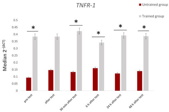
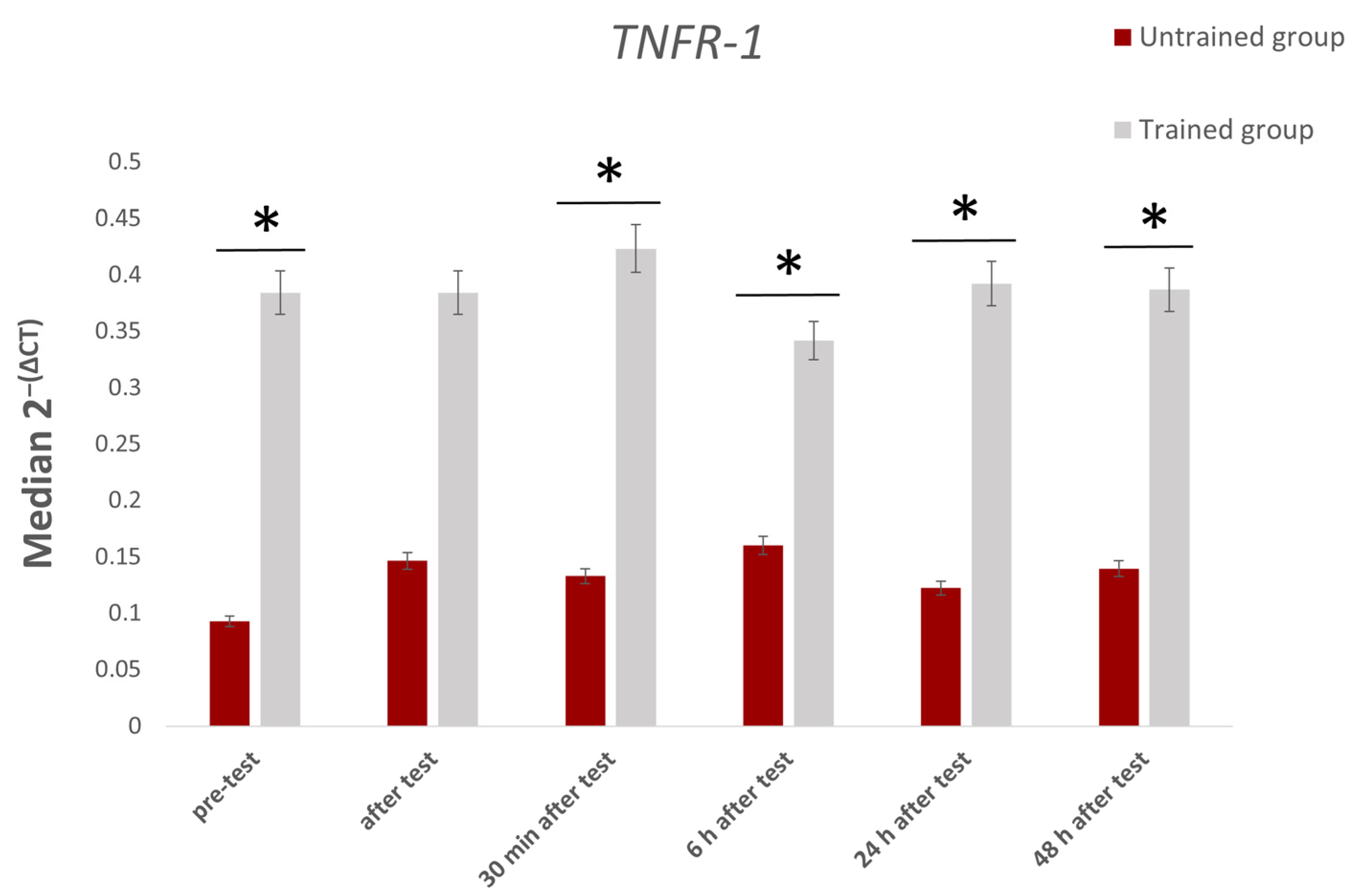
Several genes demonstrated statistically significant differences in expression between trained and untrained individuals. The most consistent divergence was observed in TNFR-1, the expression of which remained significantly lower in the untrained group across nearly all time points—including baseline (p = 0.009), 30 min (p = 0.017), 6 (p = 0.034), 24 (p = 0.021), and 48 h post-exercise (p = 0.032). The greatest contrast occurred before the physical effort (p = 0.009, Figure 1). These findings are summarized in Table 3.
Figure 1.
Median 2−(ΔCt) values of TNFR-1 expression over time between trained and untrained groups. * Indicates statistically significant differences (p < 0.05).
Table 3.
Differences in TNF-α, TNFR-1, TNFR-2, IL-4 and IL-4Rα gene expression between trained (T) and untrained (UT) groups across six time points.
In contrast, TNF-α and TNFR-2 did not differ significantly between groups at any measured time point (p > 0.05), although minor fluctuations were noted.
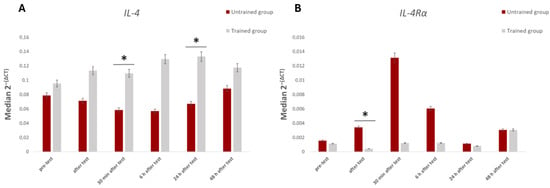
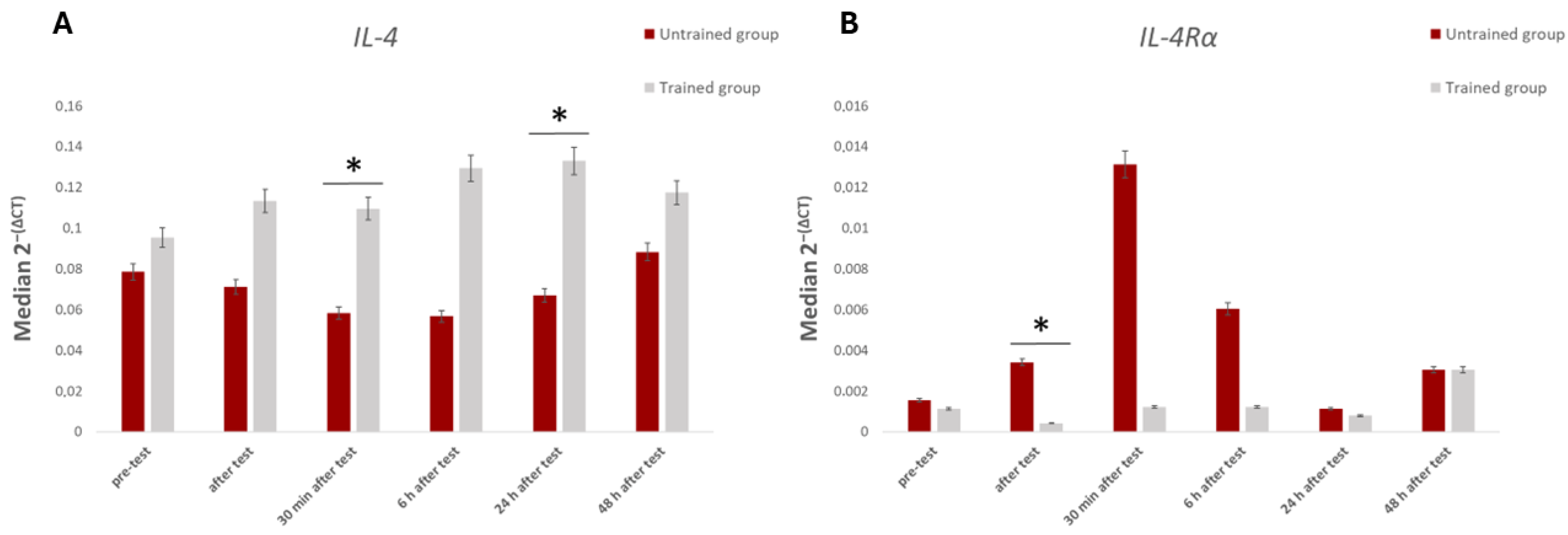
As for anti-inflammatory markers, IL-4 expression was significantly elevated in trained individuals compared to their untrained counterparts at two post-exercise time points: 30 min (p = 0.038) and 24 h (p = 0.002). The most notable difference was seen at 24 h (Figure 2A).
Figure 2.
Median 2−(ΔCt) values of IL-4 (A) and IL-4Rα (B) expression over time between trained and untrained groups. * Indicates statistically significant differences (p < 0.05).
Conversely, IL-4Rα showed a distinct pattern: its expression immediately after exercise was significantly higher in the untrained group (p = 0.027), while no other time points revealed intergroup differences (Figure 2B).
Timeline changes in the expressions of TNF-α, TNFR-1, TNFR-2, IL-4 and IL-4Rα genes relative to baseline levels were also determined. In the athlete group, no statistically significant changes were observed for any of the analyzed genes besides IL-4Rα, with expression levels remaining stable throughout the entire observation period (p > 0.05 for all comparisons).
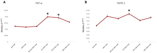
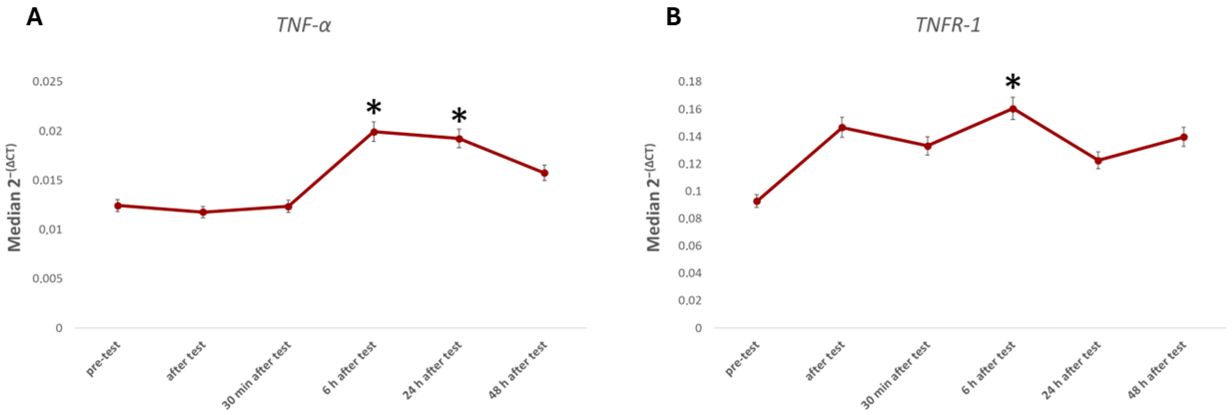
The temporal analysis of gene expression in the untrained group revealed a delayed pro-inflammatory response to the exhaustive exercise protocol. Specifically, TNF-α mRNA levels showed a gradual increase, reaching statistical significance at 6 h (p = 0.023) and 24 h post-exercise (p = 0.046) compared to the baseline (Figure 3A). This transcriptional activation was closely followed by a significant elevation in TNFR-1 expression at the 6 h mark (p = 0.023; Figure 3B), suggesting a synchronized up-regulation of both the cytokine and its primary receptor in individuals unaccustomed to high-intensity physical stress. By 48 h, these expression levels began to subside, returning toward baseline values. No statistically significant changes in TNFR-2 expression were detected at any time point.
Figure 3.
Changes in median TNF-α (A) and TNFR-1 (B) expression in the untrained group compared to the baseline. * Indicates statistically significant differences (p < 0.05).
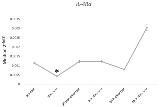
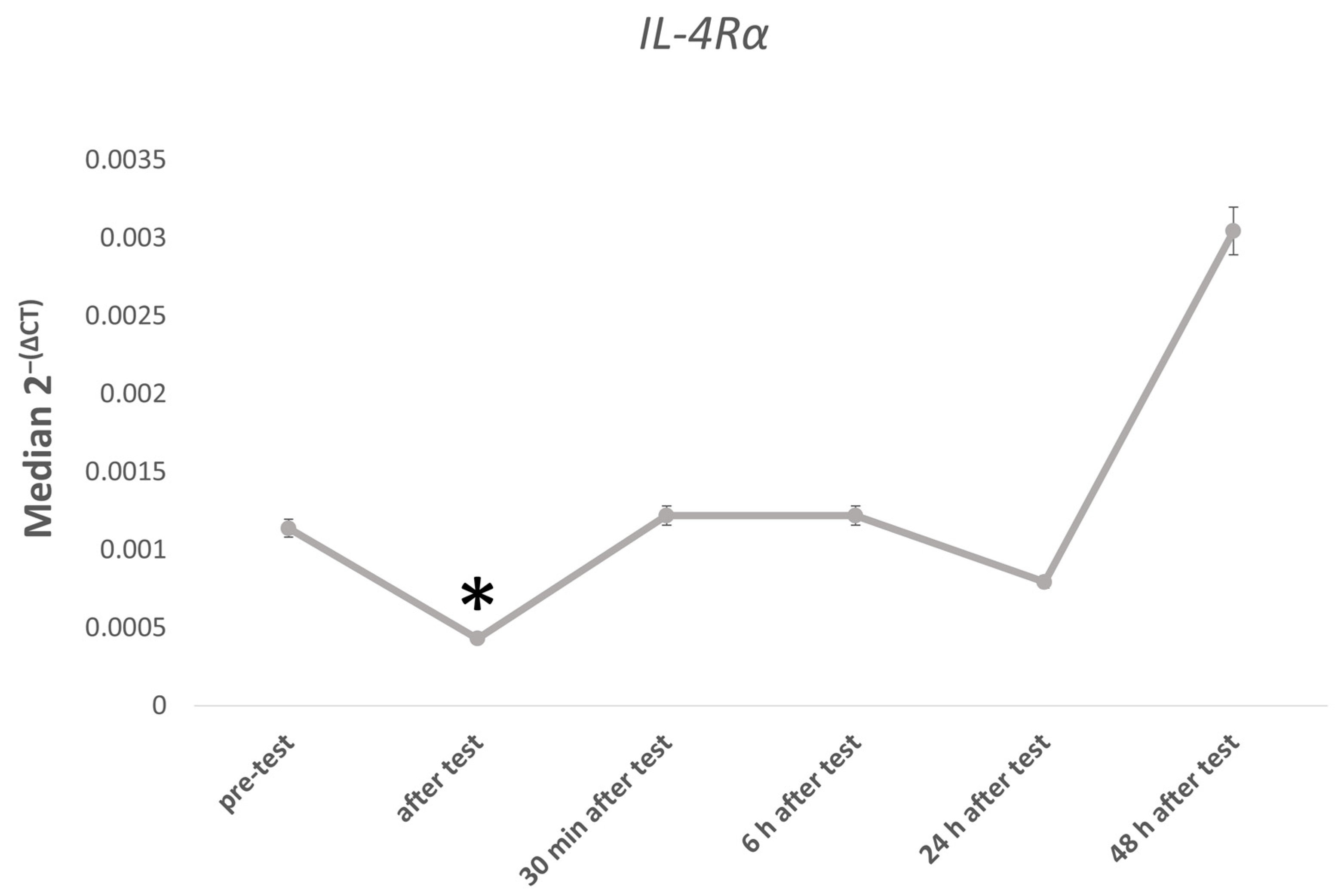
In contrast, the trained group exhibited a highly specific and transient response in the IL-4Rα gene. Unlike the stable expression observed in the untrained group, athletes showed a sharp, statistically significant reduction in IL-4Rα mRNA levels immediately after the exercise test (p = 0.041; Figure 4). This rapid suppression was short-lived, with expression levels fully recovering to baseline values by the 30 min post-exercise measurement and remaining stable thereafter. This unique “dip” in receptor expression in athletes, occurring in the absence of significant TNF-α fluctuations, highlights a distinct regulatory pattern of the anti-inflammatory pathway associated with long-term training adaptation.
Figure 4.
Changes in median IL-4Rα expression in trained group compared to baseline. * Indicates statistically significant differences (p < 0.05).
3.3. Protein Expression
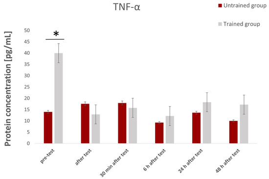
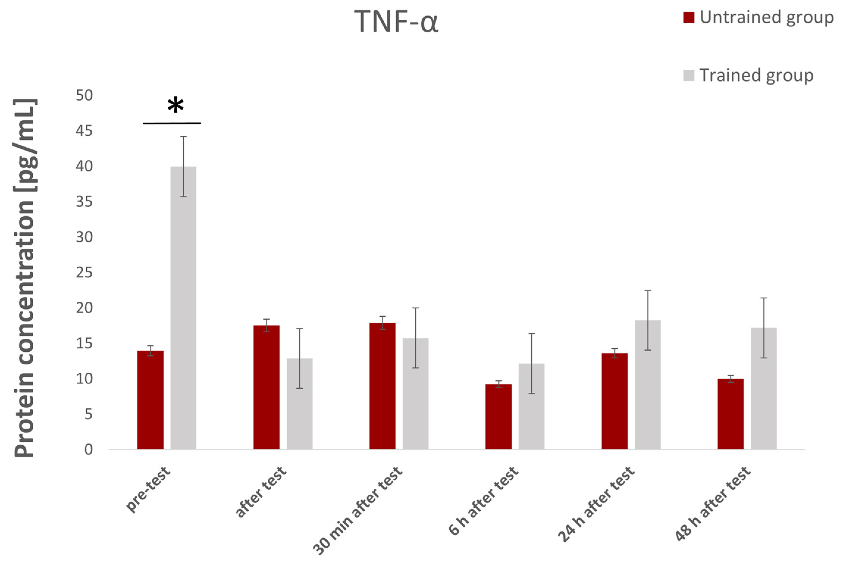
Between-group analysis using the Mann–Whitney U test revealed statistically significant differences in protein concentrations; however, these differences were specific to certain cytokines and time points, rather than universal across all measurements. For TNF-α, a significant difference between groups was observed only at the first measurement, prior to the exercise test. Baseline TNF-α levels were significantly higher (p = 0.043) in the athlete group compared to the non-athlete group (p < 0.05; Figure 5). Although higher TNF-α concentrations were observed in the non-athlete group immediately and 30 min after the test, and in the athlete group at 6, 24, and 48 h post-test, these differences were not statistically significant (p > 0.05).
Figure 5.
Visualization of TNF-α protein concentration showing significant differences between the trained and untrained groups across all time points. * Statistically significant differences (p < 0.05).
In contrast, IL-4 levels did not differ significantly between the athlete and non-athlete groups at any measured time point (p > 0.05).
This study also analyzed within-group differences in protein expression across time points separately for athletes and non-athletes.
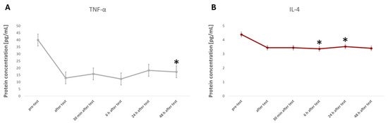
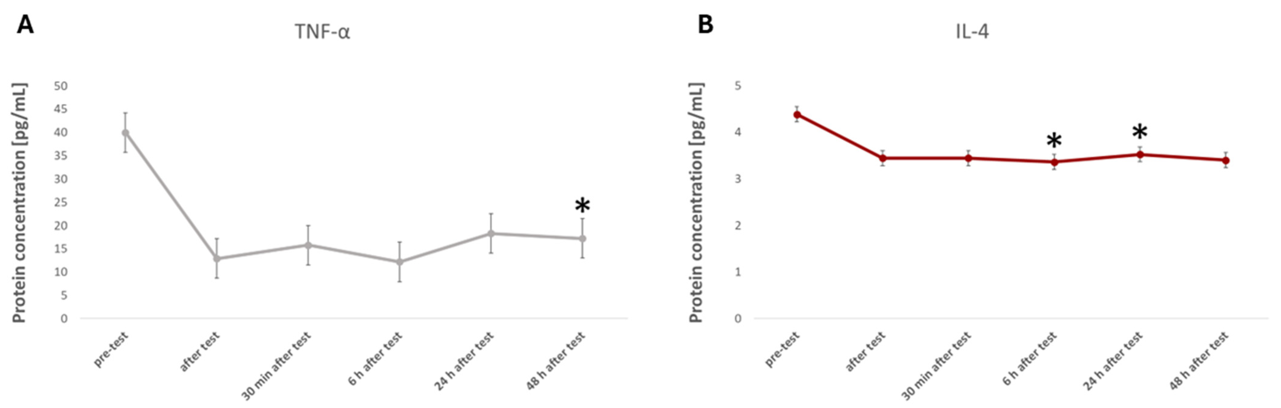
The TNF-α results revealed a statistically significant decrease in concentration 48 h after the exercise test compared to the baseline in the athlete group (p = 0.027; Figure 6A). No significant changes in TNF-α expression were observed in the athlete group at any other time point—immediately, 30 min, 6 h, or 24 h post-exercise—compared to the baseline (p > 0.05; Figure 6A). Similarly, no significant differences relative to the baseline were found in the non-athlete group.
Figure 6.
Visualization of TNF-α (A) protein concentration in the trained group and IL-4 (B) in the untrained group, showing significant changes between the baseline and the five post-exercise time points. * Statistically significant difference from baseline (p < 0.05).
Analysis of IL-4 concentrations demonstrated a significant decrease at both 6 h (p = 0.009) and 24 h (p = 0.019) post-exercise in the non-athlete group, compared to the baseline (Figure 6B). No statistically significant changes were observed at any other time points—immediately, 30 min, or 48 h after exercise—in the non-athlete group (p > 0.05; Figure 6B). In the athlete group, IL-4 protein levels remained unchanged, with no statistically significant differences at any time point compared to the baseline.
3.4. Correlation Analysis
An analysis was also conducted to assess the relationship between the expression levels of selected genes (encoding TNF-α and IL-4) and the concentrations of their corresponding protein products at each of the six time points: before the exercise test, immediately after its completion, and 30 min, 6 h, 24 h, and 48 h post-exercise. Calculations were performed separately for the athlete and non-athlete groups using Spearman’s rank correlation coefficient.
In the analyzed cohort, no statistically significant correlations were observed between gene expression and the corresponding protein concentrations (all p > 0.05), either in the non-athlete group or among athletes (Table 4).
Table 4.
Spearman’s rank correlation coefficients between mRNA expression and corresponding plasma protein concentrations for TNF-α and IL-4 across six time points.
In most cases, the correlation coefficients were low or close to zero, indicating a lack of strong association between transcript levels and protein concentrations in the samples analyzed.
An analysis was also conducted of the relationship between leukocyte indices and plasma protein concentrations (TNF-α and IL-4) at specific measurement points: before the exercise test, immediately after its completion, and 30 min, 6 h, 24 h, and 48 h after the end of exercise (Table 5).
Table 5.
Spearman’s rank correlation coefficients between leukocyte indices and plasma cytokine concentrations (TNF-α and IL-4).
4. Discussion
This study provided an in-depth examination of molecular responses triggered by acute physical exertion in two physiologically distinct groups—trained athletes and sedentary individuals. The investigation encompassed gene expression profiling of key cytokines (TNF-α and IL-4) and their receptors (TNFR-1, TNFR-2, and IL-4Rα), and quantification of serum protein levels of these molecules. These combined approaches offer new perspectives on how regular training modulates inflammatory and metabolic responses to acute exercise stress.
A critical comparative analysis of gene expression profiles underscored how training status influences the magnitude and timing of transcriptional responses to intense effort. While group comparisons revealed meaningful effect patterns, the differences should not be overinterpreted. The study provides preliminary evidence for training-associated tendencies rather than definitive mechanistic distinctions.
The results showed no significant intergroup differences in the expressions of the genes encoding TNF-α and TNFR-2 at any of the analyzed time points. At the same time, significantly higher TNFR-1 expression was found in the group of athletes compared to non-exercisers, both at rest and at each stage of the post-workout response, with the largest difference observed in the pre-exercise measurement. These results may indicate the existence of a different, permanent status of immune regulation in people who regularly engage in physical activity, regardless of its intensity. When comparing gene expressions after exercise to resting values, it was noted that TNF-α, TNFR-1, and TNFR-2 did not undergo significant changes in athletes, suggesting more effective control of the inflammatory response after exercise in this group.
The lack of differences in TNF-α expression between the groups in the presented study may indicate that the level of training does not affect TNF-α gene expression, and that the key stimulating factor is the length and intensity of training. In non-exercisers, however, TNF-α expression significantly increased 6 and 24 h post-exercise, reflecting the increased inflammation typical of individuals who are not adapted to regular physical activity [23].
A key observation in our study is that trained individuals exhibited significantly higher baseline TNF-α protein concentrations compared to sedentary controls (p = 0.043). While chronic exercise is generally associated with an anti-inflammatory systemic profile, this elevated baseline may reflect several physiological factors. First, it could be a consequence of the high cumulative training load in elite soccer players (over 10 h/week), leading to a state of chronic, low-grade metabolic and immune activation required for adaptation. Second, this elevated TNF-α “tone” might indicate ongoing subclinical muscle remodeling and satellite cell activation, where TNF-α serves as a crucial signaling molecule for tissue repair and myogenesis following repetitive mechanical stress [17,24].
Furthermore, seasonal variations in training intensity and the proximity of the last intensive training session (despite the 48 h rest period) might influence resting cytokine levels. In this context, higher baseline TNF-α in athletes should not be interpreted as maladaptive inflammation, but rather as a state of “immune readiness” or a physiological adaptation to maintain homeostatic balance under conditions of frequent strenuous exertion. This is further supported by the significant decrease in TNF-α observed in the athlete group 48 h post-exercise (p = 0.027), suggesting a highly efficient and well-coordinated resolution of the inflammatory response that is absent in untrained individuals [25].
In turn, a significant decrease in TNF-α concentration after 48 h in the group of athletes may indicate an effective mechanism of extinguishing the inflammatory response after intense exercise.
An interesting result of this study is the significantly higher expression of TNFR-1 in athletes compared to non-exercising individuals, which persisted at all time points analyzed. TNFR-1 is the main mediator of the biological effects of TNF-α, responsible for both pro-inflammatory and pro-apoptotic signaling [14], and is rapidly mobilized during acute immune activation [26]. It has consistently higher expression in trained individuals. Its higher expression in athletes may reflect a more efficient early-response system, allowing for rapid initiation and resolution of exercise-induced immune stress, and is suggestive of the potential for higher capacity in active regeneration through apoptosis of damaged muscle cells. In contrast, the lack of differences in TNFR-2 expression may indicate its lesser role in long-term adaptation to regular physical activity, which is consistent with previous reports. TNFR-2 is mainly expressed on T lymphocytes and endothelial cells, and its activation is typically associated with immunoregulatory and reparative processes, including support for Treg function, tissue remodeling, and maintenance of homeostasis following inflammatory insults [27]. It is, therefore, possible that TNFR-2 plays a more contextual and dynamic role, manifesting itself in specific pathological conditions or during prolonged immune stress. Although TNFR-2 was not directly quantified in this study, the divergent signaling pathways of the two receptors provide an interpretive framework for understanding why trained individuals exhibit faster stabilization of pro-inflammatory markers. The predominance of TNFR-1-driven early signaling followed by subsequent activation of TNFR-2-associated repair pathways may represent a characteristic adaptive response to repeated exercise stimuli.
IL-4 expression was significantly higher in athletes compared to non-exercisers at two time points—30 min and 24 h after the exercise test. IL-4 is an anti-inflammatory cytokine that plays an important role in regulating the humoral immune response, particularly by inducing Th2 lymphocyte differentiation and promoting the production of IgE and IgG1 antibodies [8]. In the context of the physiological response to physical exercise, IL-4 is considered one of the mediators of the anti-inflammatory response, counterbalancing the action of pro-inflammatory cytokines such as IL-1β [28]. The observation of the greatest difference between groups 24 h after exercise may suggest prolonged activation of pathways associated with regeneration and suppression of the inflammatory response in people who exercise regularly. Increased IL-4 expression in athletes can be interpreted as the result of training and prior immune adaptations, leading to a more effective reduction in potentially harmful inflammatory responses after exercise. In trained individuals, the immune system seems to switch more quickly from the inflammatory response phase to the regenerative phase, which may explain the difference seen after 24 h. A slightly different result was obtained for the IL-4 receptor (IL-4R), the expression of which was higher immediately after exercise in non-exercising individuals. This observation might point towards a compensatory mechanism in a population unaccustomed to exercise, in which the activation of the inflammatory response is more intense, and the increased sensitivity of cells to IL-4 may be an attempt to limit an excessive immune response. However, it is worth noting that cytokine receptor expression is often subject to dynamic and short-term regulation [24], which may explain the lack of persistent differences at other time points. When comparing time points for IL-4 and its receptor IL-4R, only a single change was observed—a decrease in IL-4R expression immediately after exercise in athletes, which may indicate a rapid, transient mechanism of anti-inflammatory response control in this group. The lack of clear changes in IL-4 expression suggests that it plays a more stable, constitutive role in regulating the immune environment after exercise. In the case of serum IL-4, significant decreases in concentration were observed only in the non-exercising group after 6 and 24 h. This observation may suggest that individuals who are not adapted to exercise show a weakened anti-inflammatory response after exercise. The lack of significant changes in IL-4 expression in both groups indicates that the level of this protein may also be regulated mainly at the translational level.
The observed lack of direct gene–protein correlations for TNF-α and IL-4 suggests that systemic cytokine output is governed by complex regulatory layers beyond mRNA abundance. For instance, TNF-α expression is known to be tightly controlled by post-transcriptional mechanisms, including microRNA-mediated interference and mRNA stability modulation via AU-rich elements. Furthermore, these molecules have divergent half-lives as the circulating TNF-α protein is rapidly cleared and its receptors (e.g., TNFR-1) exhibit various levels of stability—likely contributing to the lack of temporal synchronization between transcripts and proteins. The kinetics of protein secretion and cellular source variability (shifts in leukocyte and lymphocyte counts observed in our results) further complicate this relationship. These findings align with the recent literature [29], indicating that mRNA levels alone often do not serve as a reliable proxy for functional protein concentrations in response to exercise stress.
Analysis of the relationship between leukocyte parameters and TNF-α and IL-4 protein concentrations revealed clear differences in immune response between trained and untrained individuals. The most consistent and multi-point patterns were observed in the group of athletes, confirming that regular training modifies the way immune cells respond to endocrine and myokinetic signals associated with physical exercise [30]. In untrained individuals, correlations were rare, irregular, and did not follow any consistent pattern, suggesting a more chaotic, less synchronized immune response to physical stress.
In the case of leukocyte count, significant correlations occurred almost exclusively in athletes, especially immediately after exercise and in the late phase (24–48 h). Immediately after the test, a positive correlation was found between leukocyte count and TNF-α, which may indicate that in trained individuals, leukocyte mobilization occurs in parallel with increased expression of pro-inflammatory cytokines. TNF-α, although classified as a pro-inflammatory cytokine, also functions in signal-regulating leukocyte mobilization during the course of the early phase of regeneration in response to exercise [19]. These exclusive relationships in the trained group could reflect a more coordinated coupling between cellular and molecular responses during acute exercise stress. It is worth noting that correlations between leukocytes and IL-4 and TNF-α after 24 and 48 h were also present only in athletes, suggesting that the late phases of recovery in trained individuals are associated with the activation of both pro-inflammatory (TNF-α) and anti-inflammatory (IL-4) mechanisms. This phenomenon is consistent with the model in which chronic training leads to the alternation of pro- and anti-inflammatory pathways, which are key to adaptive processes [31]. The absence of such relationships in non-exercisers indicates that their regeneration process is less orderly and less dependent on cytokine signals.
The results for neutrophils highlight the differences between the groups even more clearly. In practice, all significant correlations concerned athletes. The appearance of a significant correlation between neutrophils and IL-4 after 48 h suggests a transition to the anti-inflammatory phase, in accordance with the mechanism of shifting the immune balance towards recovery and regeneration processes in the later post-exercise period [31].
In the case of lymphocytes, there were single significant correlations, but their pattern was different from that of neutrophils. Importantly, in contrast to the trends observed for leukocytes and neutrophils, the early phases of the lymphocyte response were more pronounced in untrained individuals. In untrained individuals, a strong negative correlation between lymphocytes and TNF-α was found before exercise, which may reflect the increased pro-inflammatory “tone” at rest characteristic of individuals with lower levels of physical activity [19]. In athletes, however, a significant lymphocyte correlation appeared only after 24 h and concerned IL-4. This indicates that in trained individuals, lymphocyte populations may play a role mainly in the later phase of anti-inflammatory regeneration, when pathways associated with tissue repair and balancing the earlier inflammatory response are activated. This is consistent with the concept that chronic training shifts the immune profile towards a more subdued and anti-inflammatory response [14].
4.1. Limitations
Despite providing novel insights into the temporal inflammatory response to exhaustive exercise, this study has several limitations that warrant consideration. First, the sample size (n = 15 per group) was relatively small, which may limit the statistical power to detect subtle effect sizes, particularly given the inter-individual variability inherent in immune signaling. Second, the exclusion of female participants and the focus on a specific age range (20–25 years) limit the generalizability of our findings to broader populations. Third, our analysis was restricted to peripheral blood, which may not fully reflect the localized inflammatory dynamics occurring within skeletal muscle or other tissues.
Furthermore, it should be noted that we did not apply formal adjustments for multiple comparisons (e.g., Bonferroni or FDR corrections). Given the exploratory nature of this study, we prioritized the identification of potential biological trends over the strict control of type I errors, though this means our results should be interpreted with caution. Finally, the observed lack of direct gene–protein correlations for TNF-α and IL-4 suggests that additional regulatory layers—such as post-transcriptional modifications, protein half-lives, and secretion kinetics—remain unaddressed in the current design. Future research involving larger, more diverse cohorts and multi-compartment sampling is necessary to validate and extend these preliminary findings.
Despite these limitations, this study provides valuable insight into training-dependent differences in acute inflammatory responses and highlights potential biomarkers relevant for evaluating adaptation and recovery processes in physically active and sedentary individuals.
4.2. Practical Implications
This research is of significant importance and provides data that can be used in practice to identify biomarkers of fatigue, adaptation, and risk of overtraining [32]. From a practical standpoint, the integration of these biomarkers into sports monitoring protocols could follow a stratified approach [33]. For instance, baseline TNF-α levels could be assessed during pre-season to establish an individual’s “immune set-point.” Subsequently, significant deviations from the 48 h recovery profile (e.g., a failure of TNF-α to decrease or a lack of IL-4 stability) could trigger a reduction in training volume for specific players. By combining high-sensitivity molecular data with routine hematology, coaching staff could move beyond subjective fatigue scales toward an objective, biologically driven model of recovery and performance optimization.
From an applied perspective, the cytokine and receptor dynamics identified in this study offer several meaningful insights for real-world training and performance environments. Faster normalization of TNF-α and TNFR-1 in trained individuals suggests a more efficient inflammatory resolution profile, which, in practical terms, may translate into greater tolerance for repeated high-intensity sessions, reduced cumulative fatigue, and shorter recovery windows. Conversely, the prolonged or more pronounced responses in untrained participants may indicate higher susceptibility to delayed onset fatigue, impaired session quality, or maladaptive load accumulation. IL-4 and IL-4Rα patterns further highlight individual variability in post-exercise immune modulation, with potential relevance for identifying athletes who may benefit from additional recovery strategies (e.g., adjusted micro-cycles, modified intensities, and nutritional support). Overall, these biomarker trajectories may complement subjective and performance-based monitoring tools and help practitioners differentiate between an adequately adapting athlete and one exhibiting early signs of excessive physiological strain.
5. Conclusions
This study offers a comprehensive insight into physiological, biochemical, and molecular responses to acute exercise in both trained athletes and sedentary individuals. By analyzing the expressions of genes (TNF-α, TNFR-1, TNFR-2, IL-4, and IL-4Rα), corresponding protein concentrations, and various blood parameters, distinct adaptation patterns associated with training status were identified.
A significant increase in TNF-α and TNFR-1 gene expressions and no significant differences in IL-4 and IL-4R expressions in the non-exercising group indicate stimulation of the TNF-α pathway and a predominance of pro-inflammatory responses in physically inactive individuals. The lack of significant differences in the expressions of TNF-α, TNFR-1, TNFR-2, IL-4, and IL-4R in athletes indicates that regular physical training suppresses the inflammatory response.
The significantly higher resting concentration of TNF-α in athletes may indicate that regular physical activity is associated with maintaining a slightly elevated level of selected pro-inflammatory cytokines as part of the body’s constant adaptation to repetitive training load. A significant decrease in TNF-α concentration after 48 h was observed in athletes, suggesting efficient activation of mechanisms that suppress the inflammatory response after exercise, which reflects the higher efficiency of regenerative processes in this group.
The significant decrease in serum IL-4 levels in the non-exercising group at 6 and 24 h after exercise may indicate that, in individuals without training adaptation, the mechanisms regulating the anti-inflammatory response are less stable and are significantly disrupted after physical exertion. At the same time, no correlation was found between mRNA levels and protein concentration, suggesting that the protein response occurs independently of transcriptional changes in blood cells.
The present study suggests that training status may influence the magnitude and temporal characteristics of the inflammatory response to acute exercise. Although several statistically significant differences were observed between trained and untrained individuals, these effects should be interpreted with caution due to the exploratory nature of the study and limited sample size. The observed patterns indicate potential training-related trends rather than definitive group-specific responses. Further research involving larger cohorts and complementary methodological approaches is warranted to confirm and extend these findings.
Author Contributions
Conceptualization, P.M., M.S. and A.M.-S.; methodology, P.M., P.T., M.T. (Marta Tkacz), K.Z., A.L. and A.M.-S.; software, P.M.; validation, P.M., P.T., M.T. (Marta Tkacz) and K.Z.; formal analysis, P.M., M.S., P.P. and M.T. (Maciej Tarnowski); investigation, P.M.; resources, A.L., R.B. and Ł.R.; data curation, P.M., M.S. and M.T. (Maciej Tarnowski); writing—original draft preparation, P.M.,M.S., P.P. and M.T. (Maciej Tarnowski); writing—review and editing, P.M. and M.S.; visualization, P.M.; supervision, M.S.; project administration, P.M.; funding acquisition, M.S. All authors have read and agreed to the published version of the manuscript.
Funding
This research received no external funding.
Institutional Review Board Statement
The study was conducted in accordance with the Declaration of Helsinki and approved by the Bioethics Committee at the University of Szczecin (approval number: US.003.5.2024, 22 May 2024).
Informed Consent Statement
Informed consent was obtained from all subjects involved in the study.
Data Availability Statement
Data are available on request due to restrictions.
Acknowledgments
Some of the research was developed during mobility at the Department of Psychology, Educational Science and Human Movement, University of Palermo, Italy as part of the Erasmus+ program.
Conflicts of Interest
The authors declare no conflicts of interest.
Abbreviations
The following abbreviations are used in this manuscript:
| TNF-α | tumor necrosis factor alpha |
| TNFR-1 | tumor necrosis factor receptor 1 |
| TNFR-2 | tumor necrosis factor receptor 2 |
| IL-4 | interleukin-4 |
| IL-4Rα | interleukin-4 receptor subunit alpha |
| VO2max | maximal oxygen uptake |
| IFN | interferon |
| CSF | colony-stimulating factor |
| TNFSF2 | TNF superfamily member 2 |
| GPAQ | Global Physical Activity Questionnaire |
| RT-qPCR | quantitative real-time polymerase chain reaction |
| GAPDH | glyceraldehyde 3-phosphate dehydrogenase |
References
- Strimbu, K.; Tavel, J.A. What Are Biomarkers? Curr. Opin. HIV AIDS 2010, 5, 463–466. [Google Scholar] [CrossRef] [Scilit] [PubMed]
- Craighead, D.H.; Heinbockel, T.C.; Freeberg, K.A.; Rossman, M.J.; Jackman, R.A.; Jankowski, L.R.; Hamilton, M.N.; Ziemba, B.P.; Reisz, J.A.; D’Alessandro, A.; et al. Time-Efficient Inspiratory Muscle Strength Training Lowers Blood Pressure and Improves Endothelial Function, NO Bioavailability, and Oxidative Stress in Midlife/Older Adults With Above-Normal Blood Pressure. J. Am. Heart Assoc. 2021, 10, e020980. [Google Scholar] [CrossRef] [Scilit]
- Tesema, G.; George, M.; Mondal, S.; Mathivana, D. Serum Cardiac Markers Are Inversely Associated with VO2max of Amateur Athletes in Response to Endurance Training Adaptations. BMJ Open Sport Exerc. Med. 2019, 5, e000537. [Google Scholar] [CrossRef] [Scilit]
- Lee, E.C.; Fragala, M.S.; Kavouras, S.A.; Queen, R.M.; Pryor, J.L.; Casa, D.J. Biomarkers in Sports and Exercise: Tracking Health, Performance, and Recovery in Athletes. J. Strength Cond. Res. 2017, 31, 2920–2937. [Google Scholar] [CrossRef] [Scilit]
- Zhang, J.-M.; An, J. Cytokines, Inflammation and Pain. Int. Anesthesiol. Clin. 2007, 45, 27–37. [Google Scholar] [CrossRef] [Scilit]
- Ferreira, V.L.; Borba, H.H.L.; Bonetti, A.d.F.; P.Leonart, L.; Pontarolo, R.; Ferreira, V.L.; Borba, H.H.L.; Bonetti, A.d.F.; Leonart, L.P.; Pontarolo, R. Cytokines and Interferons: Types and Functions; IntechOpen: London, UK, 2018. [Google Scholar]
- Sims, J.E.; Smith, D.E. The IL-1 Family: Regulators of Immunity. Nat. Rev. Immunol. 2010, 10, 89–102. [Google Scholar] [CrossRef] [Scilit] [PubMed]
- Bullens, D.M.A.; Rafiq, K.; Kasran, A.; Van Gool, S.W.; Ceuppens, J.L. Naive Human T Cells Can Be a Source of IL-4 during Primary Immune Responses. Clin. Exp. Immunol. 1999, 118, 384–391. [Google Scholar] [CrossRef] [Scilit] [PubMed]
- Skinner, S.; Nader, E.; Stauffer, E.; Robert, M.; Boisson, C.; Cibiel, A.; Foschia, C.; Feasson, L.; Robach, P.; Millet, G.Y.; et al. Differential Impacts of Trail and Ultra-Trail Running on Cytokine Profiles: An Observational Study. Clin. Hemorheol. Microcirc. 2021, 78, 301–310. [Google Scholar] [CrossRef] [Scilit] [PubMed]
- Sethi, J.K.; Hotamisligil, G.S. Metabolic Messengers: Tumour Necrosis Factor. Nat. Metab. 2021, 3, 1302–1312. [Google Scholar] [CrossRef] [Scilit]
- Jiménez-Maldonado, A.; Montero, S.; Lemus, M.; Cerna-Cortés, J.; Rodríguez-Hernández, A.; Mendoza, M.A.; Melnikov, V.; Gamboa-Domínguez, A.; Muñiz, J.; Virgen-Ortiz, A.; et al. Moderate and High Intensity Chronic Exercise Reduces Plasma Tumor Necrosis Factor Alpha and Increases the Langerhans Islet Area in Healthy Rats. J. Musculoskelet. Neuronal Interact. 2019, 19, 354–361. [Google Scholar]
- Macêdo Santiago, L.Â.; Neto, L.G.L.; Borges Pereira, G.; Leite, R.D.; Mostarda, C.T.; de Oliveira Brito Monzani, J.; Sousa, W.R.; Rodrigues Pinheiro, A.J.M.; Navarro, F. Effects of Resistance Training on Immunoinflammatory Response, TNF-Alpha Gene Expression, and Body Composition in Elderly Women. J. Aging Res. 2018, 2018, 1467025. [Google Scholar] [CrossRef] [Scilit]
- Gough, P.; Myles, I.A. Tumor Necrosis Factor Receptors: Pleiotropic Signaling Complexes and Their Differential Effects. Front. Immunol. 2020, 11, 585880. [Google Scholar] [CrossRef] [Scilit] [PubMed]
- Li, Y.; Ye, R.; Dai, H.; Lin, J.; Cheng, Y.; Zhou, Y.; Lu, Y. Exploring TNFR1: From Discovery to Targeted Therapy Development. J. Transl. Med. 2025, 23, 71. [Google Scholar] [CrossRef] [Scilit] [PubMed]
- Ducharme, J.B.; Specht, J.W.; Bailly, A.R.; Deyhle, M.R. Serum Cytokines and Their Soluble Receptors Are Differently Regulated between Trained and Untrained Men after Vigorous Endurance Exercise. Am. J. Physiol. Regul. Integr. Comp. Physiol. 2025, 328, R581–R587. [Google Scholar] [CrossRef] [Scilit] [PubMed]
- Gjevestad, G.O.; Holven, K.B.; Ulven, S.M. Effects of Exercise on Gene Expression of Inflammatory Markers in Human Peripheral Blood Cells: A Systematic Review. Curr. Cardiovasc. Risk Rep. 2015, 9, 34. [Google Scholar] [CrossRef] [Scilit] [PubMed]
- Małkowska, P.; Sawczuk, M. Cytokines as Biomarkers for Evaluating Physical Exercise in Trained and Non-Trained Individuals: A Narrative Review. Int. J. Mol. Sci. 2023, 24, 11156. [Google Scholar] [CrossRef] [Scilit] [PubMed]
- Teodoro, T.H.; Costa, K.P.M.; Prestes, J.; Campanholi, J.; Navalta, J.; Pereira, G.B. The Effects of Acute and Chronic Exercise on Immune Markers of TH1/TH2 Cells in Older Adults: A Systematic Review. Front. Physiol. 2025, 16, 201–217. [Google Scholar] [CrossRef] [Scilit]
- Nieman, D.C.; Wentz, L.M. The Compelling Link between Physical Activity and the Body’s Defense System. J. Sport Health Sci. 2019, 8, 201–217. [Google Scholar] [CrossRef] [Scilit]
- Dyken, S.J.V.; Locksley, R.M. Interleukin-4- and Interleukin-13-Mediated Alternatively Activated Macrophages: Roles in Homeostasis and Disease. Annu. Rev. Immunol. 2013, 31, 317–343. [Google Scholar] [CrossRef] [Scilit] [PubMed]
- Faustman, D.; Davis, M. TNF Receptor 2 Pathway: Drug Target for Autoimmune Diseases. Nat. Rev. Drug Discov. 2010, 9, 482–493. [Google Scholar] [CrossRef] [Scilit] [PubMed]
- Walsh, N.P.; Gleeson, M.; Shephard, R.J.; Gleeson, M.; Woods, J.A.; Bishop, N.C.; Fleshner, M.; Green, C.; Pedersen, B.K.; Hoffman-Goetz, L.; et al. Position Statement. Part One: Immune Function and Exercise. Exerc. Immunol. Rev. 2011, 17, 6–63. [Google Scholar] [PubMed]
- Gokhale, R.; Chandrashekara, S.; Vasanthakumar, K.C. Cytokine Response to Strenuous Exercise in Athletes and Non-Athletes—An Adaptive Response. Cytokine 2007, 40, 123–127. [Google Scholar] [CrossRef] [Scilit]
- Petersen, P.S.; Lei, X.; Seldin, M.M.; Rodriguez, S.; Byerly, M.S.; Wolfe, A.; Whitlock, S.; Wong, G.W. Dynamic and Extensive Metabolic State-Dependent Regulation of Cytokine Expression and Circulating Levels. Am. J. Physiol. Regul. Integr. Comp. Physiol. 2014, 307, R1458–R1470. [Google Scholar] [CrossRef] [Scilit]
- Cerqueira, É.; Marinho, D.A.; Neiva, H.P.; Lourenço, O. Inflammatory Effects of High and Moderate Intensity Exercise—A Systematic Review. Front. Physiol. 2020, 10, 1550. [Google Scholar] [CrossRef] [Scilit]
- Brenner, D.; Blaser, H.; Mak, T.W. Regulation of Tumour Necrosis Factor Signalling: Live or Let Die. Nat. Rev. Immunol. 2015, 15, 362–374. [Google Scholar] [CrossRef] [Scilit] [PubMed]
- Faustman, D.L.; Davis, M. TNF Receptor 2 and Disease: Autoimmunity and Regenerative Medicine. Front. Immunol. 2013, 4, 478. [Google Scholar] [CrossRef] [Scilit]
- Docherty, S.; Harley, R.; McAuley, J.J.; Crowe, L.A.N.; Pedret, C.; Kirwan, P.D.; Siebert, S.; Millar, N.L. The Effect of Exercise on Cytokines: Implications for Musculoskeletal Health: A Narrative Review. BMC Sports Sci. Med. Rehabil. 2022, 14, 5. [Google Scholar] [CrossRef] [Scilit]
- Bishop, D.J.; Hoffman, N.J.; Taylor, D.F.; Saner, N.J.; Lee, M.J.-C.; Hawley, J.A. Discordant Skeletal Muscle Gene and Protein Responses to Exercise. Trends Biochem. Sci. 2023, 48, 927–936. [Google Scholar] [CrossRef] [Scilit]
- Pedersen, B.K.; Febbraio, M.A. Muscles, Exercise and Obesity: Skeletal Muscle as a Secretory Organ. Nat. Rev. Endocrinol. 2012, 8, 457–465. [Google Scholar] [CrossRef] [Scilit] [PubMed]
- Peake, J.M.; Neubauer, O.; Della Gatta, P.A.; Nosaka, K. Muscle Damage and Inflammation during Recovery from Exercise. J. Appl. Physiol. 2017, 122, 559–570. [Google Scholar] [CrossRef] [Scilit] [PubMed]
- Modesti, A.; Militello, R.; Tanturli, A.; Santi, A.; Gulisano, M.; Petri, C.; Pengue, L.; Pellegrino, A.; Modesti, P.A.; Luti, S. Molecular, Physical, and Technical Performance Response After a Competitive Match in Male Professional Soccer Players. Antioxidants 2025, 14, 73. [Google Scholar] [CrossRef] [Scilit] [PubMed]
- Liu, J.; Li, Q.; Han, Y. Efficacy of Post-Exercise Recovery Strategies for Elite Soccer Players: A Network Meta-Analysis. Front. Physiol. 2026, 17, 1760392. [Google Scholar] [CrossRef] [Scilit] [PubMed]

Disclaimer/Publisher’s Note: The statements, opinions and data contained in all publications are solely those of the individual author(s) and contributor(s) and not of MDPI and/or the editor(s). MDPI and/or the editor(s) disclaim responsibility for any injury to people or property resulting from any ideas, methods, instructions or products referred to in the content. |
© 2026 by the authors. Licensee MDPI, Basel, Switzerland. This article is an open access article distributed under the terms and conditions of the Creative Commons Attribution (CC BY) license.