


Abstract
This in vitro study assessed tooth color and whiteness changes following application of a commercial LED-assisted over-the-counter (OTC) whitening system, and characterized subsequent color changes under different immersion conditions. Sixty extracted human maxillary incisors were whitened once daily for 15 consecutive days in ten six-tooth acrylic arch models. After whitening, the models were randomly allocated to five immersion conditions (two models; n = 12 teeth per condition): distilled water, black tea, coffee, cola, and red wine. Specimens were immersed in the assigned medium for 4 h daily for 15 days using a single shared container per group. Tooth color was measured with a spectrophotometer at baseline (T0), after whitening (T1), and after staining (T2). Color change was assessed using CIEDE2000 (ΔE00), the Whiteness Index for Dentistry (WI_D), and supportive ΔEab values. Mean WI_D increased from 15.66 ± 5.14 at T0 to 26.23 ± 5.67 at T1 (p < 0.001), with a mean ΔWI_D of 10.58 ± 1.97 and ΔE00 of 5.00 ± 0.87. All specimens exceeded the 50:50 acceptability thresholds for both outcomes. Descriptive post-whitening color changes varied across immersion conditions, including distilled water; however, the absence of independent container-level replication precluded valid inferential comparisons between staining media. Red wine showed the highest mean ΔE00 (16.13 ± 3.42), whereas cola showed the largest numerical loss of whiteness. The complete commercial whitening system was associated with a measurable increase in dental whiteness, whereas subsequent color changes represented descriptive laboratory observations rather than independently confirmed beverage effects.
1. Introduction
Tooth color is an important component of dental esthetics, and the growing demand for whiter teeth has contributed to the widespread use of tooth-whitening procedures [1]. In parallel, over-the-counter (OTC) whitening products have become increasingly available because of their accessibility, relatively low cost, and ease of self-administration without professional supervision. These products are marketed in various delivery forms, including whitening strips, prefabricated trays, paint-on gels, toothpastes, and mouth rinses, and may contain peroxide-based or alternative whitening agents [2]. However, OTC products vary considerably in their formulations, delivery protocols, and active-agent concentrations, and the evidence supporting the whitening effectiveness of some tray- and gel-based systems is still derived predominantly from in vitro studies [2]. Recent clinical evidence indicates that OTC bleaching products can provide satisfactory short-term whitening compared with placebo; however, the certainty of the available evidence remains low, and treatment effects vary among bleaching protocols [3].
Some OTC whitening systems incorporate light-emitting diode (LED) devices with the aim of enhancing the bleaching effect of peroxide-based gels [4,5]. However, the additional benefit of light activation appears to depend on the light source, bleaching formulation, and treatment protocol, and variable results have been reported across different systems [5,6,7]. In a recent in vitro study, Shekari et al. found that combining OTC bleaching products with their corresponding LED trays produced greater color change than product-only or light-only protocols [5]. Similar findings have been reported for an LED home-whitening system evaluated among different OTC and at-home bleaching approaches [4]. Nevertheless, the independent contribution of LED activation cannot be assumed for every commercial system.
The maintenance of tooth color after bleaching is also clinically relevant because bleached teeth are subsequently exposed to dietary chromogens that may compromise color stability. Tea, coffee, cola, and red wine are among the commonly investigated beverages associated with post-bleaching discoloration, although the magnitude of color change varies according to the staining medium and exposure conditions [8,9]. For objective assessment of these changes, the CIEDE2000 color-difference formula (ΔE00) provides a perceptually weighted measure of overall color difference, whereas the Whiteness Index for Dentistry (WI_D) specifically quantifies changes in dental whiteness [10,11,12]. Accordingly, the combined use of ΔE00 and ΔWI_D may provide complementary information when evaluating whitening efficacy and subsequent color stability.
Despite the increasing availability of LED-assisted OTC whitening systems, evidence regarding their whitening performance and post-whitening color stability after exposure to commonly consumed staining beverages remains limited and heterogeneous [2,5]. Therefore, the aim of this in vitro study was to quantify changes in tooth color and whiteness following application of a commercially available LED-assisted OTC hydrogen peroxide whitening system and to characterize post-whitening color changes observed under different immersion conditions, using ΔE00 and ΔWI_D as the principal color outcomes. The originally specified null hypotheses were that (H01) the whitening protocol would not significantly increase tooth whiteness and (H02) post-whitening color change would not differ among the tested staining media. However, because each staining medium was applied in a single immersion container, H02 could not be formally tested using independent treatment-level replication; staining-phase findings were therefore summarized descriptively.
2. Materials and Methods
2.1. Study Design and Ethical Approval
This in vitro experimental study was designed to evaluate tooth color and whiteness changes following application of a commercially available LED-assisted over-the-counter whitening system and the subsequent color stability of whitened teeth following exposure to different staining media. The experimental workflow consisted of three sequential stages: baseline color assessment, a 15-day whitening phase, and a 15-day staining phase. The study protocol was approved by the Non-Interventional Research Ethics Committee of Fırat University (approval no. 2025/07-38; 24 April 2025).
The planned tooth-level sample size was initially estimated a priori using G*Power software (version 3.1.9.7; Heinrich Heine University, Düsseldorf, Germany), based on previously reported post-bleaching ΔE00 data from a five-group beverage-staining study [8]. An effect size of f = 0.735 was calculated from the final ΔE00 group means and standard deviations reported in that study. Under the initial assumption of independent tooth-level observations, a one-way ANOVA with five groups, an alpha level of 0.05, and a statistical power of 95% indicated a nominal minimum sample size of 40 teeth (8 per group). A total of 60 teeth were included, with 12 subsequently allocated to each staining condition. However, because each immersion medium was represented by a single container, the original tooth-level power calculation did not provide independent treatment replication or support confirmatory comparisons between media. Accordingly, the staining-related outcomes were analyzed descriptively.
2.2. Tooth Selection and Specimen Preparation
Sixty human maxillary incisors extracted for periodontal or other therapeutic reasons within one month before the experiment were included in the study. After extraction, adherent calculus and residual soft-tissue deposits were removed manually using hand instruments, and the tooth surfaces were cleaned with a fluoride-free and oil-free prophylaxis paste. All teeth were examined under ×20 magnification using a Zumax operating microscope (Zumax Medical Co., Ltd., Suzhou, China). Teeth presenting caries, restorations, cracks or fractures, marked discoloration, or developmental enamel defects were excluded.
Baseline tooth shade was determined using the VITA Classical shade guide (VITA Zahnfabrik, Bad Säckingen, Germany), and only teeth presenting A2 or A3 shades were included. No grinding, sectioning, or flattening of the buccal enamel surface was performed, thereby preserving the natural enamel surface. The specimens were stored in thymol solution at room temperature until preparation.
The roots were retained intact, and the teeth were embedded in acrylic resin with the crowns and buccal surfaces exposed. The specimens were arranged in arch-shaped models to simulate the clinical positioning of teeth within the horseshoe-shaped LED mouthpiece used as part of the OTC whitening system. During the intervals between experimental procedures, the specimens were stored in Fusayama–Meyer artificial saliva at 37 °C, which was renewed daily, as previously described [13].
2.3. Color Measurement
Tooth color was measured using a VITA Easyshade V spectrophotometer (VITA Zahnfabrik, Bad Säckingen, Germany). The spectrophotometer was calibrated according to the manufacturer’s instructions before each measurement session. All color measurements were performed by the same operator on a standardized white background.
Measurements were obtained from the central region of the middle third of the buccal surface. To standardize the measurement location throughout the experiment, an individual silicone positioning guide was fabricated for each specimen at baseline. An opening corresponding to the diameter of the VITA Easyshade V measuring tip was created over the predetermined measurement area. During each assessment, the spectrophotometer probe was positioned perpendicular to the enamel surface through the guide, ensuring reproducible positioning at the same site.
Before each color assessment, the specimens were rinsed and gently blot-dried with absorbent paper to remove excess surface moisture. For the post-whitening assessment (T1), the specimens were maintained in artificial saliva at 37 °C for 24 h following the final whitening session before color measurements were performed.
Color measurements were performed at three experimental time points: before whitening (T0), after completion of the 15-day whitening protocol (T1), and after completion of the 15-day staining protocol (T2). At each time point, three consecutive measurements were obtained from each tooth, and the mean of the three readings was used for subsequent color calculations and statistical analyses.
2.4. OTC Whitening Protocol
All specimens were whitened using a commercially available LED-assisted over-the-counter whitening system (SmileShop Teeth Whitening Gel, Gold İlaç Sanayi ve Ticaret A.Ş., Silivri, İstanbul, Türkiye; series no. 2028/05). According to the product label, the whitening gel contained aqua, glycerine, propylene glycol, acrylates/C10-30 alkyl acrylate crosspolymer, triethanolamine, sodium benzoate, hydrated silica, hydrogen peroxide, menthol, sodium ascorbyl phosphate, Aloe barbadensis leaf extract, and Anthemis nobilis flower water. Although hydrogen peroxide was listed among the ingredients, its concentration was not specified on the product packaging or in the available documentation. Technical specifications of the LED unit, including wavelength, irradiance, and optical output power, were also unavailable. We contacted the company identified on the product packaging by email and telephone to request the hydrogen peroxide concentration and LED technical specifications, but the requested information was not provided.
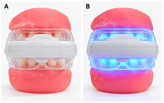
For each whitening session, the gel was applied in consistent amounts to the buccal enamel surfaces using the graduated syringe supplied with the commercial kit and evenly distributed with an applicator. The 60 specimens were arranged in ten six-tooth acrylic arch models. Two models were positioned simultaneously in the upper and lower compartments of the same horseshoe-shaped LED mouthpiece (Figure 1), requiring five sequential 15 min exposure sessions per day. The starting order of the five model pairs was rotated daily to minimize systematic order effects. The same LED device was used throughout the 15-day whitening protocol to avoid inter-device variability, and its batteries were replaced daily to minimize potential reductions in light output. The LED unit remained activated throughout each 15 min application period. All specimens underwent the combined gel-and-LED protocol; no gel-only, LED-only, or untreated control group was included during the whitening phase.
Figure 1.
Experimental setup for the LED-assisted OTC whitening procedure. (A) Positioning of the two six-tooth acrylic arch models within the LED mouthpiece before activation. (B) View of the setup during LED activation.
The whitening procedure was performed once daily for 15 consecutive days, with approximately 24 h between applications. At the end of each session, residual gel was removed by gently brushing the tooth surfaces under running water in accordance with the manufacturer’s instructions. The specimens were then stored in artificial saliva at 37 °C until the next whitening session. Following the final whitening session, the specimens were stored in artificial saliva at 37 °C for 24 h before the post-whitening color assessment (T1), using a post-bleaching measurement interval consistent with that reported by Karadas and Seven [9]. Immediately before measurement, the specimens were rinsed and gently blot-dried with absorbent paper to remove excess surface moisture.
2.5. Staining Protocol
Following the post-whitening color measurement (T1), the ten six-tooth acrylic arch models were randomly allocated using a computer-generated randomization sequence to five immersion conditions (two models per condition; n = 12 teeth): distilled water (DW; control), black tea (TEA), coffee (COF), cola (COLA), and red wine (RW). The two models assigned to each condition were immersed simultaneously in a single container containing the corresponding medium. The experimental groups and preparation of the immersion media are summarized in Table 1.
Table 1.
Experimental groups and preparation of the staining media.
After preparation, the tea and coffee solutions were allowed to reach room temperature before specimen immersion. The specimens were immersed in their assigned medium for 4 h daily for 15 consecutive days. Fresh immersion media were prepared or renewed daily. During each 4 h exposure period, the solutions were gently stirred at 1 h intervals to maintain homogeneity. The cola solution was additionally renewed every 2 h to minimize changes associated with loss of carbonation. All containers were kept closed throughout the immersion periods.
At the end of each daily immersion period, the specimens were removed from the solutions and rinsed thoroughly under running water to remove loosely adherent residues. No toothpaste, detergent, polishing agent, or other cleaning material was used during the staining phase. Between daily immersion sessions, the specimens were stored in artificial saliva at 37 °C, with the solution renewed daily.
After completion of the 15-day staining protocol, the specimens were rinsed and stored in artificial saliva at 37 °C for 24 h. Before the final color assessment, the specimens were gently blot-dried with absorbent paper to remove excess surface moisture. Final color measurements (T2) were then performed using the same standardized procedure described previously.
2.6. Color Difference and Whiteness Calculations
The mean L*, a*, and b* values obtained from the three consecutive measurements of each specimen were used for all color calculations. Color change was primarily evaluated using the CIEDE2000 color-difference formula (ΔE00). ΔE00 values were calculated according to the CIEDE2000 equation using the parametric factors kL = kC = kH = 1 [12]. The conventional CIELAB color difference (ΔEab) was retained as a supportive outcome to facilitate comparison with previous studies and was calculated as follows:
ΔEab = [(ΔL*)2 + (Δa*)2 + (Δb*)2]1/2
To quantify changes specifically related to dental whiteness, the Whiteness Index for Dentistry (WI_D) was calculated from the CIELAB coordinates according to Pérez et al. [10]:
WI_D = 0.511L* − 2.324a* − 1.100b*
Changes in whiteness (ΔWI_D) were calculated by subtracting the earlier WI_D value from the later WI_D value:
ΔWI_D = WI_D(later) − WI_D(earlier)
Accordingly, positive ΔWI_D values indicated an increase in whiteness, whereas negative values indicated a loss of whiteness. In contrast, ΔE00 and ΔEab represent the magnitude of color difference and do not indicate the direction of the color change.
Color and whiteness changes were calculated for two predefined intervals: T0–T1 to quantify changes following the whitening protocol and T1–T2 to characterize subsequent color and whiteness changes under the assigned immersion conditions.
For interpretation of color differences, the 50:50 perceptibility and acceptability thresholds for ΔE00 were set at 0.8 and 1.8, respectively [12]. The corresponding 50:50 perceptibility and acceptability thresholds for ΔWI_D were 0.72 and 2.6 WI_D units, respectively [14].
2.7. Statistical Analysis
Statistical analyses were performed using IBM SPSS Statistics version 26.0 (IBM Corp., Armonk, NY, USA). The three consecutive color measurements obtained from each tooth at each time point were averaged, and the resulting tooth-level L*, a*, and b* coordinates were used to calculate the color and whiteness outcomes. Technical repeated measurements were not treated as independent observations. Descriptive data are presented as means and standard deviations (SD) or medians and interquartile ranges (IQR), as appropriate.
Whitening-related changes between T0 and T1 were evaluated using a paired-samples t-test, following assessment of the distribution of paired differences using the Shapiro–Wilk test and Q–Q plots. This analysis was interpreted as a specimen-level comparison of color measurements before and after application of the complete commercial whitening system. Statistical significance for the within-specimen T0–T1 comparison was set at α = 0.05.
Because specimens were treated in shared LED exposure sessions, potential within-session correlations among tooth-level whitening responses could not be excluded. The paired-samples analysis did not account for this potential clustering; therefore, its statistical precision should be interpreted cautiously. As an exploratory sensitivity analysis, tooth-level T0–T1 ΔWI_D values were averaged within each of the five fixed pairs of acrylic arch models sharing the same LED exposure session. The mean of the five pair-level estimates and its 95% confidence interval were then calculated using Student’s t distribution (df = 4). This exploratory analysis did not constitute independent experimental replication or eliminate potential dependencies arising from the use of a single LED device, and its confidence interval should therefore be interpreted cautiously.
During the staining phase (T1–T2), all 12 specimens assigned to each medium shared a single immersion container. Thus, independent container-level replication was not available, and tooth-level measurements could not be considered independent experimental replications of the immersion treatment. Accordingly, ΔE00, ΔWI_D, and ΔEab outcomes were summarized descriptively without inferential comparisons between staining media. To contextualize color changes observed in the distilled-water control, differences between each group’s mean ΔWI_D and the corresponding control-group mean were also calculated as descriptive contrasts. These contrasts were not interpreted as independently estimated treatment effects.
3. Results
3.1. Color Change Following the Whitening Protocol
All specimens completed the same whitening protocol before the ten acrylic arch models were randomly allocated to the five staining media. Accordingly, T0 and T1 outcomes were evaluated for the complete specimen set without inferential comparisons among groups defined for the subsequent staining phase.
Across all 60 specimens, the mean WI_D increased significantly from 15.66 ± 5.14 at T0 to 26.23 ± 5.67 at T1 (paired t-test, t(59) = 41.59, p < 0.001). The distribution of paired WI_D differences did not show a significant departure from normality (Shapiro–Wilk W = 0.971, p = 0.162). The mean T0–T1 ΔWI_D was 10.58 ± 1.97. The corresponding unadjusted tooth-level 95% confidence interval for the mean paired WI_D change was 10.07–11.08 units. An exploratory sensitivity analysis using the mean changes in the five fixed LED-exposure model pairs yielded a mean ΔWI_D of 10.58 units (95% CI: 9.97–11.18), with positive mean changes observed in all five pairs. The mean ΔE00 was 5.00 ± 0.87. All specimens exceeded both the 50:50 perceptibility and acceptability thresholds for ΔWI_D and ΔE00. The whitening outcomes are summarized in Table 2.
Table 2.
Changes in tooth whiteness following application of the commercial LED-assisted OTC whitening system.
3.2. Post-Whitening Color Stability
Post-whitening color changes were summarized descriptively because each staining medium was applied in a single immersion container, without independent container-level replication. Post-whitening color changes according to the immersion medium are summarized in Table 3. Red wine showed the highest mean ΔE00 (16.13 ± 3.42), followed by cola (13.49 ± 3.02) and tea (11.04 ± 2.62). Coffee (2.78 ± 1.00) and distilled water (1.45 ± 0.50) showed lower mean ΔE00 values under the tested conditions. The mean L*, a*, and b* coordinates at T0, T1, and T2, together with their standard deviations and the corresponding directional changes during immersion, are provided in Supplementary Table S1. Individual tooth-level L*, a*, and b* values at T0, T1, and T2, calculated by averaging three technical readings per tooth at each time point, are provided in Supplementary Table S2.
Table 3.
Post-whitening color changes according to immersion medium (T1–T2).
All groups showed negative ΔWI_D values. Cola showed the largest numerical loss of whiteness, followed by tea, red wine, coffee, and distilled water. The median ΔWI_D values were −31.17 for cola, −24.41 for tea, −12.63 for red wine, −5.64 for coffee, and −2.79 for distilled water. As a supportive conventional CIELAB measure, cola showed the highest mean ΔEab (22.50), illustrating that the numerical ranking of the immersion media varied according to the color metric used.
The distilled-water control also exhibited post-whitening color changes (mean ΔWI_D = −2.84; mean ΔE00 = 1.45). Relative to the mean ΔWI_D observed in the distilled-water control, the descriptive differences in mean ΔWI_D were −22.74 for tea, −3.74 for coffee, −29.12 for cola, and −12.27 for red wine. These descriptive contrasts place the observed whiteness changes in the context of the distilled-water control but do not isolate beverage-specific effects or constitute independently replicated treatment comparisons.
4. Discussion
The present in vitro study showed that application of the complete commercial LED-assisted OTC whitening system was followed by a measurable increase in dental whiteness. Following the whitening protocol, all specimens exceeded the established 50:50 acceptability thresholds for both ΔWI_D and ΔE00. During the staining phase, descriptive tooth-level color changes varied across the immersion conditions; however, the lack of independent container-level replication precluded confirmatory comparisons between staining media. Red wine showed the highest mean ΔE00, while cola showed the largest numerical loss of whiteness according to ΔWI_D. Thus, the specimen-level findings did not support H01, whereas H02 could not be formally tested using independently replicated staining exposures.
The whitening protocol produced a mean increase of 10.58 ± 1.97 WI_D units and a mean ΔE00 of 5.00 ± 0.87, indicating a marked color change after 15 days of use. Statistical significance in the paired T0–T1 comparison should be distinguished from the perceptibility and acceptability thresholds, which provide a reference for interpreting the magnitude of color changes. Because these thresholds were applied to instrumental measurements in an in vitro setting, exceeding them does not independently establish clinical effectiveness or patient-perceived outcomes. These findings are consistent with previous in vitro studies demonstrating measurable whitening with peroxide-based OTC and home-whitening systems [4,15]. Clinical evidence also indicates that the whitening performance and durability of OTC systems may be lower than those of conventional dentist-supervised approaches. In a randomized controlled trial, Krayem et al. reported greater color change and better color stability with 20% carbamide peroxide at-home bleaching than with two tested OTC whitening systems [16]. Shekari et al. reported greater color change when OTC bleaching products were used with their corresponding LED trays than when the products or light sources were used alone, although the magnitude of the effect varied between commercial systems [5]. Similarly, Basheer et al. observed significant whitening with an LED home-whitening system containing peroxide-based agents [4]. More recent bleaching studies using ΔE00 and WI_D have likewise demonstrated that objective color-difference and whiteness measures provide complementary evidence of whitening performance [17]. However, the hydrogen peroxide concentration of the product evaluated in the present study and the technical specifications of its LED device were not disclosed by the manufacturer. These missing parameters prevent direct comparison of peroxide dosage and light exposure with fully characterized systems. Moreover, because gel-only, LED-only, and untreated control groups were not included, the observed increase in whiteness can only be described as a change following application of the complete commercial system. The independent contributions of the whitening gel and LED irradiation cannot be established. This cautious interpretation is also consistent with systematic evidence showing that light activation does not consistently provide an additional whitening benefit [18].
The timing of post-whitening color assessment is an important consideration. In the present study, T1 measurements were performed after 24 h of storage in artificial saliva at 37 °C following the final whitening session, rather than immediately after bleaching. This approach was consistent with the post-bleaching measurement interval reported by Karadas and Seven [9]. Hatırlı et al. demonstrated that tooth dehydration can significantly affect spectrophotometric color measurements and that a 24 h rehydration period allows substantial recovery of hydration-related color changes [19]. Although the 24 h storage period provided time for rehydration, it did not necessarily guarantee complete stabilization of the optical properties of enamel and dentin; residual transient changes could not be assessed because serial post-bleaching measurements were not performed. The same 24 h storage period in artificial saliva was also applied before the final color assessment (T2), providing a consistent post-treatment measurement interval at both time points. This consideration is also relevant to the subsequent staining phase. The distilled-water control showed a mean ΔWI_D of −2.84 and a mean ΔE00 of 1.45 between T1 and T2, indicating that color changes occurred even without exposure to a strongly staining beverage. These changes may reflect ongoing post-bleaching color stabilization or rebound, although a contribution from hydration-related changes cannot be excluded. Therefore, the observed T1–T2 color changes cannot distinguish beverage-related extrinsic staining from spontaneous post-bleaching color rebound or optical stabilization and should be interpreted as the combined consequences of the experimental storage and immersion conditions. Descriptive differences in mean ΔWI_D relative to distilled water provide additional context but cannot establish independent beverage effects.
Under the standardized immersion conditions, red wine exhibited the highest mean ΔE00, while cola and tea also showed relatively large color differences; in contrast, coffee showed a lower mean ΔE00 and was numerically closer to the distilled-water control. This pattern is partly consistent with previous studies showing greater post-bleaching discoloration with red wine, tea, and cola, whereas coffee generally produced lower color change [9,20]. Likewise, Pirolo et al. found greater post-bleaching color change after exposure to a cola-based soft drink than to coffee, with coffee producing comparatively low ΔE values at all evaluated intervals [21]. Similarly, Basheer et al. found that tea produced significantly greater discoloration than coffee after bleaching [4]. In contrast, Çam and Kütük Ömeroğlu reported that coffee and tea produced the highest ΔE00 values after one month, while cola showed a lower mean color change [8]. However, these studies differed substantially in their experimental protocols. Karadas and Seven used 10% carbamide peroxide for 6 h daily over 14 days and evaluated conventional ΔEab at several immersion intervals [9], whereas Çam and Kütük Ömeroğlu used 40% hydrogen peroxide for in-office bleaching followed by 5 min of daily beverage immersion for one month [8]. Pirolo et al. evaluated bleached bovine teeth following brief 5 min beverage exposures at different post-bleaching intervals [21], while Basheer et al. assessed color changes after 24 h of coffee or tea immersion using different bleaching regimens [4]. In comparison, the present study employed a 15 min daily commercial whitening protocol followed by 4 h of daily immersion for 15 days, with ΔE00 and ΔWI_D as complementary primary outcomes. Differences in bleaching chemistry, specimen characteristics, beverage preparation, immersion duration, storage conditions, measurement timing, and color metrics may therefore contribute to variations across studies, although the contributions of individual factors cannot be determined.
The different rankings obtained with ΔE00, ΔWI_D, and ΔEab should not be interpreted as conflicting findings, because these metrics characterize different aspects of color change. CIEDE2000 (ΔE00) provides a perceptually weighted estimate of overall color difference by incorporating changes in lightness, chroma, and hue, whereas WI_D is specifically designed to quantify dental whiteness from weighted L*, a*, and b* coordinates [10,12]. Accordingly, a staining medium may induce a large overall perceptual color difference without necessarily producing the greatest reduction in whiteness. In the present study, red wine showed the highest mean ΔE00, whereas cola showed the largest numerical decrease in ΔWI_D and also showed the highest descriptive ΔEab value. Examination of the individual CIELAB coordinates (Supplementary Table S1) showed that cola exhibited a marked decrease in L* (mean ΔL* = −14.11) and an increase in b* (mean Δb* = +17.16), whereas red wine showed a pronounced increase in a* (mean Δa* = +8.35) accompanied by a decrease in b* (mean Δb* = −9.56). These distinct directional color changes help explain the different descriptive rankings obtained with ΔE00 and ΔWI_D. Karadas and Seven similarly demonstrated that different beverages produced distinct changes across the individual CIELAB coordinates, with b* decreasing after red-wine exposure while increasing with the other tested solutions [9]. These findings support the complementary use of ΔE00 and ΔWI_D when characterizing post-whitening color stability. Nevertheless, the observed rankings should be interpreted as descriptive patterns rather than definitive comparisons of staining potential.
Several limitations should be considered when interpreting the present findings. The most important methodological limitation was the absence of independent container-level replication during the staining phase. Although the specimens provided separate tooth-level measurements, all 12 teeth within each staining group shared a single immersion environment. As a result, medium-related effects could not be separated from potential container-specific influences, including solution preparation, renewal, agitation, and handling. The original tooth-level sample-size calculation did not account for this structure, and between-medium treatment effects could not be reliably tested. Increasing the number of teeth within a single container would not overcome this limitation. Future studies should include multiple independently prepared and managed containers per staining condition, with randomization and statistical analyses accounting for the container-level experimental unit. In addition, specimens treated simultaneously within the same LED mouthpiece shared exposure conditions, potentially introducing within-session dependence during the whitening phase. Because this potential dependence was not accounted for in the paired analysis, the precision of the estimated tooth-level whitening response may have been overstated. Furthermore, the in vitro design cannot fully reproduce the complex oral environment, including salivary clearance, acquired pellicle formation, swallowing, temperature fluctuations, oral hygiene, and individual beverage-consumption behaviors [4,8]. The 4 h daily immersion protocol over 15 consecutive days (60 h of cumulative exposure) represented an accelerated laboratory staining challenge rather than typical intermittent beverage consumption, and the prolonged exposure may have amplified the observed color changes. Extended immersion protocols have also been used in previous in vitro investigations, although their limited correspondence with actual intraoral conditions has been acknowledged [22]. Consequently, the observed color changes characterize specimen behavior under the selected laboratory conditions and should not be used to predict the magnitude of clinical discoloration in patients. Clinical evidence also suggests that exposure to staining beverages does not necessarily impair whitening outcomes to the same extent observed under intensive laboratory immersion conditions, emphasizing the need for caution when extrapolating in vitro staining results to clinical behavior [23]. The inclusion of only A2- and A3-shade teeth also limits generalizability to other baseline tooth shades. Additionally, the hydrogen peroxide concentration of the whitening gel and the technical characteristics of the LED device, including wavelength, irradiance, and optical output power, were unavailable, limiting protocol reproducibility and direct comparison with fully characterized bleaching systems. In addition, the absence of gel-only and LED-only control groups prevents separation of the individual contributions of the bleaching gel and light activation. Nevertheless, standardized spectrophotometric measurements, repeated tooth-level assessments, and the complementary use of ΔE00 and ΔWI_D allowed both overall color change and changes in dental whiteness to be characterized. Future studies should evaluate fully characterized LED-assisted OTC systems using component-control groups and clinically relevant exposure conditions, with independent immersion-container replication and standardized post-whitening measurement intervals, and should confirm these findings in vivo.
5. Conclusions
Within the limitations of this in vitro study, the evaluated LED-assisted OTC whitening system was followed by an increase in instrumentally measured dental whiteness, assessed after 24 h of storage in artificial saliva at 37 °C, although the independent contribution of LED activation could not be determined. Following the intensive laboratory staining protocol, exploratory tooth-level observations revealed color and whiteness changes across the immersion conditions, including distilled water. Red wine showed the highest mean ΔE00 and cola showed the largest numerical loss of whiteness according to ΔWI_D; however, each medium was tested in a single immersion container, precluding valid inferential comparisons between media. The differing rankings obtained with these metrics highlight the value of evaluating overall color change and dental whiteness separately. Further studies using fully characterized whitening systems, component-control groups, independently replicated immersion containers, serial follow-up color assessments, and clinically relevant exposure conditions are needed to confirm these findings.
Supplementary Materials
The following supporting information can be downloaded at: https://www.mdpi.com/article/10.3390/app162110921/s1. Table S1: Mean ± standard deviation of CIELAB coordinates (L*, a*, and b*) at T0, T1, and T2, including directional changes during the staining phase. Table S2: Individual tooth-level CIELAB coordinates at T0, T1, and T2, with the corresponding technical repeated measurements.
Author Contributions
Conceptualization, A.G., H.A., and B.F.A.A.; methodology, A.G.; formal analysis, A.G.; investigation, H.A. and B.F.A.A.; data curation, A.G.; writing—original draft preparation, A.G.; writing—review and editing, A.G.; supervision, A.G.; project administration, A.G.; funding acquisition, H.A. and B.F.A.A. All authors have read and agreed to the published version of the manuscript.
Funding
This research was funded by the Scientific and Technological Research Council of Türkiye (TÜBİTAK) under the 2209-A University Students Research Projects Support Program (application no. 1919B012439729).
Institutional Review Board Statement
The study was conducted in accordance with the Declaration of Helsinki and approved by the Non-Interventional Research Ethics Committee of Fırat University (approval no. 2025/07-38; 24 April 2025).
Informed Consent Statement
Informed consent was obtained from all patients prior to tooth extraction, including consent for the use of the extracted teeth for academic and research purposes.
Data Availability Statement
Individual tooth-level CIELAB coordinates and their underlying technical readings are provided in Supplementary Table S2, while group-level summaries are included in Supplementary Table S1. Additional supporting data are available from the corresponding author upon reasonable request.
Acknowledgments
During the preparation of this work, the authors used ChatGPT (OpenAI, GPT-5.6) solely in order to improve the language, grammar, and formatting of the manuscript. After using this tool/service, the authors reviewed and edited the content as needed and take full responsibility for the final content of the publication.
Conflicts of Interest
The authors declare no conflicts of interest. The funder had no role in the design of the study; in the collection, analysis, or interpretation of data; in the writing of the manuscript; or in the decision to publish the results.
References
- Joiner, A. Tooth colour: A review of the literature. J. Dent. 2004, 32, 3–12. [Google Scholar] [CrossRef] [Scilit] [PubMed]
- Barbosa, L.M.M.; Amâncio Filha, M.B.G.; Leite, J.V.C.; Santos, J.V.N.; de Medeiros, J.M.; de Oliveira, I.L.M.; Pecho, O.E.; Meireles, S.S.; Lima, R.B.W. Over-the-counter products in tooth bleaching: A scoping review. J. Dent. 2024, 145, 104989. [Google Scholar] [CrossRef] [Scilit] [PubMed]
- de Oliveira, M.N.; Vidigal, M.T.C.; Vieira, W.; Lins-Candeiro, C.L.; Oliveira, L.M.; Nascimento, G.G.; da Silva, G.R.; Paranhos, L.R. Assessment of color changes and adverse effects of over-the-counter bleaching protocols: A systematic review and network meta-analysis. Clin. Oral Investig. 2024, 28, 189. [Google Scholar] [CrossRef] [Scilit] [PubMed]
- Basheer, R.R.; Abouelmagd, D.M.; Alnefaie, A.; Baamer, R. Effect of at-home versus over-the-counter bleaching agents on enamel color, roughness, and color stability. Cureus 2023, 15, e39036. [Google Scholar] [CrossRef] [Scilit] [PubMed]
- Shekari, T.; Agostini-Walesch, G.; Pierre-Bez, A.; Mitchell, J.C. Bleaching gels and light-emitting diodes: Comparison of tooth color change. J. Esthet. Restor. Dent. 2025, 37, 992–997. [Google Scholar] [CrossRef] [Scilit] [PubMed]
- Menezes, C.F.S.; Furtado, G.S.; Sarra, G.; Marques, M.M.; Rodrigues, V.P.; Lago, A.D.N. Violet LED dental whitening: Effectiveness and biological safety: An in vitro study. Photodiagn. Photodyn. Ther. 2022, 39, 102965. [Google Scholar] [CrossRef] [Scilit] [PubMed]
- Rossi, B.; Morimoto, S.; Tedesco, T.K.; Cunha, S.R.; Horliana, A.C.R.T.; Ramalho, K.M. Effectiveness of violet LED alone or in association with bleaching gel during dental photobleaching: A systematic review. Photodiagn. Photodyn. Ther. 2022, 38, 102813. [Google Scholar] [CrossRef] [Scilit] [PubMed]
- Çam, M.; Kütük Ömeroğlu, M. Evaluation of staining susceptibility of teeth after office bleaching. Int. Dent. Res. 2024, 14, 65–69. [Google Scholar] [CrossRef] [Scilit]
- Karadas, M.; Seven, N. The effect of different drinks on tooth color after home bleaching. Eur. J. Dent. 2014, 8, 249–253. [Google Scholar] [CrossRef] [Scilit] [PubMed]
- Pérez, M.M.; Ghinea, R.; Rivas, M.J.; Yebra, A.; Ionescu, A.M.; Paravina, R.D.; Herrera, L.J. Development of a customized whiteness index for dentistry based on CIELAB color space. Dent. Mater. 2016, 32, 461–467. [Google Scholar] [CrossRef] [Scilit] [PubMed]
- Tejada-Casado, M.; Herrera, L.J.; Carrillo-Perez, F.; Ruiz-López, J.; Ghinea, R.I.; Pérez, M.M. Exploring the CIEDE2000 thresholds for lightness, chroma, and hue differences in dentistry. J. Dent. 2024, 150, 105327. [Google Scholar] [CrossRef] [Scilit] [PubMed]
- Paravina, R.D.; Ghinea, R.; Herrera, L.J.; Della Bona, A.; Igiel, C.; Linninger, M.; Sakai, M.; Takahashi, H.; Tashkandi, E.; Pérez, M.M. Color difference thresholds in dentistry. J. Esthet. Restor. Dent. 2015, 27, S1–S9. [Google Scholar] [CrossRef] [Scilit] [PubMed]
- Ghanbarzadeh, M.; Ahrari, F.; Akbari, M.; Hamzei, H. Microhardness of demineralized enamel following home bleaching and laser-assisted in-office bleaching. J. Clin. Exp. Dent. 2015, 7, e405–e409. [Google Scholar] [CrossRef] [Scilit] [PubMed]
- Pérez, M.M.; Herrera, L.J.; Carrillo, F.; Pecho, O.E.; Dudea, D.; Gasparik, C.; Ghinea, R.; Della Bona, A. Whiteness difference thresholds in dentistry. Dent. Mater. 2019, 35, 292–297. [Google Scholar] [CrossRef] [Scilit] [PubMed]
- Müller-Heupt, L.K.; Wiesmann-Imilowski, N.; Kaya, S.; Schumann, S.; Steiger, M.; Bjelopavlovic, M.; Deschner, J.; Al-Nawas, B.; Lehmann, K.M. Effectiveness and safety of over-the-counter tooth-whitening agents compared to hydrogen peroxide in vitro. Int. J. Mol. Sci. 2023, 24, 1956. [Google Scholar] [CrossRef] [Scilit] [PubMed]
- Krayem, E.; Banerjee, A.; Milly, H. Evaluating the efficiency of two different over-the-counter tooth whitening systems: A randomised controlled clinical trial. BDJ Open 2024, 10, 41. [Google Scholar] [CrossRef] [Scilit] [PubMed]
- Cavassin, M.E.; Barchiki, N.; Barbosa, L.M.M.; Borsoi, M.X.; Pupo, Y.M.; Loguercio, A.D.; Reis, A.; de Paris Matos, T.; Favoreto, M.W. In vitro whitening performance of gold standard dental bleaching methods compared to trending alternatives from social media. J. Dent. 2026, 164, 106204. [Google Scholar] [CrossRef] [Scilit] [PubMed]
- Maran, B.M.; Burey, A.; de Paris Matos, T.; Loguercio, A.D.; Reis, A. In-office dental bleaching with light vs. without light: A systematic review and meta-analysis. J. Dent. 2018, 70, 1–13. [Google Scholar] [CrossRef] [Scilit] [PubMed]
- Hatırlı, H.; Karaarslan, E.Ş.; Yaşa, B.; Kılıç, E.; Yaylacı, A. Clinical effects of dehydration on tooth color: How much and how long? J. Esthet. Restor. Dent. 2021, 33, 364–370. [Google Scholar] [CrossRef] [Scilit] [PubMed]
- Liporoni, P.C.S.; Souto, C.M.C.; Pazinatto, R.B.; Cesar, I.C.R.; de Rego, M.A.; Mathias, P.; Cavalli, V. Enamel susceptibility to coffee and red wine staining at different intervals elapsed from bleaching: A photoreflectance spectrophotometry analysis. Photomed. Laser Surg. 2010, 28, S105–S109. [Google Scholar] [CrossRef] [Scilit] [PubMed]
- Pirolo, R.; Mondelli, R.F.L.; Correr, G.M.; Gonzaga, C.C.; Furuse, A.Y. Effect of coffee and a cola-based soft drink on the color stability of bleached bovine incisors considering the time elapsed after bleaching. J. Appl. Oral Sci. 2014, 22, 534–540. [Google Scholar] [CrossRef] [Scilit] [PubMed]
- Al Farawati, F.; Hsu, S.-M.; O’Neill, E.; Neal, D.; Clark, A.; Esquivel-Upshaw, J. Effect of carbamide peroxide bleaching on enamel characteristics and susceptibility to further discoloration. J. Prosthet. Dent. 2019, 121, 340–346. [Google Scholar] [CrossRef] [Scilit] [PubMed]
- Chen, Y.-H.; Yang, S.; Hong, D.-W.; Attin, T.; Yu, H. Short-term effects of stain-causing beverages on tooth bleaching: A randomized controlled clinical trial. J. Dent. 2020, 95, 103318. [Google Scholar] [CrossRef] [Scilit] [PubMed]

Disclaimer/Publisher’s Note: The statements, opinions and data contained in all publications are solely those of the individual author(s) and contributor(s) and not of MDPI and/or the editor(s). MDPI and/or the editor(s) disclaim responsibility for any injury to people or property resulting from any ideas, methods, instructions or products referred to in the content. |
© 2026 by the authors. Licensee MDPI, Basel, Switzerland. This article is an open access article distributed under the terms and conditions of the Creative Commons Attribution (CC BY) license.