


Abstract
Background/objective: The palmaris longus muscle is a variable and often inconsistent muscle in the anterior compartment of the forearm. This fusiform-shaped muscle originates at the medial epicondyle of the humerus bone following a long and narrow tendon that inserts at the palmar aponeurosis. That tendon is used in reconstructive surgery, and for this reason, detailed information from an ultrasound is used to detect the tendon and the possible variations in the muscle. The present study aimed to investigate the palmaris longus muscle and its variations through ultrasound, anatomical, and histological analysis with clinical and surgical applications. Methods: A total of 72 upper limbs from 33 females and 39 males, 32 right and 40 left, were evaluated in ultrasound, anatomical, and histological studies. The main objective was to prove the existence of the palmaris longus muscle and its variations, as well as to measure the tendon for surgical applications. Results: Ultrasound analysis showed that it is possible to determine the existence of the muscle (76.4%) and its variations (23.6%), as well as its absence (15.3%). The anatomical results proved the ultrasound results. The width of the tendon was between 0.4 and 0.38 mm. by ultrasound and anatomical analysis. Also, normal palmaris longus tendons were not a direct cause of compression of the median nerve. Conclusions: It is important to confirm the existence and possible variations in the palmaris longus muscle and tendon through ultrasound before surgical reconstruction and for clinical diagnostics.
1. Introduction
The palmaris longus muscle (PL) is a variable and often inconsistent structure located in the most superficial layer of the anterior compartment of the forearm [1]. It originates as a small, fusiform, and slender muscle from the anterior and inferior surface of the medial epicondyle of the humerus bone (ME) [2,3], as well as from the adjacent intermuscular septum and the antebrachial fascia (AF) [4]. Distally, the muscle continues as a long tendon that lies medial to the tendon of the flexor carpi radialis (FCR) and the median nerve (MN) [5,6]. Near its insertion, the tendon gives off fibrous slips to the flexor retinaculum (FR); however, the main portion widens, passes superficially over the FR [7], and inserts into the palmar aponeurosis (PA). The PL is innervated by the MN [2,3], and its principal motor function is to assist in wrist flexion and, to a lesser extent, in thumb abduction [8]. In addition, due to its high tendinous content, a proprioceptive role has also been suggested [9].
This muscle is present only in mammals that employ the upper limb for locomotion. It may be absent in humans and great apes, though it is consistently present in the orangutan [5]. Its presence in humans varies widely (from 1.5% to 63%) depending on population and ethnicity, and agenesia is its most frequent anatomical variation [1,7,8]. Numerous morphological variants have been reported, including accessory, diminutive, or reversed bellies, as well as tendons with atypical, bifid, or multiple insertions [10]. These variants are not merely academic; certain PL variants have been implicated in the compression of adjacent neurovascular structures, such as the median or ulnar nerve or vessels, potentially producing entrapment syndromes at the level of the wrist [6,11]. Also, it can even mimic soft-tissue tumors. In response, recent studies have proposed new classification schemes to catalog PL morphology, reflecting a growing recognition that such variations must be understood in a clinical and surgical context [6].
The clinical importance of the PL lies in its extensive use in reconstructive surgery [12,13]. Because of its favorable length, diameter, and superficial position (and owing to its vestigial function), it is often selected as a donor tendon for grafting procedures [14], including the reconstruction of digital pulleys [15], ligaments, and tendons, as well as for the restoration of the lips and chin [1,10]. For these reasons, accurate differentiation of the PL tendon from adjacent structures, particularly the MN at the level of the carpal tunnel, is essential for safe harvesting [14,16]
Indeed, knowledge of the PL’s anatomical variations is crucial for operative planning, as an anomalous or absent PL can influence the surgical approach and graft selection [16]. In this context, ultrasound (US) has emerged as a non-invasive, accessible, and reliable technique for identifying the PL and assessing its tendinous characteristics [14,15].
Histologically, the PL’s muscle belly contains a heterogeneous mix of fiber types (type I and II) [17], a feature that underscores its functional adaptability. This mix of fatigue-resistant and fast-twitch fibers may facilitate the muscle’s role in dynamic reconstructive surgeries, helping transferred tendons integrate and restore motion effectively [17]. In summary, appreciating the spectrum of PL variants (from gross anatomy to microscopic composition) is of paramount importance for clinicians utilizing this “spare” tendon in modern reconstructive surgery.
Therefore, the aim of this study is to provide comprehensive ultrasound, anatomical, and histological descriptions of the palmaris longus muscle, its tendon, and its possible variations, detailing its course from origin to insertion and its special relationships with the adjacent MN, in order to facilitate the precise identification of these structures and improve surgical planning.
2. Materials and Methods
An ultrasound, anatomical, and histological study was performed on 72 upper limbs (40 left, 32 right; 33 females, 39 males; average age 80 years) from fresh, frozen, and thawed specimens cryopreserved at −20 °C in the dissection room. No scars were observed in any of the cadavers studied. All the specimens were obtained from donated corpses for anatomic research and teaching from the dissection room of the Faculty of Medicine and Health Sciences. These specimens were donated by the donor in life, signing a written informed consent form. Every effort was made to comply with local and international ethical guidelines, including the Declaration of Helsinki and laws concerning the use of human cadaveric donors in anatomical research. Approval was granted by the Local Ethics Committee (Institutional Review Board IRB00003099, signed 28 February 2022).
2.1. Ultrasound Study
The US examinations were performed using a LOGIQ P9 ultrasound system (GE Ultrasound Korea Ltd., Seongnam, Republic of Korea) equipped with a high-frequency linear transducer (6–15 MHz). Scans were obtained in both the transverse and longitudinal planes with the upper limb positioned anatomically and the forearm in full supination.
The origin of the flexor muscles at the ME was identified, and the PL was visualized when it was present. Variations were classified as follows: Type 0 (absence of the PL); type 1 (presence of a purely tendinous structure without a muscular belly); type 2 (presence of a muscular belly located in the middle third of the forearm, with both proximal and distal tendons); type 3 (presence of a distal muscular belly).
Additional anatomical variants associated with the PL were also recorded. The relationships between the PL and the surrounding musculature or deep fascia, as well as its relationship with the MN at the wrist, were evaluated. The width of the tendon of the PL was measured at two points: 3 cm from the origin of the muscular belly and 2 cm proximal to its insertion into the PA. At the wrist, the widths of both the PL tendon and the MN were recorded.
2.2. Anatomical Study
Subsequent anatomical dissections were performed following classical procedures. A longitudinal incision was made along the midline of the anterior forearm, with transverse incisions proximal to the elbow and distal to the wrist. The existence and possible variations in the PL were assessed first. When present, its relationships with the AF were examined. After removal of the fascia, morphometric measurements were obtained using a digital vernier caliper. All measurements were performed three times by the same examiner, and the mean of the two closest values was used for analysis.
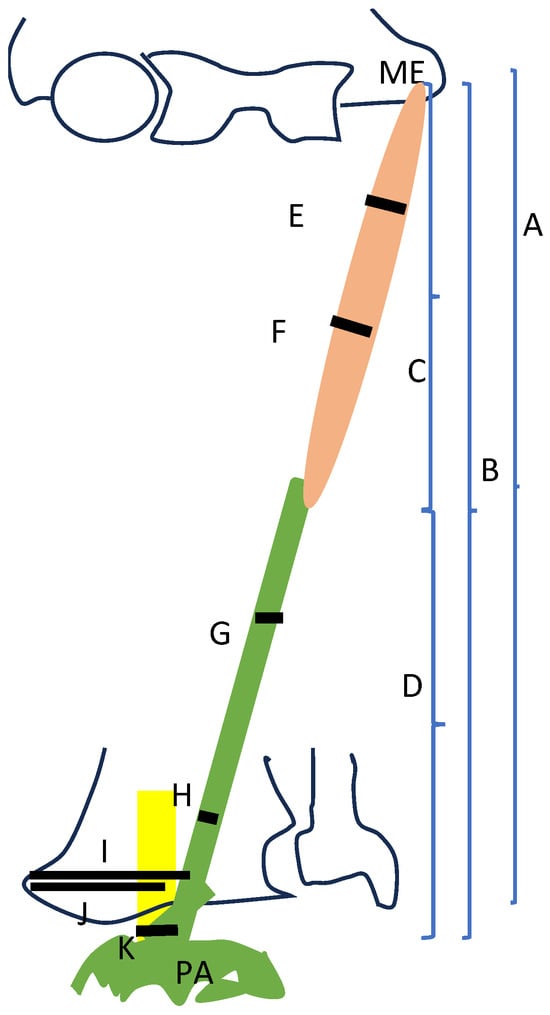
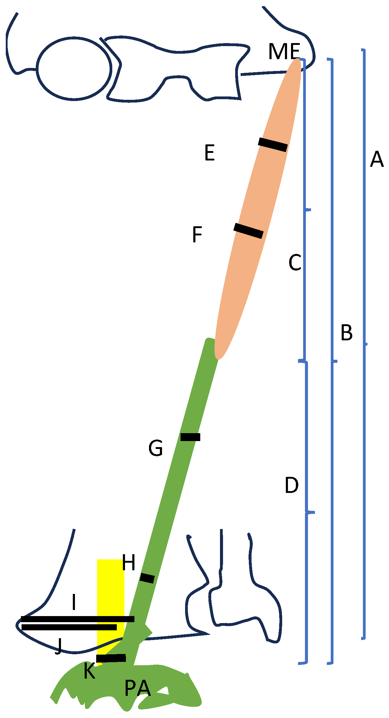
The following parameters were recorded (Figure 1):
Figure 1.
Diagram of the palmaris muscle and tendon with the different measures taken.
A. Length of the forearm (from the ME to the styloid process (SP) of the ulnar bone)
B. Length of all PL from the ME origin to the insertion at the PA.
C. Length of the muscular bell of the PL from the ME to the end of the muscular fibers.
D. Length of the tendon from the origin at the distal part of the muscular bell to the insertion at the PA.
E. Width of the muscle of PL at 3 cm from the ME.
F. Width of the muscle of PL at 6 cm from the ME.
G. Width of the tendon of PL at 3 cm from the origin (end of the belly muscle)
H. Width of the tendon of PL 2 cm before arrived at its insertion at the PA.
I. Distance from the SP of the radius bone to the lateral border of the tendon of PL
J. Distance from the styloid process of the radius bone to the medial border of MN
K. Width of the MN at the level of the wrist, before it is introduced at the carpal tunnel.
Before its insertion into the PA, the fascial expansions of the tendon and its relationships with the MN were carefully documented. All dissections were photographed using a Canon EOS 60D digital camera (Canon, Tokyo, Japan).
2.3. Statistical Study
All data were processed with Excel and SSPS 26. Qualitative variables were summarized as proportions. The concordance index (CCI) of the different measurements observed (nerve perimeter and diameter) was calculated for each anatomical point studied. If optimal CCI values were obtained, they were averaged and summarized as median and mean with their respective measures of dispersion.
2.4. Histological Study
In two randomly selected specimens, tissue examples of the PL tendon were obtained at multiple points. Both the longitudinal and transversal samples of the PL tendon were formalin-fixed and paraffin-embedded to obtain a paraffin block, from which 4 micras Haematoxilin and Eosin (H&E) slides were obtained, and a histological analysis was performed by a soft-tissue-specialized pathologist. The samples were analyzed using an optical microscope (Leica DMD 108, Leica, Wetzlar, Germany).
3. Results
A total of 72 upper limbs were studied. 39 males (54.2%) and 33 females (45.8%), with 40 from the left side (55.6%) and 32 from the right side (44.4%).
In total, 55 upper limbs presented PL (76.4%), and in 17 upper limbs, we observed anatomic variations in the PL (23.6%). The statistical study showed that the anatomical variations were distributed in the same way in males and females (Exact proof from the Fisher test = 0.408; p = 0.237) (Table 1) or sides (Exact prove de Fisher= 0.418; p = 0.279), although the male and left hands presented more variations (Table 2). The length of the forearm was 25.13 cm, SD 2.425 cm. The other parameters studied are shown in Table 3.
Table 1.
Variation in the palmaris longus muscle depending on sex.
Table 2.
Variation in the palmaris longus muscle depending on the side.
Table 3.
Measures of the variable studied in the normal palmaris longus muscle.
3.1. Ultrasound and Anatomical Study
Ultrasound study was able to identify the presence of PL in 55 forearms with 17 variations, ten being type 0, and seven other variations. Dissections proved the normal pattern visualized by US, the absence of the muscle, and the rest of the variations in the PL (Table 3).
Ultrasonography findings were consistent with the anatomical study in 98.61%. Only in one case was the US not able to detect a type 1 variation in the PL, which was classified as type 0 by US.
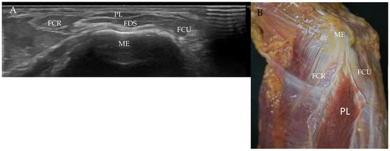
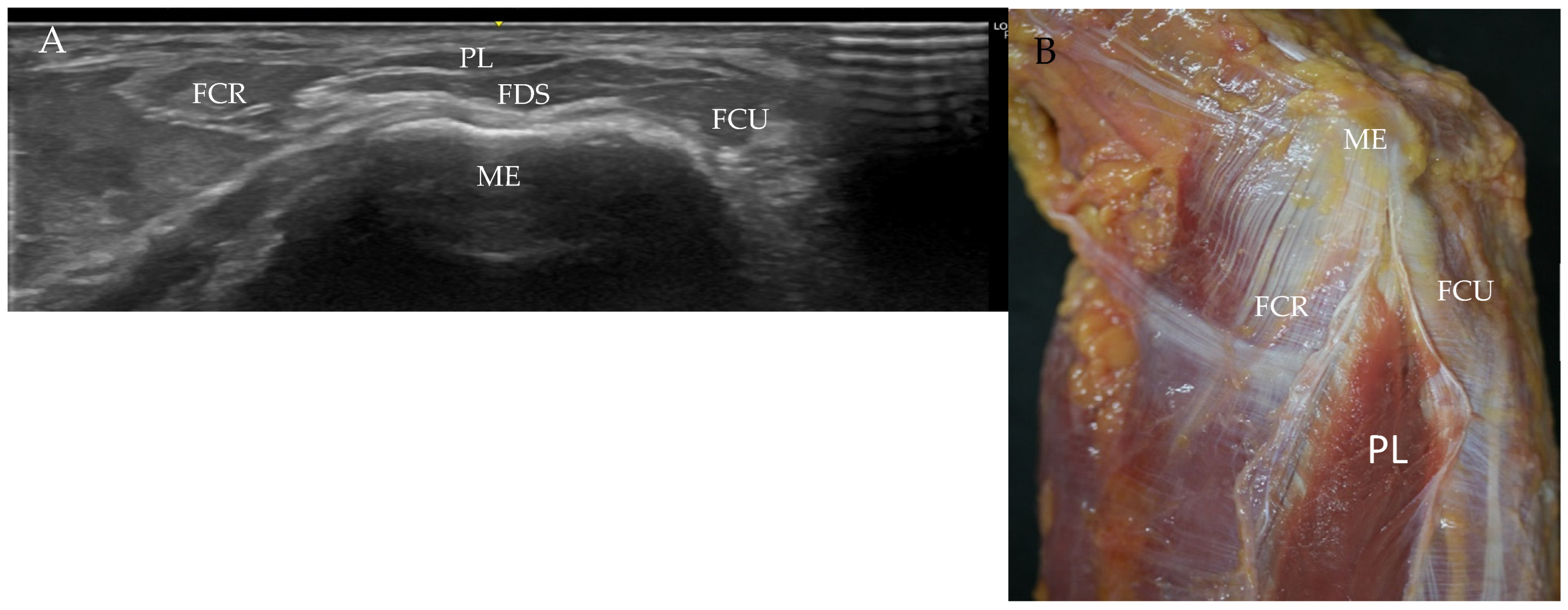
When the PL existed, a US study showed its proximal insertion at the medial epicondyle. However, it was necessary to move down slightly in the inferior direction at the medial epicondyle to observe as a small and triangular hypoechogenic structure, with a superior base and inferior vertex. It was always surrounded by a hyperechogenic line that separated it from the FCR laterally and FCU medially (Figure 2).
Figure 2.
Origin of the palmaris longus muscle (PL) (A): Ultrasound image of the PL as a small and triangular hypoechogenic structure, with base superior and vertex inferior, surrounded by the hyperechogenic line (the antebrachial fascia) and separated from the rest of the muscles. Flexor carpi radialis (FCR), flexor carpi ulnaris (FCU), flexor digitorum superficialis (FDS), and medial epicondyle (ME). (B): Anatomical view of the origin of the PL. It is possible to observe how the muscular fibers do not arrive until the medial epicondyle.
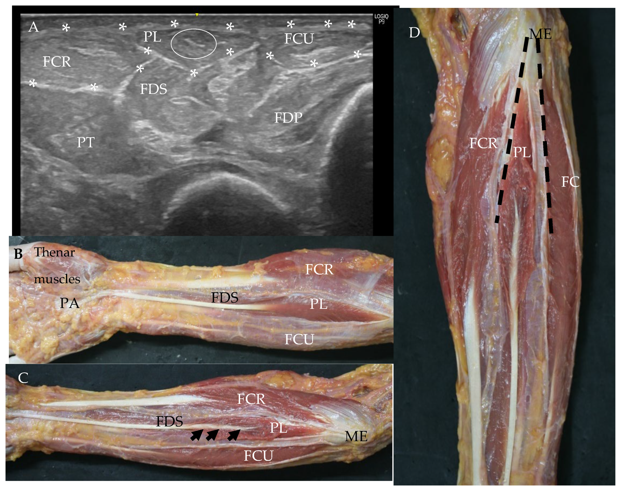
After, the belly increased, and an ovoid shape was adopted, with a characteristic and transversal hyperechogenic line inside the muscle (Figure 3). This line became the tendon that followed the muscle distally. This tendon was a uniform and fibrillar hyperechogenic structure until it arrived and inserted at the PA.
Figure 3.
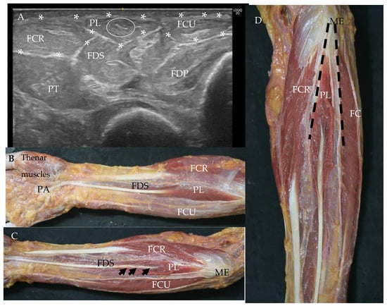
(A): Ultrasound vision of the normal palmaris longus muscle (PL) at the proximal third of the forearm. Antebrachial fascia (AF), seen as a hyperechogenic line surrounding (white *) the muscle and separating it from flexor carpi radialis (FCR), and flexor carpis ulnaris (FCU) also isolated by the AF. Inside the PL, a transverse hyperechogenic line is seen (white circle). It corresponds to the origin of the tendon of the PL. Flexor digitorum superficialis muscle (FDS), flexor digitorum profundus muscle (FDP), and pronator teris muscle (PT). (B,C): Anatomical vision of PLB: The AF has removed only over the PL. (C): The AF is cut just over the PL, FCRFCU. All of them originated at the medial epicondyle (ME). The muscular fibers of the PL were cut to see if its tendon was inside the muscle (black arrow). FDS Palmar aponeurosis (PA). (D): Anterior side of the forearm without the AF that covers FCR, PL, and FCU. The tendon of the PL is seen inside the muscle. The septum from the antebrachial fascia separates these muscles (black intermittent line).
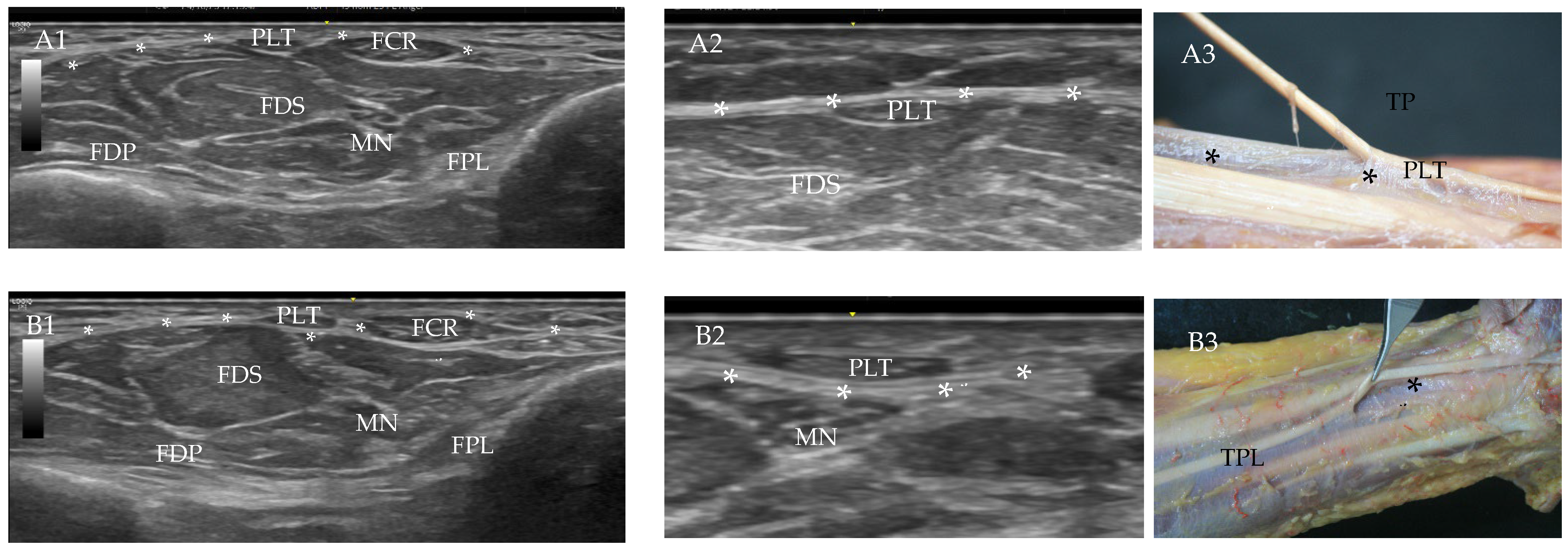
Nevertheless, the tendon had a different relationship with the AF at the proximal part of the forearm than at the distal part. First, the tendon was surrounded by the AF, as were all the anterior muscles of the forearm, and at the third distal of the forearm, the tendon was isolated and surrounded totally by the AF, as were the tendons of the FCR and flexor carpi ulnaris (FCU) (Figure 4).
Figure 4.
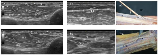
Relationship of the antebrachial fascia (AF) with the tendon of the palmaris longus muscle (PLT). (A1): Ultrasound (US) study shows that the AF (white *) covers the PTL and the rest of the anterior structures of the forearm. (A2): Enlarged image B, more specific. (A3): Anatomic image of the tendon of PL inside the AF (black *). (B1): US image of the tendon of the PL superficial and independent of the rest of the structures of the anterior side of the forearm. (B2): enlarge image. (B3): Anatomic view that shows the AF (black *) was under the tendon of PL at the distal third of the forearm surrounded the FDS, FDP, and MN.
Measurements of the width of the tendon at the two points studied were 0.39 SD 0.09 mm and 0.39 SD 0.079 mm. The width of the MN at the wrist was 0.55 SD 0.067 mm. There were no significant differences between the side of the body the limb was taken from or the sex (Table 4).
Table 4.
Measures of Figure 1 of all the upper extremities that had the palmaris longus muscle, depending on the sex. Statistically significant values are in red.
In all the cases studied, the PL kept a great relationship with the MN at the wrist before the nerve was introduced inside the carpal tunnel, just up to the nerve or medially to it.
The anatomical study showed the normal pattern of PL in 55%.
The length of the muscular bell and tendon of PL was 25.82 cm, SD 2.394 cm (Table 3).
The muscle originated at the medial epicondyle, which has a thick aponeurosis where few muscular fibers originate, and at the AF (Figure 2). We did not find a real tendon at this point of origin. The muscular bell, with a fusiform shape, was located on the anterior and superficial sides of the forearm, between the FCR and FCU, covered by the AF (Figure 3B,C). Significant differences in the length of the muscle were found depending on the sex, but not on the side studied. Samples were bigger in males (Table 4).
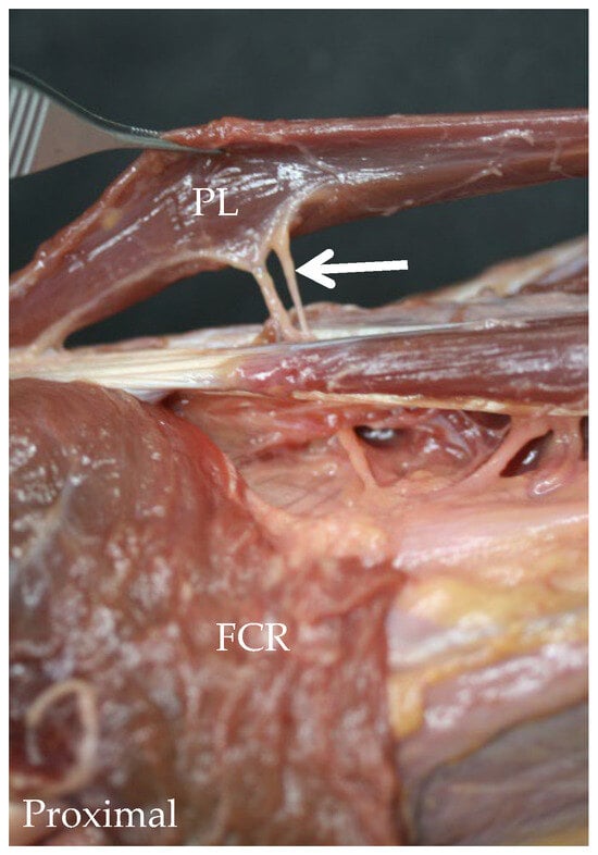
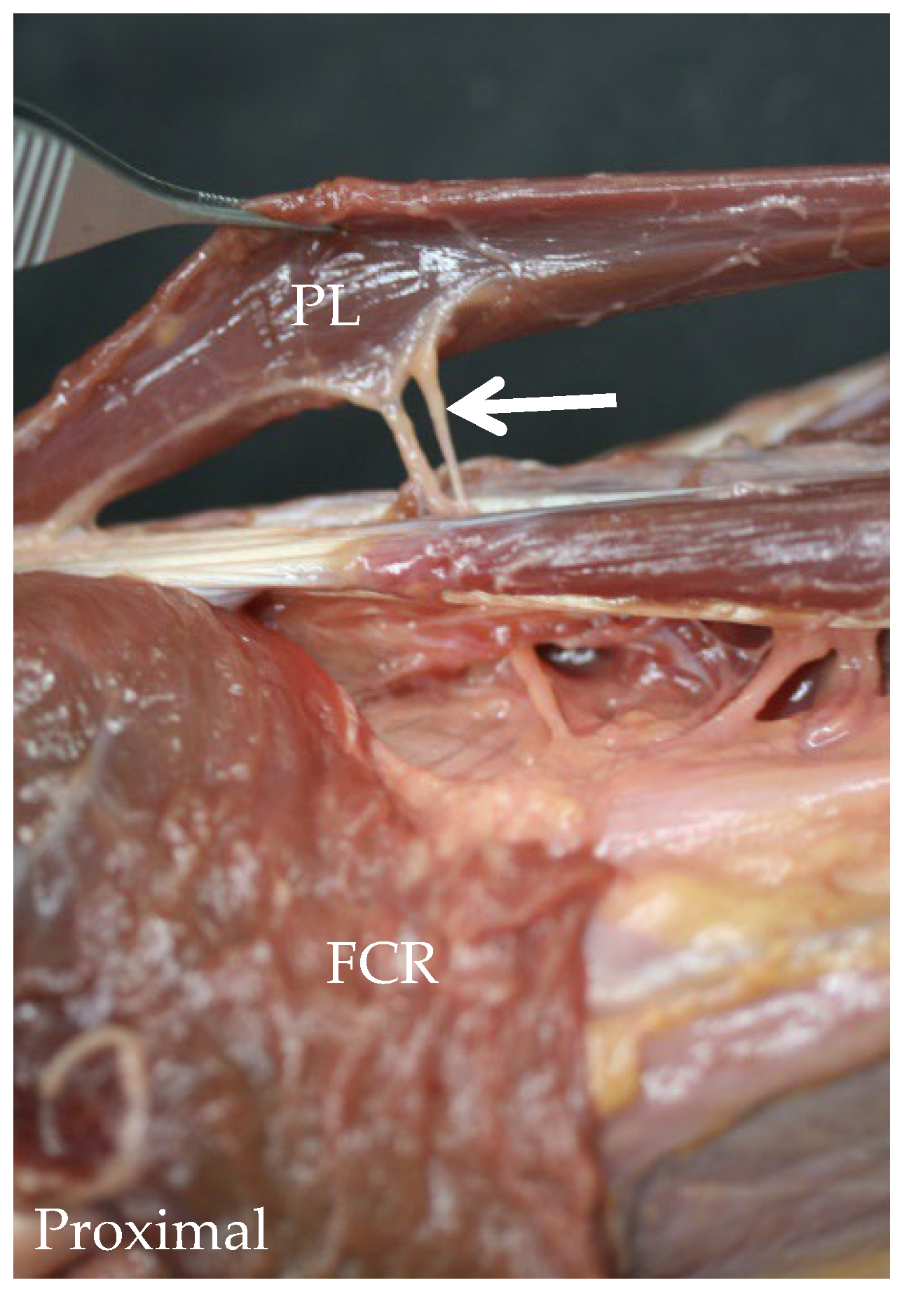
The PL was separated by a septum of dense connective tissue coming from the AF (corresponding to the septum hyperechogenic seen by US) and the FCR and FCU (Figure 3D). Although the size and length of all the bell muscle was not consistent, there was always the same pattern. The muscular belly increases its width distally until the middle of the muscle, and then it decreases its width until the muscular fibers disappear, followed by a flat tendon. The length of the PL was 12.01 cm, SD 1.708 cm, the diameter of PL at the superior point was 1.09 cm, SD 0.335 cm, and the diameter at the inferior point was 1.04 cm, SD 0.315 cm. Significant differences in the length of the muscle were found depending on the sex (Table 4). The muscle was supplied by a branch of the MN that came deeply into the muscle (Figure 5).
Figure 5.
Anatomical view of the deep side of the palmaris longus muscle (PL) after removing the flexor carpi radialis muscle (FCR). Two branches of the median nerve (white arrow) cross the flexor digitorum superficialis muscle to supply the PL.
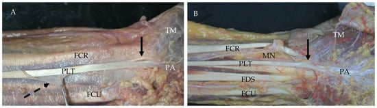
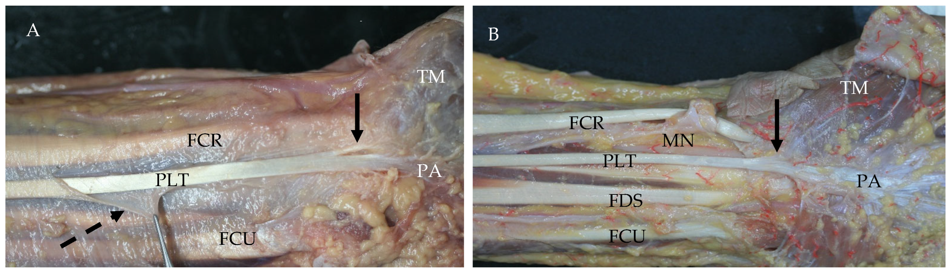
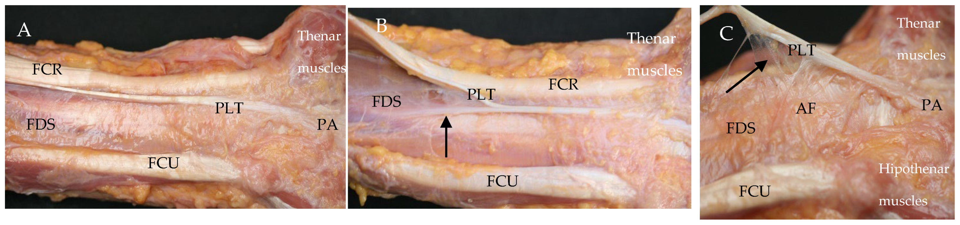
An accurate dissection of the bell revealed the presence of an intramuscular tendon that corresponded to the hyperechogenic transversal line observed inside the muscle, as the US study showed. It was followed by the tendon of the PL (Figure 3C). Distally, the tendon runs superficially along the anterior side of the forearm, covered by the muscular fascia or AF. Nevertheless, as the US study exposed, this fascia had a different behavior in the middle than in the third distal part of the forearm, where the AF covered and isolated the tendon of the PL (Figure 4 and Figure 6).
Figure 6.
Anatomical dissection of the antebrachial fascia (AF) at the anterior side and distal third of the forearm (A): the AF (intermittent black arrow) cut only on the tendon of the palmaris longus muscle (PLT). It is possible to observe that it does not affect the rest of the muscles. However, under the PLT, the AF covers the rest of the anterior structures of the forearm. Thenar muscles (TM), palmar aponeurosis (PA). (B): All the AF has been removed. The PLT inserts at the PA and expansion to the TM is seen (black arrow). Flexor carpi radialis muscle (FCR), flexor carpi ulnaris muscle (FCU), median nerve (MN), flexor digitorum superficialis (FDS).
At this point, the tendon lost its deep relationship with the epimysium of FDS and was isolated from the rest of the muscles, nerves, and vessels of the anterior side of the forearm, as were the FCR and FCU. The AF made a special sheath for these three tendons until they arrived at the hand (Figure 3). Under these sheaths, the AF followed, covering the rest of the muscles, vessels, and nerves, and even increasing this thickness.
The length of the tendon was 13.81 cm, SD 2.404 cm (Table 3). Significant differences in the total length of the tendon were observed depending on the sex, but not on the side studied (Table 5).
Table 5.
Measures of Figure 1 of all the upper extremities that have the palmaris longus muscle, depending on the side. No statistically significant results were found.
The diameter of the tendon of the PL was 0.38 mm, SD 0.065 mm, at 3 cm from its origin and 0.37 mm, SD 0.082 mm, at 2 cm before the wrist line (Table 3). No significant differences in the diameter were found depending on the sex or side.
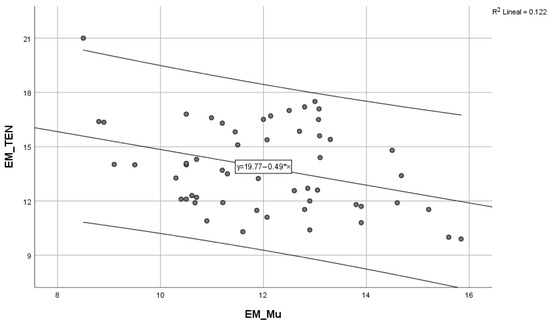
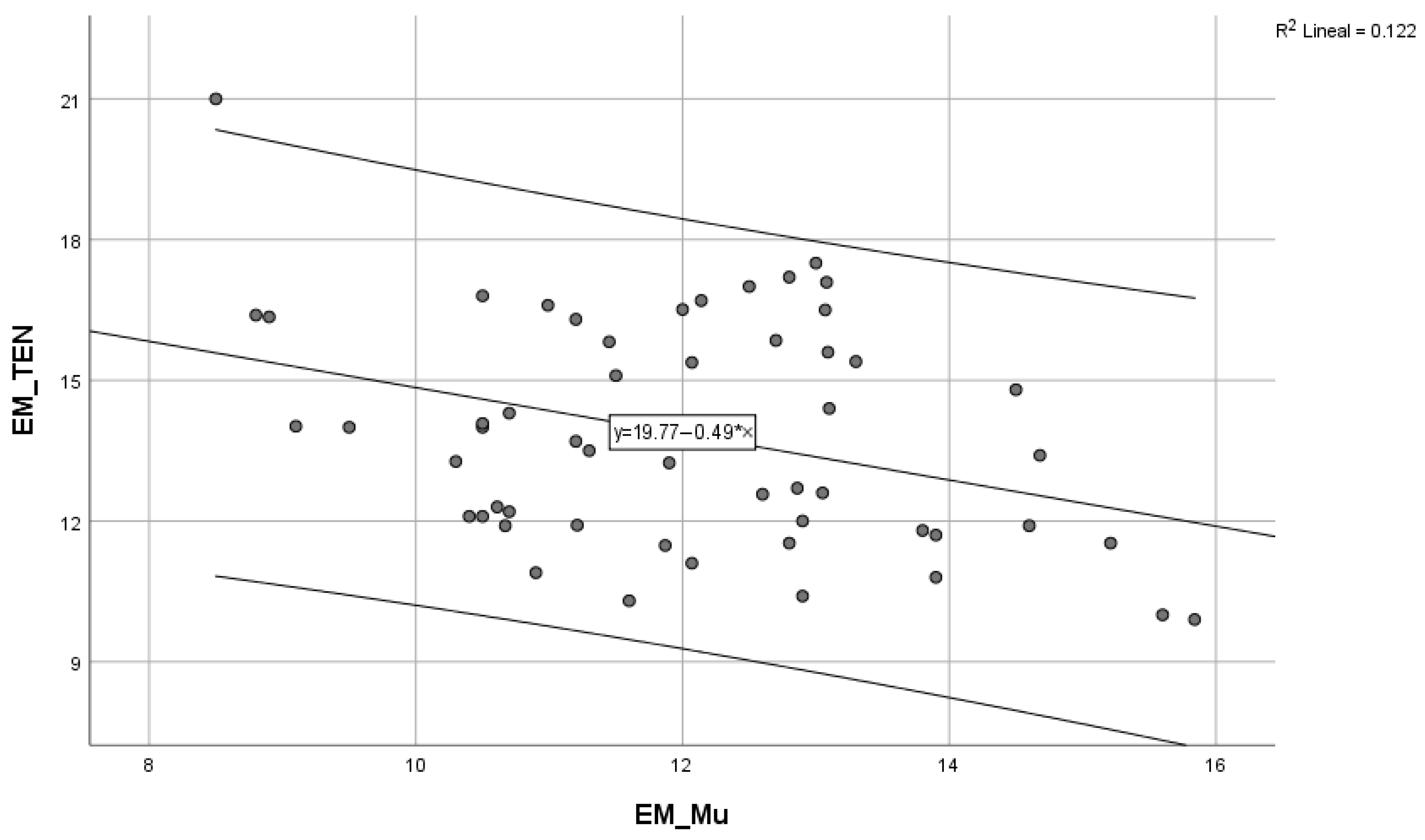
We found an inverse linear relationship between the length of the muscle and the diameter of the tendon of the PL (Spearman’s rho = −0.349; p = 0.009). However, in a simple sampling simulation (number of samples = 1000), the statistical significance of the relationship would be lost (Spearman’s rho = 0.245; p = 0.071) (Figure 7).
Figure 7.
An inverse linear relationship exists between the length of the muscle and the tendon of the palmaris longus (Rho Spearman =−0.349; p = 0.009). However, in a simulation of the sample rate (n = 1000), the statistical significance of this relationship is lost (Rho Spearman = −0.245; p = 0.071).
The distance to the SP of the radio bone to the lateral border of the tendon of the PL at the level of the wrist was 2.35 cm, SD 0.305 cm, and to the medial border of the MN, it was 2.54 cm, SD 0.315 cm. Significant differences were observed depending on sex. Samples were bigger in males (Table 5). In 71% of the cases, the results were negative. This implies that the tendon of the PL covered the MN. There were significant differences (Binomial test, exact signification p:0.003) (Table 4 and Table 5).
The width of the MN was 0.52 mm, SD 0.062 mm. No significant differences were observed depending on sex or side, although the width of the nerve in females was greater than in males. The diameter of the nerve did not vary in relation to the distance, either when measured by US (t Studdent −0.597, p = 0.553) or by anatomic study (t Studdent −0.934, p = 0.354).
When compared by the Bland–Altman plot, the agreement between the US and anatomy observations in all the measurements taken indicated that US always overestimates the measurements.
The tendon of the PL is inserted at the PA; however, we observed in 100% of the cases that this tendon expanded to the thenar muscles of the hand (Figure 6).
We found anatomical variations in 17% of the cases. Variations in the PL were detected by US in 98.61% of cases. Only in one variation of type 1 did we not detect it by US.
Variation type 0: US examination detected the absence of the PL, and anatomical study proved it in 11 upper limbs (15.27%)
Variation type 1: (two females) PL had no muscular fibers. It was only a tendon and was detected in 2.77% of cases.
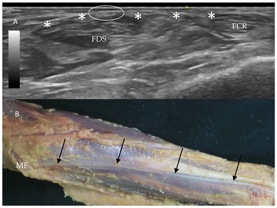
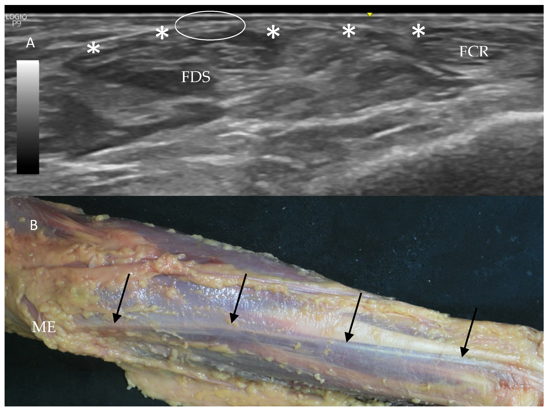
This was detected in one case by US and in two cases by the anatomical study. In the first, the US image was a slim hypoechogenic structure that originated at the ME along with the rest of the ME muscles’ origin and was inserted at the PA (Figure 8A). Also, it was surrounded and isolated by the AF in the distal third of the forearm. The width of the tendon as measured by US was 0.25 mm superiorly and 0.21 mm inferiorly. Dissection allowed us to measure the entire tendon, which was 31 cm in length; the width was 0.20 mm superiorly and 0.15 inferiorly (Figure 8B).
Figure 8.
Variation type 1: (A): Ultrasound study showed a hypoechogenic structure (white circle) compatible with a tendon just under the antebrachial fascia (white *). (B): Anatomical dissection showed, after removing the skin and adipose tissue, a variation of type 1, with only a tendon of the palmaris longus muscle (black arrows). It runs from the medial epicondyle (ME) to the palmar aponeurosis without muscular fibers, covered by the antebrachial fascia.
The second case, only detected by dissection, revealed the presence of a tendon that was inserted at the PA; however, the tendon, with a length of 15.5 cm and width of 0.20 mm, originated from the medial side of the FCR (Figure 9) Moreover, there was a very slim tendon that came from the AF and joined with it before insertion (Figure 9).
Figure 9.
Variation types by anatomical study (A): The superficial layer of the antebrachial fascia is removed. The tendon of the PL (PLT) comes from the medial border of the flexor carpi radialis muscle (FCR) and inserts at the palmar aponeurosis (PA). (B): It is possible to see another tendon (black arrow) coming from the antebrachial fascia and joining with the PLT (black arrow). (C): The deep side of the PLT provides many connections to the antebrachial fascia (AF) (black arrow) before it inserts at the (PA). The flexor digitorum superficialis muscle (FDS) is covered in all the pictures by the antebrachial fascia. Flexor carpi ulnaris (FCU).
Variation types 2 and 3 were found as hypo- or hyperechogenic structures at the medial and distal parts of the forearm, respectively, medial to the FCR, and covering the MN.
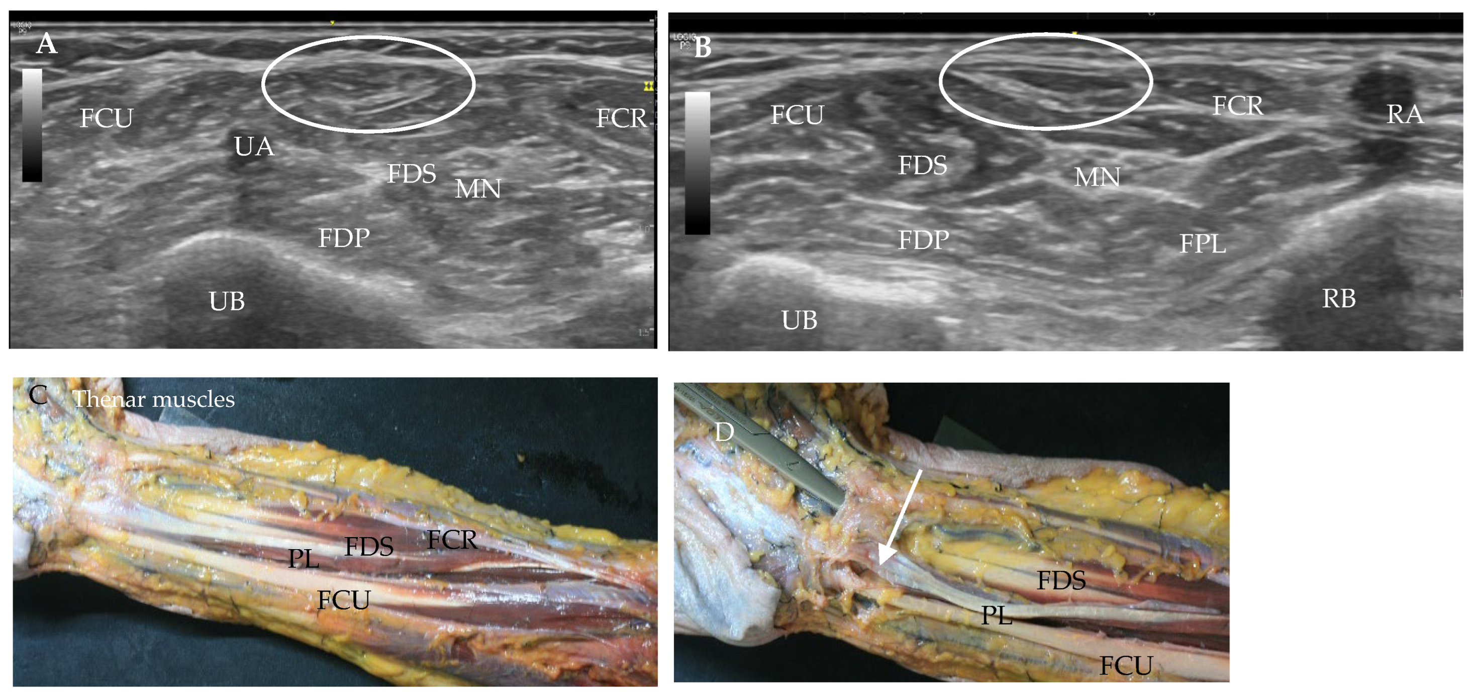
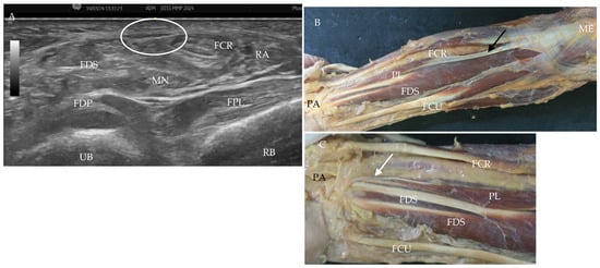
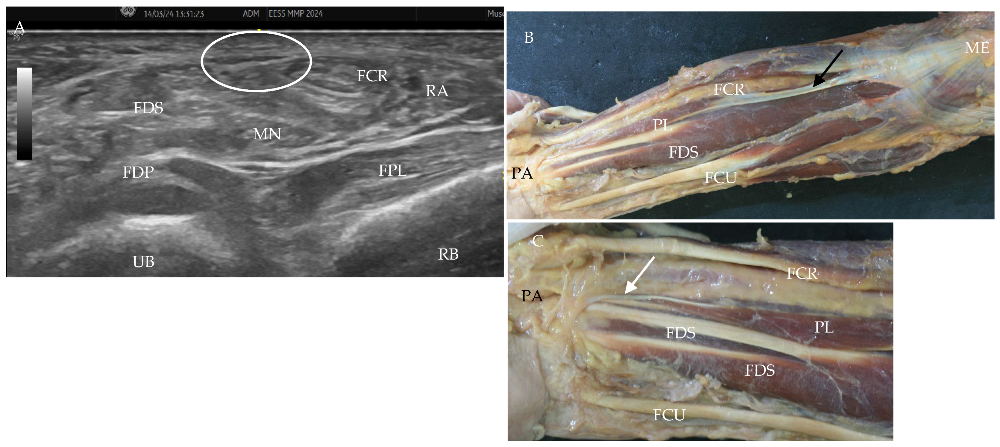
Variation type 2 (one female and one male): The muscular bell of the PL was in the middle of the forearm. The Yorigin and the insertion were a tendon. This was detected in 2.77% of cases. US images showed a broad hypoechogenic structure bigger than a normal tendon at the medial level of the forearm, similar to a normal PL, even with the intramuscular tendon that finishes with a hypoechogenic and wide structure. Dissection showed in one case that the PL originated from a tendon of 6 cm in length and 0.2 mm in width, followed by the muscular bell that had 16 cm of length and 1 cm of width. The AF surrounded the PL throughout its trajectory until it inserted at the PA (Figure 10). MN width was 0.5 cm. In some cases, the length of the tendon was 15.3 cm from the ME, followed by a muscular portion of 7.7 cm that finished at the PA with a tendon of 2.5 cm. The width of the tendon at the proximal was 0.5 mm, and at the distal part was 0.2 mm (Figure 11). The width of the MN was 0.55 mm by US and 0.52 by anatomical study.
Figure 10.
Variation type 2: (A): Ultrasound study at 9 cm from the medial epicondyle. PL has a typical muscular echogenicity and an ovoid shape (white circle). It is covered by the antebrachial fascia. It keeps its relationship with the flexor carpi radialis muscle (FCR) and the flexor carpi ulnaris muscle (FCU). Median nerve (MN), flexor digitorum superficialis (FDS), Flexor digitorum profundus (FDP), ulnar bone (UB). (B): Ultrasound image at 12 cm from the medial epicondyle is compatible with variation type 2. (C): Dissection of the forearm after removing the antebrachial fascia shows the central, long muscle of the PL with a tendinous portion. (D): Insertion of the distal tendon of PL (white arrow) under the antebrachial fascia (AF). Medial epicondyle (ME).
Figure 11.
Variation type 2. (A): Ultrasound study of the middle of the forearm shows the palmaris longus muscle (PL) as a superficial hypoechogenic structure covered (white circle) by the antebrachial fascia. It keeps the relationship with the flexor carpi radialis muscle (FCR) and the flexor carpi ulnaris muscle (FCU). The median nerve (MN), flexor digitorum superficialis (FDS), Flexor digitorum profundus (FDP), flexor pollicis longuis (FPL), radius bone (RB), and ulnar bone (UB). (B): Dissection of the forearm after removing the antebrachial fascia shows the proximal muscle of the PL (black arrow) (C): Insertion of the distal and narrow tendon of PL (white arrow). Medial epicondyle (ME), palmar aponeurosis (PA).
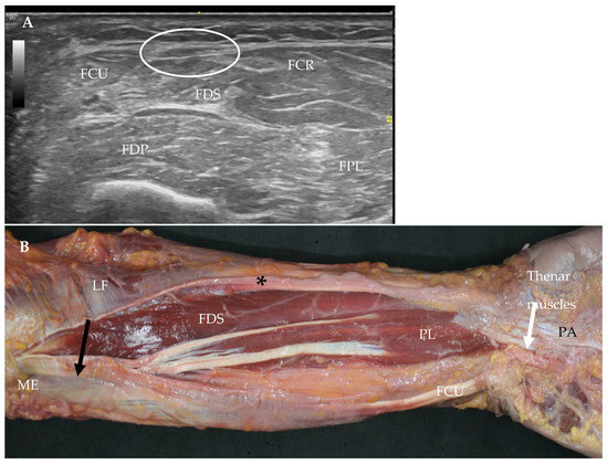
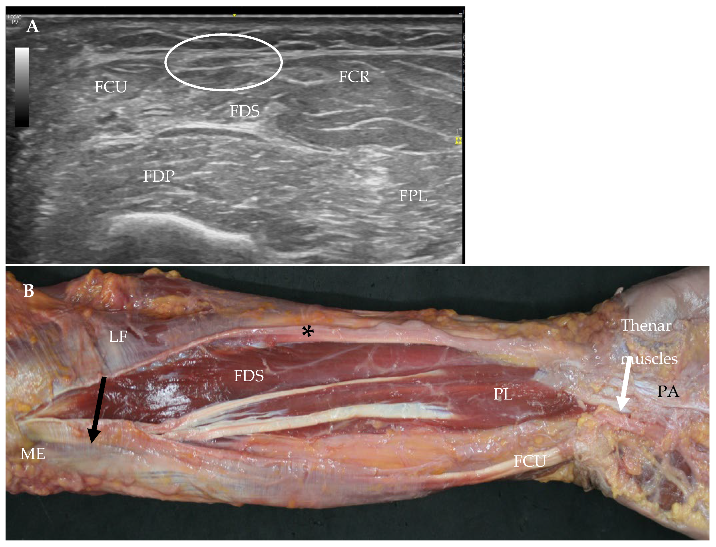
Variation type 3 (2 females): Also known as the inverted or reverse palmaris longus muscle, this variation was found in two cases (2.77%). The muscle was in the middle and distal parts of the forearm. In one case, the muscle had two origins from the ME as a wide and thin tendon, and the other, from the AF, along with the lacertus fibrosus. Muscular fibers originating from the PL were found at 8.6 cm from the ME. The length of the muscle was 15 cm, the width was 1.5 cm in the middle of the forearm, and at 2 cm from the FR. The lateral bell was inserted at the PA, and the medial bell was inserted at the medial side of the PA (Figure 12).
Figure 12.
Variation type 3. (A): Ultrasound in the middle of the forearm detects the hypoechogenic image (white circle). Flexor carpi radialis (FCR), flexor carpi ulnaris (FCU), flexor digitorum superficialis muscle (FDS), flexor digitorum profundus (FDP), flexor pollicis longus (FPL). (B): An anatomical study of the forearm after the lacertus fibrosus (LF) and the antebrachial fascia (black *) were cut. The PL has two points of origin; one is lateral and originates at the medial epicondyle (ME), and the other is medial and originates at the antebrachial fascia near the ME (black arrow). The medial bell inserts at the medial side of the palmar aponeurosis (PA) (white arrow), and the lateral bell inserts directly at the palmar aponeurosis (PA).
The innervation of the bell came from the ulnar nerve. The diameter of the MN was 0.65 mm by US and 0.62 mm as measured by the anatomical study.
In the other case, the muscle bell of PL arrived at the PA. The length of the proximal tendon was 9 cm, and the length of the muscle was 16 cm; the width at the middle of the muscle was 1 cm. Additionally, the PL coexisted with an additional abductor digiti minimi muscle going under the ulnar vessels and the other over the ulnar nerve. The distance from the MN to the SP of the radius and the PL to this process was the same, 2 cm. The diameter of the MN was 0.61 mm as measured by US and 0.59 mm as measured by the anatomical study.
3.2. Other Variations
We found two cases with a double PL, one below the other. US imaging permitted the detection of these variations. These also originated at the ME with muscular fibers that followed distally and finished in a narrow tendon parallel to the PL tendon, which inserted at the AF or FR. Although the normal PL was considered in the statistical analysis, these two variations were an additional PL with independent origin and insertion points that were excluded, as they were not included in any of the described variation types. In fact, this additional PL was covered by the AF along its course.
3.3. Histology Study
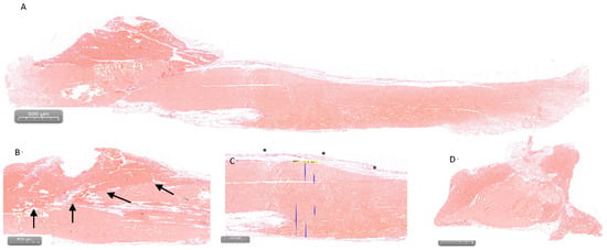
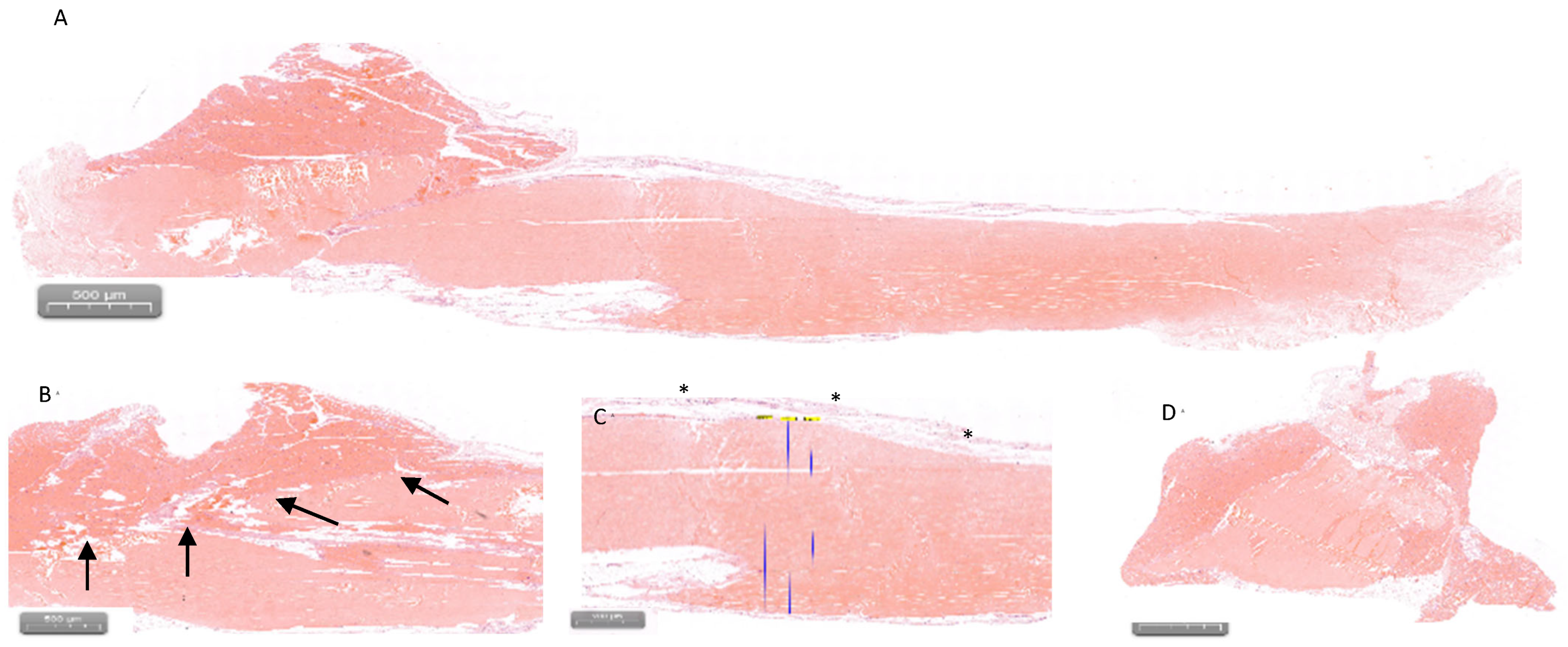
Histological study showed the continuity of the tendon with the muscular fibers, with a very close relationship (Figure 13). The study of the tendon of the PL revealed a regular and typically dense connective tissue. The width of the tendon was 0.29 mm (Figure 13).
Figure 13.
Histological view of the muscle and the tendon of the palmaris longus muscle (PL). (A): Longitudinal view of the end of the muscular portion (dark pink) and the tendon inside the muscle. (B): A view of the deep relationship between the tendon and the muscular fibers (black arrow) of the PL. The tendinous fibers are perfectly parallel. (C): Superior part of the tendon covered by the antebrachial fascia (black *). It has connections with the epitenon. Measures of the tendon were 0.29 mm. (D): Transversal view of the muscle (dark pink) and tendon at the end of the muscular portion.
4. Discussion
The PL has been extensively described by numerous authors with regard to its anatomy and variations [6,18,19] as well as its function [6,20,21]. The PL can be identified by a classical test (Schaeffer’s test) by bringing the thumb and small fingertips together (forceful opposition of the thumb) and performing a mild active flexion of the wrist joint [12]. However, the present study gives special importance to including an ultrasound study to detect the presence of the PL or its possible variations for their surgical and clinical applications and for the study of other carpal pathologies.
In agreement with previous research, our findings confirm that US is a reliable method for visualizing the PL and identifying its variants [22,23]. The US examination should begin at the ME, where a small triangular structure can typically be observed [4,22,24]. Nevertheless, it is important to move slightly distally to recognize the antebrachial fascia (AF), which appears as a hyperechogenic line and serves as an excellent reference for locating the PL. At this level, the AF clearly separates the PL from the FCR and FCU [4], allowing for their distinction in the proximal forearm. And also, to observe, as the probe is advanced distally, how the PL progressively changes its morphology from a triangular to an ovoid shape, with a typical central hyperechogenic line corresponding to the origin of the tendon of the PL. The anatomical dissection corroborates these ultrasonographic features, particularly the fusiform configuration previously reported by other authors [6,10]. Importantly, our anatomical observations emphasize that the PL originates from the ME by means of a tendinous structure, rather than by direct muscular fibers [16]. The muscle fibers do not extend to the ME itself.
The measured length of the muscular portion of the PL (12.01 ± 1.708 mm) was slightly shorter than that reported in previous studies [20], although it remained within the expected anatomical range. This can be explained by the differences between populations or even different references about the limits of the muscle.
With respect to the length of the tendon of PL (13.8 ± 2.4 cm), it was very similar to other studies [14,25]. The width of the tendon (0.39 mm), sometimes, was slightly bigger at the proximal point than the distal point as measured by US, and was also similar to some other studies [14,23] but quite different from other studies (0.88 mm) performed with living people, which may have measured at the level of the carpal bones [22]. The histological measures, although they were less than those observed with the US, and the anatomical results were very similar. Interestingly, no significant differences were observed between sexes or sides in any of the measurements obtained in this study. These measures can help with surgical applications seeking to use the PL tendon.
The histological structure of the PL tendon was consistent between the middle and distal thirds, as previously reported by other authors [20]. The tendon maintains a uniform morphology throughout its course, although the presence of abundant and large nerve fibers has been documented [26]. We have not studied them, but they can be important for movement. Another aspect to consider is the deep relation of the muscle fibers and the tendon. This could be important to preserve the function of the muscle in case of surgery, as several studies indicate that it may cause a decrease in the function of the palmar flexion [27]. Its uniformity and its length prove that it has several advantages. The most important, and in accordance with the main objective of this study, is that it is valuable for reconstruction and tendon grafting procedures [12,13]. However, we think it is mandatory to conduct a US study to prove its existence.
Of particular relevance is the relationship between the PL tendon and the AF, which has both functional and surgical implications. As demonstrated by our US and anatomical observations, the PL tendon consistently descends covered by the AF [3,20]. This relationship varies slightly between the middle and distal thirds of the forearm, as also described by other authors [4]. Such differences should be considered during surgical procedures. We recommend accessing the distal third of the forearm and incising only the sheath of the PL up to the level of the tendon. This approach preserves the AF that envelops the remaining anterior forearm muscles, as well as the MN and the ulnar neurovascular bundle, thereby minimizing the risk of iatrogenic injury. It is essential to preserve muscular function and ensure MN protection. Conversely, harvesting the PL tendon at the distal and anterior wrist crease [28,29] is not advisable, as it carries a higher risk of MN injury.
The relevance of the AF is further illustrated by other anatomical variants in which a double PL is observed; in these cases, the accessory PL inserts into the AF, potentially contributing to its tension and functional stability.
Our study also quantified the distance between the MN and the PL tendon relative to the SP of the radius bone, which provided a clear understanding of their anatomical relationship at the wrist. This information is especially useful during surgery, as the anatomical superficial PL tendon and medial position will help to distinguish the MN. However, we assert that a previous US study can be very useful to distinguish them. In many specimens, the MN was partially or completely overlain by the PL tendon, a finding that could help to explain the chronic compression of the MN caused by repetitive wrist movements, a mechanism potentially contributing to carpal tunnel syndrome [30]. Nevertheless, we do not believe that the PL tendon itself is a direct compressive factor on the MN. In our data, the average MN width was 0.55 mm, closely matching previous reports (0.59 ± 0.93 mm) [4], irrespective of tendon–nerve proximity. Additionally, no significant differences were observed between sexes or sides. We suggest that the AF, by separating the PL tendon from the MN, acts as a protective barrier, reducing potential compression. However, in type 3 variations, where muscular fibers extend distally and lie directly over the MN, contraction of these fibers may exert pressure on the nerve. In fact, in our series, the MN width was slightly greater in these cases, supporting this hypothesis.
As described in other studies, we also observed that the insertion of the PL tendon is consistently located anterior to the FR, attaching primarily to the PA and occasionally to other structures such as the AF, FR, thenar, and hypothenar regions, or carpal bones including the trapezium, scaphoid, and pisiform [7], as well as the tendons of the FCU, FCR, FDS, and FDP [16]. Despite the abundance of connective tissue in both the PL tendon and the PA, they do not share a common origin [31], which explains the wide variability of connections between them [32]. The PL appears to insert into the subcutaneous plane of the palm, continuing into the superficial layer of the PA formed by its longitudinal fibers. This anatomical configuration supports the interpretation of the PL as a tensor of the superficial fascial system of the palmar subcutaneous tissue [20].
Performing an US evaluation prior to any surgical procedure is therefore essential for detecting PL variations, given that it is among the most variable muscles in the human body [33]. US findings can guide both the feasibility assessment and surgical strategy for reconstructive interventions.
Previous authors have classified PL variations based either on tendon insertion morphology or on the full spectrum of anatomical variants [6,18]. In this study, however, we propose a more functional and clinically oriented classification, applicable to both US and surgical practice. This simplified system considers the position of the muscular belly and the insertion pattern at the PA, facilitating intraoperative identification and planning [22].
The type 0 variation, or agenesis of the PL, is the most frequent variant [22] and our findings are consistent with those of previous studies. This absence has been attributed to Mendelian inheritance [21] and varies not only among ethnic groups but also among individuals within the same population [21]. No statistically significant differences were found regarding sex or laterality [22], although other authors have reported a higher incidence in females and on the left side [1,8]. However, others report a higher incidence in males [12,34] as well as a greater frequency of bilateral rather than unilateral agenesis [22]. In general, an absence rate of approximately 10% has been reported [12,18,35], while we observed a slightly higher incidence of 15.27%, comparable to that found by other authors [8,22]; some studies even report rates as high as 25% [7]. Recognizing this type of variation is crucial to prevent misidentification of the MN during surgery, given its close anatomical proximity [35].
Type 1 variation in the PL was identified in 2.77% of the specimens. No previous studies were found in the literature for direct comparison. This variant is easily recognizable by US, as the tendon exhibits the same echogenic characteristics as the typical PL tendon, although it is slightly thinner—approximately 0.2 mm narrower. Despite its reduced caliber, we consider that this variation may still be suitable for use in reconstructive surgical procedures.
The type 2 variation was also observed in 2.77% of cases, although the two specimens presented distinct morphologies. In this variant, a proximal tendon is followed by a central muscular belly and a distal tendon [6,10]. Another possible configuration is the digastric PL, characterized by two muscular bellies [6]; these may insert independently into the PA [6], or both may converge into a single tendon that subsequently inserts into the PA [10]. In some reports, a trigastric PL has also been described, consisting of three muscular bellies with separate tendons that all insert into the PA [6], though such forms were not identified in our series. From a surgical standpoint, type 2 variations may be clinically valuable, particularly when the distal tendon is sufficiently long to be used in reconstructive procedures. In these cases, the PL tendon could be suitable for surgical grafts.
Type 3 variation is perhaps the most clinically significant variation in the PL and should be carefully assessed using US. This variant, also known as the reversed or inverted palmaris longus [6,36,37,38,39], presents a muscular belly in the distal portion of the forearm, whose morphology can vary considerably. Some authors include within this category the bifid PL [6,10] or cases with multiple muscular heads [5,40]. However, we emphasize that this type should not be confused with instances in which the muscular fibers reach the PA with a tendon, although the tendon is short. In such cases, it would be classified as a type 2 variation. We determined that this variation is not suitable for surgical treatments. Alternatively, in these variations, perhaps surgery could be indicated to avoid the compression of MN, if it is present.
In our classification, we also included cases of reversed PL coexisting with the abductor digiti minimi muscle, as previously described by other authors [6], although in our specimens, the PL inserted into the hypothenar musculature. Other authors describe the abductor digiti minimi muscle in relationship with the PL tendon. We emphasize that it is important to consider the possibility of compression of the ulnar nerve or vessels, as this could complicate surgical procedures requiring resection of the palmaris longus tendon [41,42]. Recognizing this variation is important because it is unsuitable for reconstructive surgery, given the distal position of its muscular belly and its limited tendinous length. Moreover, it carries diagnostic relevance, as it may be associated with pain in the volar aspect of the distal forearm due to compression of the MN or, less frequently, of the ulnar nerve and vessels [41]. Repetitive wrist movements may lead to hypertrophy of the distal muscle belly; owing to the rigidity of the AF, this can precipitate an effort-related compartment syndrome. In such cases, US proves highly valuable for accurate identification and differential diagnosis of this condition.
It is also worth emphasizing that the PL, owing to its high proportion of connective tissue, plays a significant role in the wrist joint proprioception, fine motor skills, and nociceptive functions of the forearm and hand. However, the last investigations confirm that the presence or absence of the PLM does not affect fine motor skills, grip strength, or wrist proprioception, suggesting it can be safely used for surgical grafting without functional loss in the hand [27]
Consequently, its morphology should be regarded as dynamic rather than regressive, reflecting an anatomical evolution more oriented toward sensory integration than pure motor performance (a transformation in structure driven by a change in function). Adopting a more holistic perspective on the anatomophysiological role of the PL could therefore expand our understanding of forearm function and contribute to improved clinical approaches to specific pathologies affecting this region.
5. Conclusions
Comprehensive knowledge of the anatomy and possible variations in the palmaris longus muscle is essential, given its importance in reconstructive surgical procedures, as the tendon of PL can be the main option for autograft tendon transfer in different areas of the body. However, it is necessary to prevent surgical complications or misidentification through a thorough understanding of its anatomical variability. For this reason, we believe it is mandatory to conduct a preoperative ultrasound assessment. This is the best technique, and is therefore indispensable for identifying the presence of the palmaris longus muscle and its variations, and ensuring safe and accurate surgical planning. Some of these variations could also be useful for surgical procedures.
Author Contributions
Conceptualization, M.M.-P., A.M.D. and A.P.-B.; Methodology, M.M.-P. and A.M.D.; Software, A.M.D., I.M. and C.M. (Carlo Martinoli); Validation, M.M.-P., A.M.D. and C.M. (Carlo Martinoli); Formal analysis, J.C.O.-S.; Investigation, M.M.-P., A.M.D., S.O.-M., S.L.-P., N.R.-R., I.M. and A.P.-B.; Resources, A.M.D., S.O.-M., S.L.-P., N.R.-R. and I.M.; Data curation, A.M.D., S.O.-M., J.C.O.-S., C.M. (Cristina Miralles), N.R.-R., I.M. and A.P.-B.; Writing—original draft, M.M.-P., S.O.-M. and A.P.-B.; Writing—review & editing, M.M.-P. and A.P.-B.; Visualization, M.M.-P., S.O.-M., S.L.-P. and C.M. (Cristina Miralles); Supervision, M.M.-P., S.O.-M., C.M. (Carlo Martinoli) and A.P.-B.; Project administration, M.M.-P. and S.O.-M.; Funding acquisition, M.M.-P. All authors have read and agreed to the published version of the manuscript.
Funding
This research received no external funding.
Institutional Review Board Statement
The study was conducted in accordance with the Declaration of Helsinki, and approved by the Institutional Review Board IRB00003099, signed 28th of Ferber of 2022).
Informed Consent Statement
Informed consent was obtains from all subjects involved in the study in life.
Data Availability Statement
The original contributions presented in this study are included in the article. Further inquiries can be directed to the corresponding author.
Acknowledgments
To all our donors.
Conflicts of Interest
The authors declare no conflict of interest.
Abbreviations
| AF | Antebrachial fascia |
| FCR | Flexor carpi radialis |
| FCU | Flexor carpi ulnaris |
| FR | Flexor retinaculum |
| ME | Medial epicondyle of humerus bone |
| MN | Median nerve |
| PA | Palmar aponeurosis |
| PL | Palmaris longus |
| SP | Styloid process |
| US | Ultrasound |
References
- Javaid, Q. Prevalence of agenesis of palmaris longus in male and female genders on clinical examination—A review. J. Pak. Med. Assoc. 2022, 72, 733–737. [Google Scholar]
- Khale, W.; Leonbardt, H.; Platzer, W. Atlas de Anatomía. Aparato Locomotor. Tomo 1; Ediciones Omega, S.A.: Barcelona, Spain, 1995; 434p. [Google Scholar]
- Schünke, M.; Schulte, E.; Schumacher, U.; Voll, M.; Wesker, K. Prometheus-Texto y Atlas de Anatomía-Tomo 1-Anatomía General y Aparato Locomotor; Panamericana, M., Ed.; Editorial Médica Panamericana S.A.: Buenos Aires, Argentina, 2016; 628p. [Google Scholar]
- Ortiz-Miguel, S.; Miguel-Pérez, M.; Navarro, J.; Möller, I.; Pérez-Bellmunt, A.; Agullo, J.L.; Ortiz-Sagristà, J.; Blasi, J.; Martinoli, C. Compartments of the antebrachial fascia of the forearm: Clinically relevant ultrasound, anatomical and histological findings. Surg. Radiol. Anat. 2021, 43, 1569–1579. [Google Scholar] [CrossRef] [Scilit]
- Sunil, V.; Rajanna, S.; Gitanjali; Kadaba, J. Variation in the insertion of the palmaris longus tendon. Singap. Med. J. 2015, 56, e7–e9. [Google Scholar] [CrossRef] [Scilit]
- Georgiev, G.P.; Iliev, A.A.; Dimitrova, I.N.; Kotov, G.N.; Malinova, L.G.; Landzhov, B.V. Palmaris Longus Muscle Variations: Clinical Significance and Proposal of New Classifications. Folia Medica 2017, 59, 289–297. [Google Scholar] [CrossRef] [Scilit]
- Bernardes, A.; Melo, C.; Pinheiro, S. Une combinaison de variations des muscles Palmaris longus et Flexor digitorum superficialis: à propos d’un cas et revue de la littérature. Morphologie 2016, 100, 245–249. [Google Scholar] [CrossRef] [Scilit]
- Javaid, Q.; Usmani, A.; Surti, A. Anatomical aberration of palmaris longus agenesis muscle through clinical examination in relation to gender and hand dominance. J. Pak. Med. Assoc. 2023, 73, 74–77. [Google Scholar]
- Garbarino, M.C.; Pisani, A.; Biggiogera, M.; Mazzarello, P. Camillo Golgi’s contributions to the anatomic basis of sensitivity in tendons. J. Neural Transm. 2024, 132, 287–293. [Google Scholar] [CrossRef] [Scilit]
- Olewnik, Ł.; Waśniewska, A.; Polguj, M.; Podgórski, M.; Łabętowicz, P.; Ruzik, K.; Topol, M. Morphological variability of the palmaris longus muscle in human fetuses. Surg. Radiol. Anat. 2018, 40, 1283–1291. [Google Scholar] [CrossRef] [Scilit]
- Ozturk, E.K.; Ozturk, S.; Taflan, S.S. Palmaris longus tendon and its relationship with the median nerve: A population-based morphometric magnetic resonance imaging study. Quant. Imaging Med. Surg. 2025, 15, 9125–9132. [Google Scholar] [CrossRef] [Scilit]
- Sambhav, K.; Krishna, H.; Singh, P.; Bhardwaj, N.; Ghatak, S. Anatomical Variations of Palmaris Longus Muscle and Its Clinical Implications: A Cadaveric Study. Maedica-A J. Clin. Med. 2025, 20, 560–565. [Google Scholar] [CrossRef] [Scilit]
- Gelaw, L.T.; Gebremariyam, Z.T.; Woldemicael, N.A.; Habtemariam, M.S. Pioneering Radial Forearm Free Flap Reconstruction for Lip Cancer in a Young Patient in Ethiopia. Plast. Reconstr. Surg.–Glob. Open 2025, 13, e7349. [Google Scholar] [CrossRef] [Scilit]
- Yammine, K.; Erić, M. Morphometric analysis and surgical adequacy of palmaris longus as a tendon graft. A systematic review of cadaveric studies. In Surgical and Radiologic Anatomy; Springer: Berlin/Heidelberg, Germany, 2020; Volume 42, pp. 259–267. [Google Scholar]
- Fritz, T.; Ducommun, P.; Pohlemann, T.; Calcagni, M.; Tschernig, T.; Menger, M.D.; Metzger, W.; Frueh, F.S. Flexor tendon grafts for pulley reconstruction–Morphological aspects. Ann. Anat. 2020, 231, 151550. [Google Scholar] [CrossRef] [Scilit]
- Yuan, J.; Dhanaraj, I.D.; Ng, H.J.H.; Rajaratnam, V. Accessory Palmaris Longus: A Concise Anatomic Review with a Relevant Case Study. Ann. Plast. Surg. 2019, 83, 363–366. [Google Scholar] [CrossRef] [Scilit]
- Moore, C.W.; Fanous, J.; Rice, C.L. Fiber type composition of contiguous palmaris longus and abductor pollicis brevis muscles: Morphological evidence of a functional synergy. J. Anat. 2021, 238, 53–62. [Google Scholar] [CrossRef] [Scilit]
- Olewnik, Ł.; Wysiadecki, G.; Polguj, M.; Podgórski, M.; Jezierski, H.; Topol, M. Anatomical variations of the palmaris longus muscle including its relation to the median nerve-A proposal for a new classification. BMC Musculoskelet. Disord. 2017, 18, 539. [Google Scholar] [CrossRef] [Scilit]
- Martinoli, C.; Perez, M.M.; Padua, L.; Valle, M.; Capaccio, E.; Altafini, L.; Michaud, J.; Tagliafico, A. Muscle variants of the upper and lower limb (with anatomical correlation). Semin. Musculoskelet. Radiol. 2010, 14, 106–121. [Google Scholar] [CrossRef] [Scilit]
- Stecco, C.; Lancerotto, L.; Porzionato, A.; Macchi, V.; Tiengo, C.; Parenti, A.; Sanudo, J.R.; De Caro, R. The palmaris longus muscle and its relations with the antebrachial fascia and the palmar aponeurosis. Clin. Anat. 2009, 22, 221–229. [Google Scholar] [CrossRef] [Scilit]
- Pai, M.M.; Prabhu, L.V.; Nayak, S.R.; Madhyastha, S.; Vadgaonkar, R.; Krishnamurthy, A.; Kumar, A. The palmaris longus muscle: Its anatomic variations and functional morphology. Rom. J. Morphol. Embryol. 2008, 49, 215–217. [Google Scholar]
- Fidan, N.; Turk, A.C.; Yetis, E.U.M.; Yucesoy, C. Determination of congenital absence of palmaris longus tendon with clinical examination and ultrasonography. Surg. Radiol. Anat. 2021, 43, 741–748. [Google Scholar] [CrossRef] [Scilit]
- Johnson, C.C.; Vutescu, E.S.; Miller, T.T.; Nwawka, O.K.; Lee, S.K.; Wolfe, S.W. Ultrasound determination ofpresence, length and diameter of the palmaris longus tendon. J. Hand Surg. Eur. Vol. 2018, 43, 948–953. [Google Scholar]
- Şahin, Z.A.; Üzel, M.; Yıldırım, F.G.K.; Tanyeli, M.E.; Ertaş, A.; Palamar, D.; Mısırlıoğlu, T.Ö.; Akgün, K.; Dirican, A. Sensitivity and specificity assessment of four clinical tests for palmaris longus muscle by ultrasound. Turk. J. Phys. Med. Rehabil. 2024, 70, 525–531. [Google Scholar] [CrossRef] [Scilit]
- Ito, M.M.; Aoki, M.; Kida, M.Y.; Ishii, S.; Kumaki, K.; Tanaka, S. Length and width of the tendinous portion of the palmaris longus: A cadaver study of adult Japanese. J. Hand Surg. 2001, 26, 706–710. [Google Scholar] [CrossRef] [Scilit]
- Jozsa, L.; Balint, J.; Kannus, P.; Lehto, M. Mechanoreceptors in human myotendinous junction. Muscle Nerve 1993, 16, 453–457. [Google Scholar] [CrossRef] [Scilit]
- Sahin, N.E.; Uysal, M.; Sahin, M.; Toy, S.; Ozmen, T. The effect of the palmaris longus muscle on fine motor skills, position sense and grip strength. J. Back Musculoskelet. Rehabil. 2025, 24, 10538127251380061. [Google Scholar] [CrossRef] [Scilit]
- Jakubietz, M.G.; Jakubietz, D.F.; Gruenert, J.G.; Zahn, R.; Meffert, R.H.; Jakubietz, R.G. Adequacy of palmaris longus and plantaris tendons for tendon grafting. J. Hand Surg. 2011, 36, 695–698. [Google Scholar] [CrossRef] [Scilit]
- Tomori, Y.; Nanno, M.; Kentaro, S.; Majima, T. Novel surgical procedure for half palmaris longus transfer during opponensplasty of the thumb for patients with carpal tunnel syndrome: A technical note. J. Nippon. Med. Sch. 2021, 88, 149–153. [Google Scholar] [CrossRef] [Scilit]
- Boltuch, A.D.; Marcotte, M.A.; Treat, C.M.; Marcotte, A.L. The Palmaris Longus and Its Association with Carpal Tunnel Syndrome. J. Wrist Surg. 2020, 9, 493–497. [Google Scholar] [CrossRef] [Scilit]
- Caughell, K.A.; McFarlane, R.M.; McGrouther, D.A.; Martin, A.H. Developmental anatomy of the palmar aponeurosis and its relationship to the palmaris longus tendon. J. Hand Surg. 1988, 13, 485–493. [Google Scholar] [CrossRef] [Scilit]
- Fahrer, M. The proximal end of the palmar aponeurosis. Hand 1980, 12, 33–38. [Google Scholar] [CrossRef] [Scilit]
- Gervasio, A.; Stelitano, C.; Bollani, P.; Giardini, A.; Vanzetti, E.; Ferrari, M. Carpal tunnel sonography. J. Ultrasound 2020, 23, 337–347. [Google Scholar] [CrossRef] [Scilit]
- Al Risi, A.M.; Al Busaidi, S.; Al Aufi, H.; Al Hashmi, L.; Sirasanagandla, S.R.; Das, S. Anatomical Study of the Palmaris Longus Muscle and Its Clinical Importance. Diagnostics 2025, 15, 304. [Google Scholar] [CrossRef] [Scilit] [PubMed]
- Woon, C.Y.L.; Gourineni, R.; Watkins, S.; Richardson, R.; Gourineni, P. That Tendon is the Median Nerve: Perils of a Palmaris Longus Deficient Wrist. J. Hand Surg. Asian-Pac. Vol. 2017, 22, 160–166. [Google Scholar] [CrossRef] [Scilit]
- Nayak, S.B.; Packiriswamy, V.; Vasudeva, S.K. An unreported variant of palmaris longus muscle. Anat. Cell Biol. 2024, 57, 605–607. [Google Scholar] [CrossRef] [Scilit]
- Behiery, A.; Bakir, M.; Dawalibi, A.; Elhossiny, A.H. Reversed Palmaris Longus: A Rare Anatomical Phenomenon Discovered. Cureus 2023, 15, e38424. [Google Scholar] [CrossRef] [Scilit] [PubMed]
- Yildiz, M.; Sener, M.; Aynaci, O. Three-headed reversed palmaris longus muscle: A case report and review of the literature. Surg. Radiol. Anat. 2000, 22, 217–219. [Google Scholar] [CrossRef] [Scilit] [PubMed]
- Longhurst, G.; Stone, D.; Mahony, N. Bilateral reversed palmaris longus muscle: A case report and systematic literature review. Surg. Radiol. Anat. 2020, 42, 289–295. [Google Scholar] [CrossRef] [Scilit]
- Raouf, H.A.; Kader, G.A.; Jaradat, A.; Dharap, A.; Fadel, R.; Salem, A.H. Frequency of palmaris longus absence and its association with other anatomical variations in the egyptian population. Clin. Anat. 2013, 26, 572–577. [Google Scholar] [CrossRef] [Scilit]
- Murabit, A.; Gnarra, M.; Mohamed, A. Reversed palmaris longus muscle: Anatomical variant-case report and literature review. Can. J. Plast. Surg. 2013, 21, 55–56. [Google Scholar] [CrossRef] [Scilit]
- Hamacher, C.D.; Browning, B.J.; Barton, J.C.; Chase, N.F.; Larson, R.E.; Green, D.J. Presence of accessory abductor digiti minimi muscle in two cadavers. Folia Morphol. 2023, 82, 216–220. [Google Scholar] [CrossRef] [Scilit]

Disclaimer/Publisher’s Note: The statements, opinions and data contained in all publications are solely those of the individual author(s) and contributor(s) and not of MDPI and/or the editor(s). MDPI and/or the editor(s) disclaim responsibility for any injury to people or property resulting from any ideas, methods, instructions or products referred to in the content. |
© 2026 by the authors. Licensee MDPI, Basel, Switzerland. This article is an open access article distributed under the terms and conditions of the Creative Commons Attribution (CC BY) license.