


Featured Application
The primary objective of this research work is the micro-structuring of titanium surfaces by means of femtosecond laser processing to form hydrophilic and hydrophobic surfaces for improving the antibacterial and cytocompatibility properties, where the potential application area is focused on orthopedic implants.
Abstract
We demonstrate the enhancement of antibacterial and cytocompatibility characteristics of femtosecond laser-treated pure titanium and Ti-6Al-4V titanium alloy samples suitable for orthopedic implant applications. We controlled the wettability of the titanium samples by tailoring the surface geometry using a femtosecond laser. To increase the hydrophobicity, laser-assisted micro-grids patterning was performed on the titanium samples, where we achieved a highest contact angle of 144.6° for a 1 µL de-ionized water droplet. In contrast, the hydrophobic Ti-6Al-4V titanium alloy surfaces were converted to hydrophilic surfaces by fabricating periodic micro-gratings on the samples’ surface, where a lowest contact angle of 19.84° was achieved. Furthermore, we assessed the biocompatibility of the micro-patterned titanium samples by investigating the antibacterial activity against Staphylococcus Aureus bacteria. Moreover, the cytocompatibility of the micro-patterned titanium samples was examined using NCTC Clone 929 (L-929) mouse fibroblasts. The laser-treated titanium samples exhibited enhanced antibacterial performance while maintaining excellent cell compatibility. The experimental results confirmed excellent correlation with the wettability of the laser-patterned samples and their antibacterial characteristics and cytocompatibility. Overall, the findings highlight femtosecond laser surface structuring as a highly effective strategy to simultaneously improve antibacterial behavior and the biocompatibility of implant materials, offering a promising way for the advanced functionalization of orthopedic implants.
1. Introduction
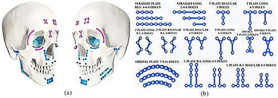
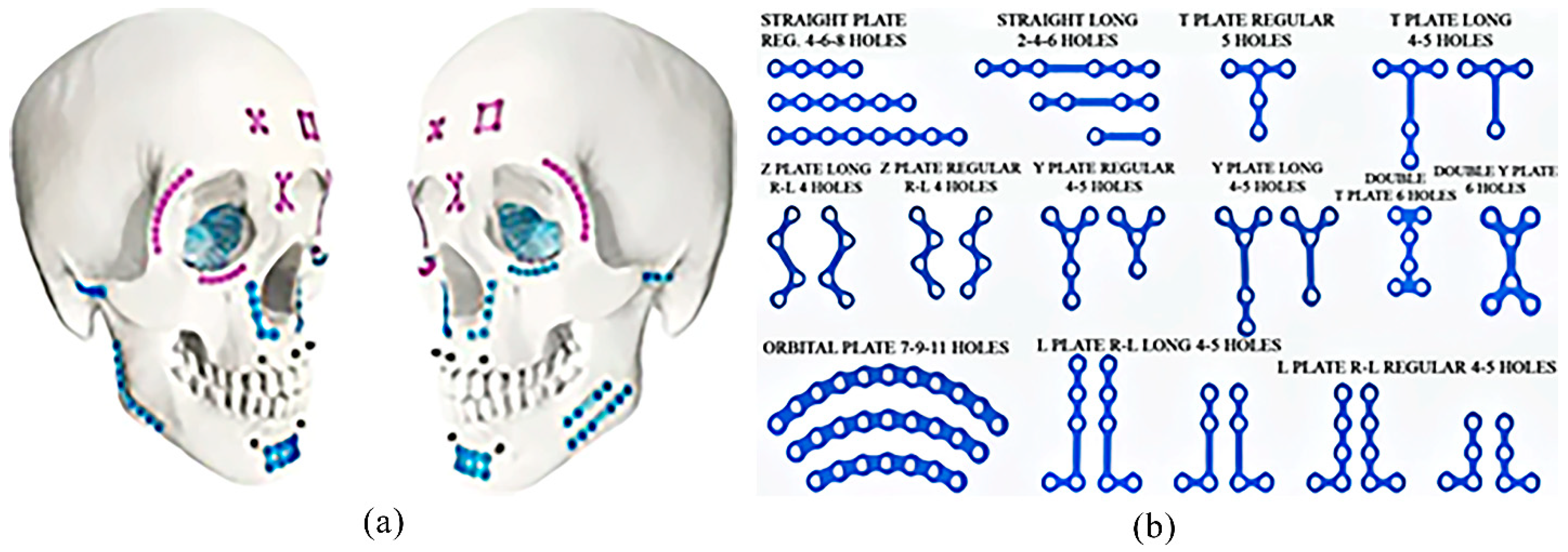
Ultrashort pulse lasers such as femtosecond laser systems are considered as a versatile tool for the precise micro/nano-metric structuring of a large variety of materials because of their extremely short laser pulse durations and localized high peak intensities. These lasers facilitate the highly precise surface structuring of diverse materials including metals, ceramics, and polymers through nonlinear interactions like multiphoton absorption and photoionization, while minimizing the thermal effects in adjacent regions. Owing to the negligible heat-affected zone, femtosecond laser processing enables the creation of micro- and nano-scale structures with excellent spatial resolution, without causing substantial changes to the substrate’s mechanical or crystalline properties [1,2,3,4]. Femtosecond laser processing, in particular, enables the creation of diverse hierarchical surface textures such as laser-induced periodic surface structures (LIPSSs), nano-spikes, and micro-grooves using a single-step fabrication approach. These patterns can be precisely controlled by modifying process parameters such as beam polarization, scanning speed, and laser fluence. This method is advantageous because it eliminates the need for masks or photolithography and can directly structure complex three-dimensional structures owing to its non-contact operation [5]. Moreover, the laser-induced oxide layers and chemical modifications formed during laser irradiation can modify surface energy, crystal phases, and chemical composition, thereby influencing not only the surface topography but also the functional characteristics of the material. Consequently, femtosecond laser micro/nano-structuring is recognized as a clean, accurate, and highly reproducible technique that offers greater flexibility than conventional mechanical or chemical surface treatment technologies. These strengths have led to growing interest in its application for biomedical materials and orthopedic implants [6,7]. Figure 1 presents common types of orthopedic plates along with their corresponding anatomical applications.
Figure 1.
Bone fixation plates in the human body. (a) Skull of human; (b) various kinds of plates suitable for fixation of skull.
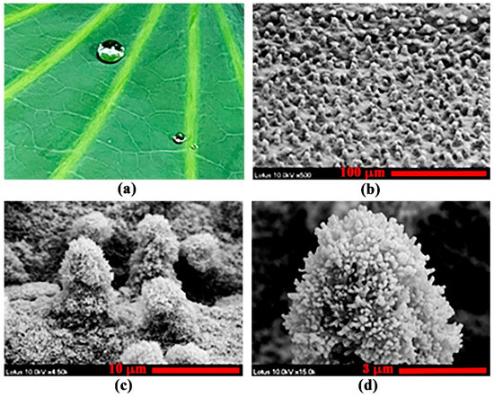
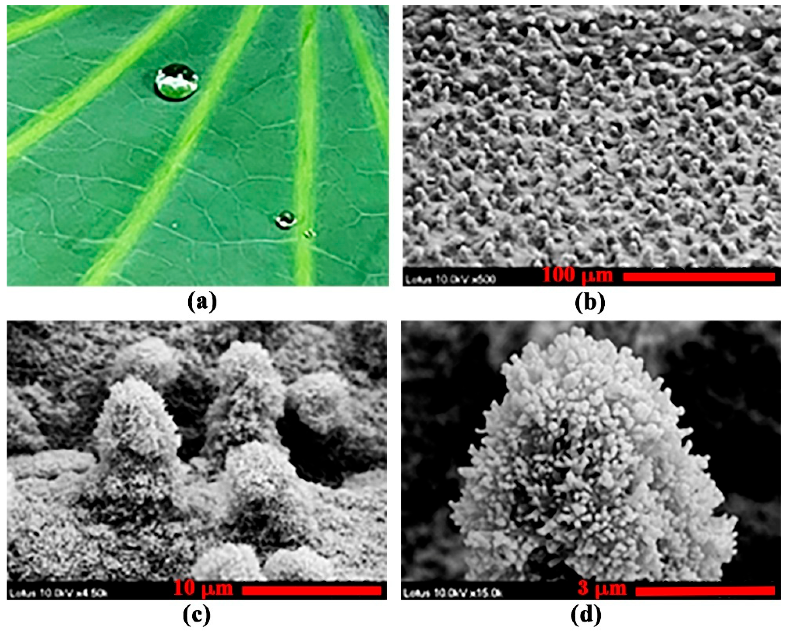
The overall performance of metallic implants depends not only on their inherent mechanical properties but also on the way their surfaces interact with surrounding biological tissues. Among these interactions, surface wettability, whether hydrophilic or hydrophobic, is a key factor influencing early biological events, including protein adsorption, cell adhesion, and bacterial attachment. Any material surface exhibiting a static liquid contact angle θ ≤ 5° is usually defined as being a superhydrophilic surface. On the other hand, when 5° < θ < 90°, we call the material surface a hydrophilic surface. A material surface is considered as a hydrophobic surface when 90° ≤ θ < 150°. In contrast, a superhydrophobic surface exhibits a contact angle greater than 150° [3,8]. Typically, hydrophilic surfaces enhance the stable adsorption of proteins from physiological fluids, thereby supporting cell attachment and subsequent proliferation. Conversely, hydrophobic surfaces often exhibit reduced protein adsorption and may induce structural changes in adsorbed proteins, which can limit cell adhesion but may suppress bacterial colonization by restricting microbial attachment [9]. These surface characteristics are influenced not only by chemical treatments but also by the formation of micro/nano-scale structures, which affect the physical roughness and surface energy. As illustrated in Figure 2, natural superhydrophobic surfaces such as lotus leaves exhibit hierarchical micro/nano-structures that alter surface tension, allowing water droplets to easily roll off. This phenomenon leads to a self-cleaning effect, which facilitates the removal of contaminants, including dust and bacteria, from the surface [10].
For implantable biomedical organs, such surface engineering can be strategically tailored to induce either hydrophilic surfaces that promote protein adsorption and cell attachment or hydrophobic/superhydrophobic surfaces that inhibit bacterial adhesion, depending on the clinical application and desired biological response. Furthermore, in the early stages of biological responses, a phenomenon known as bio-interface competition occurs where host cells and pathogenic bacteria compete for adhesion to the implant surface. In this context, the surface wettability plays a key role in either promoting the preferential attachment of host cells or inhibiting bacterial colonization, thereby contributing to infection prevention [5,11,12]. Among various materials, pure titanium (CP-Ti) and titanium alloys such as Ti-6Al-4V ELI are the most widely used materials for dental and orthopedic implants. Titanium is well recognized for its high strength-to-weight ratio, excellent corrosion resistance, bio-stability in physiological environments, and ability to integrate with human bone. Notably, its naturally formed TiO2 surface layer is chemically stable and non-toxic, with negligible ion release, thereby enhancing its biocompatibility. Ti-6Al-4V ELI, a modified alloy with reduced interstitial elements such as oxygen and carbon, exhibits improved toughness and fatigue resistance, making it suitable for high-load orthopedic applications including plates, screws, and joint replacements. However, since these materials are intrinsically bioinert, surface modification is essential to promote favorable cellular interactions. Conventional surface modification techniques such as sandblasting, acid etching, alkali treatment, and chemical deposition have been widely used to increase surface roughness for the enhancement of cells’ response. Nevertheless, these methods often generate random and non-uniform structures, having the limitations of poor uniformity and reproducibility. Moreover, chemical treatments can leave residual byproducts or impurities on the surface, raising concerns regarding biocompatibility and long-term safety [13,14]. Silver ion doping and antibiotic coatings were utilized to achieve antibacterial properties. However, these chemical coatings may leach over time, leading to potential cytotoxicity or the emergence of antibiotic-resistant strains [14]. As a result, physical surface patterning techniques that alter surface topography without additional coatings have gained attention where ultrashort pulsed laser-assisted micro/nano-structuring can be considered as a promising tool [5,6,7,11,12]. Femtosecond laser-based surface structuring can produce clean surfaces without the need for additional cleaning or post-processing steps. This technique offers excellent reproducibility and precision, enabling consistent fabrication under identical conditions. Furthermore, it allows simultaneous control of both physical structure and chemical composition in a single step, contributing to process simplification, cost efficiency, and scalability in industrial applications. On the other hand, femtosecond laser-induced micro/nano-texturing of titanium surfaces enhances cell adhesion, confers antibacterial activity, and reduces cytotoxicity. Such laser treatments enable the precise modulation of surface wettability (hydrophilicity or hydrophobicity), which in turn influences protein adsorption, osteoblast activation, and bacterial repulsion [6,14,15,16,17,18,19,20,21,22,23,24,25,26,27,28,29]. Despite these promising findings, most of the previous studies were focused on structural formation or contact angle measurement where only few research works investigated comprehensive biological responses such as cytotoxicity, antibacterial activity, and cell migration. Furthermore, issues such as fabrication reproducibility under repeated laser conditions and the in vivo stability of the structured surfaces remain underexplored.
This paper aims to reproducibly fabricate hydrophilic and hydrophobic surfaces on orthopedic-grade pure titanium and Ti-6Al-4V ELI alloy using femtosecond laser micro-structuring, and to systematically evaluate their biological responses, including cytotoxicity and antibacterial performance. We discovered an excellent correlation between the wettability of the laser-treated titanium samples with the cytocompatibility and antibacterial property, which in turn exemplified the potentials of the laser-treated titanium samples for bone fixation plates and orthopedic implants in the human body.
2. Materials and Methods
For our experiments, we considered commercially available pure titanium (CP-Ti) and Ti-6Al-4V ELI alloy, which are widely used in orthopedic implant applications. Orthopedic plates are designed to support and stabilize bones over a broad contact area. Given their extensive interface with biological tissue, minimizing bacterial adhesion and inflammatory responses are essential. To address this, a hydrophobic surface was introduced to inhibit the initial attachment of proteins and bacteria, thereby reducing the risk of inflammation. Moreover, since plates must be shaped to conform to the curvature of bone during surgery, pure titanium, with its superior ductility and formability, was chosen as the appropriate substrate material. In contrast, titanium alloy (Ti-6Al-4V ELI) is widely used for orthopedic screws, which are implanted into the bone to secure the plate in place. For screw components, enhancing the interfacial bonding with bone is critical. Therefore, hydrophilic surface characteristics were utilized to promote initial protein adsorption and osteoblast adhesion, thereby facilitating osseointegration and improving fixation strength.
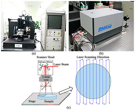
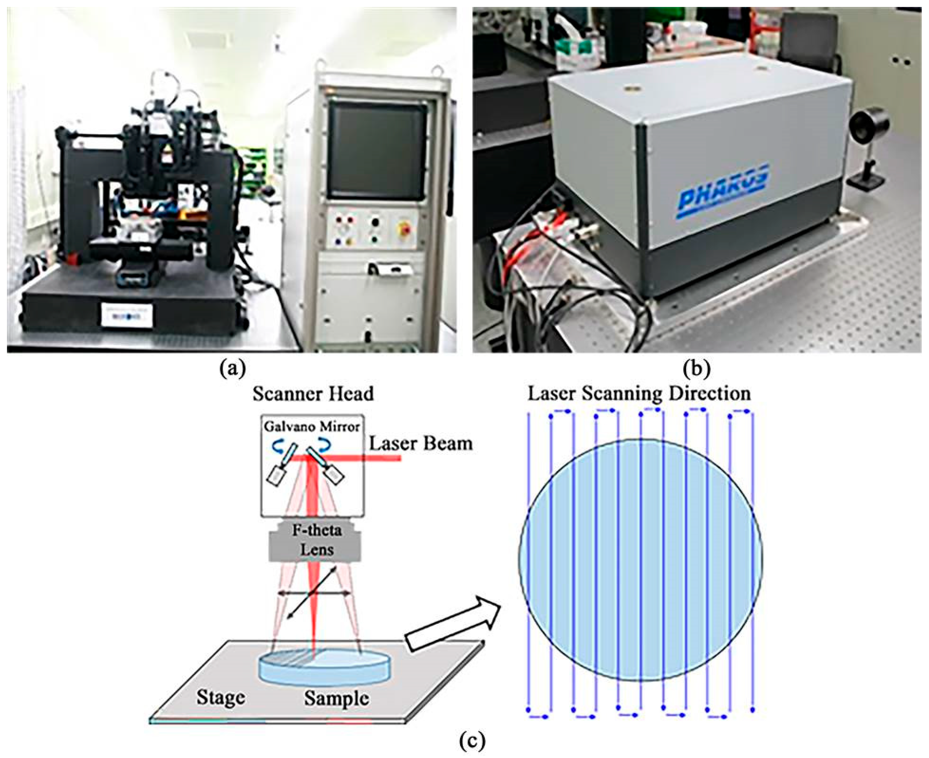
The experiments were carried out by a femtosecond laser (Light Conversion, PHAROS, Vilnius, Lithuania) operating at the center wavelength of 1030 nm with a pulse duration of 250 fs and pulse repetition rate of 100 kHz. Figure 3 presents the experimental setup of the femtosecond laser system used for the titanium surface treatment. To produce hydrophilic and hydrophobic titanium surfaces, we fabricated periodic micro-gratings and micro-grids on the samples’ surface using a femtosecond laser beam passed through a galvanometer scanner (Figure 3a). The galvanometer scanner (focal length: 100 mm) was focused directly onto the titanium surface to achieve the desired textures on the samples’ surface where the laser fluence, scanning speed, and line spacing were controlled depending on the requirement (Figure 3c). To fabricate hydrophobic pure titanium surfaces, the laser fluence was 318 J/cm2, whereas to fabricate hydrophilic Ti-6Al-4V ELI surfaces laser fluence was 509 J/cm2 and 637 J/cm2. As a result, surface patterns of different periodicity and morphology were formed to achieve specific wettability characteristics, either hydrophilic or hydrophobic, depending on the application requirements. For pure CP-Ti samples, micro-grid patterns were fabricated on the surface by varying the grid spacing from 10 to 100 µm with 10 μm increments. In contrast, the surfaces of the Ti-6Al-4V ELI samples were converted to hydrophilic surfaces by forming micro-lines with line spacing varying from 10 to 55 μm with 5 μm increments. Table 1 summarizes different parameters of the untreated and laser-treated titanium samples and the associated laser fluence.
Figure 3.
Experimental setup of the femtosecond laser processing system. (a) Laser processing system; (b) femtosecond laser; (c) surface patterning using galvanometer scanner.
Table 1.
Summary of different parameters of the untreated and laser-treated titanium samples and the corresponding laser fluence.
The surface of the titanium samples was subjected to barrel polishing as a pre-treatment step. The surface morphology of the untreated and laser-treated samples was investigated under optical microscope (ZEISS, Axioskop 40, Oberkochen, Germany) and field-emission scanning electron microscope (FE-SEM) (Hitachi, S-4700, Tokyo, Japan). Before FE-SEM examination, the titanium samples went through ultrasonic cleaning using de-ionized water for 30 min. A laser scanning confocal microscope (Olympus, OLS3100, Tokyo, Japan) was utilized to measure the roughness of the untreated and laser-treated titanium samples. To identify the elements present in the untreated and laser-treated titanium samples, we utilized energy-dispersive X-ray spectroscopy (EDS) (Thermo Fisher Scientific, Verios 5 UC, Waltham, MA, USA). The wettability of the titanium surfaces was evaluated by a contact angle measurement device (SEO Co. Ltd., Phoenix 300 Touch, Suwon, Republic of Korea) where a 1 µm diameter tip was used to dispense 4 µL of de-ionized water drop onto the sample surface and the contact angle was measured using a high-resolution camera system.
Quantitative antibacterial testing was performed using Staphylococcus Aureus as the model organism. The bacterial strain was cultured in nutrient broth, and a single colony was inoculated for pre-incubation over 16–18 h. The resulting suspension was diluted using 0.2% nutrient broth to achieve a final bacterial concentration of 2.5 × 10⁵ to 1.0 × 10⁶ CFU/mL. A volume of 0.4 mL of the prepared bacterial suspension was dispensed uniformly onto each test specimen (both pure titanium and Ti-6Al-4V ELI surfaces). A sterile 40 × 40 mm film was then gently placed over the inoculum to ensure full surface contact without spillage beyond the edges. The control group employed a sterilized polypropylene (PP) film in place of the sample under identical conditions. Following inoculation, the Petri dishes were sealed and incubated at 30 ± 2 °C for 24 h. After incubation, 20 mL of 0.2% nutrient broth was added to recover the bacterial suspension from the surface. The recovered solution was serially diluted, and viable cell counts were determined via the spread plate method. Plates exhibiting 30–300 colony forming units (CFUs) were selected for enumeration using a colony counter. All experiments were conducted in triplicate, and the average values were used for data analysis.
To evaluate the cytotoxicity of the femtosecond laser-treated pure titanium and Ti-6Al-4V ELI samples, an in vitro test was conducted in accordance with ISO 10993-5:2009 using the mouse fibroblast cell line NCTC Clone 929 (L-929) and the MTT assay. The mouse fibroblast cell line NCTC Clone 929 (L-929) was obtained from the Korean Cell Line Bank (KCLB, Seoul, South Korea), Republic of Korea (Lot No. 87059). The extracts of the samples were prepared by immersing each specimen in RPMI 1640 medium supplemented with 10% fetal bovine serum (FBS) at a ratio of 0.2 g/mL. The immersion was carried out at 37 ± 1 °C for 24 ± 2 h with constant shaking at 100 rpm, and the pH of the extraction medium was verified to be maintained within the range of 7.2 to 7.4 prior to use. Laser-treated samples were designated as the experimental group, and samples of the same material without laser treatment were used as the test material control group. To ensure the reliability and accuracy of the evaluation, three additional control groups were included in the study. The reagent control group consisted of RPMI medium without any test material, used to assess baseline cell viability. The negative control group was prepared by treating cells with an extract of a certified non-cytotoxic material under the same conditions, serving as a reference for non-toxic behavior. The positive control group was treated with a known cytotoxic substance, such as phenol, to induce cell death and confirm the sensitivity and validity of the test system. L-929 cells were seeded at a concentration of 1 × 10⁵ cells/mL into 96-well plates and cultured under standard conditions (37 ± 1 °C, 5% CO2) for at least 24 h to allow monolayer formation. After confirming uniform cell attachment and the absence of contamination under an optical microscope, 100% extract solution was added to each well and incubated again under the same conditions for 24 h. Following the incubation period, MTT solution was added and the cells were further incubated for more than 1 h to allow the formation of purple formazan crystals in viable cells. These crystals were then solubilized using dimethyl sulfoxide (DMSO), and the absorbance was measured at 570 nm using an ELISA microplate reader (BioTek, Synergy H1M, Winooski, VT, USA). Cell viability was calculated from the absorbance values and compared with the experimental and control groups.
3. Results and Discussion
In this section, we represent the experimental results and the associated analysis.
3.1. Surface Roughness of the Untreated and Laser-Treated Titanium Samples
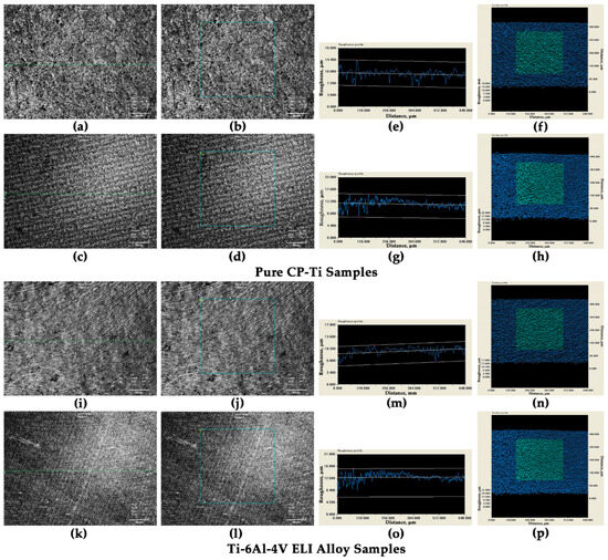
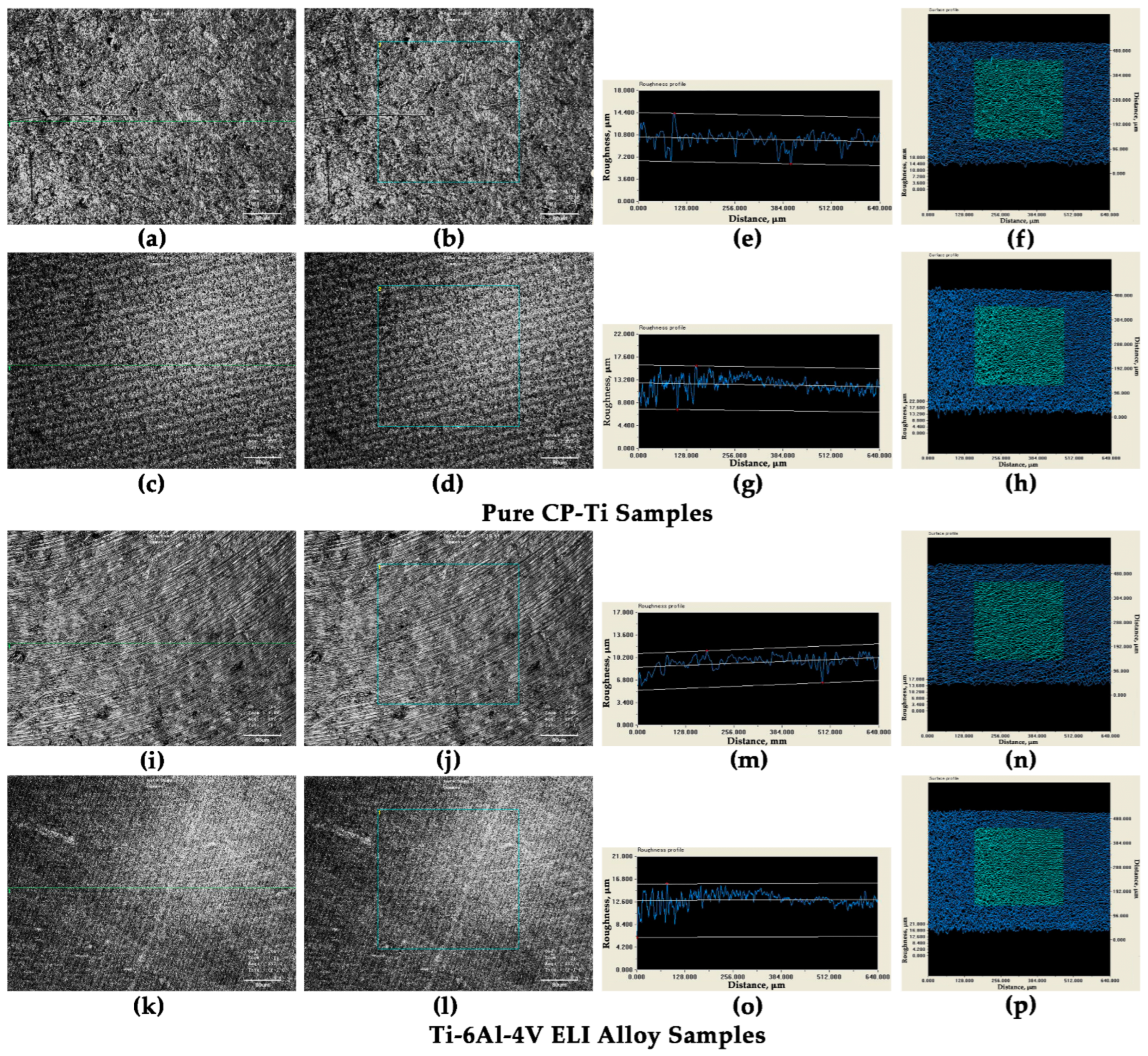
In order to investigate the impact of laser irradiation on the surface roughness of the titanium samples, we measured the surface roughness of untreated and laser-treated pure CP-Ti and Ti-6Al-4V ELI alloy samples using 2D line profile and 3D areal profile. We considered CP-Ti-G40 and Ti-6Al-4V ELI-L10 samples during the measurements, the roughness images and profile of which are represented in Figure 4. The roughness results of the titanium samples are summarized in Table 2. The 2D and 3D roughness analysis confirm a significant increase in surface roughness of both types of titanium samples after laser processing.
Figure 4.
Roughness of the untreated and laser-treated pure titanium samples. (a–h) Pure CP-Ti samples: (a) 2D line image of untreated sample (before micro-line fabrication), (b) 3D areal image of untreated sample (before micro-grid fabrication), (c) 2D line image of CP-Ti-G40 sample, (d) 3D areal image of CP-Ti-G40 sample, (e) 2D roughness profile of Figure 4a, (f) 3D roughness profile of Figure 4b, (g) 2D roughness profile of Figure 4c, (h) 3D roughness profile of Figure 4d; (i–p) Ti-6Al-4V ELI alloy samples: (i) 2D line image of untreated sample (before micro-line fabrication), (j) 3D areal image of untreated sample (before micro-grid fabrication), (k) 2D line image of Ti-6Al-4V ELI-L10 sample, (l) 3D areal image of Ti-6Al-4V ELI-L10 sample, (m) 2D roughness profile of Figure 4i, (n) 3D roughness profile of Figure 4j, (o) 2D roughness profile of Figure 4k, (p) 3D roughness image of Figure 4l.
Table 2.
The summary of the roughness of the untreated and laser-treated pure CP-Ti and Ti-6Al-4V ELI alloy samples.
3.2. Wettability Control of Titanium Surface Using Femtosecond Laser Processing
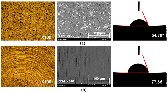
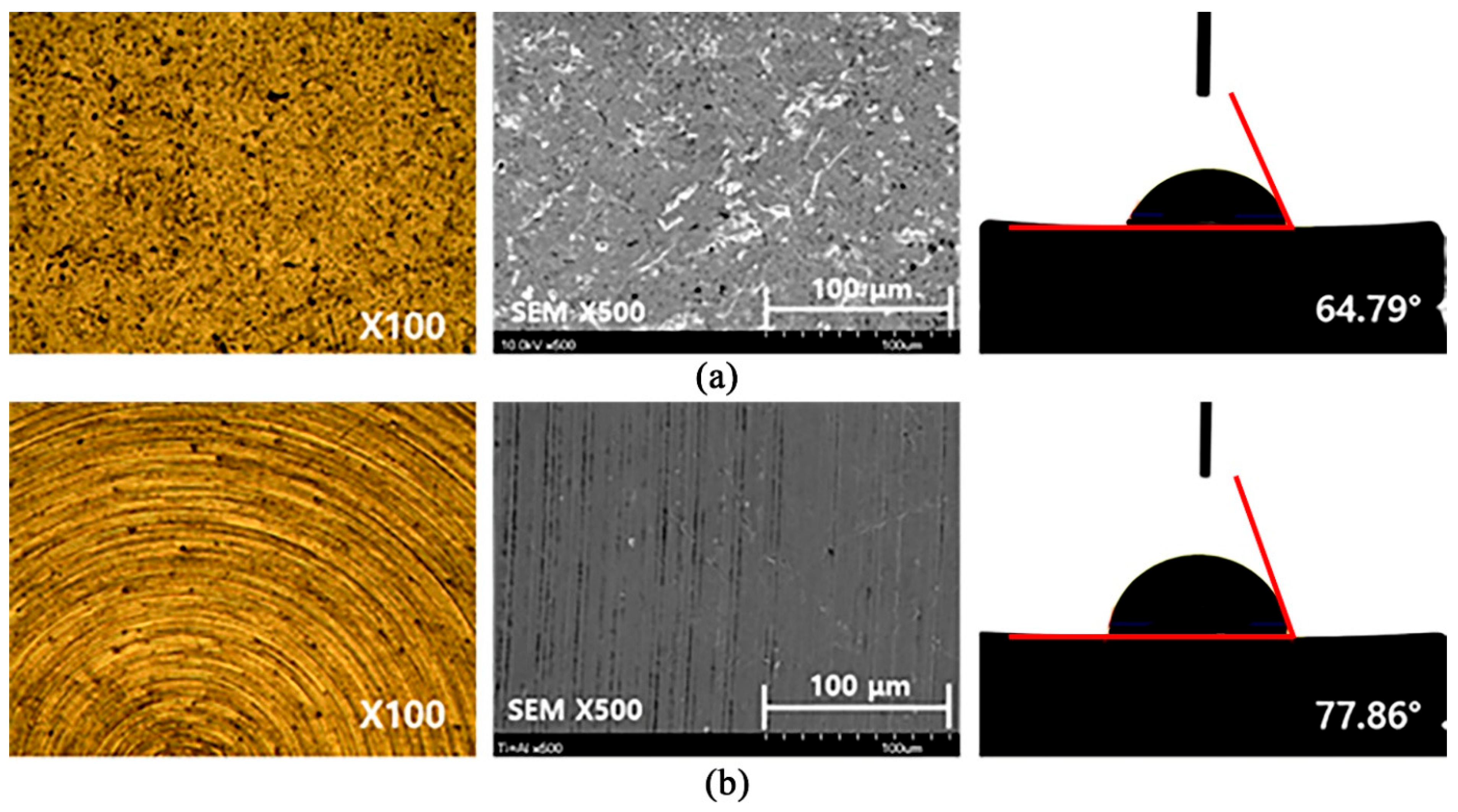
In order to control the wettability of the pure titanium and Ti-6Al-4V ELI alloy-based orthopedic implants, we fabricated periodic micro-grid and micro-line patterns on the samples’ surface using the femtosecond laser structuring technique. To control the width, depth, and period of the micro-holes/micro-grooves, we adjusted various irradiation conditions of the laser beam including laser fluence, scanning speed, and line spacing. As a result, we were able to increase the hydrophobicity of the pure CP-Ti samples and hydrophilicity of the Ti-6Al-4V ELI alloy samples. Figure 5 illustrates the optical microscope images, FE-SEM morphographs, and contact angle of the untreated pure CP-Ti and Ti-6Al-4V ELI alloy samples.
Figure 5.
Optical microscope images, FE-SEM micrographs, and contact angle measurements of untreated titanium samples. (a) Pure CP-Ti (contact angle: 64.79°); (b) Ti-6Al-4V ELI alloy (contact angle: 77.86°).
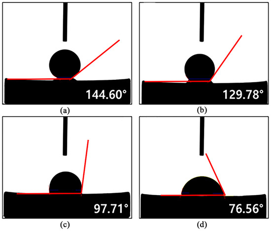
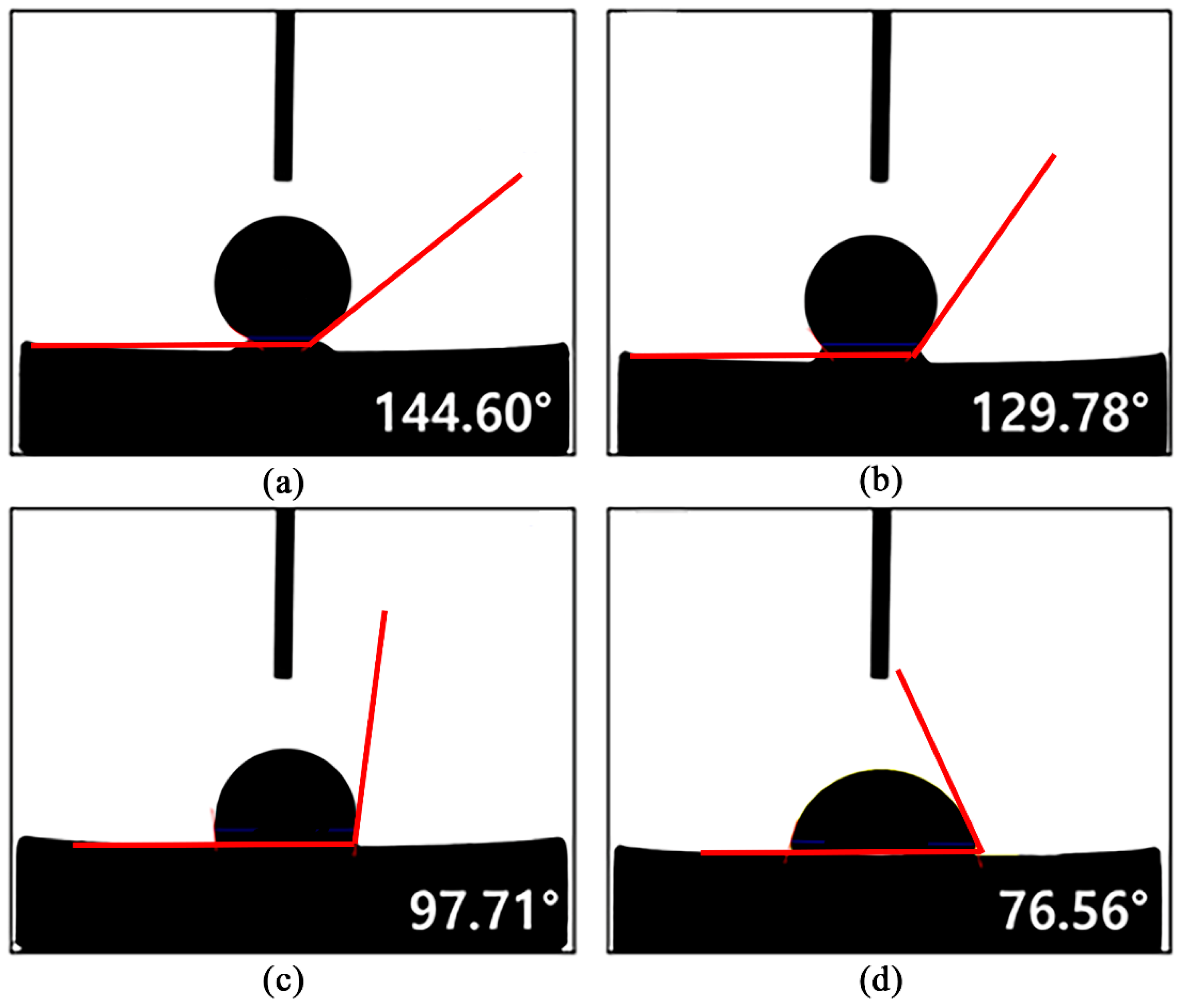
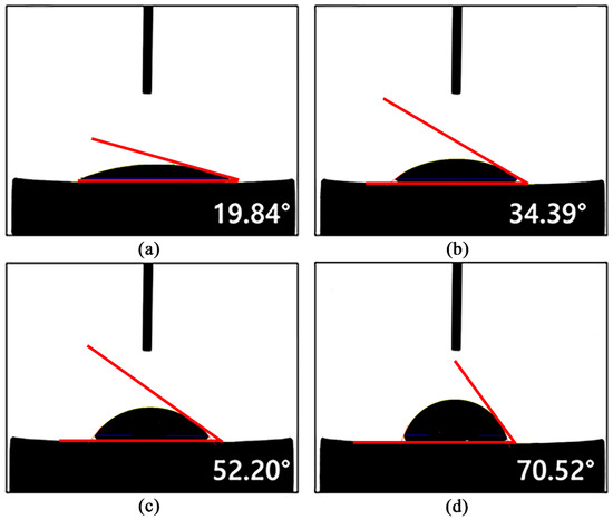
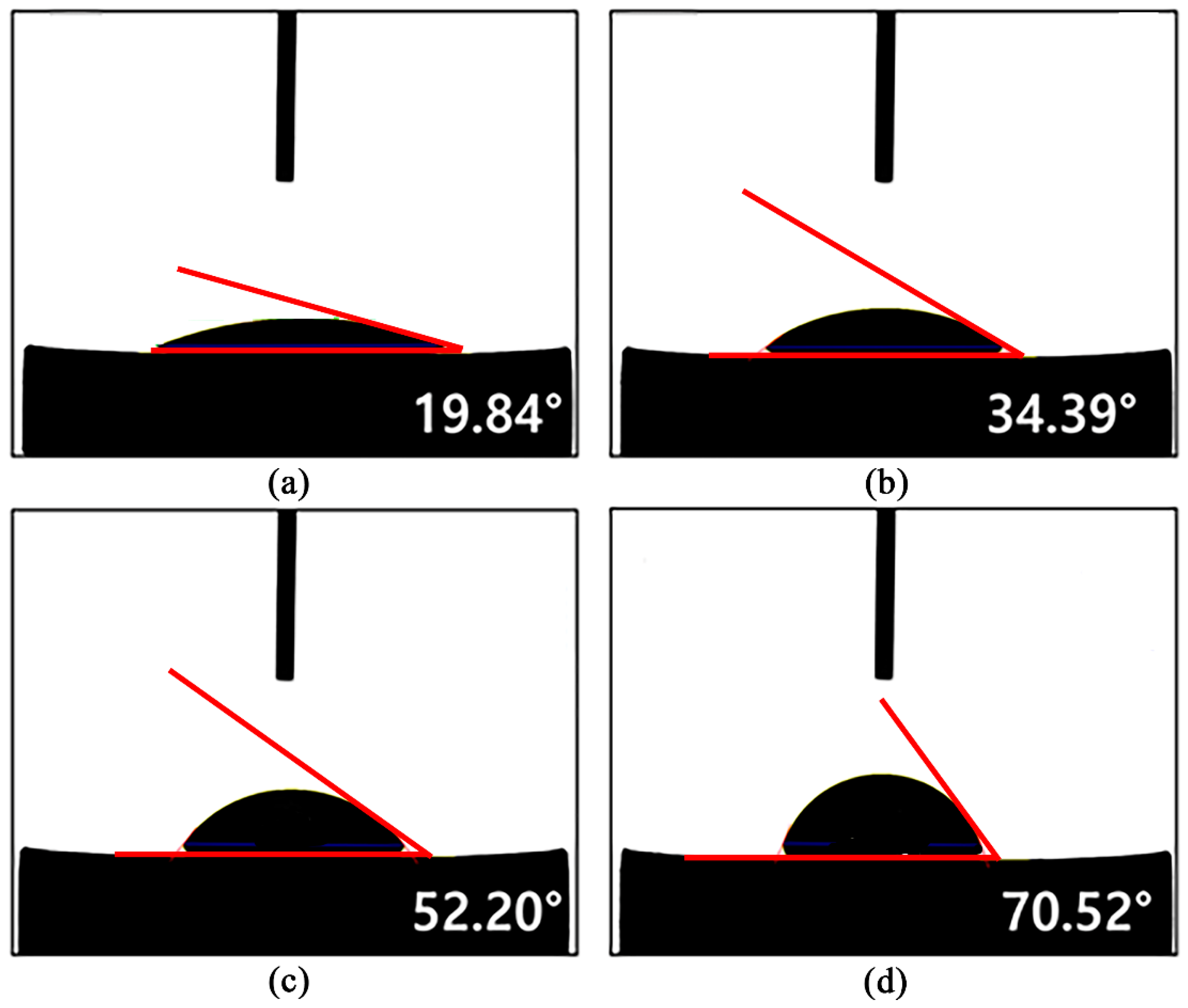
The measured contact angles were 64.79° for pure CP-Ti and 77.86° for the Ti-6Al-4V ELI alloy, indicating that both materials exhibited intrinsic hydrophilic characteristics (i.e., contact angle < 90°). Based on these initial surface conditions, femtosecond laser patterning was executed to tailor the wettability of the titanium samples. For the pure titanium samples, grid patterns were fabricated by varying the line-spacing between the laser-engraved micro-lines from 10 µm to 100 μm with 10 μm increments. The influence of grid spacing on surface wettability was systematically examined, and the results confirmed that contact angle was increased with the decrease in grid spacing, demonstrating effective control of hydrophobic behavior of the CP-Ti samples by femtosecond laser patterning. The contact angles of the micro-grid-engraved pure titanium surfaces are depicted in Figure 6. The highest and lowest contact angles for the micro-grid textured pure titanium surface were 144.6° and 76.56°. A significant increase in hydrophobicity was evident from the experimental results. For comparison, the Ti-6Al-4V ELI alloy sample surfaces were also patterned by periodic micro-lines under femtosecond laser irradiations. The line spacing varied from 10 µm to 55 μm. Unlike the hydrophobic surface generated on a pure titanium surface, the goal of the femtosecond laser-treated titanium alloy surfaces was to enhance hydrophilicity, enabling a controlled modulation of titanium surface wettability by means of linear micro-patterns. Figure 7 presents the contact angles of Ti-6Al-4V ELI alloy surfaces after micro-patterning by femtosecond laser. From the experimental results, it is evident that low periodic intervals resulted in significantly reduced contact angles. The highest contact angle was detected as 70.52° when the periodic micro-line spacing was 55 µm. On the other hand, the lowest contact angle of 19.84°, which was evident for the titanium alloy sample, was patterned by periodic micro-lines with 10 µm spacing. A significant improvement in hydrophilicity was evident for the micro-line-engraved titanium alloy sample, having 10 μm line spacing. These results demonstrate that the hydrophilicity of the titanium alloy surface can be tuned perfectly by controlling the period of the femtosecond laser-induced micro-lines. The overall findings confirm that femtosecond laser-based micro-patterned titanium surfaces enable the accurate modulation of surface wettability of both CP-Ti and Ti-6Al-4V ELI alloy. By adjusting key laser processing parameters such as scanning interval and pattern geometry, it is possible to fabricate functionally optimized titanium surfaces tailored to the specific roles of each implant material. In particular, the ability to have excellent control over the wettability of the laser-patterned titanium surfaces exemplifies femtosecond laser processing as an effective surface texturing tool for orthopedic implant applications. Figure 8 shows the surface morphology of pure titanium and Ti-6Al-4V ELI alloy samples after femtosecond laser processing, observed using optical microscopy and FE-SEM. As mentioned before, the pure titanium sample was modified by a periodic micro-grid pattern.
Figure 6.
Contact angle measurements of pure titanium surfaces after femtosecond laser patterning of micro-grids with different grid spacings. (a) 10 μm; (b) 40 μm; (c) 70 μm; (d) 100 μm.
Figure 7.
Contact angle measurements of Ti-6Al-4V ELI alloy surfaces after femtosecond laser patterning of micro-gratings with different line spacings. (a) 10 μm; (b) 25 μm; (c) 40 μm; (d) 55 μm.
Figure 8.
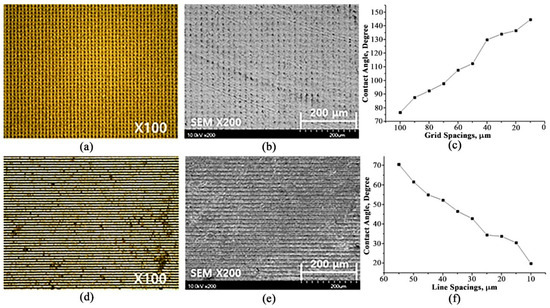
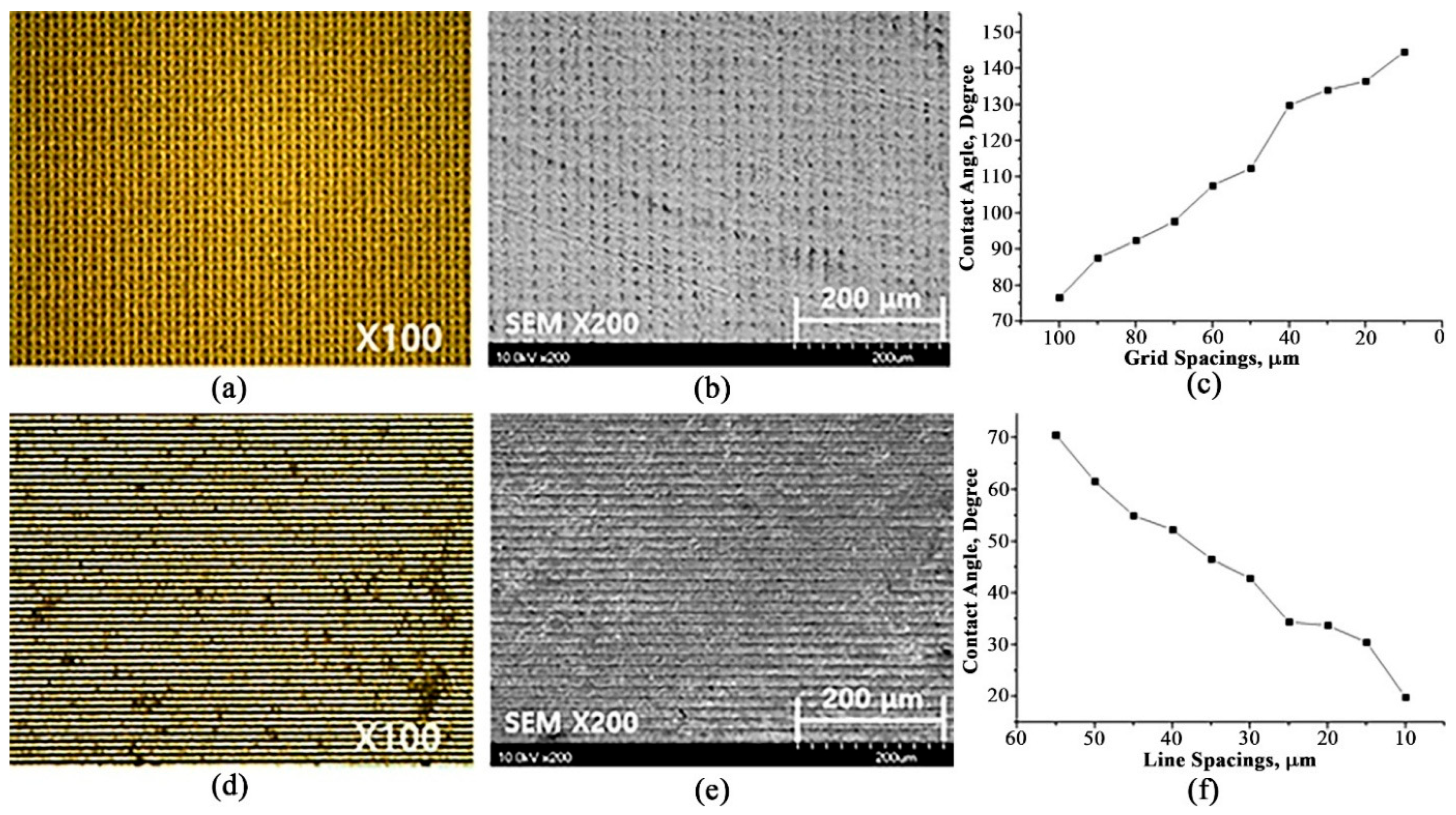
Surface analysis of pure titanium and Ti-6Al-4V ELI alloy samples after femtosecond laser patterning of micro-grids and micro-gratings with pattern spacing of 10 μm at a laser fluence of 318 J/cm2. (a,b) Grid-patterned pure titanium sample: (a) OM image, (b) FE-SEM image, (c) contact angle for different grid spacings; (d–f) micro-line patterned Ti-6Al-4V ELI alloy sample fabricated: (d) OM image, (e) FE-SEM image, (f) contact angle for different line spacings.
Figure 8a demonstrates the optical microscope (OM) image of the micro-grid-engraved pure titanium sample having grid spacing of 10 µm. The corresponding FE-SEM image is depicted in Figure 8b; the sample showed a contact angle of 144.6°, indicating a significant enhancement of hydrophobicity. Figure 8c shows the dependence of contact angle on the period of the micro-grid for CP-Ti samples. Conversely, the Ti-6Al-4V ELI alloy sample was structured with a periodic micro-line pattern, where Figure 8d represents the OM image of the titanium alloy sample having a periodic micro-line of 10 μm spacing. The corresponding FE-SEM image is illustrated in Figure 8e. The contact angle of this titanium alloy sample was 19.84°, indicating hydrophilicity. Figure 8f presents the variation in contact angle as a function of line spacing. In pure titanium, decreasing the grid spacing resulted in an increase in contact angle, thereby enhancing hydrophobic behavior. In contrast, the Ti-6Al-4V ELI alloy showed a decrease in contact angle with small line spacing, corresponding to increased hydrophilic behavior. These findings confirm that precise modulation of pattern spacing during femtosecond laser processing enables effective control of surface wettability. Furthermore, this method allows for the selective implementation of hydrophilic or hydrophobic properties depending on the application, confirming a versatile and functional surface engineering approach. For subsequent biological response assessments, the surfaces that exhibited maximum hydrophobicity in pure titanium and maximum hydrophilicity in Ti-6Al-4V ELI alloy based on the optimized pattern spacing were used as test samples. These biologically relevant surfaces were subjected to antibacterial and cytotoxicity experiments, in order to evaluate their suitability and response in orthopedic implant environments.
On flat titanium surfaces, the contact angle can be estimated using Young’s contact angle (θe) model, as defined by the following equation [3].
where , , and represent the solid–vapor interfacial energy, solid–liquid interfacial energy, and liquid–vapor interfacial energy, respectively. However, for non-flat titanium surfaces, Weznel () or Cassie–Baxter () equations are appropriate to estimate the contact angles, which can be represented as follows [3]:
Here, r denotes the roughness factor, defined as the ratio between the actual solid titanium surface area and its planar projected area corresponding to an infinitesimal displacement along the water droplet contact line. The Cassie–Baxter surface fractions, f1 and f2 (where f1 + f2 = 1), describe the area fractions of the different surface components adjacent to the droplet perimeter, as defined by the radial coordinate. Each fi corresponds to the ratio of surface type i to the total area within a small region surrounding the three-phase contact line. The parameters θ1 and θ2 represent the respective contact angles of these surface components. For titanium surfaces, patterned with micro-gratings or micro-grid structures, the contribution of f2 and θ2 in Equation (3) can be neglected. Let the pillar’s width (located between adjacent gratings), groove’s width, and groove’s height by WP, WG, and h. For a single period of micro-gratings or micro-grid structure, the true solid–liquid contact area can be expressed as SS–L = (WP + WG) × l + 2h × l, where l is the length of the contact area of the water droplet with the titanium sample. Similarly, the projected area for one structural period of micro-gratings or micro-grid structure can be estimated by SS–L(P) = (WP + WG) × l. For a single micro-grating, the projected area can be represented as SP(P) = WP × l. The roughness factor of the laser-treated titanium samples can be determined using the following equation [3].
The term f1 can be estimated as follows [3]:
The wetting height (hW) of the water droplet, indicating the depth at which the water droplet can penetrate inside the groove, can be obtained using the following equation [3].
When hW ≥ h, the water droplet is in the Weznel mode and the titanium surface is hydrophobic. For hydrophilic titanium samples, the wetting height is much higher than the groove’s height, i.e., hW >> h.
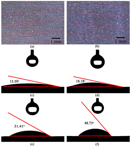
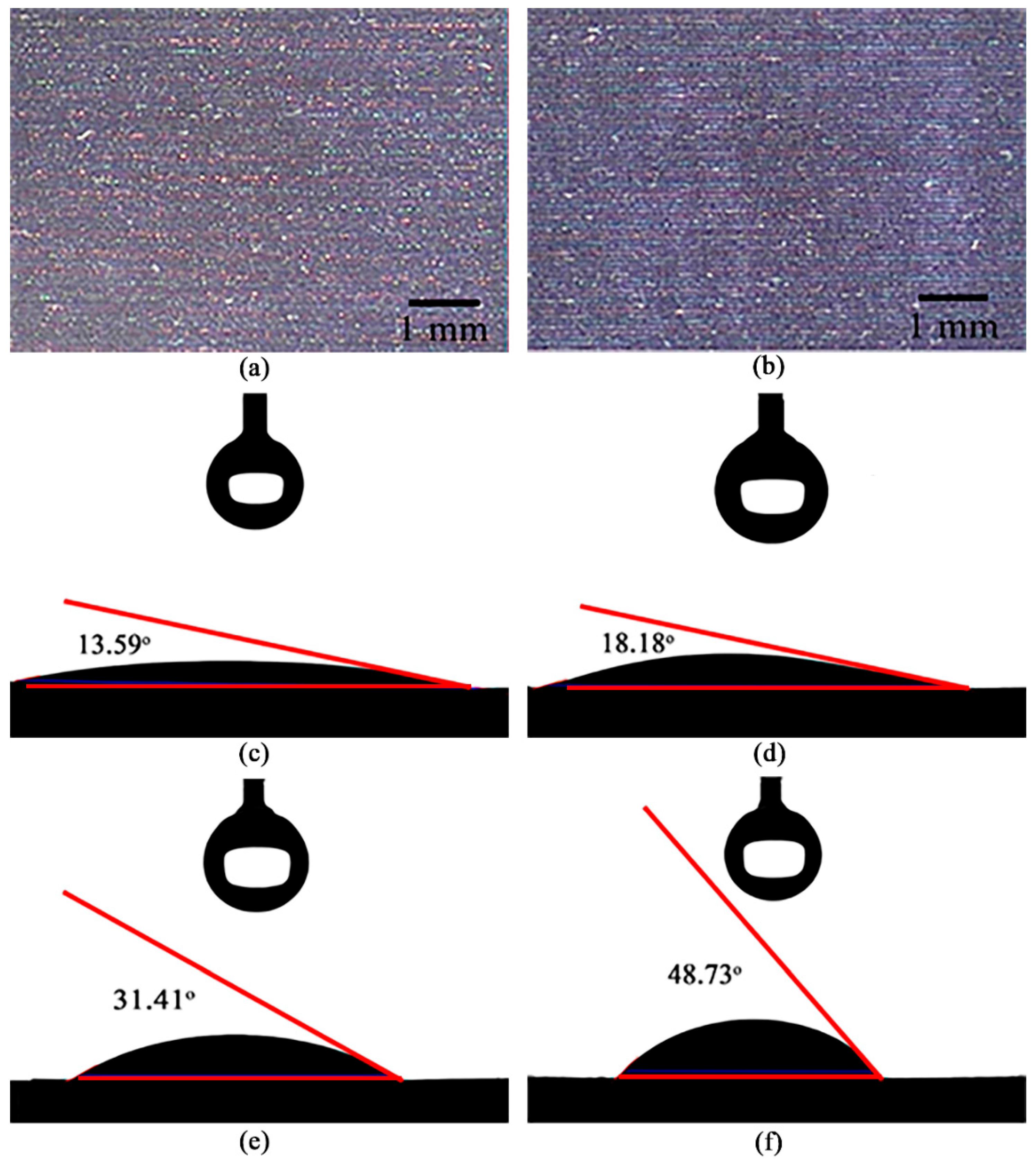
To evaluate the time-dependent impact on the quality of the hydrophilic property of the Ti-6Al-4V ELI alloy, we investigated the surface morphology of the Ti-6Al-4V ELI alloy samples fabricated under variable laser energy, as shown in Figure 9.
Figure 9.
Surface morphology of the hydrophilic titanium alloy after femtosecond laser patterning of micro-gratings with line spacing of 15 μm, fabricated under different laser energy and the corresponding contact angle over different time. (a,b) FE-SEM images: (a) E = 509 J/cm2, (b) E = 637 J/cm2; (c,d) contact angle immediately after laser pattering: (c) 13.59° for the sample of Figure 9a, (d) 18.18° for the sample of Figure 9b; (e,f) contact angle after one month: (e) 31.41° for the sample of Figure 9a, (f) 48.73° for the sample of Figure 9b.
The contact angles of the femtosecond laser-treated titanium alloy samples, immediately after laser processing, were 13.59° (for 509 J/cm2) (Figure 9c) and 18.18° (for 637 J/cm2) (Figure 9d). These results indicate that the laser-irradiated titanium alloy surfaces exhibited hydrophilicity immediately after fabrication. However, after one month, the contact angles increased to 31.41° (Figure 9e) and 48.73° (Figure 9f), respectively, indicating a significant decrease in hydrophilicity of the laser-treated titanium alloy samples over time. This increase in contact angle is attributed to the spontaneous oxidation of the titanium alloy surface during storage, resulting in the growth of a native titanium oxide (TiO2) layer. Titanium inherently forms a stable TiO2 film in air, and this process is further accelerated in the laser-patterned surfaces due to their high surface energy and activated chemical state, which promote oxygen adsorption and subsequent oxidation. The resulting oxide layer is chemically stable and alters surface charge distribution, electron affinity, and surface free energy, leading to a gradual increase in contact angle relative to the initially hydrophilic state. In other words, immediately after laser processing, enhanced hydrophilicity is primarily induced by microstructural modifications and increased surface roughness, which favor the strong adsorption of water molecules. However, as time progresses, the growth of the oxide layer along, with the adsorption of airborne hydrocarbon contaminants, reduces the effective surface free energy, thereby diminishing hydrophilicity and resulting in increased contact angles.
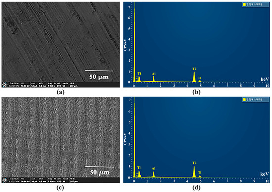
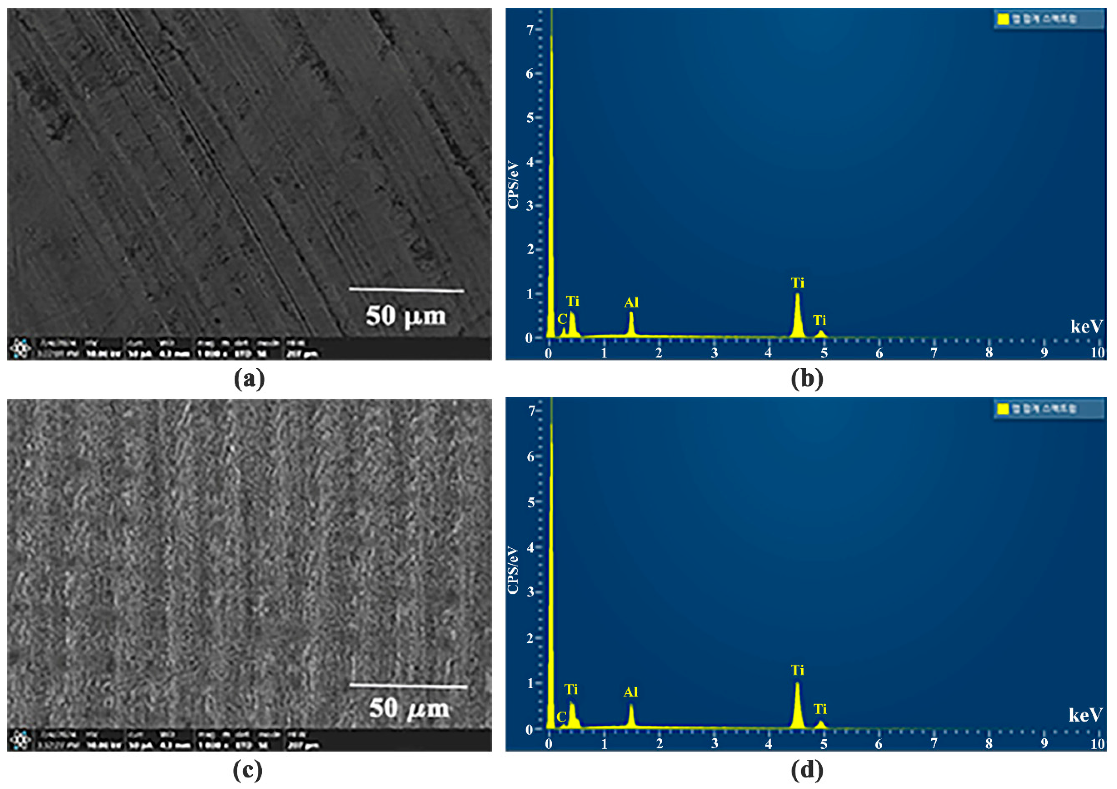
We also investigated the EDS spectrum of the titanium alloy samples before and after femtosecond laser treatment. The surface morphology of the titanium alloy sample before laser treatment is illustrated in Figure 10a, whereas the EDS spectrum is depicted in Figure 10b.
Figure 10.
Surface morphology and EDS analysis of the Ti-6Al-4V ELI samples before and after femtosecond laser processing. (a,b) Before laser processing: (a) FE-SEM image, (b) EDS spectrum; (c,d) after laser processing: (c) FE-SEM image, (d) EDS spectrum.
In contrast, the surface morphology of the titanium alloy sample after femtosecond laser treatment is shown in Figure 10c and the corresponding EDS spectrum is represented in Figure 10d. In both cases, only the major alloy constituents were detected, confirming that no new oxide compounds were formed after femtosecond laser treatment. The percentage of each element present in the untreated and laser-treated titanium samples is summarized in Table 3.
Table 3.
Percentage of each element present in the untreated and laser-treated Ti-6Al-4V ELI samples.
However, the formation of a thin titanium oxides’ layer on the titanium surface after femtosecond laser irradiation is a common phenomenon. From the EDS spectrums, we can infer that, despite high localized energy during femtosecond laser irradiation, no new phases were generated and the alloy composition remained unchanged, although some thin titanium oxides’ layers (e.g., TiO, TiO2, Ti2O3), especially the TiO2 layer, might have developed on the titanium surface [29]. However, the EDS spectrum is incapable of identifying any specific titanium oxide phase. The contact angles of the femtosecond laser-treated Ti-6Al-4V ELI alloy surfaces increased after one month of storage, indicating a gradual reduction in the initially enhanced hydrophilicity. The observed changes in contact angle can be attributed not to laser-induced compositional alterations but rather to subsequent surface oxidation and chemical modifications that occurred during the storage of the titanium samples in ambient air. Ultrashort pulse laser irradiation activates the titanium alloy surface through localized melting, rapid re-solidification, and oxidation-related chemical stabilization, which initially increases surface polarity and promotes hydrophilicity. With storage in ambient air, subsequent adsorption of airborne hydrocarbon species and further surface chemical relaxation can reduce the effective surface free energy, resulting in an increase in contact angle over time. Consistent with this interpretation, EDS analysis before and after laser processing confirms that the major alloy constituents remain unchanged, indicating that the wettability evolution is governed by surface chemical modifications rather than bulk compositional changes. Table 4 summarizes the wetting properties of the untreated and femtosecond laser-patterned titanium samples. The hydrophilicity increases with the decrease in roughness (from Equations (4) and (6)), which agrees well with our experimental results.
Table 4.
Summary of the wetting properties of the untreated and femtosecond laser-patterned titanium samples.
3.3. Biological Response Evaluation: Antibacterial Assessment
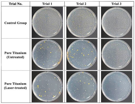
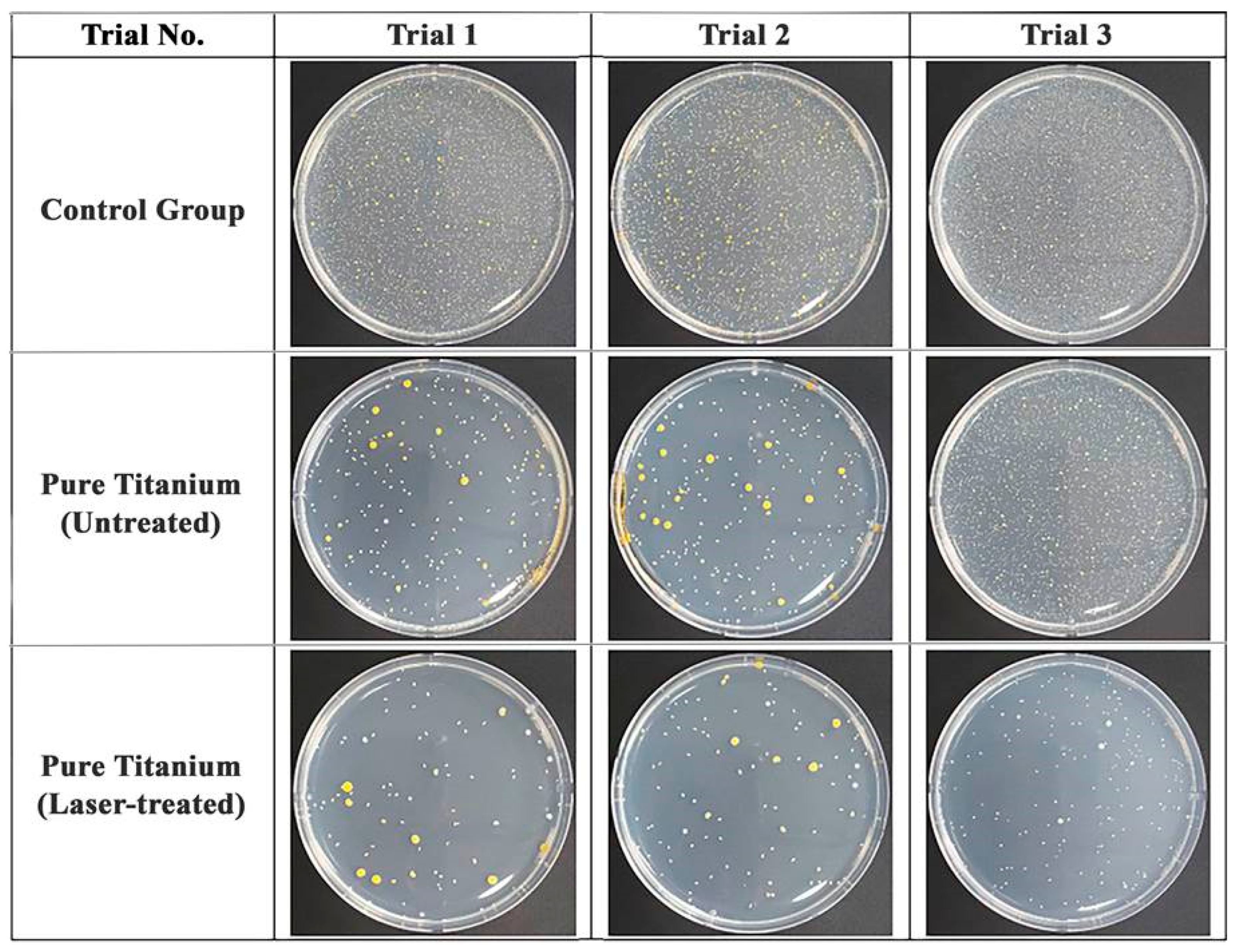
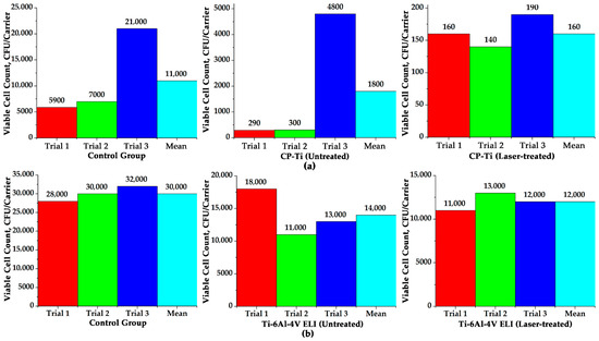
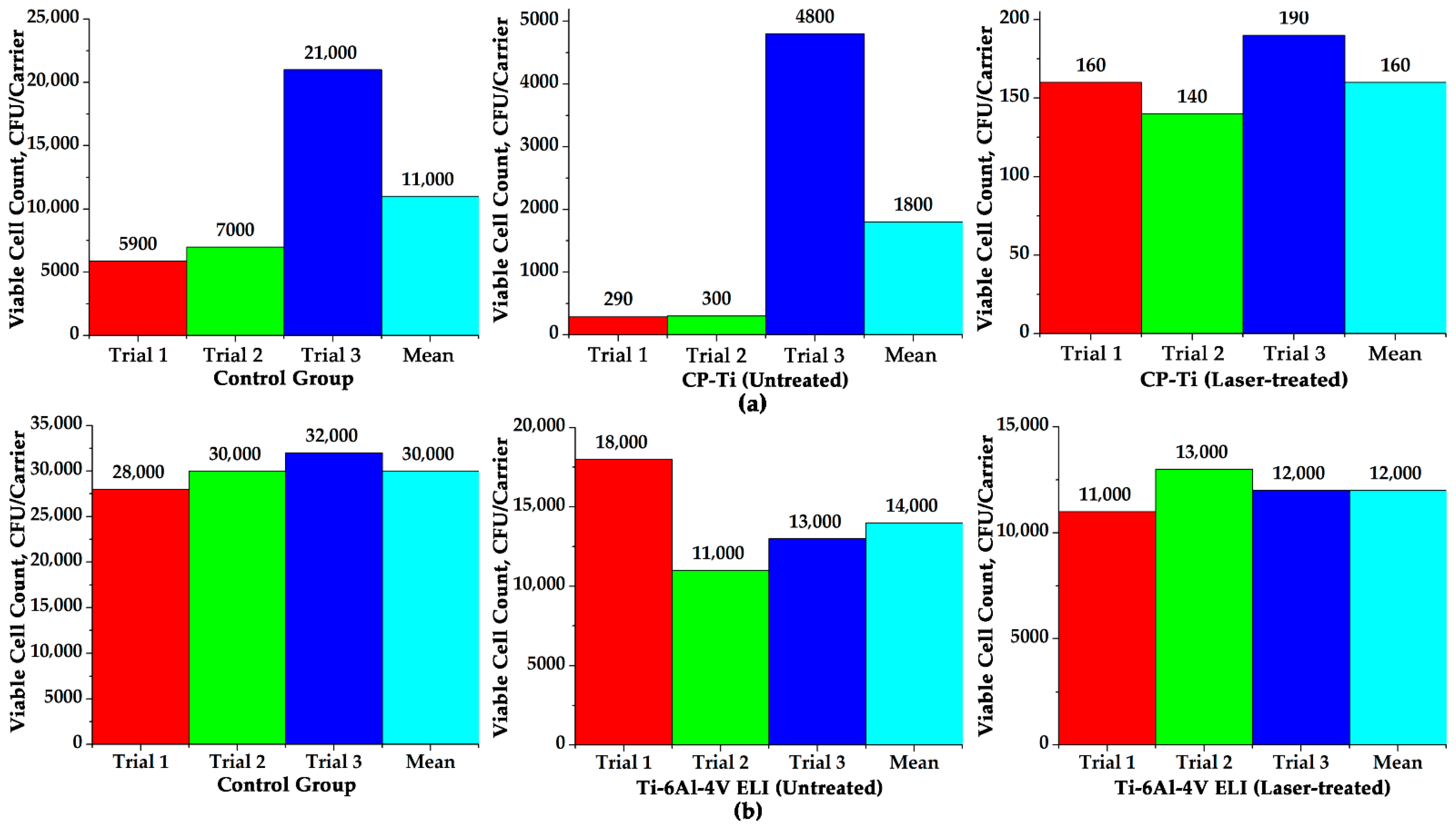
To evaluate the biological response of the laser-treated titanium surfaces, antibacterial responses were investigated where, for each type of samples, we conducted the experiment three times. As mentioned before, the pure CP-Ti sample was modified by forming periodic micro-grid structures on top of the pure titanium samples at the highest contact angle (144.6°), corresponding to the highest degree of hydrophobicity, which was evident for the micro-patterned CP-Ti sample having a periodic grid of 10 μm grid spacing. On the other hand, the Ti-6Al-4V ELI alloy sample was modified by periodic micro-line textures with 10 μm line spacing, showing a minimum contact angle of 19.84°, indicative of strong hydrophilicity. The femtosecond laser-treated pure titanium sample, exhibiting hydrophobic surface characteristics, demonstrated significantly enhanced antibacterial activity compared to both the untreated pure titanium sample and the control group (polypropylene film). Figure 11 illustrates the antibacterial performance of the untreated and laser-treated pure titanium samples. Figure 11 also confirmed a significant improvement of antibacterial performance in the femtosecond laser-patterned pure titanium samples, as compared to the untreated pure titanium samples in the presence of Staphylococcus Aureus.
Figure 11.
Antibacterial performance analysis of the untreated and femtosecond laser-patterned pure titanium samples in the presence of Staphylococcus Aureus.
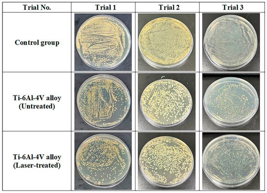
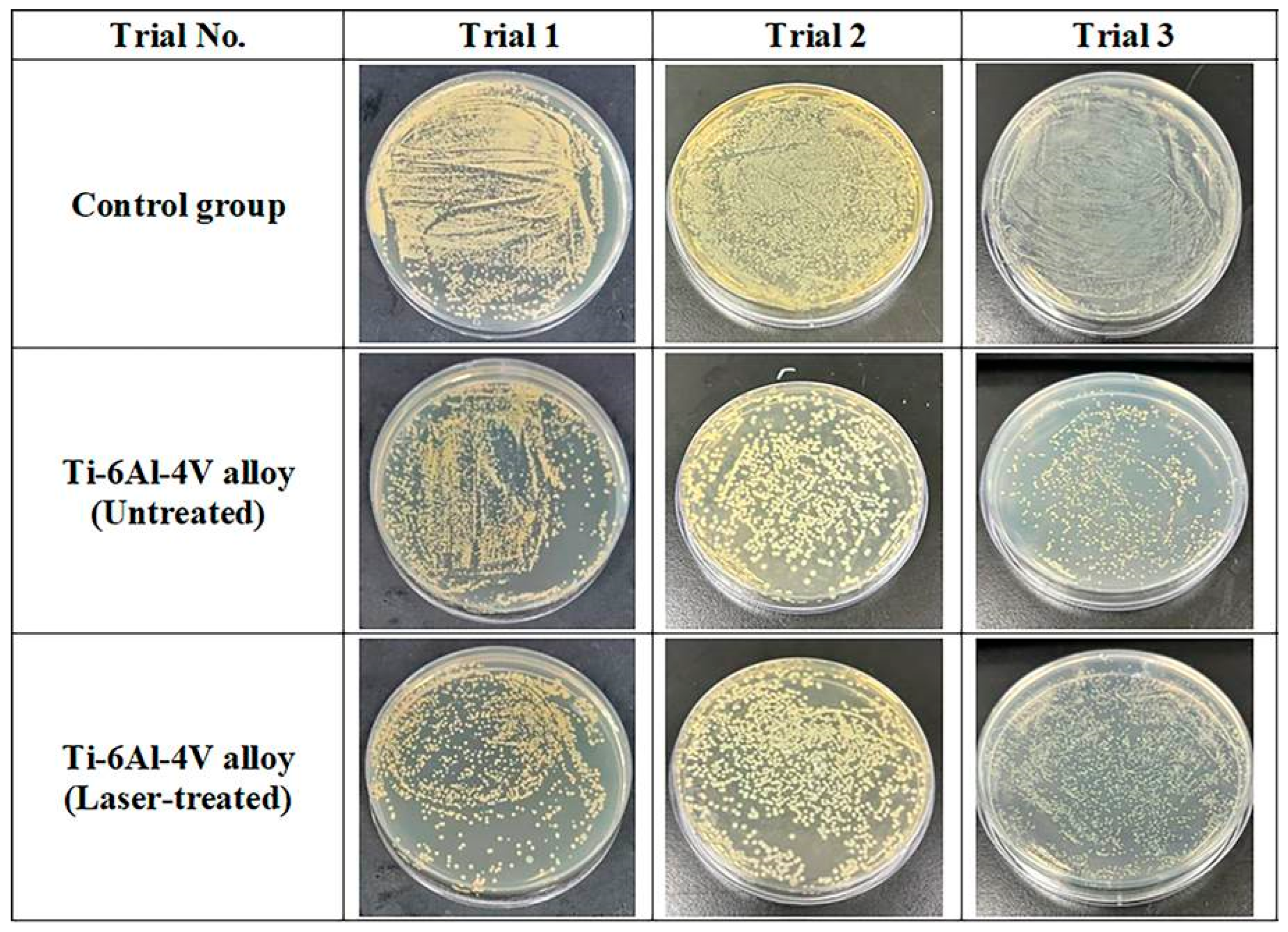
Similarly, the antibacterial evaluation of the Ti-6Al-4V ELI alloy was carried out under the same experimental conditions and procedures as those used for the pure titanium samples. However, in this case, we utilized a sterilized aluminum film as the control specimen for comparative analysis. The hydrophilic titanium alloy sample, formed by femtosecond laser processing, exhibited enhanced antibacterial activity compared to both the untreated specimen and the control group (sterilized aluminum film). Figure 12 also confirmed a significant improvement of antibacterial performance in the femtosecond laser-patterned titanium alloy samples, as compared to the untreated titanium alloy samples in the presence of Staphylococcus Aureus.
Figure 12.
Antibacterial performance analysis of the untreated and femtosecond laser-patterned titanium alloy samples in the presence of Staphylococcus Aureus.
Figure 13a shows the evaluation of antibacterial performance of the femtosecond laser-treated hydrophobic pure titanium samples in the presence of Staphylococcus Aureus bacteria. Based on viable cell count analysis, the untreated titanium sample showed an 84.0% bacterial reduction relative to the control, while the laser-treated samples exhibited a 98.6% reduction. Furthermore, a laser-treated pure titanium sample exhibited a 90.9% antibacterial performance improvement over the untreated pure titanium samples. Figure 13b presents the antibacterial test results of the Ti-6Al-4V ELI alloy samples. Quantitative analysis confirmed that the untreated titanium alloy showed a 53.3% reduction in viable bacterial count relative to the control group, while the laser-treated specimen achieved a 60.0% reduction. On the other hand, femtosecond laser-treated hydrophilic titanium samples showed a 14.28% improvement in antibacterial performance as compared to the untreated titanium alloys.
Figure 13.
Evaluation of antibacterial performance of the untreated and laser-treated hydrophobic pure CP-Ti and hydrophilic Ti-6Al-4V ELI alloy samples against Staphylococcus Aureus. (a) Pure CP-Ti samples; (b) Ti-6Al-4V ELI alloy samples.
The enhancement in antibacterial behavior is attributed to the formation of micro/nano-structures and changes in surface energy induced by the laser processing, which modified the wettability characteristics (hydrophilic or hydrophobic) of the specimen. These characteristics are interpreted as secondary contributing factors that influence bacterial attachment and viability, rather than indicators of any degradation caused by the laser treatment itself. The degree of antibacterial effect varied depending on the surface wettability. However, this variation stemmed from structural and physicochemical differences in the surface, not from the negative effects of the laser process. Notably, both femtosecond laser-treated pure titanium and titanium alloy samples demonstrated effective suppression of Staphylococcus Aureus attachment and survival, where pure titanium samples showed much better performance than the titanium alloy samples. The wettability of an implant surface, whether hydrophobic or hydrophilic, critically influences osteoblast attachment and the overall process of osteosynthesis (bone–implant integration). These effects are primarily mediated by protein adsorption, cell–surface interactions, and subsequent intracellular signaling mechanisms. Following implantation, cells do not interact directly with the bare material surface; instead, they respond to a layer of proteins adsorbed from blood and surrounding biological fluids. Hydrophobic surfaces tend to adsorb proteins strongly; however, this often leads to protein denaturation. Consequently, such surfaces exhibit reduced osteoblast attachment, weaker focal adhesion formation, and an increased likelihood of delayed cell proliferation. In contrast, hydrophilic surfaces facilitate rapid and uniform protein adsorption while preserving protein bioactivity and exposing cell-binding motifs. This promotes integrin-dependent cell adhesion, improved cell spreading, enhanced cytoskeletal organization, and the formation of stable focal adhesions. These findings suggest a high potential for femtosecond laser surface modification as a preventive strategy against implant-associated infections in orthopedic applications. The results further confirm that femtosecond laser processing can enhance the antibacterial functionality of biomaterials without compromising their inherent properties.
3.4. Biological Response Evaluation: Cytotoxicity Assessment
Table 5 and Table 6 present the cytotoxicity evaluation results for pure titanium and Ti-6Al-4V alloy samples treated with femtosecond laser surface modification. The untreated titanium samples used as the control group exhibit excellent biocompatibility. As such, the experimental results showed that the viability of mouse fibroblast cells (NCTC Clone 929) was not reduced but instead exhibited an increasing trend. In fact, both untreated pure titanium and titanium alloy samples demonstrated cell viability exceeding 98%, confirming the absence of inherent cytotoxicity in the test materials.
Table 5.
Evaluation of cytotoxicity of femtosecond laser-treated pure CP-Ti samples (hydrophobic surface) using L-929 fibroblast cells.
Table 6.
Evaluation of cytotoxicity of femtosecond laser-treated Ti-6Al-4V ELI alloy samples (hydrophilic surface) using L-929 fibroblast cells.
In contrast, the laser-treated samples showed an even higher level of cell viability. Specifically, the pure titanium samples exhibited a viability of 106.7%, while the titanium alloy samples showed 121.6%, indicating enhanced cellular proliferation compared to their untreated counterparts. These results suggest that femtosecond laser surface treatment did not generate any toxic substances. Rather, it is presumed that the micro-metric surface structures, formed during the laser processing, contributed to improved cell adhesion and proliferation.
4. Conclusions
In this study, a femtosecond laser-based micro-metric surface modification technique was employed to precisely tailor the surface characteristics of orthopedic implant materials such as pure titanium and Ti-6Al-4V ELI alloy. The primary objective of the research was to optimize the biological responses of the laser-treated samples, specifically, antibacterial activity and cytocompatibility, by controlling the surface wettability using laser-assisted micro-texturing. Unlike conventional mechanical or chemical surface treatments, which often suffer from limitations in repeatability, precision, and cleanliness, femtosecond laser processing offered a non-thermal, high-resolution fabrication method that preserved the intrinsic biocompatibility of the base materials. Periodic micro-grid patterns were developed on a pure titanium surface to convert the hydrophilic surface to a hydrophobic surface, while periodic micro-line type patterns were formed on the titanium alloy to achieve hydrophilicity. By systematically varying the pattern intervals, surface wettability was quantitatively controlled where contact angle measurements showed a maximum of 144.6° for CP-Ti-G10 (hydrophobic) and a minimum of 19.84° for Ti-6Al-4V ELI-L10 (hydrophilic) samples.
Antibacterial assessments using Staphylococcus Aureus revealed that the laser-treated pure titanium samples exhibited a 98.6% reduction in viable bacterial colonies compared to the untreated control, while the Ti-6Al-4V ELI alloy samples showed a 14.3% improvement. These results suggest that laser-induced microstructures effectively modulate surface energy, which in turn alters bacterial adhesion behavior. Hydrophobic surfaces appeared to suppress moisture and bacterial attachment, while hydrophilic surfaces facilitated competitive protein adsorption and early cell attachment, thereby inhibiting bacterial colonization. Cytotoxicity was assessed using an MTT-based in vitro assay with L-929 mouse fibroblast cells in accordance with ISO 10993-5:2009. The untreated samples exhibited cell viability levels exceeding 98%, confirming the absence of cytotoxic effects. Notably, the femtosecond laser-treated Ti-6Al-4V ELI alloy showed a markedly increased viability of 121.6%, suggesting that the laser-induced surface modifications not only maintained biocompatibility but also promoted enhanced cellular proliferation. These results exhibit that femtosecond laser processing not only avoids the induction of cytotoxicity but may also provide favorable topographies for cell growth due to its ability to generate controlled micro-scale features.
In summary, femtosecond laser surface modification has been validated as a versatile and effective tool for the functionalization of titanium-based orthopedic implants. By enabling the selective imparting of hydrophobicity (for plates using pure titanium) and hydrophilicity (for screws using Ti-6Al-4V ELI alloy), the technique addresses dual clinical needs: the prevention of bacterial infection and promotion of osseointegration. Given its precision, reproducibility, and chemical-free nature, the laser-based approach holds strong potential for broader application in other implantable medical devices, including dental implants, joint replacements, and biosensors, contributing to improved implant performance, and long-term clinical success.
Author Contributions
Conceptualization, H.-K.C. and I.-B.S.; methodology, H.-K.C., Y.-J.J., H.S. and S.K.; software, H.-K.C., H.J. and M.S.A.; validation, Y.-J.J., I.-B.S. and H.S.; formal analysis, H.-K.C., H.J., S.K., D.M. and M.S.A.; investigation, H.-K.C., I.-B.S. and D.M.; resources, I.-B.S. and S.K.; data curation, Y.-J.J., H.S., D.M. and M.S.A.; writing—original draft preparation, H.-K.C. and M.S.A.; writing, Y.-J.J., H.S., H.J. and M.S.A.; visualization, H.-K.C., H.J., S.K. and M.S.A.; supervision, I.-B.S., S.K. and D.M.; project administration, I.-B.S., H.J., S.K. and D.M.; funding acquisition, I.-B.S., S.K. and D.M. All authors have read and agreed to the published version of the manuscript.
Funding
This research work was funded by the Korea Medical Device Development Fund Grant funded by the Korea government (the Ministry of Science and ICT, the Ministry of Trade, Industry and Energy, the Ministry of Health and Welfare, the Ministry of Food and Drug Safety), project number: RS-2023-00255880. This work was supported by Gwangju Institute of Science and Technology (GIST) Research Institute (GRI) grant funded by the GIST in 2024.
Data Availability Statement
The data is available upon request.
Conflicts of Interest
Authors Harim Song, Hyeongdo Jeong, Seungpyo Kim, and Daeseon Moon were employed by KJmeditech Co., Ltd. The authors declare that the research was conducted in the absence of any commercial or financial relationships that could be construed as a potential conflict of interest.
References
- Sugioka, K.; Cheng, Y. Ultrafast lasers—Reliable tools for advanced materials processing. Light Sci. Appl. 2014, 3, e149. [Google Scholar] [CrossRef] [Scilit]
- Sohn, I.K.; Choi, H.K.; Yoo, D.; Noh, Y.C.; Noh, J.; Ahsan, M.S. Three-dimensional hologram printing by single beam femtosecond laser direct writing. Appl. Surf. Sci. 2018, 427, 396–400. [Google Scholar] [CrossRef] [Scilit]
- Ahsan, M.S.; Dwanda, F.; Lee, M.S.; Sekita, H.; Sumiyoshi, T. Formation of superhydrophobic soda-lime glass surface using femtosecond laser pulses. Appl. Surf. Sci. 2013, 265, 784–789. [Google Scholar] [CrossRef] [Scilit]
- Ahsan, M.S.; Lee, M.M.S. Formation mechanism of self-organized nanogratings on a titanium surface using femtosecond laser pulses. Opt. Eng. 2012, 51, 121815. [Google Scholar] [CrossRef] [Scilit]
- Wu, X.; Ao, H.; He, Z.; Wang, Q.; Peng, Z. Surface modification of titanium by femtosecond laser in reducing bacterial colonization. Coatings 2022, 12, 414. [Google Scholar] [CrossRef] [Scilit]
- Barylyak, A.; Nowak, R.W.; Liśkiewicz, M.K.; Krzemiński, P.; Płoch, D.; Cieniek, B.; Bobitski, Y.; Kisała, J. Photocatalytic and antibacterial activity properties of Ti surface treated by femtosecond laser–a prospective solution to peri-implant disease. Sci. Rep. 2024, 14, 20926. [Google Scholar] [CrossRef] [Scilit]
- Pawłowski, Ł.; Wawrzyniak, J.; Kopeć, A.B.; Cieślik, B.M.; Jurak, K.; Karczewski, J.; Tylingo, R.; Siuzdak, K.; Zieliński, A. Antibacterial properties of laser-encapsulated titanium oxide nanotubes decorated with nanosilver and covered with chitosan/Eudragit polymers. Biomater. Adv. 2022, 138, 212950. [Google Scholar] [CrossRef] [Scilit]
- Majhy, B.; Iqbal, R.; Sen, A.K. Facile fabrication and mechanistic understanding of a transparent reversible superhydrophobic–superhydrophilic surface. Sci. Rep. 2018, 8, 18018. [Google Scholar] [CrossRef] [Scilit]
- Gittens, R.A.; Scheideler, L.; Rupp, F.; Hyzy, S.L.; Gerstorfer, J.G.; Schwartz, Z.; Boyan, B.D. A review on the wettability of dental implant surfaces II: Biological and clinical aspects. Acta Biomater. 2014, 10, 2907–2918. [Google Scholar] [CrossRef] [Scilit]
- Saison, T.; Peroz, C.; Chauveau, V.; Berthier, S.; Sondergard, E.; Arribart, H. Replication of butterfly wing and natural lotus leaf nanostructures by nanoimprint on silica sol-gel films. Bioinspir. Biomim. 2008, 3, 046004. [Google Scholar] [CrossRef] [Scilit]
- Liu, Y.; Rui, Z.; Cheng, W.; Song, L.; Xu, Y.; Li, R.; Zhang, X. Characterization and evaluation of a femtosecond laser-induced osseointegration and an anti-inflammatory structure generated on a titanium alloy. Regen. Biomater. 2021, 8, rbab006. [Google Scholar] [CrossRef] [Scilit]
- Grase, L.; Onufrijevs, P.; Rezevska, D.; Racenis, K.; Skadins, I.; Karosas, J.; Gecys, P.; Iesalnieks, M.; Pludons, A.; Kroica, J.; et al. Effect of femtosecond laser-irradiated titanium plates on enhanced antibacterial activity and preservation of bacteriophage stability. Nanomaterials 2023, 13, 2032. [Google Scholar] [CrossRef] [Scilit] [PubMed]
- Jiang, P.; Zhang, Y.; Hu, R.; Shi, B.; Zhang, L.; Huang, Q.; Yang, Y.; Tang, P.; Lin, C. Advanced surface engineering of titanium materials for biomedical applications: From static modification to dynamic responsive regulation. Bioact. Mater. 2023, 27, 15–57. [Google Scholar] [CrossRef] [Scilit] [PubMed]
- Lijnev, A.; Val, J.E.M.S.D.; Elango, J.; Martínez, C.P.A.; Marín, J.M.G.; Scarano, A.; Gehrke, S.A. Residual-free micro-nano titanium surfaces via titanium blasting and single acid-etching: A cleaner alternative. Bioengineering 2025, 12, 735. [Google Scholar] [CrossRef] [Scilit]
- Nekleionova, A.; Moztarzadeh, J.; Wiesnerova, L.; Dvorakova, J.; Martinek, K.; Kulda, V.; Hradil, D.; Duchek, M.; Babuska, V. Comparative study of cell interaction and bacterial adhesion on titanium of different composition, structure and surfaces with various laser treatment. Mater. Res. Express 2024, 11, 055403. [Google Scholar] [CrossRef] [Scilit]
- Lausmaa, J.; Kasemo, B.; Mattsson, H. Surface spectroscopic characterization of titanium implant materials. Appl. Surf. Sci. 1990, 44, 133–146. [Google Scholar] [CrossRef] [Scilit]
- Geetha, M.; Singh, A.K.; Asokamani, R.; Gogia, A.K. Ti based biomaterials, the ultimate choice for orthopaedic implants–a review. Prog. Mater. Sci. 2009, 54, 397–425. [Google Scholar] [CrossRef] [Scilit]
- Papa, S.; Khalil, A.A.; Cognasse, H.H.; Thomas, M.; Maalouf, M.; Maio, Y.D.; Sedao, X.; Guignandon, A.; Dumas, V. Dual-functionalized titanium by ultrafast laser texturing to enhance human gingival fibroblasts adhesion and minimize Porphyromonas gingivalis colonization. Appl. Surf. Sci. 2022, 606, 154784. [Google Scholar] [CrossRef] [Scilit]
- Cui, C.; Zhao, Y.; Bai, Z.; Yan, J.; Qin, D.; Peng, H.; Liu, Y.; Tong, J.; Sun, L.; Wu, X.; et al. The effect of antibacterial-osteogenic surface modification on the osseointegration of titanium implants: A static and dynamic strategy. ACS Biomater. Sci. Eng. 2024, 10, 4093–4113. [Google Scholar] [CrossRef] [Scilit]
- Wang, Z.; Jia, B.; Li, B.; Yang, T.; Wang, D.; Wang, H. Surface modification of titanium implant by femtosecond laser to improve the biological property. Mater. Lett. 2025, 390, 138443. [Google Scholar] [CrossRef] [Scilit]
- Zhang, H.; Wu, Z.; Wang, Z.; Yan, X.; Duan, X.; Sun, H. Advanced surface modification techniques for titanium implants: A review of osteogenic and antibacterial strategies. Front. Bioeng. Biotechnol. 2025, 13, 1549439. [Google Scholar] [CrossRef] [Scilit]
- Maalouf, M.; Maio, Y.D.; Aldeiturriaga, D.P.; Sedao, X.; Hivert, L.; Papa, S.; Dalix, E.; Thomas, M.; Guignandon, A.; Dumas, V. Femtosecond laser upscaling strategy and biological validation for dental screws with improved osteogenic performance. Surf. Interfaces 2025, 66, 106546. [Google Scholar] [CrossRef] [Scilit]
- Choi, H.K.; Jung, Y.J.; Jeong, H.; Kim, S.; Moon, D.; Song, H.; Sohn, I.B. Controlling hydrophilic and hydrophobic properties of titanium bone fixation plates using femtosecond laser surface treatment. J. Surf. Sci. Eng. 2024, 57, 306–316. [Google Scholar] [CrossRef]
- Beltrán, A.M.; Giner, M.; Rodríguez, Á.; Trueba, P.; Rodríguez, A.L.M.; Vázquez, G.M.A.; Godinho, V.; Alcudia, A.; Amado, J.M.; López, S.C.; et al. Influence of femtosecond laser modification on biomechanical and biofunctional behavior of porous titanium substrates. Materials 2022, 15, 2969. [Google Scholar] [CrossRef] [Scilit]
- Shaikh, S.; Kedia, S.; Singh, D.; Subramanian, M.; Sinha, S. Surface texturing of Ti6Al4V alloy using femtosecond laser for superior antibacterial performance. J. Laser Appl. 2019, 31, 022011. [Google Scholar] [CrossRef] [Scilit]
- Wang, Y.; Yu, Z.; Li, K.; Hu, J. Study on the effect of surface characteristics of short-pulse laser-patterned titanium alloy on cell proliferation and osteogenic differentiation. Mater. Sci. Eng. C 2021, 128, 112349. [Google Scholar] [CrossRef] [Scilit] [PubMed]
- Simões, I.G.; Reis, A.C.D.; Valente, M.L.D.C. Influence of surface treatment by laser irradiation on bacterial adhesion on surfaces of titanium implants and their alloys: Systematic review. Saudi Dent. J. 2023, 35, 111–124. [Google Scholar] [CrossRef] [Scilit] [PubMed]
- Guéhennec, L.L.; Soueidan, A.; Layrolle, P.; Amouriq, Y. Surface treatments of titanium dental implants for rapid osseointegration. Dent. Mater. 2007, 23, 844–854. [Google Scholar] [CrossRef] [Scilit]
- Li, X.F.; Zhang, C.Y.; Li, H.; Dai, Q.F.; Lan, S.; Tie, S.L. Formation of 100-nm periodic structures on a titanium surface by exploiting the oxidation and third harmonic generation induced by femtosecond laser pulses. Opt. Express 2014, 22, 28086–28099. [Google Scholar] [CrossRef] [Scilit]

Disclaimer/Publisher’s Note: The statements, opinions and data contained in all publications are solely those of the individual author(s) and contributor(s) and not of MDPI and/or the editor(s). MDPI and/or the editor(s) disclaim responsibility for any injury to people or property resulting from any ideas, methods, instructions or products referred to in the content. |
© 2026 by the authors. Licensee MDPI, Basel, Switzerland. This article is an open access article distributed under the terms and conditions of the Creative Commons Attribution (CC BY) license.