


Abstract
Photodynamic therapy (PDT) is a minimally invasive therapeutic modality that relies on the activation of photosensitizers (PS) by specific wavelengths of light to generate reactive oxygen species (ROS), resulting in localized cytotoxicity with relative sparing of healthy tissues. Depending on the PS properties, light dose, and intrinsic cellular features, PDT can elicit multiple cell death pathways, including apoptosis, necrosis, and autophagy. Increasing evidence indicates that PDT is also a potent inducer of ferroptosis, an iron-dependent form of regulated cell death driven by excessive lipid peroxidation (LPO), glutathione (GSH) depletion, and inactivation of glutathione peroxidase 4 (GPX4). PDT-derived ROS promote ferroptosis both indirectly by exhausting antioxidant defenses and directly by peroxidizing PUFAs within membrane phospholipids. At the same time, intense oxidative stress generated by PDT can activate adaptive responses such as mitophagy, a selective autophagic process that removes damaged mitochondria to limit ROS production and preserve redox homeostasis. Ferroptosis and mitophagy are therefore tightly interconnected, functioning as opposing yet complementary regulators of cell fate. PDT emerges as a key upstream modulator of the ferroptosis–mitophagy balance, as spatially and temporally confined oxidative stress can shift cellular responses from adaptive mitochondrial quality control to irreversible ferroptotic injury. Despite growing interest in both PDT and ferroptosis, their mechanistic interplay, particularly in relation to mitophagy, remains underexplored. This narrative review provides an integrated overview of current knowledge on how PDT influences ferroptosis and mitophagy, highlighting the molecular mechanisms that connect these pathways and discussing their implications for improving therapeutic efficacy and overcoming resistance.
1. Introduction
Photodynamic therapy (PDT) is a minimally invasive treatment that employs a photosensitizer (PS) activated by light at wavelengths specific to their absorption spectra, resulting in the generation of reactive oxygen species (ROS) and subsequent targeted cell death. Importantly, PDT exhibits a degree of selectivity toward pathological tissue, minimizing damage to healthy tissue. This selectivity derives from multiple factors, including preferential accumulation of PS in cancer cells or microorganisms, as well as differences in redox regulation, with healthy cells and tumors exhibiting variable antioxidant capacities that influence their sensitivity to ROS [1]. Depending on treatment parameters such as PS type and concentration, subcellular localization, and cellular oxidative stress responses, PDT can activate multiple cell death modalities, including apoptosis, necrosis, and autophagy [2]. Notably, recent studies have revealed that PDT can activate ferroptosis, a distinct, iron-dependent form of regulated cell death characterized by excessive lipid peroxidation (LPO), glutathione (GSH) depletion, and impaired activity of the antioxidant enzyme glutathione peroxidase 4 (GPX4) [3,4]. By consuming intracellular oxygen, PDT promotes the generation of high levels of ROS, which, in turn, drive LPO, deplete GSH, and reduce GPX4 expression, thereby initiating ferroptosis [5,6,7,8,9]. Beyond affecting antioxidant defenses, PDT-derived ROS can also directly peroxidize polyunsaturated fatty acids (PUFAs) within membrane phospholipids, triggering non-enzymatic lipid autoxidation and further reinforcing ferroptosis [10]. These mechanisms are of particular interest because ferroptosis can bypass classical apoptosis resistance, a common hallmark of therapy-resistant cancers. At the same time, cells exposed to oxidative stress—including PDT-induced ROS—activate adaptive protective mechanisms to preserve homeostasis. Among these, mitophagy plays a central role by selectively eliminating damaged, ROS-generating mitochondria, thereby limiting oxidative injury, removing damaged mitochondria and preventing excessive LPO [11].
Ferroptosis and mitophagy are tightly interconnected processes that collectively regulate cellular redox homeostasis and survival. Mitophagy limits ferroptosis by selectively removing damaged ROS-producing mitochondria, thereby reducing LPO and iron-driven oxidative stress. Conversely, when mitophagy is insufficient, mitochondrial dysfunction amplifies ROS generation and sensitizes cells to ferroptotic death. Dysregulation of this axis shifts the response toward pathological outcomes, contributing to chronic inflammation, tissue degeneration, and therapy resistance.
Within this framework, PDT serves as a powerful upstream modulator, linking ferroptosis and mitophagy by generating spatially confined oxidative stress. Moderate PDT-induced stress may favour mitophagy, allowing for cells to adapt and survive by reducing mitochondrial ROS production. In contrast, excessive oxidative injury can overwhelm mitophagic capacity, leading to iron-driven LPO and execution of ferroptosis. Thus, PDT not only induces cell death but also reshapes the ferroptosis–mitophagy axis, tipping the balance between mitochondrial control and irreversible oxidative stress. This balance is particularly relevant in the context of cancer therapy, where exploiting ferroptosis while limiting adaptive survival pathways such as mitophagy may help overcome multidrug resistance and resistance to apoptosis. Despite numerous reviews addressing PDT and ferroptosis individually, relatively few have examined their mechanistic convergence with mitophagy. Notably, Mishchenko et al. were among the first to systematically propose and describe the synergistic interplay between PDT and ferroptosis [6]. Therefore, the aim of this narrative review is to provide an integrated overview of current knowledge on the relationships among PDT, ferroptosis, and mitophagy, with a particular focus on the molecular mechanisms governing their interactions and their implications for improving therapeutic efficacy.
2. Ferroptosis
Ferroptosis is an iron-dependent, oxidative stress–driven form of regulated cell death that results from severe disturbances in cellular metabolism and redox balance, ultimately causing excessive LPO. LPO refers to the oxidative breakdown of PUFAs within cellular membranes, leading to the buildup of phospholipid hydroperoxides (PLOOH), which are key triggers of ferroptosis. Mechanistically, ferroptosis can be initiated through two main pathways: the extrinsic (transporter-dependent) pathway and the intrinsic (enzymatic) pathway (Figure 1) [12]. The extrinsic pathway is activated by increased uptake of external iron via transporters like transferrin and lactotransferrin, resulting in intracellular iron overload. The destabilized (ferrous ion) Fe2+ then catalyzes the Fenton reaction (Fe2+ + H2O2 → Fe3+ + OH− + •OH), producing highly reactive hydroxyl radicals that intensify oxidative stress and LPO. Concurrently, the intrinsic pathway is primarily triggered by inhibition or depletion of components of the system Xc−–GSH–GPX4 axis [13]. Impairment of cystine import through system Xc−, mediated by the SLC7A11 subunit, decreases GSH synthesis, ultimately leading to the inactivation of GPX4, which allows for the uncontrolled accumulation of toxic lipid peroxides and reactive oxygen species (ROS) [14]. PUFAs play a central role in ferroptosis. They cannot spontaneously integrate into the phospholipid bilayer in an oxidizable form. Before serving as substrates for LPO, they must be activated and incorporated into membrane phospholipids. This process depends on the coordinated action of acyl-CoA synthetase long-chain family member 4 (ACSL4) and lysophosphatidylcholine acyltransferase 3 (LPCAT3), which works together as a metabolic pathway for PUFA loading. The lipid metabolic genes ACSL4 and LPCAT3 were identified by Dixon et al. in 2015 in haploid chronic myelogenous leukemia cells (KBM7) [15]. In this study, the researchers hypothesized that ACSL4 and LPCAT3 are key regulators of ferroptosis [15]. Subsequently, Doll et al. in 2016 employed genome-wide haploid and CRISPR–Cas9-based screens, revealing that two membrane-remodeling enzymes, ACSL4 and LPCAT3, are significant drivers of ferroptosis [16]. ACSL4 selectively activates long-chain PUFAs, especially arachidonic acid (AA) and adrenic acid (AdA), by attaching them to Coenzyme A (CoA) to form PUFA-CoA intermediates. This activation step is critical because, without conversion into PUFA-CoA, free PUFAs cannot enter the phospholipid biosynthetic pathway [16]. Once activated, these PUFA-CoA molecules become substrates for LPCAT3, which esterifies them into the sn-2 position of membrane phospholipids, mainly phosphatidylethanolamine (PE). This process results in the formation of PUFA-containing phospholipids, such as AA-PE and AdA-PE, which are the direct targets of LPO during ferroptosis.
Figure 1.
Mechanism of ferroptotic cell death. Ferroptosis occurs when system Xc− is inhibited or Glutathione peroxidase 4 (GPX4) is inactivated, ultimately leading to cell death. A major driver of this process is the buildup of lipid reactive oxygen species (ROS). Peroxidation of Polyunsaturated Fatty Acids (PUFAs) is a primary source of lipid ROS, while excess intracellular iron provides the catalytic basis for ferroptosis. Key proteins involved include Nuclear Receptor Coactivator 4 (NCOA4), Phosphorylase Kinase Regulatory Subunit Beta 2 (PHKB2), Heat Shock Protein Beta-1 (HSPB1), Lipoxygenases (LOXs), Iron-Responsive Element Binding Protein 2 (IREB2), Solute Carrier Family 3 Member 2 (SLC3A2), Solute Carrier Family 7 Member 11 (SLC7A11), Gamma-Glutamylcysteine Synthetase (γ-GCS), Reduced Glutathione (GSH), and Oxidized Glutathione (GS-SG) (illustration created using BioRender.com).
PUFAs are highly prone to oxidation because they contain multiple double bonds. These double bonds make the hydrogen atoms on adjacent carbons very easy to remove, turning PUFAs into ideal substrates for LPO. In non-enzymatic conditions, when ROS attack these lipids, they generate PLOOH. Normally, GPX4 detoxifies PLOOH, but during ferroptosis, PLOOH accumulates to toxic levels, damaging membrane integrity [17]. Enzymatic LPO involves the formation of PLOOH mediated by lipoxygenases (LOX) and cytochrome P450 oxidoreductase (POR) [18,19,20,21]. According to Shah et al. (2018), cells expressing 5-LOX, p12-LOX, and 15-LOX-1 were more vulnerable to ferroptosis [18]. Notably, in some contexts, simply accumulating PUFAs and incorporating them into membrane phospholipids (PUFA-PLs) can trigger ferroptosis, even without a significant increase in ROS or strong inhibition of GPX4. Cells can incorporate PUFAs from the extracellular environment, which may come from nearby cells, immune cells, pathogens, or circulating lipoproteins. The study by Dar and colleagues (2015) clearly showed that pathogens can induce ferroptosis in host cells by enzymatically oxidizing host membranes [22]. They found that a common bacterium, Pseudomonas aeruginosa, which does not produce arachidonoyl-phosphatidylethanolamine (AA-PE), can express lipoxygenase (pLoxA) to oxidize host AA-PE into 15-hydroperoxy-AA-PE (15-HOO-AA-PE) and trigger ferroptosis in human bronchial epithelial cells from patients with cystic fibrosis (CF) [22].
Importantly, PDT is closely connected to ferroptosis regulation. As a ROS-generating therapy, PDT produces significant intracellular ROS through its Type I (radical-based) and Type II (singlet oxygen-based) photochemical reactions, directly promoting non-enzymatic LPO and thereby triggering a ferroptosis-like cell death program [10]. PDT can also inhibit GPX4 activity, which further enhances the intrinsic ferroptosis pathway [14]. Conversely, tumor cells often show elevated H2O2 levels, and ferroptosis can exploit this by using endogenous H2O2 as a substrate for the Fenton reaction, producing O2 locally [23]. This not only encourages additional LPO but also helps alleviate the hypoxic tumor microenvironment (TME), ultimately boosting PDT effectiveness. Overall, ferroptosis results from a complex interaction between iron metabolism, LPO, and antioxidant defense mechanisms, with PDT acting as a strong synergistic stimulus that amplifies ferroptotic cell death in cancer treatment.
3. Mitophagy
Macroautophagy is a highly conserved intracellular degradation process that enables cells to recycle cytoplasmic components through the lysosomal system, with mitophagy representing its mitochondria-specific and selective form. Mitophagy is triggered when mitochondrial damage surpasses the cell’s innate quality control mechanisms or when increased mitochondrial turnover is needed to fulfill cellular metabolic requirements [24]. By selectively removing dysfunctional or surplus mitochondria, mitophagy maintains mitochondrial integrity and supports vital cellular processes, including energy production, redox balance, inflammatory signaling, and cell differentiation [25]. Failure to eliminate damaged mitochondria results in ATP depletion, excessive ROS production, and cell death, emphasizing the crucial balance between mitochondrial biogenesis and mitophagy in preserving cellular energy homeostasis and survival [26]. The most studied mitophagy mechanism is the PTEN-induced kinase 1 (PINK1)-Parkin pathway, where the kinase PINK1 functions as a mitochondrial damage sensor (Figure 2). Under normal conditions, PINK1 is imported into mitochondria via the translocase of the outer membrane (TOM) and translocase of the inner membrane (TIM) complexes and is quickly cleaved by mitochondrial processing peptidase (MPP) and mitochondrion-localized rhomboid (PARL), leading to its proteasomal degradation. When mitochondria depolarize, PINK1 accumulates on the outer mitochondrial membrane (OMM), phosphorylates ubiquitin and Parkin, and activates Parkin as an E3 ubiquitin ligase [27,28,29]. Activated Parkin ubiquitinates OMM proteins such as Mitofusin 1 and Mitofusin 2 (MFN1/2), Miro1, and Voltage-dependent anion-selective channel 1 (VDAC1). These proteins are recognized by selective autophagy receptors [Optineurin (OPTN), Nuclear Dot Protein 52 kDa (NDP52), Tax1 Binding Protein 1 (TAX1BP1), p62], which link mitochondria to the forming autophagosome, ensuring rapid clearance and integration with transcriptional control via Transcription Factor EB (TFEB) [30,31]. In parallel with the PINK1-Parkin axis, Parkin-independent pathways use dedicated OMM receptors such as BCL2/adenovirus E1B 19 kDa protein-interacting protein 3 (BNIP3), NIX (BNIP3L), FUN14 domain-containing protein 1 (FUNDC1), B-cell lymphoma 2-like 13 (BCL2L13), FK506-binding protein 8 (FKBP8), and Myeloid Cell Leukemia 1 (MCL-1). These receptors bind directly to LC3/GABARAP via LC3-interacting regions (LIRs) [32].
Figure 2.
Mitophagy occurs through both ubiquitin (Ub)-dependent and ubiquitin-independent pathways. In the ubiquitin-dependent pathway, mitochondrial damage stabilizes PTEN-induced kinase 1 (PINK1), which then activates Parkin and leads to the ubiquitination of outer mitochondrial membrane proteins. These proteins are recognized by autophagy receptors such as Nuclear Dot Protein 52 kDa (NDP52) and Optineurin (OPTN), which recruit Protein 1 Light Chain 3 (LC3)-positive autophagosomes. In the ubiquitin-independent pathway, mitochondrial receptors including BCL2/adenovirus E1B 19 kDa protein-interacting protein 3 (BNIP3), NIX, FUN14 domain-containing protein 2 (FUNDC2), and Prohibitin 2 (PHB2) directly bind LC3 via LC3-interacting region (LIR) motifs. These pathways can work together depending on the cellular context and stress conditions. Notably, these pathways are not mutually exclusive and can work together or compensate for each other depending on cellular context and stress conditions. Mitofusin-1/2 (Mfn1/2); Translocase of Outer Mitochondrial Membrane 20 (Tom20) (illustration created using BioRender.com).
Additional E3 ubiquitin ligases, including Mitochondrial Ubiquitin Ligase Activator of NFKB 1 (MUL1), Ariadne RBR E3 Ubiquitin Protein Ligase 1 (ARIH1), Seven in Absentia Homolog 1 (SIAH1), and HUWE1, provide fine-tuning of mitophagy. MUL1 promotes mitochondrial fission through Drp1 SUMOylation and MFN2 ubiquitination, acting upstream or parallel to Parkin. Meanwhile, AMBRA1 recruits HUWE1 to ubiquitinate OMM substrates even in Parkin-deficient conditions, exerting cytoprotective effects [33,34]. These multiple pathways ensure flexibility, allowing for cells to respond to different stressors and maintain energy homeostasis. Mitophagy is also linked to mitochondrial biogenesis via the PGC-1α–NRF1/2–TFAM axis, replacing degraded mitochondria and supporting cellular energy demands [35]. Regarding PDT, direct evidence in the literature remains limited. However, FUNDC1 has been shown to mediate mitophagy in response to ultraviolet-induced photodamage, suggesting that receptor-mediated mitophagy may play a role in regulating cellular responses to light-induced mitochondrial injury [36].
4. Balance Between Ferroptosis and Mitophagy
Mitochondria play a crucial role in iron metabolism, highlighting their function as a bridge between mitophagy and ferroptosis [37,38]. Through the Fenton reaction, mitochondrial Fe2+ can donate electrons to O2 and H2O2, generating hydroxyl and hydroperoxyl radicals that are potent inducers of LPO and ferroptosis [39,40,41]. Approximately 20–50% of cellular iron is stored in mitochondria as mitochondrial ferritin (FTMT), heme, or Fe–S clusters [42,43].
To maintain iron and metabolic balance, mitochondria constantly undergo fission and fusion, which help isolate damaged organelles and promote their selective removal through mitophagy [44].
During mitophagy, damaged mitochondria are broken down in lysosomes, and their iron is either recycled into the cytosol or safely stored in ferritin, ensuring both mitochondrial quality control and iron balance. By removing faulty mitochondria, mitophagy prevents excessive ROS production, thus protecting cells from ferroptosis. Additionally, the iron released during mitochondrial breakdown can build up in the labile iron pool or inside lysosomes, where it may engage in Fenton reactions and encourage LPO [45]. This defensive role is mediated through the activation of mitophagy by Hypoxia-Inducible Factor (HIF) signaling, PINK1/Parkin pathways, BNIP3/NIX induction, or by pharmacological triggers, which reduces mitochondrial damage, limits ROS, and decreases ferroptotic susceptibility [46,47]. Enhanced mitophagy can counteract ferroptosis caused by CDGSH Iron Sulfur Domain 3 (CISD3) knockdown and restore mitochondrial health after septic injury. Additionally, transcriptional regulators like Nrf2 promote resistance to ferroptosis by increasing key mitophagy components, including PINK1 and p62 [48,49].
However, mitophagy can also exert pro-ferroptotic effects, especially when excessively activated. Because mitochondria store large pools of iron, accelerated mitophagic degradation can release substantial amounts of free iron into the cytosol, thereby enhancing LPO and sensitizing cells to ferroptosis [50,51]. Dynamic O-GlcNAcylation has been shown to coordinate ferritinophagy and mitophagy to increase labile iron and promote ferroptosis, while FUNDC1-dependent mitophagy can either protect against or promote ferroptosis depending on the metabolic context [52]. Overall, mitophagy plays a dual role: physiological mitophagy suppresses ferroptosis by preserving mitochondrial quality and iron homeostasis, whereas excessive or dysregulated mitophagy may promote ferroptosis through mitochondrial iron release and enhanced ROS production. Given this duality, the potential off-target risk of mitophagy inhibition during PDT, especially in normal cells, requires careful consideration. While mitophagy inhibition can theoretically increase ferroptosis in healthy tissues, several features of the PDT approach minimize such off-target effects. First, PDT inherently provides spatiotemporal selectivity, as ROS generation and mitophagy modulation occur predominantly at the irradiation site. To further minimize this off-target risk several strategies are adopted such as nanoplatforms that are designed for preferential tumor accumulation via enhanced permeability and retention effects and, where applicable, tumor-targeting ligands, limiting exposure to healthy tissues. Third, tumor cells exhibit higher basal ROS levels, increased iron content, and altered redox homeostasis, rendering them significantly more susceptible to ferroptosis than normal cells. Finally, PDT parameters such as light dose and photosensitizer concentration can be precisely controlled to maximize tumor cell damage while sparing surrounding tissues. Taken together, these factors provide a mechanistic basis for the tumor-selective efficacy of our PDT–mitophagy modulation strategy, minimizing potential harm to healthy cells.
5. Susceptibility or Resistance in Cancer Cells?
Cellular factors like the expression of key enzymes (GPX4, ACSL4, LPCAT3) can determine whether PDT induces ferroptosis or other forms of cell death. Using lipidomic and biochemical techniques, Dos Santos et al., 2020 showed that both the type of photosensitizer and the cell type influence the cell death pathway triggered by PDT [53]. In this study, triple-negative breast cancer cells (MDA-MB-231) were the most sensitive to methylene blue (MB)-PDT and are characterized by a lipid profile highly enriched in PUFAs, many of which are esterified into phosphatidylethanolamine (PE), the main substrates of ferroptotic LPO [53]. In contrast, less aggressive or non-tumorigenic cells displayed lower PUFA content and higher levels of antioxidant molecules, resulting in less susceptibility to MB-PDT. Although MB-PDT reduced GPX4 levels across all cell lines, only MDA-MB-231 cells underwent ferroptotic cell death, as demonstrated by increased LPO, elevated labile iron pools, and selective protection by the ferroptosis inhibitor ferrostatin-1. This suggests that GPX4 depletion alone is not sufficient to trigger ferroptosis; rather, a permissive lipid environment rich in oxidizable PUFA-containing phospholipids is necessary. Importantly, PUFA-enriching MCF-7 cells become susceptible to MB-PDT-induced LPO and ferroptosis, further confirming that PUFA availability is a key factor in ferroptotic sensitivity. Collectively, these findings demonstrate that MB-PDT-induced cytotoxicity depends on LPO and that high PUFA content is essential for ferroptosis induction following photo-oxidative stress [53]. Other studies have also shown that not all cell types respond equally to ferroptosis. Viswanathan et al., 2017 observed that therapy-resistant cancer cells often adopt a high-mesenchymal state characterized by increased synthesis of PUFA-containing phospholipids and a strong dependence on GPX4-mediated detoxification of lipid peroxides [54]. As a result, these otherwise treatment-resistant cells become selectively vulnerable to ferroptosis when this lipid-peroxide defense pathway is inhibited [54]. Several classical tumor suppressors, including p53 and BAP1, promote ferroptosis by downregulating SLC7A11, which reduces cystine uptake and weakens the antioxidant capacity of cancer cells [55,56]. The ability of p53 to promote ferroptosis may represent one of the mechanisms through which it suppresses tumor development. However, some studies have shown that p53, by regulating different transcriptional targets, can also prevent ferroptosis [57,58]. Since p53 controls many genes involved in various biological processes, its role in ferroptosis is highly context-dependent, varying with cell type, stimuli, and metabolic state [56]. A genetic variant of p53 (the P47S polymorphism), found in individuals with higher cancer risk, makes cells resistant to ferroptosis. This indicates that certain p53 mutations might reduce its ability to promote ferroptosis, although it is unclear whether this is the only functional effect of the mutation [59]. Despite this vulnerability, many tumors develop strong ferroptosis defense systems, often by increasing SLC7A11 and other antioxidant pathways. These adaptations protect cancer cells from ferroptosis but also create a therapeutic window. Understanding the molecular basis of resistance is therefore crucial, both to identify tumors suitable for ferroptosis-based treatments and to develop strategies to prevent unintended ferroptosis in disease settings. A growing body of evidence suggests that ferroptosis may serve as a form of immunogenic cell death (ICD). Ferroptotic cancer cells release signals that stimulate the immune system, enhance dendritic cell maturation, boosting macrophage phagocytosis, and encouraging CD8+ T-cell infiltration [60]. This immunogenicity justifies combining ferroptosis induction with immunotherapies. Beyond ferroptosis, many cancers, including colorectal, lung, and breast tumors, show dysregulated mitophagy. Key regulators of this process, such as PINK1, Parkin, BNIP3, NIX, and FUNDC1, play roles that depend on the context and vary by tumor type and disease stage [61]. For example, the pro-mitophagic receptor BNIP3 has been reported to act as a tumor suppressor in certain types of cancer, such as breast cancer, renal cell carcinomas, and uterine cervical squamous cell carcinoma, by limiting mitochondrial ROS accumulation and genomic instability through the promotion of protective mitophagy [62,63]. The decreased expression of BNIP3 has been linked to tumor progression and metastasis [64,65]. In contrast, in other malignancies such as colorectal cancer, esophageal cancer, laryngeal squamous cell carcinoma, lung cancer, multiple myeloma, and pancreatic cancer, BNIP3 is often induced under hypoxic conditions in a manner dependent on HIF-1α, promoting adaptive mitophagy that supports metabolic stress tolerance, therapy resistance, and tumor growth [61]. Besides mitochondrial quality control, mitophagy is increasingly recognized as a modulator of redox homeostasis, iron metabolism, and LPO, thereby intersecting with ferroptosis regulation. By removing damaged mitochondria, mitophagy can limit mitochondrial ROS production and suppress LPO, conferring resistance to ferroptosis. Conversely, impaired or excessive mitophagy may increase oxidative stress and make cancer cells more susceptible to ferroptotic cell death. Gao et al., 2019 demonstrated that removal of mitochondria via Parkin-mediated mitophagy suppresses cysteine deprivation-induced ferroptosis [11]. This functional crosstalk highlights mitophagy as a double-edged process in cancer, capable of either promoting tumor cell survival or creating vulnerabilities that can be exploited through ferroptosis-based therapies [61]. Basal or moderate mitophagy is generally cytoprotective, allowing for cells to recycle damaged mitochondria and maintain homeostasis. However, the threshold that differentiates protective mitophagy from pro-death mitophagy remains poorly understood. Increasing evidence suggests that mitophagy plays a role in resistance to anticancer therapies, including chemotherapy and potentially PDT. Villa et al., 2017 demonstrated that the mitophagy regulator ARIH1 is widely expressed in breast and lung adenocarcinomas, helping protect cancer cells from chemotherapy-induced death [66]. Other studies suggest that mitophagy can provide resistance to PDT. Rubio et al., 2012 used murine fibrosarcoma L929 cells engineered to overexpress antioxidant enzymes such as GPX4 and SOD2, targeted at specific cellular compartments [67]. Cells were exposed to photodynamic stress using hypericin, a PS that preferentially localizes to the endoplasmic reticulum (ER), enabling spatially restricted ROS generation upon light activation. In this study, ER-restricted ROS were shown to propagate to mitochondria, where they promoted LPO and secondary ROS amplification [67]. This mitochondrial ROS amplification acted as a decision point, tipping the balance from protective mitophagy to apoptotic cell death. This experimental PDT model demonstrated accumulation of mitochondrial fragments within autophagosomes, consistent with active mitophagy following PDT. Inhibition of autophagic flux with chloroquine resulted in autophagosome accumulation, supporting the conclusion that PDT induced an active mitophagic response rather than a block in autophagosome formation [67].
6. Ferroptosis in Diseases: A Convergence of Microbial, Immune, and Metabolic Dysregulation
Physiologically, controlled ferroptosis and mitophagy work together to maintain cellular homeostasis because regulated ferroptotic signaling supports tissue renewal and immune balance. However, when this balance is disturbed, these processes become harmful rather than protective. Recent research emphasizes ferroptosis as a key mechanism connecting environmental stressors, microbial dysbiosis, and tissue degeneration across various systems [68,69].
In the periodontium, ferroptosis is increasingly recognized not only in resident structural cells but also within immune cell populations, thereby amplifying periodontal inflammation. Macrophages, neutrophils, and lymphocytes are susceptible to ferroptotic stress, although they continue to perform periodontal immune surveillance. Macrophages eliminate ferroptotic cells through TLR2-mediated recognition of oxygenated phosphatidylethanolamines, preventing excessive debris buildup [70]. Under conditions of persistent inflammation, macrophages may undergo ferroptosis, disrupt immune homeostasis and worsening tissue dysfunction. Ferroptosis influences macrophage polarization, increasing proinflammatory M1 activity [71].
Alongside immune dysregulation, ferroptosis impacts resident periodontal cells. Regulators such as Nrf2-dependent PRDX6 and butyrate-activated NCOA4 influence ferroptosis in gingival and periodontal ligament fibroblasts [72,73]. In osteocytes and osteoblasts, ferroptosis, worsened in diabetes by LPO, iron overload, and advanced glycation end products (AGEs), disrupts bone formation and increases osteoclast activity through mediators like SOST, RANKL, IL-1β, and TNF-α [74].
In other regions, the brain is susceptible to ferroptosis due to the high presence of PUFAs, increased oxygen consumption, and weak antioxidant defenses. In Parkinson’s disease, iron overload, glutathione deficiencies, and impaired mitophagy promote ROS production and LPO, making neurons more vulnerable. In Alzheimer’s disease, ferroptosis contributes to synaptic loss, while mitophagy failure exacerbates oxidative stress [75].
In acute lung injury and acute respiratory distress syndrome, mitochondrial dysfunction causes excessive mitophagy and reduces mitochondrial mass. Hyperactivation of PINK1/Parkin pathways trigger ferroptosis in alveolar epithelial cells and macrophages, leading to epithelial barrier breakdown and dysregulated inflammation [76].
Skeletal muscles are particularly vulnerable to the interaction between mitophagy and ferroptosis. During extended immobilization, or aging, excessive mitophagy leads to mitochondrial loss, impaired oxidative phosphorylation, and an increase in ROS. These effects encourage ferroptotic death of myofibers through ALOX15 activation and GPX4 suppression [77].
Literature indicates that at the pathological level, microorganisms may contribute to inducing ferroptosis. Pathogen-induced ferroptosis is a complex process in which microbial species and their virulence factors either directly activate ferroptotic signaling or interfere with intracellular iron homeostasis in host cells (Figure 3). P. gingivalis induces ferroptosis in gingival fibroblasts by suppressing the SLC7A11/GSH/GPX4 pathway, increasing interleukin 6 (IL-6), which in turn upregulates matrix metalloproteinase-1 (MMP-1), monocyte chemoattractant protein-1 (MCP-1), and IL-8. This combination promotes degradation of the extracellular matrix and enhances inflammatory infiltration [78,79,80]. Moreover, Fusobacterium nucleatum and ferroptosis-associated noncoding RNAs, especially LINC00616 and MEG3, influence ferroptosis in periodontal ligament stem cells (PDLSCs) and hinder osteogenic differentiation by causing Fe2+ buildup and miRNA-dependent regulation of TFRC and IGF1 signaling. These molecular processes jointly intensify ferroptotic cell death and lead to ongoing periodontal tissue destruction. Certain pathogens promote ferroptosis by increasing iron availability and LPO. For example, Pseudomonas aeruginosa secretes pyocyanin and pyoverdine, which increase intracellular iron levels and encourage oxidative lipid damage [22]. Pathogens can also interfere with ferritinophagy and antioxidant defenses. Salmonella enterica, through the SpiC effector, disrupts iron recycling, while P. gingivalis LPS and arginine-specific gingipains (RgpA/B) suppress GPX4 transcription [68]. Overall, these findings identify ferroptosis as a key pathogenic process that links microbial dysbiosis, immune dysfunction, and tissue damage, emphasizing its importance as both a biomarker of disease progression and a potential target for therapy.
Figure 3.
Mechanisms of bacterial-induced ferroptosis. Porphyromonas gingivalis causes ferroptosis by suppressing the Solute Carrier Family 7 Member 11 (SLC7A11)/Glutathione (GSH)/Glutathione Peroxidase 4 (GPX4) antioxidant pathway, leading to lipid peroxidation (LPO) and redox imbalance. This ferroptotic signaling is accompanied by increased interleukin 6 (IL-6) production, which then upregulates Matrix metalloproteinase 1 (MMP-1), Monocyte Chemoattractant Protein-1 (MCP-1), and interleukin 8 (IL-8), collectively promoting extracellular matrix degradation and inflammatory cell recruitment. Pseudomonas aeruginosa secretes redox-active metabolites pyocyanin and pyoverdine, enhancing intracellular iron accumulation and oxidative lipid damage. Although P. aeruginosa lacks arachidonoyl-phosphatidylethanolamine (AA-PE), it produces a bacterial pLoxA that oxidizes host AA-PE into 15-hydroperoxy-AA-PE (15-HOO-AA-PE), thereby triggering ferroptosis in host cells and amplifying tissue damage (illustration created using BioRender.com).
7. Ferroptosis Is One of the Cell Death Modalities Induced by PDT
PDT can elicit multiple cell death modalities, and the prevailing mechanism depends on multiple factors. Turubanova et al., 2019 demonstrated that the type of PS is an important factor influencing whether PDT induces ferroptosis rather than another form of cell death [60]. In their study, the authors showed that Photosens—PDT triggered both apoptosis and ferroptosis in GL261 cells, whereas photodithazine (PD)—PDT induced primarily apoptosis, highlighting how PS choice can determine the dominant cell death pathway [60]. Besides the type of PS, their concentration also can be critical in determining the mode of cell death. In a recent in vitro study, hypericin-mediated photodynamic therapy (HYP-PDT) induced distinct cell death modalities at different PS dosages. At low concentrations (≤250 nM), apoptosis was dominant, as shown by Annexin V/PI staining, detection of cleaved caspase-3, and inhibition with zVAD-fmk. High concentrations (10 μM) caused necrosis, characterized by rapid membrane rupture and nuclear swelling, which was unresponsive to ferroptosis or apoptosis inhibitors. At an intermediate concentration (2 μM), HYP-PDT induced a ferroptosis-like cell death, indicated by effective inhibition with Liproxstatin-1 and Ferrostatin-1, mitochondrial shrinkage, absence of cleaved caspase-3, and preserved intracellular ATP levels. This cell death was accompanied by a rapid depletion of GSH, likely due to its use in GPX4-mediated reduction in lipid hydroperoxides, and was linked to ER stress, as shown by CHAC1 upregulation. These results show that HYP-PDT can cause ferroptosis-like death depending on concentration, highlighting the importance of PS dosage in selecting the main cell death pathway [10]. The concentration dependence of PDT-induced ferroptosis can be mechanistically explained by the requirement to exceed a critical threshold of lipid hydroperoxides necessary to initiate irreversible lipid autoxidation [10]. Ferroptosis occurs due to the buildup of deadly lipid peroxides that disturb membrane redox balance, rather than from ROS production itself. Unlike canonical ferroptosis inducers, which act by disabling antioxidant systems such as system Xc− or GPX4, PDT generates large amounts of exogenous ROS that directly oxidize membrane lipids within minutes of light irradiation, leading to rapid lipid hydroperoxide formation and glutathione depletion. However, this process becomes ferroptotic only when a sufficient photosensitizer concentration allows for lipid peroxide levels to exceed the autoxidation threshold necessary for ferroptotic commitment. At subthreshold photosensitizer levels, LPO remains limited and adaptive antioxidant responses dominate, whereas excessively high concentrations may promote alternative, non-ferroptotic cell death pathways. These findings support the existence of an optimal photosensitizer concentration range for effective PDT-induced ferroptosis. Additionally, in some cases, the PS can act as a metabolic inducer of ferroptosis, not just as a PS combined with light. Recently, 5-aminolevulinic acid (5-ALA), one of the most used PS, was shown to induce ferroptosis and exert antitumor effects in oesophageal squamous cell carcinoma (ESCC) independently of light exposure. Shishido et al., 2020 demonstrated that 5-ALA modulates GPX4 and HMOX1, leading to increased LPO and tumor suppression both in vitro and in vivo, suggesting its potential as an inducer of ferroptosis [81]. More recently, Zhou et al., 2024 demonstrated that verteporfin (VP) can induce LPO and ferroptosis in two pancreatic cancer cell lines without any light activation [82]. Similarly, Wang et al., 2024 observed that in oral squamous carcinoma cells, VP caused ferroptosis markers such as ROS and Fe2+ accumulation, LPO, GSH and GPX4 depletion, and loss of mitochondrial membrane potential [83]. In these studies, the effects on ROS, lipid peroxides, and GPX4 downregulation occurred independently of YAP signaling and were completely blocked by ferroptosis inhibitors [82,83]. These studies suggest that some PS, like 5-ALA, may have intrinsic ferroptosis-inducing properties that are independent of light activation. The ability of photodynamic therapy to induce ferroptosis is also strongly influenced by the physicochemical and biological properties of the photosensitizer used. Photosensitizers that selectively localize to lipid-rich membranes, mitochondria, or lysosomes are more likely to promote iron-dependent LPO, a key feature of ferroptotic cell death. Additionally, photosensitizers that can generate ROS near membrane lipids, especially singlet oxygen that initiates lipid hydroperoxide formation, are more effective at triggering ferroptosis. Conversely, PS that primarily target the cytosol or nucleus, or those that produce diffusible ROS which do not effectively propagate lipid autoxidation, tend to activate alternative cell death pathways such as apoptosis or necrosis [1]. Furthermore, the lipophilicity of the PS and its capacity to disrupt redox balance, such as GSH depletion or iron mobilization, additionally influence its ferroptotic potential. These factors together explain why only certain classes of PS can effectively induce ferroptosis during PDT. Therefore, PS themselves should be investigated not only for their photodynamic effects but also as potential direct modulators of ferroptosis. This opens the possibility of repurposing or designing PS specifically to exploit ferroptotic cell death, even in contexts where light delivery is limited. In recent years, a growing number of novel PS have been shown to induce ferroptosis, expanding the traditional view of PDT-induced cell death. Many of these newly developed PS are metal-based complexes, particularly Iridium(III), Platinum(II), and Ruthenium(II) complexes, which exhibit strong photophysical properties, efficient ROS generation, and a pronounced ability to trigger iron-dependent LPO [84,85,86]. These features make them especially effective at promoting ferroptotic cell death upon light activation. Besides metal complexes, several non-metallic PS have also been reported to induce ferroptosis through different mechanisms. For example, boronic acid–derived salicylidenehydrazone (BASHY) dyes have been shown to accumulate in lipid droplets in human glioblastoma U87 cells, thereby boosting local photodynamic reactions and triggering LPO–mediated ferroptosis [87]. Together, these findings show that ferroptosis-inducing PS forms a diverse and quickly growing group of PDT agents, including both metal-based and organic compounds, and emphasize ferroptosis as an important and usable mechanism for photodynamic cancer therapy.
8. Immunological Consequences of PDT-Induced Ferroptosis and Mitophagy
Beyond direct cytotoxicity, PDT exerts significant immunomodulatory effects that play a crucial role in its therapeutic success. PDT primarily selectively destroys tumors and infections; additionally, PDT induces the release of pro-inflammatory molecules. Preclinical and clinical studies have demonstrated that PDT can activate both the innate and adaptive immune systems [88,89,90]. The oxidative stress generated by PDT not only induces tumor cell death but also shapes the tumor–immune microenvironment by releasing danger-associated molecular patterns (DAMPs), inflammatory mediators, and LPO products. In this context, ferroptosis and mitophagy might emerge as pivotal regulators of PDT-induced antitumor immunity [2]. Nevertheless, few studies have been conducted to investigate the role of ferroptosis in immune cells and to elucidate its impact within the TME (Figure 4). Recent evidence highlights the context-dependent role of ferroptosis within the TME. Zhu X et al. investigated ferroptosis in different cell types within the TME of gastric cancer tissue. Neutrophils infiltrating the TME were shown to undergo spontaneous ferroptosis, releasing oxidized lipid mediators that suppressed cytotoxic T-cell activity and impaired antitumor immunity. To address this limitation, the authors engineered a liposomal system co-encapsulating a potent PS (di-iodinated IR780, Icy7) and the ferroptosis inhibitor Liproxstatin-1. This strategy enabled photodynamic induction of immunogenic cell death (ICD) in tumor cells while simultaneously preventing ferroptosis in tumor-associated neutrophils. Thus, in parallel with increased ROS generation, inhibition of neutrophil ferroptosis reduced the accumulation of immunosuppressive oxidized lipids within the TME, restoring T-cell functionality [91]. Other authors shifted the primary subcellular target of ferroptosis induction from mitochondria to lysosomes. In a recent study, Chen Z et al. developed a novel lysosome-targeted photosensitizer (TLA) to specifically disrupt lysosomal membrane integrity upon light irradiation. Lysosomal membrane permeabilization caused the release of reduced iron, triggering cell-intrinsic Fenton reactions, mitochondrial dysfunction, and ultimately ferroptosis, while also inhibiting protective autophagy. The resulting ferroptotic cell death was immunogenic, encouraging the release of DAMPs and remodeling the immunosuppressive TME. Unlike apoptosis, ferroptosis is marked by extensive LPO of PUFAs–containing phospholipids, leading to the buildup of oxidized lipid species that serve as potent immunostimulatory signals. These LPO products, along with the release of classical DAMPs like HMGB1, ATP, and oxidized phosphatidylethanolamines, facilitate dendritic cell maturation, antigen presentation, and the subsequent activation of tumor-specific T cell responses. Importantly, combining TLA-mediated PDT with PD-L1 blockade markedly improved antitumor immune responses, emphasizing lysosome-driven ferroptosis as a promising strategy to enhance PDT when used with immunotherapy [92]. Combining immune checkpoint blockade (ICB) with PDT and modulation of ferroptosis is viewed as a relatively new and emerging strategy used by various research groups. Zhang H et al. developed a homologous magnetic-targeted exosome-based nanoplatform (SPION) for co-delivering a ferroptosis inducer and a near-infrared (NIR) PS to glioblastoma cells. The combination of ferroptosis activation and PDT disrupted redox homeostasis, caused mitochondrial damage, and increased tumor cell death, aiding in TME remodeling. This reprogramming was linked to improved tumor-specific T lymphocyte activity and modulation of macrophage phenotypes. Additionally, SPION-modified tumor-derived exosomes improved tumor targeting and blood–brain barrier penetration. When combined with anti–PD-1 therapy, this approach triggered sustained antitumor immune responses and lowered tumor recurrence [93]. Similarly, Guo et al. designed acidity-responsive nanoparticles based on M1 macrophage–derived exosomes (MEX) to combine PDT, ferroptosis induction, and immunomodulation. The nanoplatform activates itself in acidic TME, co-delivers a PS, iron ions, and catalase (CAT) to reduce tumor hypoxia, boost ROS production, and trigger immunogenic cell death. By blocking the CD47–SIRPα immune checkpoint and enhancing macrophage-mediated phagocytosis, this strategy effectively reprograms the TME, resulting in notable antitumor and antimetastatic effects across multiple preclinical models [94]. In parallel, mitophagy plays a dual and context-dependent role in shaping the immune response after PDT applications. Mitophagy is generally regarded as a cytoprotective mechanism, as it selectively removes damaged or excess mitochondria, thereby limiting excessive ROS production and preserving mitochondrial homeostasis [24,26]. Consequently, during PDT, mitophagy often acts as an adaptive survival response that attenuates oxidative stress–induced cellular damage and reduces therapeutic efficacy. However, when autophagy and mitophagy pathways are excessively activated, they can surpass the cellular tolerance threshold and trigger autophagic cell death, while simultaneously promoting the release of endogenous antigens from dying cells [95]. This process is vital for initiating and enhancing antitumor immune responses. To harness the strong immunomodulatory potential of pro-death autophagy and mitophagy, several strategies have been explored to intensify or intentionally trigger mitophagy alongside anticancer therapies. Recently, Sun q et al. developed a MnO2 shell-coated multifunctional porphyrinic metal–organic framework (MOF) loaded with carbonyl cyanide 3-chlorophenylhydrazone (CCCP). This nanoplatform was designed to improve PDT by shifting autophagy and mitophagy from promoting survival to promoting cell death. The system allowed for tumor-specific release of the mitochondrial uncoupler CCCP, reduced tumor hypoxia, and depleted intracellular GSH, thereby increasing ROS production during PDT. Excessive activation of autophagy and mitophagy caused mitochondrial dysfunction, autophagic cell death, and immunogenic cell death, which in turn stimulated T cell–mediated antitumor immunity and immune memory, leading to effective tumor elimination and reduced recurrence and metastasis in vivo [96]. Conversely, some researchers adopted an opposite strategy by blocking mitophagy to hinder mitochondrial quality control and thereby increase oxidative stress–mediated cell death. Zhang S et al. engineered a NIR–responsive nanoplatform that combines controlled ROS production, pyroptosis induction, and mitophagy inhibition. Upon NIR irradiation, the system triggers Caspase-1–dependent pyroptotic cell death while undergoing programmed disassembly and releasing lonidamine, which inhibits HK2-driven mitophagy. The combined increase in oxidative stress and suppression of mitochondrial repair mechanisms leads to enhanced immunogenic cell death, greater dendritic cell maturation, and improved infiltration of cytotoxic T lymphocytes, ultimately reprogramming the TME toward a pro-inflammatory and antitumor state [97]. Collectively, these findings position ferroptosis and mitophagy as critical immunological switches in PDT. Therapeutic strategies that selectively enhance ferroptosis while transiently restraining mitophagy may potentiate PDT-induced immunogenic cell death and improve antitumor immune responses. Conversely, fine-tuning mitophagy in immune or stromal cells may help mitigate collateral tissue damage and sustain long-term immune surveillance. Understanding how PDT orchestrates the ferroptosis–mitophagy–immunity axis will therefore be essential for optimizing PDT-based combination therapies, particularly in synergy with immunomodulatory approaches. While also elucidating how the combined modulation of ferroptosis and mitophagy impacts stromal and epithelial cell populations within the tumor microenvironment, particularly in oral tissues characterized by complex epithelial–stromal interactions.
Figure 4.
Potential mechanisms for ferroptosis mediated immune response in the TME (illustration created using BioRender.com).
9. Ferroptosis and Mitophagy in PDT-Mediated Vascular Damage
Vascular damage represents another secondary effect of PDT, complementing its direct cytotoxic and antimicrobial effects. Upon light activation of the PS, PDT generates high levels of ROS that rapidly affect the endothelial lining of blood vessels, leading to alterations in vascular permeability, perfusion, and, ultimately, tissue oxygenation [2]. Beyond direct tumor cell killing, PDT–induced oxidative stress intersects with ferroptosis and mitophagy pathways at the vascular level, where endothelial LPO and dysregulated mitochondrial quality control contribute to vascular damage, tumor ischemia, and remodeling of the immune microenvironment. PDT triggers a cascade involving vascular damage and hypoxia, which intersects with ferroptosis and immunogenic cell death to reshape the TME and enhance antitumor immune responses. PDT-induced ROS encourage iron-dependent LPO in endothelial cells, a key feature of ferroptosis. The high content of PUFAs in endothelial membranes, combined with labile iron pools and elevated metabolic activity, makes the vasculature especially vulnerable to ferroptotic damage [98]. PDT can disrupt the SLC7A11/GSH/GPX4 antioxidant axis in endothelial cells, impairing lipid peroxide detoxification and triggering ferroptotic cell death [14]. As a result, endothelial ferroptosis contributes to vascular collapse, platelet aggregation, and transient ischemia within treated tissues [98]. Vascular damage caused by PDT results in decreased blood perfusion and acute tumor hypoxia. Hypoxia-driven mitochondrial changes in endothelial cells contribute to vascular resistance, while disrupted mitophagy worsens oxidative injury and vessel dysfunction. Tumor hypoxia and vascular damage are closely linked processes within the TME, creating a self-reinforcing loop that significantly affects the success of ROS-based therapies. Because tumor hypoxia limits the effectiveness of reactive oxygen species–based treatments, some studies focus on modifying the metabolic and redox properties of the TME to improve PDT outcomes. In this context, iron-based metal–organic framework nanoplatforms combining photodynamic therapy with metabolic interventions have been proposed to simultaneously relieve hypoxia, increase oxidative stress, and trigger ferroptosis. Recently, Chen Y et al. developed a multifunctional nanoparticle, Ce6@HGMOF, that integrates photodynamic therapy, ferroptosis, and metabolic intervention to boost antitumor effects. This platform includes a photosensitizer (Ce6), glucose oxidase (GOX), a chemotherapy drug (HCPT), and an iron-based metal–organic framework. GOX-mediated glucose depletion causes “starvation therapy” while producing H2O2, which is converted into oxygen by the MOF’s catalase-like activity, reducing tumor hypoxia and boosting ROS generation during PDT. At the same time, GSH consumption and Fenton reaction–induced LPO promote ferroptosis [99]. The synergistic combination of starvation therapy, PDT, and ferroptosis effectively remodels the TME and enhances oxidative stress–mediated cell death, offering a promising integrated approach for tumor treatment. Beyond directly killing tumor cells, PDT and ferroptosis can influence the tumor vasculature, where endothelial oxidative injury and vascular shutdown worsen hypoxia but also promote immune cell extravasation. This idea is further supported by studies in non-tumor models, such as intestinal ischemia–reperfusion, where neutrophil extracellular traps (NETs) were shown to induce endothelial ferroptosis by inhibiting Fundc1-dependent mitophagy, leading to microvascular dysfunction. Interventions targeting NETs or activating mitophagy restored endothelial function, highlighting the crucial interaction between ROS, ferroptosis, and mitochondrial quality control in vascular health. Collectively, these findings emphasize that successful therapeutic strategies depend on the coordinated regulation of ROS production, ferroptosis, mitophagy, and vascular remodeling, which together boost antitumor immunity and overcome adaptive resistance in both tumor and ischemic microenvironments [100].
10. Synergistic Effect Between PDT and Ferroptosis
Since PDT interacts with several ferroptosis-related pathways, increasing evidence indicates that PDT and ferroptosis can strengthen each other, leading to better therapeutic results [10,14]. However, ferroptosis induced by PDT alone appears limited, partly because PDT-triggered DNA damage can activate DNA damage response (DDR), which counteracts ROS accumulation and reduces overall cytotoxicity. Therefore, different strategies were adopted to induce ferroptosis by combining ferroptosis-inducers and PDT, summarized in Table 1. A recent study further showed that chlorin E6 (Ce6)-mediated PDT quickly produces ROS and causes DNA double-strand breaks, which then activate ATM-dependent DDR pathways. These pathways help lung cancer cells resist PDT-induced cytotoxicity. Importantly, blocking ATM with a drug effectively prevented this DDR activation and significantly improved PDT effectiveness, confirming that DDR plays a protective role in reducing treatment response [101]. These limitations emphasize the need for combination strategies that stabilize or enhance ferroptosis, and recent research has increasingly focused on combining PDT with small-molecule ferroptosis inducers. Strategies aimed at triggering or boosting ferroptosis have shown promising synergistic effects and good tolerability in preclinical cancer models. This approach is becoming a promising method to achieve deeper and more effective ferroptosis-mediated tumor killing. Because the antioxidant axis Xc−–GSH–GPX4 plays a central role in regulating ferroptosis, selectively targeting this metabolic pathway has become a key therapeutic strategy for inducing ferroptosis (Figure 5). The protective response of DDR to PDT-induced DNA damage up-regulates GPX4, limiting the ferroptotic and cytotoxic efficacy of PDT [101,102]. Because PDT itself promotes LPO, combining PDT with GPX4 inhibitors can lead to strong synergistic cytotoxicity. Bae et al., 2025 engineered photosensitizing lipid nanoparticles (PLNPs) by incorporating a PEGylated pheophorbide-based photosensitizer (CPP) into conventional lipid nanoparticles and loading them with GPX4-targeting siRNA [103]. The authors tested the PLNPs in 4T1 and EO771 breast cancer cells, demonstrating efficient GPX4 gene silencing, robust ROS production upon light exposure, and significantly increased cytotoxicity when combining PDT with ferroptosis induction [103]. This confirmed the synergistic interaction between PDT-induced oxidative stress and siRNA-mediated ferroptosis at the cellular level. In mouse tumor models, the PLNPs exhibited prolonged intratumoral retention, effective GPX4 knockdown, and strong tumor growth inhibition following laser irradiation, all with minimal systemic side effects or toxicity. These results confirmed the therapeutic potential of the dual-function nanoplatform in a living organism [103]. Song et al., 2021 engineered intracellular-acidity-activatable dynamic nanoparticles BNP@R capable of selectively releasing the ferroptosis inducer RSL-3 and performing acid-activated PDT [104]. This nanoplatform enhanced tumor immunogenicity, promoted T-cell infiltration, and, when combined with the immunotherapy approach Programmed Death-Ligand 1 (PD-L1) blockade, effectively suppressed B16-F10 melanoma growth in vitro and 4T1 lung metastasis in vivo [104]. In detail, RSL-3 induces ferroptosis by directly inhibiting GPX4, preventing the detoxification of lipid hydroperoxides and allowing for their accumulation in cellular membranes [104]. Inhibitors of system Xc−, the cell membrane antiporter that imports cystine in exchange for glutamate, block cystine uptake, hinder GSH production, and weaken cellular antioxidant defenses, leading to increased lipid peroxide accumulation [105]. When combined with PDT, these inhibitors significantly potentiate ROS accumulation and ferroptotic cell death. Erastin and sulfasalazine are well known as potent and selective inhibitors of system Xc− [106]. A team of authors designed and tested a hybrid Erastin-Ce6 nanostructure that uses ferroptosis to sustainably produce oxygen and ROS, greatly improving PDT in tongue cancer treatment. Zhu et al., 2019 first examined the expression of the protein SLC7A11 (solute carrier family 7 member 11) in Oral Tongue Squamous Cell Carcinoma (OTSCC) samples, identifying it as a potential target due to its link to ferroptosis sensitivity [23]. They then developed a new supramolecular nanodrug called Ce6-erastin. When exposed to light, the nanoparticles significantly boosted anti-tumor effects both in vitro on CAL-27 cells and in vivo in xenograft tumor mouse models, thanks to increased intracellular ROS, higher oxygen levels, and suppression of SLC7A11 expression [23]. GSH serves as another target within the antioxidant axis Xc−–GSH–GPX4 to promote ferroptotic signaling and increases vulnerability to ROS-mediated damage. A recent study, using metal–organic frameworks (MOFs) to adjust intracellular redox balance, highlights the potential of GSH-targeted strategies to overcome ferroptosis resistance in cancer cells [107]. The researchers engineered a nanosystem BSO-MOF-HA (BMH), loading a photosensitizer (TCPP) and the glutathione synthesis inhibitor (BSO) into MOF. The MOF nanoparticle was further decorated with Hyaluronic Acid (HA) to enable active targeting by binding specifically to CD44 receptors overexpressed on many cancer cells. This strategy effectively reverses cancer ferroptosis resistance by synergistically increasing oxidative damage through PDT while simultaneously suppressing the antioxidant defenses, thereby also initiating an immune response [83]. PDT is also limited by hypoxia in the TME, caused by high oxygen (O2) consumption and distorted tumor blood vessel [108]. Wang et al., 2021 developed a hypoxia-responsive nanoreactor, BCFe@SRF, which incorporates BSA–Ce6, ferritin, and sorafenib (SRF) to facilitate synergistic PDT and ferroptosis [109]. Under hypoxic conditions, the nanoreactor releases its components to promote ROS buildup, LPO, GSH depletion, and GPX4 downregulation, leading to strong antitumor effects both in vitro and in vivo [109]. Recently, another group of researchers developed a carrier-free nanosystem called SRF@CuSO4.5H2O@IR780 (CSIR), which contains three functional components: the ferroptosis inducer SRF, copper ions (Cu2+), and the phototherapeutic agent IR780. This multi-modal approach combines nanomedicine CSIR with NIR irradiation to produce a synergistic therapeutic effect through ferroptosis, PDT, and photothermal therapy (PTT) against osteosarcoma, especially to overcome hypoxia-associated resistance [110]. Iron represents another critical element in driving ferroptosis. Excess iron intensifies oxidative stress through Fenton chemistry, enhances ROS production, and promotes iron-dependent LPO [111]. Therefore, combining PDT with iron-overloading strategies can enhance a mutual ferroptotic response. Xu et al. (2020) developed a dual-function nanoplatform called SRF@Hb-Ce6 that combines oxygen-boosted PDT and ferroptosis to address the limitations caused by tumor hypoxia [112]. This nanosystem includes Hemoglobin (Hb) to release O2 into the TME and iron, which is essential for ferroptosis. The photosensitizer Ce6 was also added, reacting with O2 to produce cytotoxic reactive oxygen species (ROS). SRF was loaded within the Hb-Ce6 platform to promote ferroptosis, mainly by depleting glutathione (GSH). An amphiphilic MMP2-responsive peptide was integrated into the nanoplatform’s structure to ensure specific drug release in the TME. Since MMP2 is often overexpressed in tumors, this improves safety. Both in vitro and in vivo studies showed that the combined treatment with RF@Hb-Ce6 resulted in a significant therapeutic effect. It was also observed that PDT enhances ferroptosis by recruiting immune cells that secrete interferon-gamma (IFN-γ), thereby sensitizing the tumor to ferroptosis [112]. Chen et al., 2021 used polylactic-co-glycolic acid (PLGA) to develop a nanosystem loaded with two active components, Ferrous Ferric Oxide (FeO4) to provide iron ions and with Ce6 [113]. This Fe3O4-PLGA-Ce6 nanosystem was engineered to dissociate into the iron ions and Ce6 specifically within the acidic environment of TME [113]. Liu et al., 2021 used red blood cells to deliver the PS Ce6 and SRF into cancer cells [114]. This nanosystem is a highly effective delivery method that utilizes erythrocyte membranes, overcoming the oxygen and iron limitations typical of TME and enhancing the combined effectiveness of PDT and ferroptosis [114]. These recent studies show how nanoparticle-based delivery systems like liposomes, polymeric nanoparticles, silica-based carriers, and inorganic platforms provide an effective way to enhance PDT/ferroptosis synergism. They are used to increase PS bioavailability, prevent aggregation, and boost tumor selectivity [115]. Indeed, efficient ROS generation in PDT depends on effective intracellular activation of PS. Because of their hydrophobic nature, many PS aggregate in aqueous environments and need organic solvents to improve their solubility and stability [1]. SRF, erastin, and Ce6 can also be engineered for co-delivery of ferroptosis modulators, offering a multifunctional strategy to boost ferroptotic activity during PDT [115]. However, nanosystems that function as excellent drug delivery platforms often incorporate metals, which, in excessive amounts, can cause potential harm, partly by triggering the Fenton reaction metals. One promising approach leverages the natural iron handling system, ferritin, which is the intracellular iron storage protein. Some recent studies focus on incorporating it directly into nanocarriers to improve iron delivery while maintaining biocompatibility [109,116]. On the other hand, ferritin is often overexpressed in malignancies as a defense mechanism [117]. Targeted ferritin degradation can be a strategy to release stored iron and promote LPO, thereby encouraging ferroptosis. Therapies combining ferritin degradation with PDT may thus achieve more effective ferroptotic activation [5]. Although preclinical studies indicate a synergy between PDT and ferroptosis and identify agents like Erastin and the clinically approved drug SRF as ferroptosis inducers, no clinical evidence to date shows a therapeutic benefit from intentionally targeting ferroptosis during PDT.
Table 1.
Synergistic formulations based on ferroptosis induction and PDT.
Figure 5.
Schematic mechanism of the synergistic interaction between PDT and ferroptosis induction. Upon light activation, PDT-excited photosensitizers generate reactive oxygen species (ROS) through Type I and Type II photochemical reactions, directly causing lipid peroxidation (LPO) and increasing intracellular oxidative stress. At the same time, ferroptosis inducers such as Erastin, Sulfasalazine, or Sorafenib inhibit the cystine/glutamate antiporter system Xc− (SLC7A11, Solute Carrier Family 7 Member 11/Solute Carrier Family 3 Member 2, SLC3A2), leading to cysteine and glutathione (GSH) depletion and suppression of GPX4 activity. Iron uptake and reduction (via transferrin and Six-Transmembrane Epithelial Antigen of Prostate 3, STEAP3) further promote Fenton reactions, increasing ROS production. The combined accumulation of lipid hydroperoxides (PLOOH), compromised antioxidant defenses, and iron-dependent oxidative damage overwhelm cellular redox balance, resulting in strong ferroptotic cell death. Enzymes involved include Acyl-CoA Synthetase Long-Chain Family Member 4 (ACAL4), Lysophosphatidylcholine Acyltransferase 3 (LPCAT3), Lipoxygenase (LOX), and NADPH-cytochrome P450 Reductase (POR) (illustration created using BioRender.com).
11. Conclusions, Limits and Future Perspectives
Altogether, ferroptosis, mitophagy, and PDT are interconnected regulators of cancer cell fate, each with unique therapeutic potential and limitations. Understanding the molecular mechanisms that control ferroptosis susceptibility, resistance, and immunogenicity, along with the context-dependent roles of mitophagy, could open new paths for precision anticancer strategies. Targeting ferroptosis to selectively destroy tumor cells, while adjusting mitophagy to maintain mitochondrial health or prevent adaptive resistance, shows great promise for future clinical use. PDT adds an extra layer of complexity: the main cell death pathway depends not only on the type and location of the PS but also on the inherent features of the target cells, such as lipid composition, antioxidant capacity, and basal ROS levels. Despite their promise, both ferroptosis and PDT have notable limitations. Ferroptosis effectiveness is limited by insufficient H2O2 availability for Fenton reactions, increased antioxidant defenses like GSH and GPX4, and the hypoxic TME, which decreases ROS production. Under normoxic conditions, PDT-induced ROS can partially support ferroptosis by increasing intracellular ROS and secondary H2O2 levels, thereby promoting iron-dependent LPO; however, under hypoxic conditions, limited oxygen availability significantly reduces PDT-mediated ROS production, decreasing H2O2 generation and diminishing its ability to support ferroptotic signaling. In addition to the type and concentration of the PS, irradiation parameters are a key factor in determining PDT effects. Although multiple studies have shown that PDT can induce ferroptosis–related LPO and improve antitumor efficacy when combined with ferroptosis inducers, the influence of irradiation parameters on ferroptosis modulation remains less systematically explored. PDT efficacy and ROS generation inherently depend on light parameters, including wavelength selection, fluence rate, and total light dose, all of which influence the magnitude and spatial distribution of ROS production. Since ROS-mediated LPO is a key trigger for ferroptotic cell death in PDT, optimizing irradiation settings is likely to significantly impact ferroptosis induction. Although explicit experimental studies on irradiation-dependent ferroptosis modulation are limited, the fundamental connection between light dose and ROS output underscores the importance of analyzing irradiation parameters in discussions of PDT–ferroptosis synergy. Similarly, mitophagy may reduce therapeutic effects by removing damaged mitochondria and limiting ROS-mediated cell damage, while PDT itself is limited by light penetration, oxygen supply, PS distribution, and cell-specific factors. Future perspectives should therefore focus on combined strategies that include ferroptosis inducers with PDT, regulate mitophagy, improve PS design for better tumor targeting, and develop methods to overcome hypoxia and resistance caused by antioxidants. By harnessing the synergistic interaction between ferroptosis, mitophagy, and PDT, it may be possible to increase cancer cell vulnerability while reducing off-target effects, ultimately paving the way for more effective and personalized anticancer treatments. Notably, in some contexts, PDT is considered an antimicrobial agent, highlighting its ability to eliminate microorganisms, which, in turn, can elicit ferroptosis in cells, leading to damage to healthy tissue [118].
Author Contributions
Conceptualization, M.P. and G.I.; methodology, E.D. and T.V.P.; software, C.C.; validation, G.I. and M.P.; writing—original draft preparation, T.V.P. and E.D.; writing—review and editing, E.D. and T.V.P.; and supervision, G.I., C.C. and M.P. All authors have read and agreed to the published version of the manuscript.
Funding
This research received no external funding.
Informed Consent Statement
Not applicable.
Data Availability Statement
All the original contributions presented in this study are included in the article. Further inquiries can be directed to the corresponding author.
Conflicts of Interest
The authors declare no conflicts of interest.
References
- Correia, J.H.; Rodrigues, J.A.; Pimenta, S.; Dong, T.; Yang, Z. Photodynamic Therapy Review: Principles, Photosensitizers, Applications, and Future Directions. Pharmaceutics 2021, 13, 1332. [Google Scholar] [CrossRef] [Scilit] [PubMed]
- Donohoe, C.; Senge, M.O.; Arnaut, L.G.; Gomes-da-Silva, L.C. Cell Death in Photodynamic Therapy: From Oxidative Stress to Anti-Tumor Immunity. Biochim. Biophys. Acta BBA Rev. Cancer 2019, 1872, 188308. [Google Scholar] [CrossRef] [Scilit] [PubMed]
- Zhang, L.; Song, A.; Yang, Q.-C.; Li, S.-J.; Wang, S.; Wan, S.-C.; Sun, J.; Kwok, R.T.K.; Lam, J.W.Y.; Deng, H.; et al. Integration of AIEgens into Covalent Organic Frameworks for Pyroptosis and Ferroptosis Primed Cancer Immunotherapy. Nat. Commun. 2023, 14, 5355. [Google Scholar] [CrossRef] [Scilit] [PubMed]
- Hu, Y.; Yin, S.; Liu, W.; Li, Z.; Chen, Y.; Li, J. Rationally Designed Monoamine Oxidase A-activatable AIE Molecular Photosensitizer for the Specific Imaging and Cellular Therapy of Tumors. Aggregate 2023, 4, e256. [Google Scholar] [CrossRef] [Scilit]
- Wang, Y.; Zhang, L.; Zhao, G.; Zhang, Y.; Zhan, F.; Chen, Z.; He, T.; Cao, Y.; Hao, L.; Wang, Z.; et al. Homologous Targeting Nanoparticles for Enhanced PDT against Osteosarcoma HOS Cells and the Related Molecular Mechanisms. J. Nanobiotechnol. 2022, 20, 83. [Google Scholar] [CrossRef] [Scilit]
- Mishchenko, T.A.; Balalaeva, I.V.; Vedunova, M.V.; Krysko, D.V. Ferroptosis and Photodynamic Therapy Synergism: Enhancing Anticancer Treatment. Trends Cancer 2021, 7, 484–487. [Google Scholar] [CrossRef] [Scilit]
- Xue, C.; Li, M.; Liu, C.; Li, Y.; Fei, Y.; Hu, Y.; Cai, K.; Zhao, Y.; Luo, Z. NIR-Actuated Remote Activation of Ferroptosis in Target Tumor Cells through a Photothermally Responsive Iron-Chelated Biopolymer Nanoplatform. Angew. Chem. Int. Ed. 2021, 60, 8938–8947. [Google Scholar] [CrossRef] [Scilit]
- He, S.; Jiang, Y.; Li, J.; Pu, K. Semiconducting Polycomplex Nanoparticles for Photothermal Ferrotherapy of Cancer. Angew. Chem. Int. Ed. 2020, 59, 10633–10638. [Google Scholar] [CrossRef] [Scilit]
- Jiang, Y.; Zhao, X.; Huang, J.; Li, J.; Upputuri, P.K.; Sun, H.; Han, X.; Pramanik, M.; Miao, Y.; Duan, H.; et al. Transformable Hybrid Semiconducting Polymer Nanozyme for Second Near-Infrared Photothermal Ferrotherapy. Nat. Commun. 2020, 11, 1857. [Google Scholar] [CrossRef] [Scilit]
- Shui, S.; Zhao, Z.; Wang, H.; Conrad, M.; Liu, G. Non-Enzymatic Lipid Peroxidation Initiated by Photodynamic Therapy Drives a Distinct Ferroptosis-like Cell Death Pathway. Redox Biol. 2021, 45, 102056. [Google Scholar] [CrossRef] [Scilit]
- Gao, M.; Yi, J.; Zhu, J.; Minikes, A.M.; Monian, P.; Thompson, C.B.; Jiang, X. Role of Mitochondria in Ferroptosis. Mol. Cell 2019, 73, 354–363.e3. [Google Scholar] [CrossRef] [Scilit]
- Xie, Y.; Hou, W.; Song, X.; Yu, Y.; Huang, J.; Sun, X.; Kang, R.; Tang, D. Ferroptosis: Process and Function. Cell Death Differ. 2016, 23, 369–379. [Google Scholar] [CrossRef] [Scilit] [PubMed]
- Conrad, M.; Pratt, D.A. The Chemical Basis of Ferroptosis. Nat. Chem. Biol. 2019, 15, 1137–1147. [Google Scholar] [CrossRef] [Scilit] [PubMed]
- Liu, J.; Kang, R.; Tang, D. Signaling Pathways and Defense Mechanisms of Ferroptosis. FEBS J. 2022, 289, 7038–7050. [Google Scholar] [CrossRef] [Scilit] [PubMed]
- Dixon, S.J.; Winter, G.E.; Musavi, L.S.; Lee, E.D.; Snijder, B.; Rebsamen, M.; Superti-Furga, G.; Stockwell, B.R. Human Haploid Cell Genetics Reveals Roles for Lipid Metabolism Genes in Nonapoptotic Cell Death. ACS Chem. Biol. 2015, 10, 1604–1609. [Google Scholar] [CrossRef] [Scilit]
- Doll, S.; Proneth, B.; Tyurina, Y.Y.; Panzilius, E.; Kobayashi, S.; Ingold, I.; Irmler, M.; Beckers, J.; Aichler, M.; Walch, A.; et al. ACSL4 Dictates Ferroptosis Sensitivity by Shaping Cellular Lipid Composition. Nat. Chem. Biol. 2017, 13, 91–98. [Google Scholar] [CrossRef] [Scilit]
- Jiang, X.; Stockwell, B.R.; Conrad, M. Ferroptosis: Mechanisms, Biology and Role in Disease. Nat. Rev. Mol. Cell Biol. 2021, 22, 266–282. [Google Scholar] [CrossRef] [Scilit]
- Shah, R.; Shchepinov, M.S.; Pratt, D.A. Resolving the Role of Lipoxygenases in the Initiation and Execution of Ferroptosis. ACS Cent. Sci. 2018, 4, 387–396. [Google Scholar] [CrossRef] [Scilit]
- Wenzel, S.E.; Tyurina, Y.Y.; Zhao, J.; St. Croix, C.M.; Dar, H.H.; Mao, G.; Tyurin, V.A.; Anthonymuthu, T.S.; Kapralov, A.A.; Amoscato, A.A.; et al. PEBP1 Wardens Ferroptosis by Enabling Lipoxygenase Generation of Lipid Death Signals. Cell 2017, 171, 628–641.e26. [Google Scholar] [CrossRef] [Scilit]
- Ghosh, M.K.; Mukhopadhyay, M.; Chatterjee, I.B. Nadph-Initiated Cytochrome P450-Dependent Free Iron-Independent Microsomal Lipid Peroxidation: Specific Prevention by Ascorbic Acid. Mol. Cell. Biochem. 1997, 166, 35–44. [Google Scholar] [CrossRef] [Scilit]
- Zou, Y.; Li, H.; Graham, E.T.; Deik, A.A.; Eaton, J.K.; Wang, W.; Sandoval-Gomez, G.; Clish, C.B.; Doench, J.G.; Schreiber, S.L. Cytochrome P450 Oxidoreductase Contributes to Phospholipid Peroxidation in Ferroptosis. Nat. Chem. Biol. 2020, 16, 302–309. [Google Scholar] [CrossRef] [Scilit]
- Dar, H.H.; Tyurina, Y.Y.; Mikulska-Ruminska, K.; Shrivastava, I.; Ting, H.-C.; Tyurin, V.A.; Krieger, J.; St Croix, C.M.; Watkins, S.; Bayir, E.; et al. Pseudomonas aeruginosa Utilizes Host Polyunsaturated Phosphatidylethanolamines to Trigger Theft-Ferroptosis in Bronchial Epithelium. J. Clin. Investig. 2018, 128, 4639–4653. [Google Scholar] [CrossRef] [Scilit]
- Zhu, T.; Shi, L.; Yu, C.; Dong, Y.; Qiu, F.; Shen, L.; Qian, Q.; Zhou, G.; Zhu, X. Ferroptosis Promotes Photodynamic Therapy: Supramolecular Photosensitizer-Inducer Nanodrug for Enhanced Cancer Treatment. Theranostics 2019, 9, 3293–3307. [Google Scholar] [CrossRef] [Scilit] [PubMed]
- Palikaras, K.; Lionaki, E.; Tavernarakis, N. Mechanisms of Mitophagy in Cellular Homeostasis, Physiology and Pathology. Nat. Cell Biol. 2018, 20, 1013–1022. [Google Scholar] [CrossRef] [Scilit] [PubMed]
- Gustafsson, Å.B.; Dorn, G.W. Evolving and Expanding the Roles of Mitophagy as a Homeostatic and Pathogenic Process. Physiol. Rev. 2019, 99, 853–892. [Google Scholar] [CrossRef] [Scilit] [PubMed]
- Ashrafi, G.; Schwarz, T.L. The Pathways of Mitophagy for Quality Control and Clearance of Mitochondria. Cell Death Differ. 2013, 20, 31–42. [Google Scholar] [CrossRef] [Scilit]
- Wang, W.; Chen, X.; Zhang, L.; Yi, J.; Ma, Q.; Yin, J.; Zhuo, W.; Gu, J.; Yang, M. Atomic Structure of Human TOM Core Complex. Cell Discov. 2020, 6, 67. [Google Scholar] [CrossRef] [Scilit]
- Rasool, S.; Veyron, S.; Soya, N.; Eldeeb, M.A.; Lukacs, G.L.; Fon, E.A.; Trempe, J.-F. Mechanism of PINK1 Activation by Autophosphorylation and Insights into Assembly on the TOM Complex. Mol. Cell 2022, 82, 44–59.e6. [Google Scholar] [CrossRef] [Scilit]
- Geisler, S.; Holmström, K.M.; Skujat, D.; Fiesel, F.C.; Rothfuss, O.C.; Kahle, P.J.; Springer, W. PINK1/Parkin-Mediated Mitophagy Is Dependent on VDAC1 and P62/SQSTM1. Nat. Cell Biol. 2010, 12, 119–131. [Google Scholar] [CrossRef] [Scilit]
- Turco, E.; Savova, A.; Gere, F.; Ferrari, L.; Romanov, J.; Schuschnig, M.; Martens, S. Reconstitution Defines the Roles of P62, NBR1 and TAX1BP1 in Ubiquitin Condensate Formation and Autophagy Initiation. Nat. Commun. 2021, 12, 5212. [Google Scholar] [CrossRef] [Scilit]
- Kim, H.J.; Joe, Y.; Rah, S.-Y.; Kim, S.-K.; Park, S.-U.; Park, J.; Kim, J.; Ryu, J.; Cho, G.J.; Surh, Y.-J.; et al. Carbon Monoxide-Induced TFEB Nuclear Translocation Enhances Mitophagy/Mitochondrial Biogenesis in Hepatocytes and Ameliorates Inflammatory Liver Injury. Cell Death Dis. 2018, 9, 1060. [Google Scholar] [CrossRef] [Scilit] [PubMed]
- Rogov, V.V.; Suzuki, H.; Marinković, M.; Lang, V.; Kato, R.; Kawasaki, M.; Buljubašić, M.; Šprung, M.; Rogova, N.; Wakatsuki, S.; et al. Phosphorylation of the Mitochondrial Autophagy Receptor Nix Enhances Its Interaction with LC3 Proteins. Sci. Rep. 2017, 7, 1131. [Google Scholar] [CrossRef] [Scilit] [PubMed]
- Igarashi, R.; Yamashita, S.; Yamashita, T.; Inoue, K.; Fukuda, T.; Fukuchi, T.; Kanki, T. Gemcitabine Induces Parkin-Independent Mitophagy through Mitochondrial-Resident E3 Ligase MUL1-Mediated Stabilization of PINK1. Sci. Rep. 2020, 10, 1465. [Google Scholar] [CrossRef] [Scilit] [PubMed]
- Lee, J.; Kim, J.; Shin, J.; Kang, Y.; Choi, J.; Cheong, H. ATG101 Degradation by HUWE1-Mediated Ubiquitination Impairs Autophagy and Reduces Survival in Cancer Cells. Int. J. Mol. Sci. 2021, 22, 9182. [Google Scholar] [CrossRef] [Scilit]
- Iwabu, M.; Yamauchi, T.; Okada-Iwabu, M.; Sato, K.; Nakagawa, T.; Funata, M.; Yamaguchi, M.; Namiki, S.; Nakayama, R.; Tabata, M.; et al. Adiponectin and AdipoR1 Regulate PGC-1α and Mitochondria by Ca2+ and AMPK/SIRT1. Nature 2010, 464, 1313–1319. [Google Scholar] [CrossRef] [Scilit]
- He, H.; Huang, W.; Xiong, L.; Ma, C.; Wang, Y.; Sun, P.; Shi, D.; Li, L.; Yan, H.; Wu, Y. FUNDC1-Mediated Mitophagy Regulates Photodamage Independently of the PINK1/Parkin-Dependent Pathway. Free Radic. Biol. Med. 2024, 225, 630–640. [Google Scholar] [CrossRef] [Scilit]
- Ru, Q.; Li, Y.; Chen, L.; Wu, Y.; Min, J.; Wang, F. Iron Homeostasis and Ferroptosis in Human Diseases: Mechanisms and Therapeutic Prospects. Signal Transduct. Target. Ther. 2024, 9, 271. [Google Scholar] [CrossRef] [Scilit]
- Lennicke, C.; Cochemé, H.M. Redox Metabolism: ROS as Specific Molecular Regulators of Cell Signaling and Function. Mol. Cell 2021, 81, 3691–3707. [Google Scholar] [CrossRef] [Scilit]
- Zhao, Z. Hydroxyl Radical Generations Form the Physiologically Relevant Fenton-like Reactions. Free Radic. Biol. Med. 2023, 208, 510–515. [Google Scholar] [CrossRef] [Scilit]
- Tang, D.; Chen, X.; Kang, R.; Kroemer, G. Ferroptosis: Molecular Mechanisms and Health Implications. Cell Res. 2021, 31, 107–125. [Google Scholar] [CrossRef] [Scilit]
- Roemhild, K.; von Maltzahn, F.; Weiskirchen, R.; Knüchel, R.; von Stillfried, S.; Lammers, T. Iron Metabolism: Pathophysiology and Pharmacology. Trends Pharmacol. Sci. 2021, 42, 640–656. [Google Scholar] [CrossRef] [Scilit] [PubMed]
- Read, A.D.; Bentley, R.E.; Archer, S.L.; Dunham-Snary, K.J. Mitochondrial Iron–Sulfur Clusters: Structure, Function, and an Emerging Role in Vascular Biology. Redox Biol. 2021, 47, 102164. [Google Scholar] [CrossRef] [Scilit]
- Galy, B.; Conrad, M.; Muckenthaler, M. Mechanisms Controlling Cellular and Systemic Iron Homeostasis. Nat. Rev. Mol. Cell Biol. 2024, 25, 133–155. [Google Scholar] [CrossRef] [Scilit] [PubMed]
- Li, X.; Zhang, W.; Xing, Z.; Hu, S.; Zhang, G.; Wang, T.; Wang, T.; Fan, Q.; Chen, G.; Cheng, J.; et al. Targeting SIRT3 Sensitizes Glioblastoma to Ferroptosis by Promoting Mitophagy and Inhibiting SLC7A11. Cell Death Dis. 2024, 15, 168. [Google Scholar] [CrossRef] [Scilit]
- Zeng, X.; Ma, X.; Bai, Y.; Li, J.; Yu, F. The Dual Function of Mitophagy in Ferroptosis. Biophys. Rep. 2025, 11, 1. [Google Scholar] [CrossRef] [Scilit]
- Ren, K.; Luan, Y.; Sun, Y.; Huang, S.; Zhang, S.; Yang, Y.; Jin, Y.; Chen, X. NPLOC4 Aggravates Heart Failure by Regulating ROS and Mitochondrial Function. Int. Immunopharmacol. 2024, 142, 113199. [Google Scholar] [CrossRef] [Scilit]
- Xu, Y.; Zhang, N.; Chen, C.; Xu, X.; Luo, A.; Yan, Y.; Lu, Y.; Liu, J.; Ou, X.; Tan, Y.; et al. Sevoflurane Induces Ferroptosis of Glioma Cells Through Activating the ATF4-CHAC1 Pathway. Front. Oncol. 2022, 12, 859621. [Google Scholar] [CrossRef] [Scilit]
- Chen, Q.M. Nrf2 for Protection against Oxidant Generation and Mitochondrial Damage in Cardiac Injury. Free Radic. Biol. Med. 2022, 179, 133–143. [Google Scholar] [CrossRef] [Scilit]
- Zhu, Q.; Combs, M.E.; Liu, J.; Bai, X.; Wang, W.B.; Herring, L.E.; Liu, J.; Locasale, J.W.; Bowles, D.E.; Gross, R.T.; et al. GRAF1 Integrates PINK1-Parkin Signaling and Actin Dynamics to Mediate Cardiac Mitochondrial Homeostasis. Nat. Commun. 2023, 14, 8187. [Google Scholar] [CrossRef] [Scilit]
- Yamashita, S.; Sugiura, Y.; Matsuoka, Y.; Maeda, R.; Inoue, K.; Furukawa, K.; Fukuda, T.; Chan, D.C.; Kanki, T. Mitophagy Mediated by BNIP3 and NIX Protects against Ferroptosis by Downregulating Mitochondrial Reactive Oxygen Species. Cell Death Differ. 2024, 31, 651–661. [Google Scholar] [CrossRef] [Scilit]
- Zhou, Q.; Ren, C.; Li, J.; Wang, L.; Duan, Y.; Yao, R.; Tian, Y.; Yao, Y. The Crosstalk between Mitochondrial Quality Control and Metal-Dependent Cell Death. Cell Death Dis. 2024, 15, 299. [Google Scholar] [CrossRef] [Scilit] [PubMed]
- Bi, Y.; Liu, S.; Qin, X.; Abudureyimu, M.; Wang, L.; Zou, R.; Ajoolabady, A.; Zhang, W.; Peng, H.; Ren, J.; et al. FUNDC1 Interacts with GPx4 to Govern Hepatic Ferroptosis and Fibrotic Injury through a Mitophagy-Dependent Manner. J. Adv. Res. 2024, 55, 45–60. [Google Scholar] [CrossRef] [Scilit] [PubMed]
- Dos Santos, A.F.; Inague, A.; Arini, G.S.; Terra, L.F.; Wailemann, R.A.M.; Pimentel, A.C.; Yoshinaga, M.Y.; Silva, R.R.; Severino, D.; de Almeida, D.R.Q.; et al. Distinct Photo-Oxidation-Induced Cell Death Pathways Lead to Selective Killing of Human Breast Cancer Cells. Cell Death Dis. 2020, 11, 1070. [Google Scholar] [CrossRef] [Scilit] [PubMed]
- Viswanathan, V.S.; Ryan, M.J.; Dhruv, H.D.; Gill, S.; Eichhoff, O.M.; Seashore-Ludlow, B.; Kaffenberger, S.D.; Eaton, J.K.; Shimada, K.; Aguirre, A.J.; et al. Dependency of a Therapy-Resistant State of Cancer Cells on a Lipid Peroxidase Pathway. Nature 2017, 547, 453–457. [Google Scholar] [CrossRef] [Scilit]
- Jiang, Z.; Yu, F.; Li, M. Upregulation of BCL2 19 KD Protein-Interacting Protein 3 (BNIP3) Is Predictive of Unfavorable Prognosis in Uveal Melanoma. Med. Sci. Monit. 2018, 24, 4711–4717. [Google Scholar] [CrossRef] [Scilit]
- Zhang, Y.; Shi, J.; Liu, X.; Feng, L.; Gong, Z.; Koppula, P.; Sirohi, K.; Li, X.; Wei, Y.; Lee, H.; et al. BAP1 Links Metabolic Regulation of Ferroptosis to Tumour Suppression. Nat. Cell Biol. 2018, 20, 1181–1192. [Google Scholar] [CrossRef] [Scilit]
- Tarangelo, A.; Magtanong, L.; Bieging-Rolett, K.T.; Li, Y.; Ye, J.; Attardi, L.D.; Dixon, S.J. P53 Suppresses Metabolic Stress-Induced Ferroptosis in Cancer Cells. Cell Rep. 2018, 22, 569–575. [Google Scholar] [CrossRef] [Scilit]
- Xie, Y.; Zhu, S.; Song, X.; Sun, X.; Fan, Y.; Liu, J.; Zhong, M.; Yuan, H.; Zhang, L.; Billiar, T.R.; et al. The Tumor Suppressor P53 Limits Ferroptosis by Blocking DPP4 Activity. Cell Rep. 2017, 20, 1692–1704. [Google Scholar] [CrossRef] [Scilit]
- Jennis, M.; Kung, C.-P.; Basu, S.; Budina-Kolomets, A.; Leu, J.I.-J.; Khaku, S.; Scott, J.P.; Cai, K.Q.; Campbell, M.R.; Porter, D.K.; et al. An African-Specific Polymorphism in the TP53 Gene Impairs P53 Tumor Suppressor Function in a Mouse Model. Genes Dev. 2016, 30, 918–930. [Google Scholar] [CrossRef] [Scilit]
- Turubanova, V.D.; Balalaeva, I.V.; Mishchenko, T.A.; Catanzaro, E.; Alzeibak, R.; Peskova, N.N.; Efimova, I.; Bachert, C.; Mitroshina, E.V.; Krysko, O.; et al. Immunogenic Cell Death Induced by a New Photodynamic Therapy Based on Photosens and Photodithazine. J. Immunother. Cancer 2019, 7, 350. [Google Scholar] [CrossRef] [Scilit]
- Vara-Perez, M.; Felipe-Abrio, B.; Agostinis, P. Mitophagy in Cancer: A Tale of Adaptation. Cells 2019, 8, 493. [Google Scholar] [CrossRef] [Scilit] [PubMed]
- Macher-Goeppinger, S.; Keith, M.; Hatiboglu, G.; Hohenfellner, M.; Schirmacher, P.; Roth, W.; Tagscherer, K.E. Expression and Functional Characterization of the BNIP3 Protein in Renal Cell Carcinomas. Transl. Oncol. 2017, 10, 869–875. [Google Scholar] [CrossRef] [Scilit] [PubMed]
- An, J.-S.; Huang, M.-N.; Song, Y.-M.; Li, N.; Wu, L.-Y.; Zhan, Q.-M. A Preliminary Study of Genes Related to Concomitant Chemoradiotherapy Resistance in Advanced Uterine Cervical Squamous Cell Carcinoma. Chin. Med. J. 2013, 126, 4109–4115. [Google Scholar] [CrossRef] [Scilit] [PubMed]
- Petrova, V.; Mancini, M.; Agostini, M.; Knight, R.A.; Annicchiarico-Petruzzelli, M.; Barlev, N.A.; Melino, G.; Amelio, I. TAp73 Transcriptionally Represses BNIP3 Expression. Cell Cycle 2015, 14, 2484–2493. [Google Scholar] [CrossRef] [Scilit]
- Niu, Y.; Lin, Z.; Wan, A.; Chen, H.; Liang, H.; Sun, L.; Wang, Y.; Li, X.; Xiong, X.; Wei, B.; et al. RNA N6-Methyladenosine Demethylase FTO Promotes Breast Tumor Progression through Inhibiting BNIP3. Mol. Cancer 2019, 18, 46. [Google Scholar] [CrossRef] [Scilit]
- Villa, E.; Proïcs, E.; Rubio-Patiño, C.; Obba, S.; Zunino, B.; Bossowski, J.P.; Rozier, R.M.; Chiche, J.; Mondragón, L.; Riley, J.S.; et al. Parkin-Independent Mitophagy Controls Chemotherapeutic Response in Cancer Cells. Cell Rep. 2017, 20, 2846–2859. [Google Scholar] [CrossRef] [Scilit]
- Rubio, N.; Coupienne, I.; Di Valentin, E.; Heirman, I.; Grooten, J.; Piette, J.; Agostinis, P. Spatiotemporal Autophagic Degradation of Oxidatively Damaged Organelles after Photodynamic Stress Is Amplified by Mitochondrial Reactive Oxygen Species. Autophagy 2012, 8, 1312–1324. [Google Scholar] [CrossRef] [Scilit]
- Guan, P.; Ruan, Q.; Li, J.; Xi, M.; Qi, W.; Ko, K.I.; Ni, J. Ferroptosis in Periodontitis: Mechanisms, Impacts, and Systemic Connections. Cell Death Discov. 2025, 11, 283. [Google Scholar] [CrossRef] [Scilit]
- Li, J.; Cao, F.; Yin, H.; Huang, Z.; Lin, Z.; Mao, N.; Sun, B.; Wang, G. Ferroptosis: Past, Present and Future. Cell Death Dis. 2020, 11, 88. [Google Scholar] [CrossRef] [Scilit]
- Yang, Y.; Wang, Y.; Guo, L.; Gao, W.; Tang, T.-L.; Yan, M. Interaction between Macrophages and Ferroptosis. Cell Death Dis. 2022, 13, 355. [Google Scholar] [CrossRef] [Scilit]
- Murray, P.J. Macrophage Polarization. Annu. Rev. Physiol. 2017, 79, 541–566. [Google Scholar] [CrossRef] [Scilit] [PubMed]
- Yang, W.-Y.; Meng, X.; Wang, Y.-R.; Wang, Q.-Q.; He, X.; Sun, X.-Y.; Cheng, N.; Zhang, L. PRDX6 Alleviates Lipopolysaccharide-Induced Inflammation and Ferroptosis in Periodontitis. Acta Odontol. Scand. 2022, 80, 535–546. [Google Scholar] [CrossRef] [Scilit] [PubMed]
- Zhao, Y.; Li, J.; Guo, W.; Li, H.; Lei, L. Periodontitis-Level Butyrate-Induced Ferroptosis in Periodontal Ligament Fibroblasts by Activation of Ferritinophagy. Cell Death Discov. 2020, 6, 119. [Google Scholar] [CrossRef] [Scilit] [PubMed]
- Wang, X.; Ma, H.; Sun, J.; Zheng, T.; Zhao, P.; Li, H.; Yang, M. Mitochondrial Ferritin Deficiency Promotes Osteoblastic Ferroptosis Via Mitophagy in Type 2 Diabetic Osteoporosis. Biol. Trace Elem. Res. 2022, 200, 298–307. [Google Scholar] [CrossRef] [Scilit]
- Hwang, S.; Disatnik, M.; Mochly-Rosen, D. Impaired GAPDH-induced Mitophagy Contributes to the Pathology of Huntington’s Disease. EMBO Mol. Med. 2015, 7, 1307–1326. [Google Scholar] [CrossRef] [Scilit]
- Luo, X.; Liu, R.; Zhang, Z.; Chen, Z.; He, J.; Liu, Y. Mitochondrial Division Inhibitor 1 Attenuates Mitophagy in a Rat Model of Acute Lung Injury. BioMed Res. Int. 2019, 2019, 1–11. [Google Scholar] [CrossRef] [Scilit]
- Laker, R.C.; Drake, J.C.; Wilson, R.J.; Lira, V.A.; Lewellen, B.M.; Ryall, K.A.; Fisher, C.C.; Zhang, M.; Saucerman, J.J.; Goodyear, L.J.; et al. Ampk Phosphorylation of Ulk1 Is Required for Targeting of Mitochondria to Lysosomes in Exercise-Induced Mitophagy. Nat. Commun. 2017, 8, 548. [Google Scholar] [CrossRef] [Scilit]
- Xing, L.; Dong, W.; Chen, Y.; Dai, W.; Xiao, X.; Liu, Z.; Zhang, X.; Bai, D.; Xu, H. Fibroblast Ferroptosis Is Involved in Periodontitis-Induced Tissue Damage and Bone Loss. Int. Immunopharmacol. 2023, 114, 109607. [Google Scholar] [CrossRef] [Scilit]
- Qiao, S.; Li, B.; Cai, Q.; Li, Z.; Yin, Z.; He, J.; Li, Y.; Meng, W. Involvement of Ferroptosis in Porphyromonas gingivalis Lipopolysaccharide-stimulated Periodontitis in Vitro and in Vivo. Oral Dis. 2023, 29, 3571–3582. [Google Scholar] [CrossRef] [Scilit]
- Apolinário Vieira, G.H.; Aparecida Rivas, A.C.; Figueiredo Costa, K.; Ferreira Oliveira, L.F.; Tanaka Suzuki, K.; Reis Messora, M.; Sprone Ricoldi, M.; Gonçalves de Almeida, A.L.; Taba, M. Specific Inhibition of IL-6 Receptor Attenuates Inflammatory Bone Loss in Experimental Periodontitis. J. Periodontol. 2021, 92, 1460–1469. [Google Scholar] [CrossRef] [Scilit]
- Shishido, Y.; Amisaki, M.; Matsumi, Y.; Yakura, H.; Nakayama, Y.; Miyauchi, W.; Miyatani, K.; Matsunaga, T.; Hanaki, T.; Kihara, K.; et al. Antitumor Effect of 5-Aminolevulinic Acid Through Ferroptosis in Esophageal Squamous Cell Carcinoma. Ann. Surg. Oncol. 2021, 28, 3996–4006. [Google Scholar] [CrossRef] [Scilit]
- Zhou, W.; Lim, A.; Elmadbouh, O.H.M.; Edderkaoui, M.; Osipov, A.; Mathison, A.J.; Urrutia, R.; Liu, T.; Wang, Q.; Pandol, S.J. Verteporfin Induces Lipid Peroxidation and Ferroptosis in Pancreatic Cancer Cells. Free Radic. Biol. Med. 2024, 212, 493–504. [Google Scholar] [CrossRef] [Scilit]
- Wang, X.-W.; Yang, Z.-Y.; Li, T.; Zhao, X.-R.; Li, X.-Z.; Wang, X.-X. Verteporfin Exerts Anticancer Effects and Reverses Resistance to Paclitaxel via Inducing Ferroptosis in Esophageal Squamous Cell Cancer Cells. Mol. Biotechnol. 2024, 66, 2558–2568. [Google Scholar] [CrossRef] [Scilit]
- Lu, N.; Deng, Z.; Gao, J.; Liang, C.; Xia, H.; Zhang, P. An Osmium-Peroxo Complex for Photoactive Therapy of Hypoxic Tumors. Nat. Commun. 2022, 13, 2245. [Google Scholar] [CrossRef] [Scilit]
- Zheng, X.; Liu, M.; Wu, Y.; Chen, Y.; He, W.; Guo, Z. An AIE-Based Monofunctional Pt(ii) Complex for Photodynamic Therapy through Synergism of Necroptosis–Ferroptosis. RSC Chem. Biol. 2024, 5, 141–147. [Google Scholar] [CrossRef] [Scilit] [PubMed]
- Ling, Y.; Wang, W.; Hao, L.; Wu, X.; Liang, J.; Zhang, H.; Mao, Z.; Tan, C. Self-Amplifying Iridium(III) Photosensitizer for Ferroptosis-Mediated Immunotherapy Against Transferrin Receptor-Overexpressing Cancer. Small 2022, 18, e2203659. [Google Scholar] [CrossRef] [Scilit] [PubMed]
- Silva, M.J.S.A.; Zhang, Y.; Vinck, R.; Santos, F.M.F.; António, J.P.M.; Gourdon-Grünewaldt, L.; Zaouter, C.; Castonguay, A.; Patten, S.A.; Cariou, K.; et al. BASHY Dyes Are Highly Efficient Lipid Droplet-Targeting Photosensitizers That Induce Ferroptosis through Lipid Peroxidation. Bioconjug. Chem. 2023, 34, 2337–2344. [Google Scholar] [CrossRef] [Scilit] [PubMed]
- Reginato, E. Immune Response after Photodynamic Therapy Increases Anti-Cancer and Anti-Bacterial Effects. World J. Immunol. 2014, 4, 1. [Google Scholar] [CrossRef] [Scilit]
- Tanaka, M.; Mroz, P.; Dai, T.; Huang, L.; Morimoto, Y.; Kinoshita, M.; Yoshihara, Y.; Nemoto, K.; Shinomiya, N.; Seki, S.; et al. Photodynamic Therapy Can Induce a Protective Innate Immune Response against Murine Bacterial Arthritis via Neutrophil Accumulation. PLoS ONE 2012, 7, e39823. [Google Scholar] [CrossRef] [Scilit]
- Kabingu, E.; Oseroff, A.R.; Wilding, G.E.; Gollnick, S.O. Enhanced Systemic Immune Reactivity to a Basal Cell Carcinoma Associated Antigen Following Photodynamic Therapy. Clin. Cancer Res. 2009, 15, 4460–4466. [Google Scholar] [CrossRef] [Scilit]
- Zhu, X.; Zheng, W.; Wang, X.; Li, Z.; Shen, X.; Chen, Q.; Lu, Y.; Chen, K.; Ai, S.; Zhu, Y.; et al. Enhanced Photodynamic Therapy Synergizing with Inhibition of Tumor Neutrophil Ferroptosis Boosts Anti-PD-1 Therapy of Gastric Cancer. Adv. Sci. 2024, 11, e2307870. [Google Scholar] [CrossRef] [Scilit] [PubMed]
- Chen, Z.; Wang, Y.; Li, Z.; Chen, M.; Li, Y.; Lu, C.; Lin, Z.; Zheng, H.; Chen, L.; Zhang, Q. Improving Ferroptosis-Mediated Immunotherapy for Colorectal Cancer through Lysosome-Targeted Photodynamic Therapy. Mater. Today Bio 2025, 31, 101552. [Google Scholar] [CrossRef] [Scilit] [PubMed]
- Zhang, H.; Feng, K.; Han, M.; Shi, Y.; Zhang, Y.; Wu, J.; Yang, W.; Wang, X.; Di, L.; Wang, R. Homologous Magnetic Targeted Immune Vesicles for Amplifying Immunotherapy via Ferroptosis Activation Augmented Photodynamic Therapy against Glioblastoma. J. Control. Release 2025, 383, 113816. [Google Scholar] [CrossRef] [Scilit] [PubMed]
- Guo, Y.; Qian, R.; Wei, X.; Yang, C.; Cao, J.; Hou, X.; Zhang, X.; Lv, T.; Bai, L.; Wei, D.; et al. PH-Activated Nanoplatform Derived from M1 Macrophages’ Exosomes for Photodynamic and Ferroptosis Synergistic Therapy to Augment Cancer Immunotherapy. Biomater. Res. 2025, 29, 0153. [Google Scholar] [CrossRef] [Scilit]
- Michaud, M.; Martins, I.; Sukkurwala, A.Q.; Adjemian, S.; Ma, Y.; Pellegatti, P.; Shen, S.; Kepp, O.; Scoazec, M.; Mignot, G.; et al. Autophagy-Dependent Anticancer Immune Responses Induced by Chemotherapeutic Agents in Mice. Science 2011, 334, 1573–1577. [Google Scholar] [CrossRef] [Scilit]
- Sun, Q.; Yang, J.; Shen, W.; Lu, H.; Hou, X.; Liu, Y.; Xu, Y.; Wu, Q.; Xuan, Z.; Yang, Y.; et al. Engineering Mitochondrial Uncoupler Synergistic Photodynamic Nanoplatform to Harness Immunostimulatory Pro-Death Autophagy/Mitophagy. Biomaterials 2022, 289, 121796. [Google Scholar] [CrossRef] [Scilit]
- Zhang, S.; Ye, M.; Han, L.; Zhang, H.; Wang, Q.; Zhang, M.; Ran, F.; Shi, X.; Xu, Z. Near-Infrared-Triggered Photoresponsive Nanobombs Overcome Tumor Immunosuppression through Coordinated Pyroptosis Activation and Mitophagy Blockade. ACS Nano 2025, 19, 40184–40197. [Google Scholar] [CrossRef] [Scilit]
- Yuan, W.; Xia, H.; Xu, Y.; Xu, C.; Chen, N.; Shao, C.; Dai, Z.; Chen, R.; Tao, A. The Role of Ferroptosis in Endothelial Cell Dysfunction. Cell Cycle 2022, 21, 1897–1914. [Google Scholar] [CrossRef] [Scilit]
- Chen, Y.; Chen, Y.; Wang, Z.; Yang, L.; Zhang, Y.; Zhang, Z.; Jia, L. Iron-Based MOF with Catalase-like Activity Improves the Synergistic Therapeutic Effect of PDT/Ferroptosis/Starvation Therapy by Reversing the Tumor Hypoxic Microenvironment. J. Nanobiotechnol. 2024, 22, 705. [Google Scholar] [CrossRef] [Scilit]
- Chu, C.; Wang, X.; Yang, C.; Chen, F.; Shi, L.; Xu, W.; Wang, K.; Liu, B.; Wang, C.; Sun, D.; et al. Neutrophil Extracellular Traps Drive Intestinal Microvascular Endothelial Ferroptosis by Impairing Fundc1-Dependent Mitophagy. Redox Biol. 2023, 67, 102906. [Google Scholar] [CrossRef] [Scilit]
- Ma, Q.-L.; Shen, M.-O.; Han, N.; Xu, H.-Z.; Peng, X.-C.; Li, Q.-R.; Yu, T.-T.; Li, L.-G.; Xu, X.; Liu, B.; et al. Chlorin E6 Mediated Photodynamic Therapy Triggers Resistance through ATM-Related DNA Damage Response in Lung Cancer Cells. Photodiagn. Photodyn. Ther. 2022, 37, 102645. [Google Scholar] [CrossRef] [Scilit]
- Han, N.; Li, L.-G.; Peng, X.-C.; Ma, Q.-L.; Yang, Z.-Y.; Wang, X.-Y.; Li, J.; Li, Q.-R.; Yu, T.-T.; Xu, H.-Z.; et al. Ferroptosis Triggered by Dihydroartemisinin Facilitates Chlorin E6 Induced Photodynamic Therapy against Lung Cancer through Inhibiting GPX4 and Enhancing ROS. Eur. J. Pharmacol. 2022, 919, 174797. [Google Scholar] [CrossRef] [Scilit] [PubMed]
- Bae, G.; Shin, S.; Park, J.D.; Koh, E.; Ko, S.; Han, J.; Park, C.G.; Kim, D.; Na, K.; Park, W. Photosensitizing Lipid Nanoparticles for Ferroptosis-Enhanced Photodynamic Cancer Therapy via GPX4 Silencing. Adv. Healthc. Mater. 2025, 12, e03748. [Google Scholar] [CrossRef] [Scilit] [PubMed]
- Song, R.; Li, T.; Ye, J.; Sun, F.; Hou, B.; Saeed, M.; Gao, J.; Wang, Y.; Zhu, Q.; Xu, Z.; et al. Acidity-Activatable Dynamic Nanoparticles Boosting Ferroptotic Cell Death for Immunotherapy of Cancer. Adv. Mater. 2021, 33, 2101155. [Google Scholar] [CrossRef] [Scilit] [PubMed]
- Dixon, S.J.; Patel, D.N.; Welsch, M.; Skouta, R.; Lee, E.D.; Hayano, M.; Thomas, A.G.; Gleason, C.E.; Tatonetti, N.P.; Slusher, B.S.; et al. Pharmacological Inhibition of Cystine-Glutamate Exchange Induces Endoplasmic Reticulum Stress and Ferroptosis. Elife 2014, 3, e02523. [Google Scholar] [CrossRef] [Scilit]
- Zhao, Y.; Li, Y.; Zhang, R.; Wang, F.; Wang, T.; Jiao, Y. The Role of Erastin in Ferroptosis and Its Prospects in Cancer Therapy. OncoTargets Ther. 2020, 13, 5429–5441. [Google Scholar] [CrossRef] [Scilit]
- Wang, C.; Wang, J.; Pan, X.; Yu, S.; Chen, M.; Gao, Y.; Song, Z.; Hu, H.; Zhao, X.; Chen, D.; et al. Reversing Ferroptosis Resistance by MOFs through Regulation Intracellular Redox Homeostasis. Asian J. Pharm. Sci. 2023, 18, 100770. [Google Scholar] [CrossRef] [Scilit]
- Dang, J.; He, H.; Chen, D.; Yin, L. Manipulating Tumor Hypoxia toward Enhanced Photodynamic Therapy (PDT). Biomater. Sci. 2017, 5, 1500–1511. [Google Scholar] [CrossRef] [Scilit]
- Wang, X.; Wu, M.; Zhang, X.; Li, F.; Zeng, Y.; Lin, X.; Liu, X.; Liu, J. Hypoxia-Responsive Nanoreactors Based on Self-Enhanced Photodynamic Sensitization and Triggered Ferroptosis for Cancer Synergistic Therapy. J. Nanobiotechnol. 2021, 19, 204. [Google Scholar] [CrossRef] [Scilit]
- Xu, T.; Ma, Q.; Zhang, C.; He, X.; Wang, Q.; Wu, Y.; Qin, K.; Liao, F.; Zhou, P.; Xu, P.; et al. A Novel Nanomedicine for Osteosarcoma Treatment: Triggering Ferroptosis through GSH Depletion and Inhibition for Enhanced Synergistic PDT/PTT Therapy. J. Nanobiotechnol. 2025, 23, 323. [Google Scholar] [CrossRef] [Scilit]
- Zheng, J.; Conrad, M. The Metabolic Underpinnings of Ferroptosis. Cell Metab. 2020, 32, 920–937. [Google Scholar] [CrossRef] [Scilit]
- Xu, T.; Ma, Y.; Yuan, Q.; Hu, H.; Hu, X.; Qian, Z.; Rolle, J.K.; Gu, Y.; Li, S. Enhanced Ferroptosis by Oxygen-Boosted Phototherapy Based on a 2-in-1 Nanoplatform of Ferrous Hemoglobin for Tumor Synergistic Therapy. ACS Nano 2020, 14, 3414–3425. [Google Scholar] [CrossRef] [Scilit] [PubMed]
- Chen, Q.; Ma, X.; Xie, L.; Chen, W.; Xu, Z.; Song, E.; Zhu, X.; Song, Y. Iron-Based Nanoparticles for MR Imaging-Guided Ferroptosis in Combination with Photodynamic Therapy to Enhance Cancer Treatment. Nanoscale 2021, 13, 4855–4870. [Google Scholar] [CrossRef] [Scilit] [PubMed]
- Liu, W.; Zhang, J.; Ding, L.; Ni, W.; Yuan, J.; Xiao, H.; Zhang, J. RBC-Derived Nanosystem with Enhanced Ferroptosis Triggered by Oxygen-Boosted Phototherapy for Synergized Tumor Treatment. Biomater. Sci. 2021, 9, 7228–7236. [Google Scholar] [CrossRef] [Scilit]
- Wang, H.; Qiao, C.; Guan, Q.; Wei, M.; Li, Z. Nanoparticle-Mediated Synergistic Anticancer Effect of Ferroptosis and Photodynamic Therapy: Novel Insights and Perspectives. Asian J. Pharm. Sci. 2023, 18, 100829. [Google Scholar] [CrossRef] [Scilit] [PubMed]
- Zhu, L.; You, Y.; Zhu, M.; Song, Y.; Zhang, J.; Hu, J.; Xu, X.; Xu, X.; Du, Y.; Ji, J. Ferritin-Hijacking Nanoparticles Spatiotemporally Directing Endogenous Ferroptosis for Synergistic Anticancer Therapy. Adv. Mater. 2022, 34, e2207174. [Google Scholar] [CrossRef] [Scilit]
- Shi, X.; Zhang, A.; Lu, J.; Wang, X.; Yi, C.; Yang, F. An Overview of Heavy Chain Ferritin in Cancer. Front. Biosci. Landmark 2023, 28, 182. [Google Scholar] [CrossRef] [Scilit]
- D’Amico, E.; Di Lodovico, S.; Pierfelice, T.V.; Tripodi, D.; Piattelli, A.; Iezzi, G.; Petrini, M.; D’Ercole, S. What Is the Impact of Antimicrobial Photodynamic Therapy on Oral Candidiasis? An In Vitro Study. Gels 2024, 10, 110. [Google Scholar] [CrossRef] [Scilit]
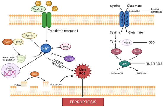
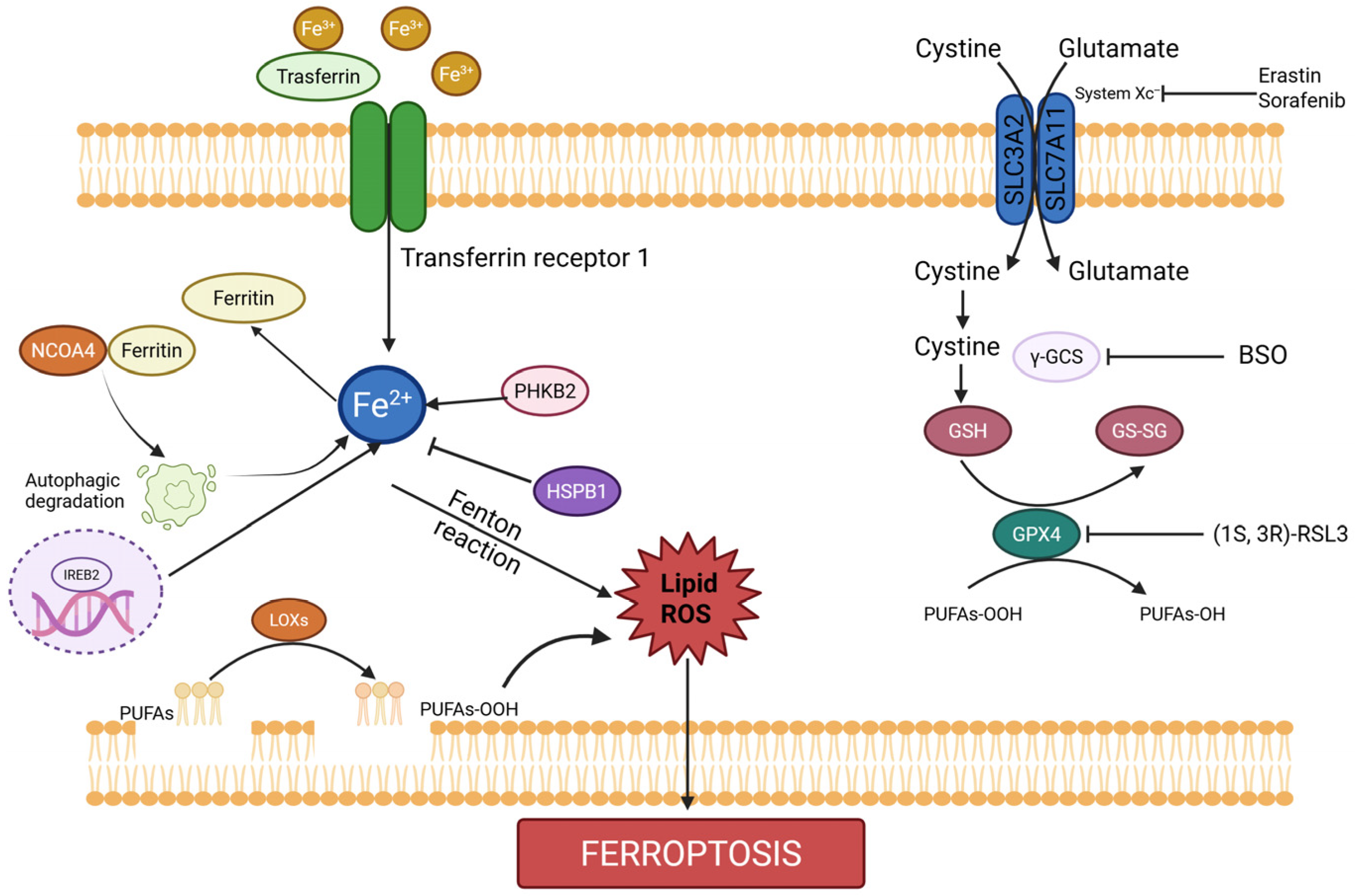
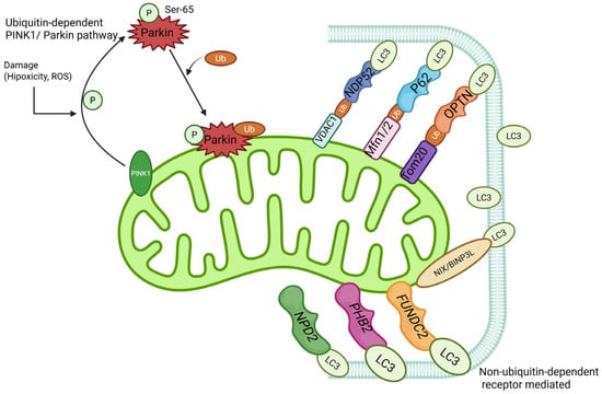
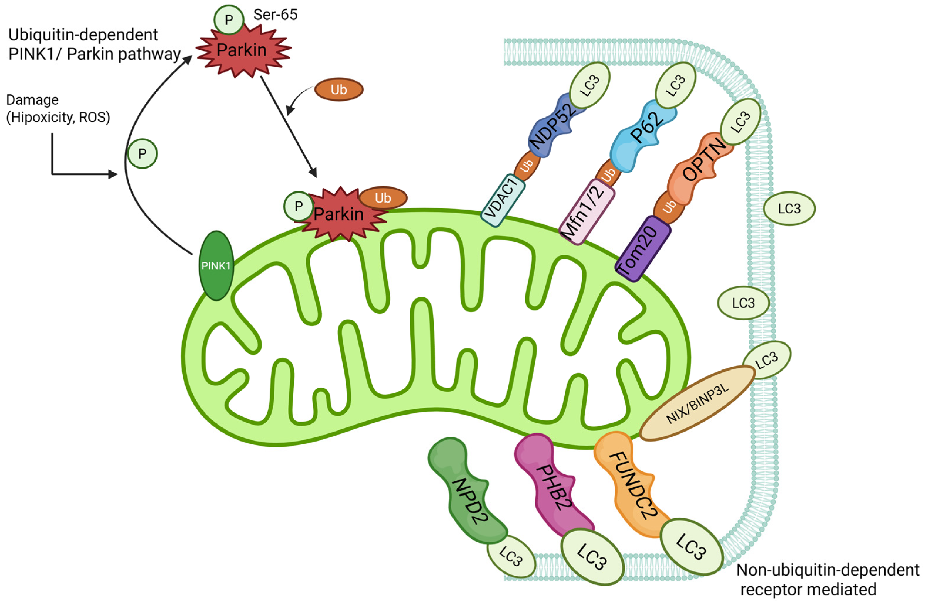
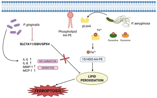
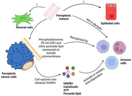
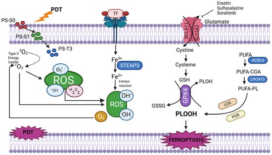
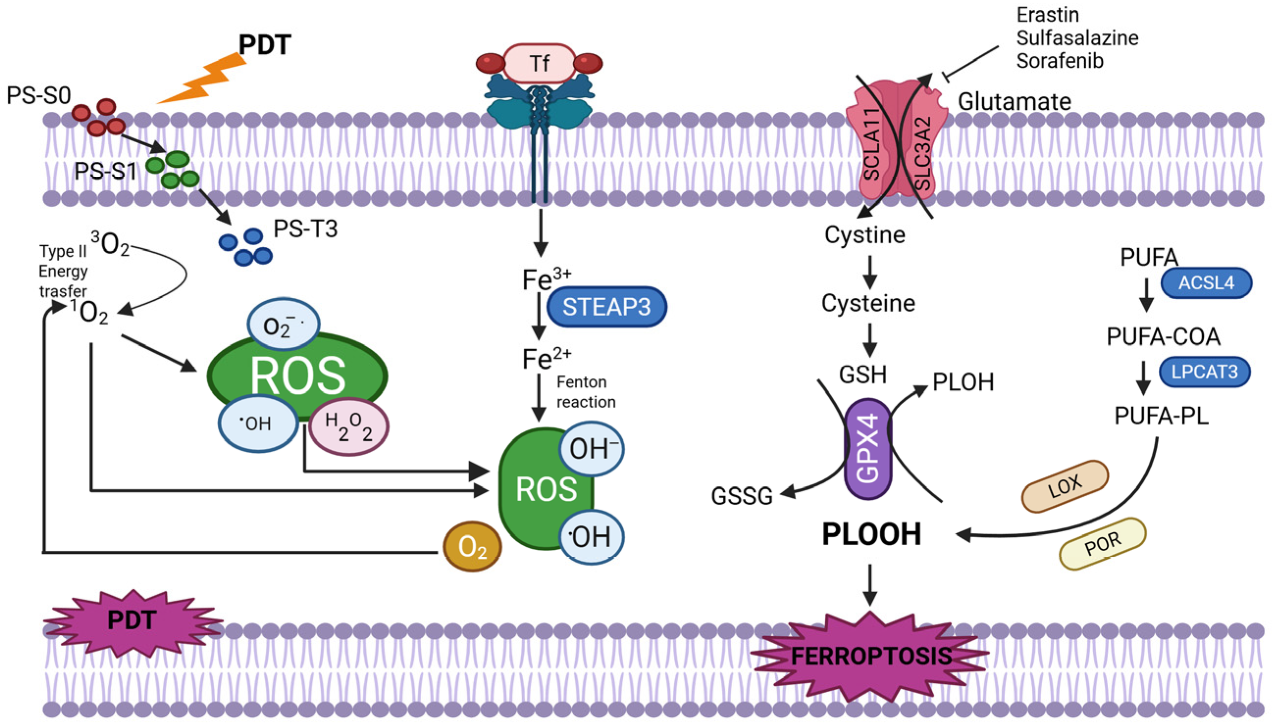

Disclaimer/Publisher’s Note: The statements, opinions and data contained in all publications are solely those of the individual author(s) and contributor(s) and not of MDPI and/or the editor(s). MDPI and/or the editor(s) disclaim responsibility for any injury to people or property resulting from any ideas, methods, instructions or products referred to in the content. |
© 2026 by the authors. Licensee MDPI, Basel, Switzerland. This article is an open access article distributed under the terms and conditions of the Creative Commons Attribution (CC BY) license.