


Abstract
Objectives: Virtual reality (VR) enables controlled, interactive, and ecologically valid visual tasks, offering potential advantages over static clinical tests. This systematic review synthesized current evidence on the use of VR technologies for assessing visual capacities across ocular, neurological, and low-vision populations, with particular attention to technological approaches, clinical domains, methodological quality, safety, and future translational needs. Methods: Following PRISMA 2020, PubMed, Scopus, and Web of Science were searched from inception to 2 August 2025, using database-specific strategies for virtual reality and visual function. Three reviewers independently screened reports, extracted data, and appraised completed studies using design-appropriate NIH Study Quality Assessment Tools or Cochrane Risk of Bias 2 Owing to methodological heterogeneity, findings were synthesized narratively. The review was not registered and no protocol was prepared. Results: Of 364 records, 36 completed studies met the eligibility criteria and were included in the narrative synthesis (publication years 2014–2024); one additional randomized-trial protocol was catalogued separately as ongoing evidence. Most completed studies employed immersive head-mounted displays, while others used semi-immersive or desktop VR. Assessed domains included visual fields, visual acuity, contrast sensitivity, oculomotor and vergence function, postural control, functional navigation, spatial orientation, and obstacle avoidance. The most mature evidence concerned VR-based perimetry and functional visual assessment in glaucoma and low-vision populations. Reported adverse events were uncommon and mild, mainly transient cybersickness, dizziness, or visual discomfort. Among completed studies, methodological appraisal was favorable for 10, intermediate for 25, and unfavorable for 1. Overall, evidence supports feasibility, user acceptability, and convergent validity versus selected conventional tools, but substantial heterogeneity remains in hardware, calibration, exposure duration, outcome metrics, and follow-up. Conclusions: This systematic review highlights the growing role of VR in assessment, showing potential for controlled, ecologically oriented evaluations across domains like perimetry and functional vision. While promising, VR is currently a complementary framework rather than a replacement for established clinical tools, requiring stronger validation, repeatability testing, and calibration standardization for routine implementation.
1. Introduction
Virtual reality (VR) refers to the simulation of real or imagined environments through computer-generated sensory experiences. Users can interact with a three-dimensional world replicating real-life or imaginary scenarios. VR technology has rapidly evolved from an experimental setting to a promising tool with broad clinical applications. Over the past three decades, technological advances in VR devices, including improvements in display resolution, motion tracking, refresh rate, portability, and computing power, have opened new possibilities for its use in healthcare settings.
VR systems can be classified into immersive (using head-mounted displays and com-plete user engagement) [1,2,3], semi-immersive (such as large-screen projections), and non-immersive forms (desktop VR) [1,2]. Each modality offers distinct advantages de-pending on the clinical objective and the patient population [4]. Immersive head-mounted displays may be particularly suitable for visual assessment because they allow the exam-iner to control binocular presentation, stimulus location, background luminance, viewing distance, head movement, and interaction with dynamic environments.
The inclusion of VR into clinical practice has been motivated by its unique capability to offer controlled, reproducible, and ecologically valid environments for evaluation and intervention. VR allows clinicians to expose patients to complex stimuli in a safe, stand-ardized, and interactive manner, facilitating both diagnosis and rehabilitation. Its appli-cations span several medical disciplines: in neurorehabilitation to promote motor recovery after stroke [5], in psychiatry for exposure therapy in anxiety and phobic disorders [6], in anesthesiology and oncology for pain distraction during procedures [7], and in surgical training for skill acquisition without risks for patients [8]. Additional emerging applica-tions include cardiology, where VR-based rehabilitation protocols aim to improve out-comes after myocardial infarction, and geriatrics, where VR tools are utilized for assessing balance and fall risk.
Nevertheless, the integration of VR into clinical workflows faces several unresolved challenges. There is a lack of standardization regarding protocols, duration, and outcome measures. Concerns such as cybersickness, a form of motion sickness induced by exposure to virtual environments, pose limitations for specific patient groups. Moreover, questions remain about the generalizability and transferability of VR-derived skills or assessments to real-world conditions [9]. These challenges are particularly relevant when VR is used not only as an engaging interface, but as a clinical measurement instrument that should pro-vide accurate, repeatable, and interpretable outcomes.
Recently, many studies have shown the broader clinical, accessibility, and multisen-sory applications of virtual reality beyond conventional visual assessment. Together, they highlight VR’s relevance to cognitive care, real-world support for people with visual im-pairment, and emerging taste-generation and biosignal-based sensory-recognition tech-nologies [10,11,12,13].
Within this context, the use of VR for the assessment of visual capacities has attracted increasing interest. Traditional methods for evaluating visual function, including visual acuity charts, perimetry for visual field assessment, and contrast sensitivity tests, are typi-cally performed in static, artificial environments that may not fully replicate the dynamic challenges of real-world vision. VR environments can overcome some of these limitations by offering interactive, three-dimensional tasks that require real-time visual processing, multisensory integration, attentional control, and motor responses.
Potential applications of VR-based visual assessment include the early detection and monitoring of diseases such as glaucoma, age-related macular degeneration, retinitis pig-mentosa, and diabetic retinopathy. This interest also parallels broader developments in portable and eye-tracking visual assessment, where mobile perimetry and VR-based func-tional tasks have shown that visual disability can be investigated beyond conventional clinic-based testing [14,15]. Furthermore, VR can be employed to assess higher-order visual functions such as navigation, spatial orientation, visuomotor coordination, and attention in patients with low vision or neurological impairments. Despite this potential, the field is still emerging, and critical issues related to validity, reliability, reproducibility, calibration, patient tolerability, and clinical utility of VR-based visual assessments must be systematically addressed.
This systematic review aims to evaluate the current state of the art regarding the use of VR technologies for the assessment of visual capacities. We summarize the types of visual functions investigated, the technological characteristics of VR systems employed, the quality of the existing studies, the main clinical and methodological limitations identified, and the future research priorities required to support translation into clinical and tele-ophthalmology settings.
2. Materials and Methods
2.1. Search Strategy and Information Sources
This systematic review was conducted and reported in accordance with the Preferred Reporting Items for Systematic Reviews and Meta-Analyses (PRISMA) 2020 statement [16]. PubMed, Scopus, and the Web of Science Core Collection were searched from database inception to 2 August 2025. No study registers, websites, organizations, reference-list searches, or other sources were used to identify eligible reports. The search was limited to publications in English, with no publication-date restriction.
The database-specific strategies were as follows: PubMed: ((“virtual reality”[All Fields] OR “VR”[All Fields]) AND (“visual capacity”[All Fields] OR “visual function”[All Fields] OR “visual ability”[All Fields] OR “visual performance”[All Fields])); Scopus: TITLE-ABS-KEY((“virtual reality” OR “VR”) AND (“visual capacity” OR “visual function” OR “visual ability” OR “visual performance”)); and Web of Science: TS = ((“virtual reality” OR “VR”) AND (“visual capacity” OR “visual function” OR “visual ability” OR “visual performance”)). The English-language limit was applied in each database. Full search strategies and database-specific record counts are provided in Supplementary Table S1.
Publications appearing after 2 August 2025, were not eligible for inclusion. Publications after the search date that are cited in the Discussion were used only as contextual background on reporting standards, recent reviews, home monitoring, or technical requirements and were not added to the evidence synthesis.
2.2. Eligibility Criteria
Eligible reports were peer-reviewed original studies involving human participants that used an immersive, semi-immersive, or non-immersive VR platform, including closely related head-mounted augmented- or mixed-reality systems, and assessed at least one visual capacity or visual-function outcome. Eligible outcomes included visual field, visual acuity, contrast sensitivity, oculomotor or binocular function, accommodation or vergence, visual search, navigation, spatial orientation, obstacle avoidance, postural control, visuomotor performance, and validated functional-vision measures. Studies using VR as an assessment platform were eligible, as were controlled VR exposure or intervention studies when they reported objective or validated visual-function outcomes relevant to feasibility, responsiveness, tolerability, or measurement performance. Observational, validation, pilot, longitudinal, randomized, and non-randomized experimental designs were considered without restrictions on age, sex, clinical condition, or care setting.
Reports were excluded if they did not assess a relevant visual outcome, did not involve VR or a closely related platform, were engineering-only studies without human visual or clinical data, case reports, editorials, reviews, conference abstracts or posters without a full original article, duplicate reports, non-English publications, or reports for which the full text could not be retrieved. Protocols without empirical outcome data were not eligible for the outcome synthesis; one crossover randomized-trial protocol identified by the search was catalogued separately to document ongoing evidence. For narrative synthesis, completed studies were grouped into five partially overlapping application areas: visual-field assessment; functional vision and mobility; oculomotor, binocular, vergence, accommodation, and visuomotor outcomes; visual acuity, contrast sensitivity, reading, and visual search; and postural control or balance.
2.3. Study Selection and Data Collection
Three reviewers (J.D., P.O., and A.C.) independently screened titles and abstracts and subsequently assessed retrieved full-text reports. The same reviewers independently extracted data using a standardized spreadsheet. Disagreements at any stage were resolved through discussion and consensus. No automation tools were used for screening or extraction, and study investigators were not contacted for additional information.
The extracted variables were study design, setting, population, sample size, age, sex, clinical condition, VR device or platform, immersion level, task characteristics, visual domain, comparator or gold-standard test when available, exposure duration, adverse events, follow-up, covariates or other relevant factors, and main outcomes. All visual outcomes and relevant time points reported within the predefined domains were considered. When several time points or analyses were available, the primary analysis and/or longest follow-up result was prioritized in Supplementary Table S2 while clinically informative intermediate findings were retained in the narrative synthesis. Missing or unclear information was recorded as not reported or not available; no values were imputed. The extraction fields and handling rules are provided in Supplementary Table S3.
2.4. Methodological Quality and Risk-of-Bias Assessment
The same three reviewers independently appraised each completed study, with disagreements resolved by consensus. Design-appropriate NIH Study Quality Assessment Tools were used for completed non-randomized studies, and Cochrane Risk of Bias 2 (RoB 2) was used for completed randomized controlled trials. The protocol report was not appraised because it contained no outcome data. NIH items were judged as yes, no, cannot determine, not reported, or not applicable, and overall judgments were assigned as Good, Fair, or Poor based on the pattern and importance of methodological limitations rather than treating the item total as a validated numerical scale. RoB 2 domain-level judgments were combined into the standard overall categories of Low risk, Some concerns, or High risk. For the descriptive cross-tool summary, NIH Good and RoB 2 Low risk were classified as favorable; NIH Fair and RoB 2 Some concerns as intermediate, and NIH Poor and RoB 2 High risk as unfavorable. No automated appraisal tools were used. Study-level judgments are reported in Supplementary Table S2.
Methodological appraisal was conducted using design-appropriate instruments. Completed non-randomized studies were evaluated using the applicable NIH Study Quality Assessment Tools. The overall judgments of “Good”, “Fair”, or “Poor” were based on a structured qualitative consideration of all relevant criteria, including study population selection, sample size and representativeness, study design, comparator appropriateness, validity and reliability of exposure and outcome measurements, blinding where applicable, follow-up and attrition, control of confounding, and statistical analysis. Item responses were not summed, no numerical quality score was calculated, and no numerical cut-offs were applied.
Completed randomized controlled trials were assessed using the Cochrane Risk of Bias 2 tool, considering bias arising from the randomization process, deviations from intended interventions, missing outcome data, measurement of the outcome, and selection of the reported result. Overall judgments were expressed as “Low risk of bias”, “Some concerns”, or “High risk of bias”. NIH and RoB 2 judgments were not converted into a common quality scale. The criteria and decision principles used for both instruments are summarized in Supplementary Table S4.
2.5. Effect Measures and Synthesis Methods
Because the included studies evaluated different populations, technologies, tasks, comparators, and outcomes, no common effect measure was prespecified. Study-specific findings were extracted and presented as reported by the original authors, including means, medians, proportions, correlations, intraclass correlation coefficients, effect sizes, confidence intervals, and p values when available. No effect measures were converted, and no missing summary statistics were imputed.
A narrative synthesis was conducted. Studies were tabulated in Supplementary Table S2 and synthesized according to participant characteristics, technological features, visual outcomes, exposure duration and follow-up, adverse events, methodological appraisal, and the five application areas defined in Section 2.2. A meta-analysis was not performed because of substantial clinical, technological, and methodological heterogeneity. Consequently, no formal statistical assessment of heterogeneity, subgroup analysis, meta-regression, sensitivity analysis, reporting-bias assessment, or certainty-of-evidence framework was applied.
2.6. Registration and Protocol
This systematic review was not registered, and no review protocol was prepared. Accordingly, there were no amendments to a registration record or protocol.
3. Results
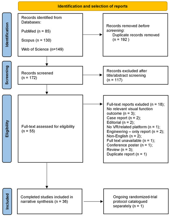
The initial search retrieved 364 records (Figure 1): 85 from PubMed, 130 from Scopus, and 149 from Web of Science. After 192 duplicate records were removed, 172 unique records underwent title and abstract screening. During this phase, 117 records were excluded because they did not involve a relevant VR application or did not address the review objective.
Figure 1.
PRISMA 2020 flow diagram for study identification and selection. Thirty-six completed studies were included in the narrative synthesis, and one ongoing trial protocol was catalogued separately.
Subsequently, 55 full-text reports were assessed. Eighteen reports were excluded: no relevant visual-function outcome (n = 3), case report (n = 2), editorial (n = 2), no VR or closely related platform (n = 1), engineering-focused report without relevant human visual or clinical data (n = 2), non-English publication (n = 2), full text not retrievable (n = 1), conference poster without a full original article (n = 1), review article (n = 3), and duplicate report (n = 1). Thirty-six completed studies met the eligibility criteria and were included in the narrative synthesis. They were published between 2010 and 2024 and involved healthy participants and populations with glaucoma, age-related macular degeneration, retinitis pigmentosa, diabetic retinopathy, low vision, binocular disorders, and visual impairment associated with neurological conditions. One additional protocol for a crossover randomized controlled trial was catalogued separately as ongoing evidence and did not contribute to the outcome or methodological-appraisal syntheses.
The 36 completed studies encompassed cross-sectional, prospective, validation, pilot, longitudinal, and experimental designs; two were completed randomized controlled trials. No case–control studies were identified. The separately catalogued report described the protocol for a randomized crossover-controlled trial and contained no empirical results.
3.1. Participants’ Characteristics
Participants’ mean age ranged from approximately 25 to 74 years across completed studies [17,18], and most samples comprised middle-aged or older adults. Most investigations were small or medium-sized and, frequently, explorative. Sex distribution varied, and many studies included both male and female participants. Study populations included ocular diseases, inherited retinal degeneration, low vision, binocular or accommodative disorders, neurological conditions, and healthy controls. Detailed characteristics and methodological appraisals are provided in Supplementary Table S2.
3.2. Technological Characteristics
The VR systems employed across completed studies varied considerably in hardware and software. Most investigations used immersive head-mounted displays, including Oc-ulus Rift, HTC Vive, smartphone-based headsets, and custom-developed devices, whereas a smaller group used projection-based, augmented-reality, or desktop environments. Several studies reported calibration procedures intended to support accurate stimulus presentation, particularly for visual-field, acuity, and contrast testing [17,19,20,21,22]. However, technological reporting was inconsistent. Luminance range, contrast calibration, field of view, refresh rate, eye-tracking availability, interpupillary-distance adjustment, fixation monitoring, and software version were not uniformly described, limiting comparability and reproducibility.
Studies rated Fair were commonly characterized by one or more limitations, including small or convenience samples, single-centre recruitment, cross-sectional or uncontrolled designs, absence of an appropriate comparator, limited blinding, incomplete control of confounding, and short or absent follow-up. Small sample size was not treated as an automatic reason for a Poor rating but was considered together with the pattern and methodological importance of the other limitations. The criteria supporting the NIH and RoB 2 judgments are summarized in Supplementary Table S4.
3.3. Outcome Measures
Visual-field assessment was the most clinically developed measurement area. Direct headset-based perimetry or VR studies reporting visual-field outcomes included clustered home perimetry, manual or gaze-based field testing, oculokinetic perimetry, and visual training studies that monitored field indices [17,19,20,23]. Conventional visual-field indices were also used as comparators, predictors, or covariates in studies of wayfinding, postural control, functional disability, and activity limitation [15,24,25,26].
Visual acuity, contrast sensitivity, and broader low-vision performance were evaluated through magnification tasks, simulated daily activities, mobility courses, psychophysical tests, and optokinetic-nystagmus-based paradigms [18,21,22,23,27,28,29]. These studies used heterogeneous metrics and test conditions, so direct quantitative comparison was not appropriate.
Binocular function, accommodation, vergence, and eye-movement performance were examined in studies of accommodative training, convergence insufficiency, binocular imbalance, vergence-accommodation conflict, dichoptic training, and developmental eye movements [30,31,32,33,34,35,36,37,38]. Other studies assessed visuospatial motion perception or visual-evoked responses in neurological populations [39,40].
Functional vision, navigation, spatial orientation, visual search, obstacle avoidance, and visually guided motor performance were investigated in glaucoma, inherited retinal degeneration, cerebral or ocular visual impairment, and ultra-low-vision populations [15,24,25,26,28,29,41,42,43,44,45,46]. Outcomes included completion time, collisions, path efficiency, task accuracy, gaze or search behavior, and postural stability.
Several completed studies combined more than one visual domain in tasks requiring visual, attentional, postural, or motor responses [15,41,42,44,45,47]. The evidence databases also included a subset of VR exposure or intervention studies that reported objective visual-function outcomes [23,30,31,32,33,36,48]. These studies were treated as supportive evidence on responsiveness, feasibility, or tolerability rather than as direct evidence of diagnostic accuracy or agreement with clinical gold standards.
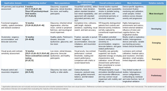
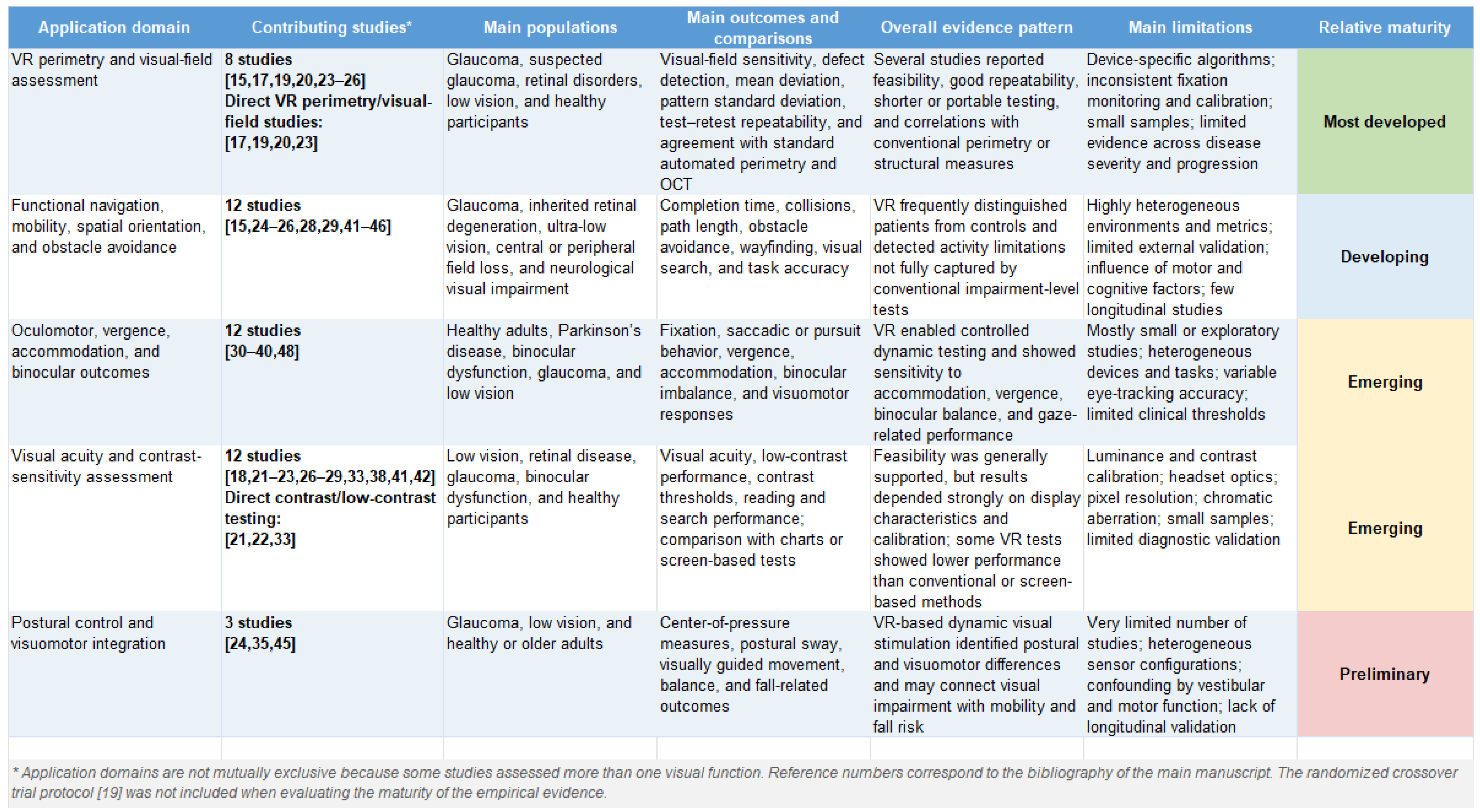
All the aforementioned information is summarized in Figure 2.
Figure 2.
Summary of the principal application domains of VR-based visual assessment.
3.4. VR Exposure Duration and Follow-Up
Exposure schedules ranged from brief single-session tasks to repeated home- or clinic-based programs delivered over several weeks. Most assessment and validation studies reported immediate or short-term outcomes, whereas intervention studies included programs lasting approximately 2 weeks to 3 months [23,30,31,32,33,36]. One postoperative randomized trial reported assessments up to 6 months [48]. Follow-up beyond 6 months was not reported in the completed studies, limiting conclusions about long-term responsiveness and safety.
3.5. Adverse Events and Safety
Safety reporting was inconsistent and was often secondary to the main study objective. Where symptoms were reported, they were generally mild and transient, including cybersickness, dizziness, visual fatigue, eyestrain, or discomfort [18,27,38,49,50,51]. No serious VR-related adverse events were described in the completed studies. However, many reports did not use a validated questionnaire or provide symptom timing, severity, recovery time, withdrawals attributable to symptoms, or susceptibility factors; absence of reporting should therefore not be interpreted as evidence that no adverse events occurred. Future studies should use standardized safety and usability outcomes [52,53].
3.6. Methodological Appraisal
Methodological appraisal varied across the 36 completed studies. Ten studies received a favorable overall judgment (NIH Good or RoB 2 Low risk), 25 received an intermediate judgment (NIH Fair or RoB 2 Some concerns), and one received an unfavorable judgment (NIH Poor). The trial protocol was not appraised because it reported no empirical outcomes. Study-level judgments and the tool applied to each report are provided in Supplementary Table S2.
Common strengths included clear description of VR protocols, standardized outcome measures, and use of objective visual assessments. Frequent limitations included small sample sizes, lack of blinding in experimental designs, single-center recruitment, heterogeneous comparators, incomplete technological reporting, and limited follow-up for assessing long-term outcomes or sensitivity to change.
3.7. Integrative Synthesis of Application Areas
The reports included did not address a single homogeneous technology or clinical problem. The synthesis therefore distinguishes direct assessment or validation studies from supportive exposure and intervention studies and organizes the completed evidence into five partially overlapping application areas. Conclusions about measurement validity are based primarily on direct assessment and validation studies; intervention studies inform responsiveness, feasibility, and safety but do not establish diagnostic equivalence. The first and most clinically developed area is VR-based visual-field assessment. This reflects the central role of perimetry in glaucoma and neuro-ophthalmology, but also the practical limitations of conventional perimeters: fixed equipment, standardized positioning, long testing time, fatigue, and dependence on trained personnel. Recent reviews and validation studies confirm that headset-based perimetry is now being compared directly with standard automated perimetry, although the strength of the evidence still varies substantially across devices, disease stages, and testing strategies [54,55].
A second group of studies examined functional vision, mobility, wayfinding, spatial orientation, and obstacle avoidance. These studies are particularly relevant because they address the gap between impairment-level measurements and daily life functioning. VR allows investigators to reproduce visually demanding situations, such as moving through a cluttered environment, detecting obstacles, orienting in unfamiliar space, or coordinating visual input with head and body movement. This domain is especially informative in glaucoma, inherited retinal degeneration, low vision, and neurological visual impairment, where conventional indices do not always explain patient-reported disability or activity restriction [15,25,29,41,42,45].
A third application area concerns oculomotor, vergence, accommodation, binocular, and visuomotor outcomes. In these studies, VR acts not only as a stimulus presentation system but also as an experimental environment in which dynamic visual behavior can be observed. The integration of eye tracking into HMDs could make this area more clinically informative by allowing fixation stability, saccades, smooth pursuit, gaze allocation, and visual search strategies to be quantified during standardized tasks [26,35,37,56].
The fourth area includes VR-based assessment of visual acuity, contrast sensitivity, metamorphopsia, reading, and search performance. These applications are attractive because they may be scalable and comparatively inexpensive, particularly when implemented through portable or smartphone-based systems. At the same time, they require careful validation: small differences in luminance, contrast rendering, headset optics, and display calibration may substantially affect psychophysical measurements [18,20,21,22,28,57].
Finally, several studies investigated postural control, balance, and visuomotor integration. This is a relevant extension of visual assessment because visual impairment contributes to mobility limitation and fall risk, especially in older adults. When combined with motion tracking, force platforms, or wearable sensors, VR may therefore connect ophthalmological assessment with broader functional evaluation [17,24,50].
4. Discussion
This systematic review demonstrates that the use of VR for assessing visual capacities is a rapidly evolving field spanning psychophysical measurement, behavioral assessment, visuomotor performance, functional mobility, and remote monitoring. The most directly relevant evidence comes from studies that used VR as an assessment or validation platform. A smaller group of exposure and intervention studies was retained because objective visual outcomes provided supportive information on responsiveness, feasibility, and tolerability; these studies should not be interpreted as validating VR for diagnosis. Immersive head-mounted displays predominate and offer potential advantages in ecological validity, stimulus control, portability, and patient engagement compared with traditional static testing environments.
4.1. From Conventional Visual Testing to Ecological Visual Function
The main added value of VR is not simply that conventional tests can be reproduced inside a headset. Its more distinctive contribution is the possibility of measuring how vision supports behavior in environments that are controlled, repeatable, and closer to real life than a chart or perimeter bowl. Standard visual acuity, contrast sensitivity, and automated perimetry remain indispensable because they are standardized and clinically interpretable. However, they are usually administered under static conditions and capture only selected components of visual function.
Everyday visual performance is more complex. Patients must combine peripheral and central vision with head movements, spatial memory, attention, balance, and motor planning. The studies included in this review show that VR can reproduce some of these demands under safe and standardized conditions. For example, VR tasks can quantify reaction time, visual search efficiency, collisions, path deviation, gaze behavior, and postural sway while the participant interacts with a visual scene [15,24,25,29,41,42,45].
For this reason, VR should currently be viewed as a complementary layer of assessment rather than a replacement for clinical gold standards. It may be most useful when conventional measures do not fully explain the patient’s reported disability, reduced mobility, or difficulty with daily activities. The strongest future applications will probably combine impairment-level outcomes, behavioral performance, and patient-reported measures instead of relying on any single VR-derived metric.
4.2. Clinical Validation and Comparison with Gold Standards
The most advanced evidence concerns VR-based perimetry. Several included studies reported encouraging reproducibility or agreement between VR-based visual field testing and standard automated perimetry. Greenfield et al. [20] found reproducibility and correlations between VR-based oculokinetic perimetry, conventional perimetry, and OCT-derived measures. Chia et al. [17] showed the feasibility of remote training and at-home testing with a clustered VR perimetry test. Daga et al. [15] and Lam et al. [25] further suggest that VR environments can reveal disability and navigation-related functional deficits in glaucoma that are not fully described by conventional perimetric indices.
Recent systematic reviews focused on VR perimetry support this cautious interpretation: the field is promising, but the evidence is not yet uniform across devices, patient groups, and clinical scenarios [54,58,59]. In particular, stronger data are needed on test–retest repeatability, diagnostic accuracy, agreement across the full spectrum of disease severity, and performance in both early and advanced defects. Device characteristics such as luminance range, dynamic contrast, fixation monitoring, field of view, and eye-tracking capability are not secondary technical details; they can directly influence whether a VR test behaves like a clinical measurement instrument [54,58,59,60].
The clinical role of VR-based visual assessment should therefore be specified carefully. In some contexts, including home monitoring, screening, low-resource settings, and patients who cannot easily access standard perimetry, VR may offer practical advantages. For diagnosis, treatment decisions, and progression monitoring, however, validation against established tools remains necessary. Future papers should distinguish clearly between feasibility, agreement, diagnostic accuracy, sensitivity to progression, and clinical utility, because these forms of evidence answer different questions.
4.3. Technological and Methodological Requirements for Clinical Translation
A recurrent limitation of the current literature is incomplete reporting of the technical conditions under which VR measurements were obtained. Visual testing in VR depends on display resolution, field of view, refresh rate, luminance range, contrast rendering, interpupillary distance adjustment, optical distortion correction, latency, background illumination, eye tracking, and response modality. If these features are omitted or described only generically, it becomes difficult to determine whether differences between studies reflect patient characteristics, task design, hardware limitations, software algorithms, or calibration procedures.
Minimum reporting requirements are therefore needed. Studies should specify the device model, display characteristics, software version, calibration procedure, test environment, stimulus properties, adaptation time, task duration, reliability criteria, and handling of invalid trials. For VR perimetry, additional details should include test strategy, stimulus size, intensity range, fixation monitoring, false-positive and false-negative responses, blind spot checks, and comparability with conventional indices such as mean deviation and pattern standard deviation. Broader work on eye tracking in VR and extended-reality visual performance standards supports the need to report accuracy, precision, latency, optical characteristics, and device-specific limitations [56,60].
Further methodological tension concerns ecological validity. A realistic VR task may better reflect daily life, but realism can introduce variability related to attention, cognition, motor ability, prior gaming or VR experience, and vestibular sensitivity. Conversely, a simplified VR task is easier to standardize but may offer little additional value over conventional testing. For this reason, each study should state whether the VR protocol is intended to reproduce an established clinical measure, complement it with functional information, or create a new digital biomarker.
4.4. Safety, Tolerability, and User Experience
Overall, the safety profile reported in the included studies was favorable. Adverse events were uncommon, mild, and transient, most often consisting of dizziness, cybersickness, or visual discomfort [25,27,40]. This is reassuring, but it should not lead to underreporting. Tolerability is a condition for clinical implementation, especially in older adults and in patients with vestibular, neurological, cognitive, or severe visual impairment.
Safety and acceptability should be assessed with standardized instruments before, during, and after exposure. The Simulator Sickness Questionnaire remains one of the most widely used scales for nausea, oculomotor discomfort, and disorientation [53], while the recent literature on cybersickness shows that symptoms depend on user susceptibility, exposure duration, visual flow, latency, and task design [52]. Ophthalmological applications should also consider eye strain, accommodation-vergence conflict, dry eye symptoms, headset weight, mask pressure, hygiene between users, and whether patients can understand and complete the task without continuous assistance.
4.5. Remote Monitoring, Tele-Ophthalmology, and Decentralized Care
Remote assessment is one of the most plausible translational uses of VR in ophthalmology. Portable HMDs could reduce dependence on fixed clinic-based equipment and allow repeated testing over time. This is relevant for chronic diseases such as glaucoma, diabetic retinopathy, inherited retinal degeneration, and age-related macular degeneration, where monitoring is longitudinal and clinic visits may be burdensome.
The included study by Chia et al. [17] supports the feasibility of remote training and at-home VR perimetry, while external evidence on home monitoring of glaucoma with virtual visual field devices shows that acceptability, retention, and compliance are now being evaluated over longer periods [61,62]. However, home-based implementation introduces issues that are less visible in supervised laboratory studies: data quality, patient adherence, device sharing, internet connectivity, cybersecurity, integration with electronic health records, automated alerts, responsibility for abnormal results, and reimbursement.
Low-cost and scalable tools could also support decentralized clinical trials or population-level screening, provided that the intended use is clearly defined. Recent smartphone-based VR visual function testing illustrates this direction for contrast sensitivity, metamorphopsia, and reading speed assessment, but such tools should not be treated as diagnostic instruments until adequate validation has been completed [57].
4.6. Future Research Agenda
The next phase of research should move beyond demonstrating that VR assessment is technically possible. Adequately powered validation studies are needed to define diagnostic accuracy, reproducibility, sensitivity to change, and clinically meaningful thresholds. Head-to-head comparisons with standard automated perimetry, validated contrast sensitivity tests, OCT, and conventional mobility assessments would help clarify whether VR provides equivalent, complementary, or genuinely additional information.
Standardization is equally important. Future protocols should report device model, display resolution, field of view, refresh rate, luminance and contrast calibration, eye-tracking availability, room conditions, task duration, adaptation time, and validity criteria. Shared protocols and normative datasets would make it easier to compare results across age groups, disease stages, and clinical populations.
Longitudinal evidence remains limited. Studies should test whether VR-derived measures can detect disease progression or treatment-related change over time in glaucoma, age-related macular degeneration, diabetic retinopathy, retinitis pigmentosa, and neuro-ophthalmological disorders. If these measures prove sensitive to clinically relevant change, they could become useful endpoints for rehabilitation studies, monitoring programs, and therapeutic trials.
Home-based assessment also requires dedicated research rather than simple extrapolation from clinic-based feasibility studies. Usability, safety, data quality, adherence, cybersecurity, and integration into care pathways should be evaluated in children, older adults, and patients with cognitive, vestibular, motor, or severe visual limitations.
Finally, VR assessment is likely to become more informative when combined with eye tracking, artificial intelligence, and multimodal digital biomarkers. Gaze allocation, fixation instability, response latency, navigation trajectories, and interaction metrics may reveal patterns that conventional tests do not capture. These approaches will be clinically useful only if algorithms are transparent, externally validated, and presented in a form that clinicians can interpret.
4.7. Strengths and Limitations of This Review
This systematic review presents several strengths. First, it provides an updated and comprehensive overview of the applications of VR in visual capacity assessment, covering a wide range of visual functions, populations, and technological approaches. The use of three major databases (PubMed, Scopus, Web of Science) and adherence to PRISMA 2020 guidelines ensure methodological rigor. Moreover, quality assessment was systematically applied, allowing for critical evaluation of the studies included.
Nevertheless, several limitations must be acknowledged. The English-language restriction, exclusion of conference abstracts and non-peer-reviewed material, and lack of supplementary citation searching may have resulted in missed evidence. The search strategy used broad visual-function terms but did not include every disease-specific or device-specific synonym. The review was not prospectively registered and no protocol was prepared. Substantial heterogeneity in study designs, populations, outcomes, and VR technologies precluded meta-analysis and formal assessment of statistical heterogeneity, reporting bias, and certainty of evidence. Although appraisal was performed independently with standardized tools, overall methodological judgments retain an element of reviewer interpretation. Finally, many completed studies were early-phase feasibility or single-center investigations, which limits generalizability and conclusions about routine clinical implementation.
5. Conclusions
This systematic review highlights the growing role of VR in the assessment of visual capacities. The available evidence shows that VR can support controlled, interactive, and ecologically oriented evaluations across several domains, including visual field testing, contrast sensitivity, visual acuity, oculomotor function, functional navigation, and visuo-motor integration. While the most mature evidence concerns VR-based perimetry and functional assessment in glaucoma, studies in low vision, inherited retinal disorders, binocular dysfunction, and neurological conditions indicate a much broader potential. Overall, VR appears promising and feasible for clinical applications; however, its translation into routine practice still requires stronger validation, rigorous repeatability testing, calibration standardization, and direct comparisons with gold-standard assessments. At present, VR should be considered a strictly complementary assessment framework rather than a substitute or replacement for established standard clinical tools. Its main promise lies in its unique ability to combine standardized stimulus control with tasks that better approximate real-world visual demands. Ultimately, successful clinical implementation will depend on overcoming these methodological limitations through clearer reporting of technical parameters, the development of standardized protocols, and the collection of robust evidence. If these requirements are rigorously addressed, VR may well become a valuable and transformative component of clinical visual assessment, rehabilitation research, and tele-ophthalmology-based monitoring.
Supplementary Materials
The following supporting information can be downloaded at: https://www.mdpi.com/article/10.3390/app16157788/s1. Table S1, database-specific search strategies; Table S2, characteristics and methodological appraisal of the 36 included studies and the separately catalogued ongoing protocol; Table S3, data-extraction fields and handling rules; and Table S4 contain the criteria supporting the NIH and RoB 2 judgments. The completed PRISMA 2020 checklist is provided separately to the Editorial Office.
Funding
This research received no external funding. No funder or sponsor had any role in the design, conduct, interpretation, or reporting of the review.
Institutional Review Board Statement
Not applicable. This systematic review used data from previously published studies and involved no new collection of data from human participants.
Informed Consent Statement
Not applicable.
Data Availability Statement
All data extracted from the included reports and all study-level methodological appraisals are provided in Supplementary Table S2. The complete database-specific search strategies and the data-extraction framework are provided in Supplementary Tables S2 and S3. No analytic code was generated because no meta-analysis was performed. Additional screening and extraction materials are available from the corresponding authors on reasonable request.
Conflicts of Interest
Author Paolo Olivetti was employed by the company Tech4Care srl. The remaining authors declare that the research was conducted in the absence of any commercial or financial relationships that could be construed as a potential conflict of interest.
Abbreviations
The following abbreviations are used in this manuscript:
| HMD | Head Mounted Display |
| VR | Virtual Reality |
References
- Radianti, J.; Majchrzak, T.A.; Fromm, J.; Wohlgenannt, I. A Systematic Review of Immersive Virtual Reality Applications for Higher Education: Design Elements, Lessons Learned, and Research Agenda. Comput. Educ. 2020, 147, 103778. [Google Scholar] [CrossRef]
- Slater, M.; Sanchez-Vives, M.V. Enhancing Our Lives with Immersive Virtual Reality. Front. Robot. AI 2016, 3, 74. [Google Scholar] [CrossRef]
- Foerster, R.M.; Poth, C.H.; Behler, C.; Botsch, M.; Schneider, W.X. Using the Virtual Reality Device Oculus Rift for Neuropsychological Assessment of Visual Processing Capabilities. Sci. Rep. 2016, 6, 37016. [Google Scholar] [CrossRef] [PubMed]
- Freeman, D.; Reeve, S.; Robinson, A.; Ehlers, A.; Clark, D.; Spanlang, B.; Slater, M. Virtual Reality in the Assessment, Understanding, and Treatment of Mental Health Disorders. Psychol. Med. 2017, 47, 2393–2400. [Google Scholar] [CrossRef] [PubMed]
- Laver, K.E.; Lange, B.; George, S.; Deutsch, J.E.; Saposnik, G.; Crotty, M. Virtual Reality for Stroke Rehabilitation. Cochrane Database Syst. Rev. 2017. [Google Scholar] [CrossRef] [PubMed]
- Carl, E.; Stein, A.T.; Levihn-Coon, A.; Pogue, J.R.; Rothbaum, B.; Emmelkamp, P.; Asmundson, G.J.G.; Carlbring, P.; Powers, M.B. Virtual Reality Exposure Therapy for Anxiety and Related Disorders: A Meta-Analysis of Randomized Controlled Trials. J. Anxiety Disord. 2019, 61, 27–36. [Google Scholar] [CrossRef] [PubMed]
- Malloy, K.M.; Milling, L.S. The Effectiveness of Virtual Reality Distraction for Pain Reduction: A Systematic Review. Clin. Psychol. Rev. 2010, 30, 1011–1018. [Google Scholar] [CrossRef] [PubMed]
- Palter, V.N.; Grantcharov, T.P. Simulation in Surgical Education. Can. Med. Assoc. J. 2010, 182, 1191–1196. [Google Scholar] [CrossRef] [PubMed]
- Howard, M.C. A Meta-Analysis and Systematic Literature Review of Virtual Reality Rehabilitation Programs. Comput. Hum. Behav. 2017, 70, 317–327. [Google Scholar] [CrossRef]
- Qin, R.; Li, Z.; Huang, K.; Luan, T.; Miao, B.; Gong, L.; Liu, W.; Chen, L. Comparative Efficacy of Various of Virtual Reality Technologies on Cognitive Function in Older Adults with Mild Cognitive Impairment: A Systematic Review and Network Meta-Analysis. J. Am. Med. Dir. Assoc. 2025, 26, 105941. [Google Scholar] [CrossRef] [PubMed]
- Qiao, G.; Shao, Z.; Gu, J.; Chen, X. Accessible Tourism for Tourists with Visual Impairment: The Role of Auditory Perception in Shaping Behavioral Attitudes. Tour. Rev. 2026, 1–24. [Google Scholar] [CrossRef]
- Ullah, A.; Liu, Y.; Wang, Y.; Gao, H.; Wang, H.; Zhang, J.; Li, G. E-Taste: Taste Sensations and Flavors Based on Tongue’s Electrical and Thermal Stimulation. Sensors 2022, 22, 4976. [Google Scholar] [CrossRef] [PubMed]
- Ullah, A.; Zhang, F.; Song, Z.; Wang, Y.; Zhao, S.; Riaz, W.; Li, G. Surface Electromyography-Based Recognition of Electronic Taste Sensations. Biosensors 2024, 14, 396. [Google Scholar] [CrossRef] [PubMed]
- Jones, P.R.; Smith, N.D.; Bi, W.; Crabb, D.P. Portable Perimetry Using Eye-Tracking on a Tablet Computer—A Feasibility Assessment. Transl. Vis. Sci. Technol. 2019, 8, 17. [Google Scholar] [CrossRef] [PubMed]
- Daga, F.B.; Macagno, E.; Stevenson, C.; Elhosseiny, A.; Diniz-Filho, A.; Boer, E.R.; Schulze, J.; Medeiros, F.A. Way-finding and Glaucoma: A Virtual Reality Experiment. Investig. Ophthalmol. Vis. Sci. 2017, 58, 3343. [Google Scholar] [CrossRef]
- Page, M.J.; McKenzie, J.E.; Bossuyt, P.M.; Boutron, I.; Hoffmann, T.C.; Mulrow, C.D.; Shamseer, L.; Tetzlaff, J.M.; Akl, E.A.; Brennan, S.E.; et al. The PRISMA 2020 Statement: An Updated Guideline for Reporting Systematic Reviews. Syst. Rev. 2021, 10, 89. [Google Scholar] [CrossRef] [PubMed]
- Chia, Z.K.; Kong, A.W.; Turner, M.L.; Saifee, M.; Damato, B.E.; Backus, B.T.; Blaha, J.J.; Schuman, J.S.; Deiner, M.S.; Ou, Y. Assessment of Remote Training, At-Home Testing, and Test-Retest Variability of a Novel Test for Clustered Virtual Reality Perimetry. Ophthalmol. Glaucoma 2024, 7, 139–147. [Google Scholar] [CrossRef] [PubMed]
- Deemer, A.D.; Swenor, B.K.; Fujiwara, K.; Deremeik, J.T.; Ross, N.C.; Natale, D.M.; Bradley, C.K.; Werblin, F.S.; Massof, R.W. Preliminary Evaluation of Two Digital Image Processing Strategies for Head-Mounted Magnification for Low Vision Patients. Transl. Vis. Sci. Technol. 2019, 8, 23. [Google Scholar] [CrossRef] [PubMed]
- Wroblewski, D.; Francis, B.A.; Sadun, A.; Vakili, G.; Chopra, V. Testing of Visual Field with Virtual Reality Goggles in Manual and Visual Grasp Modes. BioMed Res. Int. 2014, 2014, 206082. [Google Scholar] [CrossRef] [PubMed]
- Greenfield, J.A.; Deiner, M.; Nguyen, A.; Wollstein, G.; Damato, B.; Backus, B.T.; Wu, M.; Schuman, J.S.; Ou, Y. Virtual Reality Oculokinetic Perimetry Test Reproducibility and Relationship to Conventional Perimetry and OCT. Ophthalmol. Sci. 2022, 2, 100105. [Google Scholar] [CrossRef] [PubMed]
- Vivas-Mateos, G.; Boswell, S.; Livingstone, I.A.T.; Delafield-Butt, J.; Giardini, M.E. Screen and Virtual Reality-Based Testing of Contrast Sensitivity. In Proceedings of the 2020 42nd Annual International Conference of the IEEE Engineering in Medicine & Biology Society (EMBC), Montreal, QC, Canada, 20–24 July 2020; pp. 6054–6057. [Google Scholar]
- Tatiyosyan, S.A.; Rifai, K.; Wahl, S. Standalone Cooperation-Free OKN-Based Low Vision Contrast Sensitivity Estimation in VR—A Pilot Study. Restor. Neurol. Neurosci. 2020, 38, 119–129. [Google Scholar] [CrossRef] [PubMed]
- Li, B.; Chu, H.; Yan, L.; Wiederhold, B.K.; Wiederhold, M.; Lu, Y. Individualized Visual Reality Training Improves Visual Acuity and Visual Field Defects in Patients with Glaucoma: A Preliminary Study Report. Cyberpsychol. Behav. Soc. Netw. 2020, 23, 179–184. [Google Scholar] [CrossRef] [PubMed]
- Diniz-Filho, A.; Boer, E.R.; Gracitelli, C.P.B.; Abe, R.Y.; Van Driel, N.; Yang, Z.; Medeiros, F.A. Evaluation of Postural Control in Patients with Glaucoma Using a Virtual Reality Environment. Ophthalmology 2015, 122, 1131–1138. [Google Scholar] [CrossRef] [PubMed]
- Lam, A.K.N.; To, E.; Weinreb, R.N.; Yu, M.; Mak, H.; Lai, G.; Chiu, V.; Wu, K.; Zhang, X.; Cheng, T.P.H.; et al. Use of Virtual Reality Simulation to Identify Vision-Related Disability in Patients with Glaucoma. JAMA Ophthalmol. 2020, 138, 490. [Google Scholar] [CrossRef] [PubMed]
- Goh, R.L.Z.; Kong, Y.X.G.; McAlinden, C.; Liu, J.; Crowston, J.G.; Skalicky, S.E. Objective Assessment of Activity Limitation in Glaucoma with Smartphone Virtual Reality Goggles: A Pilot Study. Transl. Vis. Sci. Technol. 2018, 7, 10. [Google Scholar] [CrossRef] [PubMed]
- Gopalakrishnan, S.; Jacob, C.E.S.; Kumar, M.; Karunakaran, V.; Raman, R. Comparison of Visual Parameters Between Normal Individuals and People with Low Vision in a Virtual Environment. Cyberpsychol. Behav. Soc. Netw. 2020, 23, 171–178. [Google Scholar] [CrossRef] [PubMed]
- Authié, C.N.; Poujade, M.; Talebi, A.; Defer, A.; Zenouda, A.; Coen, C.; Mohand-Said, S.; Chaumet-Riffaud, P.; Audo, I.; Sahel, J.-A. Development and Validation of a Novel Mobility Test for Rod-Cone Dystrophies: From Reality to Virtual Reality. Am. J. Ophthalmol. 2024, 258, 43–54. [Google Scholar] [CrossRef] [PubMed]
- Gopalakrishnan, S.; Jacob, C.E.S.; Kumar, M.; Karunakaran, V.; Raman, R. Development of Immersive Virtual Reality Environment for Assessment of Functional Vision in People with Low Vision: A Pilot Study. Nepal. J. Ophthalmol. 2022, 14, 19–30. [Google Scholar] [CrossRef] [PubMed]
- Guo, D.-Y.; Zhu, M.-M.; Zhan, Y.-Y.; Shen, Y.-Y.; Wang, X.-W.; Xia, J.-H.; Jiang, B.; Gu, Y.-S.; Long, Y. Virtual Reality Training Improves Accommodative Facility and Accommodative Range. Int. J. Ophthalmol. 2022, 15, 1116–1121. [Google Scholar] [CrossRef] [PubMed]
- Li, S.; Tang, A.; Yang, B.; Wang, J.; Liu, L. Virtual Reality-Based Vision Therapy versus OBVAT in the Treatment of Convergence Insufficiency, Accommodative Dysfunction: A Pilot Randomized Controlled Trial. BMC Ophthalmol. 2022, 22, 182. [Google Scholar] [CrossRef] [PubMed]
- Munsamy, A.J.; Paruk, H.; Gopichunder, B.; Luggya, A.; Majola, T.; Khulu, S. The Effect of Gaming on Accommodative and Vergence Facilities after Exposure to Virtual Reality Head-Mounted Display. J. Optom. 2020, 13, 163–170. [Google Scholar] [CrossRef] [PubMed]
- Long, Y.; Shen, Y.; Guo, D.; Wang, X.; Gu, Y. The Effects of Consumer-Grade Virtual Reality Headsets on Adult Visual Function. Semin. Ophthalmol. 2020, 35, 170–173. [Google Scholar] [CrossRef] [PubMed]
- Xu, L.; Huang, M.; Lan, J.; Huang, W.; Wang, X.; Zhang, G.; Li, X.; Shasha, P.; Chu, H.; Wiederhold, B.K.; et al. Assessment of Binocular Imbalance with an Augmented Virtual Reality Platform in a Normal Population. Cyberpsychol. Behav. Soc. Netw. 2019, 22, 127–131. [Google Scholar] [CrossRef] [PubMed]
- McAnally, K.; Wallis, G.; Grove, P. Visually Guided Movement in Virtual Reality Is Tolerant of the Vergence-Accommodation Conflict. Displays 2024, 82, 102668. [Google Scholar] [CrossRef]
- Li, X.; Yang, C.; Zhang, G.; Zhang, Y.; Lan, J.; Chu, H.; Li, J.; Xie, W.; Wang, S.; Wiederhold, B.K.; et al. Intermittent Exotropia Treatment with Dichoptic Visual Training Using a Unique Virtual Reality Platform. Cyberpsychol. Behav. Soc. Netw. 2019, 22, 22–30. [Google Scholar] [CrossRef] [PubMed]
- Lyu, J.; Ng, C.J.; Bang, S.P.; Yoon, G. Binocular Accommodative Response with Extended Depth of Focus Under Controlled Convergences. J. Vis. 2021, 21, 21. [Google Scholar] [CrossRef] [PubMed]
- Kim, J.-H.; Son, H.-J.; Lee, S.-J.; Yun, D.-Y.; Kwon, S.-C.; Lee, S.-H. Effectiveness of a Virtual Reality Head-Mounted Display System-Based Developmental Eye Movement Test. J. Eye Mov. Res. 2016, 9, 1–14. [Google Scholar] [CrossRef]
- Beylergil, S.B.; Gupta, P.; ElKasaby, M.; Kilbane, C.; Shaikh, A.G. Does Visuospatial Motion Perception Correlate with Coexisting Movement Disorders in Parkinson’s Disease? J. Neurol. 2022, 269, 2179–2192. [Google Scholar] [CrossRef] [PubMed]
- Nakanishi, M.; Miner, A.; Jung, T.-P.; Graves, J. Novel Moving Steady-State Visual Evoked Potential Stimulus to Assess Afferent and Efferent Dysfunction in Multiple Sclerosis. IEEE Trans. Neural Syst. Rehabil. Eng. 2023, 31, 1297–1301. [Google Scholar] [CrossRef] [PubMed]
- Kartha, A.; Sadeghi, R.; Bradley, C.; Tran, C.; Gee, W.; Dagnelie, G. Measuring Visual Information Gathering in Individuals with Ultra Low Vision Using Virtual Reality. Sci. Rep. 2023, 13, 3143. [Google Scholar] [CrossRef] [PubMed]
- Bennett, J.; Aleman, E.M.; Maguire, K.H.; Nadelmann, J.; Weber, M.L.; Maguire, W.M.; Maja, A.; O’Neil, E.C.; Maguire, A.M.; Miller, A.J.; et al. Optimization and Validation of a Virtual Reality Orientation and Mobility Test for Inherited Retinal Degenerations. Transl. Vis. Sci. Technol. 2023, 12, 28. [Google Scholar] [CrossRef] [PubMed]
- Bennett, C.R.; Bailin, E.S.; Gottlieb, T.K.; Bauer, C.M.; Bex, P.J.; Merabet, L.B. Assessing Visual Search Performance in Ocular Compared to Cerebral Visual Impairment Using a Virtual Reality Simulation of Human Dynamic Movement. In Proceedings of the Technology, Mind, and Society, Washington, DC, USA, 5–7 April 2018; ACM: New York, NY, USA, 2018; pp. 1–6. [Google Scholar]
- Knopf, N.A.; Boon, M.-Y.; Suaning, G.J.; Zapf, M.P.H.; Grigg, J. Initial Mobility Behaviors of People with Visual Impairment in a Virtual Environment Using a Mixed Methods Design. In Proceedings of the 2017 IEEE Life Sciences Conference (LSC), Sydney, NSW, Australia, 13–15 December 2017; pp. 153–156. [Google Scholar]
- Kartha, A.; Sadeghi, R.; Bradley, C.; Livingston, B.; Tran, C.; Gee, W.; Dagnelie, G. Measuring Visually Guided Motor Performance in Ultra Low Vision Using Virtual Reality. Front. Neurosci. 2023, 17, 1251935. [Google Scholar] [CrossRef] [PubMed]
- Kartha, A.; Singh, R.K.; Bradley, C.; Dagnelie, G. Self-Reported Visual Ability Versus Task Performance in Individuals with Ultra-Low Vision. Transl. Vis. Sci. Technol. 2023, 12, 14. [Google Scholar] [CrossRef] [PubMed]
- Simon-Martinez, C.; Antoniou, M.-P.; Bouthour, W.; Bavelier, D.; Levi, D.; Backus, B.T.; Dornbos, B.; Blaha, J.J.; Kropp, M.; Müller, H.; et al. Stereoptic Serious Games as a Visual Rehabilitation Tool for Individuals with a Residual Amblyopia (AMBER Trial): A Protocol for a Crossover Randomized Controlled Trial. BMC Ophthalmol. 2023, 23, 220. [Google Scholar] [CrossRef] [PubMed]
- Zhang, H.; Yang, S.-H.; Chen, T.; Kang, M.-X.; Liu, D.-Y.; Wang, D.; Hao, J.; Wang, M.; Yang, Z.; Han, X.; et al. The Effect of Virtual Reality Technology in Children after Surgery for Concomitant Strabismus. Indian J. Ophthalmol. 2023, 71, 625–630. [Google Scholar] [CrossRef] [PubMed]
- Wei, Y.; Zheng, J.; So, R.H.Y. Allocating Less Attention to Central Vision During Vection Is Correlated with Less Motion Sickness. Ergonomics 2018, 61, 933–946. [Google Scholar] [CrossRef] [PubMed]
- Huang, Y.-Y.; Menozzi, M. Effects of Viewing Distance and Age on the Performance and Symptoms in a Visual Search Task in Augmented Reality. Appl. Ergon. 2022, 102, 103746. [Google Scholar] [CrossRef] [PubMed]
- Iskander, J.; Hossny, M. Measuring the Likelihood of VR Visual Fatigue through Ocular Biomechanics. Displays 2021, 70, 102105. [Google Scholar] [CrossRef]
- Simón-Vicente, L.; Rodríguez-Cano, S.; Delgado-Benito, V.; Ausín-Villaverde, V.; Cubo Delgado, E. Cybersickness. A Systematic Literature Review of Adverse Effects Related to Virtual Reality. Neurol. Engl. Ed. 2024, 39, 701–709. [Google Scholar] [CrossRef] [PubMed]
- Kennedy, R.S.; Lane, N.E.; Berbaum, K.S.; Lilienthal, M.G. Simulator Sickness Questionnaire: An Enhanced Method for Quantifying Simulator Sickness. Int. J. Aviat. Psychol. 1993, 3, 203–220. [Google Scholar] [CrossRef] [PubMed]
- Selvan, K.; Mina, M.; Abdelmeguid, H.; Gulsha, M.; Vincent, A.; Sarhan, A. Virtual Reality Headsets for Perimetry Testing: A Systematic Review. Eye 2024, 38, 1041–1064. [Google Scholar] [CrossRef] [PubMed]
- Sponsel, W.E.; Griffin, J.M.; Slagle, G.T.; Vu, T.A.; Eis, A. Prospective Comparison of VisuALL Virtual Reality Perimetry and Humphrey Automated Perimetry in Glaucoma. J. Curr. Glaucoma Pract. 2024, 18, 4–9. [Google Scholar] [CrossRef] [PubMed]
- Adhanom, I.B.; MacNeilage, P.; Folmer, E. Eye Tracking in Virtual Reality: A Broad Review of Applications and Challenges. Virtual Real. 2023, 27, 1481–1505. [Google Scholar] [CrossRef] [PubMed]
- Sozzi, F.; Groß, H.; Ljubisic, B.; Adams, J.; Gyacsok, A.; Graziano, G.; Weiß, S.; Pires, N.D. VisualR: A Novel and Scalable Solution for Assessing Visual Function Using Virtual Reality. Digit. Health 2025, 11, 20552076251365039. [Google Scholar] [CrossRef] [PubMed]
- Hekmatjah, N.; Chibututu, C.; Han, Y.; Keenan, J.D.; Oatts, J.T. Virtual Reality Perimetry Compared to Standard Automated Perimetry in Adults with Glaucoma: A Systematic Review. PLoS ONE 2025, 20, e0318074. [Google Scholar] [CrossRef] [PubMed]
- Vera, J.; Glazier, A.N.; Dunbar, M.T.; Ripkin, D.; Nafey, M. Evaluating the Clinical Validity of Commercially Available Virtual Reality Headsets for Visual Field Testing: A Systematic Review. Vision 2025, 9, 80. [Google Scholar] [CrossRef] [PubMed]
- Penczek, J. Visual Performance Standards for Virtual and Augmented Reality. Front. Virtual Real. 2025, 6, 1575870. [Google Scholar] [CrossRef]
- Hu, G.Y.; Prasad, J.; Chen, D.K.; Alcantara-Castillo, J.C.; Patel, V.N.; Al-Aswad, L.A. Home Monitoring of Glaucoma Using a Home Tonometer and a Novel Virtual Reality Visual Field Device. Ophthalmol. Glaucoma 2023, 6, 121–128. [Google Scholar] [CrossRef] [PubMed]
- Shi, R.B.; Li-Han, L.Y.; Kherani, I.N.; Trope, G.E.; Buys, Y.M.; Wong, W.; Eizenman, M. Virtual Reality Portable Perimetry and Home Monitoring of Glaucoma: Retention and Compliance over a 2-Year Period. Ophthalmol. Sci. 2025, 5, 100639. [Google Scholar] [CrossRef] [PubMed]
Disclaimer/Publisher’s Note: The statements, opinions and data contained in all publications are solely those of the individual author(s) and contributor(s) and not of MDPI and/or the editor(s). MDPI and/or the editor(s) disclaim responsibility for any injury to people or property resulting from any ideas, methods, instructions or products referred to in the content. |
© 2026 by the authors. Licensee MDPI, Basel, Switzerland. This article is an open access article distributed under the terms and conditions of the Creative Commons Attribution (CC BY) license.