


Abstract
Biopolymers have gained significant attention due to their environmental advantages, with insects emerging as a promising but underutilized source of chitin and chitosan. In this study, chitosan was extracted from the larval exuviae of Tenebrio molitor through sequential demineralization, deproteinization, and deacetylation steps. For selected analyses, the extracted chitosan was further purified via reprecipitation from an acid solution using a basic precipitant (1 M NaOH). Chitosan was then characterized using chemical and instrumental methods. The results indicated that the chitosan had a medium degree of deacetylation (72.27%) and viscosity-average molecular weight (612 kDa), along with minimal ash (0.33%) and amino acid (0.14%) content, suggesting high product quality. FTIR analysis identified characteristic functional groups present, and SEM analysis highlighted a fibrous and porous microstructure in the purified chitosan. The prepared films exhibited favorable properties, including low thickness (0.0197 mm), high swelling degree (335.07%), moderate water solubility (46.99%), and moisture content of 32.39%, supporting their practical applicability. T. molitor exuviae thus represents a sustainable and environmentally friendly source of high-quality chitosan, with beneficial structural and functional properties, supporting its use in a wide array of value-added applications.
1. Introduction
The growing demand for natural raw materials, healthy lifestyles, and environmental sustainability has driven a significant increase in interest in biopolymers in recent years. Chitin, the second most abundant natural polysaccharide (after cellulose), consists of N-acetylglucosamine units polymerized via β-1,4 linkages. It is a hard, inelastic, white, nitrogen-containing polysaccharide that serves as the principal constituent of the exoskeleton in numerous invertebrate species. Additionally, chitin plays a vital structural role in the cell walls of various fungi and yeasts [1,2]. However, due to its insolubility in aqueous solutions and most common organic solvents, its direct application is limited [1]. Chitosan is obtained by partial or complete N-deacetylation of chitin. When the degree of deacetylation reaches approximately 50%, chitin becomes soluble in acidic aqueous media and adopts the structure of chitosan [3]. Chitosan is a linear biopolyaminosaccharide with a cationic nature [4]. Unlike chitin, it is rarely found in nature and is primarily found in certain fungi [5].
Chitin is found in a wide range of living organisms, and its production in the biosphere is estimated to be approximately 1000 billion (1011) tons per year [6]. However, commercial chitin and chitosan are typically obtained from waste generated by the fishing industry, particularly from the shells of crab and shrimp [6,7,8]. Extracting these biopolymers from such sources has several limitations, including seasonal availability, localization in coastal areas (which increases transportation cost), and the ongoing debate over the sustainability of crustacean farming and wild harvesting [6,7]. Given the steadily growing demand for these biopolymers, it is necessary to find new, alternative, and sustainable sources to meet market needs. Consequently, there has been increased interest in utilizing insects as a potential source of chitin and chitosan [9]. The insect exoskeleton serves as both a protective covering and a mediator of metamorphosis, and it is particularly rich in chitin [6]. Obtaining chitin and chitosan from farmed insects offers practical advantages over crustaceans: ease of cultivation, rapid reproduction, short life cycles, not affected by seasonal fluctuations, resistance to a wide range of pathogens, and low rearing requirements [6,7,10]. Insect-derived chitin and chitosan have shown properties comparable to those of commercially available materials and are considered safe for use [8]. Moreover, the growing interest in insect farming, both for sourcing proteins for human and animal consumption and for producing agricultural fertilizers, has led to the establishment of insect farms worldwide [6,11].
Tenebrio molitor is widely reared for its high nutritional value as food and feed. Larvae are used for protein extraction because the protein fraction corresponds to 30% of the weight of larvae [12]. T. molitor belongs to the order Coleoptera and undergoes complete metamorphosis, passing through four developmental stages: egg, larva, pupa, and adult. During its development, the insect experiences multiple molting events at various stages. Molting is a biological process that enables insects to grow and renew their exoskeleton. These exuviae are regularly discarded as waste [11,12]. When this organic waste is inadequately managed, its accumulation can lead to environmental problems such as anaerobic microbial decomposition, which produces methane, a potent greenhouse gas, while simultaneously promoting pathogen proliferation, water contamination, and soil degradation [13]. In addition to its negative environmental impact, such waste also represents an underutilized resource. Innovative recycling strategies, including the conversion of insect waste into new materials, have both ecological and economic significance [14]. In this context, T. molitor exuviae stands out as a sustainable source of chitosan, enabling their valorization while simultaneously reducing the environmental burden associated with insect farming.
The physicochemical properties of chitosan are highly dependent on the raw material and the extraction methodology applied. The features that make it suitable for a wide range of applications include its biocompatibility, biodegradability, and low toxicity [15]. Chitosan has been applied in various fields such as the pharmaceutical industry (e.g., packaging materials [16], controlled drug release systems [17], excipient in formulations [18]), biomedicine (e.g., artificial skin, wound dressings, contact lenses) [17], agriculture (e.g., bio-pesticides or elicitors) [16], food industry (e.g., antimicrobial agents, coatings, additives) [19], cosmetics, and wastewater treatment [20]. In these applications, chitosan is utilized in various forms such as nanoparticles, microspheres, hydrogels, fibers, and films [17]. Among various forms, chitosan-based antibacterial films are particularly widespread. They are used as food packaging materials, in the medical field as wound care, and in the pharmaceutical drug delivery systems [21].
Given that only a few studies have addressed chitosan extraction from T. molitor, particularly from larval exuviae [11,12,22,23], this study aims to extract and comprehensively characterize chitosan from this source using both common and less frequently applied techniques, assess its film-forming ability with preliminary film characterization, and highlight its potential applications, thereby contributing to the expanding knowledge on insect-derived chitosan.
2. Materials and Methods
2.1. Chemicals, Reagents, and Materials
The exuviae (molted cuticles) of T. molitor (mealworm) were collected during the molting process. The larvae were reared in plastic containers on a diet consisting of wheat meal supplemented with carrots and cabbage as sources of moisture. After the rearing period, the exuviae were separated from the larvae, substrate, and residual frass. The collected exuviae were ground into a fine powder using a laboratory mill.
Ammonium acetate and sodium hydroxide were provided by Centrohem (Stara Pazova, Serbia), and hydrogen chloride was purchased from Zorka Pharma (Šabac, Serbia). Buffered water was used as a solvent for chitosan, and pH was adjusted to pH 5 using 0.2 M water solution of acetic acid (Zorka Pharma, Šabac, Serbia) and 0.2 M water solution of sodium acetate (Centrohem, Stara Pazova, Serbia). All chemicals were of analytical grade or higher. Commercial chitosan was obtained from Heppe Medical Chitosan GmbH (Halle, Germany). DPPH (2,2-diphenyl-1-picrylhydrazyl) was obtained from Sigma (St Louis, MO, USA).
2.2. Chitin and Chitosan Extraction
Chitin was extracted from T. molitor larval exuviae using a slightly modified method as described by Song et al. [22]. Approximately 15 g of dried exuviae was ground using a mill, then decalcified in 750 mL of 2 M HCl for 3 h at room temperature. The decalcified material was subsequently deproteinized in 250 mL of 5% NaOH (w/v) at 95 °C for 3 h. The resulting sample was filtered and washed with distilled water until a neutral pH was achieved, then dried at 70 °C for 16 h. The obtained chitin was ground and then deacetylated using 50% NaOH (w/w) at 105 °C for 5 h. After deacetylation, the sample was filtered, washed with distilled water to neutral pH, and dried again at 70 °C for 16 h to yield unpurified chitosan.
2.3. Chitosan Purification
For the purposes of sensitive analyses, the obtained chitosan was further purified from insoluble residues using a modified procedure based on Hussain et al. [24]. Chitosan was dissolved in a 1% (w/v) acetic acid solution and then filtered. A solution of 1 M NaOH was added to the resulting filtrate with continuous stirring to precipitate the chitosan. The precipitate was subsequently centrifuged (8500 rpm for 10 min), washed with distilled water until a neutral pH was achieved, and dried at 50 °C for 16 h to yield purified chitosan.
2.4. Evaluation of Raw Material Quality and Extraction Efficiency
2.4.1. Yield Analysis
The yield of chitin was calculated as the percentage of dried chitin obtained from the initial dry biomass, using Equation (1). The chitosan yield was expressed both in relation to the starting biomass and to the amount of chitin used in the deacetylation step, using Equations (2) and (3), respectively.
Chitin yield (%) = (mass of dry chitin/initial dry biomass) × 100
Chitosan yield (%) = (mass of dry chitosan/initial dry biomass) × 100
Chitosan yield (%) = (mass of dry chitosan/mass of chitin used) × 100
2.4.2. Basic Chemical Composition of Exuviae and Chitosan
Basic chemical composition of exuviae and chitosan was determined using the standard methods prescribed by the Association of Official Analytical Chemists [25], which included moisture (Official Method No. 926.5), protein (Official Method No. 950.36), and ash (Official Method No. 930.22).
2.4.3. Amino Acid Composition of Exuviae and Chitosan
Amino acid composition was determined according to Sedlar et al. [26]. All samples were hydrolyzed with 6 M HCl containing 0.5% phenol at 110 °C for 24 h. After hydrolysis, the samples were cooled, diluted with loading buffer (pH 2.2), and filtered through 0.22 μm PTFE filters. Amino acid determination was performed using a Biochrom 30 Plus Amino Acid Analyzer equipped with ion-exchange chromatography (Spackman et al. [27]). Detection was carried out at 570 nm, except for proline, which was detected at 440 nm. Amino acids were identified by comparing retention times with those of standards. Quantification was based on peak areas and standard calibration curves. Results were expressed as grams of amino acids per 100 g of sample.
2.4.4. Demineralization Efficiency
The ash content in the material generally reflects the residual mineral content. Therefore, in this study, the demineralization efficiency (DME) was calculated using the ash content of the exuviae (A0) and the ash content of the extracted chitosan (A1), according to Equation (4).
DME (%) = ((A0 − A1)/A0) × 100
This approach has also been applied in other studies, such as Hahn et al. [28] and Xiong et al. [29], where ash content was used to evaluate mineral removal. It is important to note, however, that while Hahn et al. calculated DME to reflect the isolated effect of the demineralization step, the value reported in this study represents the overall reduction in mineral content throughout the entire extraction process, including subsequent treatments.
2.4.5. Deproteinization Efficiency
Given that both chitin and chitosan inherently contain nitrogen in their structure, determining protein content based solely on total nitrogen has certain limitations. For this reason, the total amino acid content was used as an approximate indicator of protein content. This approach has also been applied in other studies; for example, Hahn et al. [28] utilized amino acid content to determine protein content. In line with this, the deproteinization efficiency (DPE) was calculated based on the total amino acid content in the exuviae (AA0) and the final chitosan product (AA1), as shown in Equation (5).
DPE (%) = ((AA0 − AA1)/AA0) × 100
It should be emphasized that, unlike in the aforementioned study by Hahn et al., where DP reflects the direct effect of the deproteinization step, the value reported here represents the cumulative removal of proteins throughout the entire extraction process.
2.5. Chitosan Characterization Methods
2.5.1. Degree of Deacetylation
The degree of deacetylation was determined by potentiometric titration, following the procedure described by Yuan et al. [30]. Approximately 0.2 g of purified and dried chitosan was dissolved in 20 mL of 0.3 N HCl with continuous stirring for 24 h. After this, 400 mL of distilled water was added. The resulting solution was titrated with 1 N NaOH, and the titration curve of pH versus volume of NaOH was recorded. The degree of deacetylation was calculated using Equation (6).
where x and y represent the volumes of NaOH corresponding to the first and second inflection points on the titration curve, respectively, and M is the mass of the chitosan sample.
NH2% = (16.1 × (y − x))/M
2.5.2. Viscosity-Average Molecular Weight
The viscosity-average molecular weight of the purified chitosan was determined using a viscometric method as described by Czechowska-Biskup et al. [31], using a Ubbelohde viscometer at 25 ± 0.1 °C. The solvent system used was a 0.2 M acetic acid/0.15 M ammonium acetate buffer (pH 4.5). Prior to viscosity measurements, the chitosan solution was filtered (Whatman GD/X syringe nylon filters, 0.45 µm pore size; Cytiva, Buckinghamshire, UK), and its concentration was corrected based on the dry matter content. Intrinsic viscosity [ƞ] was determined from plots of inherent and reduced viscosity versus chitosan concentration. The viscosity-average molecular weight was calculated using the Mark–Houwink–Sakurada Equation (7).
where K and a are constants that depend on the degree of deacetylation of chitosan, the solvent system, and the temperature. These constants were determined according to the equations provided by Kasaai et al. [32], with K = 2.2977 × 10⁻⁷ dL/g and a = 1.31.
[ƞ] = K × Mva
2.5.3. Fourier-Transform Infrared Spectroscopy
Fourier-transform infrared spectra were recorded at ambient temperature using a Nicolet iS10 spectrometer (Thermo Fisher Scientific, Waltham, MA, USA). For the analysis, a thin film (approximately 1–2 mm thick) was prepared by compressing small quantities of both purified and unpurified chitosan. Spectral data were collected over the 4000–650 cm−1 range, with a resolution of 4.0 cm−1. Instrument settings and data acquisition were managed using Omnic software version 8.1 (Thermo Fisher Scientific, Waltham, MA, USA).
2.5.4. Differential Scanning Calorimetry
Thermal behavior of the chitosan samples was assessed using an instrument: DSC 204 F1 Phoenix(NETZSCH, Selb, Germany), following the method of Soon et al. [9] with minor adjustments. Around 10 mg of sample was sealed in a semi-hermetic aluminum pan and heated from room temperature to 250 °C at a rate of 10 °C/min under a nitrogen atmosphere. To eliminate any residual thermal history, the temperature was held at 250 °C for 10 min. The samples were then cooled to −40 °C, maintained at that temperature for 10 min, and subsequently reheated to 400 °C at the same heating rate.
2.5.5. Thermogravimetric Analysis
The thermal stability was assessed using a thermogravimetric analyzer (STA 449 F5 Jupiter; NETZSCH, Selb, Germany). Approximately 10 mg of unpurified chitosan was placed in an aluminum oxide crucible and heated from 25 °C to 600 °C at a constant heating rate of 10 °C/min under a nitrogen flow of 30 mL/min.
2.5.6. Scanning Electron Microscopy
Scanning electron microscopy (SEM JEOL JSM 6460 LV; JEOL, Tokyo, Japan) was used to examine the microstructure of the chitosan samples. Prior to imaging, the samples were mounted on adhesive tape and sputter-coated with a thin layer of gold for 90 s at 30 mA using Bal-Tec SCD 001 coater (Bal-Tec, Balzers, Liechtenstein). Images were captured at different magnifications (corresponding to 100 µm and 5 µm scales) to evaluate the surface morphology of the unpurified and purified chitosan samples.
2.5.7. Rheology
Chitosan solutions (0.1%, 0.5% and 1% (w/w)) were prepared by dissolving the unpurified chitosan samples in 1% (v/v) aqueous acetic acid under constant stirring for 24 h. Rheological measurements were performed using a Haake MARS rheometer (Thermo Scientific, Karlsruhe, Germany) equipped with a parallel plate surveying geometry (35 mm diameter, 1 mm gap, titanium material). Flow curves were recorded by applying shear rates ranging from 0.001 to 400 s−1 at 20 °C, and the dependence of viscosity on shear rate was evaluated. The experimental data were fitted using the Ostwald–de Waele model, and the flow behavior index n and consistency index K were calculated using Equation (8).
τ = K × γ̇n
2.5.8. Water and Fat Binding Capacity
The water binding capacity of the chitosan sample was determined according to the method described by Wani et al. [33], with slight modifications. A total of 0.2 g of unpurified chitosan was mixed with 1 mL of distilled water at room temperature. The mixture was left to stand for 30 min, with vortexing for 5 s every 5 min. The mixture was then centrifuged (Eppendorf Mini Spin Plus; Eppendorf, Hamburg, Germany) at 4000 rpm for 20 min, and the tube was weighed after decanting the supernatant to determine the increase in mass due to bound water. The same procedure was used to determine the fat-binding capacity, except that the plant oil was used instead of distilled water. Water and fat binding capacities were calculated using Equations (9) and (10).
WBC (%) = water bound/initial chitosan weight × 100
FBC (%) = fat bound/initial chitosan weight × 100
2.5.9. Antioxidant Activity
The antioxidant activity of chitosan was evaluated using the DPPH radical scavenging assay, based on the modified method by Lubis et al. [34]. Chitosan solutions (2, 5, and 10 mg/mL in 1% acetic acid) were mixed with methanolic DPPH solution (1000 µL DPPH + 200 µL sample) and the mixture was incubated in the dark at room temperature for 30 min. After incubation, the mixtures were centrifuged at 14,500 rpm for 5 min, and the absorbance of the supernatant was measured at 520 nm using a T80/T80+ UV/Vis spectrophotometer (PG Instruments Ltd., Leicester UK). The radical scavenging activity was calculated according to Equation (11).
AA (%) = (Absblank − Abssample)/(Absblank) × 100
2.6. Preparation of Chitosan Film
To assess the film-forming ability, the extracted purified chitosan was dissolved in acetate buffer (pH 5) to prepare a 1% (w/w) chitosan solution. The solution was continuously stirred on a magnetic stirrer for 24 h, then poured into silicone molds and left to air-dry.
2.7. Film Characterization
Film thickness was measured using a digital micrometer (Mitutoyo Europe, Neuss, Germany) at five random positions, and the average value was reported. The moisture content was determined by the gravimetric method after drying the films at 105 °C to constant weight. The swelling degree was evaluated by immersing the films in distilled water for 2 h and calculating the weight increase relative to the initial dry mass. Water solubility was assessed by immersing pre-weighted dry films in distilled water for 24 h, followed by drying at 105 °C to constant weight. The solubility was expressed as the percentage of mass lost relative to the initial dry weight.
2.8. Statistical Analysis
Experimental data were analyzed using Student’s t-test. The analysis was based on mean values, standard deviations, and the number of replicates (n = 3), which were calculated from previously processed experimental results. All statistical tests were performed in R software (version 4.2.3; R Foundation for Statistical Computing, Vienna, Austria), with the significance level set at p < 0.05.
3. Results
3.1. Chitin and Chitosan Yield
The extracted chitin obtained after demineralization, deproteinization, and grinding appeared as a light brown flaky material. The resulting chitosan, obtained by deacetylation of the isolated chitin followed by grinding, had finer particles and a light beige color. The appearance of the obtained biopolymers is shown in Figure 1. The chitin yield from T. molitor larval exuviae was 12.86 ± 0.71% relative to the initial dry biomass. After the deacetylation process, a chitosan yield of 7.41 ± 0.26% was obtained, representing 57.76 ± 3.68% of the previously isolated chitin.
Figure 1.
The appearance of the obtained biopolymers: (A) chitin; (B) chitosan.
3.2. Basic Chemical and Amino Acid Composition of Exuviae and Chitosan
Basic chemical composition and amino acid content of exuviae and chitosan are represented in Table 1 and Table 2, respectively. The ash content of T. molitor exuviae was determined to be 3.84 ± 0.11%, while that of the extracted chitosan was 0.33 ± 0.21%, indicating that a high-quality chitosan was obtained. Based on these values, the calculated demineralization efficiency was 91.41%. The total amino acid content in the exuviae was 17.67%, whereas the residual amino acids in chitosan were 0.14%, yielding a deproteinization efficiency of 99.21%, indicating a high level of protein removal. The moisture content of chitosan was 7.66 ± 0.54%, and the nitrogen content was 6.59 ± 0.89%.
Table 1.
Basic chemical composition of initial biomass (exuviae) and extracted chitosan.
Table 2.
Amino acid composition of initial biomass (exuviae) and extracted chitosan.
3.3. Degree of Deacetylation and Viscosity-Average Molecular Weight
In this study, the degree of deacetylation (DD) was determined potentiometrically and found to be 72.27 ± 1.03%. This value categorizes the obtained chitosan as having a medium DD. Viscosity-average molecular weight (Mv) was determined to be 612 kDa. Based on this value, the obtained chitosan falls within the medium molecular weight category.
3.4. Chemical Structure Analysis
To gain insight into the functional groups present in the obtained chitosan, Fourier-transform infrared spectroscopy analysis (FTIR) was performed. As shown in Figure 2, the FTIR spectra of the unpurified, purified, and commercial chitosan exhibit negligible differences; therefore, the following discussion focuses on the peaks and features observed in the spectrum of the unpurified chitosan (Figure S1). The absorption band observed in the spectrum between approximately 3295–3356 cm−1 corresponds to the symmetric stretching vibrations of N-H and O-H, as well as to intramolecular hydrogen bonding [12,35]. The peak at 2879 cm−1 is attributed to the symmetric C-H stretching vibrations in the pyranose ring [12,36,37,38], while the small peak at approximately 2925 cm−1 corresponds to aliphatic C-H stretching [39]. The peak at 1652 cm−1 in our spectrum can be assigned to C=O stretching (amide I, C=O-NHR), confirming the presence of N-acetyl groups [8,35]. The absorption peak at 1589 cm−1 is attributed to N-H bending in the –NH2 group (amide II) [7,37]. Peaks at 1418 cm−1 and 1373 cm−1 correspond to CH2 bending in the CH2OH group and symmetric CH3 bending in the NHCOCH3 group, respectively [35,38]. The absorption peak at approximately 1320 cm−1 is associated with C-H bending within the pyranose ring, while the peak at 1150 cm−1 corresponds to C-O-C stretching of glycosidic bonds [38]. Additionally, the peak at 1027 cm−1 indicates C-O stretching in the secondary hydroxyl groups [35,38]. Finally, the absorption peak at 893 cm−1 suggests the presence of skeletal vibrations of the pyranose ring [38].
Figure 2.
FTIR spectra of unpurified, purified, and commercial chitosan. The yellow, purple, and red curves correspond to unpurified, purified, and commercial chitosan, respectively.
3.5. Thermal Behavior Analysis
The differential scanning calorimetry (DSC) thermograms of both unpurified and purified chitosan samples, which are presented in Figure 3, revealed two distinct thermal transitions: an endothermic event at lower temperatures and an exothermic event at higher temperatures. For the unpurified chitosan sample, the endothermic peak was observed at 155.0 °C, whereas for the purified sample, it appeared at a lower temperature of 127.1 °C. These endothermic events are typically associated with the loss of moisture from the sample, including both physically adsorbed and structurally bound water. The second thermal event, corresponding to an exothermic process, was recorded at 312.5 °C for the unpurified sample and 287.7 °C for the purified chitosan. These exothermic peaks represent the thermal decomposition of the polymer, involving complex degradation mechanisms. Both thermal transitions were distinguishable in the DSC curves, and a shift in peak temperatures between the two samples suggests that the purification process influenced the thermal behavior of chitosan. The purified sample consistently showed both endothermic and exothermic peaks at lower temperatures compared to the unpurified one.
Figure 3.
DSC thermograms of chitosan: (A) unpurified; (B) purified.
The thermogravimetric analysis (TGA) profile of unpurified chitosan is presented in Figure 4, and it also shows a two-step weight loss, which is typical for chitosan. The initial, minor weight loss (~2%) occurs in the temperature range of approximately 30–110 °C and is attributed to the evaporation of adsorbed moisture from the polysaccharide matrix. The major weight loss (~61%) is observed between approximately 250 and 400 °C, corresponding to the thermal decomposition of chitosan, including main chain scission, side group abstraction, and ring-opening reactions [40], as well as the volatilization and elimination of volatile compounds [39,41]. The temperature of maximum degradation rate (DTGmax), calculated from the first derivative (DTG) curve, was 306.69 °C. After the main decomposition phase, the TGA curve levels off, and the residual mass at 600 °C is approximately 29% of the sample’s original weight.
Figure 4.
TGA profile of unpurified chitosan.
3.6. Morphology Analysis
In this study, a scanning electron microscope (SEM) was used to examine the surface morphology of unpurified and purified chitosan samples at magnifications of 200×, and 5000× (scale bars: 100 µm and 5 µm, respectively), as shown in Figure 5. At 200× magnification, the unpurified chitosan exhibited a rough and flaky morphology with a compact texture, lacking visible pores or fibrous elements. In contrast, the purified chitosan at the same magnification showed a noticeably cleaner and more porous appearance, with visible voids in the structure and previously flaky areas appearing smoother. The surface forms a sponge-like porous matrix in which the underlying fibrous chitin network becomes more apparent. At 5000× magnification, the unpurified sample appeared compact and relatively dense, with no distinct porosity observed. On the other hand, the purified chitosan displayed a well-defined microstructure composed of interwoven fibrous elements and fine fibrils forming a porous network. The porosity is clearly visible, with micropores and spaces between fibrils. The purification process evidently influenced the microstructure of chitosan.
Figure 5.
SEM images of chitosan: (A) unpurified 200× magnification; (B) unpurified 5000× magnification; (C) purified 200× magnification; (D) purified 5000× magnification.
3.7. Flow Behavior Analysis
As shown in Figure 6, the viscosity of chitosan solutions (except the 0.1% sample) decreased with an increasing shear rate, indicating typical shear-thinning behavior. Additionally, viscosity was observed to increase with polymer concentration. The rheological behavior was further analyzed using the Ostwald–de Waele model. The flow behavior index and consistency coefficient were determined to be 1.08 and 0.002 for the 0.1% solution, 0.97 and 0.021 for the 0.5% solution, and 0.93 and 0.080 for the 1% solution, respectively. These results indicate that at the lowest concentration, the chitosan solution exhibits mild shear-thickening behavior, which transitions to shear-thinning behavior as the concentration increases. This change is consistent with the increased polymer interactions and entanglements expected at concentrations above the overlap concentration.
Figure 6.
Viscosity of chitosan solutions as a function of shear rate. The black, red, and blue correspond to 0.1%, 0.5%, and 1% chitosan solutions, respectively.
3.8. Water and Fat Binding Capacity Analysis
Water binding capacity (WBC) refers to the ability of water to associate with hydrophilic substances, while the amount of oil adsorbed onto a material is defined as its fat binding capacity (FBC) [8]. In this study, chitosan exhibited a WBC of 1158 ± 47.65% and an FBC of 1072 ± 42.89%.
3.9. DPPH Radical Scavenging Activity Analysis
Obtained chitosan exhibited concentration-dependent DPPH radical scavenging activity. The radical scavenging activity was 11.96 ± 0.49%, 32.51 ± 1.39%, and 62.75 ± 2.91% at concentrations of 2, 5, and 10 mg/mL, respectively. The IC50 value of the sample was determined to be 7.92 mg/mL, indicating that the chitosan possesses low antioxidant activity.
3.10. Film-Forming Ability of Chitosan
The 1% chitosan solution resulted in uniform and well-formed films, as shown in Figure 7. The films were slightly cream in color, transparent, and exhibited no visible surface irregularities. Measured properties included a thickness of 0.0197 ± 0.004 mm, moisture content of 32.39 ± 4.98%, swelling degree of 335.07 ± 74.85% after 2 h, and water solubility of 46.99 ± 5.18%. These results indicate successful film formation and a high affinity for water, with variability that is typical for solution-cast chitosan films due to thickness and moisture heterogeneity.
Figure 7.
Visual appearance of the obtained chitosan film.
4. Discussion
4.1. Extraction Process Efficiency
The extraction yield is a key parameter for evaluating the efficiency of the applied method and assessing the suitability of the starting biological material as a source of the targeted compound [10]. According to most available data in the literature, the chitin yield obtained from insects typically ranges from 5–15%, while the chitosan yield relative to the initial biomass is generally between 2–8%. When expressed relative to extracted chitin, chitosan yields are reported to range between 60–83% [6].
A detailed comparison of chitin and chitosan yields from various insect sources is presented in Table 3. The values obtained in this study are somewhat lower than those reported by Song et al. [22], whose extraction method was followed in this study. In their study, the chitin and chitosan yields from T. molitor larval exuviae were 18.01 and 9.2%, respectively, while yields obtained from the whole larval body were significantly lower (4.92 and 3.65%). Khatami et al. [12] did not explicitly report numerical yield values; however, based on the graphical data, it can be estimated that chitosan yields from T. molitor larvae and adults are higher than those obtained in this study, while the yield for exuviae is slightly lower (~1–4%). Azzi et al. [11] reported considerably higher chitosan yields, both from larval exuviae and adults, ranging from 19.5–24%. Nafary et al. [8] presented comparable chitosan yield for T. molitor beetles. When compared to other insect species, similar yields were reported for pupal exuviae of Hermetia illucens in the study by Triunfo et al. [7], while significantly lower values were observed for larvae and adult specimens of the same species. Additionally, the chitosan yields obtained in this study were higher than those reported by Luo et al. [36], except for cicada slough, for all insect sources.
Table 3.
Comparison of chitin and chitosan yields from various insect sources and developmental stages.
For crustacean-derived chitosan, the yield strongly depends on the extraction protocol. Reported methods vary substantially in reagent concentrations, temperatures, and processing times, including chemical and biological routes [42]. Within this context, Hossain and Iqbal [43] obtained chitin yields of 13.12–17.36% from shrimp waste, depending on the used method, and a chitosan yield of 15.4%, while Naznin [44] reported 18.2–30.6% chitin and 15.4–30% chitosan from shrimp shells. Using a fermentation-based approach, Xie et al. [45] reported a chitin yield of 16.32% from shrimp shells.
Taken together, these findings suggest that T. molitor larval exuviae represent a stable and efficient source of chitin and chitosan. Based on comparisons with other insect species and developmental stages, the potential of this material as a sustainable raw source to produce these biopolymers is evident. The reproducibility and consistency of the applied method make it suitable for further optimization and potential industrial application. Although the chitosan yield in this study was relatively low (7.41%), T. molitor exuviae offer several advantages that justify their use on an industrial scale. Economically, the raw material is a by-product of insect farming, eliminating additional costs for its generation, while the extraction process does not compete with the use of larvae for food purposes and is not subject to seasonal or geographical constraints as in the case of marine sources. Environmentally, valorizing this material for chitosan production reduces the accumulation of organic waste, decreases the pressure on marine resources, and aligns with the principles of a circular bioeconomy. Moreover, the versatility and high-value potential of insect-derived chitosan further reinforce its relevance for large-scale recovery and utilization.
The applicability of extracted chitosan largely depends on its purity, particularly its content of ash, protein, and moisture [38]. Demineralization, primarily aimed at removing calcium carbonate, is commonly achieved via acid treatment [46]. The efficiency of this process is influenced by several parameters, including the type of acid, treatment duration, and temperature [10]. Ash content, defined as the inorganic residue remaining after combustion [37], serves as a direct indicator of chitosan purity [36] and the effectiveness of demineralization. Chitosan is considered high-quality when the ash content is below 1% [38]. Luo et al. [36] reported insect-derived chitosan ash values ranging from 0.03% to 0.89%, including 0.5% for mealworm, which aligns closely with our result of 0.33%. In contrast, Marei et al. [37] reported significantly higher ash content (1.6–9.2%) in various insect sources. Ibitoye et al. [47] found 1% ash in chitosan from house crickets. For crustacean-based materials, reported ash contents from shrimp shell waste typically fall in the 0.27–1.5% range [42], placing our residual ash within the expected interval for high-purity chitosan. Soon et al. [9], whose demineralization and deproteinization protocols were adopted in this study, reported a demineralization efficiency of 32.56% when working with T. molitor exuviae. However, their result was calculated gravimetrically, based solely on mass reduction, rather than through analytical determination of ash content, as performed in the present work. In contrast, Triunfo et al. [7], who worked with Hermetia illucens and applied a similar gravimetric approach, reported higher demineralization efficiencies ranging from 82–87%, depending on the developmental stage of insects. While the calculation methods differ, it is also important to emphasize that the value reported in this study (91.41%) reflects the overall reduction in mineral content throughout the entire extraction process, not solely the effect of the acid demineralization step. This value, combined with direct analytical quantification, indicates that the demineralization achieved in this study was highly effective, resulting in a final chitosan product with low residual ash content and high purity.
The deproteinization step removes proteins and other organic materials and is essential for high-purity chitosan. Its effectiveness is influenced by the alkali concentration [10]. Soon et al. [9], whose protocol was adopted in this study, reported a deproteinization efficiency of 73.16%, whereas Triunfo et al. [7] achieved 92–97% efficiency, both using gravimetric calculations. In contrast, the value reported in this study (99.21%), calculated based on residual amino acid content, reflects the cumulative removal of proteinaceous material throughout the entire extraction process, not just the effect of the alkaline deproteinization step. The higher efficiency obtained here may be attributed to specific procedural modifications, including mechanical grinding of the sample between the steps, shorter drying times between the steps, and potential differences in the initial exuviae composition compared to previous studies. The exceptionally low amino acid content in the final chitosan indicates that protein removal was highly efficient, contributing to the overall purity of the obtained biopolymer. For comparison with crustacean sources, Naznin [44] reported protein contents of 31.84–40.88% in shrimp shell-derived chitosan depending on NaOH concentration, while a route using fermentation achieved 85.9% protein removal [45].
Regarding moisture, the chitosan in this study contained 7.66%, which is higher than values reported by Luo et al. [36] (0.07–1.8%), but lower than values observed in chitosan from different insects by Marei et al. [37] (8.8–17.6%). Ibitoye et al. [47] reported 3.33% for chitosan obtained from house cricket (Brachytrupes portentosus). Finally, the nitrogen content confirmed the chemical characteristics of the extracted chitosan, with a value of 6.59%, which is in line with literature values (e.g., Ibitoye et al. [47] 5.93%).
4.2. Chitosan Characterization
The DD of chitosan significantly influences its physicochemical and biological properties, and the optimal DD value depends on the intended application. Chitosan with high DD exhibits a pronounced cationic character, which is essential for applications such as antimicrobial agents, drug delivery systems, and flocculants in wastewater treatment. Conversely, chitosan with a lower DD tends to be more hydrophobic and is often better suited for developing biodegradable films, coatings, and controlled-release systems [10]. According to the literature, a DD in the range of 70–95% is most commonly considered appropriate for a wide range of applications [24]. In chitosan derived from insects via heterogeneous deacetylation, reported DD values typically range from 62–98% [6], while in Coleoptera species such as T. molitor, the range is 53.9–95.5% [10].
The DD value obtained in this study is notably higher than that reported by Song et al. [22], who, using the same deacetylation method, obtained a DD of 40.36% from T. molitor larval exuviae. The observed difference may stem from variations in the raw material or differences in pigmentation. Under similar deacetylation conditions, Khatami et al. [12] reported DD values ranging from 81–84% for chitosan obtained from larvae, exuviae, and adult individuals of T. molitor. Comparable results were observed by Nafary et al. [8], who obtained chitosan from adult T. molitor with a DD between 72.6–75.84%. When compared to other insect sources listed in Table 4, the DD value obtained in this study is consistent with that reported by Soon et al. [9] for chitosan extracted from Zophobas morio larvae, while slightly lower than those obtained by Triunfo et al. [7] and Luo et al. [36].
Table 4.
Comparison of the degree of deacetylation and viscosity-average molecular weight of chitosan from different insect sources and developmental stages.
Regarding crustacean sources of chitosan, the reported DD spans a wide range. For shrimp shell waste, DD values from 39.1–97% have been reported [42]. Hossain and Iqbal [43] found DD values of 45.5–81.24% for chitosans obtained from shrimp waste, while Boudouaia et al. [48] reported 88% DD for chitosan from shrimp shells. Hao et al. [49] obtained DD values of 62.2–88.5% for chitosan derived from swimming crab shells, depending on processing conditions.
Differences in DD values between studies may arise from several factors, including the type of biomass used for extraction (e.g., species and developmental stage), the extraction methodology (e.g., temperature, duration, reagent concentration, number of cycles), as well as the analytical technique used to determine DD. Considering all these variables, the results should be compared as general guidelines rather than directly.
Based on the results, it can be concluded that the chitosan characterized in this study has an adequate DD for various advanced applications, including films, bioactive coatings, and controlled-release systems. Furthermore, its potential can be expanded by modifying deacetylation conditions to achieve a higher DD, thus making it suitable for an even broader range of applications.
The molecular weight of chitosan significantly influences its physicochemical (e.g., solubility, hydrophilicity, crystallinity) and biological properties [50]. It is affected by both the type of raw material and the conditions of the deacetylation process [21], including temperature, duration, and concentration of chemical agents. Higher temperatures and longer reaction times typically result in chitosan with a lower molecular weight [10] and a higher degree of deacetylation. Chitosan derived from crustaceans usually exhibits molecular weights ranging from 100–2000 kDa, whereas insect-derived chitosan generally has lower values, due to a lower degree of chitin polymerization [12].
A comparison of the viscosity-average molecular weights (Mv) of chitosan from different insect sources is presented in Table 4. The Mv obtained in this study (612 kDa) aligns with the general range reported for insect-derived chitosan. Azzi et al. [11] reported Mv values between 83–339 kDa for chitosan obtained from T. molitor larval exuviae, although their methodology differs significantly from that employed in the present work. Additionally, Khatami et al. [12] also determined the Mv of chitosan obtained from different developmental stages of T. molitor, reporting an overall range of 234.12–799.07 kDa, and specifically 234.18–483 kDa for exuviae. Lower Mv values were observed in chitosan extracted from other insects (e.g., Hermetia illucens, cicada slough, silkworm chrysalis, and grasshopper), likely due to differences in raw material or deacetylation methods, as previously noted. Additionally, in all these cases, a higher DD was observed, which often correlates with lower Mv values.
Although there is no universally accepted classification of chitosan based on molecular weight, a commonly cited system categorizes chitosan as low molecular weight (<100 kDa), medium molecular weight (100–1000 kDa), and high molecular weight (>1000 kDa) [51]. This classification provides a useful framework for comparative analysis, although thresholds may vary between studies. High molecular weight chitosan is typically characterized by greater viscosity, superior mechanical strength, and an enhanced ability to form gels and films, while low molecular weight chitosan exhibits better solubility, higher bioactivity, and improved bioavailability [12,21]. Based on its Mv, chitosan obtained in this study falls within the medium molecular weight category. Consequently, it can be expected to exhibit balanced physicochemical properties, making it a promising candidate for applications in fields such as bioactive materials, film formation, and drug delivery systems. It is important to emphasize that viscosity-based molecular weight measurements can vary significantly depending on experimental conditions and the Mark–Houwink constants used in calculations. These constants differ across the literature and are influenced by factors such as the solvent system, degree of deacetylation, and experimental temperature. Since there is no universal standard for determining these constants, viscometrically obtained molecular weight values should be interpreted with caution. A more accurate method for determining molecular weight and its distribution is gel permeation chromatography, which may yield different results. Nevertheless, viscometry remains widely used due to its simplicity and is considered suitable for preliminary characterization, especially for comparative purposes across studies.
FTIR spectroscopy was used to analyze the chemical structure and identify characteristic functional groups of the obtained chitosan samples. The broad absorption band between approximately 3295–3356 cm−1 suggests the presence of hydroxyl and amino groups in chitosan that participate in hydrogen bonding and contribute to its crystalline structure [10]. Such interactions are commonly observed in chitosan samples, indicating partial ordering in the polymer chains. The peaks at 2879 and 2925 cm−1 are characteristic of polysaccharides and confirm the presence of the aliphatic backbone of the pyranose ring. These bands are characteristic of typical polysaccharides, such as xylan, glucans, and carrageenans [35]. These signals support the structural similarity of the obtained chitosan to other known polysaccharide materials. The peaks appearing in the 1615–1665 cm−1 region are associated with the C=O stretching vibrations of acetyl groups in chitin, whereas in chitosan they typically appear between 1635–1670 cm−1 [10]. The presence of peaks at 1652 cm−1 (amide I) and 1589 cm−1 (amide II) in this study confirms that the chitosan is only partially deacetylated, which aligns with the DD determined in this study. Residual acetyl groups are typically retained in chitosan obtained through heterogeneous deacetylation. The peaks at 1150 and 1027 cm−1 are indicative of glycosidic linkages and secondary hydroxyl groups, essential for the polymer’s chemical reactivity and solubility. Finally, the skeletal vibrations observed at the 893 cm−1 confirm that the pyranose ring structure remains intact after processing. This suggests that the overall integrity of the polysaccharide backbone was preserved during extraction and deacetylation, supporting the suitability of this chitosan for further functional applications.
The thermal properties of chitosan were examined using DSC. Polysaccharides, such as chitosan, have a strong affinity for water and are easily hydrated [52]; therefore, the endothermic peak is also referred to as the dehydration temperature, reflecting the loss of water from the chitosan sample. The presence of this peak suggests that the samples were not completely dried prior to analysis [39]. In the study by Kittur et al. [52], the endothermic peaks of different chitosan samples during the first thermal cycle were found in the range of 125–150 °C, and for a chitosan sample with a DD of 79% the peak was reported at 144.6 °C. This value closely aligns with the 155.0 °C peak observed in the unpurified sample in this work. The exothermic peak corresponds to the thermal decomposition of chitosan. High thermal energy is required for the dissociation of the chitosan structure [53], which involves depolymerization, dehydration of the saccharide ring, and breakdown of both acetylated and deacetylated units [41]. As previously observed, both the endothermic and exothermic peaks in the DSC thermogram of the purified chitosan were shifted to lower temperatures. This shift may be attributed to changes in the microstructure of chitosan caused by the reprecipitation process, which likely led to a reduction in crystallinity—an observation supported by SEM analysis. More amorphous materials generally exhibit lower thermal stability due to weaker intermolecular interactions. Additionally, the purified chitosan may have a smaller particle size and larger specific surface area, which would facilitate faster heat transfer and initiate thermal events at lower temperatures. Boudouaia et al. [48] likewise reported DSC features comparable to those observed here for shrimp shell chitosan, with an endothermic event at ~100–120 °C and an exothermic transition at ~290–300 °C, depending on the chitosan processing conditions. Soon et al. [9] reported exothermic degradation peaks ranging from 270 °C to 330 °C in the second heating cycle, further confirming that the thermal behavior of both chitosan samples in this work falls within the expected range for this polymer.
In studies involving the TGA analysis of chitosan derived from various insects, it is generally observed that the thermal degradation proceeds in multiple stages. Across all reported cases, the initial weight loss ranges from 3–13.37%, typically associated with moisture evaporation, while the second stage accounts for approximately 45.76–96% of total mass loss. The DTGmax reported for chitosan extracted from various insect species generally falls within the range of 289–317.7 °C [10]. In the present study, chitosan exhibited a slightly lower weight loss (~2%) in the first stage, indicating a lower moisture content in the sample. During the second stage, corresponding to chitosan decomposition, the weight loss (~61%) falls within the expected range. The observed temperature ranges for both degradation stages, approximately 30–110 °C for the initial stage and 250–400 °C for the second, are consistent with values reported in the literature. Notably, the DTGmax, calculated from the first derivative of the TGA curve, was 306.69 °C, which aligns well with previously reported values for chitosan. For example, Acosta-Ferreira et al. [41] and Dey et al. [39], who extracted chitosan from marine sources, reported initial mass losses in the ranges of 50–155 °C (5–7%) and 22–110 °C (~10%), respectively. In contrast, Li et al. [54] reported a minor weight loss at temperatures <100 °C, while the main degradation occurred at ~250–350 °C with a weight loss of ~51.2–59.1% for chitosan obtained from crab shells. Similarly, in the case of insect-derived chitosan, Luo et al. [36] observed two degradation steps, one occurring around 80–90 °C across all insect samples, and another around 290–300 °C in cicada slough and mealworm.
As previously mentioned, the TGA curve also indicates the formation of a residual mass, which reflects the presence of non-volatile or inorganic components remaining after thermal decomposition [10]. In this study, the residual mass of the analyzed chitosan was approximately 29%, which aligns with values reported in the literature [9,36]. Given previous measurements, specifically the ash content (0.33%) and amino acid content (0.14%), it can be concluded that this residual mass does not primarily result from impurities such as residual minerals or proteins. The fact that nearly one-third of the samples’ mass remains at 600 °C suggests formation of a substantial amount of carbonaceous char or thermally inert matter during heating. This may be attributed to residual pigments such as melanin, which are covalently bound to the polymer [55] and were not removed due to the absence of a bleaching step during extraction. However, a more plausible explanation lies in the effect of the inert nitrogen atmosphere used during TGA, which, as opposed to an oxidative environment, prevents the oxidation of carbon-rich residues, thereby leading to a higher residual mass. This phenomenon has been demonstrated in several studies [40,56,57].
The SEM analysis of the surface morphology of chitin and chitosan provides valuable insight into their microstructural characteristics. Commonly observed features include nanofibers, nanopores, smooth regions, and rough surfaces [10], with various combinations of these features reported in the literature. Insect-derived chitin is often characterized by a rough, fibrous surface with pores, whereas Triunfo et al. [7] reported that the structure becomes less fibrous following the deacetylation process, resulting in chitosan formation. Surface morphology depends not only on the biological source but also on sex, body part, and developmental stage [10], as well as on the processing conditions [6].
SEM analyses of insect-derived chitosan remain relatively scarce. However, available studies have reported similar findings. Luo et al. [36] described soft and irregular fibers in chitosan obtained from mealworms, while Triunfo et al. [7] observed a fibrillar porous structure in chitosan derived from Hermetia illucens, consistent with the results presented here. In the present study, purified chitosan exhibited a well-defined, fibrillar and porous morphology, as observed in SEM images, while the unpurified sample appeared denser and less structured. Although residual pigments or trace impurities could have initially masked the true morphology, the low ash and amino acid content determined chemically suggests that this is unlikely. A more plausible reason lies in the reorganization of the polymer structure during the reprecipitation step. According to Triunfo et al. [7], nanofibrillar chitosan structures may form upon reprecipitation of chitosan from acidic solution, which is likely the case here. The observed transformation from a dense, layered morphology to a porous, fibrillar matrix suggests a significant reduction in crystallinity and an increase in the amorphous character of the polymer, consistent with the DSC results.
The morphological characteristics of chitin and chitosan are crucial for their potential applications. For instance, fibrous chitin is suitable for textile use, whereas porous chitin is more appropriate for forming membranes or films for drug and active substance delivery [38]. Accordingly, the purified chitosan obtained in this study, with its fibrous and porous morphology, is well-suited for film production. The fibrous morphology suggests strong intermolecular entanglement of polymer chains, contributing to enhanced mechanical strength and flexibility. Additionally, the pronounced porosity and abundance of nanofibers increase the specific surface area, making this material a promising candidate for adsorption applications, including potential use in wastewater treatment.
Based on the obtained results, chitosan extracted from T. molitor exhibited shear-thickening behavior at a concentration of 0.1%, whereas higher concentrations (0.5% and 1%) showed shear-thinning (pseudoplastic) behavior. The shear-thinning behavior of chitosan is attributed to the protonation of amine groups in acidic conditions, which weakens hydrophobic interactions and hydrogen bonding between polymer chains. Under applied shear, the exposure of charged groups leads to electrostatic and steric repulsions, causing a continuous decrease in viscosity with increasing shear rate [58]. As shear increases, polymer chains align in the flow direction, reducing internal resistance and enhancing fluidity [59]. This behavior reflects dominant biopolymer–biopolymer interactions [60]. The flow index provides insight into the type of fluid behavior. Based on that, n = 1 indicates Newtonian behavior, n < 1 indicates shear-thinning, and n > 1 indicates shear-thickening [59]. As chitosan concentration increases, the flow index decreases, while the consistency coefficient increases. This trend reflects reduced hydrodynamic interactions in dilute solutions and enhanced chain entanglement at higher concentrations, which limits molecular mobility and increases resistance to flow [58]. In this study, the transition from shear-thickening to shear-thinning behavior was observed between 0.1% and 0.5%, aligning with reported overlap concentrations for chitosan. Above this threshold, polymer chains tend to entangle or aggregate, limiting their movement, whereas below it, they behave independently and remain statistically separated [61]. The consistency coefficient in our measurements ranged from 0.003 to 0.08, consistent with literature data [58].
The WBC values for commercial chitosan derived from shrimp shells reported by Cho et al. range between 458–805% [37], while FBC values range from 314–535% [62]. In contrast, the results obtained in this study show exceptionally high water and oil binding capacities (1158% and 1072%, respectively). High WBC is generally associated with a greater number of polar functional groups [63], lower crystallinity, and the presence of ionic groups or proteins [64]. For instance, Nafary et al. [8] reported WBC values of 609–623% and FBC values of 389–419% for chitosan derived from T. molitor beetles. Similarly, Marei et al. [37] reported WBC and FBC values in the ranges of 594–795% and 275–574% for all insect samples, with specific values for mealworm chitosan being 643% and 408%, respectively. The values obtained in this study indicate strong hydration and lipophilic potential. Chitosan can be used as a wound dressing in the form of hydrogels, where its ability to bind water is particularly important. These hydrogels can also serve as carriers for controlled drug release in the pharmaceutical industry, and in agriculture for slow-release fertilizers [65], while chitosan with such properties can also be applied for soil water retention [66]. Furthermore, the ability of chitosan to bind or adsorb fats is a key feature when used in food-related applications, allowing it to adsorb and remove saturated fatty acids from the body, which are associated with several health conditions [67].
Antioxidants neutralize free radicals, thereby reducing the risk of cellular damage and chronic diseases [10]. Chitosan’s DPPH radical scavenging activity generally increases with decreasing molecular weight. High molecular weight chitosan has a more compact structure, which promotes higher solution viscosity. Under these conditions, strong intramolecular hydrogen bonding reduces the activity of hydroxyl and amino groups, thereby limiting their radical-reducing capacity. Its antioxidant activity is also enhanced by higher concentration [51] and degree of deacetylation. Although the exact radical scavenging mechanism remains unclear, it is generally attributed to the amino and hydroxyl groups that react with unstable free radicals, facilitating the formation of stable macromolecular radicals [46]. In this study, the chitosan sample exhibited a radical scavenging activity of 62.75% at 10 mg/mL, with an IC50 value of 7.92 mg/mL. These results are more favorable than those reported by Soon et al. [9], who extracted chitosan from Zophobas morio larvae and obtained IC50 values ranging from 62.87–114.29 mg/mL. Kaya et al. [68] reported similar IC50 values for chitosan extracted from C. barbarus and O. decorus, at 10.68 and 10.92 mg/mL, respectively. Kusnadi et al. [69] obtained IC50 values of 4.25 and 5.2 mg/mL for extracted and commercial chitosan, respectively, which are also comparable to our findings. Taken together, these results indicate that the chitosan sample obtained in this study possesses favorable antioxidant activity, suggesting potential applications in various fields, including food and cosmetic industries [10,46].
4.3. Applicability of Chitosan Films
Chitosan is widely used in the form of films for various applications, particularly in the biomedical, pharmaceutical, agricultural, and cosmetic fields. In this study, the obtained films exhibited appropriate visual characteristics, including transparency, uniformity, and the absence of surface defects. The films were smooth to the touch, flexible, and elastic, with no signs of cracking. These properties, combined with the previously discussed characteristics of the isolated chitosan (high purity, suitable molecular weight, and acceptable degree of deacetylation), indicate the material’s potential suitability for a broad range of applications.
The applicability of chitosan films is strongly influenced by the degree of deacetylation and molecular weight, as these parameters affect biocompatibility, solubility, viscosity, mechanical properties, and drug delivery performance [70]. For instance, solubility, viscosity, and biocompatibility tend to increase with higher DD, while crystallinity and biodegradability typically decrease [71]. Studies have shown that films prepared using high molecular weight chitosan generally exhibit superior mechanical, barrier, and antimicrobial properties, whereas films from low molecular weight chitosan demonstrate greater antioxidant activity. While, for example, DD influences tensile properties with higher DD often resulting in increased tensile strength and elongation [72]. Importantly, these functional properties of chitosan, and thus the performance of the resulting films, can be tailored by modifying the extraction and processing conditions, allowing the material to be adapted for specific application requirements.
In addition to their visual and structural quality, thickness, moisture content, swelling degree and water solubility of the obtained films are particularly relevant for applications that rely on water absorption and retention. The high swelling capacity indicates excellent fluid uptake potential, which is crucial in biomedical applications such as wound dressings, where absorption of exudates supports the healing process and maintains a moist environment conducive to tissue regeneration [70]. Similarly, in pharmaceutical applications, such swelling behavior enables controlled diffusion and sustained release of active compounds, making these films suitable for use as drug delivery platforms [73].
In agriculture, they can be applied as biodegradable seed coatings to enhance germination and protect against soil-borne pathogens, or as mulching films that suppress weed growth and gradually degrade in the field. Their high swelling capacity and moisture content contribute to superior water retention, making them particularly effective for maintaining soil hydration and enhancing soil moisture near the root zone, improving plant water availability during dry periods. Moreover, the films may serve as carriers for bioactive compounds, micronutrients, or beneficial microbes in precision delivery systems, further broadening their applicability in sustainable agriculture [74]. This controlled disintegration not only supports crop health but also reduces the need for repeated field interventions, contributing to more sustainable and resource-efficient farming practices. The successful formation of stable films at low concentrations (1%) also highlights the potential for cost-effective production, which is particularly relevant in packaging or large-scale agricultural applications. Future studies should include more detailed functional testing to support the development of chitosan-based materials for specific end uses, for example, mechanical property evaluation, water vapor permeability, antimicrobial activity, and morphological analysis (SEM).
5. Conclusions
Chitosan of high purity was successfully extracted from the exuviae of T. molitor larvae using sequential acid and alkaline treatments. The efficient removal of proteins and minerals resulted in chitosan with minimal residual ash and amino acid content, indicating high product quality. Key properties of the obtained chitosan, such as the degree of deacetylation and molecular weight, were consistent with previously reported values for insect-derived chitosan, while FTIR spectroscopy confirmed the presence of characteristic functional groups. The extracted chitosan demonstrated good film-forming ability, highlighting its potential for the development of biodegradable films across various application fields, including biomedicine, pharmaceuticals, cosmetics, and agriculture. The purified chitosan exhibited a fibrous and porous microstructure, which is a desirable feature for thin-film applications. Besides their visual and structural properties, the thickness, moisture content, swelling capacity, and water solubility of the resulting films are key parameters for applications that depend on effective water uptake and retention, including those in biomedical, pharmaceutical, cosmetic, and agricultural fields. Given that certain properties of chitosan can be influenced by variations in the extraction process, future studies may focus on process optimization to enhance specific characteristics, such as increasing the degree of deacetylation. Furthermore, a more in-depth investigation into the performance of the films in specific applications will help determine their full potential.
Supplementary Materials
The following supporting information can be downloaded at: https://www.mdpi.com/article/10.3390/app15179285/s1, Figure S1: FTIR spectra of unpurified chitosan, with marked peak assignments and their full numeric peak labels.
Author Contributions
Conceptualization, J.M.B. and Ž.R.; methodology, J.M.B. and Ž.R.; investigation, J.M.B., Ž.R., T.S. and D.D.; writing—Original Draft, J.M.B. and Ž.R.; visualization, J.M.B. and M.P.P.; data curation, J.M.B.; writing—reviewing and editing, I.P., T.S., D.D. and M.P.P.; supervision, M.M. and I.P.; funding acquisition, I.P. and J.M.B.; formal analysis M.M., T.S. and D.D.; validation, M.M., T.S. and D.D.; resources, J.M.B. and I.P.; project administration, J.M.B., I.P. and M.P.P.; software, Ž.R.; All authors have read and agreed to the published version of the manuscript.
Funding
This work was financed by Ministry of Science, Technological Development, and Innovations of the Republic of Serbia (contracts nos. 451-03-136/2025-03/200134, 451-03-137/2025-03/200134, and 451-03-136/2025-03/200222), and by the Croatian Science Foundation (CSF(HRZZ)) and Slovenian Research Agency (ARIS) under the Weave initiative project nos. HRZZ IP-2022-10-8305 and ARIS N4-0346 (PROGRESS) and under CSF project HRZZ-DOK-2021-02-5517. Additional funding was provided by the European Union’s Horizon 2020 program for research and innovation under grant agreement No. 862568 (SPRINT project).
Institutional Review Board Statement
Not applicable.
Informed Consent Statement
Not applicable.
Data Availability Statement
The original contributions presented in this study are included in the article/supplementary material. Further inquiries can be directed to the corresponding authors.
Acknowledgments
The results of this paper address Sustainable Development Goal 12 (Responsible Consumption and Production) of the United Nations 2030 Agenda for Sustainable Development. Ž.R. acknowledges the scholarship support provided by the Ministry of Science, Technological Development, and Innovations of the Republic of Serbia.
Conflicts of Interest
The authors declare no conflicts of interest.
References
- Guzmán, E.; Ortega, F.; Rubio, R.G. Chitosan: A Promising Multifunctional Cosmetic Ingredient for Skin and Hair Care. Cosmetics 2022, 9, 99. [Google Scholar] [CrossRef] [Scilit]
- Dutta, P.K.; Duta, J.; Tripathi, V.S. Chitin and Chitosan: Chemistry, Properties and Applications. J. Sci. Ind. Res. 2004, 63, 20–31. [Google Scholar]
- Casadidio, C.; Peregrina, D.V.; Gigliobianco, M.R.; Deng, S.; Censi, R.; Di Martino, P. Chitin and Chitosans: Characteristics, Eco-Friendly Processes, and Applications in Cosmetic Science. Mar. Drugs 2019, 17, 369. [Google Scholar] [CrossRef] [Scilit]
- Milinković Budinčić, J. Primena Sistema Hitozan-Jonska Površinski Aktivna Materija Za Dobijanje Mikrokapsula Uljnog Sadržaja. Ph.D. Thesis, University of Novi Sad, Novi Sad, Serbia, 2019. [Google Scholar]
- Aranaz, I.; Acosta, N.; Civera, C.; Elorza, B.; Mingo, J.; Castro, C.; Gandía, M.D.l.L.; Heras Caballero, A.H. Cosmetics and Cosmeceutical Applications of Chitin, Chitosan and Their Derivatives. Polymers 2018, 10, 213. [Google Scholar] [CrossRef] [Scilit] [PubMed]
- Hahn, T.; Tafi, E.; Paul, A.; Salvia, R.; Falabella, P.; Zibek, S. Current State of Chitin Purification and Chitosan Production from Insects. J. Chem. Technol. Biotechnol. 2020, 95, 2775–2795. [Google Scholar] [CrossRef] [Scilit]
- Triunfo, M.; Tafi, E.; Guarnieri, A.; Salvia, R.; Scieuzo, C.; Hahn, T.; Zibek, S.; Gagliardini, A.; Panariello, L.; Coltelli, M.B.; et al. Characterization of Chitin and Chitosan Derived from Hermetia Illucens, a Further Step in a Circular Economy Process. Sci. Rep. 2022, 12, 1–17. [Google Scholar] [CrossRef] [Scilit] [PubMed]
- Nafary, A.; Mousavi Nezhad, S.; Jalili, S. Extraction and Characterization of Chitin and Chitosan from Tenebrio Molitor Beetles and Investigation of Its Antibacterial Effect against Pseudomonas Aeruginosa. Adv. Biomed. Res. 2023, 12, 2–7. [Google Scholar] [CrossRef] [Scilit]
- Soon, C.Y.; Tee, Y.B.; Tan, C.H.; Rosnita, A.T.; Khalina, A. Extraction and Physicochemical Characterization of Chitin and Chitosan from Zophobas Morio Larvae in Varying Sodium Hydroxide Concentration. Int. J. Biol. Macromol. 2018, 108, 135–142. [Google Scholar] [CrossRef] [Scilit]
- Mei, Z.; Kuzhir, P.; Godeau, G. Update on Chitin and Chitosan from Insects: Sources, Production, Characterization, and Biomedical Applications. Biomimetics 2024, 9, 297. [Google Scholar] [CrossRef] [Scilit]
- Azzi, M.; Elkadaoui, S.; Zim, J.; Desbrieres, J.; El Hachimi, Y.; Tolaimate, A. Tenebrio Molitor Breeding Rejects as a High Source of Pure Chitin and Chitosan: Role of the Processes, Influence of the Life Cycle Stages and Comparison with Hermetia Illucens. Int. J. Biol. Macromol. 2024, 277, 134475. [Google Scholar] [CrossRef] [Scilit]
- Khatami, N.; Guerrero, P.; Martín, P.; Quintela, E.; Ramos, V.; Saa, L.; Cortajarena, A.L.; de la Caba, K.; Camarero-Espinosa, S.; Abarrategi, A. Valorization of Biological Waste from Insect-Based Food Industry: Assessment of Chitin and Chitosan Potential. Carbohydr. Polym. 2024, 324, 121529. [Google Scholar] [CrossRef] [Scilit]
- Tepper, K.; Edwards, O.; Sunna, A.; Paulsen, I.T.; Maselko, M. Diverting Organic Waste from Landfills via Insect Biomanufacturing Using Engineered Black Soldier Flies (Hermetia illucens). Commun. Biol. 2024, 7, 1–14. [Google Scholar] [CrossRef] [Scilit]
- Beesigamukama, D.; Tanga, C.M.; Sevgan, S.; Ekesi, S.; Kelemu, S. Waste to Value: Global Perspective on the Impact of Entomocomposting on Environmental Health, Greenhouse Gas Mitigation and Soil Bioremediation. Sci. Total Environ. 2023, 902, 166067. [Google Scholar] [CrossRef] [Scilit] [PubMed]
- Zargar, V.; Asghari, M.; Dashti, A. A Review on Chitin and Chitosan Polymers: Structure, Chemistry, Solubility, Derivatives, and Applications. ChemBioEng Rev. 2015, 2, 204–226. [Google Scholar] [CrossRef] [Scilit]
- Kedir, W.M.; Abdi, G.F.; Goro, M.M.; Tolesa, L.D. Pharmaceutical and Drug Delivery Applications of Chitosan Biopolymer and Its Modified Nanocomposite: A Review. Heliyon 2022, 8, e10196. [Google Scholar] [CrossRef] [Scilit] [PubMed]
- de Alvarenga, E.S. Characterization and Properties of Chitosan. In Biotechnology of Biopolymers; Elnashar, M., Ed.; IntechOpen: Rijeka, Croatia, 2011; pp. 91–108. [Google Scholar] [CrossRef] [Scilit]
- Ahmad Khan, T.; Khiang Peh, K.; Seng Ch, H.; Khan, T.A.; Peh, K.K.; Ch’ng, H.S.; Ahmad Khan, T.; Khiang Peh, K.; Seng Ch, H. Reporting Degree of Deacetylation Values of Chitosan: The Influence of Analytical Methods. J. Pharm. Pharm. Sci. 2002, 5, 205–212. [Google Scholar]
- Haghighi, H.; Licciardello, F.; Fava, P.; Siesler, H.W.; Pulvirenti, A. Recent Advances on Chitosan-Based Films for Sustainable Food Packaging Applications. Food Packag. Shelf Life 2020, 26, 100551. [Google Scholar] [CrossRef] [Scilit]
- Rinaudo, M. Chitin and Chitosan: Properties and Applications. Prog. Polym. Sci. 2006, 31, 603–632. [Google Scholar] [CrossRef] [Scilit]
- Khubiev, O.M.; Egorov, A.R.; Kirichuk, A.A.; Khrustalev, V.N.; Tskhovrebov, A.G.; Kritchenkov, A.S. Chitosan-Based Antibacterial Films for Biomedical and Food Applications. Int. J. Mol. Sci. 2023, 24, 10738. [Google Scholar] [CrossRef] [Scilit]
- Song, Y.S.; Kim, M.W.; Moon, C.; Seo, D.J.; Han, Y.S.; Jo, Y.H.; Noh, M.Y.; Park, Y.K.; Kim, S.A.; Kim, Y.W.; et al. Extraction of Chitin and Chitosan from Larval Exuvium and Whole Body of Edible Mealworm, Tenebrio Molitor. Entomol. Res. 2018, 48, 227–233. [Google Scholar] [CrossRef] [Scilit]
- Marín-Morales, M.S.; Ibarra-Herrera, C.C.; Rivas-Arreola, M.J. Obtention and Characterization of Chitosan from Exuviae of Tenebrio Molitor and Sphenarium Purpurascens. ACS Omega 2025, 10, 17015–17023. [Google Scholar] [CrossRef] [Scilit]
- Hussain, R.; Iman, M.; Maji, T.K. Determination of Degree of Deacetylation of Chitosan and Their Effect on the Release Behavior of Essential Oil from Chitosan and Chitosan-Gelatin Complex. Int. J. Adv. Eng. Appl. 2013, 2, 4–12. [Google Scholar]
- International, A. Official Methods of Analyses, 17th ed.; AOAC International: Gaithersburg, MD, USA, 2000. [Google Scholar]
- Sedlar, T.; Pasković, I.; Dragojlović, D.; Popović, S.; Vidosavljević, S.; Goreta Ban, S.; Popović, L. Enzyme-Assisted Extraction of Proteins from Cauliflower and Broccoli Waste Leaves. Horticulturae 2025, 11, 148. [Google Scholar] [CrossRef] [Scilit]
- Spackman, D.H.; Stein, W.H.; Moore, S. Automatic Recording Apparatus for Use in Chromatography of Amino Acids. Anal. Chem. 1958, 30, 1190–1206. [Google Scholar] [CrossRef] [Scilit]
- Hahn, T.; Tafi, E.; von Seggern, N.; Falabella, P.; Salvia, R.; Thomä, J.; Febel, E.; Fijalkowska, M.; Schmitt, E.; Stegbauer, L.; et al. Purification of Chitin from Pupal Exuviae of the Black Soldier Fly. Waste Biomass Valorization 2022, 13, 1993–2008. [Google Scholar] [CrossRef] [Scilit]
- Xiong, A.; Ruan, L.; Ye, K.; Huang, Z.; Yu, C. Extraction of Chitin from Black Soldier Fly (Hermetia illucens) and Its Puparium by Using Biological Treatment. Life 2023, 13, 1424. [Google Scholar] [CrossRef] [Scilit]
- Yuan, Y.; Chesnutt, B.M.; Haggard, W.O.; Bumgardner, J.D. Deacetylation of Chitosan: Material Characterization and in Vitro Evaluation via Albumin Adsorption and Pre-Osteoblastic Cell Cultures. Materials 2011, 4, 1399–1416. [Google Scholar] [CrossRef] [Scilit]
- Czechowska-Biskup, R.; Wach, R.A.; Rosiak, J.M.; Ulański, P. PROCEDURE FOR DETERMINATION OF THE MOLECULAR WEIGHT OF CHITOSAN BY VISCOMETRY. Prog. Chem. Appl. Chitin its Deriv. 2018, XXIII, 45–54. [Google Scholar] [CrossRef] [Scilit]
- Kasaai, M.R. Calculation of Mark-Houwink-Sakurada (MHS) Equation Viscometric Constants for Chitosan in Any Solvent-Temperature System Using Experimental Reported Viscometric Constants Data. Carbohydr. Polym. 2007, 68, 477–488. [Google Scholar] [CrossRef] [Scilit]
- Wani, T.A.; Masoodi, F.A.; Akhter, R.; Sofi, F.A. Techno-Functional Characterization of Chitosan Nanoparticles Prepared through Planetary Ball Milling. Int. J. Biol. Macromol. 2020, 154, 166–172. [Google Scholar] [CrossRef] [Scilit]
- Lubis, L.D.; Prananda, A.T.; Juwita, N.A.; Nasution, M.A.; Syahputra, R.A.; Sumaiyah, S.; Lubis, R.R.; Lubis, M.F.; Astyka, R.; Atiqah, J.F. Unveiling Antioxidant Capacity of Standardized Chitosan-Tripolyphosphate Microcapsules Containing Polyphenol-Rich Extract of Portulaca Oleraceae. Heliyon 2024, 10, e29541. [Google Scholar] [CrossRef] [Scilit]
- Queiroz, M.F.; Melo, K.R.T.; Sabry, D.A.; Sassaki, G.L.; Rocha, H.A.O. Does the Use of Chitosan Contribute to Oxalate Kidney Stone Formation? Mar. Drugs 2015, 13, 141–158. [Google Scholar] [CrossRef] [Scilit]
- Luo, Q.; Wang, Y.Y.Y.Y.; Han, Q.; Ji, L.; Zhang, H.; Fei, Z.; Wang, Y.Y.Y.Y. Comparison of the Physicochemical, Rheological, and Morphologic Properties of Chitosan from Four Insects. Carbohydr. Polym. 2019, 209, 266–275. [Google Scholar] [CrossRef] [Scilit] [PubMed]
- Marei, N.H.; El-Samie, E.A.; Salah, T.; Saad, G.R.; Elwahy, A.H.M. Isolation and Characterization of Chitosan from Different Local Insects in Egypt. Int. J. Biol. Macromol. 2016, 82, 871–877. [Google Scholar] [CrossRef] [Scilit]
- Saenz-Mendoza, A.I.; Zamudio-Flores, P.B.; Tirado-Gallegos, J.M.; García-Anaya, M.C.; Velasco, C.R.; Acosta-Muñiz, C.H.; Espino-Díaz, M.; Hernández-González, M.; Vela-Gutiérrez, G.; Salgado-Delgado, R.; et al. Insects as a Potential Source of Chitin and Chitosan: Physicochemical, Morphological and Structural Characterization. -A Review. Emirates J. Food Agric. 2023, 35, 388–407. [Google Scholar] [CrossRef] [Scilit]
- Chandra Dey, S.; Al-Amin, M.; Ur Rashid, T.; Zakir Sultan, M.; Ashaduzzaman, M.; Sarker, M.; Md Shamsuddin, S. Preparation, Characterization and Performance Evaluation of Chitosan As an Adsorbent for Remazol Red. Int. J. Latest Res. Eng. Technol. 2016, 2, 52–62. [Google Scholar]
- Ziegler-Borowska, M.; Chełminiak, D.; Kaczmarek, H.; Kaczmarek-Kędziera, A. Effect of Side Substituents on Thermal Stability of the Modified Chitosan and Its Nanocomposites with Magnetite. J. Therm. Anal. Calorim. 2016, 124, 1267–1280. [Google Scholar] [CrossRef] [Scilit]
- Acosta-Ferreira, S.; Castillo, O.S.; Madera-Santana, J.T.; Mendoza-García, D.A.; Núñez-Colín, C.A.; Grijalva-Verdugo, C.; Villa-Lerma, A.G.; Morales-Vargas, A.T.; Rodríguez-Núñez, J.R. Production and Physicochemical Characterization of Chitosan for the Harvesting of Wild Microalgae Consortia. Biotechnol. Rep. 2020, 28, e00554. [Google Scholar] [CrossRef] [Scilit]
- Hosney, A.; Ullah, S.; Barčauskaitė, K. A Review of the Chemical Extraction of Chitosan from Shrimp Wastes and Prediction of Factors Affecting Chitosan Yield by Using an Artificial Neural Network. Mar. Drugs 2022, 20, 675. [Google Scholar] [CrossRef] [Scilit]
- Hossain, M.S.; Iqbal, A. Production and Characterization of Chitosan from Shrimp Shells Waste. J. Bangladesh Agril. Univ. 2014, 12, 153–160. [Google Scholar] [CrossRef] [Scilit]
- Naznin, R. Extraction of Chitin and Chitosan from Shrimp (Metapenaeus monoceros) Shell by Chemical Method. Pakistan J. Biol. Siciences 2005, 8, 1051–1054. [Google Scholar]
- Xie, J.; Xie, W.; Yu, J.; Xin, R.; Shi, Z.; Song, L.; Yang, X. Extraction of Chitin From Shrimp Shell by Successive Two-Step Fermentation of Exiguobacterium Profundum and Lactobacillus Acidophilus. Front. Microbiol. 2021, 12, 1–10. [Google Scholar] [CrossRef] [Scilit]
- Younes, I.; Rinaudo, M. Chitin and Chitosan Preparation from Marine Sources. Structure, Properties and Applications. Mar. Drugs 2015, 13, 1133–1174. [Google Scholar] [CrossRef] [Scilit]
- Ibitoye, E.B.; Lokman, I.H.; Hezmee, M.N.M.; Goh, Y.M.; Zuki, A.B.Z.; Jimoh, A.A. Extraction and Physicochemical Characterization of Chitin and Chitosan Isolated from House Cricket. Biomed. Mater. 2018, 13, 025009. [Google Scholar] [CrossRef] [Scilit]
- Boudouaia, N.; Bengharez, Z.; Jellali, S. Preparation and Characterization of Chitosan Extracted from Shrimp Shells Waste and Chitosan Film: Application for Eriochrome Black T Removal from Aqueous Solutions. Appl. Water Sci. 2019, 9, 1–12. [Google Scholar] [CrossRef] [Scilit]
- Hao, G.; Hu, Y.; Shi, L.; Chen, J.; Cui, A.; Weng, W.; Osako, K. Physicochemical Characteristics of Chitosan from Swimming Crab (Portunus trituberculatus) Shells Prepared by Subcritical Water Pretreatment. Sci. Rep. 2021, 11, 1–9. [Google Scholar] [CrossRef] [Scilit]
- ur Rehman, K.; Hollah, C.; Wiesotzki, K.; Heinz, V.; Aganovic, K.; ur Rehman, R.; Petrusan, J.I.; Zheng, L.; Zhang, J.; Sohail, S.; et al. Insect-Derived Chitin and Chitosan: A Still Unexploited Resource for the Edible Insect Sector. Sustainability 2023, 15, 4864. [Google Scholar] [CrossRef] [Scilit]
- Gonçalves, C.; Ferreira, N.; Lourenço, L. Production of Low Molecular Weight Chitosan and Chitooligosaccharides (COS): A Review. Polymers 2021, 13, 2466. [Google Scholar] [CrossRef] [Scilit]
- Kittur, F.S.; Harish Prashanth, K.V.; Udaya Sankar, K.; Tharanathan, R.N. Characterization of Chitin, Chitosan and Their Carboxymethyl Derivatives by Differential Scanning Calorimetry. Carbohydr. Polym. 2002, 49, 185–193. [Google Scholar] [CrossRef] [Scilit]
- Ramasamy, P.; Subhapradha, N.; Shanmugam, V.; Shanmugam, A. Extraction, Characterization and Antioxidant Property of Chitosan from Cuttlebone Sepia Kobiensis (Hoyle 1885). Int. J. Biol. Macromol. 2014, 64, 202–212. [Google Scholar] [CrossRef] [Scilit] [PubMed]
- Li, B.; Wu, X.; Bao, B.; Guo, R.; Wu, W. Evaluation of α-Chitosan from Crab Shell and β-Chitosan from Squid Gladius Based on Biochemistry Performance. Appl. Sci. 2021, 11, 3183. [Google Scholar] [CrossRef] [Scilit]
- A, K. The Potential of Hermetia illucens as a Source of Chitin, Chitosan and Their Melanin Complexes. Polym. Sci. Peer Rev. J. 2022, 3, 1–7. [Google Scholar] [CrossRef] [Scilit]
- Münstermann, N.; Weichold, O. A Fire-Retardant Coating for Wood Made from Chitosan Itaconate. Prog. Org. Coatings 2024, 197, 108793. [Google Scholar] [CrossRef] [Scilit]
- Barbosa, H.F.G.; Francisco, D.S.; Ferreira, A.P.G.; Cavalheiro, É.T.G. A New Look towards the Thermal Decomposition of Chitins and Chitosans with Different Degrees of Deacetylation by Coupled TG-FTIR. Carbohydr. Polym. 2019, 225, 115232. [Google Scholar] [CrossRef] [Scilit] [PubMed]
- Abu-Jdayil, B.; Ghannam, M.; Ahmed, K.A.; Djama, M. The Effect of Biopolymer Chitosan on the Rheology and Stability of Na-Bentonite Drilling Mud. Polymers 2021, 13, 3361. [Google Scholar] [CrossRef] [Scilit]
- Araújo, F.; Magalh, S.; Medronho, B.; Eivazi, A.; Dahlström, C.; Norgren, M.; Alves, L. Effect of Chitosan Properties and Dissolution State on Solution Rheology and Film Performance in Triboelectric Nanogenerators. Gels 2025, 11, 523. [Google Scholar] [CrossRef] [Scilit]
- do Amaral Sobral, P.J.; Gebremariam, G.; Drudi, F.; De Aguiar Saldanha Pinheiro, A.C.; Romani, S.; Rocculi, P.; Dalla Rosa, M. Rheological and Viscoelastic Properties of Chitosan Solutions Prepared with Different Chitosan or Acetic Acid Concentrations. Foods 2022, 11, 2692. [Google Scholar] [CrossRef] [Scilit]
- Chattopadhyay, D.P.; Inamdar, M.S. Aqueous Behaviour of Chitosan. Int. J. Polym. Sci. 2010, 2010, 939536. [Google Scholar] [CrossRef] [Scilit]
- El-araby, A.; El Ghadraoui, L.; Errachidi, F. Physicochemical Properties and Functional Characteristics of Ecologically Extracted Shrimp Chitosans with Different Organic Acids during Demineralization Step. Molecules 2022, 27, 8285. [Google Scholar] [CrossRef] [Scilit]
- Jena, K.; Ananta, S.; Akthar, J.; Patnaik, A.; Das, S.; Singh, J.; Sathyanarayana, K.; Kar, P.K.; Das, B.K.; Hassan, M.A.; et al. Physical, Biochemical and Antimicrobial Characterization of Chitosan Prepared from Tasar Silkworm Pupae Waste. Environ. Technol. Innov. 2023, 31, 103200. [Google Scholar] [CrossRef] [Scilit]
- No, H.K.; Lee, K.S.; Meyers, S.P. Correlation between Physicochemical Characteristics and Binding Capacities of Chitosan Products. J. Food Sci. 2000, 65, 1134–1137. [Google Scholar] [CrossRef] [Scilit]
- Thambiliyagodage, C.; Jayanetti, M.; Mendis, A.; Ekanayake, G. Recent Advances in Chitosan-Based Applications—A Review. Materials 2023, 16, 2073. [Google Scholar] [CrossRef] [Scilit]
- Otieno, J.O.; Ogutu, F.O.; Okumu, T.N.; Anuro, J.A. Chitosan Processing, Modification, and Multifaceted Applications across Food, Pharmaceutical, and Cosmetics Industries. J. Res. Chem. 2024, 5, 35–44. [Google Scholar] [CrossRef] [Scilit]
- Apetroaei, M.R.; Rau, I.; Paduretu, C.C.; Lilios, G.; Schroder, V. Pharmaceutical Applications of Chitosan Extracted from Local Marine Sources. Rev. Chim. 2019, 70, 2618–2621. [Google Scholar] [CrossRef] [Scilit]
- Kaya, M.; Baran, T.; Asan-Ozusaglam, M.; Cakmak, Y.S.; Tozak, K.O.; Mol, A.; Mentes, A.; Sezen, G. Extraction and Characterization of Chitin and Chitosan with Antimicrobial and Antioxidant Activities from Cosmopolitan Orthoptera Species (Insecta). Biotechnol. Bioprocess Eng. 2015, 20, 168–179. [Google Scholar] [CrossRef] [Scilit]
- Kusnadi; Purgiyanti; Kumoro, A.C.; Legowo, A.M. The Antioxidant and Antibacterial Activities of Chitosan Extract from White Shrimp Shell (Penaeus indicus) in the Waters North of Brebes, Indonesia. Biodiversitas 2022, 23, 1267–1272. [Google Scholar] [CrossRef] [Scilit]
- Shah, J.; Patel, D.; Rananavare, D.; Hudson, D.; Tran, M.; Schloss, R.; Langrana, N.; Berthiaume, F.; Kumar, S. Recent Advancements in Chitosan-Based Biomaterials for Wound Healing. J. Funct. Biomater. 2025, 16, 45. [Google Scholar] [CrossRef] [Scilit] [PubMed]
- Matica, M.A.; Aachmann, F.L.; Tøndervik, A.; Sletta, H.; Ostafe, V. Chitosan as a Wound Dressing Starting Material: Antimicrobial Properties and Mode of Action. Int. J. Mol. Sci. 2019, 20, 5889. [Google Scholar] [CrossRef] [Scilit] [PubMed]
- Stefanowska, K.; Woźniak, M.; Dobrucka, R.; Ratajczak, I. Chitosan with Natural Additives as a Potential Food Packaging. Materials 2023, 16, 1579. [Google Scholar] [CrossRef] [Scilit]
- Li, B.; Wang, J.; Gui, Q.; Yang, H. Drug-Loaded Chitosan Film Prepared via Facile Solution Casting and Air-Drying of Plain Water-Based Chitosan Solution for Ocular Drug Delivery. Bioact. Mater. 2020, 5, 577–583. [Google Scholar] [CrossRef] [Scilit]
- Román-Doval, R.; Torres-Arellanes, S.P.; Tenorio-Barajas, A.Y.; Gómez-Sánchez, A.; Valencia-Lazcano, A.A. Chitosan: Properties and Its Application in Agriculture in Context of Molecular Weight. Polymers 2023, 15, 2867. [Google Scholar] [CrossRef] [Scilit] [PubMed]
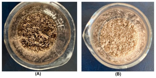
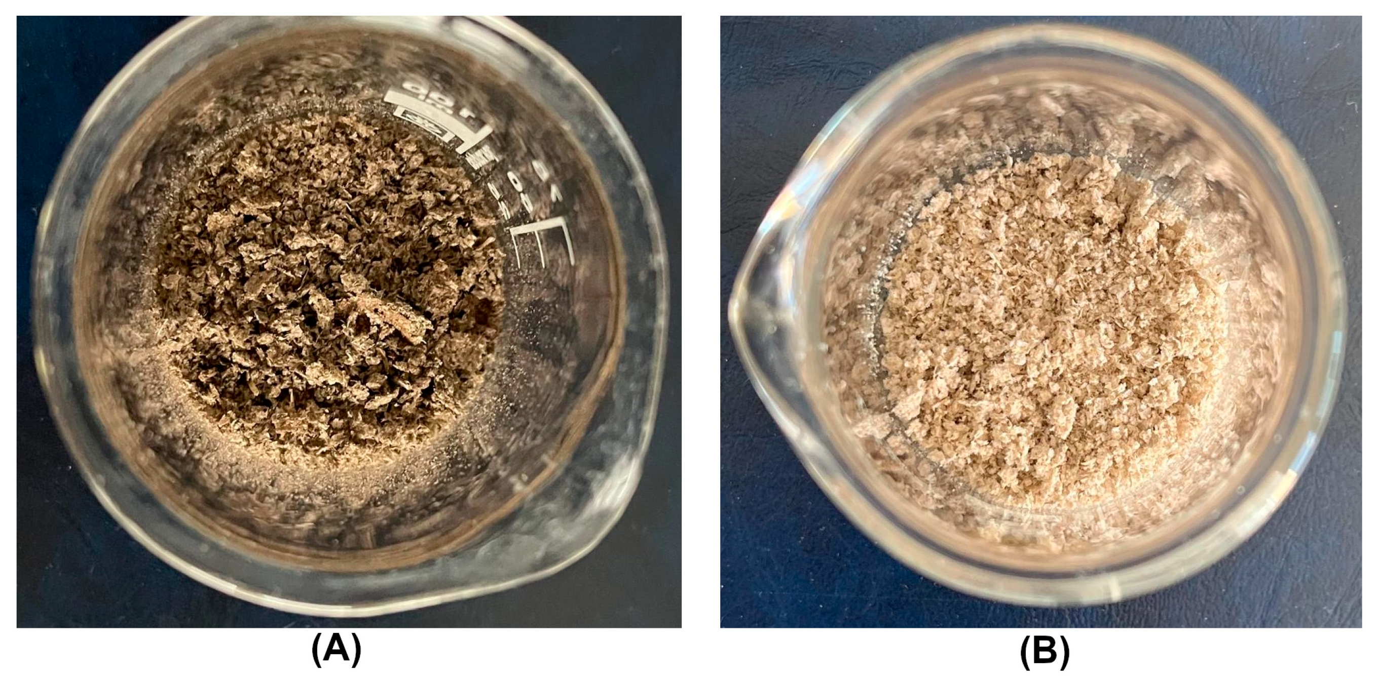
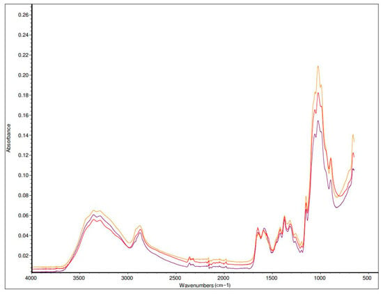
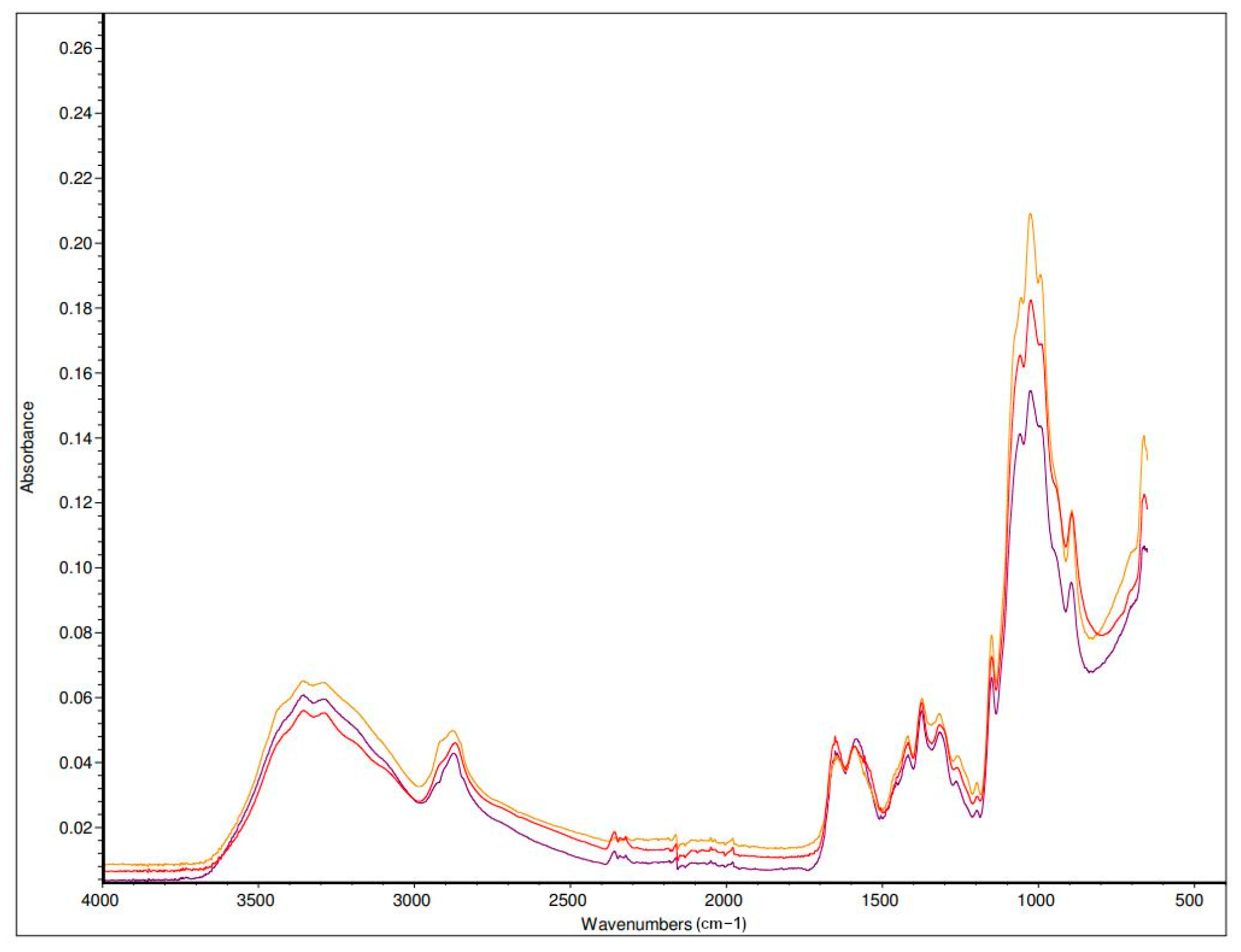
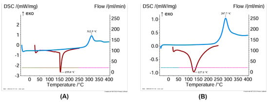
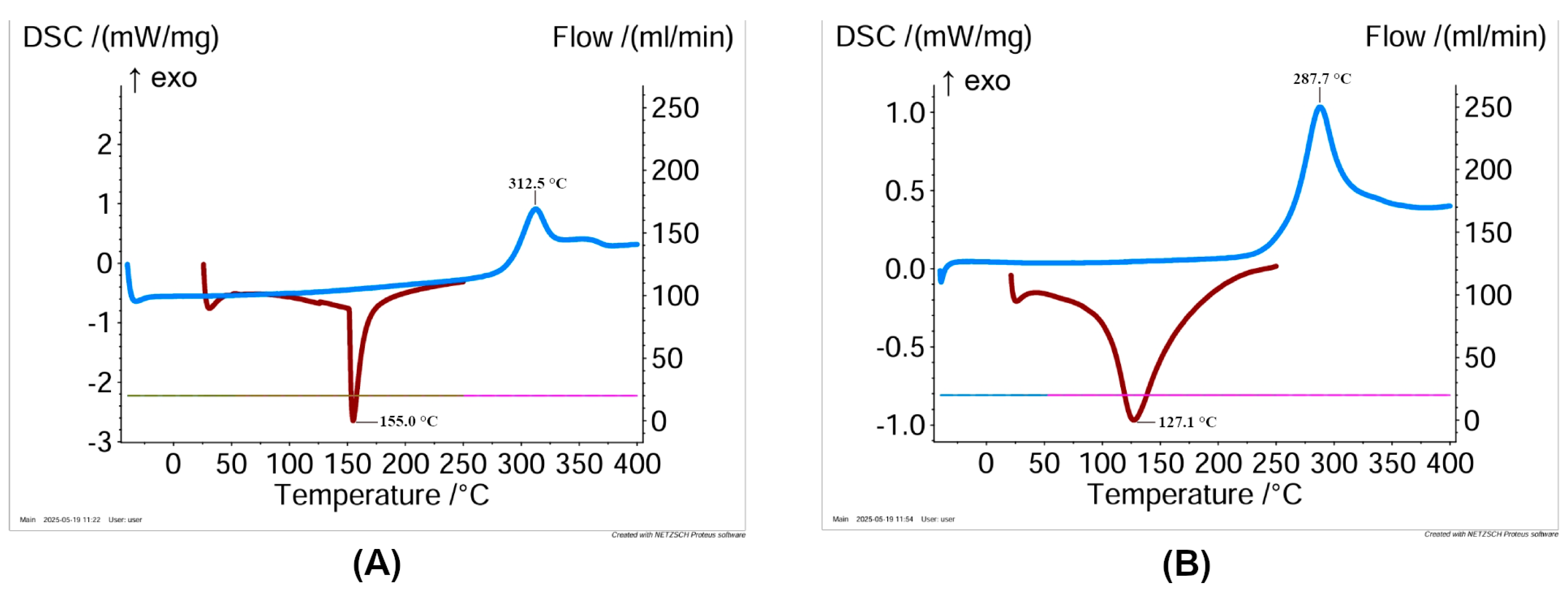
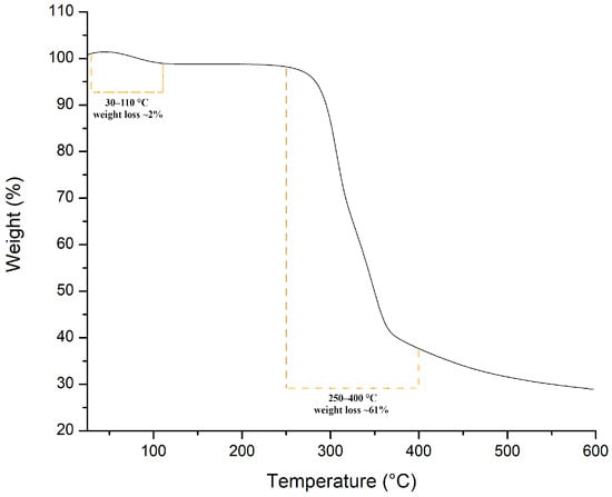
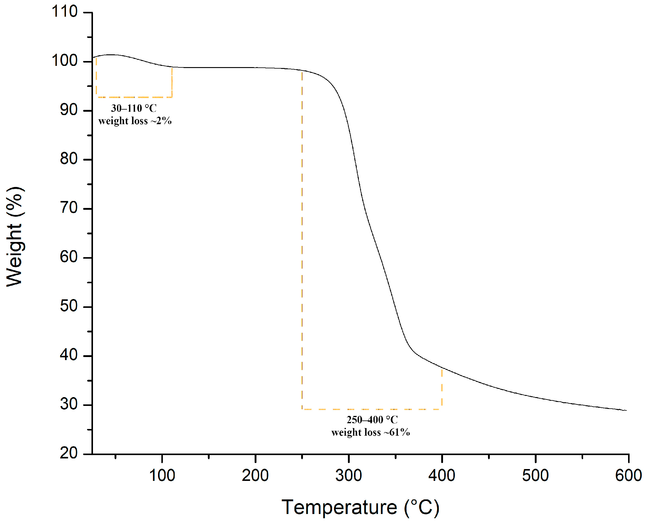
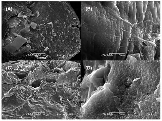
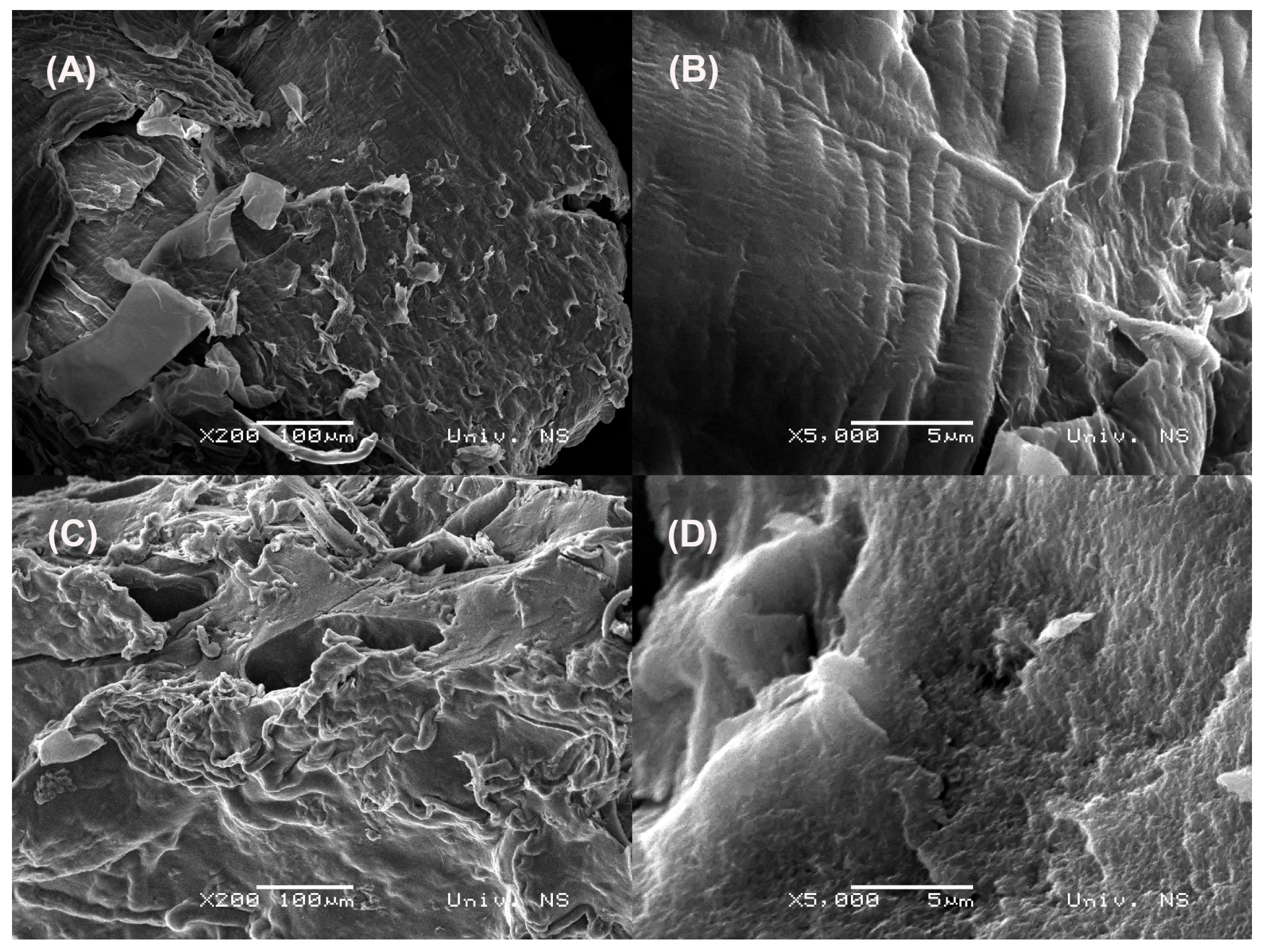
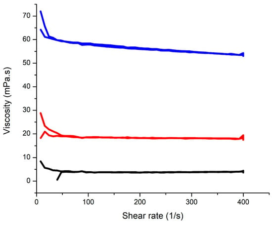
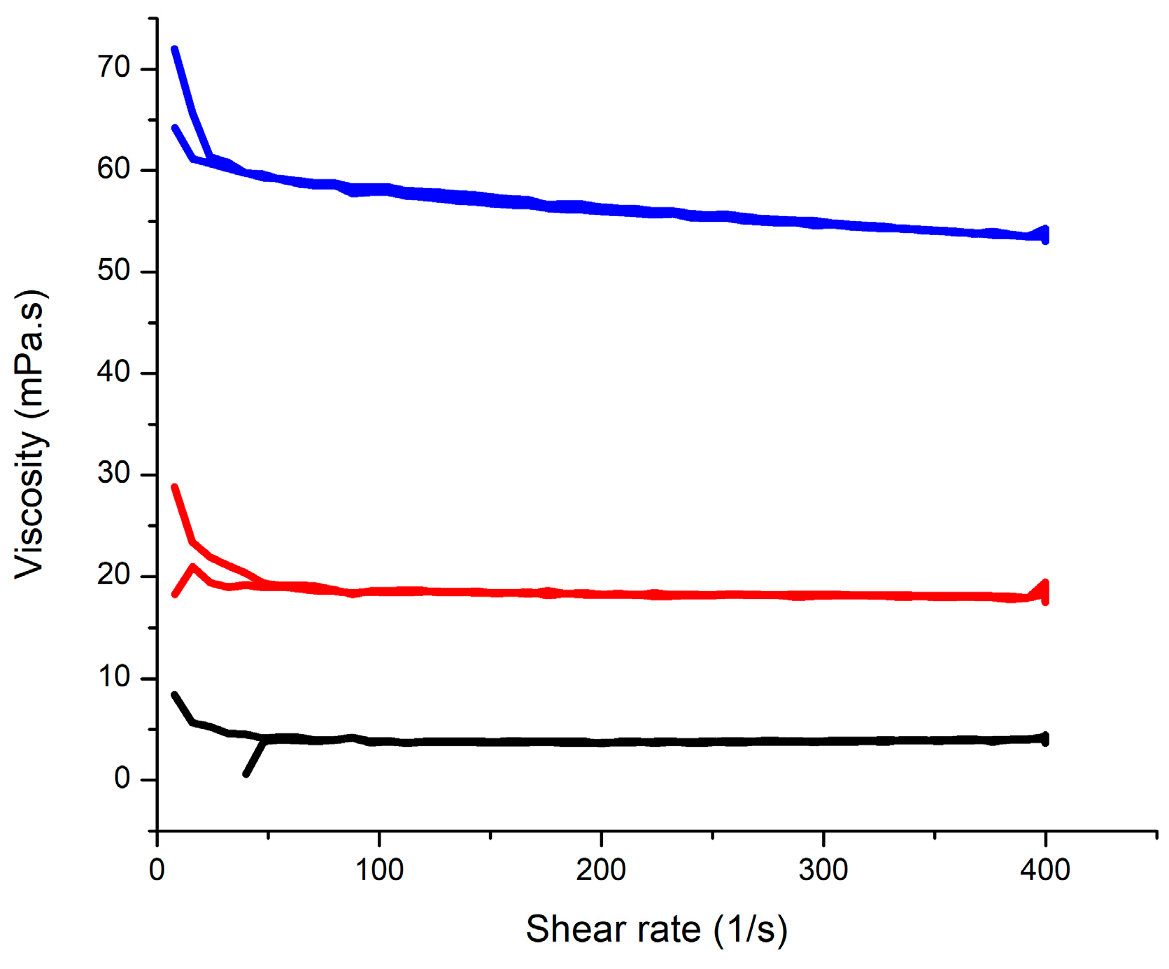
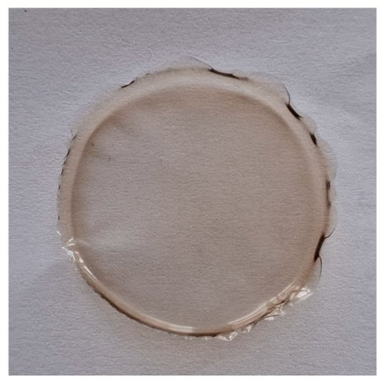
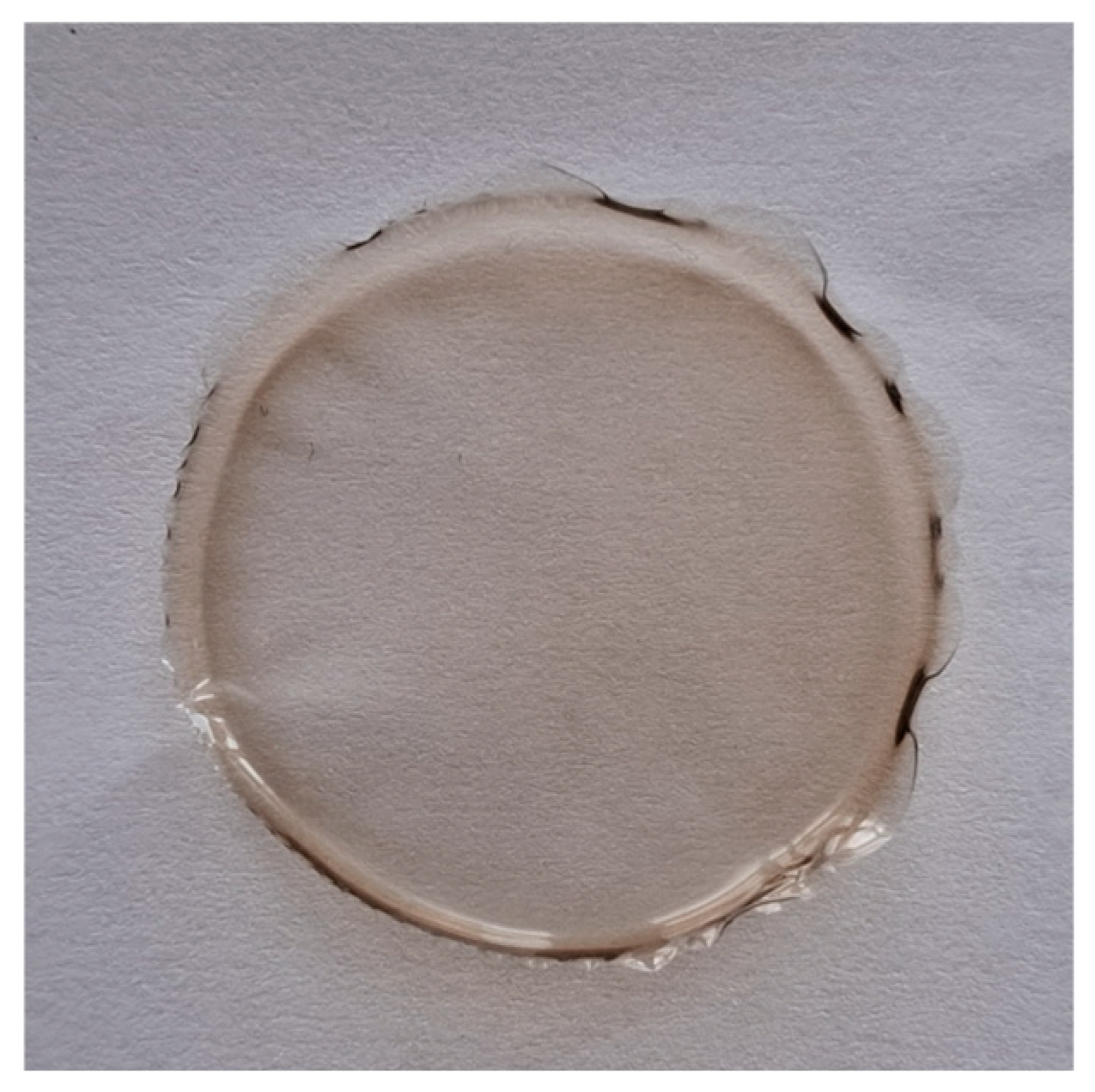

Disclaimer/Publisher’s Note: The statements, opinions and data contained in all publications are solely those of the individual author(s) and contributor(s) and not of MDPI and/or the editor(s). MDPI and/or the editor(s) disclaim responsibility for any injury to people or property resulting from any ideas, methods, instructions or products referred to in the content. |
© 2025 by the authors. Licensee MDPI, Basel, Switzerland. This article is an open access article distributed under the terms and conditions of the Creative Commons Attribution (CC BY) license (https://creativecommons.org/licenses/by/4.0/).