


Abstract
Perfluorooctanoic acid (PFOA) has been associated with various health effects, including immunotoxicity. This study aimed to examine associations between PFOA serum levels above the intervention threshold level (HBM-II, 10 µg/L) and diphtheria, tetanus and SARS-CoV-2 antibody concentrations among adult residents with increased internal PFOA. A total of 662 questionnaire data points and vaccination passports were collected and blood samples were used to determine PFOA serum levels and diphtheria, tetanus and SARS-CoV-2 antibody concentrations. Logistic regression modelling was used to examine potential associations. The median PFOA concentration was 9.17 µg/L with 45.2% exceeding the HBM-II. Of participants with at least one vaccination, 48.1% had reliable protection against diphtheria and 91.9% against tetanus. Antibodies against SARS-CoV-2 spike and nucleocapsid protein were detected in 96.4% and 53.8%, respectively. Age was a significant predictor for lack of immunity against diphtheria (OR = 3.6 (95% CI: 2.3; 5.7)) in the age group ≥60. No association was found between PFOA levels and SARS-CoV-2 N antibody concentrations. After adjusting for age group, PFOA serum concentrations above the intervention threshold were not associated with lower diphtheria or SARS-CoV-2 N antibody concentrations in the adult population. Further studies are needed to determine whether PFOA exposure has a long-term effect on the immune system.
1. Introduction
Perfluorooctanoic acid (PFOA) is a synthetic chemical compound that belongs to perfluorinated alkyl substances (PFASs), which are known as “forever chemicals” due to their persistence in the environment [1]. Contamination is widespread due to its broad use, including for surface treatment, as a lubricant or surfactant [2], and can be especially high in hotspots such as production sites and places where PFOA-containing fire-fighting foams were used [3,4]. PFOA contamination of the environment in the EU mainly occurred before it was listed as a persistent organic pollutant (POP) in EU-wide regulations in 2020. Since 2020, the production and use of PFOA is no longer permitted in the EU [5]. PFOA is extremely stable and practically non-biodegradable [6,7], which is why high concentrations can still be present in the environment, particularly at historic production sites [3]. The main routes of uptake for populations that are not occupationally exposed are drinking water and contaminated food [8,9]. PFOA can accumulate in a variety of tissues and organs [10]. Based on several studies, the half-life in humans is around two to four years, which strongly favours bioaccumulation [11,12,13].
Population studies have investigated the effects of PFOA on human health and have shown various effects, e.g., on cholesterol levels, the thyroid gland and fertility [14,15,16,17]. The immunotoxicity of PFOA was reviewed by Liang, et al. [18] in both in vitro, animal and human studies. The European Food Safety Authority (EFSA), for example, concluded based on the available scientific studies on animals and concentrations in human blood that the effect of PFASs on the immune system is the most critical aspect for risk assessment [19]. Several animal studies have demonstrated an effect of PFOA on the immune system of mice, for example, through suppression of IgM antibodies or T-cell-dependent antibody responses [20,21,22]. However, animal studies investigating the influence of PFASs on the immune system provide only limited support for a toxic effect of PFASs on the immune system. Although immunomodulation is observed after administration of PFASs, this only occurs at blood concentrations that are much higher than those of environmentally exposed humans. The high doses required for immunomodulation, the inconsistent results and the limited number of endpoints evaluated make it difficult to draw a clear conclusion about the toxic effect of PFASs on the immune system [23]. Studies on the effect of PFOA on the vaccination response in children suggested an inverse association between PFOA concentration in blood and antibodies against tetanus and diphtheria, although significance was not always given [24,25,26]. Studies on the effects of PFOA on the immune response in adults showed lower or no effects when analysing the antibody response for tetanus and diphtheria vaccination [27,28].
In order to be able to assess the health relevance of PFOA blood concentrations, the Human Biomonitoring Commission of the German Environment Agency (HBM Commission) has defined human biomonitoring values (HBM values). HBM values can use NOAEL (no observed adverse effect level) or LOAEL (lowest observed adverse effect level) as points of departure and use toxicokinetic models to obtain threshold blood concentrations in humans. Rather than focusing on estimating a critical dose, using HBM accepts that environmental exposure in the general population is often varied and will have many pathways, thus offering policy makers a threshold for the actual internal exposure [29]. The HBM-I value represents the concentration of a substance in a human body medium below which no health effects are to be expected and therefore no measures are required. According to the current assessment of the HBM Commission, the HBM-I value is 2 µg PFOA/L blood plasma or serum. In contrast, the HBM-II value is the concentration of a substance in a human body medium above which a relevant health impairment can occur and therefore measures must be taken to reduce exposure. It is 10 µg PFOA/L blood plasma or serum for the general population and 5 µg PFOA/L blood plasma or serum for women of reproductive age [30]. While these values have been derived based on the overall effect on health of PFOA above the HBM-II value, with regard to humoral immunity, more epidemiological studies are required. The association between PFOA levels exceeding the HBM-II value and antibody concentrations is the focus of this study. In the district of Altötting in Bavaria, Germany, emissions of PFOA from a fluoropolymer production plant, where PFOA was produced from 1968 until 2003 and then used for fluoropolymer production until 2008, led to environmental contamination in parts of the district. The resident population ingested the substance primarily through drinking water. Between 2007 and 2018, the drinking water supply was gradually purified using filter systems so that the drinking water was no longer an exposure source for PFOA. A human biomonitoring study in 2018 found elevated PFOA serum levels in the population [31]. In a follow-up study in 2022, in addition to the PFOA levels, antibodies against diphtheria, tetanus and SARS-CoV-2 were determined in the population’s serum. Diphtheria and tetanus antibody concentrations were included based on the previous HBM Commission conclusion that further epidemiological studies are needed for this endpoint [30]. Furthermore, given the public interest in the pandemic context and some research pointing to an association with PFAS, SARS-CoV-2 was included in the study [32]. For more detailed background information on the study, see Lahne, et al. [33].
The aim of this study was therefore to examine associations between PFOA serum levels above the intervention threshold level of 10 µg/L and antibody concentration following diphtheria, tetanus and SARS-CoV-2 vaccination or SARS-CoV-2 infection among the adult residents in the Altötting district in Bavaria, Germany.
2. Materials and Methods
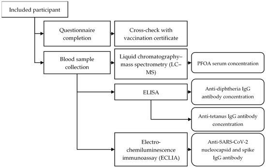
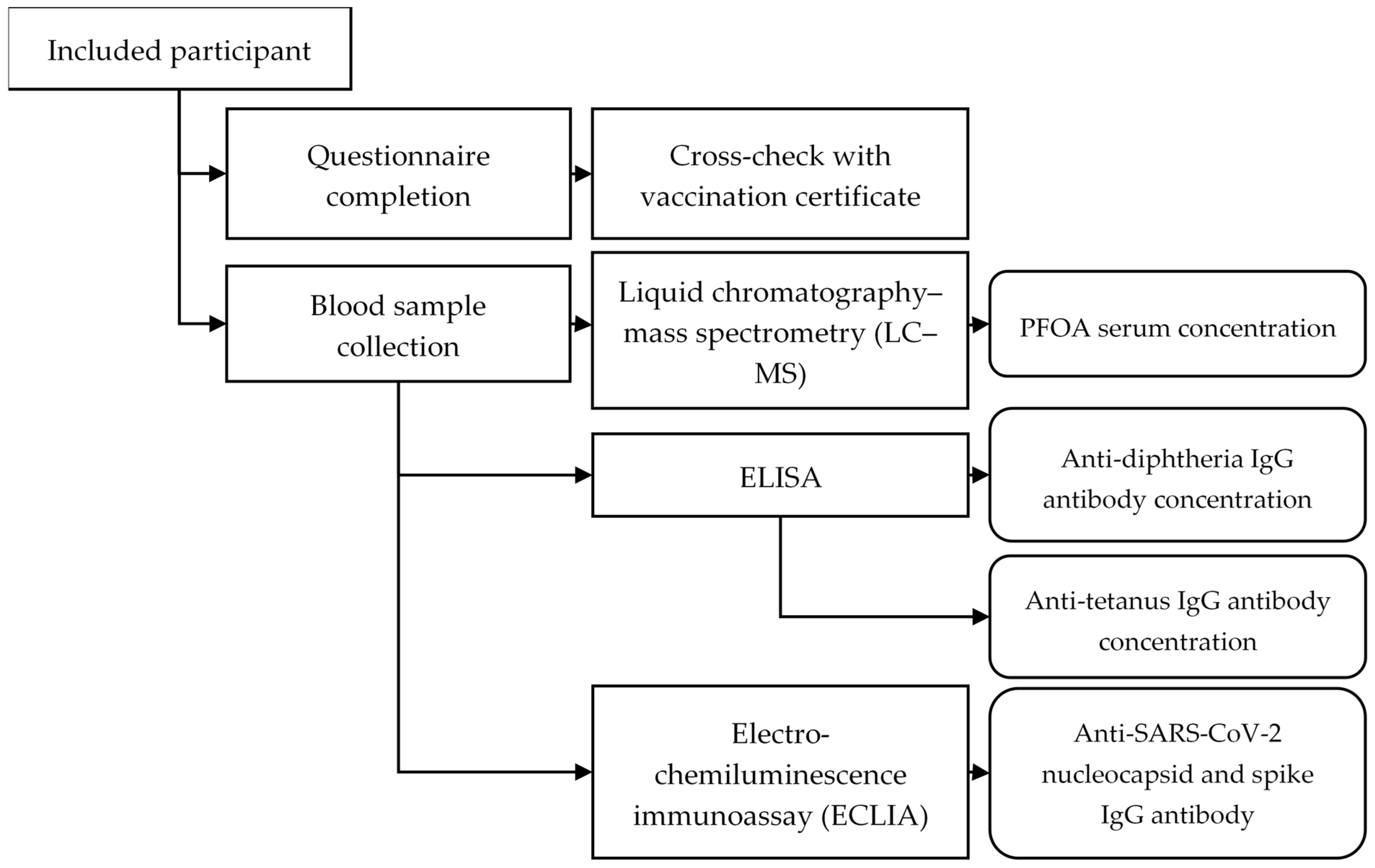
The methods of the human biomonitoring follow-up study are described in detail elsewhere [33] and are only briefly outlined here. The participants of the 2018 study were invited to participate in the 2022 study. Out of the 717 participants aged 18 years and over, 55 participants with indication of occupational exposure to PFOA or no information on occupational exposure to PFOA provided in the questionnaire were excluded from the analysis, as our study aimed at examining the association of environmental PFOA exposure of the general population and not of work-related PFOA exposure patterns (for those occupationally exposed, there are specific occupational exposure limits, which can be used to assess the health relevance). Accordingly, 662 people were included in the analyses shown in Figure 1.
Figure 1.
Flowchart of analyses carried out to measure the antibody and PFOA blood serum concentration as well as questionnaire data.
In the analyses regarding diphtheria and tetanus, all persons who reported in the questionnaire having received at least one vaccination against diphtheria or tetanus were included, which resulted in 645 persons for diphtheria and 651 persons for tetanus. With regard to SARS-CoV-2, all individuals who reported whether they had been vaccinated against SARS-CoV-2 and/or had experienced a SARS-CoV-2 infection were included in the further analyses, which was the case for 645 persons.
Blood samples were collected between June and August 2022. The questionnaire, completed by the participants, was compared with the vaccination certificate by a healthcare professional at the time of blood collection.
Liquid chromatography–mass spectrometry (LC-MS/MS) was applied for the PFOA analysis using an UltiMate 3000 (Thermo Fisher Scientific, Waltham, MA, USA) HPLC system coupled to a QTrap 5500 (SCIEX, Marlborough, MA, USA) mass spectrometer in negative mode. On-line purification was performed using a column set-up with a trap column (Oasis HLB (Waters, Milford, MA, USA), 20 × 2.1 mm, 25 µm). For separation, gradient elution (2 mM ammonium acetate and methanol) was used over the analytical column (Reprosil-Pur C18 AQ material (Dr. Maisch HPLC GmbH, Ammerbuch-Entringen, Germany), 33 × 3 mm, 5 µm), at 35 °C column oven temperature. All samples were processed and analysed in duplicate. A value of 0.25 µg/L was determined as the limit of quantification (see also [33]).
The value of 10 µg/L for PFOA was chosen as a threshold for high internal PFOA exposure, following the HBM-II value for the general population, which is the value determined by the HBM Commission above which adverse health effects may be observed. For women of reproductive age, the HBM-II value is 5 µg/L. This value was derived on the basis of available studies on the effects on fertility, birth weight and child development [30]. However, these effects do not concern the adult’s antibody formation. When considering immunity, a differentiation for this population group was not provided; thus, the HBM-II value for the general population was used for the entire study population. In addition, approximately half of the participants had a PFOA serum concentration under 10 µg/L and half had a higher one, so the two groups were well comparable.
Anti-diphtheria toxoid and anti-tetanus toxoid IgG levels were measured on the ELISA processor “DSX System” (Dynex® Technologies, Inc., Chantilly, VA, USA) using commercial ELISA kits (Anti-Diphtheria Toxoid (IgG) and Anti-Tetanus Toxoid (IgG)) (Euroimmun, Lübeck, Germany), performed as recommended by the manufacturer. According to the manufacturer’s specifications for the diphtheria antibodies, a value under 0.01 IU/mL was considered seronegative, 0.01 IU/mL to 0.09 IU/mL as no reliable immune protection and ≥0.1 IU/mL as full protection. For tetanus an antibody titre below 0.1 IU/mL was classified as seronegative, between 0.1 and 0.5 IU/mL as present protection and >0.5 IU/mL as sufficient protection.
Antibodies against the receptor-binding domain located on the S1 subunit of the SARS-CoV-2 spike protein (S) and against the nucleocapsid (N) of SARS-CoV-2 were measured on a Cobas E 411 (Roche Diagnostics GmbH, Mannheim, Germany) using Elecsys Anti-SARS-CoV-2 S electro-chemiluminescence immunoassay (ECLIA) and Elecsys Anti-SARS-CoV-2 ECLIA (Roche Diagnostics GmbH, Mannheim, Germany), respectively, performed according to the manufacturer’s instructions. For anti-SARS-CoV-2 S tests with a measurement range between 0.4 and 250 U/mL, a result under 0.8 U/mL was interpreted as negative and ≥0.8 U/mL as positive; for anti-SARS-CoV-2 N tests, a cut-off index (COI) under 1.0 was interpreted as negative and a COI ≥ 1.0 as positive. If N antibodies can be detected, it can be assumed that a SARS-CoV-2 infection has occurred in the past, regardless of the vaccination status. If S antibodies are present, this indicates that an infection has occurred or a vaccination has been administered [34].
Age, gender, vaccination rates, serum PFOA levels and antibody concentrations following diphtheria, tetanus and SARS-CoV-2 vaccination were analysed, and for PFOA and antibody concentration, the results were stratified by age. With PFOA and antibody concentration categorised as aforementioned, their association with age groups [18–39; 40–59; and ≥60] was analysed by the Spearman correlation coefficient.
For the analyses of the group that reported having ever been vaccinated, a distinction was made between people who had or had not received a base immunisation and people who had or had not received a vaccination in the last 10 years. The 10-year period is based on the vaccination recommendations of the Standing Committee on Vaccination (STIKO) at the Robert Koch Institute, which recommends that a diphtheria and tetanus booster vaccination is required every 10 years from the age of 18 [35].
Statistical analysis was conducted using a logistic regression model to estimate the odds ratio for the diphtheria antibody concentrations with PFOA dichotomised (below versus at least 10 µg/L) as the exposure. Diphtheria antibody concentration was categorised as follows: ≤0.01—no protective antibodies against diphtheria; 0.01–0.09—no reliable protection against diphtheria; ≥0.1—full protection against diphtheria. Given the aim of this research, we maintained PFOA as an independent variable for our models, while other independent variables such as gender were excluded during model selection. Age between 40 and 59, and 60 years and above, vaccination in the last 10 years and basic immunisation were taken into account as potential confounders. Additional potential confounders, such as gender, number of SARS-CoV-2 vaccinations or severity of SARS-CoV-2 infection symptoms were included during model development but were removed to improve model fit. As the focus of the study, PFOA was kept in the models, despite lack of a significant association. Because of the small number of participants in the group with low immunity against tetanus and SARS-CoV-2 S antibodies, no logistic regression was developed. The regression model with the N-antibody concentration as the dependent variable included age and number of PCR-confirmed SARS-CoV-2 infections as confounders in addition to the PFOA concentration. A regression model with categorised data was selected, as the data for the PFOA and antibody concentrations were not normally distributed. In addition, the results of the logistic regression model can be better interpreted (protected/not protected), which allows for greater clinical significance. Furthermore, there was a high proportion of individuals with antibody concentrations outside the quantification limits. Additionally, as a sensitivity analysis, linear regression models with logarithmically transformed antibody concentration and PFOA concentration was calculated. For these models, age was also included as a numeric variable.
Significance was assumed at α ≤ 0.05. The statistical programme SAS 9.4 (SAS Institute Inc, Cary, NC, USA) was used for the statistical analyses.
The study was approved by the Ethics Committee at the Medical Faculty of the Ludwig Maximilian University (21-1204 on 25 February 2022) and complies with the ethical standards of the 1964 Helsinki Declaration. Informed consent was obtained from all individual participants included in the study.
3. Results
3.1. Population Description
Analysis was based on the data of the 662 participants of the follow-up study with a mean age of 54 years (SD = 15 and range 18–87) and a proportion of females of 57.1% (n = 378) (Table 1). None of the participants reported defects of the immune system or organ or stem cell transplants.
Table 1.
Characteristics of the participants (with PFOA values for each group) (n = 662).
3.2. PFOA
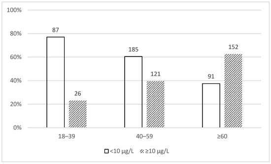
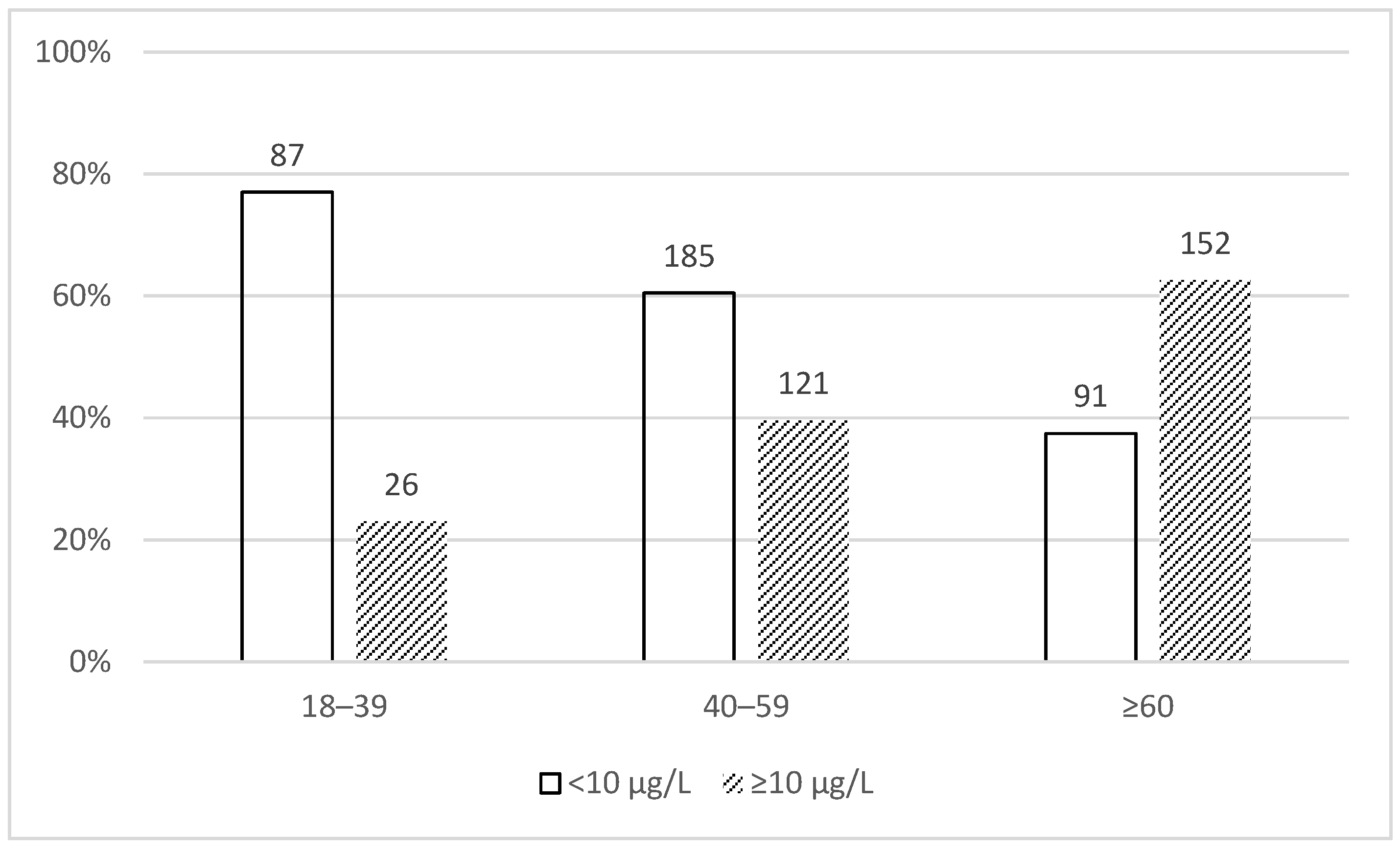
The median serum concentration of PFOA in the population was 9.17 µg/L with a range between 0.68 µg/L and 151.65 µg/L. Of the participants, 45.2% (n = 299) had a serum PFOA concentration starting from 10 µg/L. The percentage of participants with high serum PFOA concentration was higher with increasing age (Figure 2).
Figure 2.
Serum PFOA concentration stratified by age group (N = 662).
3.3. Diphtheria
Data from 645 people were available for the analyses regarding diphtheria.
3.3.1. Antibody Status
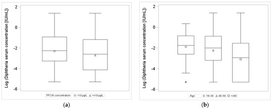
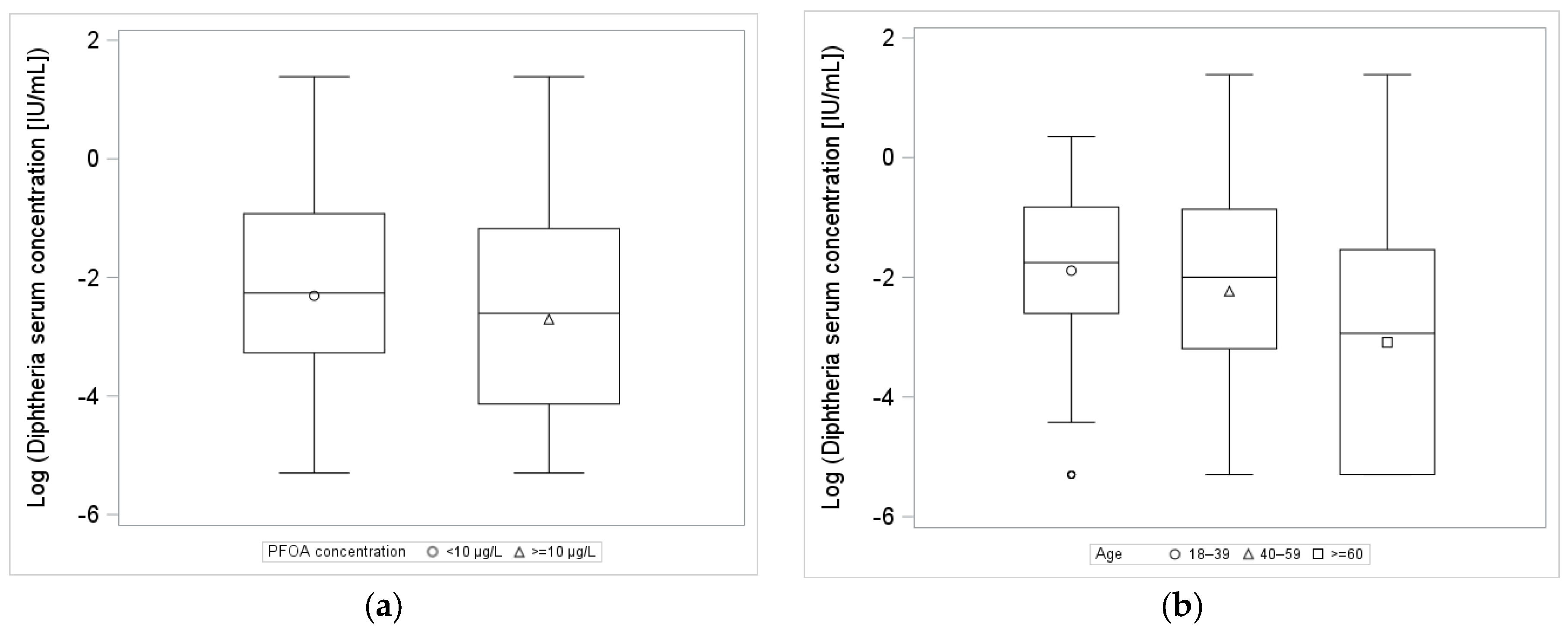
A total of 14.9% (n = 96) were seronegative (<0.01 IU/mL); 37.0% (n = 239) had no reliable protection against diphtheria (0.01 to 0.09 IU/mL) and 48.1% (n = 310) had full protection against diphtheria (≥0.1 IU/mL) according to the serum antibody titre. The distribution of log-transformed diphtheria antibody serum concentration stratified by PFOA and age category is shown in Figure 3.
Figure 3.
Box plots of log-transformed diphtheria antibody concentration by (a) serum PFOA concentration and (b) age category.
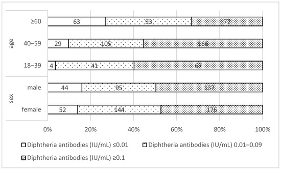
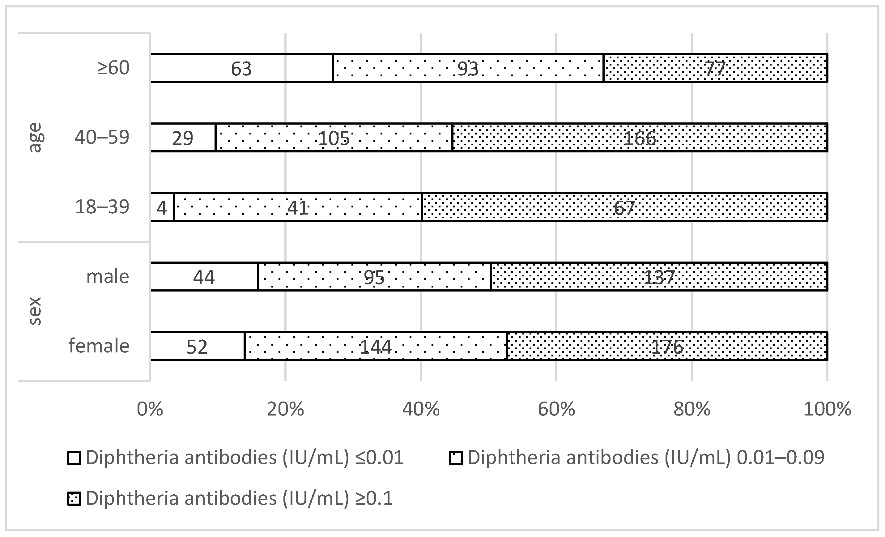
Full protection against diphtheria (≥0.1 IU/mL) according to the serum antibody titre was found in 49.1% of males and 47.3% of females. There was little difference between males and females in terms of diphtheria antibody distribution (Figure 4).
Figure 4.
Antibody status regarding diphtheria (n = 645) by sex and age category. ≤0.01: seronegative against diphtheria; 0.01–0.09: no reliable protection against diphtheria; ≥0.1: full protection against diphtheria.
With regard to the age distribution according to the antibody category, it can be seen that the group with no protective antibodies increased from 3.6% (n = 4) of the participants in the 18–39 age group to 27.0% (n = 63) of the participants over 60 years old, while the category with no reliable protection against diphtheria seemed stable with increasing age. The percentage of participants with full protection against diphtheria decreased from 59.8% to 33.1% with increasing age (Figure 4).
3.3.2. Vaccination Rates
The vaccination rates were calculated on the basis of the information provided in the questionnaire by the participants. At least one vaccination against diphtheria was reported by 97.4% (n = 645) of the participants, who were included in the subsequent analysis. Of those, 93.5% (n = 603) reported having received the base immunisation and 67.3% (n = 434) reported having received a vaccination against diphtheria within the last 10 years. With increasing age group, a higher percentage of participants did not provide any data regarding base immunisation against diphtheria (18–39: 0.9% (n = 1); 40–60: 3.6% (n = 11); ≥60: 7.8% (n = 19)) or reported that they did not receive base immunisation (18–39: 0.9% (n = 1); 40–60: 1.6% (n = 5); ≥60: 9.1% (n = 22)). Participants who reported having received both base immunisation and vaccination within the last 10 years represented 64.8% (n = 418) of included study participants, 60.8% (n = 166) and 67.7% (n = 252) of the females and males, respectively.
3.3.3. Comparison Between Base Immunisation and Vaccination in the Last 10 Years
A self-reported lack of base immunisation among those with at least one vaccination against diphtheria increased with age from 0% (n = 0/111) in the 18–39 age group to 1.0% (n = 3/293) in the 40–59 age group and 7.3% (n = 16/218) in those aged 60 years and above. The distribution of the diphtheria antibody concentration stratified by age and base immunisation is presented in Table 2. The percentage of persons with self-reported vaccination against diphtheria in the last 10 years was between 67.7% (age group 40–59) and 75.6% (age group ≥ 60) (Table 2).
Table 2.
Antibody status regarding diphtheria according to self-reported base immunisation and self-reported vaccination in the last 10 years (n = 645).
3.3.4. Results of Regression Analysis
In this analysis, data from 593 persons who provided data on base immunisation and vaccination against diphtheria in the last 10 years could be used (Table 3). The dependent variable, diphtheria antibody concentration, was categorised as described above.
Table 3.
Association of PFOA and other covariates with diphtheria antibody concentrations: results of categorical logistic regression model (n = 593).
The adjusted odds ratio of PFOA serum levels above or equal to 10 µg/L for age was 1.1 [0.8; 1.5]. Being in the age group of 60 years and above increased the likelihood of being in the seronegative diphtheria antibody group by 3.6 [2.3; 5.7] times. Similarly, not having a vaccination against diphtheria in the last 10 years and not having the base immunisation completed significantly increased the odds of having no protective antibodies against diphtheria.
The results obtained with the chosen approach were consistent with the results of the sensitivity analysis using linear regression with logarithmically transformed antibody data. Log-transformed PFOA was not significant and had a coefficient of −0.0153 [−0.1763; 0.1457] and an exponentiated coefficient of 0.98 [0.84; 1.16]. Age, having base immunisation and being vaccinated in the last 10 years were significant at 0.97 [0.96; 0.98], 0.37 [0.15; 0.87] and 0.55 [0.41; 0.72], respectively. Thus, an increase of one year is associated with 3.1% lower diphtheria antibody concentration, not being vaccinated in the last 10 years with 63.5%, and not having base immunisation with 45.5% lower diphtheria antibody concentration.
3.4. Tetanus
Data from 651 people were available for the analyses regarding tetanus.
3.4.1. Antibody Status
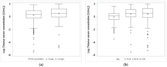
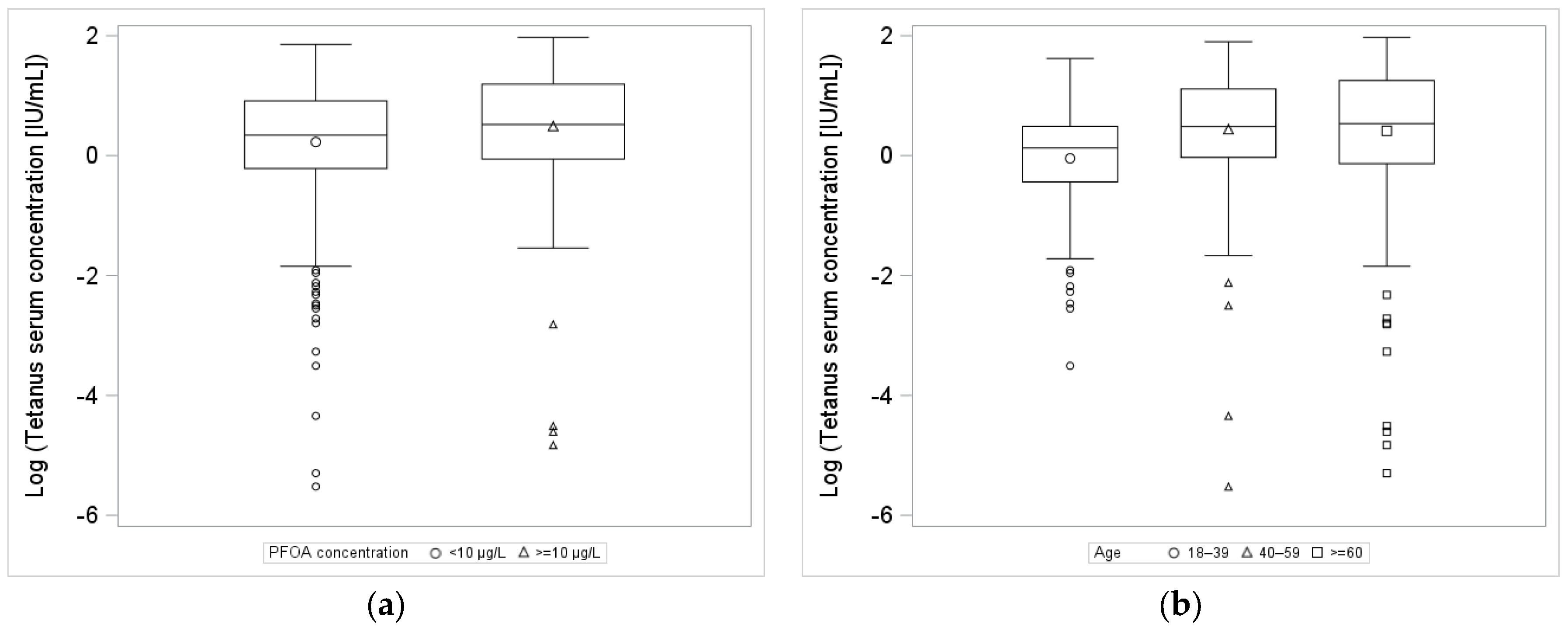
A total of 2.6% (n = 17) were seronegative (≤0.1 IU/mL); 5.5% (n = 36) had present protection against tetanus (0.1 to 0.5 IU/mL) and 91.9% (n = 598) had sufficient protection against tetanus (>0.5 IU/mL) according to the serum antibody titre. The distribution of log-transformed tetanus antibody serum concentration stratified by PFOA and age category is shown in Figure 5.
Figure 5.
Box plots of log-transformed tetanus antibody concentration by (a) serum PFOA concentration and (b) age category.
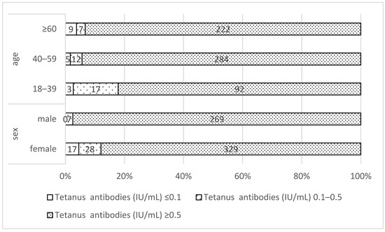
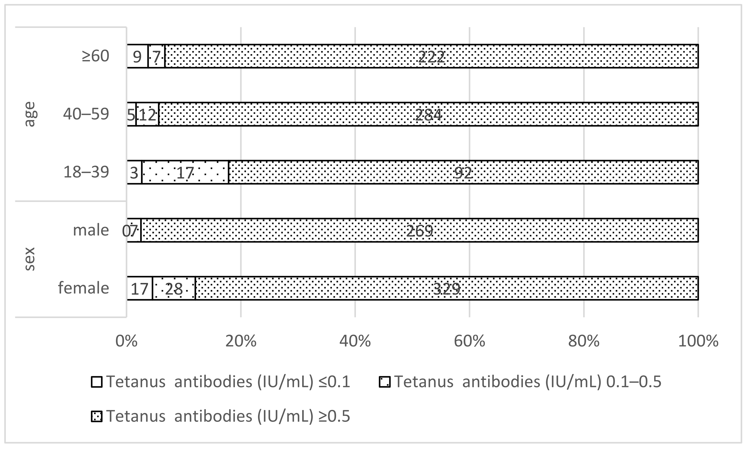
Almost all participants, 97.1% of males and 88.0% of females, had sufficient protection against tetanus (≥0.5 IU/mL). No males and 17 (4.6%) of the females were tetanus-seronegative according to the level of serum antibodies (Figure 6).
Figure 6.
Antibody status regarding tetanus (n = 651) by sex and age category. ≤0.1: seronegative against tetanus; 0.1–0.5: present protection against tetanus; ≥0.5: sufficient protection against tetanus.
The distribution of protection against tetanus is more stable with increasing age. In all three age groups, the largest proportion has sufficient protection. At 82.1%, the proportion in the 18–39 age group is the lowest compared to the two older age groups (40–59: 94.4%, ≥60: 93.3%). In the group with present protection, however, the proportion among the 18–39-year-olds (15.2%) is higher than in the other two age groups (40–59: 4.0%, ≥60: 2.9%) (Figure 6). The Spearman correlation between age and tetanus antibody serum concentration had a rho of 0.16 (p < 0.0001).
3.4.2. Vaccination Rates
Similar results were observed with the tetanus vaccination as with the diphtheria vaccination. At least one vaccination against tetanus was reported in the questionnaire by 98.3% (n = 651) of the participants, who were included in the subsequent analysis. Base immunisation schedule was completed by 94.0% (n = 612) and 72.0% (n = 469) received a vaccination within the last 10 years. With increasing age group, a higher percentage of participants did not provide any data regarding base immunisation (18–39: 0.9% (n = 1); 40–60: 2.0% (n = 6); ≥60: 5.1% (n = 12)) or reported that they did not receive base immunisation (18–39: 0% (n = 0); 40–60: 1.3% (n = 4); ≥60: 6.7% (n = 16)). Participants who reported having complete base immunisation and vaccination within the last 10 years represented 69.6% (n = 453) of the included study participants, 48.1% (n = 182) and 95.4% (n = 271) of the females and males, respectively.
3.4.3. Comparison Between Base Immunisation and Vaccination in the Last 10 Years
A self-reported lack of base immunisation among those with at least one vaccination against tetanus increased with age from 0% in the 18–39 age group to 1.4% in the 40–59 age group and 7.1% in those aged 60 years old and above. All the participants who reported not having base immunisation had sufficient protection against tetanus according to the serum antibodies (≥0.5 IU/mL) (Table 4). The percentage of persons with a self-reported vaccination against tetanus in the last 10 years was between 74.5% (age group 40–59) and 83.3% (age group ≥ 60) with a tendency for higher percentages in the categories of existing protection and seronegative for the participants who did not report tetanus vaccination in the last 10 years (Table 4).
Table 4.
Antibody status regarding tetanus according to self-reported base immunisation and (to self-reported vaccination in the last 10 years (n = 651).
3.4.4. Antibody Status Based on PFOA Serum Concentrations
Analysis of tetanus antibody status stratified by age and PFOA serum concentrations showed that the majority of participants (91.9%) across all age groups had high antibody levels (≥0.5 IU/mL), regardless of PFOA serum concentrations (see Table 5).
Table 5.
Antibody status regarding tetanus according to PFOA serum concentrations (n = 651).
Given the distribution of low numbers of participants in the category of no protective antibodies against tetanus, a logistic regression was not possible. The results obtained from the linear regression for the sensitivity analysis show no significant association with log-transformed PFOA, with a coefficient of 0.08 [−0.01; 0.16] and an exponentiated coefficient of 1.08 [0.99; 1.18]. Age and being vaccinated in the last 10 years were significant at 1.01 [1.003; 1.01] and 0.63 [0.53; 0.74], respectively. Thus, not being vaccinated in the last 10 years is associated with 37.5% lower tetanus antibody concentration.
3.5. SARS-CoV-2
3.5.1. Antibody Response Based on Vaccination Status
Irrespective of the age group and reported vaccination status, more than 80% of the participants reported having had symptoms of SARS-CoV-2 infection. A total of 42.4% of the participants reported having had at least one SARS-CoV-2 infection confirmed by PCR test, while the rest reported only a positive antibody test or did not provide any information for these questions. The distribution of log-transformed N-antibody serum concentration stratified by PFOA and age category is shown in Figure 7.
Figure 7.
Box plots of log-transformed SARS-CoV-2 N-antibody concentration by (a) serum PFOA concentration and (b) age category.
The reported vaccination rate of 92.0% is reflected in the 638 (96.4%) participants that had S antibodies above the 0.8 U/mL threshold, largely irrespective of age and PFOA serum concentration. N antibodies above or equal to 1.0 COI were found in 356 (53.8%) participants, with no significant difference between the two PFOA serum concentration groups, but there was a trend with increasing age group. A higher percentage of participants in the 18–39 age group (77.0%) were above the cut-off index compared to the 40–59 age group (57.5%) and those aged 60 years and above (38.9%). A total of 97.9% had N and/or S antibodies against SARS-CoV-2. When looking at the different age groups among those who reported a SARS-CoV-2 infection, a similar distribution was observed. The Spearman correlation between age and SARS-CoV-2 S-antibody serum concentrations was r = −0.06 (p = 0.13).
3.5.2. Vaccination Rates
Of the 662 participants included in the study, 609 (92.0%) indicated that they had received at least one vaccination against SARS-CoV-2. Of those, 85.1% (n = 518) reported three or more vaccinations, 12.8% (n = 78) two vaccinations and 2.1% (n = 13) one vaccination against SARS-CoV-2.
3.5.3. Results of Regression Analysis
With regard to the logistic regression for SARS-CoV-2, we could not use the level of S antibodies as an outcome, as only a small number of individuals in the different age groups and PFOA serum concentration groups had an S-antibody concentration below 0.8 U/mL. We used the N-antibody group as an outcome instead and only for those who had S antibodies above 0.8 U/mL (638/662). The unadjusted OR for having N antibodies below the cut-off index was 1.2 [0.9; 1.7] in those with a PFOA serum concentration ≥ 10 µg/L vs. those with a PFOA serum concentration < 10 µg/L. Similarly, after adjusting for age group and number of reported PCR-confirmed SARS-CoV-2 infections, the OR was 0.7 [0.4–1.2]. The number of infections and not being over 60 were significantly associated with having N antibodies.
The results obtained with the chosen approach were consistent with the results of the sensitivity analysis using linear regression with logarithmically transformed data, with log-transformed PFOA not being significantly associated with a coefficient of 0.2 [−0–02; 0.42]. Age and number of PCR-confirmed SARS-CoV-2 infections were significantly associated with N antibodies, with an exponentiated coefficient of 0.98 [0.96; 0.99] and 30.23 [22.15; 41.25].
3.6. Analyses with the PFOA Serum Concentrations from 2018
To compare the results, the analyses regarding the association between internal PFOA exposure and antibody status were also carried out with the PFOA serum concentrations measured in 2018. The results correspond to the results presented and are therefore not shown here.
4. Discussion
This study investigated whether there is an association between PFOA serum levels above the HBM-II value of 10 µg/L and antibody concentration following diphtheria, tetanus and SARS-CoV-2 vaccination in adult residents with increased internal PFOA exposure due to previous PFOA contamination of drinking water in the district of Altötting in Bavaria, Germany. We did not find a significant association between PFOA serum levels above the HBM-II value and anti-diphtheria and anti-SARS-CoV-2 N-antibody concentration. A significant association of age and anti-diphtheria antibody level was found but not for anti-tetanus and anti-SARS-CoV-2 S antibodies due to the high antibody titres across all age groups.
There is still no clear evidence regarding an association between serum PFOA concentration and antibody concentrations [23]. Several studies have found a negative association between PFOA serum levels and diphtheria vaccination response, meaning that potentially the antibody titre can fall faster below the susceptibility or basic protection levels [36,37]. For example, Crawford, et al. [38], who included 14 studies with a total of 4830 participants in their review, found that internal exposure to PFOA leads to a reduced level of diphtheria antibodies. However, no confounders such as age were taken into account in the analyses. In the study by Kielsen, Shamim, Ryder, Nielsen, Grandjean, Budtz-Jørgensen and Heilmann [27], which investigated the extent to which internal PFAS exposure influences the increase in antibodies after a diphtheria booster vaccination, a significant negative association was measured between PFAS serum concentrations and the rate of increase in serum antibodies. This result persisted even after adjustment for age and gender. However, several PFASs were considered together and not specifically PFOA. In a further study, in which various antibodies were measured in the blood of 101 healthy one-year-old children with internal PFOA exposure, a significant association between PFOA blood concentrations and the amount of diphtheria antibodies was also revealed [26]. However, the study only examined children with the aim of determining no observed adverse effect levels (NOAELs) for PFAS, whereas our study only included adults aged 18 and over with the aim of investigating a possible association between internal PFOA exposure and antibody concentrations, which is why the investigations are not directly comparable. Similarly, the meta-analysis of Crawford, Halperin, Dzierlenga, Skidmore, Linakis, Nakagawa and Longnecker [38] concludes that an inverse association is supported in children but clinical relevance is not shown. The present study is in accordance with another study presenting no evidence of an association between PFOA and decreased diphtheria antibody concentrations. Shih, Blomberg, Bind, Holm, Nielsen, Heilmann, Weihe and Grandjean [28] measured various antibodies in the blood of 1022 adults exposed to PFAS, and no association was found between PFOA blood levels and diphtheria antibody levels. In contrast to our study, the aforementioned study only adjusted for gender and not for age. The lack of association between PFOA and decreased antibody concentrations in adults could be influenced by the exposure timing in relation to immunisation and age-specific pharmacokinetics and immune development, similar to other possible outcomes [19,39,40,41].
The SARS-CoV-2 pandemic and the simultaneous vaccination against the virus of large parts of the population offered a unique opportunity to investigate the influence of PFAS exposure on the immune response to SARS-CoV-2. In contrast to our study, most studies investigated only the association between internal PFOA exposure and SARS-CoV-2 S antibodies rather than SARS-CoV-2 N antibodies [42,43,44,45], which was not possible in our study due to the high proportion of S-antibody-positive individuals. In one study, weak but non-significant effects were observed in an occupationally exposed population (95th percentile of PFOA serum concentration of 31.7 µg/L) for the anti-spike IgG concentration depending on the exposure to perfluorooctan sulfonic acid (PFOS) and in weaker form also for PFOA and other PFASs [44]. Another study on essential workers at increased risk of both PFAS exposure and COVID-19 infection did not show any association between the blood concentration of PFOA and other PFASs and the level of peak antibody concentration and its decrease after vaccination [45]. A significant negative correlation between elevated serum PFAS levels and anti-spike antibodies against SARS-CoV-2 was also shown for pregnant women [46]. In further studies, no effect of PFAS and specifically PFOA exposure was found on the SARS-CoV-2 antibody response after vaccination [42,43]. In a study in which the antibody status after natural infection was also examined, a reduced peak antibody response and a slower decrease in antibodies after infection associated with exposure to some PFASs, the latter also for PFOA, was found [45]. As in our investigation, the study also adjusted for age but also for other factors such as sex, ethnicity, presence of chronic diseases, occupation and location.
To eliminate exposure to PFOA in parts of the Altötting district, activated filtration for drinking water, suspected to be the main source of PFOA intake [3], was introduced in 2009. Since 2018, all PFOA drinking water values measured in the district of Altötting have been clearly below the action value recommended by the Federal Environment Agency (0.05 μg/L) for particularly sensitive population groups such as pregnant women, infants and young children. The mean age of participants included in our study was 54 (18–87), so a high percentage of participants might have been exposed to PFOA for the entire duration of PFOA exposure due to drinking water until 2018. While there are no documented historic environmental levels of PFOA for the area, it is likely that participants with increased age were exposed longer and to higher cumulative levels of PFOA due to bioaccumulation. However, PFOA serum concentration only showed a weak but significant correlation with age group in our study (rho = 0.29; p < 0.0001).
The immune response in adults is influenced by age and the associated immunosenescence, which means that the immune response declines with increasing age [47,48]. In particular, immunity to tetanus and diphtheria was found to decline with age in Europe and the United States of America [49,50]. Population groups aged 60 years or older have the highest number of susceptible individuals [51,52,53]. This could also be found in our study, as 27% of the 60+ age group were not protected against diphtheria and 3.8% were not protected against tetanus (compared with 3.6% and 2.7%, respectively, in the 18–39 age group).
The self-reported vaccination rates of 93.5% for those with base immunisation for diphtheria and 94% for tetanus were representative and consistent with the results of the WHO-reported immunisation coverage rates, which were 90% to 97% for tetanus and diphtheria among 1-year-olds between 2000 and 2020 in Germany [54]. Diphtheria and tetanus vaccination in the last 10 years was similar to the vaccination coverage previously reported by Poethko-Müller and Schmitz [55] and higher than the vaccination coverage reported by the Robert Koch-Institut (RKI) [56] of 53.4% for diphtheria and 54.4% for tetanus. The vaccination rate against SARS-CoV-2 was also higher at 92% compared to 77.9% in Germany [57]. The higher-than-expected vaccination rates in the study population resulted in low numbers in the no-immunity category, limiting our statistical analysis.
Vaccination recommendations and strategies have changed over time. Currently, in Germany, as in other European countries, base immunisation during childhood and a booster vaccination every 10 years is recommended for all adults [35,58,59], as the time interval since the last diphtheria vaccination has been identified as the most relevant factor for adequate immunity in the German population. In our study, the highest percentage of participants lacking base immunisation against diphtheria and tetanus were in the age group over 60 (7.2% and 7.1%). Nevertheless, this age group also has the highest percentage of vaccination in the last 10 years (75.6% and 83.3%). Petráš, et al. [60] found no correlation between diphtheria immunity and age in a population between 24 and 65 years old, but the immune response following booster vaccination was stronger in younger participants and only 21% of the participants had levels above 0.1 IU/mL. Therefore, the change in antibody levels with age cannot be explained by differences in vaccination patterns alone. In our study, not being vaccinated against diphtheria or tetanus in the last 10 years mainly shifted the immunity from the full/sufficient protection group to no reliable protection or present protection, respectively. With regard to tetanus immunity, the results of our study suggest that after base immunisation, antibody levels remain above the threshold of 0.1 IU/mL in most cases irrespective of age and vaccination in the last 10 years. This lasting immunity is also discussed by Hammarlund, Thomas, Poore, Amanna, Rynko, Mori, Chen and Slifka [49].
In our study we found no association between PFOA levels and SARS-CoV-2 S or N antibodies. Using a similar approach, the OR of PFOA ≥ 10 µg/L for having SARS-CoV-2 N antibodies < 1 U/mL after adjusting for age was not significant. Age was significantly associated with SARS-CoV-2 N antibodies < 1 U/mL, although the role of age in forming N antibodies needs to be further evaluated. On the one hand it could be that older age groups were not able to build and maintain natural protection to SARS-CoV-2 or received more and earlier vaccinations and thus prevented immunity by natural infection. On the other hand, it may be argued that persons over 60 were more able to isolate and thus avoid infection. Other studies suggest increased risk of COVID-19 infection [61] and mortality [32] associated with high PFAS levels. In contrast, a retrospective study focusing on the same population as the present study, the Bavarian district of Altötting, did not reveal an increased number of breakthrough infections in a population with above-average exposure to PFOA [62]. Hollister, et al. [45] found that higher PFOA levels slowed the decline in antibodies after infection but did not influence the response to vaccination.
When interpreting the results of the present study, a few limitations should be taken into account. Although the presentation of vaccination certificates was required to document vaccination status against diphtheria, tetanus and SARS-CoV-2, the documented and actual vaccination status might be different due to historically incomplete record-keeping during the lifetime of the study population. This is evident from the higher percentage of participants over 60 that could not provide any data regarding base immunisation (6.4% for diphtheria and 5.1% for tetanus) when compared to younger age groups (0.9% for diphtheria and tetanus for the age group 18–39). Misclassification in the older age groups may be one explanation for the small number of participants that have reported no base immunisation and no vaccination in the last 10 years but are positive or highly positive for antibodies against tetanus and diphtheria. Furthermore, our examination of antibody levels is a cross-sectional survey. The PFOA serum values and the antibody concentrations were determined using a single blood sample, so no statement about the course of antibody concentrations over a longer time period can be made.
One of the strengths of the study is that not only was the association between internal PFOA exposure and antibody concentration analysed, but other factors were taken into account. Thus, it was found that the PFOA serum levels were not associated with the level of antibody concentration but that age, base immunisation and vaccination in the last 10 years are factors for the level of antibody concentration. Furthermore, although the information on immunisation status was self-reported in the questionnaire, it was compared with the information in the vaccination certificate by the medical staff who took the blood samples and corrected if necessary. Even if some of the documentation in the vaccination certificate is incomplete, as mentioned above, the information is more reliable than the self-reported data alone. Although PFOA has a relatively long half-life of 2 to 3 years, a single serum measurement can offer limited insight into the lifetime exposure of the individual. For comparison, we also investigated whether there were associations between PFOA serum concentrations measured in 2018 and the antibody status determined in 2022, with similar results. As the PFOA serum concentrations were higher in 2018 compared to 2022, these results underline the 2022 results.
5. Conclusions
In the present study, we could not observe any significant association between serum PFOA levels above the intervention threshold level and vaccine antibody concentrations in an adult population previously exposed to emissions from a fluoropolymer production plant. The number of vaccinations and, for SARS-CoV-2, number of infections, as well as age, were significantly associated with antibody concentration. Overall, the study population had high immunity to diphtheria, tetanus and SARS-CoV-2, even above national averages.
Further research and study designs are required to determine if there are long-lasting effects of PFOA exposure on the immune system, such as by comparing groups with and without PFOA exposure or obtaining antibody concentrations before and after re-vaccination. As our study focused only on the adult population, other groups such as children and infants should be considered.
Author Contributions
Conceptualization, W.V., W.S., C.H., C.Q. and S.H.; methodology, V.F., B.A. and H.L.; software, M.Z.; validation, D.G., J.R. and H.L.; formal analysis, M.Z. and H.L.; investigation, H.L., B.A., V.F. and D.G.; resources, C.Q., S.H., W.V. and W.S.; data curation, M.Z. and H.L.; writing—original draft preparation, M.Z.; writing—review and editing, D.G., H.L., J.R., V.F., B.A., C.R., W.V., W.S., S.H., C.H. and C.Q.; supervision, C.Q. and S.H.; project administration, C.Q. and S.H. All authors have read and agreed to the published version of the manuscript.
Funding
This research was funded by the Bavarian State Ministry of Health, Care and Prevention, which had no involvement in the writing or publication of this paper.
Institutional Review Board Statement
The study was conducted in accordance with the Declaration of Helsinki and approved by the Ethics Committee of The Medical Faculty of the Ludwig Maximilian University (21-1204, 25 February 2022).
Informed Consent Statement
Informed consent was obtained from all subjects involved in the study.
Data Availability Statement
The datasets generated and/or analysed during the current study are not publicly available, as they contain private medical information and their distribution and use has been restricted with the study participants’ agreement. Requests to access the datasets should be directed to the corresponding author.
Acknowledgments
We would like to thank all participants for taking part in the study.
Conflicts of Interest
The authors declare no conflicts of interest.
Abbreviations
The following abbreviations are used in this manuscript:
| CDC | Centers for Disease Control and Prevention |
| EFSA | European Food Safety Authority |
| HBM | Human BioMonitoring |
| PFAS | Perfluorinated alkyl substances |
| PFOA | Perfluorooctanoic acid |
| RKI | Robert Koch Institute |
| STIKO | Standing Committee on Vaccination |
| WHO | World Health Organization |
References
- Brunn, H.; Arnold, G.; Körner, W.; Rippen, G.; Steinhäuser, K.G.; Valentin, I. PFAS: Forever chemicals—Persistent, bioaccumulative and mobile. Reviewing the status and the need for their phase out and remediation of contaminated sites. Environ. Sci. Eur. 2023, 35, 20. [Google Scholar] [CrossRef] [Scilit]
- Glüge, J.; Scheringer, M.; Cousins, I.T.; DeWitt, J.C.; Goldenman, G.; Herzke, D.; Lohmann, R.; Ng, C.A.; Trier, X.; Wang, Z. An overview of the uses of per-and polyfluoroalkyl substances (PFAS). Environ. Sci. Process. Impacts 2020, 22, 2345–2373. [Google Scholar] [CrossRef] [Scilit] [PubMed]
- Fromme, H.; Wöckner, M.; Roscher, E.; Völkel, W. ADONA and perfluoroalkylated substances in plasma samples of German blood donors living in South Germany. Int. J. Hyg. Environ. Health 2017, 220, 455–460. [Google Scholar] [CrossRef] [Scilit] [PubMed]
- Kärrman, A.; Elgh-Dalgren, K.; Lafossas, C.; Møskeland, T. Environmental levels and distribution of structural isomers of perfluoroalkyl acids after aqueous fire-fighting foam (AFFF) contamination. Environ. Chem. 2011, 8, 372–380. [Google Scholar] [CrossRef] [Scilit]
- European Comission. Commission Delegated Regulation (EU) 2020/784 of 8 April 2020 amending Annex I to Regulation (EU) 2019/1021 of the European Parliament and of the Council as regards the listing of perfluorooctanoic acid (PFOA), its salts and PFOA-related compounds. Off. J. Eur. Union 2020, L 188, I/1–I/5. [Google Scholar]
- Key, B.D.; Howell, R.D.; Criddle, C.S. Fluorinated organics in the biosphere. Environ. Sci. Technol. 1997, 31, 2445–2454. [Google Scholar] [CrossRef] [Scilit]
- Prescher, D.; Gross, U.; Wotzka, J.; Txcheu-Schlueter, M.; Starke, W. Environmental behavior of fluoro surfactants: Part 2: Study on biochemical degradability. Acta Hydrochim. Hydrobiol. 1985, 13, 17–24. [Google Scholar] [CrossRef] [Scilit]
- De Silva, A.O.; Armitage, J.M.; Bruton, T.A.; Dassuncao, C.; Heiger-Bernays, W.; Hu, X.C.; Kärrman, A.; Kelly, B.; Ng, C.; Robuck, A.; et al. PFAS exposure pathways for humans and wildlife: A synthesis of current knowledge and key gaps in understanding. Environ. Toxicol. Chem. 2021, 40, 631–657. [Google Scholar] [CrossRef] [Scilit]
- Sunderland, E.M.; Hu, X.C.; Dassuncao, C.; Tokranov, A.K.; Wagner, C.C.; Allen, J.G. A review of the pathways of human exposure to poly-and perfluoroalkyl substances (PFASs) and present understanding of health effects. J. Expo. Sci. Environ. Epidemiol. 2019, 29, 131–147. [Google Scholar] [CrossRef] [Scilit]
- Lau, C.; Anitole, K.; Hodes, C.; Lai, D.; Pfahles-Hutchens, A.; Seed, J. Perfluoroalkyl acids: A review of monitoring and toxicological findings. Toxicol. Sci. 2007, 99, 366–394. [Google Scholar] [CrossRef] [Scilit]
- Gomis, M.I.; Vestergren, R.; MacLeod, M.; Mueller, J.F.; Cousins, I.T. Historical human exposure to perfluoroalkyl acids in the United States and Australia reconstructed from biomonitoring data using population-based pharmacokinetic modelling. Environ. Int. 2017, 108, 92–102. [Google Scholar] [CrossRef] [Scilit] [PubMed]
- Russell, M.H.; Waterland, R.L.; Wong, F. Calculation of chemical elimination half-life from blood with an ongoing exposure source: The example of perfluorooctanoic acid (PFOA). Chemosphere 2015, 129, 210–216. [Google Scholar] [CrossRef] [Scilit] [PubMed]
- Worley, R.R.; Moore, S.M.; Tierney, B.C.; Ye, X.; Calafat, A.M.; Campbell, S.; Woudneh, M.B.; Fisher, J. Per-and polyfluoroalkyl substances in human serum and urine samples from a residentially exposed community. Environ. Int. 2017, 106, 135–143. [Google Scholar] [CrossRef] [Scilit] [PubMed]
- Wang, W.; Hong, X.; Zhao, F.; Wu, J.; Wang, B. The effects of perfluoroalkyl and polyfluoroalkyl substances on female fertility: A systematic review and meta-analysis. Environ. Res. 2023, 216, 114718. [Google Scholar] [CrossRef] [Scilit]
- Melzer, D.; Rice, N.; Depledge, M.H.; Henley, W.E.; Galloway, T.S. Association between serum perfluorooctanoic acid (PFOA) and thyroid disease in the US National Health and Nutrition Examination Survey. Environ. Health Perspect. 2010, 118, 686–692. [Google Scholar] [CrossRef] [Scilit]
- Geiger, S.D.; Xiao, J.; Ducatman, A.; Frisbee, S.; Innes, K.; Shankar, A. The association between PFOA, PFOS and serum lipid levels in adolescents. Chemosphere 2014, 98, 78–83. [Google Scholar] [CrossRef] [Scilit]
- Li, Y.; Barregard, L.; Xu, Y.; Scott, K.; Pineda, D.; Lindh, C.H.; Jakobsson, K.; Fletcher, T. Associations between perfluoroalkyl substances and serum lipids in a Swedish adult population with contaminated drinking water. Environ. Health 2020, 19, 33. [Google Scholar] [CrossRef] [Scilit]
- Liang, L.; Pan, Y.; Bin, L.; Liu, Y.; Huang, W.; Li, R.; Lai, K.P. Immunotoxicity mechanisms of perfluorinated compounds PFOA and PFOS. Chemosphere 2022, 291, 132892. [Google Scholar] [CrossRef] [Scilit]
- Schrenk, D.; Bignami, M.; Bodin, L.; Chipman, J.K.; Del Mazo, J.; Grasl-Kraupp, B.; Hogstrand, C.; Hoogenboom, L.R.; Leblanc, J.C.; Nebbia, C.S.; et al. Risk to human health related to the presence of perfluoroalkyl substances in food. EFSA J. 2020, 18, e06223. [Google Scholar] [CrossRef] [Scilit]
- Dewitt, J.C.; Copeland, C.B.; Strynar, M.J.; Luebke, R.W. Perfluorooctanoic acid-induced immunomodulation in adult C57BL/6J or C57BL/6N female mice. Environ. Health Perspect. 2008, 116, 644–650. [Google Scholar] [CrossRef] [Scilit]
- DeWitt, J.C.; Williams, W.C.; Creech, N.J.; Luebke, R.W. Suppression of antigen-specific antibody responses in mice exposed to perfluorooctanoic acid: Role of PPARα and T- and B-cell targeting. J. Immunotoxicol. 2016, 13, 38–45. [Google Scholar] [CrossRef] [Scilit]
- Vetvicka, V.; Vetvickova, J. Reversal of perfluorooctanesulfonate-induced immunotoxicity by a glucan-resveratrol-vitamin C combination. Orient. Pharm. Exp. Med. 2013, 13, 77–84. [Google Scholar] [CrossRef] [Scilit]
- Antoniou, E.; Colnot, T.; Zeegers, M.; Dekant, W. Immunomodulation and exposure to per- and polyfluoroalkyl substances: An overview of the current evidence from animal and human studies. Arch. Toxicol. 2022, 96, 2261–2285. [Google Scholar] [CrossRef] [Scilit]
- Grandjean, P.; Andersen, E.W.; Budtz-Jørgensen, E.; Nielsen, F.; Mølbak, K.; Weihe, P.; Heilmann, C. Serum vaccine antibody concentrations in children exposed to perfluorinated compounds. JAMA 2012, 307, 391–397. [Google Scholar] [CrossRef] [Scilit] [PubMed]
- Grandjean, P.; Heilmann, C.; Weihe, P.; Nielsen, F.; Mogensen, U.B.; Budtz-Jørgensen, E. Serum Vaccine Antibody Concentrations in Adolescents Exposed to Perfluorinated Compounds. Environ. Health Perspect. 2017, 125, 077018. [Google Scholar] [CrossRef] [Scilit] [PubMed]
- Abraham, K.; Mielke, H.; Fromme, H.; Völkel, W.; Menzel, J.; Peiser, M.; Zepp, F.; Willich, S.N.; Weikert, C. Internal exposure to perfluoroalkyl substances (PFASs) and biological markers in 101 healthy 1-year-old children: Associations between levels of perfluorooctanoic acid (PFOA) and vaccine response. Arch. Toxicol. 2020, 94, 2131–2147. [Google Scholar] [CrossRef] [Scilit] [PubMed]
- Kielsen, K.; Shamim, Z.; Ryder, L.P.; Nielsen, F.; Grandjean, P.; Budtz-Jørgensen, E.; Heilmann, C. Antibody response to booster vaccination with tetanus and diphtheria in adults exposed to perfluorinated alkylates. J. Immunotoxicol. 2016, 13, 270–273. [Google Scholar] [CrossRef] [Scilit]
- Shih, Y.-H.; Blomberg, A.J.; Bind, M.-A.; Holm, D.; Nielsen, F.; Heilmann, C.; Weihe, P.; Grandjean, P. Serum vaccine antibody concentrations in adults exposed to per-and polyfluoroalkyl substances: A birth cohort in the Faroe Islands. J. Immunotoxicol. 2021, 18, 85–92. [Google Scholar] [CrossRef] [Scilit]
- Angerer, J.; Aylward, L.L.; Hays, S.M.; Heinzow, B.; Wilhelm, M. Human biomonitoring assessment values: Approaches and data requirements. Int. J. Hyg. Environ. Health 2011, 214, 348–360. [Google Scholar] [CrossRef] [Scilit]
- Schümann, M.; Lilienthal, H.; Hölzer, J. Human biomonitoring (HBM)-II values for perfluorooctanoic acid (PFOA) and perfluorooctane sulfonic acid (PFOS)—Description, derivation and discussion. Regul. Toxicol. Pharmacol. 2021, 121, 104868. [Google Scholar] [CrossRef] [Scilit]
- Bavarian Health and Food Safety Authority. Human-Biomonitoring von Perfluorierten Substanzen in Teilen des Landkreises Altötting. Abschlussbericht; Bayerisches Landesamt für Gesundheit und Lebensmittelsicherheit (LGL): München, Germany, 2018. [Google Scholar]
- Catelan, D.; Biggeri, A.; Russo, F.; Gregori, D.; Pitter, G.; Da Re, F.; Fletcher, T.; Canova, C. Exposure to Perfluoroalkyl Substances and Mortality for COVID-19: A Spatial Ecological Analysis in the Veneto Region (Italy). Int. J. Environ. Res. Public Health 2021, 18, 2734. [Google Scholar] [CrossRef] [Scilit] [PubMed]
- Lahne, H.; Gerstner, D.; Völkel, W.; Schober, W.; Aschenbrenner, B.; Herr, C.; Heinze, S.; Quartucci, C. Human biomonitoring follow-up study on PFOA contamination and investigation of possible influencing factors on PFOA exposure in a German population originally exposed to emissions from a fluoropolymer production plant. Int. J. Hyg. Environ. Health 2024, 259, 114387. [Google Scholar] [CrossRef] [Scilit] [PubMed]
- Centers for Disease Control and Prevention (CDC). Interim Guidelines for COVID-19 Antibody Testing. Available online: https://archive.cdc.gov/www_cdc_gov/coronavirus/2019-ncov/hcp/testing/antibody-tests-guidelines.html (accessed on 24 October 2024).
- Ständige Impfkommission (STIKO). Empfehlungen der Ständigen Impfkommission (STIKO) beim Robert Koch-Institut 2024—Impfkalender. Epidemiol. Bull. 2024, 4, 4–66. [Google Scholar]
- Borella-Venturini, M.; Frasson, C.; Paluan, F.; De Nuzzo, D.; Di Masi, G.; Giraldo, M.; Chiara, F.; Trevisan, A. Tetanus vaccination, antibody persistence and decennial booster: A serosurvey of university students and at-risk workers. Epidemiol. Infect. 2017, 145, 1757–1762. [Google Scholar] [CrossRef] [Scilit]
- Le, T.V.; Nguyen, V.T.T.; Nguyen, Q.H.; Nguyen, T.T.T.; Duong, T.T.N.; Ly, T.T.T.; Pham, T.N.; Nguyen, V.L.; Vien, C.C. The evaluation of anti-diphtheria toxoid antibodies in healthy population in Kon Tum, Vietnam: A population-based study. IJID Reg. 2022, 3, 171–176. [Google Scholar] [CrossRef] [Scilit]
- Crawford, L.; Halperin, S.A.; Dzierlenga, M.W.; Skidmore, B.; Linakis, M.W.; Nakagawa, S.; Longnecker, M.P. Systematic review and meta-analysis of epidemiologic data on vaccine response in relation to exposure to five principal perfluoroalkyl substances. Environ. Int. 2023, 172, 107734. [Google Scholar] [CrossRef] [Scilit]
- Starling, A.P.; Adgate, J.L.; Hamman, R.F.; Kechris, K.; Calafat, A.M.; Dabelea, D. Prenatal exposure to per- and polyfluoroalkyl substances and infant growth and adiposity: The Healthy Start Study. Environ. Int. 2019, 131, 104983. [Google Scholar] [CrossRef] [Scilit]
- Wu, X.; Braun, J.M.; Papandonatos, G.D.; Tong, J.; Gan, H.; Tao, S.; Geng, M.; Liang, C.; Chen, A.; Yolton, K.; et al. Does the timing of sample collection confound the association between prenatal serum PFAS concentrations and birthweight: Results from two prospective cohort studies. Environ. Int. 2026, 207, 109990. [Google Scholar] [CrossRef] [Scilit]
- Cavicchio, L.; Zenesini, C.; Righetto, M.L.; Rosato, I.; Fletcher, T.; Sera, F.; Canova, C. Exposure to per- and polyfluoroalkyl substances (PFAS) and timing of menarche: A systematic review and meta-analysis. Environ. Res. 2026, 291, 123596. [Google Scholar] [CrossRef] [Scilit]
- Andersson, A.G.; Lundgren, A.; Xu, Y.; Nielsen, C.; Lindh, C.H.; Pineda, D.; Cederlund, J.; Pataridou, E.; Søgaard Tøttenborg, S.; Ugelvig Petersen, K.; et al. High Exposure to Perfluoroalkyl Substances and Antibody Responses to SARS-CoV-2 mRNA Vaccine—An Observational Study in Adults from Ronneby, Sweden. Environ. Health Perspect. 2023, 131, 87007. [Google Scholar] [CrossRef] [Scilit]
- Bailey, J.M.; Wang, L.; McDonald, J.M.; Gray, J.S.; Petrie, J.G.; Martin, E.T.; Savitz, D.A.; Karrer, T.A.; Fisher, K.A.; Geiger, M.J.; et al. Immune response to COVID-19 vaccination in a population with a history of elevated exposure to per- and polyfluoroalkyl substances (PFAS) through drinking water. J. Expo. Sci. Environ. Epidemiol. 2023, 33, 725–736. [Google Scholar] [CrossRef] [Scilit] [PubMed]
- Porter, A.K.; Kleinschmidt, S.E.; Andres, K.L.; Reusch, C.N.; Krisko, R.M.; Taiwo, O.A.; Olsen, G.W.; Longnecker, M.P. Antibody response to COVID-19 vaccines among workers with a wide range of exposure to per- and polyfluoroalkyl substances. Environ. Int. 2022, 169, 107537. [Google Scholar] [CrossRef] [Scilit] [PubMed]
- Hollister, J.; Caban-Martinez, A.J.; Ellingson, K.D.; Beitel, S.; Fowlkes, A.L.; Lutrick, K.; Tyner, H.L.; Naleway, A.L.; Yoon, S.K.; Gaglani, M.; et al. Serum per- and polyfluoroalkyl substance concentrations and longitudinal change in post-infection and post-vaccination SARS-CoV-2 antibodies. Environ. Res. 2023, 239, 117297. [Google Scholar] [CrossRef] [Scilit] [PubMed]
- Kaur, K.; Lesseur, C.; Chen, L.; Andra, S.S.; Narasimhan, S.; Pulivarthi, D.; Midya, V.; Ma, Y.; Ibroci, E.; Gigase, F.; et al. Cross-sectional associations of maternal PFAS exposure on SARS-CoV-2 IgG antibody levels during pregnancy. Environ. Res. 2023, 219, 115067. [Google Scholar] [CrossRef] [Scilit]
- Pera, A.; Campos, C.; López, N.; Hassouneh, F.; Alonso, C.; Tarazona, R.; Solana, R. Immunosenescence: Implications for response to infection and vaccination in older people. Maturitas 2015, 82, 50–55. [Google Scholar] [CrossRef] [Scilit]
- Crooke, S.N.; Ovsyannikova, I.G.; Poland, G.A.; Kennedy, R.B. Immunosenescence and human vaccine immune responses. Immun. Ageing 2019, 16, 25. [Google Scholar] [CrossRef] [Scilit]
- Hammarlund, E.; Thomas, A.; Poore, E.A.; Amanna, I.J.; Rynko, A.E.; Mori, M.; Chen, Z.; Slifka, M.K. Durability of vaccine-induced immunity against tetanus and diphtheria toxins: A cross-sectional analysis. Clin. Infect. Dis. 2016, 62, 1111–1118. [Google Scholar] [CrossRef] [Scilit]
- Weinberger, B.; Keller, M.; Putzer, C.; Breitenberger, D.; Koller, B.; Fiegl, S.; Moreno-Villanueva, M.; Bernhardt, J.; Franceschi, C.; Voutetakis, K.; et al. Protection against Tetanus and Diphtheria in Europe: The impact of age, gender and country of origin based on data from the MARK-AGE Study. Exp. Gerontol. 2018, 105, 109–112. [Google Scholar] [CrossRef] [Scilit]
- Wagner, K.S.; White, J.M.; Andrews, N.J.; Borrow, R.; Stanford, E.; Newton, E.; Pebody, R.G. Immunity to tetanus and diphtheria in the UK in 2009. Vaccine 2012, 30, 7111–7117. [Google Scholar] [CrossRef] [Scilit]
- Weinberger, B. Adult vaccination against tetanus and diphtheria: The European perspective. Clin. Exp. Immunol. 2017, 187, 93–99. [Google Scholar] [CrossRef] [Scilit]
- di Giovine, P.; Kafatos, G.; Nardone, A.; Andrews, N.; Ölander, R.M.; Alfarone, G.; Broughton, K.; Cohen, D.; Kriz, B.; Mikova, I.; et al. Comparative seroepidemiology of diphtheria in six European countries and Israel. Epidemiol. Infect. 2013, 141, 132–142. [Google Scholar] [CrossRef] [Scilit]
- World Health Organization. Diphtheria-Tetanus-Pertussis (DTP3) Immunization Coverage Among 1-Year-Olds (%). Available online: https://data.who.int/indicators/i/48D7D19/F8E084C (accessed on 16 August 2024).
- Poethko-Müller, C.; Schmitz, R. Vaccination coverage in German adults: Results of the German Health Interview and Examination Survey for Adults (DEGS1). Bundesgesundheitsblatt Gesundheitsforschung Gesundheitsschutz 2013, 56, 845–857. [Google Scholar] [CrossRef] [Scilit]
- RKI. Impfquoten bei Erwachsenen in Deutschland. Available online: https://www.rki.de/DE/Content/Infekt/EpidBull/Archiv/2022/49/Art_01.html (accessed on 20 May 2024).
- RKI. Digitales Impfquotenmonitoring des Robert-Koch-Instituts zur COVID-19-Impfung; RKI: Berlin, Germany, 2023. [Google Scholar]
- Kanitz, E.E.; Wu, L.A.; Giambi, C.; Strikas, R.A.; Levy-Bruhl, D.; Stefanoff, P.; Mereckiene, J.; Appelgren, E.; D’Ancona, F. Variation in adult vaccination policies across Europe: An overview from VENICE network on vaccine recommendations, funding and coverage. Vaccine 2012, 30, 5222–5228. [Google Scholar] [CrossRef] [Scilit]
- Hasselhorn, H.-M.; Nübling, M.; Tiller, F.W.; Hofmann, F. Factors influencing immunity against diphtheria in adults. Vaccine 1998, 16, 70–75. [Google Scholar] [CrossRef] [Scilit]
- Petráš, M.; Oleár, V.; Molitorisová, M.; Dáňová, J.; Čelko, A.M.; Nováková, E.; Štefkovičová, M.; Krištúfková, Z.; Malinová, J.; Králová Lesná, I. Factors Influencing Persistence of Diphtheria Immunity and Immune Response to a Booster Dose in Healthy Slovak Adults. Vaccines 2019, 7, 139. [Google Scholar] [CrossRef] [Scilit]
- Ji, J.; Song, L.; Wang, J.; Yang, Z.; Yan, H.; Li, T.; Yu, L.; Jian, L.; Jiang, F.; Li, J.; et al. Association between urinary per- and poly-fluoroalkyl substances and COVID-19 susceptibility. Environ. Int. 2021, 153, 106524. [Google Scholar] [CrossRef] [Scilit]
- Beyerlein, A.; Heinze, S.; Quartucci, C.; Katz, K. No increased rates of COVID-19 breakthrough infections in Altötting, a Bavarian district with a history of environmental PFOA contamination—Results from a retrospective observational study. Infection 2024, 52, 1639–1642. [Google Scholar] [CrossRef] [Scilit]

Disclaimer/Publisher’s Note: The statements, opinions and data contained in all publications are solely those of the individual author(s) and contributor(s) and not of MDPI and/or the editor(s). MDPI and/or the editor(s) disclaim responsibility for any injury to people or property resulting from any ideas, methods, instructions or products referred to in the content. |
© 2026 by the authors. Licensee MDPI, Basel, Switzerland. This article is an open access article distributed under the terms and conditions of the Creative Commons Attribution (CC BY) license.