


Abstract
The study aimed to evaluate bottom sediments as a promising source of probiotic bacteria for aquaculture applications. Bacillus strains were selected as the most suitable bacterial species for application in the food industry. Initially, seven Bacillus spp. strains were isolated from the intestinal contents of healthy sterlet specimens; however, none of them demonstrated high potential probiotic properties. Subsequently, bottom sediments were considered as a source of probiotic strains. In the bottom sediments, bacilli exist in a vegetative form and constitute an integral part of the microbial community. A total of 120 Bacillus spp. strains were isolated and comprehensively analyzed. Proteolytic and amylolytic activities were detected at moderate levels in almost all isolated strains. Most isolates exhibited low or negligible antioxidant, DNA-protective, and antimicrobial activities; however, a small group of strains showed high values of these properties. Principal component and cluster analyses indicated the co-existence of three life strategies of bacilli in bottom sediments. These findings highlight the high probiotic potential of bacilli from bottom sediments and support their suitability as novel probiotics for enhancing the health and productivity of aquatic organisms in aquaculture.
1. Introduction
The development of aquaculture is currently a critical task for ensuring food security, especially given the disruption of marine and river ecosystems due to pollution and climate change [1,2,3]. To enhance the efficiency of aquaculture, alternative strategies that reduce the negative impacts of artificial fish farming and the use of artificial feeds are essential. One of these factors is the application of probiotics.
Utilizing probiotics in aquaculture can increase biomass growth and reduce feed conversion rates [4,5,6]. Probiotics enhance fish immunity, increase disease resistance, improve food digestion, maintain intestinal integrity, and reduce stress levels [4,5,6,7,8,9].
Although the exact mechanisms of action of specific probiotic strains are not always fully understood, known mechanisms include competitive adhesion, the production of digestive enzymes, vitamins, antioxidants, antimicrobial compounds, and biologically active compounds that modulate the host’s physiology [4].
The application of probiotics in aquaculture presents certain challenges, such as the need to consider the low body temperature of fish, which may reduce the physiological activity of many strains. Additionally, fish are characterized by a rapid food passage rate through the intestines, meaning some strains may not have sufficient time to act [10,11].
Bacteria isolated from bottom sediment deposits are increasingly employed in aquaculture. They are primarily used as water additives or within biofilters to improve water quality, particularly in closed-loop aquaculture systems. Such systems lack a fundamental component of natural ecosystems—native sediments with their established microbiota—and these additives are therefore intended to mitigate this deficiency [12]. Additionally, these bacteria can be applied in pond aquaculture systems [13]. In aquaculture, sediment-derived bacteria are mainly utilized to enhance water quality by accelerating the decomposition of organic waste and reducing concentrations of harmful nitrogenous compounds, such as ammonia and nitrites [14].
Among the bacteria used for these purposes, species of Bacillus are the most common, owing to their capacity to lower ammonia and nitrite levels, degrade organic waste, and improve water clarity. These effects contribute to the achievement of a healthier environment for fish farming. Furthermore, the introduction of Bacillus species helps balance microbial communities and suppress pathogens, thereby indirectly enhancing the health of fish and crustaceans [12,15,16].
Previous studies [17] indicated that bacteria with probiotic potential may be present within the microbiota of bottom sediment. These include bacterial genera already recognized for their probiotic activity, such as Bacillus, as well as emerging genera that could serve as novel specialized probiotics for aquaculture, including Priestia, Lysinibacillus, Kocuria, and Heyndrickxia [17].
It has been demonstrated that Bacillus species isolated from bottom sediments can inhibit fish pathogens such as Aeromonas and Pseudomonas [18]. Other Bacillus species exhibit both antagonistic and enzymatic activities, highlighting their high potential as probiotic strains [15,19]. Furthermore, it has been observed that the addition of bacteria as sediment improvers can enhance the productivity of aquatic organisms, even when the water purification effect is limited [20].
This study aimed to compare the probiotic potential of Bacillus spp. strains isolated from fish intestines and bottom sediments, and to assess the suitability of bottom sediments as a promising source of probiotic bacilli for aquaculture. A comprehensive phenotypic characterization of the isolated Bacillus spp. strains was performed, including evaluation of their proteolytic and amylolytic activities, antioxidant capacity, and DNA-protective effects. Additionally, the antimicrobial activity of these strains was assessed. To integratively characterize Bacillus spp. strains from bottom sediments, principal component analysis (PCA) was applied, allowing identification of key parameters that define the functional potential of the isolates.
2. Materials and Methods
2.1. Isolation of Potential Probiotics from the Intestinal Contents of Fish
Bacillus spp. strains were isolated from the intestinal contents of healthy adult sterlets (Acipenser ruthenus Linnaeus, 1758). The fish were raised in recirculating aquaculture system (RAS) conditions at the Experimental Research Center of the Southern Scientific Center of the Russian Academy of Sciences in Kagalnik, Rostov region, Russia. Intestinal contents from five individuals were used in the study. The intestines were delivered to the laboratory within two hours in a cooled state. The intestinal contents were extracted using sterile instruments and placed into sterile plastic tubes and weighed. Then, equal volumes from each individual were taken and pooled to obtain a composite sample.
The composite sample was diluted with 10 mL of sterile physiological solution and heated to 95 °C for 5 min. Subsequently, a series of consecutive decimal dilutions were prepared, and the suspension was plated on solid Luria–Bertani (LB) medium. The plates were incubated at 37 °C for 24 h. The resulting colonies were isolated using a streak method on solid LB medium and evaluated for potential probiotic properties.
2.2. Isolation of Potential Probiotics from the Natural Fish Habitat
Sediment samples were collected from the Don River at the Experimental Research Center of the Southern Scientific Center of the Russian Academy of Sciences in Kagalnik, Rostov region, Russia, at a depth of 0–5 cm.
To determine the abundance of Bacillus spp. cells in vegetative and sporulated forms, a method described in [21] was employed. Immediately after receiving the sediment samples, a portion of sediments was pasteurized (95 °C, 5 min). Subsequently, a series of consecutive decimal dilutions were prepared, and samples were inoculated onto solid Luria–Bertani medium to determine the number of Bacillus spp. cells in sporulated form. The sediment samples were then maintained at +4 °C for 2 days to promote sporulation of Bacillus spp. After pasteurization, the total bacterial count, including those in spores and those transitioning to spores upon cooling, was determined. The difference between these counts represented the colony-forming units (CFU) of vegetative cells.
2.3. Isolation of Potential Probiotic Strains with Specified Enzymatic Activity from Bottom Sediment Samples
Proteolytic activity was assessed on solid Luria–Bertani (LB) agar supplemented with 200 mL of skim milk on 1 L of media. Following incubation, strains showing colonies with clear zones in the medium were selected.
Amylolytic activity was tested on solid Luria–Bertani (LB) agar supplemented with 10 g/L starch. After incubation, strains displaying colonies surrounded by clear zones were selected.
Lipolytic and lecithinase activities were tested on solid Luria–Bertani (LB) agar supplemented with a 1:1 suspension of raw chicken egg yolk. The egg yolk suspension was prepared by mixing 90 g of yolks with 210 mL of saline at 50 °C. After incubation, strains with colonies surrounded by an iridescent halo were selected. Lecithinase activity was indicated by the formation of a white opaque zone around the colony.
In each case, we measured only the hydrolysis zone, without accounting for the diameter of the colony itself.
Bacterial colonies exhibiting the targeted activities were streaked onto solid Luria–Bertani (LB) agar for purity assessment.
2.4. Isolation of Potential Probiotic Strains with Antimicrobial Activity Against Fish Pathogens
For strains isolated from intestinal contents, plates from Section 2.1 were employed. For strains from sediment samples, plates used for enzymatic activity testing (Section 2.3) were employed. Plates with developed colonies were carefully inverted using sterile spatulas and forceps such that colonies faced downward and the agar surface was on top. Pathogens from the laboratory collection, Pseudomonas aeruginosa and Aeromonas veronii, were inoculated onto this agar surface. The plates were incubated at 37 °C for 24 h.
After 24 h, zones of inhibited growth were observed on the pathogen-inoculated agar above colonies exhibiting antimicrobial activity. Agar from above such colonies was then excised with a loop, bacterial biomass collected, and transferred to Luria–Bertani agar. Bacterial colonies exhibiting the targeted activities were streaked onto solid Luria–Bertani (LB) agar for purity assessment.
2.5. Cross-Assessment of Enzymatic Activity of Strains
All isolated strains exhibiting any of the potential probiotic activities described above (proteolytic, amylolytic, lipolytic, antimicrobial) were further tested for amylase, protease, and lipase activities. All strains isolated from fish intestines were also examined. Using the media described in Section 2.3, each strain was inoculated and incubated at 37 °C for 48 h. Any resulting clearance zones were recorded. In each case, we measured only the hydrolysis zone, without accounting for the diameter of the colony itself. Strains from the laboratory collection with previously confirmed probiotic activity, Bacillus amyloliquefaciens B1895a and Bacillus subtilis KATMIRA 1933, were used as controls.
2.6. Evaluation of Antimicrobial Activity of Strains
Strains showing antimicrobial activity were examined using the method of consecutive two-fold dilutions. Overnight cultures of all fish pathogen test strains (Pseudomonas aeruginosa, Aeromonas veronii) were prepared in their respective liquid nutrient media and incubated at 37 °C with shaking.
Overnight cultures of bacilli were also prepared. For this, 5 mL of liquid LB medium was added to a test tube, inoculated with a loopful of the strain biomass, and placed at an angle to increase the surface area of the medium. Biofilm formed on the surface in all samples. After 24 h, the test tubes were centrifuged (5 min, 3900 rpm) and the supernatant was filtered through a 0.22 µm cellulose filter (MilliporeSigma, Burlington, MA, USA) to separate it from remaining bacilli cells.
In a sterile 96-well microplate (Greiner Bio-One GmbH, Kremsmünster, Upper Austria, Austria), 200 uL of sterile supernatant was pipetted in each well. Then, 100 µL was taken from it and transferred to the next well. To this well, 100 µL of nutrient medium was added and thoroughly mixed to obtain a 1:2 dilution. Subsequently, 100 µL of the two-fold diluted sample was taken and transferred to the next well; this allowed us to obtain a 1:4 dilution with consecutive two-fold dilutions. Wells containing nutrient medium without supernatant served as controls.
The biomass of pathogen strains from the overnight culture was centrifuged (5 min, 3600 rpm) and resuspended in fresh nutrient medium to an OD600 of 0.1. Then, 100 µL of the resulting suspension was added to each well. Each plate was sealed with a sterile plate seal and incubated at 37 °C for 24 h.
After 24 h, growth of the pathogen strains in the presence of each supernatant concentration was evaluated. The degree of cell growth was assessed by measuring the optical density at 600 nm using a multimodal plate reader FLUOstar Omega (version 5.70 R2 BMG LABTECH, GmbH, Ortenberg, Germany).
2.7. Determination of Antioxidant and Genoprotective Activity
All strains isolated in the previous stages were examined.
The suspensions of the tested strains were prepared as described in Section 2.6.
Lux biosensors were used to determine the antioxidant and antimutagenic activity. These biosensors are living bacterial cells containing a hybrid plasmid with two main elements embedded: a regulatory region (promoter and operator) and reporter genes. LuxCDABE genes, isolated from the genomes of luminescent bacteria and encoding luciferase and reductase, were used as reporters.
E. coli strain MG1655 pKatG (Lux biosensor of oxidative stress, detecting the presence of hydrogen peroxide and organic peroxides in the environment) was used to determine antioxidant activity, while E. coli strain MG1655 pRecA (Lux biosensor of factors causing DNA damage and SOS response) was utilized to determine genoprotective activity, using an approach described in our earlier work [22].
Strains of biosensors were cultivated in a complete Luria–Bertani (LB) medium supplemented with ampicillin at 100 µg/mL. An overnight culture was diluted in fresh LB with ampicillin to a density of 0.1 (for E. coli MG1655 pKatG) and 0.01 (for E. coli MG1655 pRecA) MacFarland units using a DEN-1B densitometer (Biosan), followed by incubation in a thermostat at 37 °C for 2 h.
For the negative control, a 160 µL aliquot of the biosensor culture was transferred to sterile microplate wells and supplemented with 20 µL of liquid LB medium and 20 µL of distilled water. For the positive control, 20 µL of liquid LB medium and 20 µL of inducers were added to the aliquot: 10−2 M hydrogen peroxide (equivalent to 10−3 M in the well) for E. coli MG1655 pKatG, and 2.25∙10−4 M dioxidin (corresponding to 2.25∙10−5 M in the well) for E. coli MG1655 pRecA.
As a control sample for pro-oxidant and promutagenic activities, 20 uL, 20 µL of the supernatant of the tested strains and 20 µL of distilled water were added. For antioxidant and genoprotective activity determination, 20 µL of the supernatant and 20 µL of the corresponding inducer were added.
Microplates were placed in a FLUOstar Omega (version 5.70 R2 BMG LABTECH, GmbH, Ortenberg, Germany) multimodal reader and incubated at 37 °C. The bioluminescence intensity was measured every 10 min for 2 h.
The induction coefficient (IC) of the SOS response (IS) was calculated using the following formula:
where Le—luminescence intensity of the sample with the inducer (in arbitrary units);
Lc—luminescence intensity of the control sample (in arbitrary units).
Statistical significance of the SOS induction effect was determined by the significant increase in Le over Lc, assessed using the t-criterion. The standard deviation of induction was calculated as the statistical error of the ratio using the following formula:
where indices e and c refer to the experiment and control, respectively.
Statistically significant differences from the control were identified based on the values of confidence intervals calculated using the following formula:
where Ī is the mean value of the induction coefficient; tkp is determined from Student’s distribution table.
Protective activity was calculated using the following formula:
where Ie—induction coefficient of the sample with the inducer (in arbitrary units); Ic—induction coefficient of the control sample (in arbitrary units).
2.8. Statistical Processing
All measurements were performed in triplicate. Violin plots were created in R 4.4.2 using the ggplot2 package with the geom_violin function. Principal component analysis (PCA) and cluster analysis were performed using the FactoMineR and pcaMethods packages.
3. Results
3.1. Bacteria from Fish Intestines and Their Probiotic Potential
During the isolation of potential probiotic Bacillus spp. strains from natural sources and intestinal contents, colony morphology on solid media was carefully evaluated. Bacillus species display considerable variability in colony characteristics [23,24,25,26]. Accordingly, colonies grown on the same nutrient medium differed in size, edge shape, elevation, color, density, surface texture, presence, abundance, thickness, height of wrinkles and spatial distribution. To avoid redundancy, morphologically identical strains originating from the same sample were excluded from further selection.
Sterlet intestinal contents contained 2.3 ± 0.3 × 10³ CFU/g of Bacillus spp. belonging to 7 morphotypes. All of these were isolated and tested (Strains BT1-7). The sediment showed higher Bacillus spp. concentrations, with most bacteria in the vegetative state (Table 1).
Table 1.
Bacillus spp. Abundance in Bottom Sediments in Vegetative and Sporulated Forms.
All seven Bacillus spp. strains isolated from fish intestines were evaluated for potential probiotic properties, including proteolytic, amylolytic, and lipolytic activities, as well as antioxidant and antimutagenic capacities. However, none of the seven isolated strains displayed significant probiotic potential. Consequently, the search for probiotic Bacillus spp. strains was extended to isolates from sediments within the natural habitats of sterlet (Acipenser ruthenus).
3.2. Bacteria from Sediments and Their Probiotic Potential
To isolate only Bacillus strains with at least one potentially interesting probiotic property from all the bacilli present, a suspension from the bottom sediment was directly inoculated onto plates containing test media to determine enzymatic activity. This method allowed selection of strains capable of secreting at least one group of exoenzymes. A total of 120 strains were selected: 61 strains secreting exoproteases, 52 strains secreting exoamylases, and 5 strains secreting exolipases.
After selecting these strains, agar test plates with the remaining colonies were inverted and inoculated with a suspension of fish pathogens. The pathogens grew as a continuous lawn, with clear zones appearing over Bacillus spp. colonies exhibiting antimicrobial activity. This process resulted in the selection of 10 additional strains with antimicrobial activity against at least one of the test pathogens.
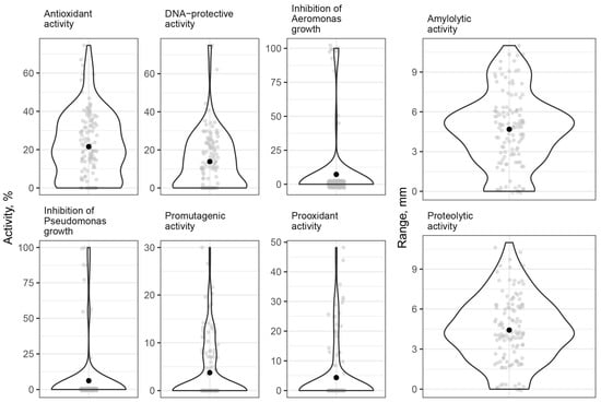
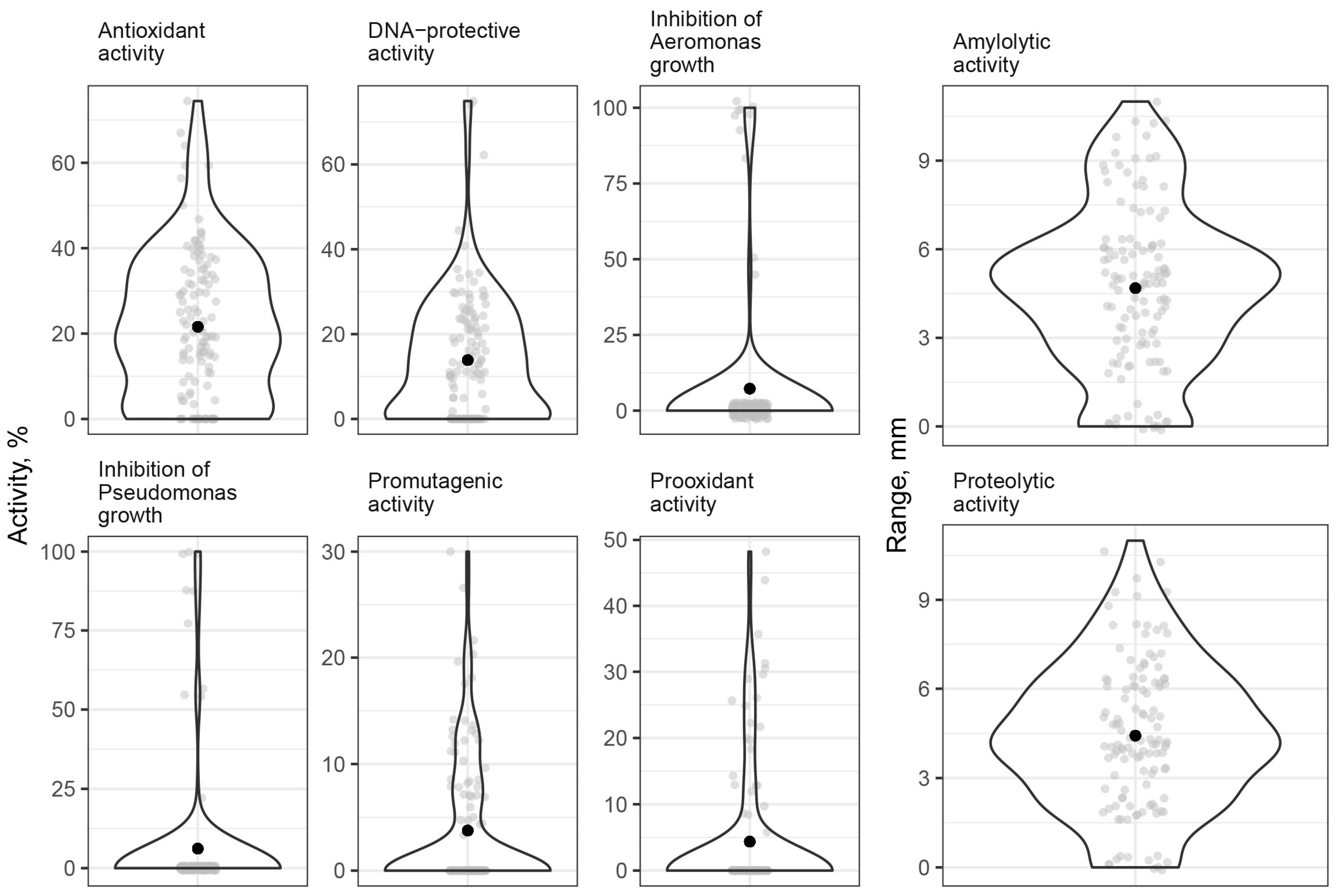
In total, 120 strains were isolated from the sediments (strains MT1-116, MT141, MT142, MT155, MT186). After purification, each strain was tested for all potential probiotic properties: exoenzyme secretion, antimicrobial activity, antioxidant activity, and antimutagenic activity (Figure 1). These strains were compared to two reference laboratory strains, B. subtilis KATMIRA 1933 and B. amyloliquefaciens B1895, which have demonstrated probiotic efficacy in in vivo studies. The reference strains exhibit both enzymatic and antioxidant activities and have been reported to enhance productivity in broiler chickens and laying hens [27,28].
Figure 1.
Evaluation of the extent of potential probiotic properties in Bacillus spp. strains isolated from bottom sediments. Grey dots represent values for individual strains, and black dots indicate the mean value averaged over all strains.
The objective of our study was to isolate strains exhibiting probiotic activities comparable to or exceeding those of the reference strains, while being naturally adapted to the fish intestinal environment and aquatic conditions.
Data for each strain can be found in the Supplementary Materials. Comparative data for two strains with confirmed probiotic activity, B. subtilis KATMIRA 1933 and B. amyloliquefaciens B1895, are also provided.
Proteolytic and amylolytic activities among the strains isolated from sediment exhibited considerable variability, with their distribution approaching a normal pattern within the population. Of the 120 strains analyzed, some showed no notable exoenzyme activity (the assessment method specifically measures exoenzyme activity), whereas others demonstrated strong enzymatic activity, characterized by a clearance zone exceeding 9 mm around the colony. The majority of strains exhibited moderate activity, with clearance zones ranging between 3 and 6 mm.
Lipolytic activity was generally low among the Bacillus spp. strains isolated from bottom sediments. Only 5 out of 120 strains exhibited detectable lipolytic activity, with the width of the iridescent halo surrounding the colonies not exceeding 2 mm.
LUX-biosensor assays enable the detection of both antioxidant and pro-oxidant activities. Antioxidant activity is reflected by a decrease in catalase expression in the LUX-biosensor E. coli MG1655 pKatG in the presence of hydrogen peroxide upon addition of metabolites produced by the test strain. Conversely, pro-oxidant activity is indicated by increased catalase expression in the presence of metabolites produced by the test strain alone.
Most strains produced metabolites with varying degrees of antioxidant activity. However, a small subset (7 strains) exhibited antioxidant activity exceeding 50–55%, as clearly indicated by the upper peak in the violin plot (Figure 1). The majority of strains either lacked pro-oxidant activity or displayed only weak pro-oxidant activity (up to 10%). Nonetheless, some individual Bacillus spp. isolates (20 strains) showed more pronounced pro-oxidant activity, reaching 40–50%, as reflected by the narrow spike in the plot (Figure 1).
DNA-protective and pro-mutagenic activities were evaluated using the same approach as for antioxidant and pro-oxidant activities, assessing the expression level of the Rec operon. Most Bacillus spp. strains isolated from bottom sediments exhibited limited DNA-protective activity, not exceeding 40%, while only a few strains (5) demonstrated high DNA-protective activity, reaching 60–70%. Similarly, most strains showed either no or weak pro-mutagenic activity (up to 10%). However, a small subgroup (21 strains) displayed elevated pro-mutagenic activity, reaching up to 30%.
Most of the isolated strains did not exhibit antimicrobial activity against Gram-negative fish pathogens. Among the 120 strains tested, only 10 demonstrated varying degrees of antimicrobial activity against Pseudomonas aeruginosa and Aeromonas veronii. These strains and their respective inhibition levels are summarized in Table 2.
Table 2.
Antimicrobial activity of cell-free supernatants from potential probiotic strains against fish pathogens.
Thus, all isolated strains were characterized. However, to describe the Bacillus community as a whole, principal component analysis (PCA) was performed on the obtained data.
3.3. Principal Component Analysis of Bacillus spp. Properties
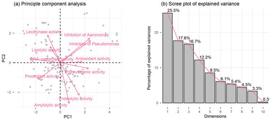
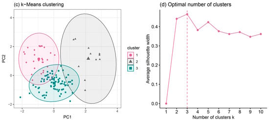
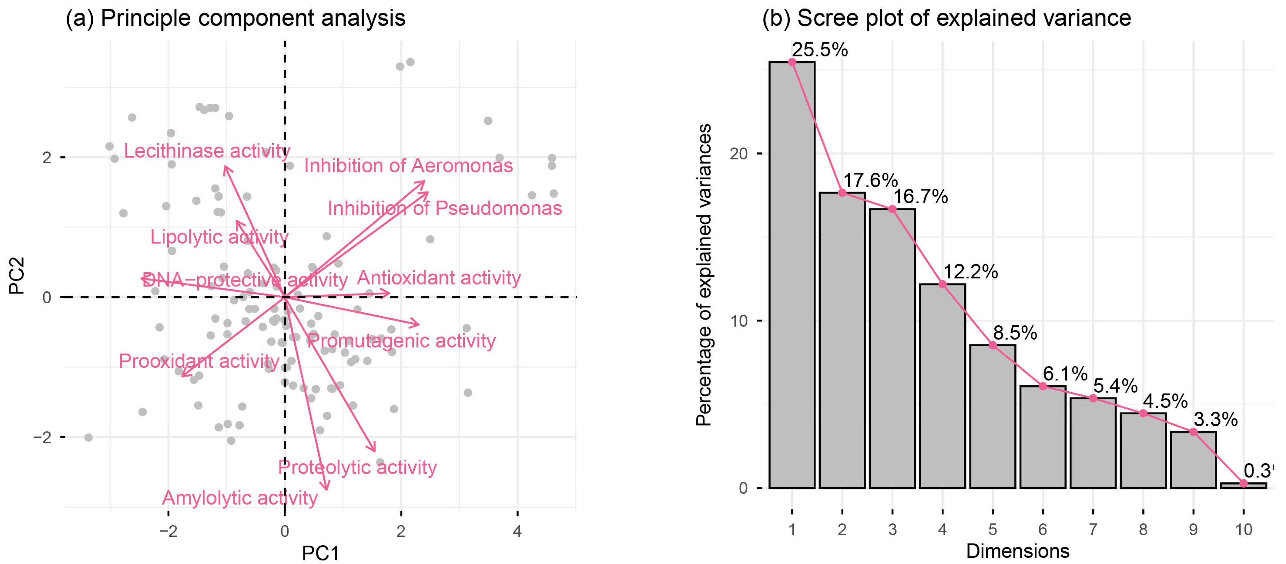
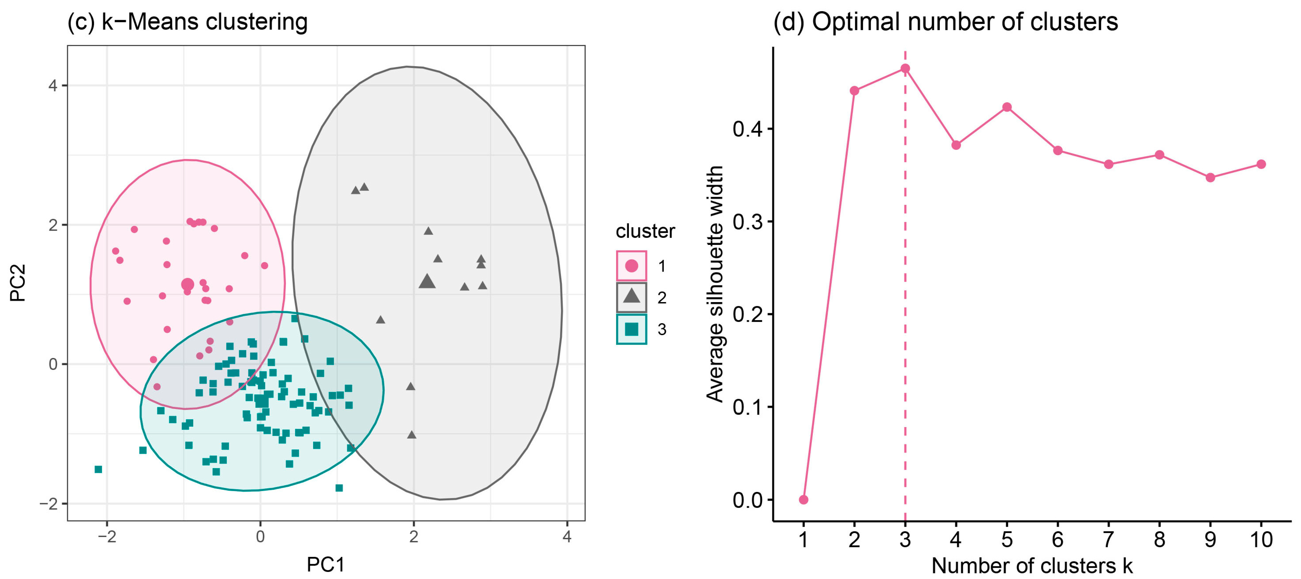
For a comprehensive analysis of the enzymatic, antioxidant and pro-oxidant, antimutagenic and promutagenic, and antimicrobial properties of the strains isolated from bottom sediments, principal component analysis (PCA) was employed (Figure 2). The first principal component (PC1) accounted for 25.5% of the total variance, the second principal component (PC2) explained 17.6%, and the third principal component (PC3) accounted for 16.7%. Thus, the three principal components collectively explained approximately 60% of the total variability. The identified strains clustered into three distinct groups according to PCA.
Figure 2.
Probiotic properties of the strains from bottom sediments via principal component analysis (PCA). (a) Loading plot. Dots represent individual strains. (b) Scree plot. (c) Score plot. (d) Optimal number of clusters plot.
4. Discussion
4.1. Potential Probiotics Isolated from the Fish Intestine
When identifying potential probiotics, it is crucial to define the specific function the probiotic is intended to perform within the host organism and the challenges it aims to mitigate. Based on prior research, the following probiotic functions and properties can be distinguished:
1. Enhancement of feed digestibility during dietary transitions in livestock and poultry [5], during weaning and shifts in diet composition in fish fry [29,30,31,32], as well as during phases of intensive growth in meat-producing animals and poultry [33,34]. In these contexts, selection should prioritize probiotics exhibiting enzymatic activities, particularly the production of proteases, lipases, amylases, and related enzymes.
2. Prevention of bacterial and fungal infections, notably gastrointestinal diseases. Here, probiotics with antibacterial and antifungal activities are sought. Mechanisms include direct inhibition through antimicrobial agents of both specific (e.g., antibiotics, bacteriocins) and nonspecific (e.g., organic acids, reactive oxygen species) nature, as well as interference with quorum sensing and competitive exclusion of pathogenic bacteria [4,5,6,7,8,9].
3. Protection against adverse environmental factors, including exposure to feed contaminated with mycotoxins, bacterial toxins, herbicide residues, and similar compounds. Probiotics with antioxidant and antimutagenic properties [32], alongside the capacity to degrade toxins enzymatically [33,34], are particularly valuable in this context.
4. Modulation of host biochemical processes. The role of gut microbiota in influencing the host’s immune and nervous systems is well established. Although direct bacterial signaling pathways affecting the host remain incompletely understood, several molecular mechanisms have been elucidated. Therefore, it is imperative to screen probiotic candidates for the production of relevant signaling molecules using ELISA techniques or for the presence of genes implicated in the biosynthesis of such molecules [4,5,6,7,8,9].
In this study, the search for potential probiotics for aquaculture focused primarily on the first three criteria, given the limited current understanding of the mechanisms by which gut microbiota influence fish biochemistry.
A critical factor in selecting probiotic strains is their accessibility to end users. This consideration is particularly important when probiotics are intended for large-scale use in agriculture rather than individual medical applications. Such probiotic strains must exhibit a combination of practical traits beyond their immediate functional benefits. These include robust and rapid growth, the ability to proliferate on inexpensive nutrient media, low drying and processing costs, independence from stringent storage conditions, and long shelf life. Collectively, these traits contribute to reducing the overall production costs of probiotic formulations, a major factor in sectors such as aquaculture. Consequently, Bacillus spp. strains exhibiting these characteristics are of particular industrial interest [35,36].
The objective of this study was to identify potential probiotic strains suitable for aquaculture. Initially, we intended to isolate strains from the gut of healthy sterlet individuals. Gut-derived strains are inherently adapted to the gastrointestinal environment, having survived exposure to gastric acidity, digestive enzymes and metabolism adapted to low oxygen levels [37]. The intestinal microbiota is regarded as one of the most promising sources of probiotic candidates [38]. However, the abundance of Bacillus spp. in the intestinal contents of sterlets was low—2.3 ± 0.3 × 10³ CFU/g—and their morphological diversity was limited.
To evaluate the probiotic potential of the isolated strains, their activities were compared with those of B. subtilis KATMIRA 1933 and B. amyloliquefaciens B1895, which served as reference strains. These Bacillus strains have demonstrated probiotic efficacy in previous studies with poultry, enhancing broiler weight gain, increasing egg production in laying hens, and protecting mitochondrial DNA [27,28,29,30,31,32,33,34,35,36,37,38,39].
Unfortunately, none of the strains isolated from the fish gut exhibited probiotic properties comparable to or exceeding those of the reference strains. Measurements of lytic, antioxidant, and DNA-protective activities were uniformly low across all isolates, leading to their exclusion from subsequent investigations.
Consequently, alternative sources of Bacillus were sought for probiotic screening, leading to the examination of microbiota from river bottom sediments.
4.2. Potential Probiotic Bacteria Isolated from Bottom Sediments
It is well established that Bacillus spp. strains isolated from bottom sediments are used as so-called “probiotics” for aquatic environments. Bacilli efficiently absorb organic matter from water and are capable of assimilating excess nitrogen by converting it into a bound form within their own biomass. For this reason, they are commonly utilized in high-density fishpond systems and recirculating aquaculture setups, either as water additives or as part of the biofilter microbial community [12,15,16]. Certain studies have demonstrated that Bacillus-based supplements achieve a less pronounced reduction in detrimental water quality parameters compared to chemical treatments, but concurrently promoting the growth and productivity of aquatic organisms [20]. In this context, Bacillus likely functioned not only as a water purification agent but also as probiotics for crustaceans.
The authors propose that, due to the characteristics of aquatic environments, a unified fish–water–sediment microbiota system exists in aquaculture species, particularly among freshwater and marine coastal and benthic dwellers. Fish harbor microbiomes in their intestines, on their skin, and gills. These microorganisms are released into the surrounding water and subsequently settle in the sediment, thereby becoming integral components of the sediment microbiota. Over time, these bacteria may be resuspended into the water column, especially through the activities of benthic organisms that disturb the sediment. Subsequently, bacteria can recolonize the fish skin, gills, and intestines. This dynamic process supports the concept of a continuous exchange of bacteria between the environment and the associated biota.
Metagenomic data further support the existence of such a unified system. It has been shown that the composition of the microbiota of bottom sediments, water, and the microbiota of aquatic organisms inhabiting the water body largely overlaps [39]. Similarity is lower in fish residing in the water column and predatory species; however, in some cases, the intestinal microbiota and bottom sediments share more than 70% similarity in benthic fish species [40]. Additionally, filter-feeding shrimps from both freshwater and saltwater environments show a high degree of similarity between their intestinal microbiota and that of bottom sediments [41].
Zhang X et al. demonstrated that the fish microbiota is one of the primary sources of bacteria for the microbiota of bottom sediment [42]. Conversely, Zhang B. et al. reported that the microbiota of water and bottom sediments plays a significant role in shaping the microbiota of fish inhabiting these environments. For certain species of fish and crustaceans, the microbiota of bottom sediment serves as the main source for the establishment of intestinal microbiota [40,41].
Therefore, if under natural conditions the microbiota of bottom sediments constitutes a source of intestinal microbiota for fish, the utilization of bottom sediments as a resource for isolating novel probiotics for aquaculture is not only justified but also desirable.
A total of 120 Bacillus spp. strains were isolated from bottom sediments of the Don River, Russia. Only 11% of the population was in the spore form, with the remainder existing in the vegetative state. This suggests that bacilli in bottom sediments are metabolically active and represent an important part of the benthic microbial community.
All 120 isolated strains were tested for potential probiotic properties. It was found that, based on the pattern of property distribution within the Bacillus population, the properties of bacterial isolates can be divided in two groups.
In the first group of properties (lytic activities), the property was present to some extent in all members of the population, and its value distribution was close to a normal distribution. This is illustrated by proteolytic and amylolytic activities, where most of the isolated Bacillus spp. strains exhibited average levels of these properties. Since bacilli are typical r-strategists, focused on rapid consumption of large amounts of organic substrates and fast growth until resource depletion, the presence of extracellular enzyme is essential for their survival [43].
In the second group of properties (antioxidant and DNA-protective activities antimicrobial activity), most of the population either lacked the trait entirely or exhibited weak activity, with only a small subset of strains (6–17%) displaying high activity for the trait. This pattern is characteristic of both advantageous probiotic properties, such as antioxidant and DNA-protective activities and antimicrobial activity, and undesirable features, such as pro-oxidant and pro-mutagenic activities. These traits do not appear to be essential for microbial survival but may offer adaptive advantages under specific environmental conditions.
4.3. Principal Component Analysis of Properties of Bacillus spp. Strains Isolated from Bottom Sediments
To characterize the entire collection of Bacillus spp. strains isolated and to evaluate the relationships among their various potential probiotic properties, principal component analysis (PCA) and cluster analysis were performed.
Principal component analysis provides an effective overview of large datasets and has been widely used in the visualization of metagenomic, meta-transcriptomic, meta-proteomic, and metabolomic data [44,45,46,47].
Among its advantages, PCA enables the identification of correlations between individual traits. The loading plot displays the contributions of variables to the first two principal components. As shown in Figure 2a, antimicrobial activities against Pseudomonas aeruginosa and Aeromonas veronii are strongly correlated. Given that both bacteria are Gram-negative, it is likely that antimicrobial strains produce compounds effective against a broad range of Gram-negative bacteria.
Proteolytic and amylolytic activities exhibited a moderate positive correlation Interestingly, total lipolytic and lecithinase activities were positively correlated with each other but negatively correlated with proteolytic and amylolytic activities. The isolated Bacillus spp. strains generally displayed low lipolytic activity, observed in only a small subset of strains, as was the case for lecithinase activity. Lecithinase belongs to the lipase group (also known as phospholipase C), which explains the correlation between lecithinase activity and total lipolytic activity. Additionally, lecithinase is recognized as a virulence factor [48]. This suggests two alternative life strategies of Bacillus: either active uptake of nutrients or adaptation to pathogenicity.
Antioxidant and pro-oxidant activities were not correlated. At first glance, this may seem unusual; however, some strains simultaneously produced both antioxidant and pro-oxidant metabolites, which was reflected in the arrangement of variable vectors in the loading plot.
Cluster analysis showed that Bacillus spp. strains isolated from bottom sediments can be classified into three distinct clusters. Cluster #2 (Figure 2c) comprised antimicrobial bacilli and strains exhibiting pronounced antioxidant properties. Cluster #3 (#3 in Figure 2c) comprised bacilli with high levels of extracellular enzyme. The last cluster (#1 in Figure 2c) consisted of strains lacking prominent traits, although many displayed lipolytic and lecithinase activities.
These findings suggest the existence of three potential ecological strategies among Bacillus spp. from bottom sediments: (1) growth and development driven by active exoenzyme production, enabling rapid utilization of complex organic compounds entering the sediment; (2) competitive advantage through the expression of additional traits, including antimicrobial activities targeting competitors or antioxidant activities conferring protection against reactive oxygen species; and (3) advantage via conditional pathogenicity and the capacity to utilize other organisms as habitats. Our study focused on Bacillus belonging to the first two groups.
4.4. Efficacy of Strains in In Vivo Experiments
For further studies, eleven strains exhibiting either enzymatic activity, high antioxidant and antimutagenic activity, or antimicrobial properties were selected. These strains correspond to the prominent peaks in the violin plot (Figure 1), surpassing test strains B. subtilis KATMIRA 1933 and B. amyloliquefaciens B1895 in the manifestation of potential probiotic properties.
Some of the selected strains underwent safety testing and sequencing and were used for the formulation of probiotic preparations. These preparations were evaluated in experiments on sterlet (Acipenser ruthenus Linnaeus, 1758) and carp (Cyprinus carpio Linnaeus, 1758) [49,50]. Although comprehensive results are not presented here and can be found in the cited references, it is notable that B. velezensis strains MT14, MT42, MT141, and MT142—characterized by pronounced proteolytic and amylolytic activities—significantly increased biomass growth in carp juveniles. B. velezensis strains MT14 and MT42, as well as B. subtilis MT48 and MT74, promoted biomass increase in juvenile sterlet, accompanied by upregulated expression of the interleukin-1 beta (IL-1β) gene, which is associated with anti-inflammatory effects. Additionally, B. subtilis strains MT48 and MT74 reduced the expression of genes encoding insulin-like growth factor 1 (IGF-1), 70 kDa heat shock proteins (HSP70), and glutathione S-transferases (GST), suggesting a potential role of their antioxidant activity in stress alleviation.
These findings indicate that Bacillus spp. strains isolated from bottom sediments possess significant probiotic potential and may exert stimulatory effects in in vivo tests with live fish.
5. Conclusions
This study evaluated the potential of bottom sediments as a promising source of probiotic Bacillus strains, offering advantages over isolation from fish intestines. We conducted a comprehensive phenotypic analysis of the isolated Bacillus spp. strains, including assessments of their proteolytic and amylolytic activities, as well as antioxidant and DNA-protective properties. Additionally, the antimicrobial activity of these microorganisms was investigated. Principal component analysis was employed for integrative data description and dimensionality reduction, allowing the identification of key parameters characterizing the functional potential of Bacillus strains from bottom sediments. Based on the results, it can be concluded that bottom sediments represent a valuable and efficient reservoir for isolating probiotic Bacillus with a broad spectrum of beneficial biological properties.
Supplementary Materials
The following supporting information can be downloaded at https://www.mdpi.com/article/10.3390/environments13010013/s1, Table S1: Probiotic Properties of individual strain isolated from sediment deposits.
Author Contributions
Conceptualization, M.S.M. and D.V.R.; methodology, M.S.M., E.V.M. and S.A.E.; investigation, M.S.M., E.V.M. and S.A.E.; resources, D.V.R.; writing—original draft preparation, M.S.M. and S.V.K.; project administration, D.V.R.; funding acquisition, D.V.R. All authors have read and agreed to the published version of the manuscript.
Funding
The research was supported by the Russian Science Foundation, grant No. 23-76-30006.
Institutional Review Board Statement
The study was approved by the Local Independent Ethics Committee of Don State Technical University (Rostov-on-Don, Russian Federation). The approval date is 9 July 2024. During the experiments, recommendations for the humane and ethical treatment of laboratory animals were followed, including those in accordance with Directive 2010\/63\/EU of the European Parliament and of the Council of 22 September 2010 on the protection of animals used for scientific purposes.
Data Availability Statement
The data presented in this study are available on request from the corresponding author.
Acknowledgments
The authors express their deep gratitude to Konstantin Demin for assistance with the bioinformatic analysis.
Conflicts of Interest
The authors declare no conflicts of interest.
References
- Servili, A.; Canario, A.V.M.; Mouchel, O.; Muñoz-Cueto, J.A. Climate Change Impacts on Fish Reproduction Are Mediated at Multiple Levels of the Brain-Pituitary-Gonad Axis. Gen. Comp. Endocrinol. 2020, 291, 113439. [Google Scholar] [CrossRef] [Scilit] [PubMed]
- Scherer, L.; Boom, H.A.; Barbarossa, V.; van Bodegom, P.M. Climate Change Threats to the Global Functional Diversity of Freshwater Fish. Glob. Change Biol. 2023, 29, 3781–3793. [Google Scholar] [CrossRef] [Scilit]
- Elgendy, M.Y.; Ali, S.E.; Abbas, W.T.; Algammal, A.M.; Abdelsalam, M. The Role of Marine Pollution on the Emergence of Fish Bacterial Diseases. Chemosphere 2023, 344, 140366. [Google Scholar] [CrossRef] [Scilit]
- El-Saadony, M.T.; Alagawany, M.; Patra, A.K.; Kar, I.; Tiwari, R.; Dawood, M.A.O.; Dhama, K.; Abdel-Latif, H.M.R. The Functionality of Probiotics in Aquaculture: An Overview. Fish Shellfish Immunol. 2021, 117, 36–52. [Google Scholar] [CrossRef] [Scilit] [PubMed]
- Olmos, J.; Acosta, M.; Mendoza, G.; Pitones, V. Bacillus Subtilis, an Ideal Probiotic Bacterium to Shrimp and Fish Aquaculture That Increase Feed Digestibility, Prevent Microbial Diseases, and Avoid Water Pollution. Arch. Microbiol. 2020, 202, 427–435. [Google Scholar] [CrossRef] [Scilit] [PubMed]
- Hai, N.V. The Use of Probiotics in Aquaculture. J. Appl. Microbiol. 2015, 119, 917–935. [Google Scholar] [CrossRef] [Scilit]
- Lee, Y.; Nguyen, T.L.; Roh, H.; Kim, A.; Park, J.; Lee, J.Y.; Kang, Y.R.; Kang, H.; Sohn, M.Y.; Park, C.I. Mechanisms Underlying Probiotic Effects on Neurotransmission and Stress Resilience in Fish via Transcriptomic Profiling. Fish Shellfish Immunol. 2023, 141, 109063. [Google Scholar] [CrossRef] [Scilit]
- Tarnecki, A.M.; Wafapoor, M.; Phillips, R.N.; Rhody, N.R. Benefits of a Bacillus Probiotic to Larval Fish Survival and Transport Stress Resistance. Sci. Rep. 2019, 9, 4892. [Google Scholar] [CrossRef] [Scilit]
- Langlois, L.; Akhtar, N.; Tam, K.C.; Dixon, B.; Reid, G. Fishing for the Right Probiotic: Host-Microbe Interactions at the Interface of Effective Aquaculture Strategies. FEMS Microbiol. Rev. 2021, 45, fuab030. [Google Scholar] [CrossRef] [Scilit]
- Farrell, A.; Brauner, C.; Randall, D.J.; Eliason, E. The 50th Anniversary Issue of Fish Physiology: Physiological Systems and Development; Elsevier: Amsterdam, The Netherlands, 2023. [Google Scholar]
- Hoar, W.S.; Randall, D.J.; Donaldson, E.M. Fish Physiology; Academic Press: London, UK, 1983. [Google Scholar]
- Yi, M.; Wang, C.; Wang, H.; Zhu, X.; Liu, Z.; Gao, F.; Lu, M. The In Situ Remediation of Aquaculture Water and Sediment by Commercial Probiotics Immobilized on Different Carriers. Water Reuse 2021, 11, 572–585. [Google Scholar]
- Wang, Y.; He, Z. Effect of Probiotics on Alkaline Phosphatase Activity and Nutrient Level in Sediment of Shrimp, Penaeus vannamei, Ponds. Aquaculture 2009, 287, 94–97. [Google Scholar] [CrossRef] [Scilit]
- Zhang, D.; Li, H.; Liu, Y.; Qiao, G.; Chi, S.; Song, J. Screening and Identification of Organics-Degrading Bacteria from the Sediment of Sea Cucumber Apostichopus japonicus Ponds. Aquac. Int. 2016, 24, 373–384. [Google Scholar] [CrossRef] [Scilit]
- Truong, Q.P.; Phan, T.C.; Vu, H.H.; Pham, T.T.; Huynh, T.G.; Vu, N.U. Isolation of Potential Probiotic Bacillus subtilis CM3.1 and Its Effects on the Water Quality and Growth Performance of the Whiteleg Shrimp Litopenaeus vannamei in the Mekong Delta, Vietnam. Aquac. Aquar. Conserv. Legis. 2021, 14, 3347–3357. [Google Scholar]
- Rudoy, D.; Meskhi, B.; Olshevskaya, A.; Kozyrev, D.; Shevchenko, V.; Odabashyan, M.; Rybak, A. Analysis of Edaphic Factors on the Role of Probiotics in the Development of Sustainable and Productive Aquaculture. Fishes 2025, 10, 457. [Google Scholar] [CrossRef] [Scilit]
- Rwezawula, P.; Waiswa Mwanja, W.; Vereecke, N.; Bossier, P.; Vanrompay, D. Advancing Aquaculture Probiotic Discovery via an Innovative Protocol for Isolation of Indigenous, Heat and Salt Tolerant, Quorum Quenching Probiotic Candidates. Front. Microbiol. 2025, 16, 1558238. [Google Scholar] [CrossRef] [Scilit]
- Morozova, M.; Gorovcov, A.; Prazdnova, E.; Basankina, V.; Chistyakov, V.; Pepoyan, A.; Miralimova, S.; Grigoriyev, V. Bacteria of Genus Bacillus as Antagonists of Pathogens in Aquaculture. Vestnik Astrakhan State Tech. Univ. Ser. Fish. Ind. 2023, 1, 89–97. [Google Scholar]
- James, G.; Radhakrishnan, V.; Marathippallam Jamal, J.; Prasannan Geetha, P.; Pillai, D.; Vattiringal Jayadradhan, R.K. Exploring the Probiotic Potential of B. cereus and B. velezensis Associated with Mangrove Sediments in Aquaculture. Discov. Ocean. 2025, 2, 15. [Google Scholar] [CrossRef] [Scilit]
- Wu, X.; Wu, H.; Wu, Y.; Xu, Z.; Shan, H.; Gao, T. Effects of Different Sediment Improvers on the Growth Environment, Innate Immune Responses, and Intestinal Health of Procambarus clarkii. Biology 2025, 14, 407. [Google Scholar] [CrossRef] [Scilit]
- Popov, I.V.; Skripkin, V.S.; Mazanko, M.S.; Epimakhova, E.E.; Prazdnova, E.V.; Dilekova, O.V.; Dannikov, S.P.; Popov, I.V.; Trukhachev, V.I.; Rastovarov, E.I.; et al. Effects of Spore-Forming Bacillus Probiotics on Growth Performance, Intestinal Morphology, and Immune System of Broilers Housed on Deep Litter. J. Appl. Poult. Res. 2024, 33, 100396. [Google Scholar] [CrossRef] [Scilit]
- Chistyakov, V.A.; Prazdnova, E.V.; Mazanko, M.S.; Bren, A.B. The Use of Biosensors to Explore the Potential of Probiotic Strains to Reduce the SOS Response and Mutagenesis in Bacteria. Biosensors 2018, 8, 25. [Google Scholar] [CrossRef] [Scilit]
- Li, J.; Wu, J.; Wang, J.; Wang, X. Phenotypic Variations Induced Emergence of Orientation Order and Morphology in Bacillus subtilis Biofilm Growth. Biochem. Biophys. Res. Commun. 2023, 686, 149198. [Google Scholar] [CrossRef] [Scilit] [PubMed]
- Wang, X.; Kong, Y.; Zhao, H.; Yan, X. Dependence of the Bacillus subtilis Biofilm Expansion Rate on Phenotypes and the Morphology under Different Growing Conditions. Dev. Growth Differ. 2019, 61, 431–443. [Google Scholar] [CrossRef] [Scilit] [PubMed]
- Li, X.; Kong, R.; Wang, J.; Wu, J.; He, K.; Wang, X. The Formation Mechanism of Bacillus subtilis Biofilm Surface Morphology under Competitive Environment. Can. J. Microbiol. 2023, 69, 251–261. [Google Scholar] [CrossRef] [Scilit] [PubMed]
- Wang, J.; Li, X.; Kong, R.; Wu, J.; Wang, X. Fractal Morphology Facilitates Bacillus subtilis Biofilm Growth. Environ. Sci. Pollut. Res. Int. 2022, 29, 56168–56177. [Google Scholar] [CrossRef] [Scilit]
- Mazanko, M.S.; Gorlov, I.F.; Prazdnova, E.V.; Makarenko, M.S.; Usatov, A.V.; Bren, A.B.; Chistyakov, V.A.; Tutelyan, A.V.; Komarova, Z.B.; Mosolova, N.I.; et al. Bacillus Probiotic Supplementations Improve Laying Performance, Egg Quality, Hatching of Laying Hens, and Sperm Quality of Roosters. Probiotics Antimicrob. Proteins 2018, 10, 367–373. [Google Scholar] [CrossRef] [Scilit]
- Prazdnova, E.V.; Chistyakov, V.A.; Churilov, M.N.; Mazanko, M.S.; Bren, A.B.; Volski, A.; Chikindas, M.L. DNA-Protection and Antioxidant Properties of Fermentates from Bacillus amyloliquefaciens B-1895 and Bacillus subtilis KATMIRA1933. Lett. Appl. Microbiol. 2015, 61, 549–554. [Google Scholar] [CrossRef] [Scilit]
- Salehi, M.; Bagheri, D.; Sotoudeh, E.; Ghasemi, A.; Mozanzadeh, M.T. The Combined Effects of Propionic Acid and a Mixture of Bacillus spp. Probiotic in a Plant Protein-Rich Diet on Growth, Digestive Enzyme Activities, Antioxidant Capacity, and Immune-Related Genes mRNA Transcript Abundance in Lates calcarifer Fry. Probiotics Antimicrob. Proteins 2023, 15, 655–667. [Google Scholar] [CrossRef] [Scilit]
- Yadav, M.K.; Kumari, I.; Singh, B.; Sharma, K.K.; Tiwari, S.K. Probiotics, Prebiotics and Synbiotics: Safe Options for Next-Generation Therapeutics. Appl. Microbiol. Biotechnol. 2022, 106, 505–521. [Google Scholar] [CrossRef] [Scilit]
- Ayalew, H.; Zhang, H.; Wang, J.; Wu, S.; Qiu, K.; Qi, G.; Tekeste, A.; Wassie, T.; Chanie, D. Potential Feed Additives as Antibiotic Alternatives in Broiler Production. Front. Vet. Sci. 2022, 9, 916473. [Google Scholar] [CrossRef] [Scilit]
- Prazdnova, E.V.; Mazanko, M.S.; Chistyakov, V.A.; Bogdanova, A.A.; Refeld, A.G.; Kharchenko, E.Y.; Chikindas, M.L. Antimutagenic Activity as a Criterion of Potential Probiotic Properties. Probiotics Antimicrob. Proteins 2022, 14, 1094–1109. [Google Scholar] [CrossRef] [Scilit]
- Liu, L.; Xie, M.; Wei, D. Biological Detoxification of Mycotoxins: Current Status and Future Advances. Int. J. Mol. Sci. 2022, 23, 1064. [Google Scholar] [CrossRef] [Scilit]
- Pop, O.L.; Suharoschi, R.; Gabbianelli, R. Biodetoxification and Protective Properties of Probiotics. Microorganisms 2022, 10, 1278. [Google Scholar] [CrossRef] [Scilit] [PubMed]
- Jeżewska-Frąckowiak, J.; Seroczyńska, K.; Banaszczyk, J.; Jedrzejczak, G.; Żylicz-Stachula, A.; Skowron, P.M. The Promises and Risks of Probiotic Bacillus Species. Acta Biochim. Pol. 2018, 65, 509–519. [Google Scholar] [CrossRef] [Scilit] [PubMed]
- Kimura, K.; Yokoyama, S. Trends in the Application of Bacillus in Fermented Foods. Curr. Opin. Biotechnol. 2019, 56, 36–42. [Google Scholar] [CrossRef] [Scilit]
- Nakano, M.M.; Hulett, F.M. Adaptation of Bacillus subtilis to Oxygen Limitation. FEMS Microbiol. Lett. 1997, 157, 1–7. [Google Scholar] [CrossRef] [PubMed]
- Mazanko, M.S.; Popov, I.V.; Prazdnova, E.V.; Refeld, A.G.; Bren, A.B.; Zelenkova, G.A.; Chistyakov, V.A.; Algburi, A.; Weeks, R.M.; Ermakov, A.M.; et al. Beneficial Effects of Spore-Forming Bacillus Probiotic Bacteria Isolated from Poultry Microbiota on Broilers’ Health, Growth Performance, and Immune System. Front. Vet. Sci. 2022, 9, 877360. [Google Scholar] [CrossRef] [Scilit]
- Huang, Z.; Hou, D.; Zhou, R.; Zeng, S.; Xing, C.; Wei, D.; He, J. Environmental Water and Sediment Microbial Communities Shape Intestine Microbiota for Host Health: The Central Dogma in an Anthropogenic Aquaculture Ecosystem. Front. Microbiol. 2021, 12, 772149. [Google Scholar] [CrossRef] [Scilit]
- Zhang, B.; Xiao, J.; Liu, H.; Zhai, D.; Wang, Y.; Liu, S.; Xiong, F.; Xia, M. Vertical Habitat Preferences Shape the Fish Gut Microbiota in a Shallow Lake. Front. Microbiol. 2024, 15, 1341303. [Google Scholar] [CrossRef] [Scilit]
- Fan, L.; Wang, Z.; Chen, M.; Qu, Y.; Li, J.; Zhou, A.; Zou, J. Microbiota Comparison of Pacific White Shrimp Intestine and Sediment at Freshwater and Marine Cultured Environment. Sci. Total Environ. 2019, 657, 1194–1204. [Google Scholar] [CrossRef] [Scilit]
- Zhang, X.; You, Y.; Peng, F.; Tang, X.; Zhou, Y.; Liu, J.; Lin, D.; Zhou, Y. Interaction of Microbiota between Fish and the Environment of an In-Pond Raceway System in a Lake. Microorganisms 2022, 10, 1143. [Google Scholar] [CrossRef] [Scilit]
- Fierer, N.; Bradford, M.A.; Jackson, R.B. Toward an Ecological Classification of Soil Bacteria. Ecology 2007, 88, 1354–1364. [Google Scholar] [CrossRef] [Scilit]
- Wintermans, B.; Brandt, B.; Vandenbroucke-Grauls, C.; Budding, A. TreeSeq, a Fast and Intuitive Tool for Analysis of Whole Genome and Metagenomic Sequence Data. PLoS ONE 2015, 10, e0123851. [Google Scholar] [CrossRef] [Scilit]
- Mansfeldt, C.B.; Rowe, A.R.; Heavner, G.L.W.; Zinder, S.H.; Richardson, R.E. Meta-Analyses of Dehalococcoides mccartyi Strain 195 Transcriptomic Profiles Identify a Respiration Rate-Related Gene Expression Transition Point and Interoperon Recruitment of a Key Oxidoreductase Subunit. Appl. Environ. Microbiol. 2014, 80, 6062–6072. [Google Scholar] [CrossRef] [Scilit] [PubMed]
- Vahedi, M.; Kabiri, M.; Salami, S.A.; Rezadoost, H.; Mirzaie, M.; Kanani, M.R. Quantitative HPLC-Based Metabolomics of Some Iranian Saffron (Crocus sativus L.) Accessions. Ind. Crops Prod. 2018, 118, 26–29. [Google Scholar] [CrossRef] [Scilit]
- Zhang, L.; Qu, H.; Liu, X.; Li, Q.; Liu, Y.; Wang, W.; Chen, D.; Xiao, L.; Gu, R. Comparison and Selection of Probiotic Lactobacillus from Human Intestinal Tract and Traditional Fermented Food In Vitro via PCA, Unsupervised Clustering Algorithm, and Heat-Map Analysis. Food Sci. Nutr. 2022, 10, 4247–4257. [Google Scholar] [CrossRef] [Scilit] [PubMed]
- Sharaf, E.F.; El-Sayed, W.S.; Abosaif, R.M. Lecithinase-Producing Bacteria in Commercial and Home-Made Foods: Evaluation of Toxic Properties and Identification of Potent Producers. J. Taibah Univ. Sci. 2014, 8, 207–215. [Google Scholar] [CrossRef] [Scilit]
- Prazdnova, E.V.; Mazanko, M.S.; Shevchenko, V.N.; Skripnichenko, R.V.; Kulikov, M.P.; Golovko, L.S.; Rudoy, D.V. Genomic Characterization of Four Novel Probiotic Strains with Enzymatic Activity and Their Effects on Carp (Cyprinus carpio). Animals 2025, 15, 1998. [Google Scholar] [CrossRef] [Scilit]
- Prazdnova, E.V.; Mazanko, M.S.; Kulikov, M.P.; Shevchenko, V.N.; Emelyantsev, S.A.; Chistyakov, V.A.; Grigoryev, V.A.; Kulikova, D.B.; Vibe, V.V.; Chikindas, M.L.; et al. Evaluation of Effects and Genomic Analysis of Aquaculture Probiotic Strains with Antioxidant and Enzymatic Activity. Mol. Nutr. Food Res. 2025, e70277. [Google Scholar] [CrossRef] [Scilit]

Disclaimer/Publisher’s Note: The statements, opinions and data contained in all publications are solely those of the individual author(s) and contributor(s) and not of MDPI and/or the editor(s). MDPI and/or the editor(s) disclaim responsibility for any injury to people or property resulting from any ideas, methods, instructions or products referred to in the content. |
© 2025 by the authors. Licensee MDPI, Basel, Switzerland. This article is an open access article distributed under the terms and conditions of the Creative Commons Attribution (CC BY) license.