


Highlights
- This study examines the effects of simulated space-like proton irradiation on olivine and pyroxene in three meteorites (NWA 4560, NWA 5838 and Dho 007) using LV-SEM and Raman spectroscopy.
- High-fluence irradiation with protons up to 12 keV (1019 ions/cm2 fluence, ~30 h duration) caused severe lattice damage and extensive amorphization in all silicates.
- Raman spectral shifts and peak broadening indicate mineral-dependent irradiation responses, with similar behavior in NWA olivines and more variable changes in Dho 007 pyroxenes.
- The appearance of three characteristic amorphous Raman bands (~550–1000 cm−1, ~1100–1700 cm−1, and ~1700–1850 cm−1) confirms widespread amorphization under intense irradiation.
- Elemental fractionation reflects preferential sputtering, leading to Mg loss and Fe enrichment in olivines and Ca depletion with compositional redistribution in pyroxenes.
Abstract
This study reveals the effects of artificial space-like proton irradiation on three meteorite samples that are Northwest Africa (NWA) 4560 LL3.2 and NWA 5838 H6 chondrite meteorites, as well as the Dhofar (Dho) 007 eucrite. We used low-vacuum scanning electron microscopy (LV-SEM) and Raman Spectroscopy to examine the structure and composition of olivine and pyroxene grains in the meteorites before and after the irradiation events. This article focuses on the strongest and most intense irradiation, which was performed by protons up to 12 keV with a fluence value of 1019 ions/cm2 that lasted ~30 h. According to the Raman spectra, significant lattice disruption in all analyzed silicates occurred, and a more extensive amorphous, glassy layer developed under the strongest irradiation conditions. Relative to the second irradiation, peak 1 (820.0 cm−1) shifts slightly negatively (–0.46 cm−1) with a small FWHM increase (+0.88 cm−1), while peak 2 (850.3 cm−1) shifts positively in both parameters (+0.40 and +4.04 cm−1) in NWA 4560 olivines. In NWA 5838 olivines, both olivine peaks (820.5 and 850.8 cm−1) shift positively (+7.40 and +7.90 cm−1) and broaden (+2.75 and +4.29 cm−1). In Dho 007 pyroxenes, peak 1 (997.1 cm−1) shifts positively (+3.01 cm−1) with an FWHM decrease (−0.46 cm−1), peak 2 (669.7 cm−1) shifts slightly negatively (−0.75 cm−1) while broadening strongly (+29.23 cm−1), and peak 3 (327.7 cm−1) shifts positively (+0.86 cm−1) with reduced FWHM (−4.55 cm−1). Three characteristic amorphous bands appear in all examined meteorite silicates, located at ~550–1000 cm−1, ~1100–1700 cm−1, and ~1700–1850 cm−1. Olivines in NWA 4560 and NWA 5838 exhibited similar responses across all irradiation events. In contrast, Dho 007 pyroxenes showed variable compositional changes without a consistent or well-defined pattern in our SEM dataset. The Fo decrease in our experiments likely results from preferential Mg sputtering in the olivine lattice, leading to relative Fe enrichment, similar to but more pronounced than after the first irradiation. Pyroxenes exhibit a comparable response, with Fs and En increasing and Wo sharply decreasing, reflecting preferential Ca loss relative to Mg alongside Fe enrichment. Investigating these processes improves the interpretation of planetary remote sensing data and advances our understanding of planetary surface evolution, while also clarifying how surface materials respond to space environmental conditions.
1. Introduction
Space weathering is a complex and dynamic process that alters the surfaces of airless planetary bodies through irradiation, particle implantation, and sputtering mechanisms driven by galactic and solar energetic particles, ultraviolet (UV) irradiation, and daily temperature fluctuations. Among these processes, the primary agents of space weathering are solar wind irradiation and micrometeorite impacts (e.g., [1,2,3,4]). These cumulative processes progressively modify the spectral and optical appearance of the surface [5,6,7,8], as well as the underlying mineral structure and chemical composition of asteroid regolith. The alterations occur through microscale fracturing, localized melting, ablation, ion implantation, and the progressive destruction of the crystalline lattice, ultimately leading to partial or complete amorphization (e.g., [9,10,11,12]). In addition, the interaction of these space-weathering agents contributes to the formation of nanophase metallic iron and other optically active submicroscopic particles, which further influences the observed reflectance spectra and albedo properties of asteroid and lunar surfaces [13].
The investigation of space weathering uses mostly VIS-NIR spectroscopic techniques; here we applied Fourier-transform infrared (FTIR) spectroscopy and Raman spectroscopy to understand the crystalline changes and what is essential for elucidating how cosmic radiation, solar wind particles [14,15,16,17,18,19], and micrometeorite impacts modify the physical and chemical properties of planetary surfaces, particularly those of airless bodies like the Moon and asteroids (e.g., [20,21]). By systematically examining space-weathering effects through multiple spectroscopic methods, researchers can refine the interpretation of remote sensing data collected by planetary and space missions (e.g., [22,23,24]) and thereby gain a more comprehensive understanding of the processes that govern planetary surface evolution [1]. Moreover, these techniques provide critical information on how surface materials respond to the extreme environmental conditions of space, including vacuum exposure, temperature fluctuations, and high-energy particle interactions, which together shape the long-term spectral and compositional characteristics of planetary regoliths. Raman spectroscopy enables high-resolution analysis of crystal structures, revealing subtle structural distortions and vibrational mode variations associated with irradiation and micrometeorite-induced alterations [25].
This study summarizes the results of artificially simulated solar wind irradiation experiments (three steps of proton irradiation events) conducted on three distinct meteorite samples (i.e., Northwest Africa (NWA) 4560 LL3.2 and NWA 5838 H6 ordinary chondrites, as well as Dhofar (Dho) 007 eucrite), with the objective of investigating and understanding the mineral-scale consequences of such exposure (particularly the third, strongest step).
The background and preliminary results of our recent research in similar irradiation tests are established in [26], in which we examined the above-mentioned three meteorites in a two-step, weaker-intensity proton irradiation experiment with the same method. In this study, we went beyond the former irradiations by an order of magnitude to generate major crystal lattice redistribution inside the meteorite minerals. In [26], we found that olivine (NWA 4560 and NWA 5838) compositions showed significant variations following each proton irradiation event, whereas pigeonitic pyroxenes (Dho 007) exhibited greater compositional stability with smaller fluctuations. In NWA 4560 olivines, a slight Fo-content decrease (−2.93 mol%) occurred after weaker irradiation, likely due to metastable alteration, followed by a pronounced increase (+8.71 mol%) under stronger irradiation, associated with amorphization-driven structural change. Raman spectral analyses revealed negative peak shifts after the first irradiation and divergent behaviors among samples after the second irradiation, indicating complex structural responses. The observed peak broadening after the first irradiation and subsequent peak narrowing in some samples suggested partial recovery or reorganization of the crystal lattice. Decreasing band intensities, particularly in olivine doublets, were linked to amorphization processes, consistent with previous findings by [27,28]. These spectral changes correlated with compositional alterations, such as Mg sputtering leading to Fe enrichment and metallic Fe formation during reduction processes. Pyroxenes displayed similar but less pronounced irradiation-induced effects, implying a parallelism in the structural evolution of both silicate minerals. Overall, the results indicated that increasing irradiation intensity promotes amorphization, compositional redistribution, and potential reorganization within the silicate crystal lattice structures.
The outcomes provide valuable insights that can enhance the interpretation of space-weathered asteroid spectra and contribute to a more comprehensive understanding of location-dependent surface alteration processes in the Solar System, which are stronger closer to the Sun and scale with the incoming flux. These results are significant not only from a scientific perspective but also in the context of developing mitigation strategies for Near-Earth Asteroids (NEAs) [29], where the physical and chemical characteristics of the regolith may strongly influence the practical mode or technology of realization of various deflection or surface interaction methods as well as the reaction of the regolith composed of grains [30].
2. Samples and Analytical Methods
During the laboratory investigations, low-vacuum scanning electron microscopy (LV-SEM) and Raman spectroscopy were employed to analyze the samples (fragments cut on one side) before and after the irradiation procedures.
2.1. Samples
The three analyzed samples in this work were the NWA 4560 LL3.2 and NWA 5838 H6 ordinary chondrites, as well as the Dho 007 Eucrite-cm achondrite [31,32]. The mineralogical, elemental and isotopic compositions, petrological characteristics and other additional properties of these meteorites have been summarized and described in detail in [26].
During the analysis, it is important to measure exactly (as much as possible) the same location as previously, e.g., before the first or the previous irradiation event. In this work, at the SEM measurements, the locations could be identified in the back-scattered electron (BSE) images. Only such grains (or phases) were selected for measurements at the beginning, which were large enough to clearly measure the given phase without their surrounding grains (the different grains could be well separated in BSE images). These locations were identified and found clearly using formerly recorded and saved images. For Raman measurements, the same locations were identified as much as possible by the instrument capacity; however, in the case of Raman data, only averages were used, which decreases any uncertainty.
2.2. Irradiation Method
During the laboratory experiments, 1 keV protons generated by the ECR ion source at the HUN-REN Institute for Nuclear Research (ATOMKI) were employed with fluence values of 1017 and 1019 ions/cm2 for the first and second irradiation [26,33], while protons up to 12 keV were produced with a fluence value of 1019 ions/cm2 for the third and strongest irradiation that lasted ~30 h. Irradiations were performed under high-vacuum conditions at pressures below 1 × 10⁻⁷ mbar within the experimental chamber on the cut but not polished surface of meteorite slices. The ion beam current ranged from 20 nA to 3 A, corresponding to fluxes between 3 × 10¹⁰ and 4.5 × 10¹² ions/cm2/s over an irradiated area of 4 cm2 total. During the irradiation, the sliced samples were positioned on a 10 mm thick aluminum plate, and their temperatures was monitored by a K-type thermocouple, and the meteorites never reached +45 °C.
Based on the above-mentioned values, our samples received a total fluence of 1019 ions/cm2 during the 30 h irradiation period. According to the analysis presented in [34], the flux of the protons up to 12 keV (10−2 MeV) is around the scale of 103 particles/cm2/s at the lunar surface. Using our total dose, the irradiation is roughly equal to 1019/103 = 1016 s, i.e., about 317 Ma; however, due to the possible uncertainties, we adopt a conservative timescale of 100 Ma in this work. Because laboratory exposure times are orders of magnitude shorter than typical astrophysical timescales, substantially higher fluxes than those occurring under natural space-weathering conditions are applied to compensate for this discrepancy. Through the irradiation events, the same samples were irradiated consecutively three times using the above-described different conditions.
2.3. Low-Vacuum Scanning Electron Microscopy (LV-SEM)
Low-vacuum scanning electron microscopy (LV-SEM) was carried out using a JEOL JSM-IT700 HR scanning electron microscope equipped with an Aztec X-ACT Premium SDD spectrometer (Poway, CA, USA) at the Institute for Geological and Geochemical Research, Research Centre for Astronomy and Earth Sciences, HUN-REN. The instrument was operated at an accelerating voltage of 20 keV and a beam current of 6 nA, with a focused beam diameter of 1 μm. Acquisition times were 40 s for analyses and 100 s for areal measurements. Quantification of the collected EDS spectra was performed using natural standards supplied by the C. M. Taylor Corporation (olivine, albite, and wollastonite). All analytical results were normalized to a total of 100 wt%. Detailed LV-SEM data are presented in Table S1. A degree of uncertainty is inherent in the LV-SEM method (e.g., due to the absence of carbon coating on the samples); however, data points where such effects would significantly influence the outcomes, considering the trends of changes in mineralogy, were excluded from the final evaluation.
2.4. Raman Spectroscopy Method
Raman analyses were conducted using a Thermo Scientific DXR Raman spectrometer (Waltham, MA, USA) equipped with a 780 nm excitation laser, delivering 1.0 mW of power at a spot size of ~1.0 μm. Spectra were collected with integration times of 15 × 3 s. Data processing and curve fitting were performed with the Fityk 1.3.1 software. Before the measurements, wavelength, laser frequency, and white light calibration were performed using the atomic spectral lines of a neon light source and the vibrational bands of the breathing mode of the aromatic ring of polystyrene.
For each meteorite sample, more than 100 individual spectra were acquired both prior to and following irradiation. From these datasets, 25 point measurements of olivine in NWA 4560 and pyroxene (pigeonite) in Dho 007 were selected for detailed evaluation. In the case of NWA 5838 olivines, 19 spectra were analyzed before the first irradiation, 21 after the first irradiation, 22 following the second irradiation, and 25 after the third irradiation. Raman spectra were evaluated in the 50–1850 cm−1 wavenumber range. Raman datasets are provided in Table S2, while Figures S1 and S2 present representative Fityk 1.3.1 peak fittings and averaged spectra of the studied meteoritic minerals. A Voigt peak fitting approach was applied to all measured spectra.
3. Results
Here we focus on the structural and compositional changes that are connected to the third, strongest irradiation, as it produced the most space condition-relevant changes; however, the results of the first and second irradiations are described briefly.
3.1. LV-SEM
Overall, the olivines of NWA 4560 and NWA 5838 responded similarly after all of the irradiation events (see numerical results detailed in Table 1). After the first and third irradiations, the FeO content relatively increased (NWA 4560: +3.32 wt% FeO, −0.44 wt% MgO, −2.93 mol% Fo (1st irradiation), +3.49 wt% FeO, −3.40 wt% MgO, −4.96 mol% Fo (3rd irradiation); NWA 5838: +0.86 wt% FeO, −0.12 wt% MgO, −0.82 mol% Fo (1st irradiation), +2.28 wt% FeO, −1.91 wt% MgO, −2.94 mol% Fo (3rd irradiation)), but following the second irradiation olivines became relatively enriched in MgO (NWA 4560: −5.33 wt% FeO, +6.50 wt% MgO, +8.71 mol% Fo; NWA 5838: −2.42 wt% FeO, +5.05 wt% MgO, +5.05 mol% Fo). On the other hand, pyroxenes of Dho 007 do not show similar changes, the composition changes follow each other in an undefined manner.
Table 1.
Composition of olivines in the NWA 4560 and NWA 5838 meteorites using LV-SEM measurements before the first irradiation and after the three irradiation events with the produced changes. Oxides (FeO, MgO) in wt%, while Fo in mol%. Standard deviations in brackets.
Following the first irradiation event on NWA 4560, an increase in FeO content (+3.32 wt%) was observed together with a decrease in MgO content (−0.44 wt%) and a corresponding reduction in the Fo (forsterite) value (−2.93 mol%). After the second irradiation, the MgO content exhibited an increase (+6.50 wt%) consistent with the rise in average Fo content (+8.71 mol%), whereas the FeO content declined (−5.33 wt%). After the third irradiation, the analyzed values behaved exactly the same as following the first irradiation, the FeO content increased (+3.49 wt%), while the MgO content and Fo value decreased (−3.40 wt% and −4.96 mol%, respectively). The overall compositional variation trends remained relatively consistent across the individual measurement points, as presented in Table S1.
For comprehensive numerical data on the compositions of NWA 5838 before and after irradiation, refer to Table S1. The observed compositional variation trends appear relatively consistent across the analyzed measurement points. The olivine grains in the NWA 5838 meteorite exhibited similar post-irradiation behavior to those observed in the NWA 4560 sample (Table 1). In particular, the FeO content increased (+2.28 wt%), while the MgO content and Fo value decreased (−1.91 wt% and −2.94 mol%, respectively) following the third, strongest irradiation.
Regarding Dho 007, an increase in the En (enstatite) and Wo (wollastonite) values was observed together with a decrease in Fs (ferrosilite) value after the first irradiation (Table 2). However, following the second irradiation, the En value showed a decrease, while the Wo and Fs values exhibited an increase. Following the third irradiation, En and Fs values showed a major increase (+3.18 and +4.63 mol%, respectively), while the Wo value exhibited a major drop (−7.81 mol%). For detailed numerical data on the compositions before and after the irradiation experiments, refer to Table S1. The identified compositional variation trends were relatively consistent across the individual measurement points, as shown in Table S1.
Table 2.
Composition of Dho 007 pyroxenes before the first irradiation and after each irradiation event with the produced changes. Oxides in wt%, while end members in mol%. Standard deviations in brackets.
3.2. Raman Spectroscopy
We analyzed the characteristic high-intensity Raman doublet of olivines and the three most intense pyroxene peaks. Comparative analysis of the pre- and post-irradiation spectra revealed noticeable modifications, which are attributed to irradiation-induced mineralogical and structural transformations. These spectral changes, along with their implications for understanding the effects of solar wind exposure on asteroid surfaces, are discussed and evaluated in detail in this work.
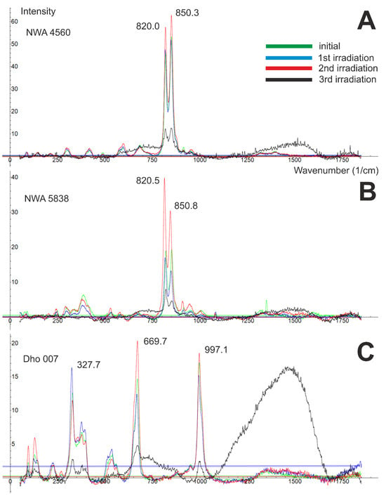
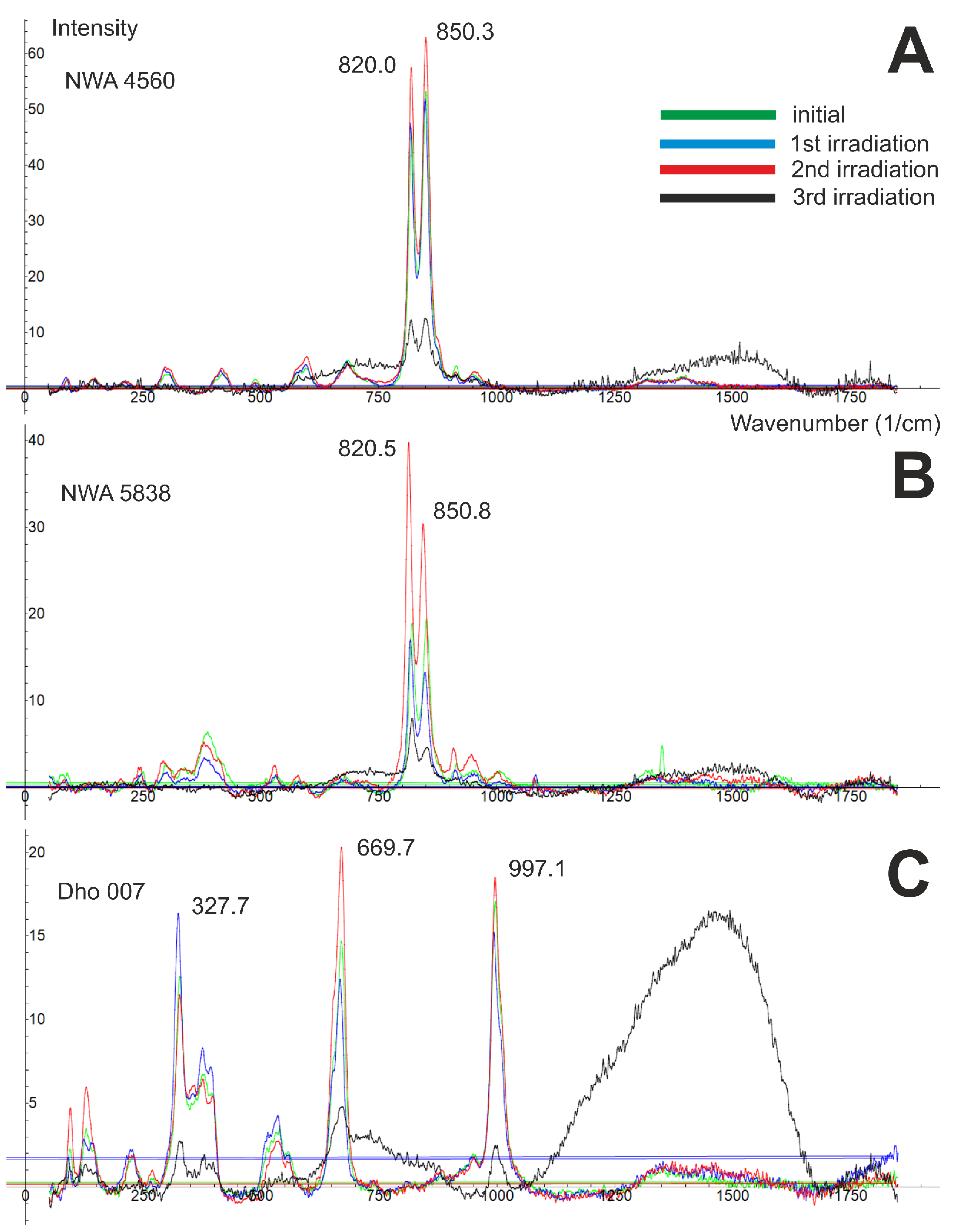
For the NWA 4560 olivines, the initial average peak positions were 820.0 ± 0.2 and 850.3 ± 0.4 cm−1, with corresponding average FWHM values of 15.5 ± 1.0 and 21.3 ± 1.9 cm−1 (Figure 1). After the first irradiation, both peaks show negative shifts in position (−1.9 and −1.7 cm−1) and in FWHM (−0.6 and −1.4 cm−1). Between the first and second irradiations, peak positions shift positively (1.56 and 1.73 cm−1), while FWHM decreases for peak 1 (−0.62 cm−1) and increases for peak 2 (1.21 cm−1; Table 3). After the third irradiation, the peak positions are 819.23 ± 0.78 and 850.79 ± 0.73 cm−1, with FWHM values of 15.22 ± 3.59 and 25.13 ± 6.65 cm−1. Relative to the second irradiation, peak 1 shows a slight negative shift in position (−0.46 cm−1) and a small increase in FWHM (0.88 cm−1), whereas peak 2 shifts positively in both parameters (0.40 and 4.04 cm−1). Three broad amorphous bands appear consistently at ~550–1000, ~1250–1650, and ~1700–1850 cm−1 (Figure 1).
Figure 1.
Differences in the averaged Raman spectra of the three examined meteorites are shown (top to bottom: NWA 4560 olivines (A), NWA 5838 olivines (B), and Dho 007 pyroxenes (pigeonite) (C)) prior to irradiation (green spectra) and following each irradiation event (blue spectra after the 1st irradiation; red spectra after the 2nd irradiation; black spectra after the 3rd irradiation). Numbers indicate the analyzed peaks. Wavenumbers are given in cm−1.
Table 3.
Raman band positions and FWHM values of peaks of the silicate minerals in the examined meteorites before the irradiation and the produced changes in the values (i.e., peak shift and FWHM) after the three irradiation events. Standard deviations in brackets.
For NWA 5838 olivines, the initial average peak positions were 820 ± 0.4 and 850 ± 0.4 cm−1, with FWHM values of 15.7 ± 1.1 and 22.4 ± 3.5 cm−1. The first irradiation produced negative shifts in peak position (−3.0 and −3.5 cm−1) and slight decreases in FWHM (−0.8 and −0.2 cm−1). Between the first and second irradiations, peak positions continue to shift negatively (−3.87 and −3.68 cm−1), while FWHM increases slightly (0.17 and 2.37 cm−1). Following the third irradiation, the peak positions move to 820.97 ± 1.98 and 851.52 ± 2.50 cm−1, with FWHM values of 17.83 ± 11.47 and 28.84 ± 8.54 cm−1. Compared to the second irradiation, both peaks shift positively in position (7.40 and 7.90 cm−1) and show increases in FWHM (2.75 and 4.29 cm−1). Three amorphous bands appear in all spectra at ~600–950, ~1250–1600, and ~1750–1850 cm−1 (Figure 1).
For the Dho 007 pyroxenes, the initial average peak positions were 997.1 ± 2.1, 669.7 ± 0.7, 327.7 ± 0.7 cm−1, with FWHM values of 21.5 ± 4.7, 20.0 ± 1.4, and 21.3 ± 2.3 cm−1. The first irradiation causes negative shifts in both peak position (−3.1, −2.7, and −3.3 cm−1) and FWHM (−0.3, −0.6, and −1.2 cm−1) for all three peaks. Following the second irradiation, peak positions shift positively (2.28, 2.77, and 2.99 cm−1), while FWHM decreases for peak 1 (−1.08 cm−1) and increases for peaks 2 and 3 (0.58 and 3.90 cm−1). After the third irradiation, the peak positions are 999.33 ± 1.87, 669.02 ± 6.45, and 328.26 ± 1.12 cm−1, with FWHM values of 19.70 ± 5.92, 49.22 ± 25.38, and 19.44 ± 7.98 cm−1. Relative to the second irradiation, peak 1 shows a small positive shift in position (3.01 cm−1) and a decrease in average FWHM (−0.46 cm−1); peak 2 shifts slightly negatively in position (−0.75 cm−1) and strongly positively in FWHM (29.23 cm−1); and peak 3 shifts positively in position (0.86 cm−1) and negatively in FWHM (−4.55 cm−1). All spectra exhibit three amorphous bands at ~600–900, ~1100–1700, and ~1700–1850 cm−1 (Figure 1).
Following the first irradiation, all peaks shifted negatively in all studied meteorites. After the second irradiation, silicates from NWA 4560 and Dho 007 exhibited positive shifts, whereas NWA 5838 olivines shifted further negatively. Peak broadening occurred after the first irradiation, but during the second one, several NWA 4560 olivine and Dho 007 pyroxene peaks narrowed, as did both components of the NWA 5838 olivine doublet.
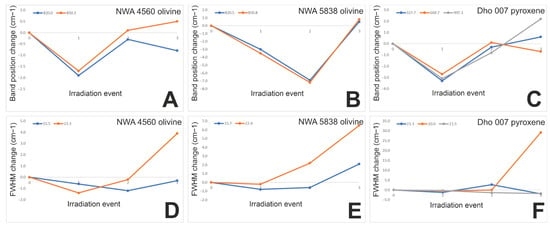
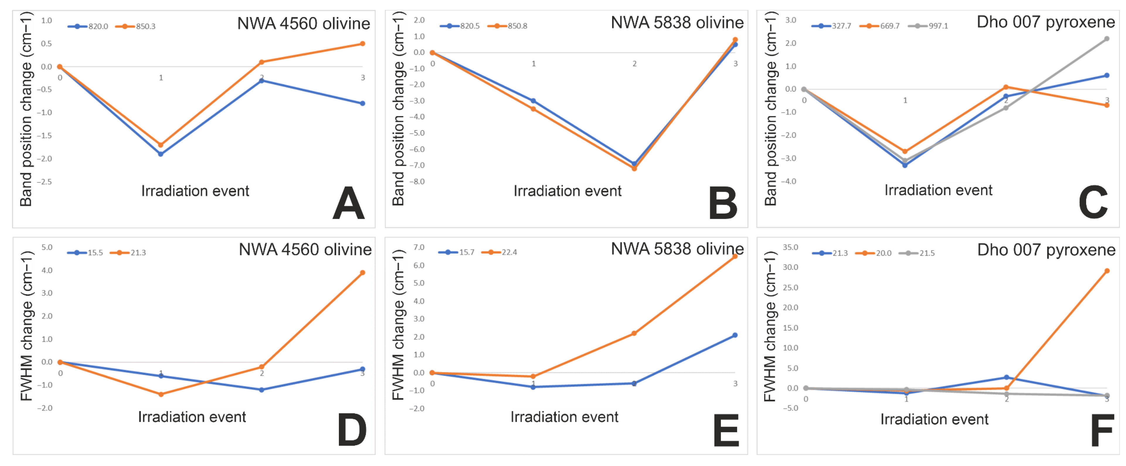
In the case of NWA 4560, the initial peak positions (820.0 and 850.3 cm−1) shifted in the same direction and by a similar magnitude up to the third irradiation (Figure 2). After the third irradiation, the shift occurred in the opposite direction but with a comparable scale. Following the first irradiation, the peaks exhibited a negative shift, whereas after the second irradiation, the shift was positive. The initial FWHM values (15.5 and 21.3 cm−1) decreased (narrowing) after the first irradiation, behave differently after the second irradiation, and increased (broadening) after the third irradiation.
Figure 2.
Raman band position ((A): NWA 4560, (B): NWA 5838 and (C): Dho 007) and FWHM ((D): NWA 4560, (E): NWA 5838 and (F): Dho 007) changes in the examined doublet of NWA 4560 and NWA 5838 olivines, as well as the changes in the analyzed peaks in the case of pyroxenes in Dho 007 after the irradiation events.
The initial peak positions (820.5 and 850.8 cm−1) exhibited correlated shifts in very similar magnitude under irradiation in the case of NWA 5838 (Figure 2). The first and second irradiations caused negative shifts, while the third irradiation resulted in a positive shift. The initial FWHM values (15.7 and 22.4 cm−1) showed changes in comparable direction and magnitude. After the first irradiation, a negative change was observed, whereas the second and third irradiations led to positive changes.
In the case of Dho 007, the initial peak positions (327.7, 669.7 and 997.1 cm−1) also exhibited similar shifts under irradiation; however, after the third irradiation, more variable movements were observed, differing in magnitude (Figure 2). Following the first irradiation, the shifts were negative, whereas after the second irradiation, they became positive. The initial FWHM values (21.3, 20.0 and 21.5 cm−1) decreased similarly (negative change) up to the first irradiation, but following the second and third irradiations, they changed in a non-correlated and dissimilar manner.
4. Discussion
In this section, we discuss the results of LV-SEM and Raman spectroscopy, as well as compare them with data from other authors. Before discussing our recent results, we briefly summarize the main findings on the effects of the first two weaker irradiation events and their interpretation at the beginning of each subsection.
4.1. Mineral Composition Changes
The olivine compositions in NWA 4560 and NWA 5838 (initial Fo-content were 64.99 mol% and 69.02 mol%, respectively) varied widely after the first two irradiations (Fo composition changes: −2.93 and +8.71 mol%, as well as −0.82 and +5.05 mol%, respectively), whereas the pyroxenes in Dho 007 remained comparatively stable, showing only minor compositional fluctuations (En: +0.73 and −3.63 mol%, Wo: +1.55 and +1.09 mol%, Fs: −2.27 and +2.55 mol%, respectively) under proton exposure. The highest variation in the composition of olivine among the NWA chondrites occurred after the second irradiation (i.e., +8.71 mol% change in the Fo content) in NWA 4560. The Fo content of olivine shows a decrease after the first and third irradiations in both NWA 4560 and NWA 5838, whereas after the second irradiation an increase in the Fo content was observed. This implies that after the second irradiation, metastable alteration occurred, but the third irradiation stabilized the Mg loss, which was similarly observed by other researchers [17,18,19]. In NWA 4560, the average Fo content of olivines followed a similar pattern, an initial slight decrease under weaker irradiation (metastable alteration), followed by a pronounced increase at higher doses, with amorphization-induced structural change. In the NWA 5838 sample, the first irradiation produced weak changes in composition (decrease in Fo content by 0.82 mol%), which was followed by the increase in Fo content by +5.05 mol% after the second irradiation, and finally, the decrease in the Fo content by −2.94 mol%.
In the case of the Dho 007 meteorite, the En content increased after the first and third irradiations, the Wo content increased after the first and second irradiation, while the Fs content increased after the second and third irradiations in pyroxene. In the case of pyroxene, the first and second irradiations show minor variation in Mg-Ca-Fe (En-Wo-Fs) composition, but after the third irradiation, the composition shows the highest variation as follows: +3.18 mol% (En), −7.81 mol% (Wo), +4.63 mol% (Fs).
Ref. [35] irradiated ultra-thin olivine with He+ ions to investigate chemical and structural evolution of silicates in the interstellar medium. Using energies of 4, 10 and 50 keV and fluences of 5 × 10¹⁵–1018 ions/cm2—conditions comparable to supernova shock waves—they observed amorphization at low energies and chemical modifications driven by differential sputtering. This process removes Mg and O, enriching the surface of Si and Fe, and promotes metallic iron formation through reduction. Under 4 and 10 keV irradiation, sputtering decreased the atomic ratios (i.e., O/Si and Mg/Si). In our experiments, the Fo content of olivines in both NWA meteorites decreased significantly following the third, most intense irradiation. The decrease in Fo values after the third irradiation may be the result of preferential sputtering of Mg in the olivine structure, leading to a relative enrichment of Fe over Mg. This process is similar to what we observed after the first irradiation, but it shows greater changes. The pyroxenes of Dho 007 behave similar as the olivines after the last irradiation event, because their Fs values increased remarkably, while the En content also emerged, as well as the Wo values decreased steeply. This habit can be connected with the preferential depletion of Ca over Mg, besides the relative Fe enrichment.
4.2. Spectroscopic Property Changes
The FWHM values of the analyzed peaks in the olivines of the NWA 4560 and NWA 5838 meteorites, as well as in the pyroxenes of the Dho 007 meteorite, exhibited similar trends during irradiation. A pronounced difference, however, was observed in the changes in the peak positions of the olivines from NWA 4560 and NWA 5838 after the second irradiation. In this case, the peak positions in NWA 4560 shifted in a positive direction, whereas those in NWA 5838 shifted markedly in a negative direction (Figure 2).
As a result of the third and most intense irradiation event, three major amorphous bands (Figure 1) emerged in the Raman spectra of all analyzed meteorite silicates (i.e., NWA 4560: ~550–1000 cm−1, ~1250–1650 cm−1, ~1700–1850 cm−1; NWA 5838: ~600–950 cm−1, ~1250–1600 cm−1, ~1750–1850 cm−1; Dho 007: ~600–900 cm−1, ~1100–1700 cm−1, ~1700–1850 cm−1). These broad bands should be caused by the presence of glassy, amorphous structured materials (typically with a few tens to hundreds nm thickness) that were produced during the irradiation (simulating space weathering) besides nanophase iron [36,37,38]. We can note that the three bands rise almost the same wavenumber in both olivines and pyroxenes, between ~550–1000 cm−1, ~1100–1700 cm−1 and ~1700–1850 cm−1. Therefore, our experiments show a transitional state of olivine and pyroxene grains that are covered with a very thin layer of the silicate glass.
The thickness of such amorphous rims can vary between 60 and 200 nm in the case of lunar soil grains [36,39], or 30–60 nm around Itokawa particles [37] and can be divided into four categories (amorphous, inclusion-rich (with nm sized Fe grains), multiple or vesicular). These solar wind-damaged particle rims show preferential sputtering of cations and the inclusion-rich rims can collect non-indigenous elements from impact-generated vapors [36]. Due to our strongest, third irradiation, it is possible that in some cases, around the silicates, thicker and highly compositionally modified rims developed.
Ref. [27] irradiated crystalline San Carlos olivine single crystals (Mg1.8Fe0.2SiO4) with 10 keV He+ ions with 1018 ions/cm2 fluence. They found that the irradiation-induced amorphization affects only a very thin surface layer of olivine—about 90 ± 10 nm—beneath which lies a mixed crystalline–amorphous zone. In the upper, amorphous layer, crystalline olivine is converted to a glassy phase with pyroxene composition by high-energy particle bombardment [40]. In our experiments, we used protons up to 12 keV with 1019 ions/cm2 fluence for the third irradiation which had a ~30 h duration. These conditions caused major modifications in the crystal lattice in the silicates of all examined meteorites and might allow them to develop a thicker amorphous zone with an even thicker crystalline-amorphous layer. It can be deduced from the conditions (higher energy and fluence combined with longer duration) of our irradiation method compared to the one [27] used. This observation on the thickness can explain the obviously intense amorphization-induced changes on some of our Raman spectra (e.g., NWA 5838 olivines) with a strong trend.
Considering Mg loss-related crystalline structure changes, the observed preferential loss from olivine can be explained by the crystal structure of forsterite (Mg2SiO4), in which Mg occupies relatively open octahedral sites (M1 and M2) linking isolated SiO4 tetrahedra [41,42]. These Mg–O bonds are weaker than the Si–O bonds of the tetrahedral framework, making Mg more susceptible to ion-induced sputtering and radiolytic loss under energetic irradiation [43,44]. Mg depletion is accompanied by surface enrichment in Fe and Si, as well as the formation of metallic iron through reduction processes [43,44,45].
In contrast, enstatite (MgSiO3) consists of single chains of corner-sharing SiO4 tetrahedra, within which Mg is held in more structurally constrained octahedral sites [46]. As a result, Mg sputtering in enstatite is generally less efficient and shows a stronger dependence on crystallographic orientation and irradiation conditions compared to olivine [47,48]. This structural difference explains why significant compositional changes in pyroxenes are mainly observed after the most intense irradiation events.
Wollastonite (CaSiO3) differs fundamentally from Mg-bearing silicates due to the presence of Ca in large, distorted coordination sites [49] characterized by longer and weaker Ca–O bonds. Laboratory sputtering experiments on wollastonite [50,51] and Ca-bearing silicates have shown that Ca is preferentially removed under ion irradiation, leading to a rapid decrease in the wollastonite component [51]. These crystal-chemical and bonding differences therefore explain why Mg preferentially sputters in forsterite, whereas Ca sputtering dominates in wollastonite, consistent with the compositional trends observed in our experiments.
The observed trend of Raman intensity (i.e., lower after the first, higher after the second and the lowest after the third irradiation) reflects the competition between structural disordering processes that suppress Raman activity, defect-related processes that can enhance it, and eventual amorphization that diminishes it again [52].
4.3. Joint Interpretation of the Spectroscopic and Compositional Changes
The observed changes in Raman peak positions and FWHM values can be attributed to compositional modifications of minerals. The decrease in Fo-values and peak broadening following the first irradiation may result from Mg sputtering in olivine, leading to relative Fe enrichment over Mg. In contrast, the increase in Fo-values and peak narrowing after the second (more intense) irradiation could be due to the formation of metallic Fe inclusions, arising from a reduction process that releases Fe from the olivine crystal lattice. This interpretation is supported by laboratory analyses of returned asteroid samples on Earth [53]. Although studies on irradiation-induced chemical and structural changes in pyroxenes remain limited, pyroxenes are likely to respond similarly to olivines as they also have a silicate structure [54]. This parallel behavior is evident when comparing the olivines of NWA 4560 with the Dho 007 pigeonitic pyroxenes, as their peak shifts and FWHM variations exhibit similar trends, likely reflecting amorphization [27,28].
Following the third, most intense irradiation, a thick amorphous layer started to develop in the silicates, which caused preferential sputtering of Mg in the olivine structure, leading to a relative enrichment of Fe over Mg, while in the case of pyroxenes, Ca depleted over Mg with relative Fe enrichment which triggered emerging of Fs and En content besides the decreasing of Wo content. This process generally led to a positive shift in peak positions and broadened the examined peaks. The higher mobility of Ca and Mg compared to Fe originates from their larger ionic radii (Ca2+ ~1.00 Å, Mg2+ ~0.72 Å vs. Fe2+ ~0.61 Å) [55], weaker metal–oxygen bonding energies (Ca–O ~340 kJ/mol, Mg–O ~390 kJ/mol vs. Fe–O ~410–420 kJ/mol) [56], and consequently lower migration activation energies (≤1.1 eV for Ca/Mg compared to ≥1.2 eV for Fe) [57]. Moreover, in contrast with the first two irradiation events, the third and strongest irradiation gave rise to three major amorphous bands in the silicate Raman spectra (~550–1000 cm−1, ~1100–1700 cm−1 and ~1700–1850 cm−1) without the entire transformation of the structure.
In the case of pyroxene of Dho 007, the variation was higher in Fe-Mg-Ca after the third irradiation. The olivine is characterized by weak change in Fo content after the first irradiation, and the highest variation occurred after the second irradiation. The decrease in Fo content after the third irradiation indicates Mg loss from the crystal structure of olivine. The amorphization of the crystal structure is indicated by increasing FWHM of almost all bands of olivine in the Raman spectra. The pyroxene shows the highest variation after the third irradiation in the case of Fe-Mg-Ca due to the distortion of the cation octahedra of the crystal lattice, which is supported by a high FWHM value (+29.2 cm−1).
Our experiments show that strong compositional and structural changes can occur in silicates as a result of the location-dependent space radiation environment and thus strengthen the conclusions of [14,27,58], whose work unfolded that ion irradiation can efficiently modify both the structure and composition of the silicate grains—however we used stronger irradiation with protons (characteristic for solar wind) instead of heavier nuclei in other tests.
4.4. Consequences of Space Weathering in the Context of Remote Sensing
Space weathering on airless bodies not only modifies the optical behavior of surface minerals but also drives measurable structural and compositional changes in olivines and pyroxenes. Solar wind irradiation (besides micrometeorite bombardment) produces amorphous, melt-rich rims on individual grains, mobilizes Fe and other cations, and generates nanophase metallic iron particles within these rims [1]. As our experiments show, in olivine, this leads to partial disordering of the crystal lattice and local Fe-enrichment in amorphized zones, while in pyroxenes it promotes lattice distortion and Ca–Mg–Fe redistribution. These nanoscale alterations reduce the integrity and symmetry of the silicate frameworks, which shift and broaden the characteristic absorption bands that form the basis of remote mineral identification.
SEM and Raman spectroscopy provide direct evidence for these space-weathering-induced transformations, revealing compositional modifications, presence of iron inclusions, and structural disorder that correlate with spectral darkening and reddening. Raman spectra show peak broadening, reduced intensity, and peak position shifts in key vibrational modes (e.g., Si–O stretching doublet of olivine, chain-stretching bands of pyroxene), all markers of lattice damage and compositional modification. For remote sensing of asteroid surfaces, these small-scale alterations introduce systematic biases: diagnostic bands are weakened and amorphous bands emerge. Accurate interpretation therefore requires modeling calibrated with SEM- and Raman-characterized laboratory analogs, enabling the separation of original mineralogical signatures from weathering-induced spectral overprints.
Our observations through Raman spectroscopy can give an opportunity to better estimate the space-weathering effects on the surface of asteroids and, furthermore, to describe exposure ages by remote sensing in the future. Using the lunar example, exposure age is proportional to the amount of np-Fe (nanophase iron) particles increase and is shown in reflectance spectroscopy [59]. The third irradiation causes noticeable changes in the Raman spectra of the studied silicates. In NWA 4560 olivines, peak 1 shows a slight negative shift with minor broadening, while peak 2 shifts positively with increased width. In NWA 5838 olivines, both components of the olivine doublet shift positively and become significantly broader compared to the previous irradiation. For Dho 007 pyroxenes, the first peak shifts positively with narrowing, the second shows a slight negative shift accompanied by strong broadening, and the third exhibits a small positive shift with reduced width. Additionally, all analyzed silicates display the emergence of three broad amorphous bands in the Raman spectra, indicating irradiation-induced structural disorder (Figure 1).
The third, most intense irradiation, which is likely consistent with the energy and duration of the energetic particles of several CMEs (Coronal Mass Ejections), caused major changes in the composition and structure of silicates within the scope of our investigation. Overall, it produced positive peak shifts and peak broadening along with the development of amorphous bands, as well as Ca–Mg–Fe redistribution (Fa and Fs increasing against Fo and En-Wo) as a consequence of the weaker bonding energy, bigger ion size and higher migration capability of Ca and Mg over Fe.
4.5. Instrument Relevant Suggestions
Having results on the irradiation-driven spectral changes, we could provide suggestions for infrared detector channel arrangement optimization on asteroid-visiting cubesats, which will probably carry simple and low-cost detectors in the future.
All of the examined peaks of olivines and pyroxenes are strong and stable enough to use them for phase analyses. The other, less intense and more unstable peaks vanish during the strong irradiation, and they become part of one of the three prominent amorphous Raman bands that appear in all examined meteorite silicates at ~550–1000 cm−1, ~1100–1700 cm−1, and ~1700–1850 cm−1.
The results of this article should be combined in the future with Fourier Transform Infrared Spectroscopy (FTIR) and Transmission Electron Microscopy (TEM) to better understand the distinct trends of some peaks during certain irradiation events, for example the opposite peak position changes in doublet in NWA 4560 (positive shift) and NWA 5838 (negative shift) olivines. In cases like this, Laser Ablation Inductively Coupled Plasma Mass Spectrometry (LA-ICP-MS) could also be a useful technique to better understand the trace element migration and their behavior in the vicinity of crystalline lattice defects.
5. Conclusions
We examined the olivines of two NWA ordinary chondrites (NWA 4560 and NWA 5838) and the pigeonitic pyroxenes of the Dho 007 eucrite achondrite with LV-SEM and Raman spectroscopic methods, following three different fluences from 1017 ions/cm2 to 1019 ions/cm2 by proton irradiation (1 keV for the first and second and up to 12 keV for the third irradiation) to simulate the structural and compositional evolution of minerals under space-like weathering conditions, roughly at a 100 million year timescale.
The olivine compositions in NWA 4560 and NWA 5838 exhibit substantial variation after the first two irradiation steps. Both samples display Fo depletion following a metastable alteration, and the third stabilizes Mg loss, consistent with previous observations [17,18,19], producing a relative Fe enrichment.
The pyroxenes in Dho 007 remain comparatively stable, showing only minor shifts in En, Wo, and Fs components following the first two weaker irradiation events. However, the third irradiation produces the largest shifts in Mg-Ca-Fe components, with notable increases in Fs (+4.63 mol%) and En (+3.18 mol%) and a sharp decline in Wo (−7.81 mol%). These shifts indicate preferential Ca depletion relative to Mg, accompanied by enhanced Fe enrichment.
We also examined the Raman characteristics of olivine. After the first irradiation, all examined silicate peaks exhibited negative shifts and peak broadening after the first irradiation, whereas the second irradiation caused peak narrowing. Following the third irradiation, NWA 4560 and NWA 5838 olivines exhibit various peak changes. In Dho 007 pyroxenes, peak 3 shifts positively (+0.86 cm−1) with reduced FWHM (−4.55 cm−1) at the strongest irradiation.
In general, three prominent amorphous Raman bands appeared in all examined meteorite silicates, located at ~550–1000 cm−1, ~1100–1700 cm−1, and ~1700–1850 cm−1. These broad features are consistent with the formation of glassy, amorphous material generated during irradiation alongside nanophase iron. Under the strongest irradiation conditions, it is likely that some silicates developed thicker rims with substantial compositional modification. The intensity and duration of this final irradiation produced significant lattice disruption in all analyzed silicates and facilitated the formation of a more extensive amorphous layer, providing a coherent explanation for the strong spectral changes observed in several Raman measurements.
Supplementary Materials
The following supporting information can be downloaded at: https://www.mdpi.com/article/10.3390/geosciences16020072/s1, Table S1. The initial and irradiation-modified mineral compositional data (SEM EDS) were obtained for olivines in the NWA 4560 and NWA 5838 chondrites, as well as for pyroxenes in the Dho 007 eucrite. Reported parameters include cation numbers expressed in atoms per formula unit (apfu), oxide concentrations in weight percent (wt%), and end-member proportions (i.e., Fo, Fa for olivine; En, Wo, Fs for pyroxene) in mol%. Table S2. The initial and irradiation-modified Raman datasets were obtained for olivines in the NWA 4560 and NWA 5838 chondrites, as well as for pyroxenes in the Dho 007 eucrite. The table presents the peak center, height, area, and full width at half maximum (FWHM) values of the analyzed bands, together with their average, standard deviation (σ), minimum and maximum values, as well as the variations observed following the irradiation events. Figure S1. The plotted Raman datasets (blue dots) together with the fitted (red curves) and combined (green curves) peaks are shown for representative measurements of olivines from the NWA 4560 and NWA 5838 chondrites, as well as pyroxenes from the Dho 007 eucrite, both prior to and following irradiation. The X-axis represents the wavenumber (cm−1), while the Y-axis denotes dimensionless intensity. Figure S2. Averaged Raman spectra of individual measurements of olivines from the NWA 4560 and NWA 5838 chondrites, as well as pyroxenes from the Dho 007 eucrite, obtained before and after irradiation. The blue curve represents the averaged spectrum, whereas the red curves indicate the minimum and maximum values. The X-axis corresponds to the wavenumber (cm−1), while the Y-axis denotes dimensionless intensity.
Author Contributions
Conceptualization, D.R., I.G. and Á.K.; Methodology, D.R., I.G., S.B., K.F., Z.J., R.R., B.S., M.S., B.D.P. and Á.K.; Investigation, D.R., S.B., K.F., Z.J., R.R., B.S. and M.S.; Data curation, D.R. and I.G.; Writing—original draft, D.R.; Writing—review and editing, B.D.P. and Á.K.; Visualization, D.R.; Supervision, Á.K.; Project administration, Á.K. All authors have read and agreed to the published version of the manuscript.
Funding
This project was supported by the K-138594 and the 151196-ADVANCED projects of NKFIH. The authors also acknowledge the support from the Europlanet 2024 RI which has been funded by the European Union’s Horizon 2020 Research Innovation Programme under grant agreement No. 871149.
Data Availability Statement
The data presented in this study are available on request from the corresponding author.
Conflicts of Interest
The authors declare no conflicts of interest.
References
- Chapman, C.R. Space Weathering of Asteroid Surfaces. Annu. Rev. Earth Planet. Sci. 2004, 32, 539–567. [Google Scholar] [CrossRef] [Scilit]
- Bennett, C.J.; Pirim, C.; Orlando, T.M. Space-Weathering of Solar System Bodies: A Laboratory Perspective. Chem. Rev. 2013, 113, 9086–9150. [Google Scholar] [CrossRef] [Scilit]
- Brunetto, R.; Loeffler, M.J.; Nesvorný, D.; Sasaki, S.; Strazzulla, G.J. Asteroid Surface Alteration by Space Weathering Processes; Asteroids, I.V., Michel, P., DeMeo, F.E., Bottke, W.F., Eds.; University of Arizona: Tucson, AZ, USA, 2015; pp. 597–616. [Google Scholar]
- Pieters, C.M.; Noble, S.K. Space Weathering on Airless Bodies. J. Geophys. Res. Planets 2016, 121, 1865–1884. [Google Scholar] [CrossRef] [Scilit]
- Gaffey, M.J. Space Weathering and the Interpretation of Asteroid Reflectance Spectra. Icarus 2010, 209, 564–574. [Google Scholar] [CrossRef] [Scilit]
- Kánuchová, Z.; Brunetto, R.; Melita, M.; Strazzulla, G. Space Weathering and the Color Indexes of Minor Bodies in the Outer Solar System. Icarus 2012, 221, 12–19. [Google Scholar] [CrossRef] [Scilit]
- Hasegawa, S.; DeMeo, F.E.; Marsset, M.; Hanus, J.; Avdellidou, C.; Delbo, M.; Bus, S.J.; Hanayama, H.; Horiuchi, T.; Takir, D.; et al. Spectral Evolution of Dark Asteroid Surfaces Induced by Space Weathering over a Decade. Astrophys. J. Lett. 2022, 939, L9. [Google Scholar] [CrossRef] [Scilit]
- Clark, B.E.; Sen, A.; Zou, X.D.; Dellagiustina, D.N.; Sugita, S.; Sakatani, N.; Thompson, M.; Trang, D.; Tatsumi, E.; Barucci, M.; et al. Overview of the Search for Signs of Space Weathering on the Low-Albedo Asteroid (101955) Bennu. Icarus 2023, 400, 115563. [Google Scholar] [CrossRef] [Scilit]
- Poppe, A.R.; Farrell, W.M.; Halekas, J.S. Formation Timescales of Amorphous Rims on Lunar Grains Derived from ARTEMIS Observations. J. Geophys. Res. Planets 2018, 123, 37–46. [Google Scholar] [CrossRef] [Scilit]
- Nenon, Q.; Poppe, A.R. On the Long-Term Weathering of Airless Body Surfaces by the Heavy Minor Ions of the Solar Wind: Inputs from Ion Observations and SRIM Simulations. Planet. Sci. J. 2020, 1, 69. [Google Scholar] [CrossRef] [Scilit]
- Jordan, A.P. Reevaluating How Charged Particles Cause Space Weathering on Airless Bodies. Icarus 2022, 376, 114878. [Google Scholar] [CrossRef] [Scilit]
- Bissbort, T.; Jiang, Q.; Becker, H.W.; Foteinou, V.; Chakraborty, S. An experimental study of proton implantation in olivine. Phys. Chem. Miner. 2023, 50, 12. [Google Scholar] [CrossRef] [Scilit]
- Penttilä, A.; Väisänen, T.; Markkanen, J.; Martikainen, J.; Kohout, T.; Videen, G.; Muinonen, K. Rigorous light-scattering simulations of nanophase iron space-weathering effects on reflectance spectra of olivine grains. Icarus 2020, 345, 113727. [Google Scholar] [CrossRef] [Scilit]
- Dukes, C.A.; Baragiola, R.A.; McFadden, L.A. Surface modification of olivine by H+ and He+ bombardment. J. Geophys. Res. Planets 1999, 104, 1865–1872. [Google Scholar] [CrossRef] [Scilit]
- Lazzarin, M.; Marchi, S.; Moroz, L.V.; Brunetto, R.; Magrin, S.; Paolicchi, P.; Strazzulla, G. Space weathering in the main asteroid belt: The big picture. Astrophys. J. 2006, 647, L179. [Google Scholar] [CrossRef] [Scilit]
- Vernazza, P.; Fulvio, D.; Brunetto, R.; Emery, J.P.; Dukes, C.A.; Cipriani, F.; Witasse, O.; Schaible, M.J.; Zanda, B.; Strazzulla, G.; et al. Paucity of Tagish Lake-like Parent Bodies in the Asteroid Belt and Among Jupiter Trojans. Icarus 2013, 225, 517–525. [Google Scholar] [CrossRef] [Scilit]
- Brunetto, R.; Lantz, C.; Ledu, D.; Baklouti, D.; Barucci, M.A.; Beck, P.; Delauche, L.; Dionnet, Z.; Dumas, P.; Duprat, J.; et al. Ion irradiation of Allende meteorite probed by visible, IR, and Raman spectroscopies. Icarus 2014, 237, 278–292. [Google Scholar] [CrossRef] [Scilit]
- Lantz, C.; Brunetto, R.; Barucci, M.A.; Dartois, E.; Duprat, J.; Engrand, C.; Godard, M.; Ledu, D.; Quirico, E. Ion irradiation of the Murchison meteorite: Visible to mid-infrared spectroscopic results. Astron. Astrophys. 2015, 577, A41. [Google Scholar] [CrossRef] [Scilit]
- Lantz, C.; Brunetto, R.; Barucci, M.A.; Fornasier, S.; Baklouti, D.; Bourçois, J.; Godard, M. Ion irradiation of carbonaceous chondrites: A new view of space weathering on primitive asteroids. Icarus 2017, 285, 43–57. [Google Scholar] [CrossRef] [Scilit]
- Taylor, L.A.; Pieters, C.; Patchen, A.; Taylor, D.H.S.; Morris, R.V.; Keller, L.P.; McKay, D.S. Mineralogical and Chemical Characterization of Lunar Highland Soils: Insights into the Space Weathering of Soils on Airless Bodies. J. Geophys. Res. Planets 2010, 115, E02002. [Google Scholar] [CrossRef] [Scilit]
- Grice, A.M.; Stancil, P.C.; Ghafariasl, M.; Singh, S.; Gamage, S.; Schaible, M.J.; Abate, Y.; Lang, K.; Orlando, T.M. Nanoscale Characterization of Space Weathering in Lunar Samples. Sci. Rep. 2025, 15, 301. [Google Scholar] [CrossRef] [Scilit]
- Chrbolková, K.; Halodová, P.; Kohout, T.; Ďurech, J.; Mizohata, K.; Malý, P.; Dědič, V.; Penttilä, A.; Trojánek, F.; Jarugula, R. Sub-surface alteration and related change in reflectance spectra of space-weathered materials. Astron. Astrophys. 2022, 665, A14. [Google Scholar] [CrossRef] [Scilit]
- Denevi, B.W.; Noble, S.K.; Christoffersen, R.; Thompson, M.S.; Glotch, T.D.; Blewett, D.T.; Garrick-Bethell, I.; Gillis-Davis, J.J.; Greenhagen, B.T.; Hendrix, A.R.; et al. Space weathering at the Moon. Rev. Mineral. Geochem. 2023, 89, 611–650. [Google Scholar] [CrossRef] [Scilit]
- Thompson, M.S.; Jurewicz, A.; Noguchi, T. Space weathering: Clear with a chance of solar wind and micrometeoroid showers. Elements 2025, 21, 346–351. [Google Scholar] [CrossRef] [Scilit]
- Bonal, L.; Brunetto, R.; Beck, P.; Dartois, E.; Dionnet, Z.; Djouadi, Z.; Duprat, J.; Füri, E.; Kakazu, Y.; Montagnac, G.; et al. Visible-IR and Raman Microspectroscopic Investigation of Three Itokawa Particles Collected by Hayabusa: Mineralogy and Degree of Space Weathering Based on Nondestructive Analyses. Meteorit. Planet. Sci. 2015, 50, 1562–1576. [Google Scholar] [CrossRef] [Scilit]
- Rezes, D.; Gyollai, I.; Biri, S.; Fintor, K.; Juhász, Z.; Rácz, R.; Sulik, B.; Szabó, M.; Kereszturi, Á. Comparison of three proton irradiated meteorite samples to better understand the solar wind-based space weathering. Meteorit. Planet. Sci. 2025, 60, 2297–2310. [Google Scholar] [CrossRef] [Scilit]
- Demyk, K.; Carrez, P.; Leroux, H.; Cordier, P.; Jones, A.P.; Borg, J.; Quirico, E.; Raynal, P.I.; d’Hendecourt, L. Structural and Chemical Alteration of Crystalline Olivine Under Low Energy He+ Irradiation. Astron. Astrophys. 2001, 368, L38–L41. [Google Scholar] [CrossRef] [Scilit]
- Brunetto, R.; Vernazza, P.; Marchi, S.; Birlan, M.; Fulchignoni, M.; Orofino, V.; Strazzulla, G. Modeling Asteroid Surfaces from Observations and Irradiation Experiments: The Case of 832 Karin. Icarus 2006, 184, 327–337. [Google Scholar] [CrossRef] [Scilit]
- Harris, A.W.; Chodas, P.W. The Population of Near-Earth Asteroids Revisited and Updated. Icarus 2021, 365, 114452. [Google Scholar] [CrossRef] [Scilit]
- Cheng, A.F.; Agrusa, H.F.; Barbee, B.W.; Meyer, A.J.; Farnham, T.L.; Raducan, S.D.; Dotto, E.; Zinzi, A.; Della Corte, V.; Statler, T.S.; et al. Momentum transfer from the DART mission kinetic impact on asteroid Dimorphos. Nature 2023, 616, 457–460. [Google Scholar] [CrossRef] [Scilit]
- Grossman, J.N. The meteoritical bulletin, No. 84, 2000 August. Meteorit. Planet. Sci. 2000, 35, A199–A225. [Google Scholar] [CrossRef] [Scilit]
- Weisberg, M.K.; Smith, C.; Benedix, G.; Herd, C.D.K.; Righter, K.; Haack, H.; Yamaguchi, A.; Chennaoui Aoudjehane, H.; Grossman, J.N. The meteoritical bulletin, No. 97. Meteorit. Planet. Sci. 2010, 45, 449–493. [Google Scholar] [CrossRef] [Scilit]
- Kereszturi, A.; Gyollai, I.; Biri, S.; Juhász, Z.; Király, C.; Pál, B.D.; Rácz, R.; Rezes, D.; Sulik, B.; Szabó, M.; et al. Evaluation of simulated space weathering-based meteorite alteration and potential influence on mechanical deformation of rubble pile asteroids. Front. Astron. Space Sci. 2025, 12, 1427387. [Google Scholar] [CrossRef] [Scilit]
- Nénon, Q.; Raines, J.M.; Poppe, A.R. The Long-Term Flux of the Solar Wind Suprathermal Ions That Precipitate on the Lunar Surface. J. Geophys. Res. Planets 2023, 128, e2023JE007958. [Google Scholar] [CrossRef] [Scilit]
- Carrez, P.; Demyk, K.; Cordier, P.; Gengembre, L.; Grimblot, J.; d’Hendecourt, L.; Jones, P.A.; Leroux, H. Low-energy helium ion irradiation-induced amorphization and chemical changes in olivine: Insights for silicate dust evolution in the interstellar medium. Meteorit. Planet. Sci. 2002, 37, 1599–1614. [Google Scholar] [CrossRef] [Scilit]
- Keller, L.P.; McKay, D.S. The nature and origin of rims on lunar soil grains. Geochim. Cosmochim. Acta 1997, 61, 2331–2341. [Google Scholar] [CrossRef] [Scilit]
- Noguchi, T.; Nakamura, T.; Kimura, M.; Zolensky, M.E.; Tanaka, M.; Hashimoto, T.; Konno, M.; Nakato, A.; Ogami, T.; Fujimura, A.; et al. Incipient space weathering observed on the surface of Itokawa dust particles. Science 2011, 333, 1121–1125. [Google Scholar] [CrossRef] [Scilit]
- Yang, Y.; Zhang, H.; Wang, Z.; Yuan, Y.; Li, S.; Hsu, W.; Liu, C. Optical spectroscopic characterizations of laser irradiated olivine grains. Astron. Astrophys. 2017, 597, A50. [Google Scholar] [CrossRef] [Scilit]
- Uvarova, A.V.; Slyuta, E.N.; Agapkin, I.A. Geotechnical Properties of Lunar Soil Analogue VI-75 in Dry and Frozen State. Sol. Syst. Res. 2025, 59, 89. [Google Scholar] [CrossRef] [Scilit]
- Rietmeijer, F.J. The irradiation-induced olivine to amorphous pyroxene transformation preserved in an interplanetary dust particle. Astrophys. J. 2009, 705, 791. [Google Scholar] [CrossRef] [Scilit]
- Smyth, J.R.; Hazen, R.M. The crystal structures of forsterite and hortonolite at several temperatures up to 900 C. Am. Mineral. J. Earth Planet. Mater. 1973, 58, 588–593. [Google Scholar]
- Deer, W.A.; Howie, R.A.; Zussman, J. An Introduction to the Rock-Forming Minerals; Mineralogical Society of Great Britain and Ireland: London, UK, 2013. [Google Scholar]
- Loeffler, M.J.; Dukes, C.A.; Baragiola, R.A. Irradiation of olivine by 4 keV He+: Simulation of space weathering by the solar wind. J. Geophys. Res. Planets 2009, 114, E3. [Google Scholar] [CrossRef] [Scilit]
- Loeffler, M.J.; Dukes, C.A.; Christoffersen, R.; Baragiola, R.A. Space weathering of silicates simulated by successive laser irradiation: In situ reflectance measurements of Fo90, Fo99+, and SiO2. Meteorit. Planet. Sci. 2016, 51, 261–275. [Google Scholar] [CrossRef] [Scilit]
- Cantando, E.D.; Dukes, C.A.; Loeffler, M.J.; Baragiola, R.A. Aqueous depletion of Mg from olivine surfaces enhanced by ion irradiation. J. Geophys. Res. Planets 2008, 113, E9. [Google Scholar] [CrossRef] [Scilit]
- Cameron, M.; Papike, J.J. Structural and chemical variations in pyroxenes. Am. Mineral. 1981, 66, 1–50. [Google Scholar]
- Schrempel, F.; Jäger, C.; Fabian, D.; Dorschner, J.; Henning, T.; Wesch, W. Study of the amorphization process of MgSiO3 by ion irradiation as a form of dust processing in astrophysical environments. Nucl. Instrum. Methods Phys. Res. Sect. B Beam Interact. Mater. At. 2002, 191, 411–415. [Google Scholar] [CrossRef] [Scilit]
- Jäger, C.; Fabian, D.; Schrempel, F.; Dorschner, J.; Henning, T.; Wesch, W. Structural processing of enstatite by ion bombardment. Astron. Astrophys. 2003, 401, 57–65. [Google Scholar] [CrossRef] [Scilit]
- Buerger, M.J.; Prewitt, C.T. The crystal structures of wollastonite and pectolite. Proc. Natl. Acad. Sci. USA 1961, 47, 1884–1888. [Google Scholar] [CrossRef] [Scilit]
- Szabo, P.S.; Chiba, R.; Biber, H.; Stadlmayr, R.; Berger, B.M.; Mayer, D.; Mutzke, A.; Doppler, M.; Sauer, M.; Appenroth, J.; et al. Solar wind sputtering of wollastonite as a lunar analogue material—Comparisons between experiments and simulations. Icarus 2018, 314, 98–105. [Google Scholar] [CrossRef] [Scilit]
- Szabo, P.S.; Biber, H.; Jäggi, N.; Brenner, M.; Weichselbaum, D.; Niggas, A.; Stadlmayr, R.; Primetzhofer, D.; Nenning, A.; Mutzke, A.; et al. Dynamic potential sputtering of lunar analog material by solar wind ions. Astrophys. J. 2020, 891, 100. [Google Scholar] [CrossRef] [Scilit]
- Sidorov, A.I.; Kirpichenko, D.A.; Yurina, U.V.; Podsvirov, O.A. Structural changes in silica glass under the action of electron beam irradiation: The effect of irradiation dose. Glass Phys. Chem. 2021, 47, 118–125. [Google Scholar] [CrossRef] [Scilit]
- Nakamura, T.; Matsumoto, M.; Amano, K.; Enokido, Y.; Zolensky, M.E.; Mikouchi, T.; Genda, H.; Tanaka, S.; Zolotov, M.Y.; Kurosawa, K.; et al. Formation and evolution of carbonaceous asteroid Ryugu: Direct evidence from returned samples. Science 2022, 379, eabn8671. [Google Scholar] [CrossRef] [Scilit]
- Caminiti, E.; Lantz, C.; Besse, S.; Brunetto, R.; Carli, C.; Serrano, L.; Mari, N.; Vincendon, M.; Doressoundiram, A. Effects of ion irradiation on Mercury terrestrial analogues in the visible to mid-infrared. Icarus 2024, 420, 116191. [Google Scholar] [CrossRef] [Scilit]
- Shannon, R.D. Revised effective ionic radii and systematic studies of interatomic distances in halides and chalcogenides. Found. Crystallogr. 1976, 32, 751–767. [Google Scholar] [CrossRef] [Scilit]
- Huheey, J.E.; Keiter, E.A.; Keiter, R.L.; Medhi, O.K. Inorganic Chemistry: Principles of Structure and Reactivity, 4th ed.; Pearson Educ.: London, UK, 2006; p. 808. [Google Scholar]
- Brady, J.B.; Cherniak, D.J. Diffusion in minerals: An overview of published experimental diffusion data. Rev. Mineral. Geochem. 2010, 72, 899–920. [Google Scholar] [CrossRef] [Scilit]
- Bradley, J.P. Chemically anomalous, preaccretionally irradiated grains in interplanetary dust from comets. Science 1994, 265, 925–929. [Google Scholar] [CrossRef] [Scilit]
- Morris, R.V. Surface exposure indices of lunar soils—A comparative FMR study. In Proceedings of the Seventh Lunar Science Conference, Houston, TX, USA, 15–19 March 1976; Volume 1, pp. 315–335. [Google Scholar]

Disclaimer/Publisher’s Note: The statements, opinions and data contained in all publications are solely those of the individual author(s) and contributor(s) and not of MDPI and/or the editor(s). MDPI and/or the editor(s) disclaim responsibility for any injury to people or property resulting from any ideas, methods, instructions or products referred to in the content. |
© 2026 by the authors. Licensee MDPI, Basel, Switzerland. This article is an open access article distributed under the terms and conditions of the Creative Commons Attribution (CC BY) license.