


Simple Summary
Postpartum endometritis is a widespread disease in dairy cows and reduces their ability to become pregnant. Antibiotics are commonly used for treatment, yet drug-resistant bacteria and ongoing fertility problems remain difficult issues. This study investigated the relationship between the traditional veterinary concept of blood stasis and cow endometritis using clinical observations, rat models, protein analysis and bioinformatic tools. We found that more than half of diseased dairy cows showed signs of blood stasis syndrome, which could be recognized by changed blood clotting indicators. Experiments using a rat blood stasis syndrome model further demonstrated uterine inflammatory changes, as well as alterations in molecular signaling involved in vascular regulation and inflammatory responses. These observations point to a potential link between blood stasis-related abnormalities and uterine inflammation. This work offers new clues for developing better therapies to protect dairy cow reproductive health and reduce economic losses in dairy farming.
Abstract
Endometritis, inflammation of the endometrium caused primarily by bacterial infection, can pose a major threat to the reproductive health and fertility of dairy cows if left untreated. Current antibiotic therapies for endometritis in dairy cows face challenges, such as bacterial resistance, microbial dysbiosis, and impaired fertility, necessitating novel treatment strategies. In this study, we explored potential molecular signals associated with the crosstalk between blood stasis syndrome (BSS) and endometritis via an integrated approach involving veterinary investigations, an adrenaline-induced rat BSS model, bioinformatics, and proteomic profiling. We identified shortened fibrinogen time (FIBT) and elevated fibrinogen (FIB) concentrations as coagulation parameters associated with the study-defined BSS phenotype, suggesting a hypercoagulable state. Clinical data from dairy cows showed that 53.49% of endometritis cases were BSS-associated. Compared with healthy cows, cows in the endometritis with blood stasis syndrome (EBSS) group showed elevated rates of retained placenta and severe vaginal mucosal congestion. Experiments in the rat BSS model provided complementary observations of uterine inflammatory changes in this combined model, with histopathology revealing uterine vascular remodeling, neutrophilic infiltration, and upregulation of IL-1β expression. Molecular analyses revealed BSS-associated changes in HIF-1α-related VEGF/EGFR signaling and KLKB1-related plasmin and CACNA2D1 levels, along with alterations in pathways involved in coagulation, angiogenesis, and vascular contraction. Inflammatory mediator levels were also altered. As the mechanistic investigations were conducted primarily in a rat model, the current findings suggest a potential association between blood stasis and inflammation rather than establishing a fully defined signaling cascade. This study provides a basis for further investigation of endometritis pathogenesis and the evaluation of potential therapeutic strategies.
1. Introduction
Postpartum endometritis is a major reproductive disease with an incidence rate ranging from 37 to 45% on intensive dairy farms [1,2]. The clinical manifestations are heterogeneous, as different pathogenic bacteria result in different vaginal discharge characteristics [3]. Pathological features include endometrial congestion and edema, destruction of glandular structures, and inflammatory cell infiltration [4,5]. It causes decreased pregnancy rates, prolonged calving intervals, and premature culling of approximately 15 to 25% of affected cows due to reproductive failure, resulting in substantial economic losses (a decrease in profit of approximately $512 per dairy cow) [6,7,8].
Modern veterinary medicine widely recognizes endometritis as an inflammatory lesion caused by exogenous pathogenic bacterial infections, such as Escherichia coli, Mycoplasma spp., and Staphylococcus aureus. Thus, current treatment protocols primarily rely on broad-spectrum antibiotics [9,10]. However, drug resistance continues to rise: the rate of E. coli resistance to fluoroquinolones exceeds 60%, and the rate of Streptococcus resistance to tetracyclines is as high as 45% [11,12]. Crucially, while antibiotics effectively eliminate pathogens, a large proportion of treated cows still fail to conceive again [13]. Histopathological analyses have revealed persistent post-treatment uterine fibrosis, reduced microvascular density, and abnormal expression of endometrial receptivity-related molecules, indicating a fundamental disconnect between pathogen clearance and functional uterine recovery [14].
Based on the holistic view of Traditional Chinese Veterinary Medicine (TCVM), endometritis arises from the combined effects of internal deficiency and external pathogenic invasion [15]. Its pathogenesis involves Qi deficiency and blood stasis as the root cause and external pathogenic invasion as the secondary factor [16]. On the one hand, parturition leads to substantial depletion of vital energy (Qi), while lactation and postpartum recovery further consume Qi, resulting in a state of negative energy balance and compromised immunity [16,17]. On the other hand, the physiological hypercoagulable state during pregnancy, if unresolved during the early postpartum period, disrupts uterine microcirculation [18], resulting in a pathological basis for blood stasis syndrome (BSS) [19].
In TCVM, BSS refers to a pathological state caused by factors such as trauma, Qi deficiency or stagnation, or cold or heat congelation, leading to internal blockage of stagnant blood and impaired blood flow, either systemically or locally [20]. Core manifestations include stabbing pain, purple masses, dark-purple bleeding, and cyanotic mucosa and tongue. Fundamentally, BSS reflects the holistic macro-pathological concept of Qi-blood dysfunction [17]. BSS differs fundamentally from the Western medical concept of “thrombosis.” Thrombosis specifically denotes a localized physical obstruction within blood vessels composed of fibrin and blood cells and is classified as distinct organic pathological change [21]. Although chronic BSS may increase thrombosis risk and although post-thrombotic manifestations may align partially with BSS, the TCVM concept has a broader scope (e.g., microcirculatory disorders and metabolic stagnation). Its definition is based on the holistic Qi-blood theory rather than tangible material blockages.
Given that blood flow is systemic, blood in a hypercoagulable state can also spread to the local circulation of specific organs through systemic blood circulation, thereby contributing to tissue-specific diseases [22,23]. For instance, cardiovascular stasis may induce angina pectoris [24], hepatobiliary stasis correlates with cholestatic hepatitis [25], and uterine stasis causes dysmenorrhea, amenorrhea, and infertility [26]. Notably, clinical observations reveal that many antibiotic-treated cows, despite their infections being resolved, continue to have pregnancy difficulties, accompanied by BSS symptoms, such as abnormal uterine artery pulsatility indices and elevated plasma D-dimer levels, suggesting that persistent BSS may be a critical barrier to full recovery [27].
Although TCVM formulations containing blood-activating and stasis-resolving herbs are frequently used in China to treat endometritis, pharmacodynamic evaluations have predominantly focused on inflammation-related indicators. Similarly, mechanistic studies have primarily concentrated on inflammation-associated signaling pathways [28]. Our previous study demonstrated that the Chinese herb Salvia miltiorrhiza can treat endometritis in dairy cows by promoting Qi and blood circulation [16]; however, direct molecular biological evidence linking blood stasis to the pathogenesis of endometritis is lacking. Despite clinical evidence showing that disrupted blood circulation is responsible for various inflammatory lesions, thus implicating blood stasis in the pathogenesis of endometritis, quantitative validation is lacking [22,23]. This study evaluated postpartum BSS and endometritis using modern molecular biology techniques to identify specific molecular pathways through which blood stasis induces endometrial inflammation. The findings can provide a scientific basis for the direct relationship between BSS and endometritis, potentially contributing to more comprehensive treatment protocols for clinical endometritis in dairy cows.
2. Materials and Methods
2.1. Animal Management
In total, 2346 postpartum Holstein cows from three large farms in Lingwu City, Ningxia, were included in this study. The cows were 22–44 months old and were in their first or second parity. Detailed information on the cows included in this study is recorded in Table S1. Uterine secretions and jugular venous blood were collected from live postpartum Holstein cows as routine non-terminal sampling procedures. Therefore, no anesthesia or euthanasia was performed in the cow study. Continuous monitoring was initiated on the day of parturition, with blood stasis-related indicators (blood coagulation parameters; vaginal mucus, placenta and vulva condition) serially assessed from parturition through day 14 postpartum. Endometritis diagnoses were established at day 42 postpartum. All experimental cows were housed in barn stalls with sand bedding and had access to the same forage diet (corn silage, alfalfa hay, whole cottonseed, beet granules, corn flakes, corn meal, soybean meal, extruded soybean, corn gluten powder, rapeseed meal, corn distilled soluble grain, calcium fatty acid, sodium bicarbonate, and urea). The cows were withheld from all drug treatments during the study period and were allowed ad libitum feeding.
Female Sprague-Dawley rats, aged 6–7 weeks, were purchased from Chengdu Dashuo Experimental Animal Co., Ltd. (Xianyang, Shaanxi, China). A total of 20 rats were used in this study. After one week of acclimatization, the rats were randomly assigned to a control group and a BSS group (n = 10 per group). The rats were maintained under standard laboratory conditions with free access to food and water.
All experimental procedures were approved by the Animal Ethical and Welfare Committee of Northwest A&F University (Approval No. XN2024-0403, approval date: [11 March 2024]).
2.2. Diagnosis of BSS in Cows
BSS was assessed during postpartum days 1–14. In veterinary medicine, there are currently no standardized diagnostic criteria for BSS. Drawing upon established diagnostic criteria from human medicine adapted to veterinary clinical realities (Table S2), the specific diagnostic criteria for cows were defined as follows: (1) congested and swollen vaginal mucosa with a dark-red or -purple coloration and a swollen, protruding vulva; (2) retained fetal membranes or abortion; and (3) a hypercoagulable blood state. The presence of two or more of these criteria was considered to indicate BSS in cows [29].
2.3. Diagnosis of Endometritis in Cows
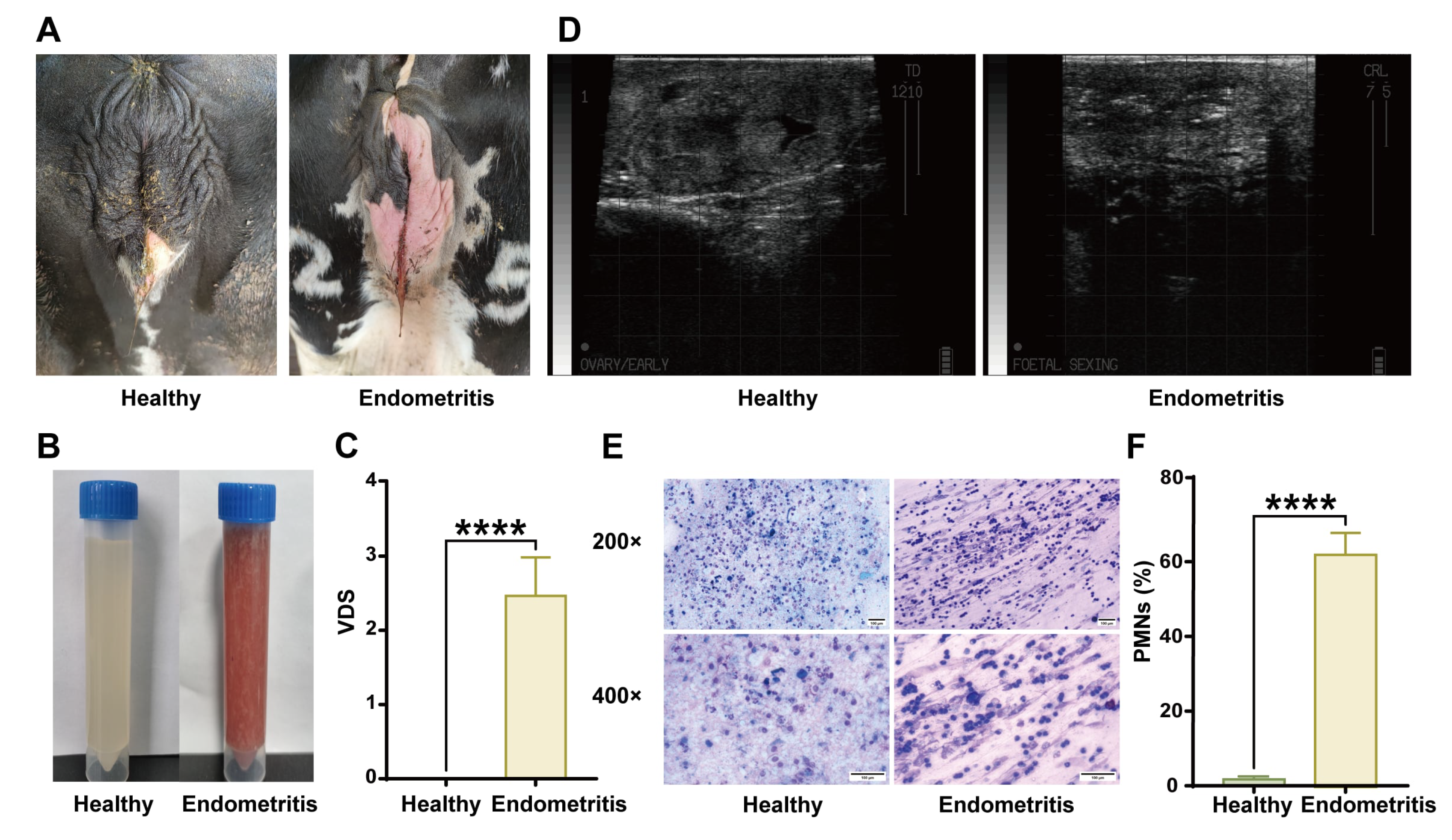
Endometritis was diagnosed in dairy cows on postpartum day 42. Due to the lack of consensus diagnostic standard, an endometritis diagnosis in dairy cows is difficult to establish. In this study, dairy cows were mainly diagnosed by both vaginal discharge score (VDS) and polymorphonuclear neutrophil (PMN) count. B-ultrasound (IMV Imaging, Easi-Scan, Bellshill, UK) was used as an auxiliary diagnostic method. VDS was scored by evaluating discharged uterine mucus as follows: 0, completely transparent or translucent mucus; 1, mucus containing white or grayish-white purulent spots; 2, mucus containing less than 50% pus; and 3, mucus containing over 50% pus. For PMN% evaluation, vaginal discharge was smeared on a slide and observed at 400× magnification. A total of 300 representative epithelial cells and PMNs were counted in several fields, and the percentage of PMNs was calculated. Cows with VDS ≥ 2, PMN% > 5%, and ultrasonographic imaging showing abnormal echogenicity and fluid accumulation were diagnosed as having endometritis [16]. Healthy cows were identified by VDS = 0, PMN% < 5%, and ultrasonographic imaging showing no signs of puerperal disease. None of the cows received any medical therapy at the time of experimentation.
2.4. Survey of Postpartum Endometritis and BSS
A field survey was conducted on postpartum cows from three large-scale dairy farms in Lingwu City to investigate the association between BSS and endometritis. Based on the diagnostic criteria of BSS and endometritis, the proportions of EBSS and ENBSS were determined.
2.5. Assessment of Blood Coagulation
To assess blood coagulation indices—including prothrombin time (PT), activated partial thromboplastin time (APTT), thrombin time (TT), fibrinogen time (FIBT), and FIB concentration—blood was collected from the jugular vein of dairy cows or the abdominal aorta of rats. Samples were anticoagulated with sodium citrate (sodium citrate–blood ratio = 1:9) and centrifuged (800 g, 10 min). After centrifugation, the upper layer containing the plasma was isolated. PT, APTT, TT, FIBT, and FIB concentrations in the plasma were detected using a Coagulator (MD PACIFIC, Tianjin, China).
2.6. Bioinformatics Analysis
Disease-related genes were identified in the GeneCards (https://www.genecards.org/ accessed on 4 November 2026) and OMIM databases (https://www.omim.org/ accessed on 4 November 2026) using the keywords “Endometritis in dairy cows” and “BSS in dairy cows.” The retrieved genes were mapped to Bos taurus orthologs using g:Orth in g:Profiler (https://biit.cs.ut.ee/gprofiler/orth accessed on 4 November 2026). The potential disease-related genes mentioned above were uploaded to the Venny2.1 platform (https://bioinfogp.cnb.csic.es/tools/venny/index.html accessed on 4 November 2026) to obtain the intersecting target genes. These intersecting target genes were then imported into the STRING database (https://cn.string-db.org/ accessed on 4 November 2026), with the species restricted to Bos taurus proteins. The obtained data were input into Cytoscape software (version 3.7.1) to construct a PPI map. The minimum required interaction score was set to 0.400. Gene Ontology (GO) and Kyoto Encyclopedia of Genes and Genomes (KEGG) pathway enrichment analyses of the core target genes were conducted using the Database for Annotation, Visualization, and Integrated Discovery (DAVID 6.8). The significance level was set at p < 0.05, and the top 10 terms were selected for each category: biological processes (BPs), cellular compounds (CCs), molecular functions (MFs), and KEGG pathways. These selected processes or pathways were then imported into a bioinformatics platform for data visualization and plotting [30].
2.7. Plasma Isolation
Blood was collected from the jugular vein of dairy cows or the abdominal aorta of rats using heparin as an anticoagulant. Then, the blood was centrifuged (300 g, 10 min). Afterward, the upper layer—platelet-rich plasma—was centrifuged (300 g) for another 10 min, and the plasma was collected from the supernatant.
2.8. Proteomics Analysis
For comparative proteomic analysis, plasma samples were collected from 15 healthy dairy cows (H group) and 15 cows diagnosed with endometritis accompanied by blood stasis syndrome (EBSS group). Within each group, plasma samples from five individual cows were pooled to generate one composite sample, resulting in three independent pooled samples per group (n = 3 pooled samples per group). Bovine plasma proteomic analysis was performed with high-abundance protein depletion using commercial kits (ProteoMiner) to unmask low-abundance biomarkers. Protein extraction involved ultrasonication in urea-based lysis buffer (8 M urea, protease inhibitors) at 4 °C, followed by centrifugation (12,000 g, 10 min) and BCA quantification. After acetone-washed trichloroacetic acid precipitation, tryptic digestion (1:50 ratio) was performed, followed by reduction (5 mM dithiothreitol, 56 °C) and alkylation (11 mM iodoacetamide in the dark). Next, enrichment was carried out via affinity chromatography: peptides bound to NP-40-equilibrated resins were washed (NaCl/Tris-HCl buffer) and eluted with 0.1% TFA prior to C18 desalting. The peptides were subjected to LC–MS/MS analysis using a timsTOF Pro system with a 60 min UHPLC gradient (2–90% acetonitrile/0.1% FA, 450 nL/min) and PASEF acquisition (100–1700 m/z range, 24 s dynamic exclusion). Database searches against the Bos taurus proteome (UniProt 2023) were performed in Proteome Discoverer 2.5, followed by STRING-based PPI network construction and DAVID-driven KEGG pathway annotation. The quantitative proteomic dataset was analyzed using biological replicates. A total of 1009 proteins were identified, among which 1002 proteins were quantitatively analyzed. Three pooled samples were included for each experimental group. Protein abundance differences between groups were statistically evaluated using Student’s t-test. Differentially expressed proteins were screened based on fold change (FC) and p value criteria. Quantitative data were normalized using the built-in total area normalization algorithm in Proteome Discoverer 2.5; missing values were not imputed, and samples were not processed in separate batches. Because FDR correction was not performed, the identified proteins were considered exploratory candidate proteins.
2.9. Establishment of a Rat BSS Model
Rats were injected subcutaneously with epinephrine hydrochloride (0.8 mg/kg). Two hours later, the tails were clamped for 30 min. After another 90 min, the rats were again injected subcutaneously with epinephrine hydrochloride (0.8 mg/kg). Two hours after the last injection, the rats were deeply anesthetized by isoflurane inhalation. After adequate anesthesia had been confirmed by loss of the righting reflex, complete absence of response to firm toe pinch, and slow, deep, regular respiration, blood from the abdominal aorta and uterine tissues was collected for analysis [31].
2.10. H&E Staining
Rat uterine tissues were washed with PBS and fixed in 4% paraformaldehyde for 24 h. Dehydration and paraffin embedding were performed as described previously. After H&E staining, the sections were mounted with neutral gum and observed using an upright fluorescence microscope (Nikon, Tokyo, Japan).
2.11. Western Blot Analysis
Rat uterine tissues were lysed on ice using ultrasonic disruption and RIPA buffer (No. R0010, Solarbio, Beijing, China) containing protease and phosphatase inhibitors (No. P1260, Solarbio, China). Total protein in the lysates was quantified using a BCA protein assay kit (Solarbio, Beijing, China), and equal amounts of protein were subjected to SDS–polyacrylamide gel electrophoresis. After they were transferred to a polyvinylidene difluoride membrane, the proteins were detected with primary antibodies against HIF-1α, PAI-1, CACNA2D1, MyD88, NFKB1, VEGFA, EGFR, AKT, and mTOR. The antibody against HIF-1α was from Affinity Biosciences, Ltd. (Liyang, Jiangsu, China). The antibodies against AKT, MyD88, mTOR, and PAI-1 were from Cell Signaling Technology (Danvers, MA, USA). The antibodies against CACNA2D1, VEGFA, and NFKB1 were from Proteintech (Wuhan, China), and the EGFR antibody was from Abcam (Cambridge, CA, USA). HRP-conjugated secondary antibodies (goat anti-rabbit and goat anti-mouse IgG) were purchased from Proteintech (Wuhan, China), and Immobilon Western detection reagents were purchased from Suzhou You Yi LAN Di Biology Science and Technology Co., Ltd. (Suzhou, China). Chemiluminescence images were obtained and analyzed using a chemiluminescence gel imaging system (P&G Science & Technology, Shanghai, China). The intensity of each protein band was quantified using ImageJ software (1.54k) and normalized to β-actin expression.
2.12. Immunofluorescence Staining
Rat uterine tissue specimens were fixed in 4% paraformaldehyde (24 h, 4 °C), paraffin-embedded using standard protocols, and sectioned at a 4 μm thickness. Antigen retrieval was performed by immersing the sections in citrate buffer (pH 6.0) and microwaving them at 95 °C for 15 min. After blocking with 5% BSA/0.3% Triton X-100 (1 h, RT), the sections were incubated overnight at 4 °C with the same primary antibodies as those used for Western blotting. After washing, Alexa Fluor 594-conjugated secondary antibodies (1:500, Invitrogen, Waltham, MA, USA) were applied, with the sections protected from light, followed by DAPI nuclear counterstaining. The sections were mounted on slides using anti-fade medium (ProLong Gold, Thermo Fisher Scientific, Waltham, MA, USA) and imaged via laser scanning confocal microscopy with ZEN imaging software (v2.3, LSM 880, Zeiss, Jena, Germany). Positive signals were quantified across three non-consecutive sections per specimen using ImageJ (v1.53) intensity threshold analysis. Images from all experimental groups were acquired using identical microscope settings to ensure comparable fluorescence intensity analysis.
2.13. Hemorheological Profiling
The viscosity of heparin-anticoagulated whole rat blood was measured at 37 °C using a South 990JK-1000 automatic blood viscosity dynamic analyzer (CSNCE, South 990JK, Chongqing, China) according to the manufacturer’s instructions. Next, the whole blood samples were centrifuged (3500 rpm, 5 min) to separate the plasma, and the plasma viscosity was measured using a South 990JK-1000 automatic blood viscosity dynamic analysis instrument.
2.14. Cell Culture, Viability Assessment, siRNA Transfection and Drug Treatment
Bovine endometrial epithelial cells (BEECs), purchased from BeNa Culture Collection Co., Ltd. (Beijing, China), were cultured in Dulbecco’s Modified Eagle Medium (DMEM) supplemented with 10% newborn bovine serum under a humidified atmosphere of 5% CO2 at 37 °C. Cell viability was assessed using a CCK-8 assay (Solarbio, Beijing, China) according to the manufacturer’s instructions. Cells were treated with thrombin at 2.5, 5, 10, or 20 U/mL for 24 h, or with 10 U/mL thrombin for 6, 12, 24, 36, or 48 h. HIF-1α-targeting siRNA, KLKB1-targeting siRNA, and scrambled negative control (NC) siRNA were designed and synthesized by Suzhou Jima Gene Co., Ltd. (Suzhou, China) The specific sequences of siRNAs are shown in Table S3. Transfection with siRNAs was performed using SiRNA-Mate Plus Transfection Reagent according to the manufacturer’s instructions. Subsequently, transfected BEECs were treated with thrombin at 10 U/mL for 24 h, and the culture supernatants and cell lysates were collected for subsequent experiments [32,33].
2.15. Quantitative Real-Time PCR
Total RNA was isolated from BEECs using TRIzol reagent (Thermo Fisher Scientific, MA, USA). Gene expression was subsequently measured by quantitative real-time PCR (qRT-PCR) using the GoTaq 1-Step qRT-PCR System (Promega, Madison, WI, USA) on a CFX96 Real-Time PCR Detection System (Bio-Rad, Hercules, CA, USA). Verified mRNA primers for target genes are listed in Supplementary Table S4. The cycling conditions were as follows: pre-denaturation at 95 °C for 10 min, followed by 40 cycles of denaturation at 95 °C for 10 s, annealing at 60 °C for 30 s, and extension at 72 °C for 30 s. Relative expression levels of target genes and differentially expressed miRNAs were normalized to β-actin expression in each sample and calculated using the 2−ΔΔCq method (CFX Manager Software, version 1.6, Bio-Rad, Hercules, CA, USA).
2.16. ELISA
Rat and bovine plasma were directly used for protein concentration determination. The rat uterine tissue was lysed with tissue lysis buffer, homogenized with a homogenizer, and centrifuged at 2500 rpm for 20 min at 4 °C, after which the sediment was discarded, and the sample was immediately analyzed. Following relevant gene knockdown and thrombin treatment, BEECs were lysed, and the cell lysates were centrifuged at 12,000 rpm for 5 min at 4 °C; the resulting supernatant was collected for protein concentration determination. Rat and bovine IL-1β, IL-1α, C3, C1QC, KLKB1, and plasmin levels were evaluated using ELISA kits purchased from Shanghai Fanke Detection Technology Co., Ltd. (Shanghai, China). The ELISA kits used in this study were selected according to the species-specific validation information provided by the manufacturers and were applicable for bovine or rat samples.
2.17. Statistical Analysis
All statistical analyses were performed using GraphPad Prism 10.1.2 and SPSS 27.0. Statistical methods included two-way repeated-measures analysis of variance (ANOVA), Geisser–Greenhouse (G–G) correction, Sidak’s multiple comparisons test, unpaired Student’s t-test, one-way ANOVA followed by Dunnett’s multiple comparisons test, and binomial logistic regression, as appropriate. The experimental data are presented as mean ± SEM, with p < 0.05 considered statistically significant (* p < 0.05, ** p < 0.01, *** p < 0.001, and **** p < 0.0001).
3. Results
3.1. Diagnosis of BSS in Dairy Cows
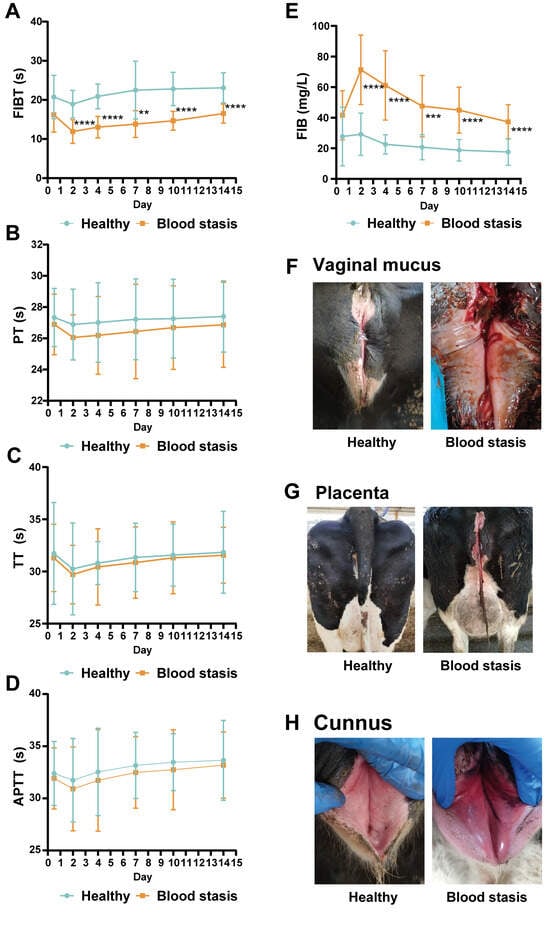
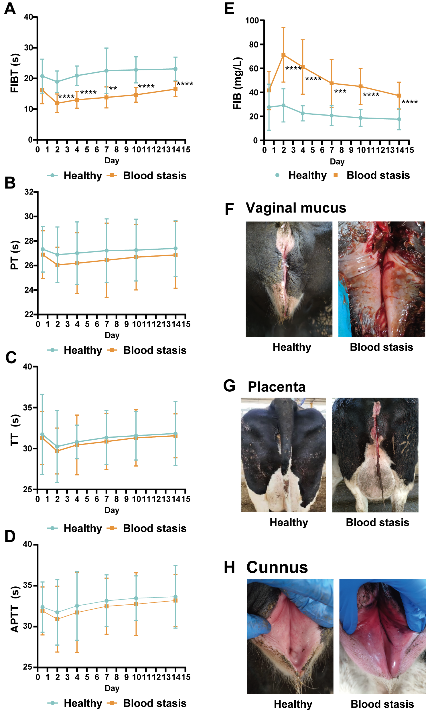
All postpartum cows exhibited enhanced plasma coagulation on day 2 after parturition, including shortened FIBT, PT, TT, and APTT, as well as elevated FIB concentration (Figure 1A–E). These parameters gradually returned to normal levels within 14 days postpartum. Compared with the healthy group, no significant differences were observed in PT, TT, or APTT values in the BSS group (Figure 1B–D). However, FIBT and FIB concentrations in the BSS group were significantly different from those in the healthy group throughout the observation period (Figure 1A,E).
Figure 1.
Diagnosis of blood stasis syndrome (BSS) in dairy cows. Blood coagulation parameters (fibrinogen time, FIBT; prothrombin time, PT; thrombin time, TT; activated partial thromboplastin time, APTT) and fibrinogen (FIB) concentrations were measured in healthy (H) and BSS cows from days 1 to 14 postpartum (A–E). Diagnostic criteria for BSS included clinical observations of the vaginal mucus (F), placenta (G), and vulva (H) (n = 15). ** p < 0.01, *** p < 0.001, **** p < 0.0001 vs. the healthy group.
Cows in the BSS group displayed turbid and abnormally viscous mucus, retained placenta, and redness and swelling of the vulva (Figure 1F–H).
3.2. Diagnosis of Endometritis in Dairy Cows
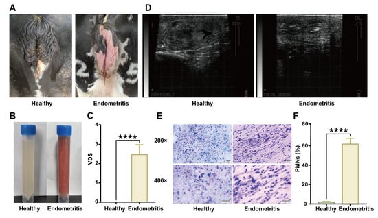
Comparative analysis revealed significant differences in vulvar and vaginal discharge characteristics between healthy cows and those with endometritis. In the healthy group, the vulva remained clean and dry with no significant discharge, and any secretions observed were clear or translucent. In contrast, cows with endometritis exhibited a turbid, viscous, red, or brown vaginal discharge (Figure 2A,B). The VDS was significantly elevated in the endometritis group compared with the healthy group, confirming the severity of uterine inflammation (Figure 2C). Ultrasonographic imaging of the uterine cavity further showed uterine inflammation, including abnormal echogenicity and fluid accumulation, in the cows with endometritis (Figure 2D). Microscopic observation of uterine secretions revealed that the PMN percentage was considerably higher in the endometritis group than in the healthy group (Figure 2E,F). This neutrophilic infiltration aligns with the inflammatory nature of the disease and correlates with clinical observations of mucosal damage and stromal edema. Endometritis can be diagnosed systematically via analysis of an integrated set of indicators, including vulvar status, vaginal discharge properties (turbidity, viscosity), ultrasonographic uterine abnormalities, VDS, and PMN%.
Figure 2.
Diagnosis of endometritis in dairy cows. Comparative assessment of vulvar condition (A), vaginal discharge (B), and uterine ultrasound imaging (D) between the healthy and endometritis groups. Vaginal discharge scores (VDSs) were assigned based on vaginal discharge status (C). Endometrial cytology smears stained with Diff-Quik revealed polymorphonuclear leukocyte (PMN) infiltration (E), and PMN percentages were quantified by calculating the PMN–epithelial cell ratio (F) (n = 15). **** p < 0.0001 vs. the healthy group. TD, transverse diameter; CRL, crown–rump length.
3.3. Prevalence of Postpartum BSS and Its Association with Subsequent Endometritis in Dairy Cows
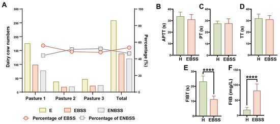
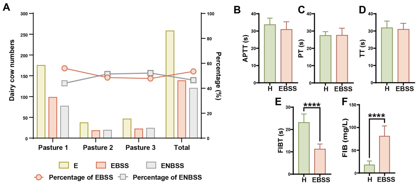
Analysis of 258 endometritis cases across three dairy farms revealed that 53.49% were accompanied by BSS. Among these, Farm 1 had the highest proportion of EBSS(56.00%), significantly exceeding those of Farm 2 (48.65%) and Farm 3 (47.83%) (Figure 3A). Among all cows included in the study, endometritis at 42 days postpartum occurred in 138 of 336 cows in the BSS group (41.07%) and 120 of 2010 cows in the Non-BSS group (5.97%). After adjustment for farm, BSS status was significantly associated with higher odds of endometritis (Table 1). There were no significant differences in APTT, PT, or TT between the EBSS and healthy groups (Figure 3B–D). However, the EBSS group exhibited significantly shortened FIBT and abnormally elevated FIB concentrations compared to the healthy group (Figure 3E,F). The above results indicate that endometritis in the examined cows was frequently accompanied by the study-defined BSS phenotype, and that shortened FIBT and elevated FIB concentrations were associated with this phenotype.
Figure 3.
Distribution and coagulation profiles of cows with endometritis with BSS (EBSS) across dairy farms. The number (bars) and proportions (lines) of cows with endometritis (E), endometritis with BSS (EBSS), and endometritis without BSS (ENBSS) (A). Coagulation parameters (APTT, PT, TT, FIBT, and FIB concentration) were compared between the H and EBSS groups (B–F) (n = 15). **** p < 0.0001 vs. the H group.
Table 1.
Association between postpartum blood stasis syndrome (BSS) and endometritis at 42 days postpartum.
3.4. Uterine Inflammation in a Rat Model of BSS
To eliminate confounding factors associated with dairy cow husbandry, we used a rat model of BSS to investigate the direct association between BSS and endometritis. Adrenaline hydrochloride injection, a widely used method for inducing BSS in animal models, was employed to induce BSS in Sprague-Dawley rats.
In the BSS group, rats exhibited distinct tail ecchymosis, pale claws, and dark sublingual vasculature (Figure 4B). In the BSS group, reduced whole blood viscosity and whole blood viscosity were significantly higher than in the healthy group (Figure 4C,D). The BSS and healthy groups showed no significant differences in PT, APTT, or TT (Figure 4E–G). However, FIBT was significantly shortened and FIB concentration was considerably elevated in the BSS group compared to the healthy group (Figure 4H,I).
Figure 4.
A rat model of BSS exhibiting uterine inflammation. Schematic of the establishment of the BSS model (SC: subcutaneous injection) (A). Morphological differences in the tails, paws, and sublingual veins between H and BSS rats, with arrows highlighting petechiae (B). Reduced whole blood viscosity (RWBV) and whole blood viscosity (WBV) measured at shear rates of 200/s, 30/s, and 1/s (C,D). Comparison of coagulation parameters between the H and BSS groups (E–I). Representative images of uterine tissue sections (J). Hematoxylin–eosin-stained uterine sections (arrows highlight inflammatory cell infiltration) (K). IL-1β levels in uterine tissues (L). (n = 10). * p < 0.05, ** p < 0.01, **** p < 0.0001 vs. the H group.
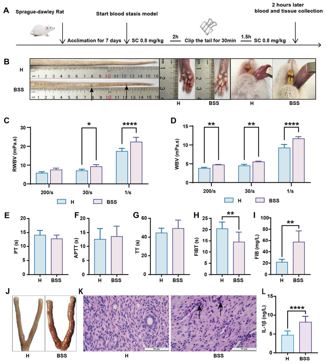
Morphologically, multiple grayish-white suppurative foci were observed on the serosal surface of the uterus in the BSS group (Figure 4J). Histopathological examination of the uterine tissues showed marked inflammatory lesions in the endometrium and myometrium in the BSS group. The endometrium contained some neutrophils that had infiltrated the interstitium. The endometrial blood vessels were moderately congested. The endometrial stroma was distended, with clear spaces separating the blood vessels and endometrial glands (edema). In the myometrium, we observed dense aggregates of neutrophils infiltrating interconnecting bundles of smooth muscle and the surrounding vessels (Figure 4K). Additionally, IL-1β concentrations were significantly higher in the uterine tissues of the BSS group compared to the healthy group (Figure 4L). These findings indicate that experimentally induced BSS was accompanied by uterine inflammatory changes in rats, supporting a potential association between BSS-related abnormalities and uterine inflammation.
3.5. Bioinformatics Analysis of the Correlation Between BSS and Endometritis
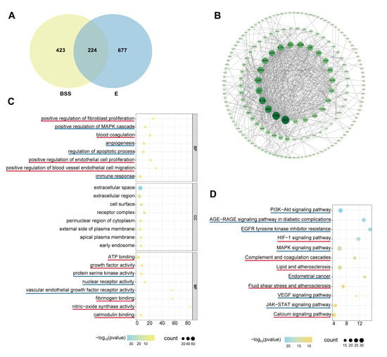
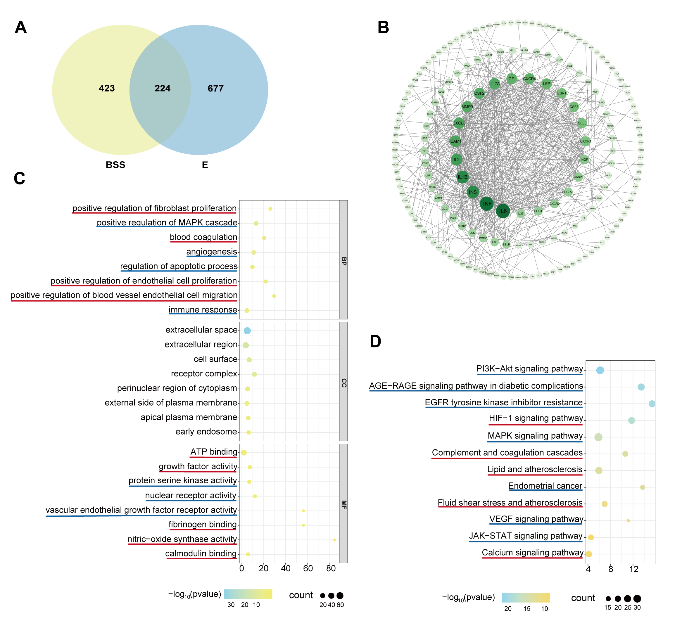
Analysis of target genes associated with both BSS and endometritis identified 224 disease common targets (DCTs) that likely mediate endometritis induction by BSS (Figure 5A) [34]. These 224 target genes were used to form a complex PPI network (Figure 5B). They are enriched in 390 biological processes (BPs), 60 cellular compounds (CCs), and 82 molecular functions (MFs), with the top 10 of each category shown in Figure 5C. KEGG pathway analysis further indicated that the common targets are enriched in 152 pathways, the top 12 of which are shown in Figure 5D. The bioinformatics results show that a significant number of DCTs between BSS and endometritis were enriched in blood circulation- and inflammation-related pathways, further validating the correlation between BSS and endometritis.
Figure 5.
Bioinformatics analysis of the correlation between BSS and endometritis. Venn diagram showing disease-specific targets of BSS, endometritis (E), and their 224 disease common targets (DCTs) (A). Protein–protein interaction (PPI) network of the 224 DCTs constructed using the STRING database (B). GO analysis of the 224 DCTs performed to identify the biological processes, cellular components, and molecular functions they were enriched in (C). KEGG pathway enrichment analysis results of the DCTs (D). In (C,D), pathways directly linked to blood stasis and inflammation are indicated in red and blue, respectively.
3.6. Proteomic Profiling of the Correlation Between BSS and Endometritis
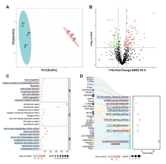
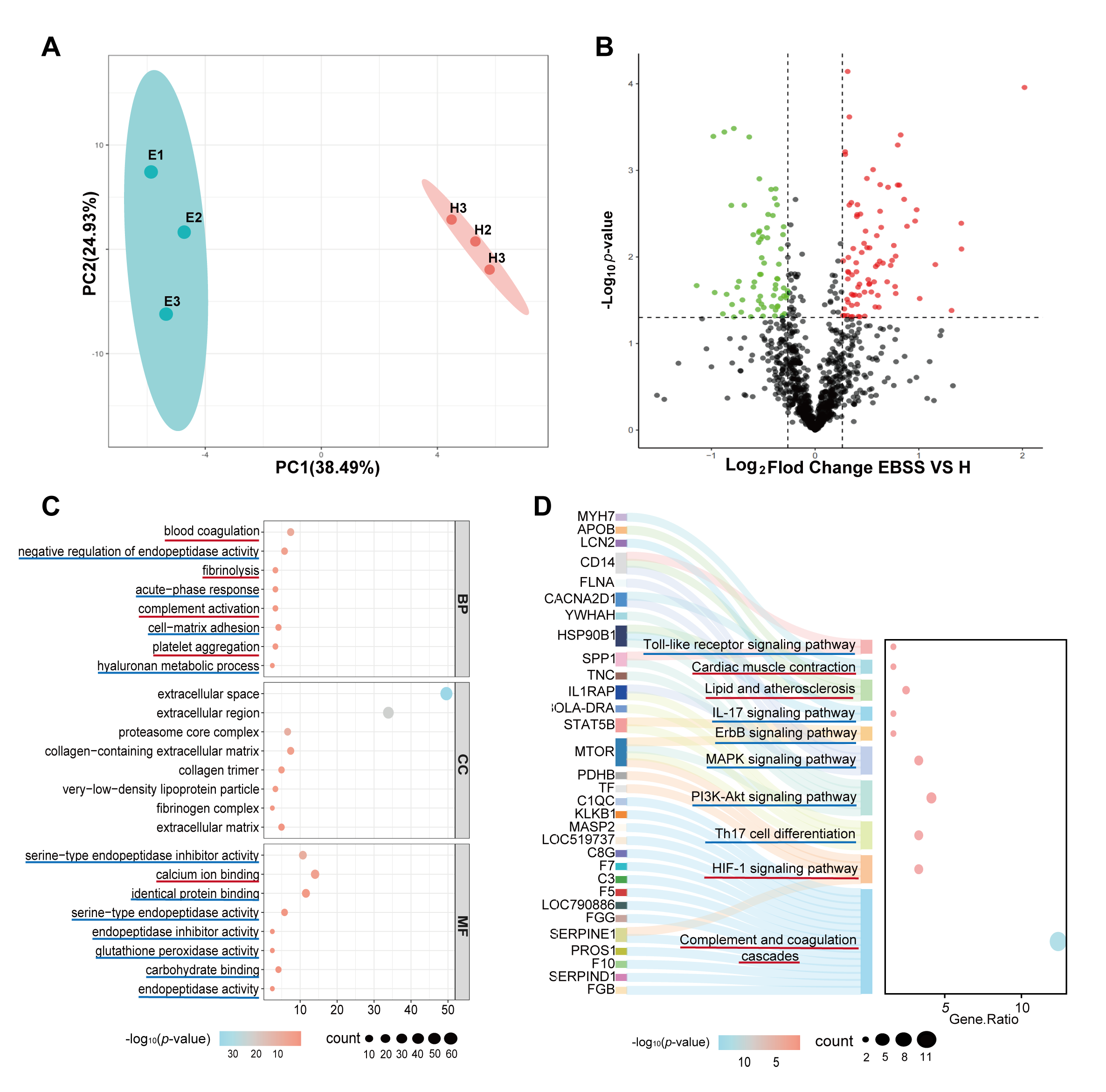
Significant differences in blood protein expression profiles were observed between dairy cows in the healthy and EBSS groups. Principal component analysis (PCA) revealed a distinct separation between the healthy and EBSS groups in PC1 (variance contribution rate of 38.49%) and PC2 (variance contribution rate of 24.93%), indicating that disease status significantly influenced protein expression patterns (Figure 6A). Exploratory candidate proteins were further identified via volcano plot analysis, with 84 upregulated and 66 downregulated candidate proteins under the threshold of |log2FC| > 1 and an unadjusted p < 0.05 (Figure 6B). GO enrichment analysis showed that these candidate proteins were enriched in 32 BPs, 16 CCs, and 21 MFs, with the top 10 of each shown in Figure 6C. KEGG analysis further indicated that these candidate proteins were enriched in 185 pathways, with the top 10 shown in Figure 6D. As FDR correction was not performed, these findings should be interpreted as exploratory.
Figure 6.
Proteomic signature of BSS-related endometritis. (A) Principal component analysis (PCA) was performed to compare protein expression profiles between the healthy and EBSS groups, with PC1 and PC2 accounting for 38.49 and 24.93% of variance, respectively. (B) Volcano plot displaying exploratory candidate proteins (red: upregulated; green: downregulated; black: non-significant). GO (C) and KEGG (D) enrichment analysis results; red and blue lines highlight pathways directly linked to blood stasis and inflammation, respectively.
3.7. Predicting the Molecular Links Between BSS and Endometritis
The enriched pathways identified by GO and KEGG analysis were highly consistent between those based on DCT analysis and those based on candidate proteins analysis. To explore potential molecular signals associated with the crosstalk between BSS and endometritis, we focused on the pathways shown in Figures S2–S13, in which the DCTs and candidate proteins were marked with red asterisks. By integrating DCTs and candidate proteins within these pathways, we investigated potential molecular mechanisms linking blood stasis to endometritis, including: (1) HIF-1α and KLKB1 activating the PAI-1/FIB → C1QC/plasmin → complement C3 and CACNA2D1 → NFKB1 → MYD88 → IL-1α/IL-1β pathways and (2) HIF-1α activating the VEGF/EGFR → AKT → mTOR/NFKB1 → MYD88 → IL-1α/IL-1β pathway. These proposed relationships were inferred from integrated bioinformatics analyses and the previous literature.
3.8. Confirmation of Complement and Coagulation Cascade Activation in Cows with Endometritis with BSS
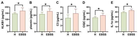
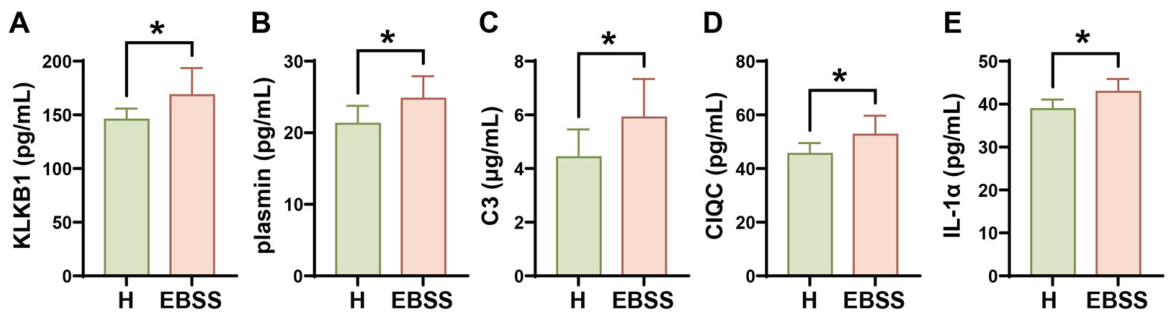
Compared with those in the healthy group, the levels of KLKB1, plasmin, complement C3, and C1QC were significantly elevated in the EBSS group (Figure 7A–D), indicating alterations in coagulation and complement-related markers in EBSS cows. Additionally, the IL-1α concentration in the EBSS group was significantly higher than that in the healthy group (Figure 7E), confirming inflammation in EBSS cows. These results confirmed that EBSS cows exhibited both hypercoagulable states and inflammatory lesions.
Figure 7.
Confirmation of molecular signals indicating blood stasis and inflammation in the plasma of EBSS cows. Plasma concentrations of KLKB1 (A), plasmin (B), C3 (C), C1QC (D), and IL-1α (E) (n = 10). * p < 0.05, vs. the H group.
3.9. Confirmation of HIF-1α- and KLKB1-Mediated Activation of the Complement and Coagulation Cascades and Cardiac Muscle Contraction Signaling in BSS Rats
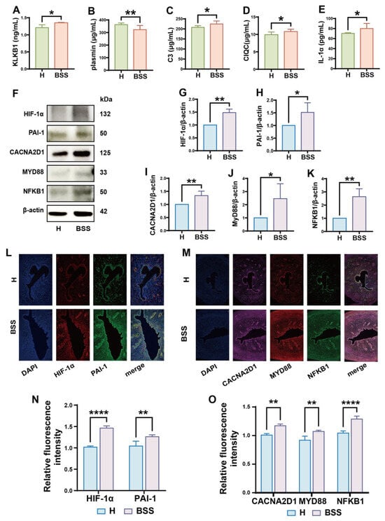
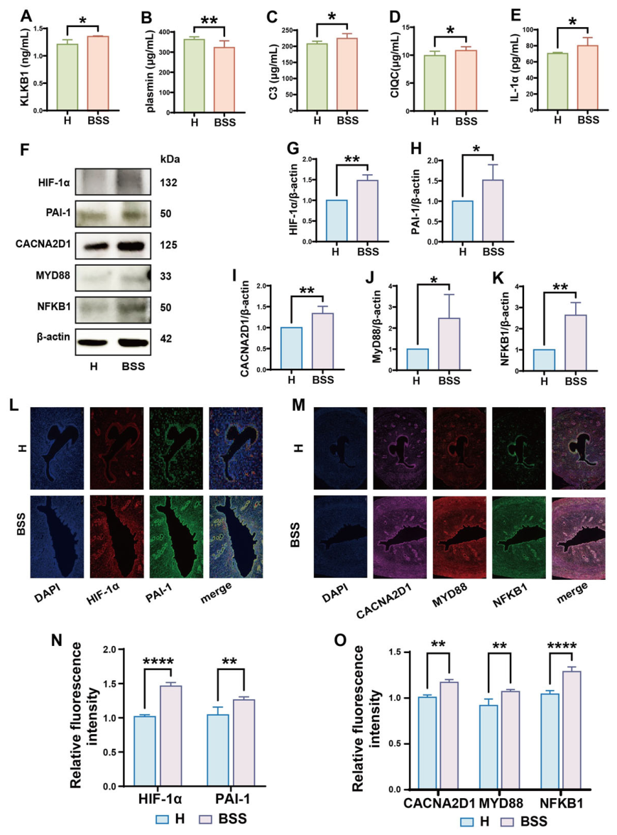
The ELISA results showed that KLKB1, complement C3, C1QC, and IL-1α concentrations in uterine tissues were significantly elevated in the BSS group compared with the healthy group, whereas plasmin levels were significantly decreased (Figure 8A–E). The Western blot results indicated that HIF-1α, PAI-1, CACNA2D1, MYD88, and NFKB1 expression levels in the uterine tissues of BSS group rats were significantly higher than those in the healthy group (Figure 8F–K). Immunofluorescence staining further confirmed that the HIF-1α, PAI-1, CACNA2D1, MYD88, and NFKB1 fluorescence signal intensities in the endometrial epithelium and endometrial gland epithelium of the BSS group were significantly increased compared with those in the healthy group (Figure 8L–O). These results confirm that adrenaline-induced BSS in rats is closely associated with HIF-1α- and KLKB1-mediated activation of the complement and coagulation cascades and cardiac muscle contraction signaling.
Figure 8.
Role of the HIF-1α–PAI-1 and KLKB1-plasmin/CACNA2D1 signaling pathways in generating blood stasis. KLKB1 (A), plasmin (B), C3 (C), C1QC (D), and IL-1α (E) levels in uterine tissues. Western blot of HIF-1α, PAI-1, CACNA2D1, MYD88, and NFKB1 expression in the H and BSS groups (F). HIF-1α (G), PAI-1 (H), CACNA2D1 (I), MYD88 (J), and NFKB1 (K) band density ratios normalized to β-actin. Immunofluorescence staining of paraffin-embedded uterine tissues was performed to assess spatial differences in HIF-1α, PAI-1, CACNA2D1, MYD88, and NFKB1 expression (L,M). Quantification of the relative fluorescence intensity of the data shown in (L–O) (n = 5). * p < 0.05, ** p < 0.01, **** p < 0.0001 vs. the H group.
3.10. Confirmation of HIF-1α-Mediated Activation of VEGF and EGFR Signaling in Rats with BSS
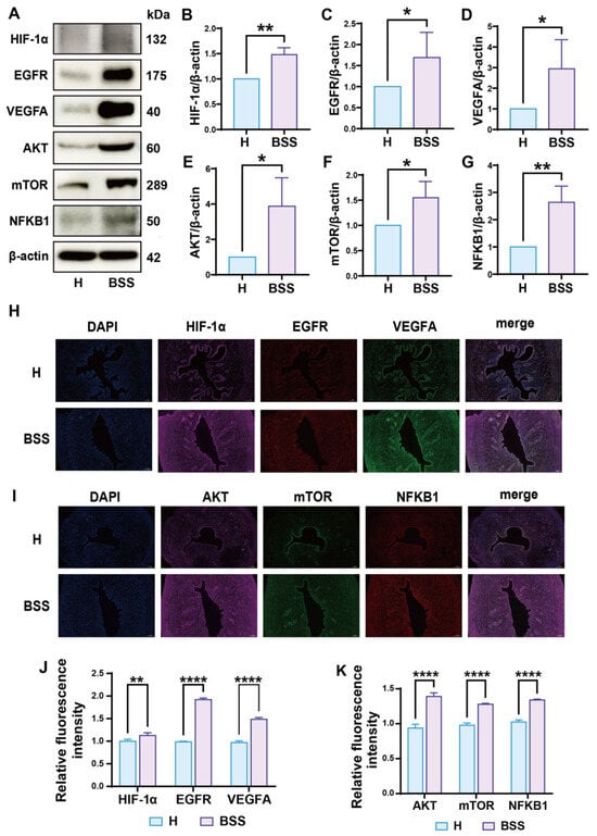
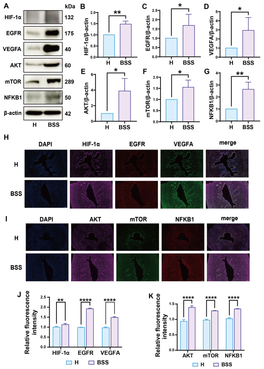
Western blotting confirmed that HIF-1α, EGFR, VEGFA, AKT, mTOR, and NFKB1 levels in the uterine tissue of rats in the BSS group were significantly higher than those in the healthy group (Figure 9A–G). Immunofluorescence staining further confirmed that the fluorescence signal intensities of all of the above proteins were significantly increased in the endometrial epithelium and endometrial gland epithelium of the BSS group compared with those of the healthy group (Figure 9H–K). These results suggest that BSS-associated uterine inflammation is closely related to abnormal activation of the VEGF and EGFR signaling pathways by HIF-1α.
Figure 9.
The role of the HIF-1α signaling pathway in regulating inflammation. Western blot of HIF-1α, EGFR, VEGFA, AKT, mTOR, and NFKB1 expression in the H and BSS groups (A). HIF-1α (B), EGFR (C), VEGFA (D), AKT (E), mTOR (F), and NFKB1 (G) band density ratios normalized to β-actin. Immunofluorescence staining of paraffin-embedded uterine tissues was performed to assess spatial differences in HIF-1α, EGFR, VEGFA, AKT, mTOR, and NFKB1 expression (H,I). Quantification of the relative fluorescence intensity of the data shown in (H–K) (n = 5). * p < 0.05, ** p < 0.01, **** p < 0.0001 vs. the H group.
3.11. Potential Involvement of HIF-1α and KLKB1 in Thrombin-Induced Inflammatory Responses in BEECs
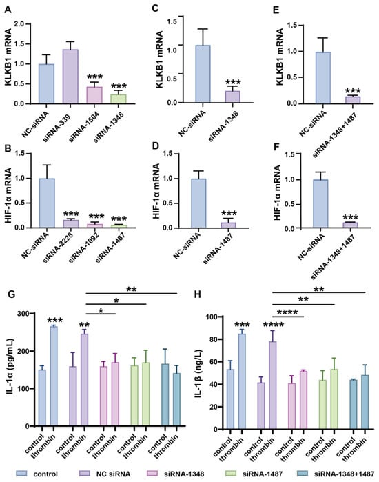
To investigate the critical role of KLKB1 and HIF-1α in mediating blood stasis-induced inflammation during the progression of dairy cow endometritis, NC-siRNA, three KLKB1 siRNAs, and three HIF-1α siRNAs were individually transfected into BEECs. The results show that KLKB1 siRNA-1348 and HIF-1α siRNA-1487 exhibited the highest knockdown efficiency (Figure 10A,B). Thus, these two siRNAs were selected for subsequent experiments. The selected siRNAs were transfected into BEECs individually or in combination, and the mRNA levels of KLKB1 and HIF-1α were successfully knocked down in both single and combined transfection groups (Figure 10C–F).
Figure 10.
The role of KLKB1 and HIF-1α in mediating thrombin-induced inflammation. Analysis of KLKB1 and HIF-1α mRNA knockdown efficiency in bovine endometrial epithelial cells (BEECs) transfected with KLKB1 siRNAs (A) and HIF-1α siRNAs (B). Validation of KLKB1 and HIF-1α mRNA levels in BEECs transfected with siRNA-1348 (C), siRNA-1487 (D), or their combination (E,F). ELISA detection of IL-1α (G) and IL-1β (H) concentrations in the cell culture supernatants (n = 3). * p < 0.05, ** p < 0.01, *** p < 0.001, **** p < 0.0001 vs. the NC-siRNA group.
To mimic blood stasis-related inflammation, BEECs were treated with thrombin—a key activated factor in the blood coagulation process. CCK-8 assays confirmed that thrombin treatment at 10 U/mL for 24 h did not significantly affect BEEC viability (Figure S13). Following thrombin treatment, the secretion of IL-1α and IL-1β was significantly upregulated in non-transfected and NC-siRNA-transfected BEECs, demonstrating that thrombin can induce cellular inflammation. In contrast, IL-1α and IL-1β secretion remained unaffected in BEECs transfected with siRNA-1348, siRNA-1487, or their combination (Figure 10G,H). These findings suggest that KLKB1 and HIF-1α are involved in regulating thrombin-induced inflammatory responses in bovine endometrial epithelial cells.
4. Discussion
Our findings revealed an association between BSS and clinical endometritis, supporting the TCVM hypothesis that links BSS to postpartum endometritis. The elevated FIB concentrations and shortened FIBT observed in cows with BSS indicated a hypercoagulable state, consistent with prior reports demonstrating that impaired uterine microcirculation drives fibrin deposition and tissue hypoxia. This pathological microenvironment of “static blood retention” may compromise endometrial barrier integrity and facilitate bacterial colonization, consistent with published evidence showing that microthrombi and endothelial dysfunction accelerate inflammation [35,36,37]. Proteomic signatures further suggested a potential association between coagulation abnormalities and uterine inflammatory cascades. Further validation of potential mechanisms, including thrombin-mediated protease-activated receptor (PAR) signaling and HIF-1α-related signaling, is therefore warranted. Nevertheless, other risk factors could also contribute to the development of endometritis, such as parity, farm management practices, and genetic predisposition.
The observation of blood stasis-related features, including coagulation abnormalities and uterine inflammatory changes, in the adrenaline-induced rat model provides complementary experimental evidence supporting a potential relationship between blood stasis and uterine inflammation. Previous studies confirm that adrenaline triggers profound α-adrenergic vasoconstriction and platelet activation, inducing microcirculatory failure that pathologically mirrors TCVM-defined BSS [38,39]. Our findings align with published research demonstrating that elevated KLKB1 (encoding plasma prekallikrein) initiates contact system activation—amplifying both coagulation (via FIB cleavage) and inflammation (via complement C3/C1QC cleavage and IL-1α release) [40,41,42,43]. This supports prior mechanistic work in which KLKB1-driven bradykinin and thrombin generation promotes vascular leakage and leukocyte recruitment [41,44]. Crucially, the uterine inflammation observed in our BSS rat model supports clinical–pathological correlations reported in cattle, where microthrombi and fibrin deposition disrupt endometrial integrity, providing an already compromised environment for bacterial or other pathogenic factors to cause damage to the uterus. Collectively, these findings suggest a potential association between blood stasis-related perturbations and uterine inflammation.
Not all cows with clinical endometritis had BSS, indicating that blood stasis is not a necessary condition for endometritis. However, blood stasis was associated with the occurrence and progression of endometritis in over 50% of the postpartum cows evaluated in this study [45]. Early detection and therapeutic intervention for cows with blood stasis during the initial postpartum period might significantly reduce the incidence and severity of endometritis. Our results suggest that coagulation–complement crosstalk may be involved in uterine inflammation, as indicated by the synchronized increases in KLKB1, FIB, C3, and IL-1α in both bovine EBSS and rat BSS models. Multiple studies have demonstrated that blood-activating herbs (e.g., Salvia miltiorrhiza and Carthamus tinctorius) disrupt this cascade by inhibiting KLKB1-driven contact activation, suppressing thrombin-induced fibrin deposition (reducing FIB) and blocking C3 convertase formation [35,46,47]. Results from our proteomic analyses are consistent with the TCVM hypothesis that blood stasis is associated with endometritis [48]. These findings suggest that therapeutic approaches targeting both coagulation-related abnormalities and inflammatory responses may warrant further investigation in dairy cows with endometritis. Nevertheless, blood stasis is only one of the factors potentially associated with endometritis. In the prevention and control of endometritis in clinical dairy cows, preventing bacterial infection remains one of the most effective methods.
The present study supports the potential involvement of HIF-1α and KLKB1 in thrombin-induced BEEC inflammation via knockdown of these two genes. This finding is consistent with previous studies. Blood stasis-induced microcirculatory disturbances activate HIF-1α via hypoxia. HIF-1α subsequently regulates the coagulation–inflammation axis and induces metabolic reprogramming, forming a vicious “hypoxia–thrombosis–inflammation” cycle [49]. Consistently, we found that HIF-1α may be involved in hypercoagulability through VEGF and integrin signaling [50,51]. Notably, HIF-1α overexpression is associated with endometriosis, while its deficiency correlates with intrauterine adhesions, underscoring its critical role in uterine health [52]. KLKB1 has also been shown to cleave protease-activated receptors in vascular smooth muscle cells, thus regulating the expression of epidermal growth factor receptors [53]. These blood stasis-related pathways may be associated with inflammatory responses involving AKT → mTOR/NFKB1 → IL-1 → MYD88 signaling [37,54]. Prior mechanistic studies have confirmed that HIF-1α serves as a master regulator of hypoxic adaptation, directly transactivating VEGF and downstream angiogenic cascades [55]. Complementary work further supports the notion that KLKB1 amplifies coagulation via factor XII activation, consistent with our observation of KLKB1-driven coagulation–complement crosstalk [56]. Critically, our findings converge at the AKT → mTOR/NFKB1 → IL-1 → MYD88 signaling hub: HIF-1α may be involved in NFKB1-related signaling, while thrombin can cleave pro-IL-1α into its active form, potentially contributing to endometrial inflammation [36,57]. These findings suggest that HIF-1α and KLKB1 may be involved in thrombin-induced inflammatory responses in BEECs.
In this study, we found that a series of blood circulation- and inflammation-related signaling pathways were activated in the context of BSS. Furthermore, signaling crosstalk between the blood and uterine tissue through tissue microcirculation has the potential to form a “blood stasis–inflammation” loop. Prior mechanistic studies confirm that hypoxic stress activates HIF-1α in endometrial cells, stimulating VEGF and coagulation cascades that further impair microcirculation. Concurrently, KLKB1-driven contact system activation cleaves complement C3 and releases bradykinin, increasing vascular permeability and neutrophil infiltration, resulting in inflammation [42]. These studies support the possibility that uterine tissue-derived IL-1β feeds back into circulation, activating endothelial PAR-1 receptors to exacerbate coagulation via thrombin generation, thereby reinforcing blood stasis [58]. The present study highlights the potential relevance of BSS-associated inflammation to the prevention and treatment of clinical endometritis. Additionally, the confirmation of the hypothesis that inflammation causes microcirculation disorders, and thus leads to the formation of a “blood stasis–inflammation” vicious cycle, will be of great significance for explaining long-term endometritis in dairy cows [59,60]. The “blood stasis–inflammation” loop is a cyclical process involving signal amplification, and it is difficult to determine which signal initiates the cycle. Previous studies have suggested that blood stasis-induced endometrial microcirculatory disturbances may persistently impair endometrial receptivity, hindering embryo implantation even after inflammation subsides [61,62]. Recurrent chronic endometritis might lead to tubal mucosal fibrosis and luminal adhesions, and blood stasis-related coagulation abnormalities could accelerate these pathological processes.
5. Conclusions
Our clinical studies showed that postpartum BSS in cows could be intrinsically correlated with endometritis occurrence. The rat model provided complementary experimental evidence regarding uterine inflammation associated with experimentally induced BSS. In BSS model rats, uterine inflammatory changes were associated with alterations in HIF-1α- and KLKB1-related signaling, including increased expression of VEGFA and EGFR and elevated levels of CACNA2D1. These molecular changes were accompanied by increased expression of inflammation-related factors (NFKB1, MYD88, IL-1α, and IL-1β), suggesting potential interactions between coagulation-related signaling and uterine inflammation. The potential therapeutic value of targeting both coagulation-related abnormalities and inflammatory responses in bovine endometritis warrants further investigation.
6. Limitations
The findings of the present study should be interpreted in the context of the diagnostic criteria, study design, and experimental models employed.
Firstly, standardized and validated clinical diagnostic criteria for BSS in dairy cows are currently lacking. The criteria used in this study were developed with reference to diagnostic criteria established in human medicine and the relevant literature, taking into account the practical conditions of the participating dairy farms. Although reference intervals for coagulation parameters have been established in healthy human populations, comparable standardized and validated reference intervals for dairy cows remain limited. Therefore, potential diagnostic misclassification, diagnostic overlap, and incorporation bias cannot be completely excluded, which may affect the accuracy of group classification and the estimated strength of the association between BSS and endometritis. Future studies should consider the physiological and pathological characteristics of dairy cows at different postpartum stages, identify candidate diagnostic indicators based on clinical signs, physical findings, and laboratory parameters, and establish standardized BSS diagnostic criteria through expert consensus and multicenter clinical investigations, followed by validation in independent cattle populations. Furthermore, as this was an observational clinical study, causal inferences cannot be drawn. Although farm effects were adjusted for in the analysis, other unmeasured or insufficiently controlled confounding factors, such as parity, retained placenta, and calving-related complications, may still have affected the observed association between BSS and endometritis. To better address these limitations, prospective studies should collect comprehensive individual-level clinical data and account for relevant confounding factors in multivariable analyses.
Secondly, the proteomic findings of this study should be interpreted cautiously. The use of pooled samples limited the assessment of individual biological variability, while the absence of FDR correction may have increased the risk of false-positive findings during differential protein screening. In this study, the proteomic analysis was limited to a comparison between healthy cows and cows with EBSS, without including groups with BSS alone or NEBSS. Nevertheless, the rat model provided complementary experimental evidence suggesting a potential association between BSS alone and uterine inflammation, and several candidate proteins identified by proteomic analysis were further validated in rat uterine tissues. Despite these complementary findings, the current data cannot determine whether the identified protein changes are specifically associated with BSS, endometritis, or their coexistence.
Regarding the animal model, the findings provided preliminary evidence of a potential association between blood stasis and uterine inflammation. However, the model primarily reflected their coexistence, and the absence of separate epinephrine-only and tail-clamping-only groups precluded distinction of the independent effects of these two interventions. Moreover, microbiological characterization was not performed, and the model may not fully reflect the complex pathological features of endometritis in dairy cows. Further studies should compare BSS alone, NEBSS, and EBSS using proteomic analyses and corresponding animal models, incorporating microbiological characterization to distinguish disease-specific and shared molecular changes.
Finally, although the experimental findings suggest that HIF-1α and KLKB1 may be involved in thrombin-induced inflammatory responses in bovine endometrial epithelial cells, the current evidence is insufficient to establish the signaling cascade of the proposed network. Further systematic studies involving sequential siRNA-mediated knockdown and inhibitor-based interventions are needed to clarify the regulatory hierarchy and signaling cascade among these components.
Therefore, the potential therapeutic value of these findings requires further evaluation through functional validation and interventional studies. Larger-scale clinical studies are also needed to further investigate the associations among postpartum BSS, endometritis, and subsequent reproductive outcomes.
Supplementary Materials
The following supporting information can be downloaded at https://www.mdpi.com/article/10.3390/ani16223665/s1, Table S1. Basic information on experimental dairy cows. Table S2. International Diagnostic Guidelines for Blood Stasis Syndrome. Table S3. Specific Sequences of siRNA. Table S4. qRT-PCR primer sequences used in the study. Figure S1. KEGG analysis of disease common targets (DCTs) and candidate proteins associated with complement and coagulation cascades. Figure S2. KEGG analysis of DCTs and candidate proteins associated with the VEGF signaling pathway. Figure S3. KEGG analysis of DCTs and candidate proteins associated with EGFR tyrosine kinase inhibitor resistance. Figure S4. KEGG analysis of DCTs and candidate proteins associated with lipids and atherosclerosis. Figure S5. KEGG analysis of DCTs and candidate proteins associated with cardiac muscle contraction. Figure S6. KEGG analysis of DCTs and candidate proteins associated with the HIF-1α signaling pathway. Figure S7. KEGG analysis of DCTs and candidate proteins associated with the MAPK signaling pathway. Figure S8. KEGG analysis of DCTs and candidate proteins associated with the PI3K–AKT signaling pathway. Figure S9. KEGG analysis of DCTs and candidate proteins associated with the Toll-like receptor signaling pathway. Figure S10. KEGG analysis of DCTs and candidate proteins associated with the IL-17 signaling pathway. Figure S11. KEGG analysis of DCTs and candidate proteins associated with the ERBB signaling pathway. Figure S12. KEGG analysis of DCTs and candidate proteins associated with Th-17 cell differentiation. Figure S13. Effect of thrombin on BEEC viability.
Author Contributions
Conceptualization, J.J. and Y.L.; methodology, J.J. and Z.L.; validation, J.J., Z.L., Z.B., M.L., Y.F., L.C., W.Z. and W.W.; formal analysis, J.J. and Z.L.; investigation, J.J., Z.L., Z.B. and M.L.; resources, Y.L.; data curation, J.J., Z.L. and Z.B.; writing—original draft preparation, J.J. and Z.L.; writing—review and editing, J.J., Z.L. and Y.L.; visualization, J.J. and Z.L.; supervision, Y.F., L.C., W.Z., W.W. and Y.L.; project administration, Y.L.; funding acquisition, Y.L. All authors have read and agreed to the published version of the manuscript.
Funding
This research was supported by the Shaanxi Province Agricultural Key Core Technology Project (Grant No. 2024NYGG005; 2025NYGG004), the National Natural Science Foundation of China (Grant No. 31802230), and the Natural Science Basic Research Project of the Shaanxi Provincial Department of Science and Technology (Grant No. 2019JQ-281).
Institutional Review Board Statement
All experimental procedures were approved by the Animal Ethical and Welfare Committee of Northwest A&F University (Approval No. XN2024-0403; approved on 11 March 2024).
Informed Consent Statement
Written informed consent was obtained from the owner of the animals involved in this study.
Data Availability Statement
The original contributions presented in this study are included in the article/Supplementary Materials. Further inquiries can be directed to the corresponding author.
Acknowledgments
We thank LetPub (www.letpub.com.cn accessed on 4 November 2026) for linguistic assistance and pre-submission expert review.
Conflicts of Interest
The authors declare no conflicts of interest.
Abbreviations
| TCVM | Traditional Chinese Veterinary Medicine |
| Qi | vital energy |
| BSS | blood stasis syndrome |
| FIBT | fibrinogen time |
| PT | prothrombin time |
| TT | thrombin time |
| APTT | activated partial thromboplastin time |
| FIB | fibrinogen concentration |
| VDS | vaginal discharge score |
| PMN | polymorphonuclear leukocyte |
| EBSS | endometritis with blood stasis syndrome |
| ENBSS | endometritis without blood stasis syndrome |
| RWBV | reduced whole blood viscosity |
| WBV | whole blood viscosity |
| DCT | disease common target |
| BP | biological process |
| CC | cellular compound |
| MF | molecular function |
| GO | Gene Ontology |
| KEGG | Kyoto Encyclopedia of Genes and Genomes |
| PPI | protein–protein interaction |
| PCA | principal component analysis |
| PAR | protease-activated receptor |
| BEECs | bovine endometrial epithelial cells |
| DMEM | Dulbecco’s Modified Eagle Medium |
| NC | negative control |
| qRT-PCR | quantitative real-time PCR |
References
- Druker, S.A.; Sicsic, R.; van Straten, M.; Goshen, T.; Kedmi, M.; Raz, T. Cytological endometritis diagnosis in primiparous versus multiparous dairy cows. J. Dairy Sci. 2022, 105, 665–683. [Google Scholar] [CrossRef] [Scilit]
- Sheldon, I.M.; Cronin, J.G.; Bromfield, J.J. Bromfield, Tolerance and Innate Immunity Shape the Development of Postpartum Uterine Disease and the Impact of Endometritis in Dairy Cattle. Annu. Rev. Anim. Biosci. 2019, 7, 361–384. [Google Scholar] [CrossRef] [Scilit]
- Sheldon, I.M.; Lewis, G.S.; LeBlanc, S.; Gilbert, R.O. Defining postpartum uterine disease in cattle. Theriogenology 2006, 65, 1516–1530. [Google Scholar] [CrossRef] [Scilit]
- McKay, C.; Viora, L.; Denholm, K.; Cook, J.; Belandria, R.V. Risk factors for ultrasound-diagnosed endometritis and its impact on fertility in Scottish dairy cattle herds. Vet. Rec. 2023, 193, e3168. [Google Scholar] [CrossRef] [Scilit]
- LeBlanc, S.J.; Duffield, T.F.; Leslie, K.E.; Bateman, K.G.; Keefe, G.P.; Walton, J.S.; Johnson, W. The effect of treatment of clinical endometritis on reproductive performance in dairy cows. J. Dairy Sci. 2002, 85, 2237–2249. [Google Scholar] [CrossRef] [Scilit]
- Silva, T.V.; de Oliveira, E.B.; Pérez-Báez, J.; Risco, C.A.; Chebel, R.C.; Cunha, F.; Daetz, R.; Santos, J.; Lima, F.; Jeong, K.; et al. Economic comparison between ceftiofur-treated and nontreated dairy cows with metritis. J. Dairy Sci. 2021, 104, 8918–8930. [Google Scholar] [CrossRef] [Scilit]
- Pérez-Báez, J.; Silva, T.V.; Risco, C.A.; Chebel, R.C.; Cunha, F.; De Vries, A.; Santos, J.; Lima, F.; Pinedo, P.; Schuenemann, G.; et al. The economic cost of metritis in dairy herds. J. Dairy Sci. 2021, 104, 3158–3168. [Google Scholar] [CrossRef] [Scilit]
- Amin, Y.A.; Abdelaziz, S.G.; Said, A.H. Treatment of postpartum endometritis induced by multidrug-resistant bacterial infection in dairy cattle by green synthesized zinc oxide nanoparticles and in vivo evaluation of its broad spectrum antimicrobial activity in cow uteri. Res. Vet. Sci. 2023, 165, 105074. [Google Scholar] [CrossRef] [Scilit]
- Cai, X.S.; Jiang, H.; Xiao, J.; Yan, X.; Xie, P.; Yu, W.; Lv, W.-F.; Wang, J.; Meng, X.; Chen, C.-Z.; et al. Changes in bacterial community composition in the uterus of Holstein cow with endometritis before and after treatment with oxytetracycline. Sci. Rep. 2024, 14, 9511. [Google Scholar] [CrossRef] [Scilit]
- Denis-Robichaud, J.; Dubuc, J. Randomized clinical trial of intrauterine cephapirin infusion in dairy cows for the treatment of purulent vaginal discharge and cytological endometritis. J. Dairy Sci. 2015, 98, 6856–6864. [Google Scholar] [CrossRef] [Scilit]
- Moghnieh, R.A.; Kanafani, Z.A.; Tabaja, H.Z.; Sharara, S.L.; Awad, L.S.; Kanj, S.S. Epidemiology of common resistant bacterial pathogens in the countries of the Arab League. Lancet Infect. Dis. 2018, 18, e379–e394. [Google Scholar] [CrossRef] [Scilit]
- Haimerl, P.; Arlt, S.; Borchardt, S.; Heuwieser, W. Antibiotic treatment of metritis in dairy cows—A meta-analysis. J. Dairy Sci. 2017, 100, 3783–3795. [Google Scholar] [CrossRef] [Scilit]
- LeBlanc, S.J. Review: Postpartum reproductive disease and fertility in dairy cows. Animal 2023, 17, 100781. [Google Scholar] [CrossRef] [Scilit]
- Ang, C.J.; Skokan, T.D.; McKinley, K.L.; Skokan, K.L. McKinley, Mechanisms of Regeneration and Fibrosis in the Endometrium. Annu. Rev. Cell Dev. Biol. 2023, 39, 197–221. [Google Scholar] [CrossRef] [Scilit]
- Zhang, J.H.; Xiong, Y.H.; Luo, X.Q.; Chen, J.J.; Shao, J.H.; Gong, C.Y. Effect on Treatment of Addition and Subtraction Therapy of Zhuyu Zhixuetang to Chronic and Subtraction Endometritis with Syndrome of Qi Deficiency and Blood Stasis. Chin. J. Exp. Tradit. Med. Formulae 2020, 7, 110–115. [Google Scholar]
- Tian, S.; Liu, T.; Jiang, J.; Zhao, X.; Fan, Y.; Zhang, W.; Ma, W.; Guo, T.; Wang, W.; Liu, Y. Salvia miltiorrhiza ameliorates endometritis in dairy cows by relieving inflammation, energy deficiency and blood stasis. Front. Pharmacol. 2024, 5, 1349139. [Google Scholar] [CrossRef] [Scilit]
- Bao, G.C. The idealist and pragmatist view of qi in tai chi and qigong: A narrative commentary and review. J. Integr. Med. 2020, 18, 363–368. [Google Scholar] [CrossRef] [Scilit]
- Bardan, C.R.; Ioniță, I.; Iordache, M.; Călămar-Popovici, D.; Todorescu, V.; Popescu, R.; Bernad, B.C.; Bardan, R.; Bernad, E.S. Epigenetic Biomarkers in Thrombophilia-Related Pregnancy Complications: Mechanisms, Diagnostic Potential, and Therapeutic Implications: A Narrative Review. Int. J. Mol. Sci. 2024, 25, 13634. [Google Scholar] [CrossRef] [Scilit]
- Li, L.P.; Zhuang, A.W.; Bao, Y.H.; Chu, J.M.; Dou, X.Q. Clinical research on acupoint catgut implantation in the prevention and treatment of postpartum pain of uterine contraction with qi and blood deficiency. Zhongguo Zhen Jiu 2014, 34, 34–36. [Google Scholar]
- Qiu, Y.; Xu, H. Formulation and Interpretation of International Diagnostic Guidelines for Blood-Stasis Syndrome. Chin. J. Integr. Med. 2022, 28, 291–296. [Google Scholar] [CrossRef] [Scilit]
- Sowa, M.A.; Hannemann, C.; Pinos, I.; Ferreira, E.; Biwas, B.; Dai, M.; Corr, E.M.; Cornwell, M.G.; Drenkova, K.; Lee, A.H.; et al. Tetraspanin CD37 regulates platelet hyperreactivity and thrombosis. Cardiovasc. Res. 2025, 21, 943–956. [Google Scholar] [CrossRef] [Scilit]
- Li, Y.; Hu, Z.; Xie, L.; Chen, L.; Xiong, T.; Ma, L.; Zhang, Y.; Bai, Y.; Li, W.; Ding, H.; et al. Buyang huanwu decoction protects against cerebral ischemia-reperfusion injury by inhibiting complement and coagulation cascades: A proteomics and experimental study. Phytomedicine 2025, 147, 157205. [Google Scholar] [CrossRef] [Scilit]
- Hashimoto, Y.; Tsuzuki-Nakao, T.; Kida, N.; Matsuo, Y.; Maruyama, T.; Okada, H.; Hirota, K. Inflammatory Cytokine-Induced HIF-1 Activation Promotes Epithelial-Mesenchymal Transition in Endometrial Epithelial Cells. Biomedicines 2023, 11, 210. [Google Scholar] [CrossRef] [Scilit]
- Antman, E.M.; Braunwald, E. Managing Stable Ischemic Heart Disease. N. Engl. J. Med. 2020, 82, 1468–1470. [Google Scholar] [CrossRef] [Scilit]
- Zou, M.; Wang, A.; Wei, J.; Cai, H.; Yu, Z.; Zhang, L.; Wang, X. An insight into the mechanism and molecular basis of dysfunctional immune response involved in cholestasis. Int. Immunopharmacol. 2021, 92, 107328. [Google Scholar] [CrossRef] [Scilit]
- Guan, F.; Zhang, S.; Fan, L.; Sun, Y.; Ma, Y.; Cao, C.; Zhang, Y.; He, M.; Du, H. Kunling Wan improves oocyte quality by regulating the PKC/Keap1/Nrf2 pathway to inhibit oxidative damage caused by repeated controlled ovarian hyperstimulation. J. Ethnopharmacol. 2023, 301, 115777. [Google Scholar] [CrossRef] [Scilit]
- Lindsay, C.V.; Potter, J.A.; Grimshaw, A.A.; Abrahams, V.M.; Tong, M. Endometrial responses to bacterial and viral infection: A scoping review. Hum. Reprod. Update 2023, 29, 675–693. [Google Scholar] [CrossRef] [Scilit]
- Dai, O.; Fan, Y.; Zhou, Q.; Liu, J.; Zuo, J.; Wang, F.; Li, L.; Wang, F.; Xiong, L. Effect of Leonurus japonicus alkaloids on endometrial inflammation and its mechanisms. J. Ethnopharmacol. 2025, 342, 119432. [Google Scholar] [CrossRef] [Scilit]
- Meridians, B.O. International Diagnostic Guidelines for Blood-Stasis Syndrome. Chin. J. Integr. Med. 2022, 28, 297–303. [Google Scholar] [CrossRef] [Scilit]
- Gao, Y.; Shang, B.; He, Y.; Deng, W.; Wang, L.; Sui, S. The mechanism of Gejie Zhilao Pill in treating tuberculosis based on network pharmacology and molecular docking verification. Front. Cell Infect. Microbiol. 2024, 14, 1405627. [Google Scholar] [CrossRef] [Scilit]
- Liu, Y.; Park, J.M.; Chang, K.H.; Chin, Y.W.; Lee, M.Y. α- and γ-mangostin cause shape changes, inhibit aggregation and induce cytolysis of rat platelets. Chem. Biol. Interact. 2015, 240, 240–248. [Google Scholar] [CrossRef] [Scilit]
- Kawano, Y.; Furukawa, Y.; Kawano, Y.; Nasu, K.; Narahara, H. Thrombin-induced chemokine production in endometrial stromal cells. Hum. Reprod. 2011, 26, 407–413. [Google Scholar] [CrossRef] [Scilit]
- Kusama, K.; Ashihara, M.; Okita, M.; Yoshida, K.; Suzuki, M.; Suzuki, K.; Hosokawa, R.; Yoshie, M.; Kojima, J.; Mizuno, Y.; et al. Role of Transcription Factor, LIM Homeobox 9 (LHX9) in Inflammation Associated with Endometriosis. Mol. Reprod. Dev. 2025, 92, e70046. [Google Scholar] [CrossRef] [Scilit]
- Liu, T.; Chen, Q.; Wu, H.; Li, J.; Xian, M.; Lu, K.; Zhu, C.; Wei, J. Exploring Potential Causal Links: How Parkinson’s Disease Brain Damage Impacts Liver Health-A Mendelian Randomization and Transcriptomic Study. J. Inflamm. Res. 2025, 18, 8173–8197. [Google Scholar] [CrossRef] [Scilit]
- Vasuthas, K.; Kjesbu, J.S.; Brambilla, A.; Levitan, M.; Coron, A.E.; Fonseca, D.M.; Strand, B.L.; Slupphaug, G.; Rokstad, A.M.A. Fucoidan alginate and sulfated alginate microbeads induce distinct coagulation, inflammatory and fibrotic responses. Mater. Today Bio 2025, 31, 101474. [Google Scholar] [CrossRef] [Scilit]
- Burzynski, L.C.; Humphry, M.; Pyrillou, K.; Wiggins, K.A.; Chan, J.N.E.; Figg, N.; Kitt, L.L.; Summers, C.; Tatham, K.C.; Martin, P.B.; et al. The Coagulation and Immune Systems Are Directly Linked through the Activation of Interleukin-1α by Thrombin. Immunity 2019, 50, 1033–1042.e6. [Google Scholar] [CrossRef] [Scilit]
- Zhang, Y.M.; Miao, Z.M.; Chen, Y.P.; Song, Z.B.; Li, Y.Y.; Liu, Z.W.; Zhou, G.C.; Li, J.; Shi, L.L.; Chen, Y.; et al. Ononin promotes radiosensitivity in lung cancer by inhibiting HIF-1α/VEGF pathway. Phytomedicine 2024, 125, 155290. [Google Scholar] [CrossRef] [Scilit]
- Odawara, A.; Kikkawa, K.; Katoh, M.; Toryu, H.; Shimazaki, T.; Sasaki, Y. Inhibitory effects of TA-993, a new 1,5-benzothiazepine derivative, on platelet aggregation. Circ. Res. 1996, 78, 643–649. [Google Scholar] [CrossRef] [Scilit]
- Valenzuela Leon, P.C.; Ring, M.E.; Nishiduka, E.; Bonilla, B.; Cruz, P.; Martinez, E.P.; Percopo, C.; Calvo, E. Guianensin, a Blackfly Salivary Protein, Inhibits the Lectin Pathway of Complement. J. Med. Chem. 2025, 68, 4663–4671. [Google Scholar] [CrossRef] [Scilit]
- Lan, T.; Zeng, Q.; Fan, Y.; Liu, T.; Yao, P.; Liang, Z.; Dang, X.; Zhu, H.; Li, Y.; Jiang, W.; et al. Proteomics Analysis of Serum Reveals Potential Biomarkers for Heart Failure Patients with Phlegm-Blood Stasis Syndrome. J. Proteome Res. 2024, 23, 226–237. [Google Scholar] [CrossRef] [Scilit]
- Moellmer, S.A.; Puy, C.; McCarty, O.J.T. Biology of factor XI. Blood 2024, 143, 1445–1454. [Google Scholar] [CrossRef] [Scilit]
- Sohaei, D.; Hollenberg, M.; Janket, S.J.; Diamandis, E.P.; Poda, G.; Prassas, I. The therapeutic relevance of the Kallikrein-Kinin axis in SARS-cov-2-induced vascular pathology. Crit. Rev. Clin. Lab. Sci. 2023, 60, 25–40. [Google Scholar] [CrossRef] [Scilit]
- Cohn, D.M.; Gurugama, P.; Magerl, M.; Katelaris, C.H.; Launay, D.; Bouillet, L.; Petersen, R.S.; Lindsay, K.; Aygören-Pürsün, E.; Maag, D.; et al. CRISPR-Based Therapy for Hereditary Angioedema. N. Engl. J. Med. 2025, 392, 458–467. [Google Scholar] [CrossRef] [Scilit]
- Riedl, M.A.; Bordone, L.; Revenko, A.; Newman, K.B.; Cohn, D.M. Clinical Progress in Hepatic Targeting for Novel Prophylactic Therapies in Hereditary Angioedema. J. Allergy Clin. Immunol. Pract. 2024, 12, 911–918. [Google Scholar] [CrossRef] [Scilit]
- Lean, I.J.; LeBlanc, S.J.; Sheedy, D.B.; Duffield, T.; Santos, J.E.P.; Golder, H.M. Associations of parity with health disorders and blood metabolite concentrations in Holstein cows in different production systems. J. Dairy Sci. 2023, 106, 500–518. [Google Scholar] [CrossRef] [Scilit]
- Guo, P.; Wang, X.; Chen, Q.; Dong, X.; Qin, Z.; Liu, J. Integrative metabolomics and proteomics reveal the effects and mechanisms of Salvia miltiorrhiza in alleviating traumatic blood stasis syndrome. Front. Vet. Sci. 2025, 12, 1579790. [Google Scholar] [CrossRef] [Scilit]
- Hayama, T.; Kamio, N.; Okabe, T.; Muromachi, K.; Matsushima, K. Kallikrein Promotes Inflammation in Human Dental Pulp Cells Via Protease-Activated Receptor-1. J. Cell Biochem. 2016, 117, 1522–1528. [Google Scholar] [CrossRef] [Scilit]
- Stark, K.; Massberg, S. Interplay between inflammation and thrombosis in cardiovascular pathology. Nat. Rev. Cardiol. 2021, 18, 666–682. [Google Scholar] [CrossRef] [Scilit]
- Shaposhnikov, M.; Thakar, J.; Berk, B.C. Value of Bioinformatics Models for Predicting Translational Control of Angiogenesis. Circ. Res. 2025, 136, 1147–1165. [Google Scholar] [CrossRef] [Scilit]
- Deng, H.; Xu, Q.; Li, X.T.; Huang, X.; Liu, J.Y.; Yan, R.; Quan, Z.S.; Shen, Q.K.; Guo, H.Y. Design, synthesis, and evaluation of antitumor activity in Pseudolaric acid B Azole derivatives: Novel and potent angiogenesis inhibitor via regulation of the PI3K/AKT and MAPK mediated HIF-1/VEGF signaling pathway. Eur. J. Med. Chem. 2024, 278, 116813. [Google Scholar] [CrossRef] [Scilit]
- Li, M.; Wang, Y.; Li, M.; Wu, X.; Setrerrahmane, S.; Xu, H. Integrins as attractive targets for cancer therapeutics. Acta Pharm. Sin. B 2021, 11, 2726–2737. [Google Scholar] [CrossRef] [Scilit]
- Tang, X.; Zhao, H.; Ding, Y.; Qin, Y.; Yang, X.; Jiang, X.; Zhou, H.; Liu, B. Enhancement of endometrial receptivity by Bushen Zhuyun Decoction via cryptotanshinone-mediated TRIM28 induction and HIF-1α suppression. J. Ethnopharmacol. 2025, 350, 119943. [Google Scholar] [CrossRef] [Scilit]
- Klein, D.; Goldberg, S.; Theile, C.S.; Dambra, R.; Haskell, K.; Kuhar, E.; Lin, T.; Parmar, R.; Manoharan, M.; Richter, M.; et al. Centyrin ligands for extrahepatic delivery of siRNA. Mol. Ther. 2021, 29, 2053–2066. [Google Scholar] [CrossRef] [Scilit]
- Tan, C.C.; Cao, S.J.; Chao, W.H.; Zhang, B.; Deng, X.Y.; Wang, L.N.; Kang, N.; Qiu, F. Podophyllum hexandrum Royle mitigates perimenopausal symptoms in an OVX rat model by activating the PI3K/AKT/mTOR pathway and enhancing estrogen receptor expression. J. Ethnopharmacol. 2025, 349, 119968. [Google Scholar] [CrossRef] [Scilit]
- Konieczny, P.; Xing, Y.; Sidhu, I.; Subudhi, I.; Mansfield, K.P.; Hsieh, B.; Biancur, D.E.; Larsen, S.B.; Cammer, M.; Li, D.; et al. Interleukin-17 governs hypoxic adaptation of injured epithelium. Science 2022, 377, eabg9302. [Google Scholar] [CrossRef] [Scilit]
- Simão, F.; Ustunkaya, T.; Clermont, A.C.; Feener, E.P. Plasma kallikrein mediates brain hemorrhage and edema caused by tissue plasminogen activator therapy in mice after stroke. Blood 2017, 129, 2280–2290. [Google Scholar] [CrossRef] [Scilit]
- Wang, G.; Mao, W.; Zhang, Y.; Yang, H.; Zhu, M.; Li, Y.; Chen, W.; Chen, Y.; Lou, C.; Li, P.; et al. Multiomics and Systematic Analyses Reveal the Roles of Hemoglobin and the HIF-1 Pathway in Polycystic Ovary Syndrome. Adv. Sci. 2025, 12, e2411679. [Google Scholar] [CrossRef] [Scilit]
- Niu, X.Y.; Xie, X.X.; Tuo, H.Z.; Lv, C.P.; Huang, Y.R.; Zhu, J.; Liang, S.-Y.; Du, X.-Y.; Yang, C.-G.; Hou, S.-J.; et al. Thrombomodulin reduces α-synuclein generation and ameliorates neuropathology in a mouse model of Parkinson’s disease. Cell Death Discov. 2024, 10, 167. [Google Scholar] [CrossRef] [Scilit]
- Qian, C.; Huang, Y.; Zhang, S.; Yang, C.; Zheng, W.; Tang, W.; Wan, G.; Wang, A.; Lu, Y.; Zhao, Y. Integrated identification and mechanism exploration of bioactive ingredients from Salvia miltiorrhiza to induce vascular normalization. Phytomedicine 2025, 138, 56427. [Google Scholar] [CrossRef] [Scilit]
- Pontecorvi, P.; Banki, M.A.; Zampieri, C.; Zalfa, C.; Azmoon, P.; Kounnas, M.Z.; Marchese, C.; Gonias, S.L.; Mantuano, E. Fibrinolysis protease receptors promote activation of astrocytes to express pro-inflammatory cytokines. J. Neuroinflamm. 2019, 16, 257. [Google Scholar] [CrossRef] [Scilit]
- Zhou, W.; Van Sinderen, M.; Rainczuk, K.; Menkhorst, E.; Sorby, K.; Osianlis, T.; Pangestu, M.; Santos, L.; Rombauts, L.; Rosello-Diez, A.; et al. Dysregulated miR-124-3p in endometrial epithelial cells reduces endometrial receptivity by altering polarity and adhesion. Proc. Natl. Acad. Sci. USA 2024, 121, e2401071121. [Google Scholar] [CrossRef] [Scilit]
- Siebert, A.E.; Maroney, S.A.; Martinez, N.D.; Soares, M.J.; Mast, A.E. Placental Vascular Defects and Embryonic Lethality Triggered by TFPIα Deficiency in Factor V Leiden Mice. Arter. Thromb. Vasc. Biol. 2025, 45, 1266–1276. [Google Scholar] [CrossRef] [Scilit]

Disclaimer/Publisher’s Note: The statements, opinions and data contained in all publications are solely those of the individual author(s) and contributor(s) and not of MDPI and/or the editor(s). MDPI and/or the editor(s) disclaim responsibility for any injury to people or property resulting from any ideas, methods, instructions or products referred to in the content. |
© 2026 by the authors. Licensee MDPI, Basel, Switzerland. This article is an open access article distributed under the terms and conditions of the Creative Commons Attribution (CC BY) license.