Review

Role of Gut Microbiota in Bridging Vitamin D Deficiency and Type 2 Diabetes Mellitus Pathogenesis

1
School of Public Health, Shanghai Jiao Tong University School of Medicine, Shanghai 201318, China
2
The College of Medical Technology, Shanghai University of Medicine and Health Sciences, Shanghai 201318, China
3
College of Public Health, Shanghai University of Medicine and Health Sciences, Shanghai 201318, China
*
Author to whom correspondence should be addressed.
Microorganisms 2026, 14(3), 628; https://doi.org/10.3390/microorganisms14030628
Submission received: 2 February 2026 / Revised: 2 March 2026 / Accepted: 4 March 2026 / Published: 11 March 2026
(This article belongs to the Special Issue Effects of Diet and Nutrition on Gut Microbiota)

Abstract

Type 2 diabetes mellitus (T2DM) is a complex metabolic disorder. The nutritional status of vitamin D, an essential micronutrient, is closely linked to the onset and progression of T2DM. A growing body of research has shown that gut microbiota and its metabolites are emerging as a biological link connecting vitamin D and systemic glucose metabolism. Gut dysbiosis is prevalent in T2DM patients, which is characterized by reduced gut microbial diversity, increased abundance of pathogenic bacteria, and abnormal production of key metabolites such as short-chain fatty acids, bile acids and tryptophan derivatives. These abnormal changes in gut microecology and metabolites can impair intestinal barrier integrity and induce chronic low-grade inflammation in the body, and vitamin D deficiency may further exacerbate these abnormal processes. The evidence suggests that the regulatory effect of vitamin D on systemic glucose metabolism may be partially achieved through gut microbiota-related pathways. This review aims to explore whether, and by what mechanisms, the gut microbiota mediates the regulatory effect of vitamin D on T2DM. It also intends to conduct an analysis of the potential molecular mechanisms underlying the interactions between vitamin D, gut microbiota and T2DM, so as to provide a new theoretical basis and research ideas for the prevention and intervention of T2DM.

1. Introduction

Diabetes is a chronic disease marked by hyperglycemia. It has become one of the most important public health challenges worldwide [1]. As of 2024, the global number of adults with diabetes aged 20–79 years reached 589 million. It is projected to rise to 853 million by 2050. Type 2 diabetes mellitus (T2DM) accounts for more than 85% of this number [1,2]. T2DM is driven by insulin resistance (IR), impaired β-cell function and persistent hyperglycemia. Current studies show that the development of T2DM results from long-term interactions between genetic susceptibility and environmental factors [3]. Unhealthy lifestyles, excessive energy intake and micronutrient deficiencies are all regarded as important environmental risk factors [4]. Among the many nutrients, vitamin D is important. A growing body of evidence suggests that vitamin D status is closely linked to insulin resistance and T2DM risk [5,6,7]. The association between vitamin D and T2DM cannot be explained by a single pathway alone. Gut microbiota may play an important role in this association [8]; however, how gut microbiota mediate the effect of vitamin D on T2DM is not yet fully clear.
In T2DM patients, the composition and diversity of gut microbiota change significantly. This suggests that gut microbial dysbiosis may be involved in the development and progression of IR and T2DM [9,10,11]. T2DM patients generally have reduced microbial diversity, an increased proportion of pathogenic bacteria and disrupted microbial metabolic functions. They also have an impaired intestinal barrier and chronic low-grade inflammation [12,13]. Therefore, gut microecology is regarded as an important pathological link in T2DM. Changes in the structure and diversity of gut microbiota can directly or indirectly affect the levels of its functional molecules and metabolites. These include short-chain fatty acids (SCFAs), tryptophan (Trp) derivatives, bile acids (BAs) and branched-chain amino acids (BCAAs) [14]. These gut metabolites can act as key signaling molecules in the host’s metabolism. They regulate insulin sensitivity, glucose homeostasis and immune responses [14,15,16,17]. Hence, regulating gut microbiota composition and restoring the balance of its metabolites to maintain intestinal homeostasis is of great significance for improving T2DM-related metabolic disorders.
Individuals with vitamin D deficiency also exhibit analogous patterns of gut dysbiosis to those seen in patients with T2DM [18,19]. The vitamin D/vitamin D receptor (VD/VDR) signaling pathway not only modulates the expression of antimicrobial peptides, preserves intestinal barrier integrity and impacts mucosal immunity, but also changes the abundance and composition of gut microbiota [8]. This further exerts an indirect effect on the production of microbial metabolites. There is a correlation between vitamin D’s role in ameliorating T2DM and its regulatory effect on gut microbiota. The potential protective effect of vitamin D against T2DM is most likely achieved through its modulation of gut microbiota and their metabolites [7,8,15,20,21,22]. Although numerous studies have independently explored the associations among vitamin D, gut microbiota, and T2DM, comprehensive summaries that conceptualize these three components as an integrated regulatory axis remain scarce, and findings from human, animal, and in vitro studies have yet to be integrated into a unified mechanistic framework [8,19]. Therefore, by summarizing the existing evidence, we found that the gut microbiome can serve as an important mediating factor in the association between vitamin D and T2DM. between vitamin D and T2DM association.
There may be a close connection between vitamin D, gut microbiota, and T2DM, but the mechanism of how vitamin D influences T2DM through the gut microbiota is still not clear. In this review article, we discuss in detail how vitamin D and T2DM interact through intestinal barrier function, inflammation–immune modulation, and microbial metabolite-mediated pathways, thereby affecting the regulation of glucose metabolism.

2. Literature Search and Study Selection

This review was conducted as a structured narrative review. PubMed, Web of Science, and Google Scholar databases were searched for reports until 15 December 2025 using the following terms: “vitamin D” OR “25-hydroxyvitamin D” AND “type 2 diabetes mellitus” OR “T2DM”, and “microbiota*” AND “type 2 diabetes mellitus” OR “T2DM”, and “vitamin D” or “25-hydroxyvitamin D” AND “microbiota*”. Studies were included if they: (1) Investigated the relationship between vitamin D and gut microbiota; (2) examined T2DM or metabolic-related outcomes; and (3) were original research articles involving human subjects, animal models, or in vitro experiments. Reviews, conference abstracts, non-English publications, and duplicate records were excluded. Titles and abstracts were screened for relevance, followed by full-text evaluation when necessary. A total of 38 human studies, 22 animal studies, and 14 in vitro studies were included in the final synthesis. Serum 25(OH)D concentrations are uniformly expressed in ng/mL throughout this review (1 ng/mL = 2.5 nmol/L). Study-specific cutoff definitions for vitamin D deficiency or insufficiency were retained as originally reported, acknowledging heterogeneity in threshold criteria across studies.

3. Vitamin D Deficiency and Type 2 Diabetes Mellitus Risk

Substantial observational evidence has demonstrated that vitamin D deficiency, defined by reduced serum 25(OH)D levels, is associated with a higher risk of T2DM [23]. A Danish population study involving 222,311 participants reported a significant inverse association between 25(OH)D levels and type 2 diabetes risk, with each 10 nmol/L reduction in 25(OH)D corresponding to a 15% increase in diabetes risk [24]. Consistent findings were reported in the Spanish SUN project, where predicted serum vitamin D levels were negatively correlated with the risk of developing T2DM [25]. Data from NHANES also indicate that optimal 25(OH)D concentrations (~30 ng/mL; originally reported as ~75 nmol/L; 1 ng/mL = 2.5 nmol/L) are associated with the lowest T2DM risk [26].
Observational evidence suggests that vitamin D deficiency is associated with a high risk of T2DM, indicating that vitamin D deficiency is one of the many factors contributing to an increased risk of T2DM. However, evidence from randomized controlled trials (RCTs) remains heterogeneous. Some trials have reported improvements in glycemic parameters. For example, six months of high-dose vitamin D supplementation has been shown to significantly improve peripheral insulin sensitivity and β-cell function [27]. Another RCT with a 30-month intervention period also found that vitamin D supplementation significantly reduced levels of fasting insulin and homeostasis model assessment of insulin resistance (HOMA-IR) in middle-aged and elderly patients with T2DM [28]. Nevertheless, a recent large-scale trial in older adults found no significant reduction in incidences of T2DM with vitamin D supplementation, although pooled analyses across multiple RCTs suggested a modest protective effect [29]. Overall, findings from RCTs are inconsistent. This is probably because of differences in supplementation dose, baseline vitamin D status, ethnicity and participants’ metabolic characteristics. People with near-normal baseline 25(OH)D usually get little benefit from supplementation. In contrast, those with severe vitamin D deficiency are most likely to show a response [21,29,30,31].
Despite consistent evidence from various study designs indicating a strong association between vitamin D status and the risk of T2DM, classical endocrine pathways alone are insufficient to fully explain its metabolic effects. Increasing research suggests that the regulatory role of vitamin D on insulin sensitivity and glucose homeostasis may be partly mediated through its influence on the gut microbiota and their derived metabolites [8], providing new theoretical support for a potential vitamin D–gut microbiota–T2DM regulatory axis.

4. Vitamin D and Gut Microbiota in Type 2 Diabetes Mellitus

4.1. Effects of Vitamin D on Gut Microbiota Composition and Diversity

An increasing body of evidence suggests that vitamin D status is closely associated with gut microbiota diversity and composition (Table 1). Observational studies have shown that in infants, those with vitamin D deficiency exhibited significantly lower Pielou’s evenness and Shannon diversity at six months compared with vitamin D-sufficient infants, indicating that vitamin D deficiency may restrict the establishment and development of early intestinal microbiota diversity [32]. In postmenopausal women, the Observed, Chao1, and ACE indices in a low vitamin D level group were all lower than those in a high vitamin D level group, suggesting that low vitamin D status is associated with decreased intestinal microbiota richness. At the level of microbiota composition, the high vitamin D (HVD) group had enriched abundances of Christensenellaceae, Eggerthellaceae, and Cloacibacillus in the intestine, while the low vitamin D (LVD) group was dominated by Bifidobacterium, Gemella, and Lachnoclostridium [33]. Studies on obese populations have found that individuals with optimal serum 25(OH)D levels had significantly higher intestinal microbiota diversity than those with low levels, and there were intergroup differences in the relative abundances of Bacteroides, Prevotella, and some Clostridium species [34]. Therefore, the aforementioned observational studies across different populations indicate that low vitamin D status is closely associated with reduced gut microbiota diversity and dysbiosis.
Interventional studies further support the association between vitamin D and gut microbiota. In healthy individuals, moderate-dose vitamin D supplementation significantly increased the microbial abundance of Gemella, Fournierella, and Ruminococcaceae UCG-010, while the percentage change in 25(OH)D was negatively correlated with gut microbial stability [35]. In healthy women with vitamin D deficiency, vitamin D supplementation not only significantly enhanced gut microbial diversity but also increased the ratio of Bacteroidetes to Firmicutes and promoted the enrichment of beneficial probiotics such as Akkermansia and Bifidobacterium [36]. In specific populations, in a trial involving vitamin D-deficient overweight or obese adults, vitamin D supplementation increased the abundance of Granulicatella in the gut of the participant, accompanied by a decrease in the abundance of Ruminococcus [37]. Yet, not all results were positive [38,39].
Animal studies further indicate that vitamin D supplementation increases unclassified Muribaculaceae and Lachnospiraceae_NK4A136_group, while reducing the relative abundance of Lactobacillus and Odoribacter in KKay mice [40]. Particularly in a high-fat diet-induced dysbiosis mouse model, vitamin D supplementation increased the relative abundance of Prevotella and Porphyromonadaceae, while reducing Mucispirillum, Acetatifactor, Desulfovibrio, and Oscillospira, thereby alleviating dysbiosis. This was accompanied by a reduced Firmicutes/Bacteroidetes (F/B) ratio and enhanced α-diversity, thereby improving the dysbiotic state induced by a high-fat diet [41,42].
Combining the results of diverse studies, vitamin D supplementation is associated with the growth of SCFA-producing bacteria (e.g., Anaerostipes, Akkermansia) in the intestine and reduces the abundance of lipopolysaccharide (LPS)-positive bacteria (e.g., EscherichiaShigella, Desulfovibrio). Other microbial taxa may exhibit heterogeneous responses, potentially influenced by baseline vitamin D status, metabolic condition, supplementation dose and duration, as well as differences in sequencing and analytical methodologies. Since SCFA-producing bacteria can maintain intestinal barrier integrity by producing propionate and butyrate, while LPS-positive bacteria may induce intestinal inflammation, vitamin D may thus improve the host’s intestinal microecological environment and metabolic status by regulating the balance of intestinal microbiota. These may also provide a potential mechanistic explanation for the protective effect of vitamin D in metabolic-related diseases; however, further well-designed studies are required to clarify the context-dependent nature and causality of these associations.
Table 1. Gut microbiota changes in relation to vitamin D status or supplementation.
StudyStudy
Design
Subjects/ModelVitamin D
Exposure
Microbial Taxa (↑/↓)
Song et al. [32]Cohort study87 mother–infant pairs
VD deficient (n = 59) and VD sufficient (n = 28).
Infant stool samples at 1 month (M1) and 6 months (M6)
Maternal VD insufficiency vs. sufficiencyM1: Campylobacter ↓; Bacteroides ↑.
M6: Clostridium and Morganella ↑; Epulopiscium and Proteus ↓.
Gong et al. [33]Cross-sectional study88 postmenopausal women
HVD (n = 44) and LVD group (n = 44)
HVD group with 25(OH)D levels ≥ 20 ng/mL and a LVD group with 25(OH)D levels < 20 ng/mLIn the HVD group: Christensenellaceae, Eggerthellaceae, Cloacibacillus
In the LVD group: Bifidobacterium, Bacillus, F0332, Jeotgalibaca, Lachnospiraceae (unclassified), Lachnospira (UC5_1_2E3 group), Ruminococcus gnavus group ↑.
Boughanem et al. [34]A nested cross-sectional and prospective study91 adults with obesity and metabolic syndrome
Optimal 25(OH)D (n = 45) and Low 25(OH)D (n = 46)
Optimal 25(OH)D > 24.05 ng/mL
Low 25(OH)D ≤ 24.05 ng/mL
Baseline differences in Low 25(OH)D group: Bacteroides, Prevotella, Clostridiales feature 1, Clostridiales feature 2 ↑.
Wyat et al. [35]RCTVitamin D3 group (n = 20) vs. Placebo group (n = 21)Vitamin D3 group: 4000 IU/day for 12 weeks
Placebo group:
Matching placebo for 16 weeks.
In Vitamin D3 group: Bifidobacterium, Firmicutes, Anaerostipes, Erysipelotrichaceae UCG-003 ↑;
Bacteroides, Faecalibacterium, Prevotella, Eubacterium coprostanoligenic ↓.
Naderpoor et al. [37]RCTOverweight/obese, vitamin D-deficient adults (25(OH)D ≤ 20 ng/mL) Vitamin D group (n = 14)
Placebo group (n = 12)
Vitamin D group: 100,000 IU cholecalciferol, followed by 4000 IU/day for 16 weeks
Placebo group:
Matching placebo for 16 weeks
After supplementation: Lachnospira ↑ and Blautia ↓.
In participants achieving higher vitamin D status (>75 nmol/L): Coprococcus ↑ and Ruminococcus ↓.
Singh et al. [36]Single-arm studyHealthy vitamin D-deficient women (n = 80)Received 50,000 IU of oral vitamin D3 weekly for 12 weeks.Akkermansia, Bifidobacterium, Bacteroidetes/Firmicutes ratio and gut microbial diversity ↑.
Zhang et al. [40]Animal studyKKay miceWeekly intraperitoneal Vitamin D3 at different doses vs. PEX168 vs. vehicleAfter Vitamin D3 supplementation: Muribaculaceae (unclassified), Lachnospiraceae_NK4A136_group ↑.
Lactobacillus, Odoribacter ↓.
Zhang et al. [41]Animal studyRats with HFD-induced NAFLDVitamin D injection, twice weekly, 12 weeksAfter Vitamin D supplement: α-diversity, Prevotella, Porphyromonadaceae ↑.
Firmicutes/Bacteroidetes ratio, Mucispirillum, Acetatifactor, Desulfovibrio, Oscillospira ↓.
Xiang et al. [42]Animal studyMale C57BL/6J mice with HFD-induced obesityDietary Vitamin D3 supplementation, 5650–11,300 IU/kg, 8 weeksAfter Vitamin D3 supplementation: Bacteroidetes, Proteobacteria, Desulfobacterota, Dehalobacterota, Odoribacterota, Parabacteroides and α-diversity ↑
Firmicutes, Ruminococcus and Firmicutes/Bacteroidetes ratio ↓.
Liu et al. [43]Animal studyRats with early-life (0–8 weeks) VD deficiencyVitamin D deficiency from 0 to 8 weeks (F1), normal diet afterwards; F2 fed normallyIn Vitamin D deficiency group: Desulfovibrio, Roseburia, Ruminiclostridium, Lachnoclostridium, A2, GCA-900066575, Peptococcus, Lachnospiraceae_FCS020_group, Bilophila
Blautia ↓.
Microbial taxa show genera, species, or families with significant changes. “↑” indicates increased abundance, “↓” indicates decreased abundance.

4.2. Gut Microbiota Alterations in Type 2 Diabetes Mellitus

A growing body of evidence indicates that patients with T2DM exhibit significant alterations in gut microbiota composition compared with healthy individuals (Table 2). Overall, it has been frequently reported that the relative abundance of Firmicutes increases and Bacteroidetes decreases in individuals with prediabetes and T2DM, leading to an elevated F/B ratio, a feature linked to insulin resistance [44,45,46]. At the genus and species levels, gut microbial biomarkers are often altered in T2DM patients. For instance, the abundances of Bacteroides, Methanobrevibacter, Paraprevotella and Halomonadaceae are reduced, while Prevotella, Megasphaera, Ligilactobacillus and Lachnoclostridium are increased [47]. Additional metagenomic studies across multiple countries have shown notable enrichment of Clostridium citroniae, Enterocloster bolteae and Escherichia coli in T2DM patients. By contrast, Coprococcus eutactus and Ruminococcus sanguinis are found in significantly lower levels in these patients [48]. Such microbial changes appear to shift progressively from the normoglycemic state to prediabetes and overt T2DM. This suggests these taxa may act as potential early indicators for T2DM onset. In addition to compositional alterations, gut dysbiosis in patients with T2DM may influence metabolic regulation through multiple interconnected pathways. Changes in microbial communities have been linked to impaired intestinal barrier integrity, altered immune–inflammatory responses, and dysregulated production of key microbial metabolites. These alterations are thought to influence insulin signaling and glucose metabolism, potentially contributing to the onset and progression of T2DM [48,49,50,51].
Ethnicity, geographic region, and dietary differences are important factors contributing to the heterogeneity of gut microbiota research findings in T2DM. Microbiome profiles exhibit significant regional variation. Asian T2DM patients exhibit increased abundances of Bifidobacterium, Streptococcus, and Prevotella, while Bacteroides, Faecalibacterium, and Blautia abundance is reduced [52]. In contrast, American T2DM patients exhibit relatively higher abundances of Bacteroides, Faecalibacterium, and Blautia, alongside lower Alistipes abundance [53]. In Finnish populations, the enrichment of Clostridium citroniae, Enterocloster bolteae, Tyzzerella, and Ruminococcus gnavus were consistently associated with T2DM [48,49]. Additionally, several population-specific studies have reported a decreased Firmicutes/Bacteroidetes (F/B) ratio, which further confirms the regulatory effects of dietary patterns and population backgrounds on gut microbiota characteristics [54,55]. Moreover, obesity, as the most common comorbidity of T2DM, can independently modulate the patterns of gut microbiota dysbiosis. A German study demonstrated that T2DM patients with comorbid obesity only exhibited mild enrichment of EscherichiaShigella. On the contrary, in non-diabetic obese individuals, the abundances of gut bacterial genera such as Akkermansia, Faecalibacterium, Oscillibacter, and Alistipes were significantly reduced [56]. These findings suggest distinct dysbiosis patterns between obesity alone and obesity-related T2DM.
Collectively, despite some heterogeneity among studies, converging evidence supports the existence of pronounced alterations in the gut microbiota of T2DM patients, potentially emerging even before clinical onset. Host dietary habits, genetic background, and environmental exposures collectively shape these disease-specific microbial signatures.
Table 2. Summary of gut microbiota alterations reported in type 2 diabetes mellitus.
StudyStudy Subjects/CountryMicrobial TaxaChange in T2DM (↑/↓)
Wu et al. [52]3378 healthy individuals and 551 T2DM patients from six Asian studiesET-L: Escherichia fergusonii, Collinsella aerofaciens, Enterococcus faecalis, Bifidobacterium longum.
ET-P: Escherichia fergusonii, Megasphaera elsdenii, Oscillibacter valericigenes.
ET-L: Phocaeicola vulgatus, Bacteroides uniformis, Faecalibacterium prausnitzii
ET-P: Bacteroides koreensis, Faecalibacterium prausnitzii.
Park et al. [53]1039 T2DM patients and 872 healthy controls from the United StatesEnterocloster bolteae, Faecalicatena fissicatena, Clostridium symbiosum, Faecalibacterium prausnitzii.
Bacteroides koreensis, Oscillibacter ruminantium, Bacteroides uniformis, Blautia wexlerae.
Ruuskanen et al. [49]5572 healthy individuals, including 432 who developed T2DM during follow-up, from Finland.Clostridium citroniae, Enterocloster bolteae, Tyzzerella nexilis, Ruminococcus gnavus.
Alistipes spp.
Larsen et al. [55]18 T2DM patients and 18 non-diabetic individuals from DenmarkBacteroides–Prevotella group, Lactobacillus group, EscherichiaShigella.
Faecalibacterium prausnitzii, Roseburia, C. coccoides–E. rectale group.
Thinghol et al. [56]633 lean individuals without diabetes, 494 obese individuals without diabetes, and 153 obese individuals with T2DM from GermanyEscherichiaShigella.
Mei et al. [48]8117 individuals from 10 cohorts in the USA, Europe, Israel, and China (T2DM: n = 1851; prediabetes: n = 2770; normoglycemia: n = 2277)Enterocloster bolteae.
Coprococcus eutactus, Turicibacter sanguinis, Ruminococcus lactaris, Bacteroides plebeius, Butyrivibrio crossotus.
Doumatey et al. [57]98 T2DM patients and 193 controls from AfricaPrevotella, Peptostreptococcus, Desulfovibrio piger, Eubacterium.
Clostridiaceae, Peptostreptococcaceae, Clostridium butyricum, Ruminococcus lactaris, Anaerostipes, Cellulosilyticum ruminicola.
Qin et al. [9]71 T2DM patients and 74 healthy
controls from China
Bacteroides caccae, Clostridium hathewayi, Clostridium ramosum, Clostridium symbiosum, Eggerthella lenta and Escherichia coli, Akkermansia muciniphila, and Desulfovibrio.
Clostridiales sp. SS3/4, Eubacterium rectale, Faecalibacterium prausnitzii, Roseburia intestinalis, and Roseburia inulinivorans.
Alvarez-Silva et al. [58]279 Danish individuals (138 normoglycemic, 141 T2DM) and 294 Indian individuals (137 normoglycemic, 157 T2DM)Lachnoclostridium.
Subdoligranulum, Butyricicoccus, Anaerosporobacter.
Karlsson et al. [59]European women with T2DM (n = 53), IGT (n = 49), NGT (n = 43)Lactobacillus gasseri, Streptococcus mutans.
Roseburia, Eubacterium eligens, Bacteroides intestinalis, Coriobacteriaceae.
Morsy et al. [60]10 T2DM patients and 10 non-diabetic individuals from EgyptBacteroides, Blautia, and Lachnospiraceae_FCS020_group.
Faecalibacterium and Roseburia.
Letchumanan et al. [61]45 T2DM patients and 45 non-T2DM individuals from MalaysiaEscherichia–Shigella.
Anaerostipes and Romboutsia.
Change in T2DM: Relative abundance of taxa in T2DM patients compared with healthy controls, “↑”: Increased; “↓”: Decreased.

4.3. Evidence for the Potential Regulatory Axis of Vitamin D Modulating Type 2 Diabetes Mellitus via Gut Microbiota

Accumulating evidence indicates that the influence of vitamin D on T2DM may extend beyond its classical roles in insulin secretion, insulin sensitivity, and calcium homeostasis, and may partly rely on its regulatory effects on the gut microbiota [8]. Dysbiosis of the gut microbiota is recognized as a typical hallmark of T2DM. It is characterized by a reduction in the abundance of Bifidobacterium, Akkermansia, Anaerostipes, and Lactobacillus, alongside elevated levels of EscherichiaShigella, Enterococcus, Desulfovibrio, and Enterobacter. These changes may be accompanied by an elevated F/B ratio [9,52,59,61,62,63]. Vitamin D deficiency has been associated in some studies with similar microbial shifts, including decreased abundance of Bifidobacterium, Akkermansia, and Anaerostipes, increased abundance of Desulfovibrio and EscherichiaShigella, and an increased F/B ratio [8,35,36,41,64,65,66,67]. Therefore, the intestinal barrier injury and microbiota imbalance caused by vitamin D deficiency are highly consistent with the microbiota dysbiosis commonly observed in T2DM patients. This suggests that vitamin D may participate in the pathogenesis and progression of T2DM through the pathway of regulating the gut microbiota.
Direct evidence demonstrating that vitamin D reduces the risk of T2DM through gut microbiota-mediated effects (including human cohort mediation analyses, causal inference studies, mechanistic fecal microbiota transplantation experiments, and VDR-targeted intervention studies) remains limited. To date, insufficient evidence is available to confirm the causal and mediating relationships among these three factors. One animal experiment has provided relatively direct clues. In KKay mice, vitamin D supplementation not only improved glucose metabolism, restored the expression of intestinal tight junction proteins, and reduced metabolic endotoxemia and inflammation, but also produced metabolic benefits that could be transferred via fecal microbiota transplantation [40]. This strongly supports the potential mediating role of gut microbiota in the regulation of T2DM by vitamin D. In terms of supportive evidence, a human study found that individuals with low serum 25(OH)D levels (<32 ng/mL) exhibited a typical T2DM-associated gut microbiota dysbiosis. In contrast, those with higher serum 25(OH)D levels showed a more balanced gut microbiota structure, characterized by increased abundance of butyrate-producing bacteria and decreased abundance of pro-inflammatory bacteria [63]. An animal experiment further confirmed that early-life vitamin D deficiency can induce the remodeling of gut microbiota structure in rats, and the variation in the abundance of Blautia is significantly correlated with glucose tolerance indices in adult rats. These findings suggest that the metabolic effects of vitamin D deficiency are microbiota-dependent and may have potential intergenerational effects [43]. Meanwhile, another mechanistic study indicated that vitamin D may alleviate T2DM-related metabolic disorders by regulating the gut microbiota–endocannabinoid system signaling pathway [68]. In summary, these studies reveal that the gut microbiota likely serves as a mediator in the effects of vitamin D on T2DM progression. Understanding this regulatory axis provides a useful perspective on nutrition and opens new doors for treating metabolic diseases. The mechanism through which Vitamin D regulates T2DM via gut microbial modulation attracts increasing attention.

5. Potential Mechanisms Linking Vitamin D, Gut Microbiota, and Type 2 Diabetes Mellitus

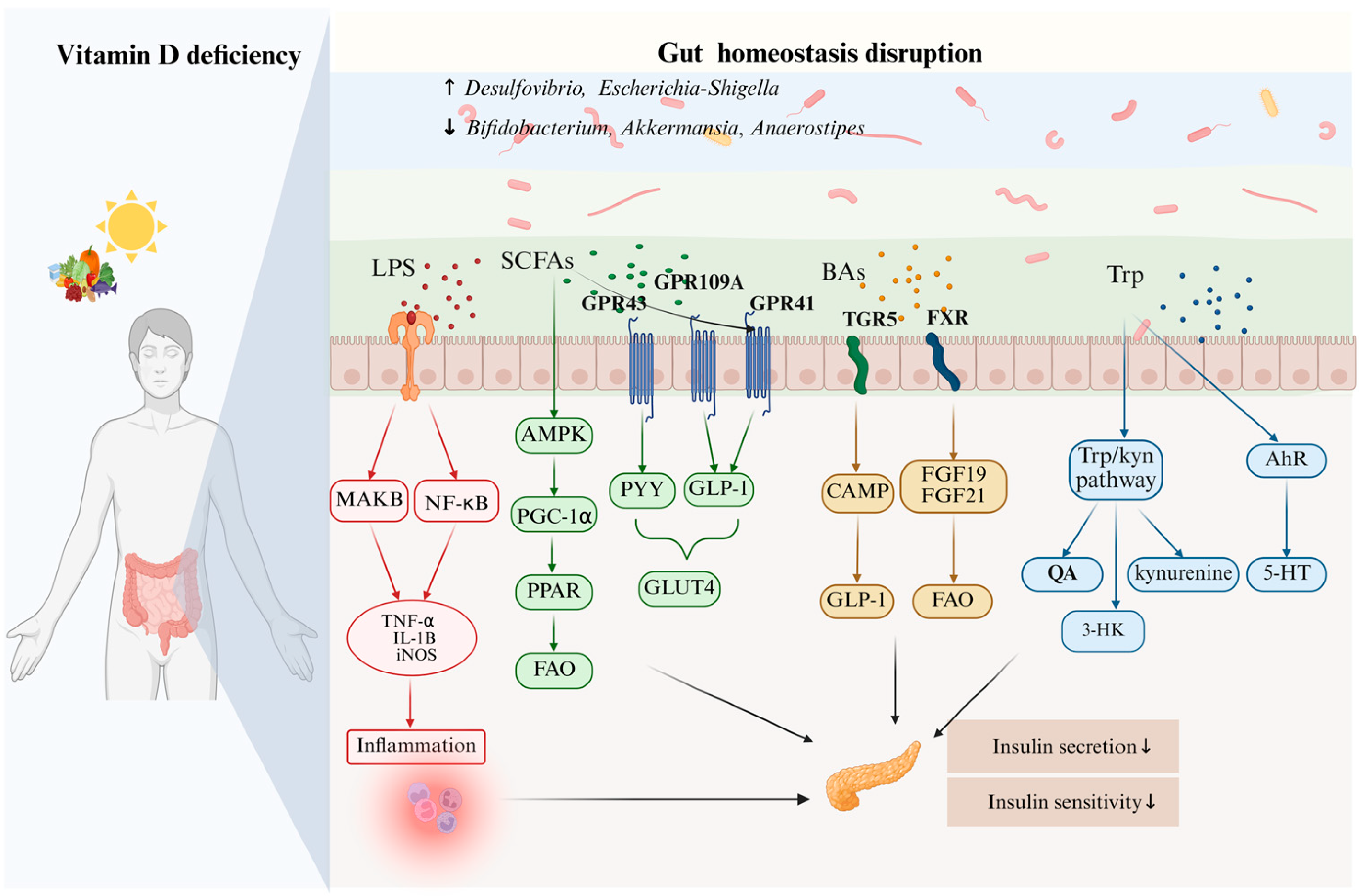
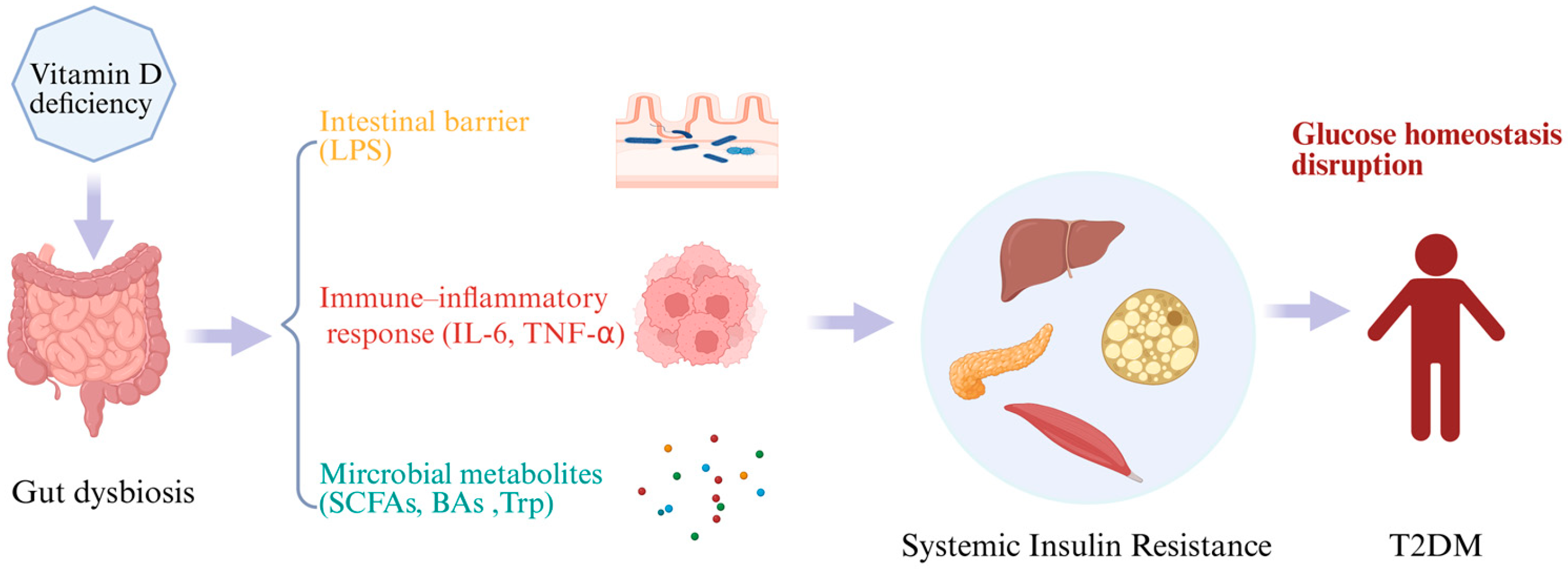
By reviewing current studies, it appears that gut microbiota may act as an intermediary and play an important role in how vitamin D influences the onset and progression of T2DM. In this section, we summarize the potential mechanisms by which vitamin D may regulate T2DM through the gut microbiota from three aspects: the intestinal barrier, immune–inflammatory responses, and microbial metabolites (Figure 1).

5.1. Intestinal Barrier Function and Endotoxin Translocation

Vitamin D activates its receptor VDR. It promotes the expression of tight junction proteins, such as occludin, claudin-1 and ZO-1, to strengthen the physical barrier between epithelial cells [69]. It also upregulates the secretion of antimicrobial peptides (e.g., cathelicidin and β-defensin) in intestinal epithelial cells. These peptides kill pathogenic bacteria and inhibit the overproliferation of opportunistic pathogens [70]. In this dual way, vitamin D maintains the integrity of the intestinal epithelial barrier and the homeostasis of intestinal flora. Vitamin D deficiency reduces defensin production, and increases intestinal permeability as a result. This allows intestinal bacteria and their metabolites, especially lipopolysaccharide (LPS), to cross the epithelial barrier and enter the circulatory system more easily [68,71]. Once in the system, LPS binds to lipopolysaccharide-binding protein (LBP). It then activates the Toll-like receptor 4 (TLR4)/myeloid differentiation factor 2 (MD2) complex on the surface of immune cells. This triggers the nuclear translocation of the nuclear factor kappa B (NF-κB) pathway [72]. Consequently, pro-inflammatory cytokines such as tumor necrosis factor-α (TNF-α) and interleukin-6 (IL-6) are released. These cytokines not only directly inhibit the phosphorylation of insulin receptor substrates and block insulin signal transduction. They also damage the function of pancreatic β-cells. Eventually, insulin resistance is induced [22]. LPS translocation caused by barrier damage is recognized as a critical early event linking intestinal microecological imbalance to metabolic disorders [73]. Low vitamin D levels are associated with elevated serum LPS levels. Supplementing vitamin D can reduce serum LPS and pro-inflammatory cytokine levels in T2DM patients. It can also improve their insulin sensitivity [40,42,63,74]. Therefore, LPS translocation is often regarded as a key factor in T2DM-related inflammation and insulin resistance. Its important role in the axis linking vitamin D-modulated intestinal flora to T2DM deserves close attention.

5.2. Immune Modulation and Inflammatory Regulation

Vitamin D modulates gut microbiota composition by regulating the body’s immune system. It also regulates immune and inflammatory responses in a reverse manner when gut microbiota status is altered [75]. These regulatory actions go on to affect how type 2 diabetes mellitus develops and progresses. Patients with T2DM usually have chronic low-grade inflammation. This condition is marked by higher levels of pro-inflammatory factors like TNF-α, IL-6 and C-reactive protein [76]. These inflammatory mediators interfere with insulin signaling pathways.
They damage post-receptor signaling and reduce the body’s uptake and use of glucose. Eventually, this leads to insulin resistance [77,78]. An imbalance in immune cell subsets is a major cause of worsening inflammation. This kind of imbalance disrupts the immune homeostasis of the intestinal mucosa. It also directly shows that intestinal homeostasis and systemic immune–inflammatory responses are closely connected [51]. Vitamin D has two key effects on immune and inflammatory responses, and this is mainly achieved by regulating gut microbiota. On the one hand, the VD/VDR signaling pathway can help probiotics like Bacteroides and Lactobacillus colonize the gut. It also prevents opportunistic pathogens from overgrowing, which in turn maintains the stability of the gut’s microecological balance [35,79]. Restoring gut homeostasis not only eases local and systemic inflammation, but can also adjust glucose metabolism by reducing insulin resistance [51]. On the other hand, this signaling pathway is involved in controlling the differentiation and function of immune cells. When the pathway is activated, it promotes the production of regulatory T cells (Tregs) and prevents pro-inflammatory cell subsets such as Th1 and Th17 from being overactivated [80]. This maintains the Th17/Treg immune balance. This balance is essential for keeping the intestinal mucosal homeostasis intact [51]. When intestinal homeostasis is well maintained, the body produces more SCFAs. SCFAs, especially butyrate, can enhance the function of Tregs and increase the expression of anti-inflammatory genes [81,82]. It does this by maintaining the right ratio of Th17/Treg cells and promoting SCFA production. These actions then have an impact on glucose metabolism. In contrast, low vitamin D levels often lead to gut dysbiosis [18]. This is usually accompanied by endotoxemia and more severe inflammation. It impairs TNF-α-induced TLR4/NF-κB signaling and disrupts the PI3K/AKT insulin signaling pathway [22]. Vitamin D lessens these inflammatory processes by inhibiting NF-κB and the NOD-like receptor family pyrin domain-containing 3 (NLRP3) inflammasome. This reduction then brings down the levels of IL-6, TNF-α and other inflammatory mediators [42]. Therefore, these findings show that vitamin D and gut microbiota work together to regulate immune responses. These joint regulatory effects ultimately affect the health of the body’s glucose metabolism.

5.3. The Mediating Role of Microbial Metabolites in the Vitamin D–T2DM Axis

Gut microbiota metabolites, such as SCFAs, BAs and Trp, play a key role in regulating systemic glucose metabolism [50,83]. They may also act as mediators in the potential regulatory axis. This axis connects vitamin D, gut microbiota and T2DM regulation. Disturbance to this axis does more than reduce the production of beneficial metabolites like SCFAs. It also promotes the buildup of harmful substances such as LPS. Furthermore, it directly damages the integrity of the intestinal barrier and may cause intestinal permeability [35,68]. This subsequently triggers chronic low-grade inflammation and insulin resistance, ultimately accelerating the pathological progression of T2DM. Accordingly, these metabolites serve as the central hub that links the interplay between vitamin D, gut microbiota and T2DM (Figure 2).
SCFAs include acetate, propionate and butyrate. They are key metabolites produced by intestinal flora through dietary fiber fermentation. Vitamin D binds to its receptor VDR to upregulate the expression of mucins (MUC2, MUC3) and tight junction proteins (ZO-1, occludin) in intestinal epithelial cells. This process enhances the integrity of the intestinal mucosal barrier [69]. It also inhibits the release of intestinal pro-inflammatory factors. In this way, the intestinal immune microenvironment is improved [70]. These changes help maintain intestinal microecological homeostasis. They create a favorable ecological niche for SCFA-producing bacteria such as Faecalibacterium prausnitzii, Roseburia and Bifidobacterium to survive stably and exert their metabolic functions [84,85]. SCFAs can activate multiple G protein-coupled receptors (GPRs), including GPR41, GPR43 and GPR109A. They synergistically regulate energy metabolism and inflammatory responses in the intestine, adipose tissue and immune system [86]. In adipose tissue, GPR41 activation alleviates lipotoxicity. In the immune system, GPR109A activation helps inhibit macrophage-mediated chronic inflammation [87]. Thus, SCFAs participate in regulating the development and progression of T2DM. When propionate activates GPR41/GPR43 on intestinal L cells, they stimulate the secretion of glucagon-like peptide-1 (GLP-1) and peptide YY (PYY). GLP-1 promotes insulin secretion and improves insulin sensitivity. PYY helps suppress appetite [65,88]. Propionate is linked to methylation at the cg26345888 locus, which subsequently suppresses the expression of disabled homolog 1 (DAB1) associated with vitamin D deficiency. This could constitute a propionate-mediated vitamin D epigenetic regulatory pathway, influencing the progression of diabetes [89]. Butyrate stands out for its role in improving immune responses. It can regulate the number and function of regulatory T cells. This reduces inflammation in adipose tissue. It also promotes GLP-1 production, thereby enhancing insulin sensitivity. Meanwhile, butyrate serves as the primary energy source for colonic epithelial cells [86,90]. Furthermore, a synergistic interaction may exist between butyrate and vitamin D signaling. Butyrate inhibits histone deacetylases (HDACs). This enhances histone acetylation at the VDR gene promoter region. It then promotes VDR transcription and protein expression, indirectly boosting VDR activity [91]. On the other hand, vitamin D binds to VDR to promote the differentiation of Treg and inhibit pro-inflammatory factor release [79,91,92]. The two may form a positive feedback loop that jointly maintains immune homeostasis and metabolic balance in the body and plays a regulatory role in the development of T2DM.
Bile acids, including primary and secondary bile acids, are important signaling molecules that link hepatic metabolism, gut microbiota, and host energy homeostasis [93]. Vitamin D exerts a multi-level regulatory effect on bile acid metabolism. When vitamin D binds to VDR, it upregulates the expression of small heterodimer partner (SHP) in the liver. SHP then interacts with hepatocyte nuclear factor 4α, a key protein that controls how bile acids are made. This interaction slows down the activity of cholesterol 7α-hydroxylase (CYP7A1). CYP7A1 is the main enzyme that limits how much bile acid the body produces [94,95]. Activation of the VD/VDR axis also elevates the expression of bile acid efflux transporters, including the bile salt export pump (BSEP) and multidrug resistance-associated protein 2 (MRP2). Meanwhile, it modulates the expression of intestinal bile acid transporters such as the apical sodium-dependent bile acid transporter (ASBT) and organic solute transporter α/β (OSTα/β). These coordinated regulatory changes contribute to the maintenance of bile acid enterohepatic circulation [93,95]. Beyond its direct modulation of host bile acid synthesis and transport, vitamin D indirectly influences bile acid transformation by enhancing intestinal barrier integrity and sustaining gut microbial homeostasis [96]. Adequate vitamin D levels in the body promote the enrichment of commensal bacteria, including Ruminococcus and Eubacterium species [33]. This bacterial enrichment is accompanied by increased production of bile salt hydrolase (BSH) and enhanced activity of 7α-dehydroxylase. Such alterations accelerate the deconjugation and dehydroxylation of bile acids, thereby facilitating the generation of secondary bile acids like lithocholic acid (LCA) [97]. Altered bile acid composition not only modulates enterohepatic metabolic homeostasis but also regulates metabolic signaling via specific bile acid receptors [93]. Secondary bile acids, particularly LCA, can activate the Takeda G protein-coupled receptor 5 (TGR5) receptor on intestinal L cells, which further triggers the secretion of GLP-1. TGR5 activation in pancreatic β cells also enhances glucose-dependent insulin secretion [98]. Moreover, TGR5 signaling inhibits M1 polarization of macrophages, alleviates local pancreatic inflammation, and thus plays a role in preserving the functional integrity of pancreatic β cells [99,100]. Bile acids are also capable of activating the farnesoid X receptor (FXR) signaling pathway, which collaborates with other signaling cascades to regulate inflammatory responses and maintain energy metabolic balance [100]. LCA can also act as an endogenous ligand for VDR to directly activate VDR-mediated signaling. This finding implies the existence of a bidirectional regulatory feedback loop between bile acids and vitamin D [101,102]. In states of vitamin D deficiency, bile acid synthesis, transport, and microbiota-mediated transformation may become dysregulated, resulting in an abnormal proportion of secondary bile acids. This imbalance may weaken FXR- and TGR5-mediated signaling and indirectly impair VDR activation, ultimately leading to reduced GLP-1 secretion, decreased insulin sensitivity, and disruption of glucose metabolic homeostasis [50,103,104].
Trp metabolism mainly involves the kynurenine, serotonin, and indole pathways, with the indole pathway being the one most closely linked to the gut microbial ecosystem [105]. After binding to the nuclear VDR, vitamin D can directly interact with vitamin D response elements (VDREs) in the indoleamine 2,3-dioxygenase 1 (IDO1) gene promoter region to inhibit the transcription and protein expression of IDO1 [106]. Meanwhile, it competitively blocks the IDO1 activation pathway induced by inflammatory factors (e.g., IFN-γ) [107], because kynurenine metabolites may promote inflammation, oxidative stress, and β-cell damage [105]. This prevents excessive Trp shunting into the kynurenine pathway and reduces the accumulation of immunosuppressive or neurotoxic metabolites, such as 3-hydroxykynurenine and quinolinic acid [108,109]. At the same time, VDR activation upregulates the expression of intestinal epithelial mucins (e.g., MUC2) [69]. This regulatory effect helps establish a stable colonization niche for symbiotic tryptophanase-producing bacteria, such as Lactobacillus and Bifidobacterium species. It also facilitates the conversion of tryptophan into indole metabolites, including indole, indole-3-propionic acid, and indole-3-acetic acid [110,111]. These indole metabolites strengthen intestinal barrier function, decrease LPS translocation, and suppress systemic inflammatory responses by activating the aryl hydrocarbon receptor (AhR) or pregnane X receptor [84,111]. When indole derivatives bind to AhR, they induce nuclear translocation and heterodimer formation of the receptor [105]. This process upregulates the expression of downstream genes involved in anti-inflammatory responses and intestinal barrier repair, thereby alleviating intestinal inflammation and reducing systemic insulin resistance induced by endotoxemia [107,112]. In this way, vitamin D modulates T2DMby re-establishing the balance of Trp metabolic pathways.
Beyond these metabolites, vitamin D may also impact the development and progression of T2DM through other microbial metabolites. Trimethylamine N-oxide (TMAO) forms when the liver oxidizes trimethylamine (TMA). TMA is a byproduct of dietary choline and carnitine metabolism, produced by gut microbiota. Higher TMAO levels show a close link to inflammation, oxidative stress and insulin resistance [113]. Vitamin D can modify gut microbiota composition. This change reduces the abundance of bacteria that produce TMA, and in turn lowers TMAO levels in the body [114]. Similarly, elevated BCAA levels in plasma correlate positively with insulin resistance. BCAAs include leucine, isoleucine and valine. The gut microbiota have an important role in synthesizing and degrading these amino acids [115,116,117]. Vitamin D may ease the abnormal buildup of BCAAs by improving microbial homeostasis and energy metabolism. This action may also help improve T2DM symptoms [118].

6. Conclusions

To sum up, available evidence shows that vitamin D can regulate the integrity of the intestinal barrier, intestinal inflammation, immune responses and the profile of gut-derived metabolites through multiple pathways. In this way, it affects the host’s metabolic homeostasis of glucose and lipids, which may reduce the risk of T2DM. Among these factors, gut metabolites such as SCFAs, BAs and Trp play a key mediating role between vitamin D and metabolic phenotypes. Vitamin D deficiency may cause intestinal dysbiosis, impaired intestinal barrier function and low-grade systemic inflammation. These changes further reduce insulin sensitivity and promote metabolic disorders. In contrast, sufficient vitamin D levels help maintain the homeostasis of intestinal microecology and participate in the regulation of glucose metabolism through the above key metabolites.
However, current studies still have several limitations. These include the heterogeneity of study subjects and study designs, inconsistent methods for assessing vitamin D status and intervention protocols, as well as high diversity of outcome indicators related to intestinal microbiota. In addition, T2DM is a metabolic disease driven by multiple factors: its complex etiological characteristics, together with the limitations of extrapolating results from rodent models to human studies, have to some extent restricted the interpretability of existing evidence.
Future research needs to further identify the optimal dosage and intervention timing of vitamin D supplementation. Combined with the characteristics of individualized intestinal microbiota, researchers should explore precise metabolic regulation strategies based on the vitamin D–gut microbiota axis. Although accumulating evidence supports vitamin D as a potential adjunctive intervention for cardiometabolic diseases, its clinical translational value needs to be verified by well-designed large-scale randomized controlled trials.

Author Contributions

L.N. and Y.Z. conceptualized the manuscript. Y.Z. wrote the manuscript. J.L. and Q.D. edited the manuscript. All authors have read and agreed to the published version of the manuscript.

Funding

This research was funded by the General Program of Natural Science Foundation of Shanghai Municipal Science and Technology Commission, Grant No. 25ZR1401165.

Institutional Review Board Statement

Not applicable.

Informed Consent Statement

Not applicable.

Data Availability Statement

No new data were created or analyzed in this study. Data sharing is not applicable to this article.

Conflicts of Interest

The authors declare no conflicts of interest.

References

  1. Lytrivi, M.; Tong, Y.; Virgilio, E.; Yi, X.; Cnop, M. Diabetes mellitus and the key role of endoplasmic reticulum stress in pancreatic β cells. Nat. Rev. Endocrinol. 2025, 21, 546–563. [Google Scholar] [CrossRef] [Scilit]
  2. Duncan, B.B.; Magliano, D.J.; Boyko, E.J. IDF diabetes atlas 11th edition 2025: Global prevalence and projections for 2050. Nephrol. Dial. Transplant. 2025, 41, 7–9. [Google Scholar] [CrossRef] [Scilit]
  3. Suzuki, K.; Hatzikotoulas, K.; Southam, L.; Taylor, H.J.; Yin, X.; Lorenz, K.M.; Mandla, R.; Huerta-Chagoya, A.; Melloni, G.E.M.; Kanoni, S.; et al. Genetic drivers of heterogeneity in type 2 diabetes pathophysiology. Nature 2024, 627, 347–357. [Google Scholar] [CrossRef] [Scilit]
  4. Accili, D.; Deng, Z.; Liu, Q. Insulin resistance in type 2 diabetes mellitus. Nat. Rev. Endocrinol. 2025, 21, 413–426. [Google Scholar] [CrossRef] [Scilit]
  5. Giustina, A.; Bilezikian, J.P.; Adler, R.A.; Banfi, G.; Bikle, D.D.; Binkley, N.C.; Bollerslev, J.; Bouillon, R.; Brandi, M.L.; Casanueva, F.F.; et al. Consensus Statement on Vitamin D Status Assessment and Supplementation: Whys, Whens, and Hows. Endocr. Rev. 2024, 45, 625–654. [Google Scholar] [CrossRef] [Scilit]
  6. Bouillon, R.; Marcocci, C.; Carmeliet, G.; Bikle, D.; White, J.H.; Dawson-Hughes, B.; Lips, P.; Munns, C.F.; Lazaretti-Castro, M.; Giustina, A.; et al. Skeletal and Extraskeletal Actions of Vitamin D: Current Evidence and Outstanding Questions. Endocr. Rev. 2019, 40, 1109–1151. [Google Scholar] [CrossRef] [Scilit] [PubMed]
  7. Ortiz-Prado, E.; Vasconez-Gonzalez, J.; Izquierdo-Condoy, J.S.; Suárez-Sangucho, I.A.; Prieto-Marín, J.G.; Villarreal-Burbano, K.B.; Barriga-Collantes, M.A.; Altamirano-Castillo, J.A.; Borja-Mendoza, D.A.; Pazmiño-Almeida, J.C.; et al. Cholecalciferol (vitamin D3): Efficacy, safety, and implications in public health. Front. Nutr. 2025, 12, 1579957. [Google Scholar] [CrossRef] [Scilit] [PubMed]
  8. Sukik, A.; Alalwani, J.; Ganji, V. Vitamin D, Gut Microbiota, and Cardiometabolic Diseases—A Possible Three-Way Axis. Int. J. Mol. Sci. 2023, 24, 940. [Google Scholar] [CrossRef] [Scilit] [PubMed]
  9. Qin, J.; Li, Y.; Cai, Z.; Li, S.; Zhu, J.; Zhang, F.; Liang, S.; Zhang, W.; Guan, Y.; Shen, D.; et al. A metagenome-wide association study of gut microbiota in type 2 diabetes. Nature 2012, 490, 55–60. [Google Scholar] [CrossRef] [Scilit]
  10. Canfora, E.E.; Meex, R.C.R.; Venema, K.; Blaak, E.E. Gut microbial metabolites in obesity, NAFLD and T2DM. Nat. Rev. Endocrinol. 2019, 15, 261–273. [Google Scholar] [CrossRef] [Scilit]
  11. Ge, X.; Liu, T.; Wang, Y.; Wen, H.; Huang, Z.; Chen, L.; Xu, J.; Zhou, H.; Wu, Q.; Zhao, C.; et al. Porous starch microspheres loaded with luteolin exhibit hypoglycemic activities and alter gut microbial communities in type 2 diabetes mellitus mice. Food Funct. 2025, 16, 54–70. [Google Scholar] [CrossRef] [Scilit] [PubMed]
  12. Cheng, L.; Lv, C.; Xue, L.; Zhang, C.; Wang, L.; Wang, X.; Chen, S.; Li, X.; Feng, W.; Xie, H.; et al. The prevention and improvement effects of vitamin D on type 2 diabetes mellitus: Evidence from an umbrella review on Meta-analyses of cohort studies and randomized controlled trials. Front. Nutr. 2024, 11, 1462535. [Google Scholar] [CrossRef] [Scilit]
  13. Zhang, Y.; Xu, D.; Cai, X.; Xing, X.; Shao, X.; Yin, A.; Zhao, Y.; Wang, M.; Fan, Y.N.; Liu, B.; et al. Gut Commensal Barnesiella intestinihominis Ameliorates Hyperglycemia and Liver Metabolic Disorders. Adv. Sci. 2025, 12, e2411181. [Google Scholar] [CrossRef] [Scilit]
  14. Chong, S.; Lin, M.; Chong, D.; Jensen, S.; Lau, N.S. A systematic review on gut microbiota in type 2 diabetes mellitus. Front. Endocrinol. 2024, 15, 1486793. [Google Scholar] [CrossRef] [Scilit]
  15. Fuentes-Barría, H.; Aguilera-Eguía, R.; Flores-Fernández, C.; Angarita-Davila, L.; Rojas-Gómez, D.; Alarcón-Rivera, M.; López-Soto, O.; Maureira-Sánchez, J. Vitamin D and Type 2 Diabetes Mellitus: Molecular Mechanisms and Clinical Implications—A Narrative Review. Int. J. Mol. Sci. 2025, 26, 2153. [Google Scholar] [CrossRef] [Scilit]
  16. Lin, K.; Zhu, L.; Yang, L. Gut and obesity/metabolic disease: Focus on microbiota metabolites. MedComm 2022, 3, e171. [Google Scholar] [CrossRef] [Scilit]
  17. Warmbrunn, M.V.; Attaye, I.; Aron-Wisnewsky, J.; Rampanelli, E.; van der Vossen, E.W.J.; Hao, Y.; Koopen, A.; Bergh, P.O.; Stols-Gonçalves, D.; Mohamed, N.; et al. Oral histidine affects gut microbiota and MAIT cells improving glycemic control in type 2 diabetes patients. Gut Microbes 2024, 16, 2370616. [Google Scholar] [CrossRef] [Scilit]
  18. Tangestani, H.; Boroujeni, H.K.; Djafarian, K.; Emamat, H.; Shab-Bidar, S. Vitamin D and The Gut Microbiota: A Narrative Literature Review. Clin. Nutr. Res. 2021, 10, 181–191. [Google Scholar] [CrossRef] [Scilit] [PubMed]
  19. Zeb, F.; Osaili, T.; Hashim, M.; Alkalbani, N.; Papandreou, D.; Cheikh Ismail, L.; Naja, F.; Radwan, H.; Hasan, H.; Obaid, R.S.; et al. Effect of Vitamin D Supplementation on Human Gut Microbiota: A Systematic Review of Randomized Controlled Trials. Nutr. Rev. 2025. online ahead of print. [Google Scholar] [CrossRef] [Scilit]
  20. Mitri, J.; Dawson-Hughes, B.; Hu, F.B.; Pittas, A.G. Effects of vitamin D and calcium supplementation on pancreatic β cell function, insulin sensitivity, and glycemia in adults at high risk of diabetes: The Calcium and Vitamin D for Diabetes Mellitus (CaDDM) randomized controlled trial. Am. J. Clin. Nutr. 2011, 94, 486–494. [Google Scholar] [CrossRef] [Scilit]
  21. Pittas, A.G.; Dawson-Hughes, B.; Sheehan, P.; Ware, J.H.; Knowler, W.C.; Aroda, V.R.; Brodsky, I.; Ceglia, L.; Chadha, C.; Chatterjee, R.; et al. Vitamin D Supplementation and Prevention of Type 2 Diabetes. N. Engl. J. Med. 2019, 381, 520–530. [Google Scholar] [CrossRef] [Scilit] [PubMed]
  22. Zhang, Y.; Ni, P.; Miao, Y.; Chen, H.; Tang, L.; Song, H.; Li, W.; Li, X. Vitamin D(3) improves glucose metabolism and attenuates inflammation in prediabetic human and mice. J. Nutr. Biochem. 2024, 130, 109659. [Google Scholar] [CrossRef] [Scilit]
  23. Pramono, A.; Jocken, J.W.E.; Blaak, E.E.; van Baak, M.A. The Effect of Vitamin D Supplementation on Insulin Sensitivity: A Systematic Review and Meta-analysis. Diabetes Care 2020, 43, 1659–1669. [Google Scholar] [CrossRef] [Scilit]
  24. Rohold, C.K.; Jørgensen, H.L.; Vojdeman, F.J.; Madsen, C.M.; Olsen, A.; Heegaard, A.M.; Lind, B.S.; Tjønneland, A.; Schwarz, P.; Gæde, P.H. Levels of plasma 25-hydroxy vitamin D and risk of developing type 2 diabetes in a large Danish primary health care population. Acta Diabetol. 2025, 62, 397–404. [Google Scholar] [CrossRef] [Scilit] [PubMed]
  25. Valer-Martinez, A.; Sayon-Orea, C.; Martinez, J.A.; Basterra-Gortari, F.J.; Martinez-Gonzalez, M.A.; Bes-Rastrollo, M. Vitamin D and risk of developing type 2 diabetes in the SUN project: A prospective cohort study. J. Endocrinol. Investig. 2024, 47, 2313–2323. [Google Scholar] [CrossRef] [Scilit]
  26. Zhang, J.; Wu, K.; Dong, X. Exploring the association between serum vitamin D levels and type 2 diabetes risk in US adults. Ann. Med. 2025, 57, 2522320. [Google Scholar] [CrossRef] [Scilit]
  27. Lemieux, P.; Weisnagel, S.J.; Caron, A.Z.; Julien, A.S.; Morisset, A.S.; Carreau, A.M.; Poirier, J.; Tchernof, A.; Robitaille, J.; Bergeron, J.; et al. Effects of 6-month vitamin D supplementation on insulin sensitivity and secretion: A randomised, placebo-controlled trial. Eur. J. Endocrinol. 2019, 181, 287–299. [Google Scholar] [CrossRef] [Scilit] [PubMed]
  28. Hu, Z.; Zhi, X.; Li, J.; Li, B.; Wang, J.; Zhu, J.; Zhang, Z. Effects of long-term vitamin D supplementation on metabolic profile in middle-aged and elderly patients with type 2 diabetes. J. Steroid Biochem. Mol. Biol. 2023, 225, 106198. [Google Scholar] [CrossRef] [Scilit]
  29. Tobias, D.K.; Pradhan, A.D.; Duran, E.K.; Li, C.; Song, Y.; Buring, J.E.; Cook, N.R.; Mora, S.; Manson, J.E. Vitamin D supplementation vs. placebo and incident type 2 diabetes in an ancillary study of the randomized Vitamin D and Omega-3 Trial. Nat. Commun. 2025, 16, 3332. [Google Scholar] [CrossRef] [Scilit] [PubMed]
  30. Virtanen, J.K.; Hantunen, S.; Kallio, N.; Lamberg-Allardt, C.; Manson, J.E.; Nurmi, T.; Pihlajamäki, J.; Uusitupa, M.; Voutilainen, A.; Tuomainen, T.P. The effect of vitamin D(3) supplementation on the incidence of type 2 diabetes in healthy older adults not at high risk for diabetes (FIND): A randomised controlled trial. Diabetologia 2025, 68, 715–726. [Google Scholar] [CrossRef] [Scilit]
  31. Kawahara, T.; Suzuki, G.; Mizuno, S.; Inazu, T.; Kasagi, F.; Kawahara, C.; Okada, Y.; Tanaka, Y. Effect of active vitamin D treatment on development of type 2 diabetes: DPVD randomised controlled trial in Japanese population. BMJ 2022, 377, e066222. [Google Scholar] [CrossRef] [Scilit]
  32. Song, Q.; Li, Y.; Zhou, T.; Xiao, M.; Xiao, B.; Wang, M.; Zhu, Y. Maternal vitamin D status during pregnancy and infant’s gut microbiota: A prospective cohort study. Front. Nutr. 2024, 11, 1428356. [Google Scholar] [CrossRef] [Scilit]
  33. Gong, J.; He, L.; Zou, Q.; Zhao, Y.; Zhang, B.; Xia, R.; Chen, B.; Cao, M.; Gong, W.; Lin, L.; et al. Association of serum 25-hydroxyvitamin D (25(OH)D) levels with the gut microbiota and metabolites in postmenopausal women in China. Microb. Cell Fact. 2022, 21, 137. [Google Scholar] [CrossRef] [Scilit]
  34. Boughanem, H.; Ruiz-Limón, P.; Pilo, J.; Lisbona-Montañez, J.M.; Tinahones, F.J.; Moreno Indias, I.; Macías-González, M. Linking serum vitamin D levels with gut microbiota after 1-year lifestyle intervention with Mediterranean diet in patients with obesity and metabolic syndrome: A nested cross-sectional and prospective study. Gut Microbes 2023, 15, 2249150. [Google Scholar] [CrossRef] [Scilit]
  35. Wyatt, M.; Choudhury, A.; Von Dohlen, G.; Heileson, J.L.; Forsse, J.S.; Rajakaruna, S.; Zec, M.; Tfaily, M.M.; Greathouse, L. Randomized control trial of moderate dose vitamin D alters microbiota stability and metabolite networks in healthy adults. Microbiol. Spectr. 2024, 12, e0008324. [Google Scholar] [CrossRef] [Scilit]
  36. Singh, P.; Rawat, A.; Alwakeel, M.; Sharif, E.; Al Khodor, S. The potential role of vitamin D supplementation as a gut microbiota modifier in healthy individuals. Sci. Rep. 2020, 10, 21641. [Google Scholar] [CrossRef] [Scilit]
  37. Naderpoor, N.; Mousa, A.; Fernanda Gomez Arango, L.; Barrett, H.L.; Dekker Nitert, M.; de Courten, B. Effect of Vitamin D Supplementation on Faecal Microbiota: A Randomised Clinical Trial. Nutrients 2019, 11, 2888. [Google Scholar] [CrossRef] [Scilit]
  38. Pham, H.; Waterhouse, M.; Rahman, S.; Baxter, C.; Duarte Romero, B.; McLeod, D.S.A.; Ebeling, P.R.; English, D.R.; Hartel, G.; O’Connell, R.L.; et al. The effect of vitamin D supplementation on the gut microbiome in older Australians—Results from analyses of the D-Health Trial. Gut Microbes 2023, 15, 2221429. [Google Scholar] [CrossRef] [Scilit]
  39. Shieh, A.; Lee, S.M.; Lagishetty, V.; Gottleib, C.; Jacobs, J.P.; Adams, J.S. Pilot Trial of Vitamin D3 and Calcifediol in Healthy Vitamin D Deficient Adults: Does It Change the Fecal Microbiome? J. Clin. Endocrinol. Metab. 2021, 106, 3464–3476. [Google Scholar] [CrossRef] [Scilit] [PubMed]
  40. Zhang, Y.; Ni, P.; Chen, H.; Tang, L.; Song, H.; Wen, H.; Miao, Y.; Li, W.; Li, X. Vitamin D(3) ameliorates hyperglycemia by modulating gut microbiota and metabolites in prediabetic KKay mice. Food Res. Int. 2025, 211, 116369. [Google Scholar] [CrossRef] [Scilit] [PubMed]
  41. Zhang, X.L.; Chen, L.; Yang, J.; Zhao, S.S.; Jin, S.; Ao, N.; Yang, J.; Liu, H.X.; Du, J. Vitamin D alleviates non-alcoholic fatty liver disease via restoring gut microbiota and metabolism. Front. Microbiol. 2023, 14, 1117644. [Google Scholar] [CrossRef] [Scilit] [PubMed]
  42. Xiang, L.; Du, T.; Zhang, J.; Zhang, Y.; Zhou, Y.; Zhao, Y.; Zhou, Y.; Ma, L. Vitamin D(3) supplementation shapes the composition of gut microbiota and improves some obesity parameters induced by high-fat diet in mice. Eur. J. Nutr. 2024, 63, 155–172. [Google Scholar] [CrossRef] [Scilit] [PubMed]
  43. Liu, J.; Liu, J.; Zhang, J.; Liu, C.; Qu, C.; Na, L. Vitamin D deficiency in early life regulates gut microbiome composition and leads to impaired glucose tolerance in adult and offspring rats. Food Funct. 2023, 14, 5768–5786. [Google Scholar] [CrossRef] [Scilit]
  44. Zhao, L.; Lou, H.; Peng, Y.; Chen, S.; Zhang, Y.; Li, X. Comprehensive relationships between gut microbiome and faecal metabolome in individuals with type 2 diabetes and its complications. Endocrine 2019, 66, 526–537. [Google Scholar] [CrossRef] [Scilit]
  45. Nuli, R.; Cai, J.; Kadeer, A.; Zhang, Y.; Mohemaiti, P. Integrative Analysis Toward Different Glucose Tolerance-Related Gut Microbiota and Diet. Front. Endocrinol. 2019, 10, 295. [Google Scholar] [CrossRef] [Scilit]
  46. Gaike, A.H.; Paul, D.; Bhute, S.; Dhotre, D.P.; Pande, P.; Upadhyaya, S.; Reddy, Y.; Sampath, R.; Ghosh, D.; Chandraprabha, D.; et al. The Gut Microbial Diversity of Newly Diagnosed Diabetics but Not of Prediabetics Is Significantly Different from That of Healthy Nondiabetics. mSystems 2020, 5, e00578-19. [Google Scholar] [CrossRef] [Scilit] [PubMed]
  47. Ebrahimi, F.; Maleki, H.; Ebrahimi, M.; Beiki, A.H. A novel approach to finding the compositional differences and biomarkers in gut microbiota in type 2 diabetic patients via meta-analysis, data-mining, and multivariate analysis. Endocrinol. Diabetes Nutr. (Engl. Ed.) 2025, 72, 501561. [Google Scholar] [CrossRef] [Scilit] [PubMed]
  48. Mei, Z.; Wang, F.; Bhosle, A.; Dong, D.; Mehta, R.; Ghazi, A.; Zhang, Y.; Liu, Y.; Rinott, E.; Ma, S.; et al. Strain-specific gut microbial signatures in type 2 diabetes identified in a cross-cohort analysis of 8,117 metagenomes. Nat. Med. 2024, 30, 2265–2276. [Google Scholar] [CrossRef] [Scilit]
  49. Ruuskanen, M.O.; Erawijantari, P.P.; Havulinna, A.S.; Liu, Y.; Méric, G.; Tuomilehto, J.; Inouye, M.; Jousilahti, P.; Salomaa, V.; Jain, M.; et al. Gut Microbiome Composition Is Predictive of Incident Type 2 Diabetes in a Population Cohort of 5,572 Finnish Adults. Diabetes Care 2022, 45, 811–818. [Google Scholar] [CrossRef] [Scilit]
  50. Yu, Y.; Ding, Y.; Wang, S.; Jiang, L. Gut Microbiota Dysbiosis and Its Impact on Type 2 Diabetes: From Pathogenesis to Therapeutic Strategies. Metabolites 2025, 15, 397. [Google Scholar] [CrossRef] [Scilit]
  51. Takeuchi, T.; Kubota, T.; Nakanishi, Y.; Tsugawa, H.; Suda, W.; Kwon, A.T.; Yazaki, J.; Ikeda, K.; Nemoto, S.; Mochizuki, Y.; et al. Gut microbial carbohydrate metabolism contributes to insulin resistance. Nature 2023, 621, 389–395. [Google Scholar] [CrossRef] [Scilit]
  52. Wu, X.; Park, S. Fecal Bacterial Community and Metagenome Function in Asians with Type 2 Diabetes, According to Enterotypes. Biomedicines 2022, 10, 2998. [Google Scholar] [CrossRef] [Scilit]
  53. Park, S.; Zhang, T.; Kang, S. Fecal Microbiota Composition, Their Interactions, and Metagenome Function in US Adults with Type 2 Diabetes According to Enterotypes. Int. J. Mol. Sci. 2023, 24, 9533. [Google Scholar] [CrossRef] [Scilit]
  54. Chávez-Carbajal, A.; Pizano-Zárate, M.L.; Hernández-Quiroz, F.; Ortiz-Luna, G.F.; Morales-Hernández, R.M.; De Sales-Millán, A.; Hernández-Trejo, M.; García-Vite, A.; Beltrán-Lagunes, L.; Hoyo-Vadillo, C.; et al. Characterization of the Gut Microbiota of Individuals at Different T2D Stages Reveals a Complex Relationship with the Host. Microorganisms 2020, 8, 94. [Google Scholar] [CrossRef] [Scilit]
  55. Larsen, N.; Vogensen, F.K.; van den Berg, F.W.; Nielsen, D.S.; Andreasen, A.S.; Pedersen, B.K.; Al-Soud, W.A.; Sørensen, S.J.; Hansen, L.H.; Jakobsen, M. Gut microbiota in human adults with type 2 diabetes differs from non-diabetic adults. PLoS ONE 2010, 5, e9085. [Google Scholar] [CrossRef] [Scilit]
  56. Thingholm, L.B.; Rühlemann, M.C.; Koch, M.; Fuqua, B.; Laucke, G.; Boehm, R.; Bang, C.; Franzosa, E.A.; Hübenthal, M.; Rahnavard, A.; et al. Obese Individuals with and without Type 2 Diabetes Show Different Gut Microbial Functional Capacity and Composition. Cell Host Microbe 2019, 26, 252–264.e10. [Google Scholar] [CrossRef] [Scilit] [PubMed]
  57. Doumatey, A.P.; Adeyemo, A.; Zhou, J.; Lei, L.; Adebamowo, S.N.; Adebamowo, C.; Rotimi, C.N. Gut Microbiome Profiles Are Associated With Type 2 Diabetes in Urban Africans. Front. Cell Infect. Microbiol. 2020, 10, 63. [Google Scholar] [CrossRef] [Scilit]
  58. Alvarez-Silva, C.; Kashani, A.; Hansen, T.H.; Pinna, N.K.; Anjana, R.M.; Dutta, A.; Saxena, S.; Støy, J.; Kampmann, U.; Nielsen, T.; et al. Trans-ethnic gut microbiota signatures of type 2 diabetes in Denmark and India. Genome Med. 2021, 13, 37. [Google Scholar] [CrossRef] [Scilit] [PubMed]
  59. Karlsson, F.H.; Tremaroli, V.; Nookaew, I.; Bergström, G.; Behre, C.J.; Fagerberg, B.; Nielsen, J.; Bäckhed, F. Gut metagenome in European women with normal, impaired and diabetic glucose control. Nature 2013, 498, 99–103. [Google Scholar] [CrossRef] [Scilit]
  60. Morsy, Y.; Shafie, N.S.; Mostafa, M.; Ali, K.A.; Afife, O.N.; Zedan, S.; Darwish, B.A.; Nour, H.T.; Ahmed, M.M.; Ghaly, M.T.; et al. Integrative analysis of gut microbiota and metabolic pathways reveals key microbial and metabolomic alterations in diabetes. Sci. Rep. 2025, 15, 30686. [Google Scholar] [CrossRef] [Scilit] [PubMed]
  61. Letchumanan, G.; Marlini, M.; Baharom, N.; Lawley, B.; Syed Mohideen, F.B.; Jogulu, S.R.; Addnan, F.H.; Nur Fariha, M.M.; Omar, M.R.; Pathmanathan, S.G. Ethnicity-matched case-control study reveals significant gut microbiota differences in Malaysian adults with type 2 diabetes. J. Med. Microbiol. 2025, 74, 001963. [Google Scholar] [CrossRef] [Scilit]
  62. Das, T.; Jayasudha, R.; Chakravarthy, S.; Prashanthi, G.S.; Bhargava, A.; Tyagi, M.; Rani, P.K.; Pappuru, R.R.; Sharma, S.; Shivaji, S. Alterations in the gut bacterial microbiome in people with type 2 diabetes mellitus and diabetic retinopathy. Sci. Rep. 2021, 11, 2738. [Google Scholar] [CrossRef] [Scilit] [PubMed]
  63. Velizarova, M.; Yanachkova, V.; Boneva, T.; Giragosyan, S.; Mihaleva, I.; Andreeva-Gateva, P.; Svinarov, D.; Dimova, I. Relationship between Vitamin D status and microbiome changes in Bulgarian patients with type 2 diabetes mellitus. Biotechnol. Biotechnol. Equip. 2023, 37, 2209662. [Google Scholar] [CrossRef] [Scilit]
  64. Arora, T.; Tremaroli, V. Therapeutic Potential of Butyrate for Treatment of Type 2 Diabetes. Front. Endocrinol. 2021, 12, 761834. [Google Scholar] [CrossRef] [Scilit] [PubMed]
  65. Qin, L.; Fan, B.; Zhou, Y.; Zheng, J.; Diao, R.; Wang, F.; Liu, J. Targeted gut microbiome therapy: Applications and prospects of probiotics, fecal microbiota transplantation and natural products in the management of type 2 diabetes. Pharmacol. Res. 2025, 213, 107625. [Google Scholar] [CrossRef] [Scilit]
  66. Byndloss, M.; Devkota, S.; Duca, F.; Hendrik Niess, J.; Nieuwdorp, M.; Orho-Melander, M.; Sanz, Y.; Tremaroli, V.; Zhao, L. The Gut Microbiota and Diabetes: Research, Translation, and Clinical Applications-2023 Diabetes, Diabetes Care, and Diabetologia Expert Forum. Diabetes Care 2024, 47, 1491–1508. [Google Scholar] [CrossRef] [Scilit] [PubMed]
  67. Wyatt, M.; Choudhary, A.; Dohlen, E.V.; Heileson, J.L.; Forsse, J.S.; Greathouse, L. Vitamin D3 Supplementation Alters Gut Microbiome Composition And Increases Stability In Healthy Individuals. Med. Sci. Sports Exerc. 2024, 56, 489. [Google Scholar] [CrossRef] [Scilit]
  68. Hussein, H.M.; Elyamany, M.F.; Rashed, L.A.; Sallam, N.A. Vitamin D mitigates diabetes-associated metabolic and cognitive dysfunction by modulating gut microbiota and colonic cannabinoid receptor 1. Eur. J. Pharm. Sci. 2022, 170, 106105. [Google Scholar] [CrossRef] [Scilit]
  69. Wang, J.; Mei, L.; Hao, Y.; Xu, Y.; Yang, Q.; Dai, Z.; Yang, Y.; Wu, Z.; Ji, Y. Contemporary Perspectives on the Role of Vitamin D in Enhancing Gut Health and Its Implications for Preventing and Managing Intestinal Diseases. Nutrients 2024, 16, 2352. [Google Scholar] [CrossRef] [Scilit]
  70. Yan, Y.; Guo, Y.; Li, Y.; Jiang, Q.; Yuan, C.; Zhao, L.; Mao, S. Vitamin D, Gut Microbiota, and Cancer Immunotherapy-A Potentially Effective Crosstalk. Int. J. Mol. Sci. 2025, 26, 7052. [Google Scholar] [CrossRef] [Scilit]
  71. Verma, R.; Kim, J.Y. 1,25-Dihydroxyvitamin D3 Facilitates M2 Polarization and Upregulates TLR10 Expression on Human Microglial Cells. Neuroimmunomodulation 2016, 23, 75–80. [Google Scholar] [CrossRef] [Scilit]
  72. Luo, R.; Yao, Y.; Chen, Z.; Sun, X. An examination of the LPS-TLR4 immune response through the analysis of molecular structures and protein-protein interactions. Cell Commun. Signal. 2025, 23, 142. [Google Scholar] [CrossRef] [Scilit]
  73. Winer, D.A.; Luck, H.; Tsai, S.; Winer, S. The Intestinal Immune System in Obesity and Insulin Resistance. Cell Metab. 2016, 23, 413–426. [Google Scholar] [CrossRef] [Scilit]
  74. Probosari, E.; Subagio, H.W.; Heri, N.; Rachmawati, B.; Muis, S.F.; Tjandra, K.C.; Adiningsih, D.; Winarni, T.I. The Impact of Vitamin D Supplementation on Fasting Plasma Glucose, Insulin Sensitivity, and Inflammation in Type 2 Diabetes Mellitus: A Systematic Review and Meta-Analysis. Nutrients 2025, 17, 2489. [Google Scholar] [CrossRef] [Scilit]
  75. Sun, J.; Zhang, Y.G. Vitamin D Receptor Influences Intestinal Barriers in Health and Disease. Cells 2022, 11, 1129. [Google Scholar] [CrossRef] [Scilit]
  76. Delalat, S.; Sultana, I.; Osman, H.; Sieme, M.; Zhazykbayeva, S.; Herwig, M.; Budde, H.; Kovács, Á.; Kaçmaz, M.; Göztepe, E.; et al. Dysregulated inflammation, oxidative stress, and protein quality control in diabetic HFpEF: Unraveling mechanisms and therapeutic targets. Cardiovasc. Diabetol. 2025, 24, 211. [Google Scholar] [CrossRef] [Scilit]
  77. de Deus, I.J.; Martins-Silva, A.F.; Fagundes, M.M.A.; Paula-Gomes, S.; Silva, F.; da Cruz, L.L.; de Abreu, A.R.R.; de Queiroz, K.B. Role of NLRP3 inflammasome and oxidative stress in hepatic insulin resistance and the ameliorative effect of phytochemical intervention. Front. Pharmacol. 2023, 14, 1188829. [Google Scholar] [CrossRef] [Scilit]
  78. Sorski, L.; Gidron, Y. The Vagal Nerve, Inflammation, and Diabetes—A Holy Triangle. Cells 2023, 12, 1632. [Google Scholar] [CrossRef] [Scilit]
  79. Wu, S.; Zhang, Y.G.; Lu, R.; Xia, Y.; Zhou, D.; Petrof, E.O.; Claud, E.C.; Chen, D.; Chang, E.B.; Carmeliet, G.; et al. Intestinal epithelial vitamin D receptor deletion leads to defective autophagy in colitis. Gut 2015, 64, 1082–1094. [Google Scholar] [CrossRef] [Scilit] [PubMed]
  80. Cutolo, M.; Smith, V.; Paolino, S.; Gotelli, E. Involvement of the secosteroid vitamin D in autoimmune rheumatic diseases and COVID-19. Nat. Rev. Rheumatol. 2023, 19, 265–287. [Google Scholar] [CrossRef] [Scilit] [PubMed]
  81. Recharla, N.; Geesala, R.; Shi, X.Z. Gut Microbial Metabolite Butyrate and Its Therapeutic Role in Inflammatory Bowel Disease: A Literature Review. Nutrients 2023, 15, 2275. [Google Scholar] [CrossRef] [Scilit] [PubMed]
  82. Prasad, S.; Singh, S.; Menge, S.; Mohapatra, I.; Kim, S.; Helland, L.; Singh, G.; Singh, A. Gut redox and microbiome: Charting the roadmap to T-cell regulation. Front. Immunol. 2024, 15, 1387903. [Google Scholar] [CrossRef] [Scilit]
  83. Murugesan, R.; Kumar, J.; Leela, K.V.; Meenakshi, S.; Srivijayan, A.; Thiruselvam, S.; Satheesan, A.; Chaithanya, V. The role of gut microbiota and bacterial translocation in the pathogenesis and management of type 2 diabetes mellitus: Mechanisms, impacts, and dietary therapeutic strategies. Physiol. Behav. 2025, 293, 114838. [Google Scholar] [CrossRef] [Scilit]
  84. Bellerba, F.; Muzio, V.; Gnagnarella, P.; Facciotti, F.; Chiocca, S.; Bossi, P.; Cortinovis, D.; Chiaradonna, F.; Serrano, D.; Raimondi, S.; et al. The Association between Vitamin D and Gut Microbiota: A Systematic Review of Human Studies. Nutrients 2021, 13, 3378. [Google Scholar] [CrossRef] [Scilit]
  85. Zhang, Y.; Jiang, H.; Peng, X.H.; Zhao, Y.L.; Huang, X.J.; Yuan, K.; Yang, Y.F.; Du, Y.; Ji, S.; Tang, D.Q. Mulberry leaf improves type 2 diabetes in mice via gut microbiota-SCFAs-GPRs axis and AMPK signaling pathway. Phytomedicine 2025, 145, 156970. [Google Scholar] [CrossRef] [Scilit] [PubMed]
  86. Wu, J.; Yang, K.; Fan, H.; Wei, M.; Xiong, Q. Targeting the gut microbiota and its metabolites for type 2 diabetes mellitus. Front. Endocrinol. 2023, 14, 1114424. [Google Scholar] [CrossRef] [Scilit]
  87. Guo, W.; Li, W.; Su, Y.; Liu, S.; Kan, X.; Ran, X.; Cao, Y.; Fu, S.; Liu, J. GPR109A alleviate mastitis and enhances the blood milk barrier by activating AMPK/Nrf2 and autophagy. Int. J. Biol. Sci. 2021, 17, 4271–4284. [Google Scholar] [CrossRef] [Scilit]
  88. Zhang, Y.; Li, X.; Huang, G.; Wang, H.; Chen, H.; Su, Y.; Yu, K.; Zhu, W. Propionate stimulates the secretion of satiety hormones and reduces acute appetite in a cecal fistula pig model. Anim. Nutr. 2022, 10, 390–398. [Google Scholar] [CrossRef] [Scilit]
  89. Guo, W.; Zhang, Z.; Li, L.; Liang, X.; Wu, Y.; Wang, X.; Ma, H.; Cheng, J.; Zhang, A.; Tang, P.; et al. Gut microbiota induces DNA methylation via SCFAs predisposing obesity-prone individuals to diabetes. Pharmacol. Res. 2022, 182, 106355. [Google Scholar] [CrossRef] [Scilit] [PubMed]
  90. Pham, N.H.T.; Joglekar, M.V.; Wong, W.K.M.; Nassif, N.T.; Simpson, A.M.; Hardikar, A.A. Short-chain fatty acids and insulin sensitivity: A systematic review and meta-analysis. Nutr. Rev. 2024, 82, 193–209. [Google Scholar] [CrossRef] [Scilit] [PubMed]
  91. Liao, M.; Zhu, Y.; Cui, Q.; Yue, M.; Li, J.; Gao, L.; He, Y.; Guo, Y.; Zhang, W.; Wei, Z.; et al. Intestinal Butyrate Ameliorates Rheumatoid Arthritis Through Promoting the Expression of Cortistatin in Ileum via HDAC3-Vitamin D Receptor Pathway. Immunology 2025, 175, 373–390. [Google Scholar] [CrossRef] [Scilit]
  92. von Essen, M.R.; Kongsbak, M.; Schjerling, P.; Olgaard, K.; Odum, N.; Geisler, C. Vitamin D controls T cell antigen receptor signaling and activation of human T cells. Nat. Immunol. 2010, 11, 344–349. [Google Scholar] [CrossRef] [Scilit] [PubMed]
  93. Fleishman, J.S.; Kumar, S. Bile acid metabolism and signaling in health and disease: Molecular mechanisms and therapeutic targets. Signal Transduct. Target. Ther. 2024, 9, 97. [Google Scholar] [CrossRef] [Scilit] [PubMed]
  94. Clifford, B.L.; Sedgeman, L.R.; Williams, K.J.; Morand, P.; Cheng, A.; Jarrett, K.E.; Chan, A.P.; Brearley-Sholto, M.C.; Wahlström, A.; Ashby, J.W.; et al. FXR activation protects against NAFLD via bile-acid-dependent reductions in lipid absorption. Cell Metab. 2021, 33, 1671–1684.e4. [Google Scholar] [CrossRef] [Scilit]
  95. Chow, E.C.; Magomedova, L.; Quach, H.P.; Patel, R.; Durk, M.R.; Fan, J.; Maeng, H.J.; Irondi, K.; Anakk, S.; Moore, D.D.; et al. Vitamin D receptor activation down-regulates the small heterodimer partner and increases CYP7A1 to lower cholesterol. Gastroenterology 2014, 146, 1048–1059. [Google Scholar] [CrossRef] [Scilit] [PubMed]
  96. Fu, Y.; Lyu, J.; Wang, S. The role of intestinal microbes on intestinal barrier function and host immunity from a metabolite perspective. Front. Immunol. 2023, 14, 1277102. [Google Scholar] [CrossRef] [Scilit]
  97. Yu, J.; Zhang, H.; Chen, L.; Ruan, Y.; Chen, Y.; Liu, Q. Disease-Associated Gut Microbiota Reduces the Profile of Secondary Bile Acids in Pediatric Nonalcoholic Fatty Liver Disease. Front. Cell Infect. Microbiol. 2021, 11, 698852. [Google Scholar] [CrossRef] [Scilit]
  98. Zheng, X.; Chen, T.; Jiang, R.; Zhao, A.; Wu, Q.; Kuang, J.; Sun, D.; Ren, Z.; Li, M.; Zhao, M.; et al. Hyocholic acid species improve glucose homeostasis through a distinct TGR5 and FXR signaling mechanism. Cell Metab. 2021, 33, 791–803.e7. [Google Scholar] [CrossRef] [Scilit]
  99. Jia, W.; Xie, G.; Jia, W. Bile acid-microbiota crosstalk in gastrointestinal inflammation and carcinogenesis. Nat. Rev. Gastroenterol. Hepatol. 2018, 15, 111–128. [Google Scholar] [CrossRef] [Scilit]
  100. Pathak, P.; Xie, C.; Nichols, R.G.; Ferrell, J.M.; Boehme, S.; Krausz, K.W.; Patterson, A.D.; Gonzalez, F.J.; Chiang, J.Y.L. Intestine farnesoid X receptor agonist and the gut microbiota activate G-protein bile acid receptor-1 signaling to improve metabolism. Hepatology 2018, 68, 1574–1588. [Google Scholar] [CrossRef] [Scilit]
  101. Chen, Q.; Zhao, L.; Mei, L.; Zhao, X.; Han, P.; Liu, J.; Meng, C.; Li, R.; Zhong, R.; Wang, K.; et al. Vitamin C and vitamin D(3) alleviate metabolic-associated fatty liver disease by regulating the gut microbiota and bile acid metabolism via the gut-liver axis. Front. Pharmacol. 2023, 14, 1163694. [Google Scholar] [CrossRef] [Scilit]
  102. Zhang, D.; Lv, W.; Xu, Y.; Zhang, Z.; Zeng, S.; Zhang, W.; Gong, L.; Shao, L.; Zhang, M.; He, T.; et al. Microbial bile acid metabolite ameliorates mycophenolate mofetil-induced gastrointestinal toxicity through vitamin D3 receptor. Am. J. Transplant. 2024, 24, 1132–1145. [Google Scholar] [CrossRef] [Scilit]
  103. Nie, Q.; Luo, X.; Wang, K.; Ding, Y.; Jia, S.; Zhao, Q.; Li, M.; Zhang, J.; Zhuo, Y.; Lin, J.; et al. Gut symbionts alleviate MASH through a secondary bile acid biosynthetic pathway. Cell 2024, 187, 2717–2734.e33. [Google Scholar] [CrossRef] [Scilit] [PubMed]
  104. Qu, Q.; Chen, Y.; Wang, Y.; Wang, W.; Long, S.; Yang, H.Y.; Wu, J.; Li, M.; Tian, X.; Wei, X.; et al. Lithocholic acid binds TULP3 to activate sirtuins and AMPK to slow down ageing. Nature 2025, 643, 201–209. [Google Scholar] [CrossRef] [Scilit]
  105. Xue, C.; Li, G.; Zheng, Q.; Gu, X.; Shi, Q.; Su, Y.; Chu, Q.; Yuan, X.; Bao, Z.; Lu, J.; et al. Tryptophan metabolism in health and disease. Cell Metab. 2023, 35, 1304–1326. [Google Scholar] [CrossRef] [Scilit]
  106. Tan, Y.; Liu, M.; Li, M.; Chen, Y.; Ren, M. Indoleamine 2, 3-dioxygenase 1 inhibitory compounds from natural sources. Front. Pharmacol. 2022, 13, 1046818. [Google Scholar] [CrossRef] [Scilit]
  107. Seo, S.-K.; Kwon, B. Immune regulation through tryptophan metabolism. Exp. Mol. Med. 2023, 55, 1371–1379. [Google Scholar] [CrossRef] [Scilit]
  108. Nguyen, D.T.; Kleczko, E.K.; Dwivedi, N.; Monaghan, M.T.; Gitomer, B.Y.; Chonchol, M.B.; Clambey, E.T.; Nemenoff, R.A.; Klawitter, J.; Hopp, K. The tryptophan-metabolizing enzyme indoleamine 2,3-dioxygenase 1 regulates polycystic kidney disease progression. JCI Insight 2023, 8, 154773. [Google Scholar] [CrossRef] [Scilit]
  109. Li, M.; Wu, Y.; Xu, Y.; Huang, X.; Gao, K.; Hu, N.; Zhu, S.; Wang, C.; Liang, S. Peripheral tryptophan-kynurenine pathway dysfunction in first-episode schizophrenia. Sci. Rep. 2025, 15, 2432. [Google Scholar] [CrossRef] [Scilit] [PubMed]
  110. Li, C.; Peng, K.; Xiao, S.; Long, Y.; Yu, Q. The role of Lactobacillus in inflammatory bowel disease: From actualities to prospects. Cell Death Discov. 2023, 9, 361. [Google Scholar] [CrossRef] [Scilit]
  111. Li, S. Modulation of immunity by tryptophan microbial metabolites. Front. Nutr. 2023, 10, 1209613. [Google Scholar] [CrossRef] [Scilit] [PubMed]
  112. Fukunaga, M.; Yamamoto, Y.; Kawasoe, M.; Arioka, Y.; Murakami, Y.; Hoshi, M.; Saito, K. Studies on tissue and cellular distribution of indoleamine 2,3-dioxygenase 2: The absence of IDO1 upregulates IDO2 expression in the epididymis. J. Histochem. Cytochem. 2012, 60, 854–860. [Google Scholar] [CrossRef] [Scilit]
  113. Jaworska, K.; Kuś, M.; Ufnal, M. TMAO and diabetes: From the gut feeling to the heart of the problem. Nutr. Diabetes 2025, 15, 21. [Google Scholar] [CrossRef] [Scilit]
  114. Zheng, X.; Huang, Y.; Yang, M.; Jin, L.; Zhang, X.; Zhang, R.; Wu, Y.; Yan, C.; Gao, Y.; Zeng, M.; et al. Vitamin D is involved in the effects of the intestinal flora and its related metabolite TMAO on perirenal fat and kidneys in mice with DKD. Nutr. Diabetes 2024, 14, 42. [Google Scholar] [CrossRef] [Scilit] [PubMed]
  115. Zhou, L.; Mei, S.; Ma, X.; Wuyun, Q.; Cai, Z.; Chen, C.; Ding, H.; Yan, J. Multi-omics insights into the pathogenesis of diabetic cardiomyopathy: Epigenetic and metabolic profiles. Epigenomics 2025, 17, 33–48. [Google Scholar] [CrossRef] [Scilit] [PubMed]
  116. Chen, Y.; Jiang, S.; Wang, H.; Si, M.; Wu, H.; Liang, X.; Yao, S.; Zhang, Y.; Wen, X.; Yang, J. The Probiotic Parabacteroides johnsonii Ameliorates Metabolic Disorders Through Promoting BCAAs to BSCFAs Conversion. Adv. Sci. 2025, 12, e02624. [Google Scholar] [CrossRef] [Scilit] [PubMed]
  117. Mansoori, S.; Ho, M.Y.; Ng, K.K.; Cheng, K.K. Branched-chain amino acid metabolism: Pathophysiological mechanism and therapeutic intervention in metabolic diseases. Obes. Rev. 2025, 26, e13856. [Google Scholar] [CrossRef] [Scilit]
  118. Alfaqih, M.A.; Melhem, N.Y.; Khabour, O.F.; Al-Dwairi, A.; Elsalem, L.; Alsaqer, T.G.; Allouh, M.Z. Normalization of Vitamin D Serum Levels in Patients with Type Two Diabetes Mellitus Reduces Levels of Branched Chain Amino Acids. Medicina 2022, 58, 1267. [Google Scholar] [CrossRef] [Scilit]
Figure 1. Proposed mechanisms linking vitamin D deficiency, gut microbiota dysbiosis, and type 2 diabetes mellitus. Above is a schematic illustration of the proposed mechanisms by which vitamin D deficiency may contribute to the development of type 2 diabetes mellitus (T2DM) through alterations in the gut microbiota. Vitamin D deficiency is associated with gut dysbiosis, characterized by reduced microbial diversity and compositional imbalance. This dysbiotic state may impair intestinal barrier integrity, leading to increased lipopolysaccharide (LPS) translocation and metabolic endotoxemia. Consequently, activation of immune–inflammatory pathways, including elevated pro-inflammatory cytokines such as interleukin-6 (IL-6) and tumor necrosis factor-α (TNF-α), may promote systemic low-grade inflammation. In parallel, alterations in gut microbiota-derived metabolites, including short-chain fatty acids (SCFAs), bile acids (BAs), and tryptophan (Trp) metabolites may further disturb host metabolic regulation. These combined effects impair insulin signaling in major insulin-sensitive organs, including the liver, skeletal muscle, adipose tissue, and pancreatic β cells, leading to systemic insulin resistance. Persistent insulin resistance ultimately results in glucose homeostasis disruption and facilitates the development of T2DM.
Microorganisms 14 00628 g001
Figure 2. Mechanistic links between vitamin D deficiency, gut microbiota dysregulation, and insulin resistance. Vitamin D deficiency, arising from inadequate sunlight exposure or insufficient dietary intake, has been associated in some studies with shifts in gut microbiota composition, including reduced levels of Bifidobacterium, Akkermansia, and Anaerostipes and increased abundance of Desulfovibrio and Escherichia-Shigella. Dysbiosis compromises gut barrier integrity and promotes lipopolysaccharide (LPS) translocation, activating MAPK and NF-κB pathways and increasing pro-inflammatory cytokines (TNF-α, IL-1β, iNOS), leading to systemic inflammation. Reduced short-chain fatty acids (SCFAs) weaken GPR43/GPR41/GPR109A signaling, resulting in decreased PYY, GLP-1, and GLUT4 expression, AMPK–PGC-1α–PPAR activation, and fatty acid oxidation (FAO). Altered bile acid (BA) composition impairs TGR5 and FXR signaling, lowering CAMP, FGF19, FGF21, and GLP-1 responses. Disrupted tryptophan (Trp) metabolism shifts toward the kynurenine pathway, increasing kynurenine, QA, and 3-HK while reducing 5-HT and AhR activation. Abbreviations: SCFAs, short-chain fatty acids; BAs, bile acids; LPS, lipopolysaccharide; MAPK, mitogen-activated protein kinase; NF-κB, nuclear factor κB; TNF-α, tumor necrosis factor-α; IL-1β, interleukin-1β; iNOS, inducible nitric oxide synthase; AMPK, AMP-activated protein kinase; PGC-1α, peroxisome proliferator-activated receptor gamma coactivator-1α; PPAR, peroxisome proliferator-activated receptor; FAO, fatty acid oxidation; PYY, peptide YY; GLP-1, glucagon-like peptide-1; BA, bile acid; CAMP, cathelicidin antimicrobial peptide; FGF19/FGF21, fibroblast growth factor 19/21; Trp, tryptophan; QA, quinolinic acid; 3-HK, 3-hydroxykynurenine; 5-HT, serotonin; AhR, aryl hydrocarbon receptor.
Microorganisms 14 00628 g002
Disclaimer/Publisher’s Note: The statements, opinions and data contained in all publications are solely those of the individual author(s) and contributor(s) and not of MDPI and/or the editor(s). MDPI and/or the editor(s) disclaim responsibility for any injury to people or property resulting from any ideas, methods, instructions or products referred to in the content.