


Abstract
The open reading frame 3 (ORF3) protein of the swine hepatitis E virus (SHEV) is a critical virulence factor implicated in viral infection, yet its precise mechanisms remain poorly understood. Circular RNAs (circRNAs) have emerged as key regulators of gene expression during viral infections by functioning as miRNA sponges. This study aimed to identify key circRNAs and construct a potential circRNA-miRNA-mRNA regulatory network associated with the viral infection pathway in HepG2 cells expressing genotype IV SHEV ORF3. Based on our previous high-throughput circRNA and transcriptome sequencing data from HepG2 cells with adenovirus-mediated ORF3 overexpression, we screened for differentially expressed circRNAs and mRNAs linked to viral infection pathways. Using bioinformatic tools, we predicted miRNAs targeted by these mRNAs and those that could bind to the circRNAs, ultimately constructing a competing endogenous RNA (ceRNA) network with Cytoscape. We identified 31 differentially expressed circRNAs and 7 mRNAs (HSPA8, HSPA1B, EGR2, CXCR4, SOCS3, NOTCH3, and ZNF527) related to viral infection. A potential ceRNA network comprising 32 circRNAs, 23 miRNAs, and the 7 mRNAs was constructed. Core circRNAs, including ciRNA203, circRNA14936, and circRNA5562, may act as miRNA sponges to regulate the expression of these mRNAs. This network suggests a novel mechanism by which SHEV ORF3 might modulate host cell functions to facilitate viral infection.
1. Introduction
Hepatitis E Virus (HEV) is a single-stranded, positive-sense RNA virus belonging to the genus Orthohepevirus within the family Hepeviridae. With a genome of approximately 7.2 kilobases, HEV is a significant human pathogen [1]. Infection primarily leads to self-limiting acute viral hepatitis; however, it can also cause chronic hepatitis in immunocompromised individuals [2,3]. Currently, HEV comprises eight distinct genotypes (HEV1–8) and 36 subtypes. HEV1 and HEV2 are typically restricted to human-to-human transmission and are endemic in developing regions of Africa and Asia, manifesting as sporadic or localized outbreaks [4]. In contrast, HEV3 and HEV4 are recognized as zoonotic pathogens, frequently isolated from both humans and animals, suggesting their role in cross-species transmission [5]. Regarding swine hepatitis E virus (SHEV), four genotypes have been identified to date, all classified within the species Orthohepevirus A [6]. SHEV-3 strains are predominantly prevalent in Europe, Asia, and the Americas, whereas SHEV-4 exhibits a circulation pattern primarily in Europe and Asia. In mainland China, SHEV-1 remains the predominant genotype, followed by SHEV-4 [7,8].
The swine hepatitis E virus has three overlapping open reading frames (ORFs): ORF1, ORF2, and ORF3 [9]. The SHEV open reading frame 3 (ORF3) is an important virulence protein that can encode the small multifunctional phosphoprotein [10]. Although ORF3 is the smallest open reading frame in the SHEV genome, it contains recognition sequences for various protein kinases, which are thought to play a crucial role in signal transduction and the release of virulence factors. All three ORFs of SHEV regulate many cellular signaling pathways and suppress host immune responses to promote the survival of infected cells, influencing viral replication and release, lipid metabolism, and the onset of hepatitis [11].
Circular RNA (circRNA) is a type of closed loop RNA molecule formed through back splicing. The absence of its 5′ cap and 3′ poly (A) tail, as well as its covalently closed circular structure, endow it with unique exonuclease resistance, making its stability and half-life significantly better than linear RNA in cells [12]. Although the presence of circRNA was first observed in plant viruses in the 1970s, due to technological limitations, such molecules have long been misunderstood as byproducts of transcription or splicing errors [13]. Until 2012, with the breakthrough development of high-throughput sequencing technology and bioinformatics analysis, research revealed that circRNA is widely present in eukaryotes and exhibits tissue-specific expression patterns. Its function is no longer limited to the old cognition of “molecular redundancy”—studies have shown that circRNA can finely regulate gene expression networks by adsorbing miRNA, binding to RNA binding proteins, or directly encoding small peptides, becoming a cutting-edge research direction in the field of non-coding RNA. In recent years, many studies have shown that circRNA is involved in the regulation of viral infection mechanisms, such as circVAMP3 restricting the replication of influenza A virus by interfering with NP and NS1 proteins [14], and circBACH1 promoting hepatitis B virus replication and liver cancer development by regulating the miR-200a-3p/MAP3K2 axis [15]. In our previous research, we have analyzed the circRNA expression profile in HepG2 cells overexpressing swine hepatitis E virus ORF3, but we have not discussed in detail the relationship between differentially expressed circRNA and ORF3 on the mechanism of HEV infection [16]. Therefore, in this study, based on previous circRNA and transcriptome research, we constructed a potential circRNA-miRNA-mRNA regulatory network targeting viral infection related pathways, laying the foundation for revealing the function of ORF3 in viral infection, explaining the interaction mechanism between SHEV and target cells, and providing scientific basis for the prevention and treatment of SHE.
2. Materials and Methods
2.1. Overexpression of Genotype IV SHEV pORF3 by Recombinant Adenovirus AD-ORF3 in HepG2 Cells and High-Throughput Sequencing of circRNA and Transcriptome
Gene type IV SHEV pORF3 was overexpressed in HepG2 cells by recombinant adenovirus AD-ORF3. The human hepatocellular carcinoma cell line HepG2 (purchased from the Shanghai Cell Bank of the Chinese Academy of Sciences, Shanghai, China) was routinely cultured in DMEM medium containing 10% fetal bovine serum (37 °C, 5% CO2). The experimental design was divided into 2 groups: recombinant adenovirus-infected group (AD-ORF3) and control virus group (AD-GFP, which carries a green fluorescent protein reporter gene). HepG2 cells were infected with the recombinant adenovirus AD-ORF3 or the control virus AD-GFP at a multiplicity of infection (MOI) of 5 for 24 h to ensure efficient gene delivery and overexpression. And high-throughput sequencing of circRNA and transcriptome was performed. Total RNA was extracted from three biological replicates per group and used for ribosomal RNA-depleted, strand-specific RNA sequencing on the Illumina HiSeq 4000 platform (LC Bio, Hangzhou, China). Sequencing reads were aligned to the Homo sapiens reference genome (Ensemble release-96), and circRNAs were identified using a combined approach with CIRCExplorer2 and CIRI under the following criteria: back-spliced junction reads ≥ 1, mismatch ≤ 2, and distance between splice sites < 100 kb. Differentially expressed circRNAs were identified using edgeR with a threshold of |log2(fold change)| > 1.0 and p-value < 0.05. Functional annotation of circRNA host genes was performed through GO and KEGG enrichment analyses. To explore potential post-transcriptional regulatory mechanisms, circRNA—miRNA interactions were predicted using TargetScan (v7.2) (context++ score percentile ≥ 50) and miRanda (v3.3a) (max free energy < −10 kcal/mol). The details and results of this method are described in our previously published article.
2.2. Screening for Differentially Expressed mRNAs Associated with Viral Infection Pathways
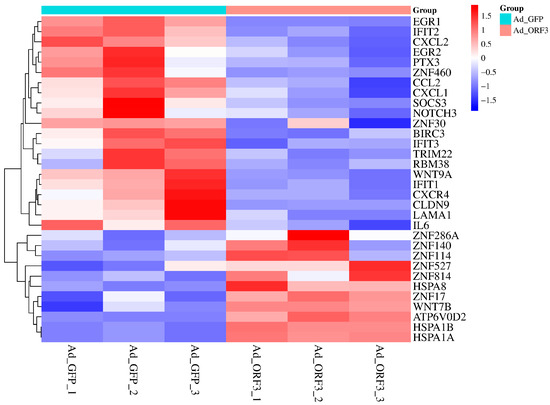
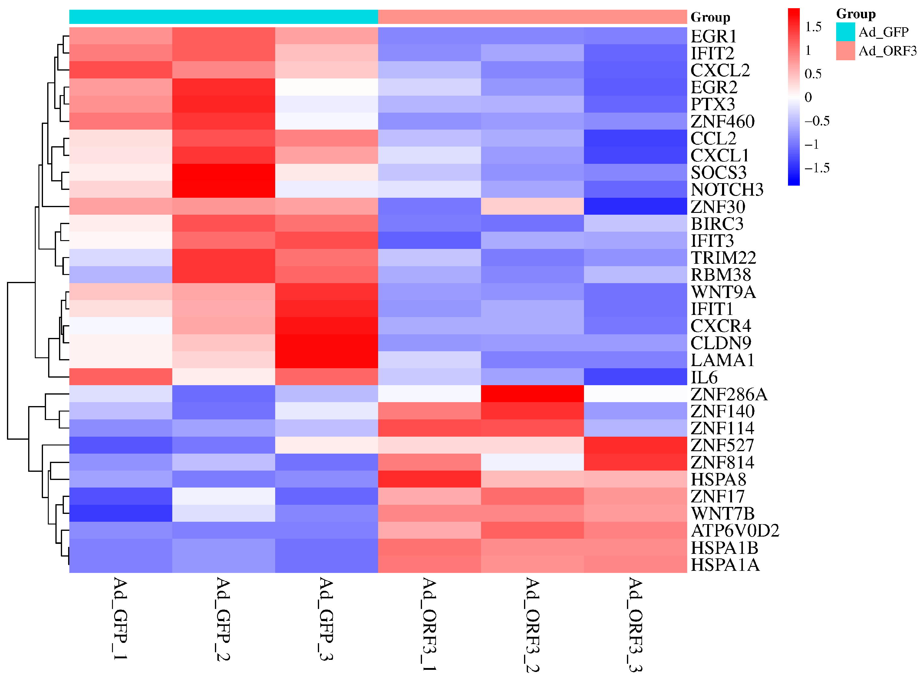
Based on gene expression profiles from six samples (Ad_GFP1, Ad_GFP2, Ad_GFP3, Ad_ORF3_1, Ad_ORF3_2, Ad_ORF3_3), differentially expressed genes associated with viral infection-related pathways were identified through KEGG functional annotation enrichment analysis, with the threshold set at |log2(fold change)| > 1.0 and a p-value < 0.05. The differential genes involved in viral infection pathways were visualized using a heatmap (Figure 1), where the horizontal axis represents the samples and the vertical axis represents the mRNAs. Distinct colors indicate different levels of mRNA expression: red denotes highly expressed mRNAs, and dark blue indicates lowly expressed mRNAs.
Figure 1.
The mRNA of the pathways associated with viral infection that were selected based on transcriptome sequencing was differentially expressed.
2.3. Prediction of miRNAs Associated with Differentially Expressed mRNAs of Viral Infection-Related Pathways
The screened differentially expressed mRNAs were used to predict potentially binding miRNAs through an online tool (https://jingege.shinyapps.io/jingle_molecular/ (accessed on 16 March 2025)), which integrates a total of 11 databases. These include three experimentally validated databases (miRecords, miRTarBase, and TarBase) and eight prediction databases (DIANA-microT, ElMMo, MicroCosm, miRanda, miRDB, PicTar, PITA, and TargetScan). Only miRNAs supported by five or more of these databases were selected for subsequent analysis.
2.4. Screening for Differentially Expressed circRNAs Associated with Viral Infection Pathways
The predicted miRNAs were subsequently used for circRNA prediction through both TargetScan (http://www.targetscan.org/ (accessed on 17 March 2025)) and miRanda (https://www.bioinformatics.com.cn/local_miranda_miRNA_target_prediction_120 (accessed on 17 March 2025)), with thresholds set as follows: a maximum free energy of < −10 kcal/mol in miRanda and a context++ score percentile ≥ 50 in TargetScan. The intersection between these prediction results and the experimentally identified differentially expressed circRNAs from sequencing data (filtered at |log2FC| > 1.0 and p-value < 0.05) was taken to identify circRNAs associated with viral infection-related pathways.
2.5. Prediction of circRNA-miRNA-mRNA Regulatory Network Associated with Viral Infection in Genotype IV SHEV ORF3 in HepG2 Cells
To identify relevant miRNA interactions, the screened differentially expressed circRNAs were analyzed using both TargetScan and miRanda. The resulting miRNA predictions were then intersected with the miRNA sets previously derived from mRNA-based prediction. Finally, the associations among the filtered circRNAs, miRNAs, and mRNAs were integrated and visualized as a comprehensive circRNA–miRNA–mRNA regulatory network using Cytoscape version 3.10.0.
3. Results
3.1. Screening of Differentially Expressed mRNAs of Viral Infection-Related Pathways
Differentially expressed mRNAs were screened by the following GO and KEGG functional annotations of viral infection-related pathways (Table 1 and Table 2), and a total of 32 differentially expressed mRNAs (Figure 1) were screened (Table 3).
Table 1.
GO Pathway related to viral infection.
Table 2.
KEGG pathway related to viral infection.
Table 3.
The sequencing data of 32 mRNAs related to the differential expression of viral infection pathways were screened.
3.2. Prediction of miRNAs Interacting with Differentially Expressed mRNAs Associated with Viral Infection Pathways
The 32 mRNAs screened above were used to predict their binding miRNAs through an online website tool, with 11 prediction databases including 3 experimental validation databases: mirecords, mirtarbase, tarbase, and 8 prediction databases: diana_microt, elmmo, microcosm, miranda, mirdb, pictar, pita, targetscan), a total of 6480 results were predicted, and 51 results were obtained by filtering miRNAs that could be predicted by 5 or more databases (Table S1).
3.3. Screening for Differentially Expressed circRNAs Associated with Viral Infection Pathways
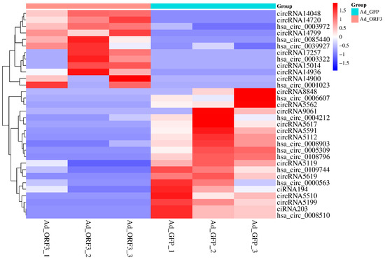
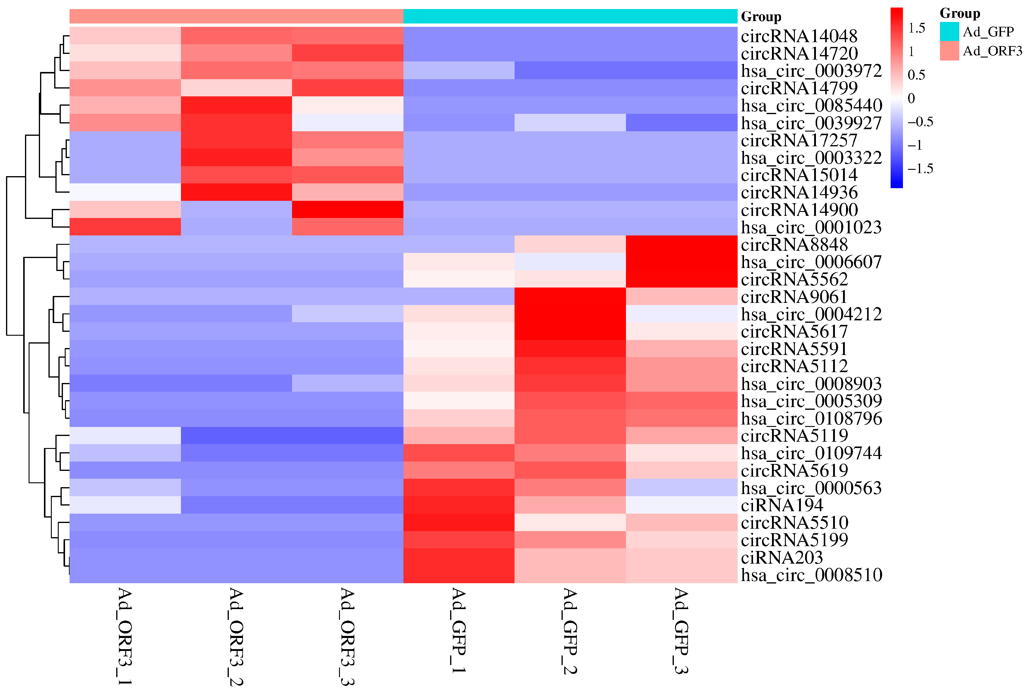
CircRNAs act as microRNA sponges associated with related miRNAs, which together constitute the circRNA-miRNA axis involved in disease pathogenesis. Using the 51 miRNAs obtained after post-prediction screening, circRNA prediction was performed by Targetscan and miRanda, and the intersection of the prediction results with the actual differentially expressed circRNAs in the sequencing data was taken to obtain the following 32 circRNAs (Figure 2 & Table 4).
Figure 2.
Based on circRNA sequencing, 31 differential expression circRNA related to viral infection pathways were screened out.
Table 4.
The sequencing data of circRNA related to the differential expression of viral infection pathways were screened. (The inf values in the log2(fold change) column represent infinite fold changes, which occur when the expression level in one group is undetectable or below the detection limit of the assay).
3.4. Prediction of Viral Infection Pathway-Associated circRNA-miRNA-mRNA Regulatory Network
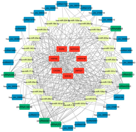
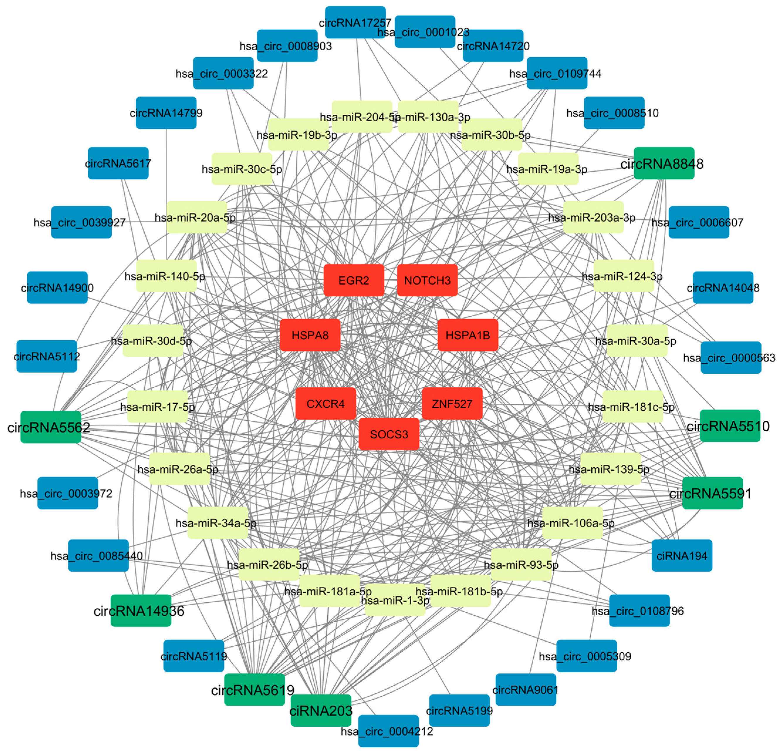
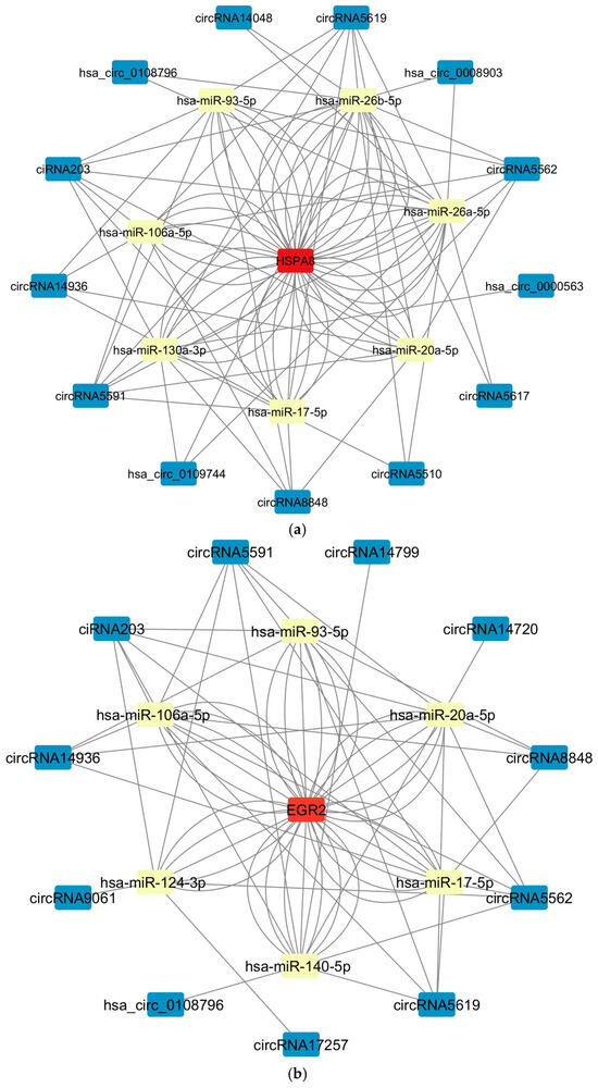
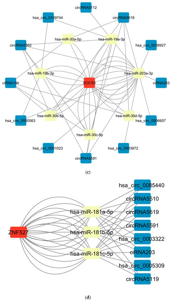
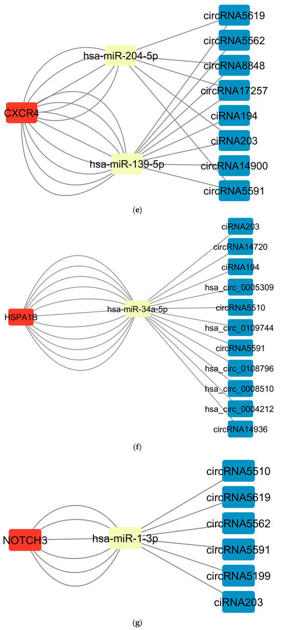
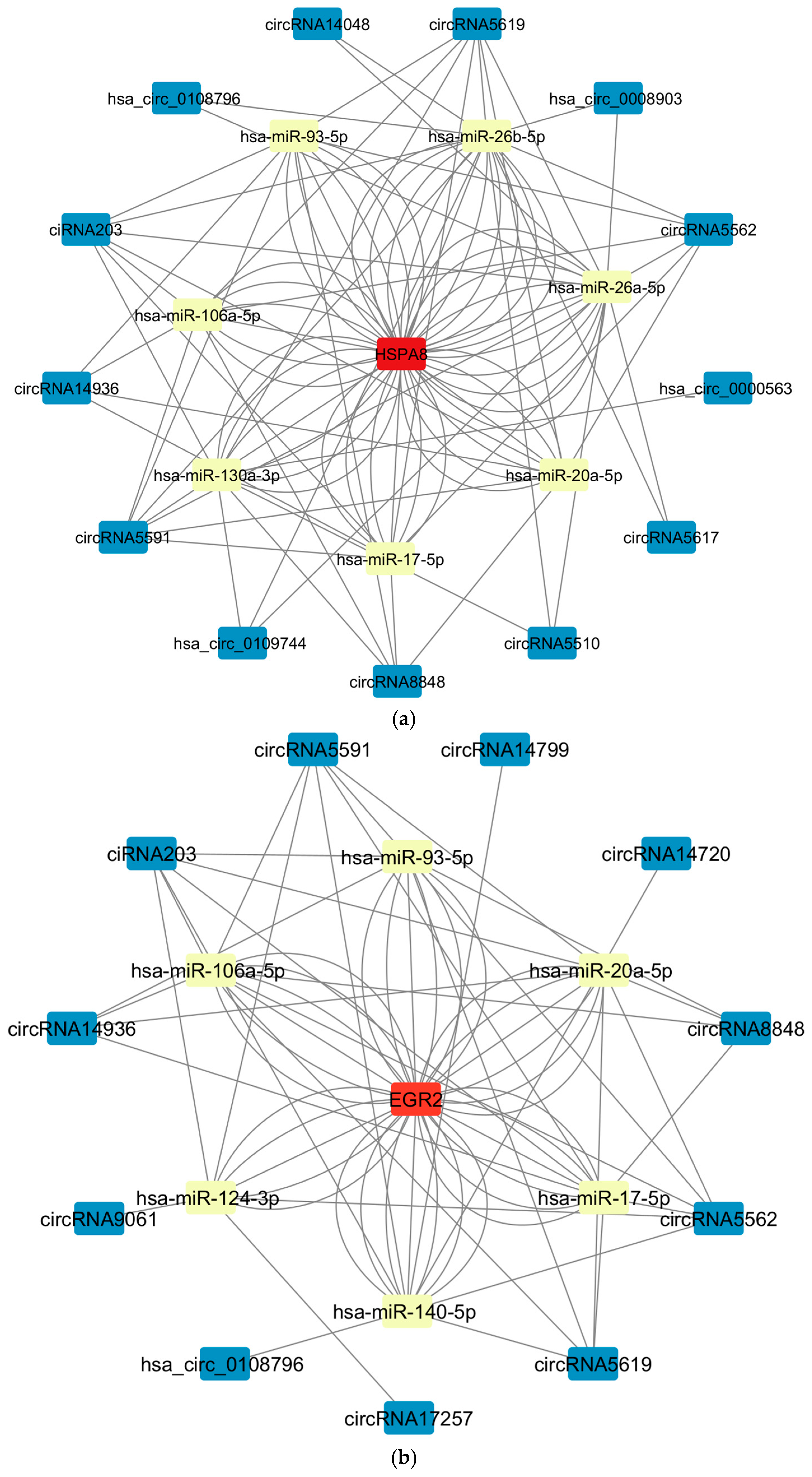
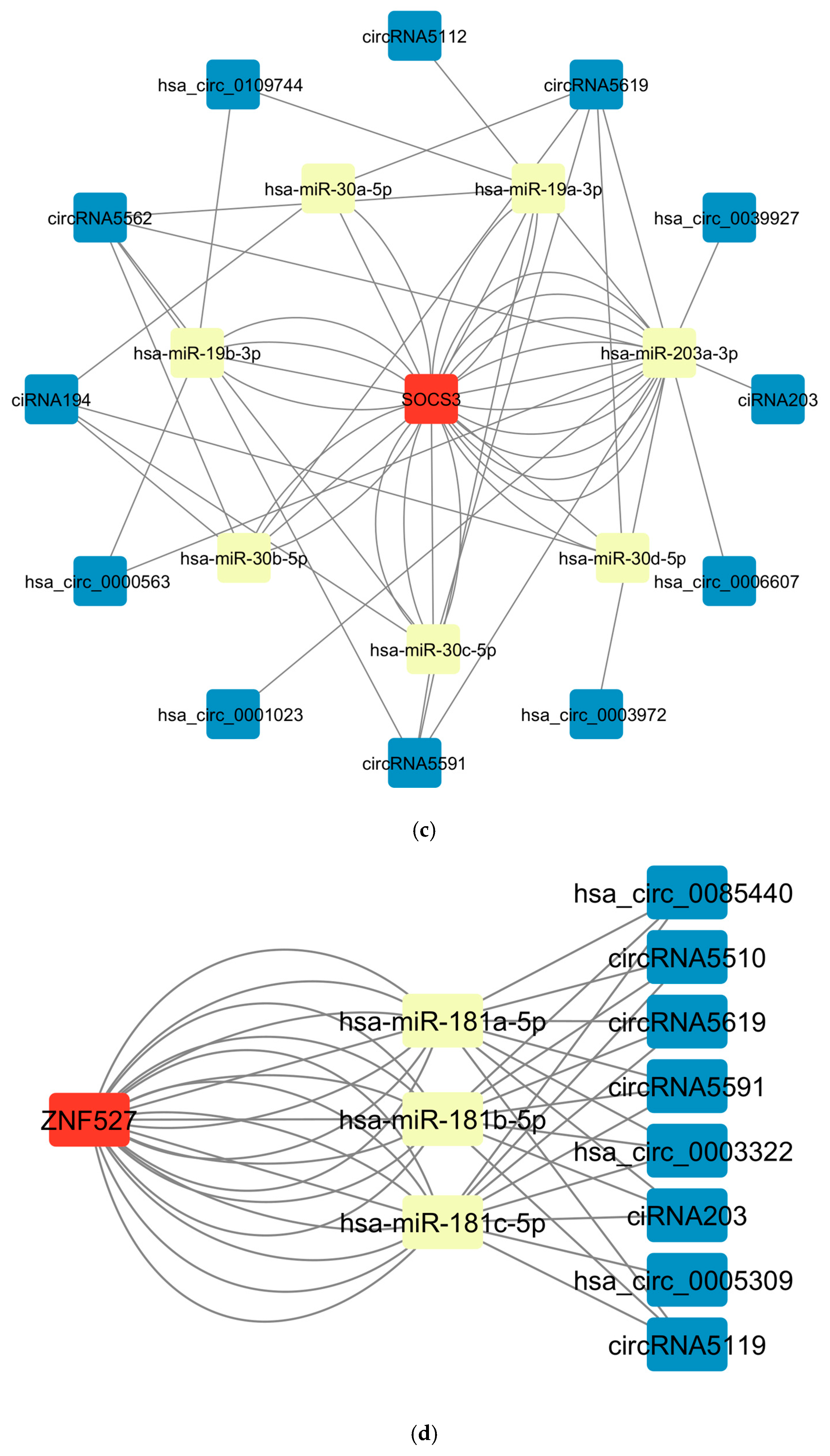
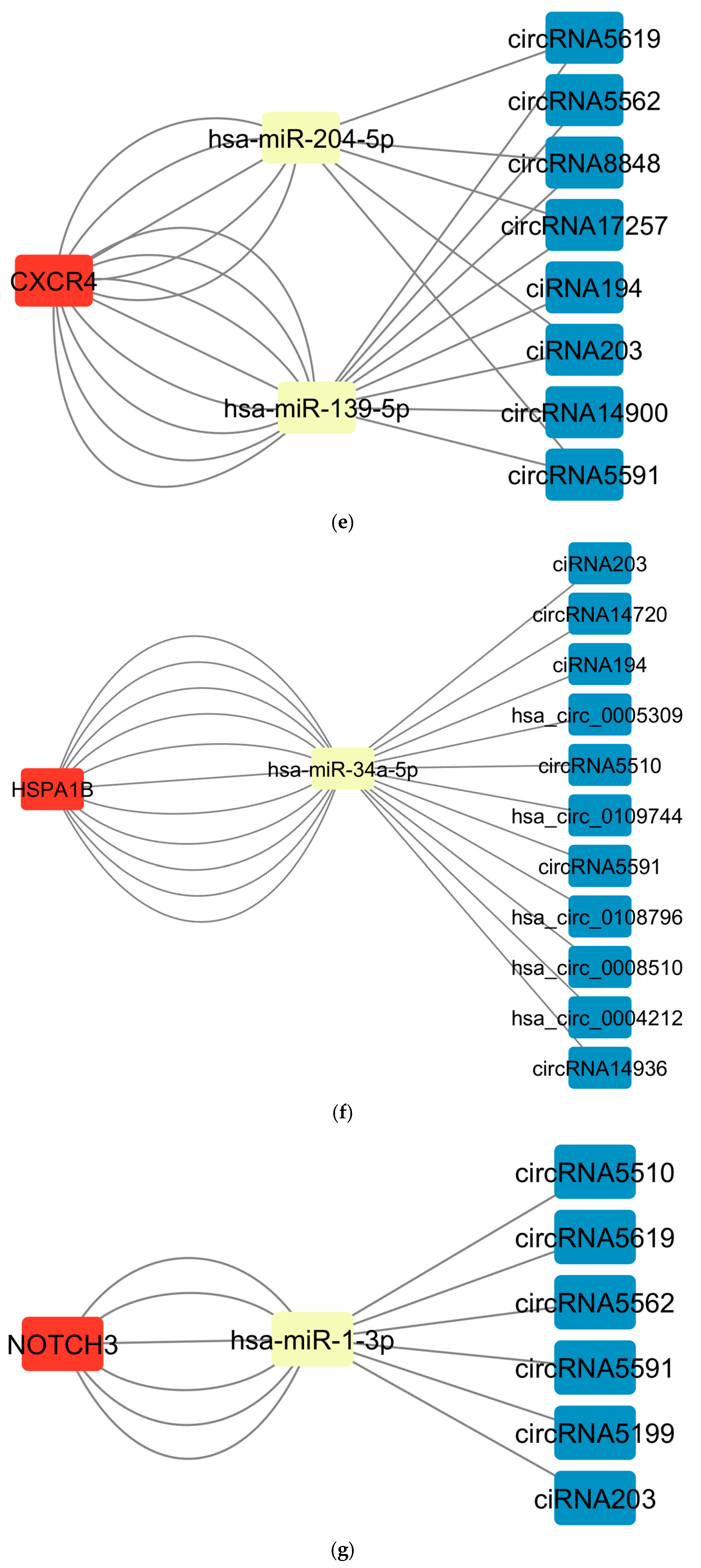
The 32 circular RNAs were screened for miRNA targeting prediction (combined with TargetScan and miRanda tools), and the miRNAs predicted by the differentially expressed mRNA related to the virus infection pathway were crossed, and finally 23 miRNAs were obtained. The obtained 23 miRNAs were associated with the corresponding 7 differentially expressed mRNAs (Table S2), and then a potential circRNA-miRNA-mRNA regulatory network containing 32 circular RNAs, 23 miRNAs and 7 mRNAs was constructed through Cytoscape (Figure 3). Among them, a total of 167 regulatory pathways were screened (Table S3), with 51 associated with HSPA8 (Figure 4a), 36 associated with EGR2 (Figure 4b), 28 associated with SOCS3 (Figure 4c), 22 associated with ZNF527 (Figure 4d), and CXCR4 associated with 13 (Figure 4e), 11 associated with HSPA1B (Figure 4f), and 6 associated with NOTCH3 (Figure 4g). Among them, circRNA5591, circRNA5619, ciRNA203, circRNA5562, and circRNA8848 may be shared circRNAs of multiple regulatory pathways, which appeared 98 times in 167 pathways. hsa-miR-106a-5p, hsa-miR-17-5p, hsa-miR -20a-5p, hsa-miR-93-5p, hsa-miR-34a-5p, hsa-miR-203a-3p, hsa-miR-26a-5p, and hsa-miR-26b-5p may be shared miRNAs for multiple regulatory pathways, appearing 89 times in 167 pathways.
Figure 3.
The potential circRNA-miRNA-mRNA regulatory network associated with viral infection was constructed using Cytoscape. Within this network, mRNA nodes are represented in red, miRNA nodes in yellow, and circRNA nodes in blue and green. Notably, the potential hub circRNAs identified in this study are highlighted as green nodes with enlarged font size.
Figure 4.
The potential circRNA-miRNA-mRNA regulatory network of each gene was constructed by Cytoscape. Within this network, mRNA nodes are represented in red, miRNA nodes in yellow, and circRNA nodes in blue. (a) CircRNA-miRNA-mRNA regulatory pathways of HSPA8 were demonstrated using network diagram. (b) CircRNA-miRNA-mRNA regulatory pathways of EGR2 were demonstrated using network diagram. (c) CircRNA-miRNA-mRNA regulatory pathways of SOCS3 were demonstrated using network diagram. (d) CircRNA-miRNA-mRNA regulatory pathways of ZNF527 were demonstrated using network diagram. (e) CircRNA-miRNA-mRNA regulatory pathways of CXCR4 were demonstrated using network diagram. (f) CircRNA-miRNA-mRNA regulatory pathways of HSPA1B were demonstrated using network diagram. (g) CircRNA-miRNA-mRNA regulatory pathways of NOTCH3 were demonstrated using network diagram.
4. Discussion
In this study, based on previous circRNAomics and transcriptomics sequencing data, we analyzed the changes in expression profiles of porcine hepatitis E virus ORF3 overexpression adenovirus after transfection of HepG2 cells, and firstly screened out 32 differentially expressed mRNAs related to the viral infection pathway. Subsequently, we predicted potential miRNA targets of these 32 mRNAs by using an online tool to obtain a total of 51 candidate miRNAs, and further predicted the circRNAs that these miRNAs might bind using TargetScan and miRanda software. Combined with the actual differentially expressed circRNAs in the sequencing data, 32 candidate circRNAs associated with viral regulation were screened out. Next, the potential miRNAs of these circRNAs were predicted in reverse, and compared with pre mRNA-associated miRNAs to take the intersection, and finally obtained 23 core miRNAs. Based on the above analysis, we integrated the 32 circRNAs, 23 miRNAs and the key 7 mRNAs among them, and constructed a circRNA-miRNA-mRNA regulatory network by using Cytoscape to systematically reveal the role of the hepatitis E virus ORF3 protein in viral regulation and the role of the hepatitis virus ORF3 protein in hepatocytes.
It is important to note that this study employed an adenovirus-mediated ORF3 overexpression system in HepG2 cells, a well-established model for investigating specific viral protein functions. While this approach enables precise examination of ORF3-specific effects, it presents inherent methodological considerations including non-physiological expression levels and separation from the complete viral replication context. These aspects should be considered when interpreting the identified regulatory networks.
The circRNA–miRNA–mRNA regulatory network constructed in this study encompasses seven differentially expressed mRNAs (HSPA8, HSPA1B, EGR2, SOCS3, ZNF527, CXCR4, NOTCH3). Both HSPA8 and HSPA1B belong to the heat shock protein family. Literature reports indicate that HSPA8 may interact with PRRSV GP4 protein to participate in viral attachment and internalization [17], and enhance PRRSV replication by promoting RAB18/PLIN2 interactions through chaperone-mediated autophagy [18]. HSPA8 has also been implicated in viral RNA release during JEV infection [19]. In HBV-related hepatocellular carcinoma, HSPA8 appears to exert dual functions, potentially regulating viral replication and ferroptosis pathways [20]. Similarly, HSPA1B has been shown to be required for efficient replication in ECTV infection [21], while exhibiting inhibitory effects on viral proliferation during the mid-to-late stages of ORFV infection [22], suggesting virus-specific functionalities within this protein family. Our bioinformatic predictions suggest that circRNAs such as circRNA5591, ciRNA203, and circRNA5619 may potentially influence the expression levels of HSPA8 and HSPA1B by sequestering miRNAs including hsa-miR-34a-5p and hsa-miR-26a-5p. This regulatory relationship might be associated with the progression of SHEV infection.
The early growth response transcription factor EGR2 participates in host defense through its negative regulation of T cell activation. Studies have demonstrated that EGR2 deficiency significantly reduces resistance in influenza virus-infected mice [23]. Our bioinformatic predictions suggest that circRNAs including circRNA5562, circRNA5591, and ciRNA203 might influence EGR2 expression through interactions with miRNAs such as hsa-miR-140-5p and hsa-miR-106a-5p. This potential regulatory relationship indicates that downregulation of EGR2 expression could be somewhat correlated with alterations in host defense mechanisms against SHEV.
SOCS3 functions as a negative immune regulator and demonstrates differential regulation patterns across viral infections: HSV-1 upregulates SOCS3 via STAT3 activation to suppress JAK/STAT antiviral signaling [24]; influenza A virus induces SOCS3 through an NF-κB-dependent pathway to attenuate type I interferon responses [25]; while HIV-1 downregulates SOCS3 to trigger persistent immune activation that promotes viral replication [26]. PRRSV can also promote viral replication by inducing SOCS3 expression through the p38/AP-1 signaling pathway [27]. Our predictive analysis suggests that circRNAs including circRNA5562, circRNA5619, and circRNA5591 might potentially affect SOCS3 expression by sequestering miRNAs such as hsa-miR-203a-3p and hsa-miR-19b-3p. This regulatory pattern could potentially create a favorable environment for viral infection.
Although direct evidence for ZNF527 in viral infection remains limited, zinc finger protein family members typically participate in antiviral immune responses. Research indicates that ZNF268 may contribute to antiviral immunity by promoting NF-κB signaling through maintaining IKK complex stability [28], while certain ZNF proteins might potentially inhibit SARS-CoV-2 infection by enhancing immune cell activity and abundance [29]. Our predictive results suggest that circRNAs including circRNA5119, circRNA5510, and circRNA5591 might influence ZNF527 expression by sequestering miRNAs such as hsa-miR-181c-5p and hsa-miR-181a-5p. This regulatory relationship could potentially be linked to host anti-SHEV immune responses.
The chemokine receptor CXCR4 serves as a key co-receptor for HIV-1 entry into CD4+ T cells [30]. Studies show that viruses such as HSV-1 and EBV can interfere with immune cell function by downregulating CXCR4 expression [31,32]. Our bioinformatic predictions suggest that circRNAs including circRNA17257, circRNA5591, and circRNA5619 might potentially affect CXCR4 expression by sequestering hsa-miR-139-5p and hsa-miR-204-5p. Such expression changes might be correlated with alterations in immune cell chemotactic function, potentially providing favorable conditions for SHEV infection establishment.
NOTCH3 signaling activation has been associated with various viral infection pathologies: HIV Tat protein drives neuroinflammation by enhancing NOTCH3 signaling [33]; SARS-CoV-2 infection in alveolar foci is accompanied by NOTCH3 upregulation [34]; and HCV NS3 protein promotes persistent infection by modulating the Notch pathway through SRCAP/p400 [15]. Our analysis suggests that circRNAs including ciRNA203, circRNA5199, and circRNA5510 might potentially regulate NOTCH3 expression by sequestering hsa-miR-1-3p. These predictive results imply that the NOTCH3 signaling pathway might be involved in SHEV–host cell interactions.
5. Conclusions
In summary, this study utilized transcriptomic and circRNA sequencing data from SHEV ORF3-overexpressing adenovirus-infected HepG2 cells to construct a circRNA-miRNA-mRNA regulatory network. Through differential expression analysis and bioinformatics prediction, the network was identified to center on seven core mRNAs (HSPA8, HSPA1B, EGR2, SOCS3, ZNF527, CXCR4, and NOTCH3). In this study, we showed that ciRNA203, circRNA14936, circRNA5510, circRNA5562, circRNA5591, circRNA5619, and circRNA8848 may act as potential key molecules in the process of SHEV infection of HepG2 cells, and through competitive binding of miRNAs that targeted regulation of the transcription of seven differentially expressed mRNAs, including HSPA8, HSPA1B, and SOCS3 (Table S4). Although our study proposes a theoretical framework for the circRNA-miRNA-mRNA interactions of viral infection-related pathways in SHEV ORF3-overexpressing HepG2 cells, the molecular mechanism still needs to be further confirmed by target validation experiments. Moving forward, we plan to conduct both in vitro and in vivo functional experiments—such as RNA pulldown, dual-luciferase reporter assays, and gene knockdown/overexpression studies—to validate the key molecular interactions within this network. These efforts will help clarify the biological functions and molecular mechanisms of these candidates during SHEV infection, thereby contributing to a more comprehensive understanding of the virus–host interaction network.
Supplementary Materials
The following supporting information can be downloaded at: https://www.mdpi.com/article/10.3390/microorganisms13122654/s1, Table S1: miRNA predicted by differential expression of mRNA; Table S2: Seven identified mRNAs and their involved viral infection pathways and related sequencing data; Table S3: Screen out the relevant circRNA-miRNA-mRNA; Table S4: Summary of the diverse functional roles of the seven identified mRNAs in virus-host interactions.
Author Contributions
H.J., L.W. and S.W. contributed to the conception and design of the study. H.J., Y.Q. and J.G. contributed to data acquisition and data analysis. J.L., L.C., C.M. and Y.Z. contributed to the interpretation of the data. H.J., L.C. and J.J.W. drafted the manuscript. The manuscript was revised by H.J., J.J.W. and F.W. All authors have read and agreed to the published version of the manuscript.
Funding
This study was financially supported by the Natural Science Foundation of Chongqing (CSTB2022NSCQ-MSX0311, cstc2020jcyj-msxmX0446), the General Fund of Guizhou Agricultural Science and Technology (2024-27), and the Fundamental Research Funds for the Central Universities (SWU-KT22013).
Institutional Review Board Statement
Not applicable.
Informed Consent Statement
Not applicable.
Data Availability Statement
The original contributions presented in this study are included in the article and Supplementary Materials. Further inquiries can be directed to the corresponding authors.
Conflicts of Interest
The authors declare no conflicts of interest.
References
- Aslan, T.; Balaban, H.Y. Hepatitis E virus: Epidemiology, diagnosis, clinical manifestations, and treatment. World J. Gastroenterol. 2020, 26, 5543–5560. [Google Scholar] [CrossRef] [Scilit] [PubMed]
- Alexandrova, R.; Tsachev, I.; Kirov, P.; Abudalleh, A.; Hristov, H.; Zhivkova, T.; Dyakova, L.; Baymakova, M. Hepatitis E Virus (HEV) Infection Among Immunocompromised Individuals: A Brief Narrative Review. Infect. Drug Resist. 2024, 17, 1021–1040. [Google Scholar] [CrossRef] [Scilit] [PubMed]
- Zhou, X.; Lu, H. Advances in Acute Severe Hepatitis of Unknown Etiology in Children. Zoonoses 2022, 2, 981. [Google Scholar] [CrossRef] [Scilit]
- Songtanin, B.; Molehin, A.J.; Brittan, K.; Manatsathit, W.; Nugent, K. Hepatitis E Virus Infections: Epidemiology, Genetic Diversity, and Clinical Considerations. Viruses 2023, 5, 1389. [Google Scholar] [CrossRef] [Scilit]
- Sooryanarain, H.; Meng, X.-J. Swine hepatitis E virus: Cross-species infection, pork safety and chronic infection. Virus Res. 2020, 284, 197985. [Google Scholar] [CrossRef] [Scilit]
- Salines, M.; Andraud, M.; Rose, N. From the epidemiology of hepatitis E virus (HEV) within the swine reservoir to public health risk mitigation strategies: A comprehensive review. Vet. Res. 2017, 48, 31. [Google Scholar] [CrossRef] [Scilit]
- Bahoussi, A.N.; Guo, Y.-Y.; Wang, P.-H.; Dahdouh, A.; Wu, C.; Xing, L. Genomic characteristics and recombination patterns of swine hepatitis E virus in China. Transbound. Emerg. Dis. 2022, 69, e3273–e3281. [Google Scholar] [CrossRef] [Scilit]
- Jiao, H.; Meng, C.; Jiao, F.; Zhou, G.; Wang, L.; Wu, S.; Fan, C.; Li, J.; Cao, L.; Zhao, Y.; et al. Descriptive Comparative Transcriptomic Analysis of Genotype IV SHEV ORF3-Expressing HepG2 Cells. Microorganisms 2025, 13, 412. [Google Scholar] [CrossRef] [Scilit]
- Yang, Y.-L.; Nan, Y.-C. Open reading frame 3 protein of hepatitis E virus: Multi-function protein with endless potential. World J. Gastroenterol. 2021, 27, 2458–2473. [Google Scholar] [CrossRef] [Scilit]
- Ding, Q.; Heller, B.; Capuccino, J.M.V.; Song, B.; Nimgaonkar, I.; Hrebikova, G.; Contreras, J.E.; Ploss, A. Hepatitis E virus ORF3 is a functional ion channel required for release of infectious particles. Proc. Natl. Acad. Sci. USA 2017, 114, 1147–1152, Erratum in Proc. Natl. Acad. Sci. USA 2017, 114, E4897. [Google Scholar] [CrossRef] [Scilit] [PubMed]
- Jiao, F.; Zhao, Y.; Zhou, G.; Meng, C.; Wang, L.; Wu, S.; Li, J.; Cao, L.; Zhou, B.; Luo, Y.; et al. Multiple Functions of Hepatitis E Virus ORF3. Microorganisms 2024, 12, 1405. [Google Scholar] [CrossRef] [Scilit]
- Li, H. circRNA: A promising all-around star in the future. Epigenomics 2023, 15, 677–685. [Google Scholar] [CrossRef] [Scilit]
- Zhang, P.; Dai, M. CircRNA: A rising star in plant biology. J. Genet. Genom. 2022, 49, 1081–1092. [Google Scholar] [CrossRef] [Scilit]
- Min, J.; Li, Y.; Li, X.; Wang, M.; Li, H.; Bi, Y.; Xu, P.; Liu, W.; Ye, X.; Li, J. The circRNA circVAMP3 restricts influenza A virus replication by interfering with NP and NS1 proteins. PLoS Pathog. 2023, 19, e1011577. [Google Scholar] [CrossRef] [Scilit]
- Iwai, A.; Takegami, T.; Shiozaki, T.; Miyazaki, T. Hepatitis C Virus NS3 Protein Can Activate the Notch-Signaling Pathway through Binding to a Transcription Factor, SRCAP. PLoS ONE 2011, 6, e20718. [Google Scholar] [CrossRef] [Scilit] [PubMed]
- Jiao, H.; Zhao, Y.; Zhou, Z.; Li, W.; Li, B.; Gu, G.; Luo, Y.; Shuai, X.; Fan, C.; Wu, L.; et al. Identifying Circular RNAs in HepG2 Expressing Genotype IV Swine Hepatitis E Virus ORF3 Via Whole Genome Sequencing. Cell Transpl. 2021, 30, 9636897211055042. [Google Scholar] [CrossRef] [Scilit] [PubMed]
- Wang, L.; Li, R.; Geng, R.; Zhang, L.; Chen, X.-X.; Qiao, S.; Zhang, G. Heat Shock Protein Member 8 (HSPA8) Is Involved in Porcine Reproductive and Respiratory Syndrome Virus Attachment and Internalization. Microbiol. Spectr. 2022, 10, e0186021. [Google Scholar] [CrossRef] [Scilit]
- Li, G.-L.; Han, Y.-Q.; Su, B.-Q.; Yu, H.-S.; Zhang, S.; Yang, G.-Y.; Wang, J.; Liu, F.; Ming, S.-L.; Chu, B.-B. Porcine reproductive and respiratory syndrome virus 2 hijacks CMA-mediated lipolysis through upregulation of small GTPase RAB18. PLoS Pathog. 2024, 20, e1012123. [Google Scholar] [CrossRef] [Scilit] [PubMed]
- Chuang, C.-K.; Yang, T.-H.; Chen, T.-H.; Yang, C.-F.; Chen, W.-J. Heat shock cognate protein 70 isoform D is required for clathrin-dependent endocytosis of Japanese encephalitis virus in C6/36 cells. J. Gen. Virol. 2015, 96 Pt 4, 793–803. [Google Scholar] [CrossRef] [Scilit]
- Wang, Y.; Zhao, M.; Zhao, L.; Geng, Y.; Li, G.; Chen, L.; Yu, J.; Yuan, H.; Zhang, H.; Yun, H.; et al. HBx-Induced HSPA8 Stimulates HBV Replication and Suppresses Ferroptosis to Support Liver Cancer Progression. Cancer Res. 2023, 83, 1048–1061. [Google Scholar] [CrossRef] [Scilit]
- Cheng, W.; Jia, H.; Wang, X.; He, X.; Jin, Q.; Cao, J.; Jing, Z. Ectromelia virus upregulates the expression of heat shock protein 70 to promote viral replication. Int. J. Mol. Med. 2018, 42, 1044–1053. [Google Scholar] [CrossRef] [Scilit]
- Hao, J.-H.; Kong, H.-J.; Yan, M.-H.; Shen, C.-C.; Xu, G.-W.; Zhang, D.-J.; Zhang, K.-S.; Zheng, H.-X.; Liu, X.-T. Inhibition of orf virus replication in goat skin fibroblast cells by the HSPA1B protein, as demonstrated by iTRAQ-based quantitative proteome analysis. Arch. Virol. 2020, 165, 2561–2587. [Google Scholar] [CrossRef] [Scilit]
- Du, N.; Kwon, H.; Li, P.; West, E.E.; Oh, J.; Liao, W.; Yu, Z.; Ren, M.; Leonard, W.J. EGR2 is critical for peripheral naïve T-cell differentiation and the T-cell response to influenza. Proc. Natl. Acad. Sci. USA 2014, 111, 16484–16489. [Google Scholar] [CrossRef] [Scilit]
- Yokota, S.-I.; Yokosawa, N.; Okabayashi, T.; Suzutani, T.; Miura, S.; Jimbow, K.; Fujii, N. Induction of suppressor of cytokine signaling-3 by herpes simplex virus type 1 contributes to inhibition of the interferon signaling pathway. J. Virol. 2004, 78, 6282–6286. [Google Scholar] [CrossRef] [Scilit]
- Pauli, E.-K.; Schmolke, M.; Wolff, T.; Viemann, D.; Roth, J.; Bode, J.G.; Ludwig, S. Influenza A virus inhibits type I IFN signaling via NF-kappaB-dependent induction of SOCS-3 expression. PLoS Pathog. 2008, 4, e1000196. [Google Scholar] [CrossRef] [Scilit] [PubMed]
- Miller, R.C.; Schlaepfer, E.; Baenziger, S.; Crameri, R.; Zeller, S.; Byland, R.; Audigé, A.; Nadal, D.; Speck, R.F. HIV interferes with SOCS-1 and -3 expression levels driving immune activation. Eur. J. Immunol. 2011, 41, 1058–1069. [Google Scholar] [CrossRef] [Scilit] [PubMed]
- Luo, X.; Chen, X.-X.; Qiao, S.; Li, R.; Lu, Q.; Geng, R.; Wang, L.; Zhou, E.-M.; Zhang, G. Porcine reproductive and respiratory syndrome virus increases SOCS3 production via activation of p38/AP-1 signaling pathway to promote viral replication. Vet. Microbiol. 2021, 257, 109075. [Google Scholar] [CrossRef] [Scilit] [PubMed]
- Wang, L.; Qiao, X.; Zhang, S.; Qin, Y.; Guo, T.; Hao, Z.; Sun, L.; Wang, X.; Wang, Y.; Jiang, Y.; et al. Porcine transmissible gastroenteritis virus nonstructural protein 2 contributes to inflammation via NF-κB activation. Virulence 2018, 9, 1685–1698. [Google Scholar] [CrossRef] [Scilit] [PubMed]
- Qin, S.; Xu, W.; Wang, C.; Jiang, S.; Dai, W.; Yang, Y.; Shen, J.; Jin, P.; Ma, F.; Xia, X. Analyzing master regulators and scRNA-seq of COVID-19 patients reveals an underlying anti-SARS-CoV-2 mechanism of ZNF proteins. Brief. Bioinform. 2021, 22, bbab118. [Google Scholar] [CrossRef] [Scilit]
- Huang, L.S.M.; Snyder, E.Y.; Schooley, R.T. Strategies and Progress in CXCR4-Targeted Anti-Human Immunodeficiency Virus (HIV) Therapeutic Development. Clin. Infect. Dis. 2021, 73, 919–924. [Google Scholar] [CrossRef] [Scilit]
- Prechtel, A.T.; Turza, N.M.; Kobelt, D.J.; Eisemann, J.I.; Coffin, R.S.; McGrath, Y.; Hacker, C.; Ju, X.; Zenke, M.; Steinkasserer, A. Infection of mature dendritic cells with herpes simplex virus type 1 dramatically reduces lymphoid chemokine-mediated migration. J. Gen. Virol. 2005, 86 Pt 6, 1645–1657. [Google Scholar] [CrossRef] [Scilit] [PubMed]
- Ehlin-Henriksson, B.; Mowafi, F.; Klein, G.; Nilsson, A. Epstein-Barr virus infection negatively impacts the CXCR4-dependent migration of tonsillar B cells. Immunology 2006, 117, 379–385. [Google Scholar] [CrossRef] [Scilit] [PubMed]
- Gao, L.; Sun, W.; Zhang, D.; Shang, Y.; Li, L.; Tao, W.; Zhang, L.; Liu, H. HIV-1 subtype B Tat enhances NOTCH3 signaling in astrocytes to mediate oxidative stress, inflammatory response, and neuronal apoptosis. J. Neurovirol. 2023, 29, 479–491. [Google Scholar] [CrossRef] [Scilit]
- Li, C.; Xiao, N.; Song, W.; Lam, A.H.-C.; Liu, F.; Cui, X.; Ye, Z.; Chen, Y.; Ren, P.; Cai, J.; et al. Chronic lung inflammation and CK14+ basal cell proliferation induce persistent alveolar-bronchiolization in SARS-CoV-2-infected hamsters. EBioMedicine 2024, 108, 105363. [Google Scholar] [CrossRef] [Scilit] [PubMed]

Disclaimer/Publisher’s Note: The statements, opinions and data contained in all publications are solely those of the individual author(s) and contributor(s) and not of MDPI and/or the editor(s). MDPI and/or the editor(s) disclaim responsibility for any injury to people or property resulting from any ideas, methods, instructions or products referred to in the content. |
© 2025 by the authors. Licensee MDPI, Basel, Switzerland. This article is an open access article distributed under the terms and conditions of the Creative Commons Attribution (CC BY) license (https://creativecommons.org/licenses/by/4.0/).