


Abstract
Microtubules constitute fundamental cytoskeletal polymers that orchestrate cell division, intracellular trafficking, and the maintenance of cellular architecture across eukaryotic lineages. The biogenesis, folding, and steady-state homeostasis of αβ-tubulin heterodimers are tightly regulated by an evolutionarily conserved cohort of dedicated molecular chaperones, designated tubulin-specific chaperones (TBCs; TBCA–TBCE), acting in concert with the small GTPase Arl2. While this intricate assembly pathway has been extensively dissected in mammalian and plant systems, its existence, molecular architecture, and physiological relevance within the malaria parasite Plasmodium falciparum have not been examined systematically, although the parasite’s wider chaperone network, including the prefoldin and TRiC/CCT complexes that act upstream of the TBCs, has been described. In the present study, we undertake a rigorous multi-layered in silico interrogation of the P. falciparum genome to identify and functionally contextualize the parasite’s putative tubulin-specific chaperones (PfTBCs). Through integrated sequence homology analyses, domain architecture mapping, and phylogenetic reconstruction, we establish the presence of clear orthologs corresponding to the canonical TBC repertoire. Subsequent functional annotation, coupled with protein–protein interaction network predictions, strongly implicates the role of PfTBCs in the coordinated assembly of αβ-tubulin heterodimers and the broader regulation of microtubule dynamics. Given the indispensable contribution of microtubule dynamics to parasite growth, asexual replication, gametocytogenesis, motility, and host cell invasion, the identification of a conserved tubulin-folding machinery represents a significant conceptual advance. This work furnishes the first systematic computational evidence for a conserved TBC (TBCA–TBCE) module of the tubulin-folding pathway in P. falciparum, thereby laying a robust computational foundation for targeted experimental validation. Moreover, by highlighting PfTBCs as potentially essential and selectively vulnerable components of parasite cytoskeletal homeostasis, this study nominates these proteins as candidates for experimental evaluation as antimalarial targets. All conclusions presented are computational predictions that await experimental validation.
1. Introduction
Microtubules are highly dynamic polymers that are formed as a result of polymerization and depolymerization of tubulin dimers, and are essential molecules for maintaining cytoskeletal architecture, intracellular transport, and cell division in eukaryotes [1,2]. The biogenesis of αβ-tubulin heterodimers is a highly regulated process as neither tubulin can fold autonomously; instead, they require a committed multistep pathway involving a conserved network of molecular chaperones to achieve their functional conformation [3,4,5]. The pathway is initiated as soon as tubulin emerges from the ribosome in an unfolded state and is captured by the chaperone prefoldin, which subsequently transfers it to the cytosolic chaperonin TRiC for proper folding [3,5,6]. TRiC encapsulates tubulin within its chamber as it undergoes a closed conformation upon ATP hydrolysis [6,7]. Post this, polymerization-competent αβ-tubulin heterodimers are formed in a highly coordinated series of reactions mediated by a specific group of tubulin-specific chaperones (TBCs), also known as tubulin-folding cofactors (TBCA–TBCE), and the small GTPase Arl2 [4,5,8]. Altogether, coordinated activity of these chaperones along with Arl2 provide quality control machinery for the precise regulation of αβ-tubulin heterodimer homeostasis [4,5,8].
Early biochemical reports suggested that TBCs act sequentially to stabilize αβ-tubulin intermediates and facilitate hetero-dimerization via GTP-dependent hydrolysis [9,10,11]. Quasi-native folding intermediates of αβ-tubulin are captured and stabilized by free exchange between TBCA and TBCD (in case of β-tubulin), and between TBCB and TBCE (in case of α tubulin), resulting in the formation of TBCD/β and TBCE/α, respectively, which latter form a supercomplex (TBCE/α/TBCD/β) [9,10]. Post this, TBCC interacts with supercomplex and triggers GTP hydrolysis in the E-site thereby releasing newly formed α/β tubulin heterodimers. Following spontaneous E-site exchange with GTP, α β heterodimers acquire competency to form the microtubule polymer [10,11]. The tubulin-specific chaperones thus act as a tubulin assembly machine regulating not only de novo folding but also the recycling and turnover of the tubulin heterodimer [4,5,11].
A recent report by Seong et al. proposed a new molecular mechanism by which TBCs maintain tubulin homeostasis [12]. The authors used cryo-electron microscopy to determine structures of the human TBCs at six stages under defined nucleotide conditions. The data suggest that TBCD, TBCE, and Arl2 form a cage-like TBC-DEG complex that encapsulates α- and β-tubulin in partially assembled states [12]. TBCC subsequently acts as a molecular clamp and triggers the conformational rearrangement that allows α- and β-tubulin interfaces for productive GTP hydrolysis that ultimately leads to formation of α β heterodimers [12]. The authors also highlighted TBCs as central regulators capable of both promoting assembly and driving disassembly of α β heterodimers under specific cellular conditions instead of merely functioning as molecular chaperones [12]. This dual role underscores the significance of TBCs in maintaining tubulin homeostasis, particularly in organisms where microtubule dynamics are highly responsive to stress and developmental cues [12].
In Plasmodium, microtubule-based structures underpin essentially every proliferative, invasive and transmission step of its life cycle, and are built from a strikingly small repertoire of two α-tubulin genes and a single β-tubulin gene [13,14,15]. Apicomplexan division is itself unusual: blood-stage parasites proliferate by schizogony, in which repeated asynchronous rounds of closed nuclear division precede a single segmentation event, and related apicomplexans employ a broad spectrum of budding and fission modes [16,17,18], while recent ultrastructure expansion microscopy atlases have resolved the centriolar plaque, hemispindle and mitotic spindles at each of these steps [19] and α-tubulin levels have been shown to set microtubule number and sporozoite infectivity [20]. This demand peaks during gametogenesis, when the activated male gametocyte completes three rounds of endomitosis and assembles eight 9+2 axonemes within minutes at a bipartite microtubule-organizing center spanning the nuclear envelope, a process requiring de novo basal body formation and the motor kinesin-8B [21,22,23,24,25]; such rapid polymerization can only proceed if a sufficient pool of assembly-competent heterodimers is available, and therefore depends on the upstream folding machinery. Parasite tubulin and its biogenesis are validated as druggable, since dinitroaniline and phosphorothioamidate herbicides act through a site that is not conserved in human tubulin [26]. Purified P. falciparum tubulin has enabled the identification of parasite-selective microtubule inhibitors [27], and the parasite chaperonin TRiC/CCT is the target of clemastine, which lowers parasite tubulin levels and disrupts mitotic spindles without affecting host microtubules [28,29]. The tubulin-specific chaperones themselves, however, remain unexplored in apicomplexans: the only parasite data come from kinetoplastids, where TBC-B has been structurally characterized [30], the TBCC-domain protein TBCCD1 is essential for cytoskeletal organization [31] and ARL2 is required for cytokinesis and α-tubulin acetylation [32], leaving the recent cryo-EM reconstructions of the human and yeast cofactor machinery [12,33] as the only mechanistic framework available for interpreting candidate parasite orthologs.
In light of the above facts and several other studies in mammalian models and plants, TBCs are considered evolutionarily conserved essential molecules required for the biogenesis, maintenance, and degradation of tubulin heterodimers [5,34,35,36]. While the canonical tubulin-specific chaperones (TBCs A–E) are well-known in most eukaryotes, the information of these specialized proteins within the Plasmodium falciparum genome remains totally unidentified. Tubulins form essential building blocks for several specialized microtubule-based structures that govern the malaria parasite’s complex life cycle in human and mosquito hosts [13,14,15]. These structures are vital for maintaining parasite-specific shapes, driving unique forms of gliding motility, and ensuring successful host invasion as well as transmission [13,14,15]. Microtubule function is governed not only by the assembly of α- and β-tubulin heterodimers but also by a diverse repertoire of post-translational modifications (PTMs), collectively referred to as the “tubulin code” [37,38]. Consequently, any change in tubulin PTMs can influence the structural integrity, stability, and interaction network of mature tubulin heterodimers, thereby affecting downstream cytoskeletal organization [37,38]. This suggests that the efficiency of tubulin folding and maturation mediated by TBCs may determine the availability of correctly assembled tubulin substrates for subsequent post-translational modification. Since TBCs play a pivotal role in the tubulin biogenesis pathway and considering the significance of tubulins in parasite biology, the present study represents the first comprehensive identification and in silico characterization of the tubulin-specific chaperone (PfTBC) family in the P. falciparum genome. Using an integrative bioinformatics approach, we identified putative PfTBC proteins and systematically analyzed their sequence features, conserved domains, structural organization, evolutionary relationships, expression profiles, and predicted interaction networks. By establishing a foundational framework for understanding the tubulin-folding machinery in P. falciparum, this study provides novel insights into parasite cytoskeletal biology and highlights PfTBCs as components of the parasite proteostasis network that, in contrast to the prefoldin and TRiC/CCT chaperones [39,40], have not previously been characterized. Furthermore, our study suggests PfTBCs as candidate drug targets that require experimental validation and the development of selective antimalarial strategies targeting parasite-specific mechanisms of microtubule homeostasis.
2. Materials and Methods
2.1. Sequence Retrieval and Domain Organization Analysis
The FASTA sequences of PfTBCs were retrieved from the PlasmoDB database [41]. Putative candidate genes were subjected to conserved domain analysis using the Conserved Domain Database (CDD) [42] and InterProScan [43] to verify the presence of hallmark tubulin-specific chaperone (TBC) domains. BLASTp against specific organisms was used to identify homologs of PfTBCs [44], whereas multiple sequence alignment and phylogenetic analysis were performed using Clustal Omega [45], which uses seeded guide trees and HMM profile–profile techniques to generate alignments using default settings. A separate alignment was generated for each TBC family (TBCA–TBCE) at the EBI Clustal Omega server and used without trimming. Phylogenetic relationships were inferred with the neighbor-joining tree option of the same server, which builds the tree from the pairwise distances of the alignment and therefore uses no explicit substitution model; bootstrap resampling was not performed, and the trees are displayed as unrooted cladograms, so branch lengths and node support should not be interpreted quantitatively. Candidate identification used the following criteria: UniProt and PlasmoDB were queried by keyword (“tubulin-specific chaperone”, “tubulin-binding cofactor”) restricted to P. falciparum 3D7 (taxon 36329), and organism-restricted BLASTp searches were run on the NCBI server (BLAST+ 2.17.0) with the BLOSUM62 matrix, gap costs 11/1, expected threshold as 0.05 and conditional compositional score matrix adjustment (NCBI web defaults). Hits were accepted as putative orthologs only when CDD and InterProScan confirmed the hallmark TBC domain and the best reciprocal BLASTp hit was the original query.
2.2. Gene Expression Analysis
Transcriptomic and proteomic data for identified PfTBCs were retrieved from publicly available datasets deposited in PlasmoDB [41]. Asexual stage expression was taken from the “Intraerythrocytic development cycle transcriptome (2018)” dataset (Toenhake et al., 2018 [46]; DS_ee861a9187). This is strand-specific RNA-Seq of synchronized P. falciparum 3D7 parasites sampled at eight time points across the intraerythrocytic developmental cycle (5–40 h post-invasion, at 5 h intervals). Sexual stage expression was taken from the “Gametocyte Transcriptomes” dataset (Lasonder et al., 2016 [47]; DS_a518f45975), which is directional RNA-Seq of separated male and female gametocytes of P. falciparum NF54. For both datasets, sense strand transcript abundance was recorded in transcripts per million (TPM), together with the percentile rank of each gene relative to all other genes in the same sample. Several genes belong to multigene families, so only values from uniquely mapped reads were used. Values were therefore used as deposited, without further normalization or scaling, and comparisons were made within each dataset and not between them. Heat maps and expression plots were generated using GraphPad Prism 10.6.1 (GraphPad Software, Boston, MA, USA) to visualize relative expression trends.
2.3. Essentiality Profiling and Protein–Protein Interaction Network Analysis
Essentiality of putative tubulin-specific chaperone (TBC) genes in Plasmodium falciparum was assessed using phenotype-based functional genomics data retrieved from PlasmoDB [41] and the PhenoPlasm database [48], which integrate results from large-scale genetic screens, including saturation transposon mutagenesis and targeted gene disruption studies. PhenoPlasm records each P. falciparum phenotype against a defined life cycle stage together with a “mutant viability” attribute, with every entry linked to its source publication via a PubMed identifier. A gene was classed as essential when repeated attempts at classical gene disruption failed to yield a viable mutant (recorded as refractory to deletion), or when conditional knockdown or knockout produced a lethal or severe asexual growth arrest phenotype. Genes were classed as dispensable where a viable disruptant with wild-type or near-wild-type asexual growth was reported. Protein–protein interaction networks for identified PfTBCs were constructed using the STRING database [49], incorporating both experimentally validated and predicted interactions, with a confidence score threshold of ≥0.4. Gene Ontology enrichment analyses were performed to identify key interacting partners involved in tubulin folding, cytoskeletal organization, and cell division.
2.4. Structure Modeling and Complex Studies
Protein–protein docking calculations were performed using the PIPER (protein–protein interaction prediction engine) algorithm integrated within Schrödinger Maestro (Schrödinger Release 2023-4) [50,51]. Three-dimensional structures of all proteins were derived from Plasmodium falciparum protein sequences obtained from PlasmoDB [41] and predicted using AlphaFold Server (AlphaFold 3) [52] with structure templates enabled; each protein was submitted as a single chain (monomer prediction), and the top-ranked of five models was used. Model confidence was assessed from pTM, pLDDT and PAE (Supplementary Table S1, Supplementary Figure S1). No multimer or complex prediction was performed, and all complexes were obtained solely by docking. Rigid-body protein–protein docking was performed with PIPER in Schrödinger Release 2023-4, with PfTBC-B and PfTBC-D defined as the receptor and Pf α-tubulin and Pf β-tubulin as the ligand. No attraction/repulsion or distance restraints and no active-site information were applied (blind docking), and PIPER default parameters were used. The ligand was rotated into a large number of orientations relative to the receptor, the top 1000 rotations were clustered by the RMS distance between matching atoms, and the pose with the most neighbors in each cluster was taken as its representative, giving 30 poses per complex. Poses were ordered by cluster population, and the cluster size, pose energy and score of all poses are given in Supplementary Tables S2 and S3. PIPER pose energy is the weighted sum of van der Waals (repulsive and attractive), electrostatic and DARS desolvation terms, and PIPER pose score is the weighted DARS term alone. Negative control and decoy docking were not performed. Post-docking processing included clustering analysis based on RMSD criteria and energy ranking. Binding interface analysis was conducted using Maestro’s integrated tools: hydrogen bond detection (distance cutoff ≤ 3.2 Å), salt bridge analysis (distance ≤ 4.0 Å), hydrophobic contact identification (distance ≤ 4.0 Å), and buried surface area (SASA) calculations. Surface complementarity was calculated using shape-based complementarity metrics. van der Waals clash analysis identified close contacts. All calculations employed standard pH 7.4 conditions. Structural superimposition of the protein models was performed using the align command in PyMOL (version 2.5) [53].
2.5. Molecular Dynamics Simulations
The docked complexes of PfTBC-B with Pf α-tubulin and PfTBC-D with Pf β-tubulin were subjected to all-atom molecular dynamics (MD) simulations using Desmond (Schrödinger Release 2023-4) [51,54]. Each complex was prepared with the Protein Preparation Wizard and placed in an orthorhombic simulation box with a 10 Å buffer distance from the solute, solvated with SPC water molecules, and neutralized with Na+/Cl− ions, with additional salt added to a concentration of 0.15 M NaCl. The system was modeled with the OPLS4 force field. Before the production run, each system was energy-minimized and equilibrated using the default Desmond relaxation protocol. Production simulations were run for 50 ns in the NPT ensemble at 300 K and 1.01325 bar, using the Nosé–Hoover chain thermostat and Martyna–Tobias–Klein barostat, with a 2 fs integration time step, a 9 Å short-range cutoff for non-bonded interactions, and long-range electrostatics treated with the u-series/particle mesh Ewald method. Coordinates were saved every 50 ps, giving 1002 frames per trajectory. One trajectory was generated for each complex. Trajectories were analyzed for the Cα root mean square deviation (RMSD) of the whole complex and of each partner after superposition on the tubulin Cα atoms, per-residue root mean square fluctuation (RMSF), and interface properties, including inter-chain heavy atom contacts within 4.5 Å, N/O pairs within 3.5 Å, acid–base pairs within 4 Å, and the solvent accessible surface area buried on the complex formation (1.4 Å probe). The persistence of each residue pair contact was defined as the fraction of frames in which it was present. Acid–base pairs were counted between Asp/Glu carboxylate oxygen and Lys/Arg nitrogen atoms, and the buried surface area was sampled every 2.5 ns.
3. Results
3.1. Identification and Comparative Evolutionary Analysis of TBCs in the Malaria Parasite
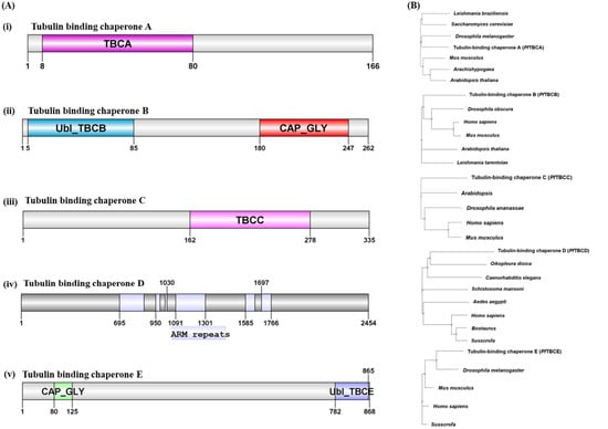
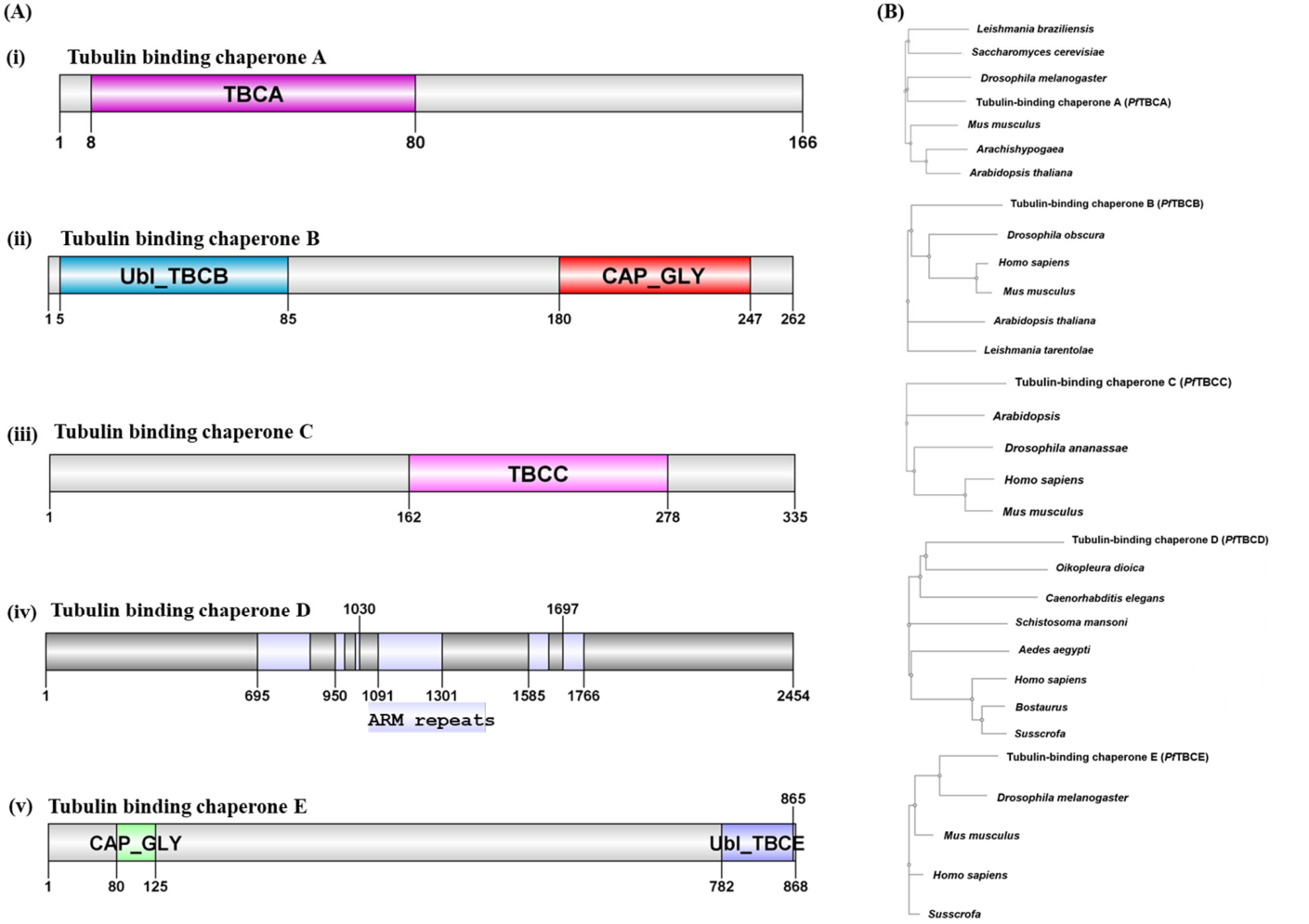
In the malaria parasite Plasmodium falciparum, the folding and regulation of tubulin is of particular interest, as microtubules drive essential processes including schizogony, merozoite morphogenesis, and gametocyte development. Parasite-specific adaptations in tubulin cofactors may represent unexplored vulnerabilities for antimalarial intervention, especially because the parasite’s cytoskeletal machinery diverges significantly from that of humans. However, identification and comprehensive characterization of TBCs in parasites remain totally unexplored, and understanding their structural and functional divergence may contribute to fill the key knowledge gaps and uncover novel therapeutic targets. Uniprot [55] was used to search the P. falciparum genome for the presence of tubulin specific chaperones genes in P. falciparum (PfTBCs) and three genes viz. PF3D7_0109400 (Uniprot ID: Q8I250_PLAF7), PF3D7_0906910 (Uniprot Id: Q8I381_PLAF7) and PF3D7_1015700 (Uniprot Id: C6S3C6_PLAF7) for tubulin-specific chaperone A, tubulin-specific chaperone, and tubulin binding cofactor c respectively were identified. The Plasmodb database [41] was then searched using their Plasmodb IDs (PF3D7_0109400; PF3D7_0906910 and PF3D7_1015700), and putative proteins were identified. Domain analysis using the CDD database [42] demonstrated the presence of a domain characteristic of TBCA, TBCB, and TBCC families in PF3D7_0109400, PF3D7_0906910 and PF3D7_1015700 respectively (Figure 1A). Interestingly, our search in Uniprot provided a hit named PI3K/PI4K catalytic domain containing protein (Uniprot Id: Q8IKD7_PLAF7; Plasmodb Id: PF3D7_1470100), and sequence analysis using InterPro database [43] demonstrated centrally located beta tubulin cofactor D domain in this protein (Figure 1A). To look for the presence of TBCE in Pf, an organism-specific Blastp search [44] was performed using a human TBCE sequence that provided the most reliable homolog in Pf. Detailed domain organization analysis using CDD clearly demonstrated the presence of the C terminal domain characteristic of the TBCE protein family (Figure 1A). The presence of PfTBCs homologs in other species was next explored. Organism-specific Blastp search using PfTBCs sequences provided their most reliable homologs in Homo sapiens, Saccharomyces cerevesisia, Drosophila melanogaster, Mus musculus, Arabidopsis thaliana, etc. All TBC sequences were subjected to multiple sequence alignment, and the resulting alignment was used to construct a phylogenetic tree to infer evolutionary and sequence-level relationships among the TBCs (Figure 1B).
Figure 1.
Identification, domain architecture, and evolutionary relationships of tubulin-binding chaperones (PfTBCs) in Plasmodium falciparum. (A) Schematic representation of the conserved domain organization of the five putative P. falciparum tubulin-binding chaperones (PfTBCs). (i) PfTBCA (PF3D7_0109400) contains the characteristic TBCA domain as depicted. (ii) PfTBCB (PF3D7_0906910) possesses an N-terminal Ubl_TBCB (ubiquitin-like) domain and a C-terminal CAP_GLY (cytoskeleton-associated protein glycine-rich) domain. (iii) PfTBCC (PF3D7_1015700) contains the conserved TBCC domain towards C-terminal. (iv) PfTBCD (PF3D7_1470100) is a large multi-domain protein containing the characteristic Armadillo (ARM) repeat region associated with tubulin-binding cofactor D. (v) PfTBCE (PF3D7_0708000) comprises an N-terminal CAP_GLY domain and a C-terminal Ubl_TBCE (ubiquitin-like) domain. Conserved domains were identified using the Conserved Domain Database (CDD) and InterProScan, and their relative positions within each protein are indicated. (B) Phylogenetic analysis of the identified PfTBC proteins and their homologs from other species including Homo sapiens, Mus musculus, Arabidopsis thaliana, Drosophila melanogaster, Saccharomyces cerevisiae, and Leishmania braziliensis. Multiple sequence alignment was performed using Clustal Omega, and the phylogenetic tree was constructed to analyze evolutionary relationships. Each PfTBC clustered with its respective orthologs while exhibiting clear evolutionary divergence from mammalian homologs, supporting the conservation of the tubulin-folding machinery across eukaryotes alongside parasite-specific adaptations.
3.2. Expression Analysis of TBCs in Asexual and Sexual Stages
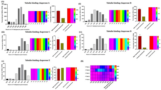
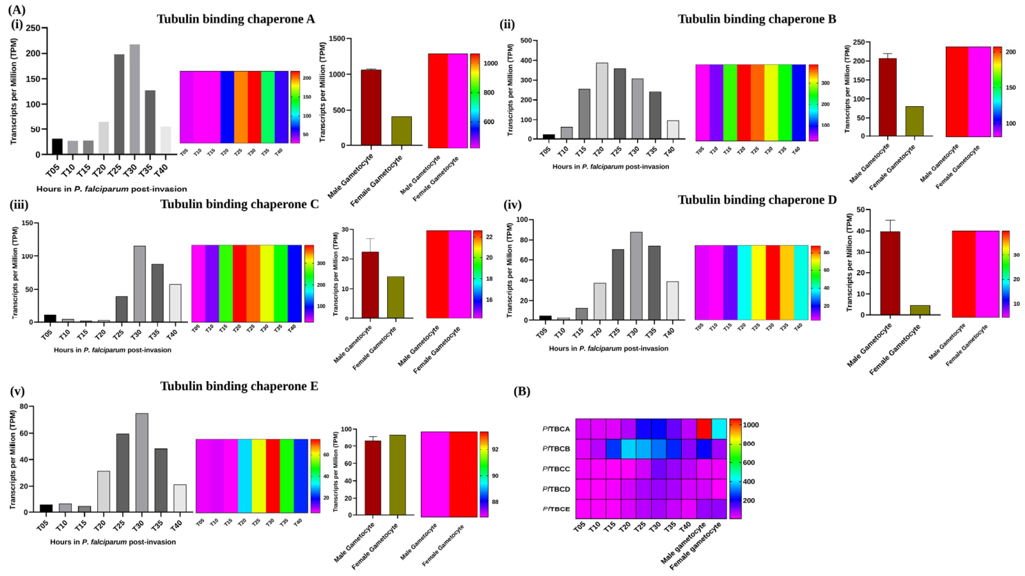
To look into the mechanistic insights of the PfTBC families in the malaria parasite, we next explored their expression analysis by digging the transcriptomics datasets from Plasmodb [41] during the asexual and sexual stages. Interestingly, we observed up-regulation in all PfTBCs’ expression during the intraerythrocytic development cycle of the malaria parasite, with notable expression peaks at the trophozoite stage, suggesting its possible role in development-related processes (Figure 2A). Moreover, PfTBCs show a prominent increase in expression, particularly during gametocytogenesis, with male gametocytes showing significantly higher transcripts compared with females (Figure 2B). This stage-specific variation indicates their potential role in sexual differentiation processes. Overall, these data underscore the gene’s essential role in parasite development and adaptation.
Figure 2.
Stage-specific expression profiling of putative tubulin-binding cofactors (PfTBCs) during the asexual intraerythrocytic developmental cycle and sexual stages of Plasmodium falciparum. (A) Transcriptomic expression profiles of the five identified PfTBCs across the parasite life cycle. Panels (i–v) represent PfTBCA (PF3D7_0109400), PfTBCB (PF3D7_0906910), PfTBCC (PF3D7_1015700), PfTBCD (PF3D7_1470100), and PfTBCE (PF3D7_0708000), respectively. For each protein, the left panel shows transcript abundance during the intra-erythrocytic developmental cycle (IDC) at different hours post-invasion (HPI), depicting dynamic stage-specific expression. The middle panels represent heatmaps showing the transcript expression patterns of each protein during the asexual blood stages at different hours post-invasion (HPI). The right panels represent transcript expression across sexual developmental stages viz. activated male and female gametocytes, highlighting differential regulation during gametocytogenesis along with their heat maps. Collectively, PfTBCs exhibit increased expression during the trophozoite and schizont stages of asexual development, with pronounced enrichment during male gametocyte differentiation, suggesting an important role in parasite proliferation, microtubule biogenesis, and transmission-stage development. (B) Heatmap summarizing the transcript expression patterns of all five PfTBC genes across sexual and asexual developmental stages of P. falciparum. The heatmap demonstrates coordinated up-regulation of PfTBCs during late gametocyte maturation, particularly in activated male gametocytes, consistent with the increased requirement for tubulin folding and microtubule assembly during rapid axoneme formation and gamete development.
3.3. Essentiality Profiling of PfTBCs and Their Interaction Networks in the Malaria Parasite
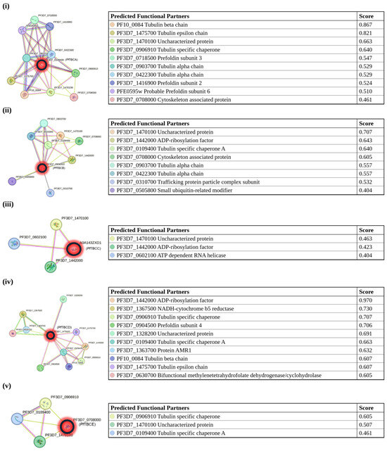
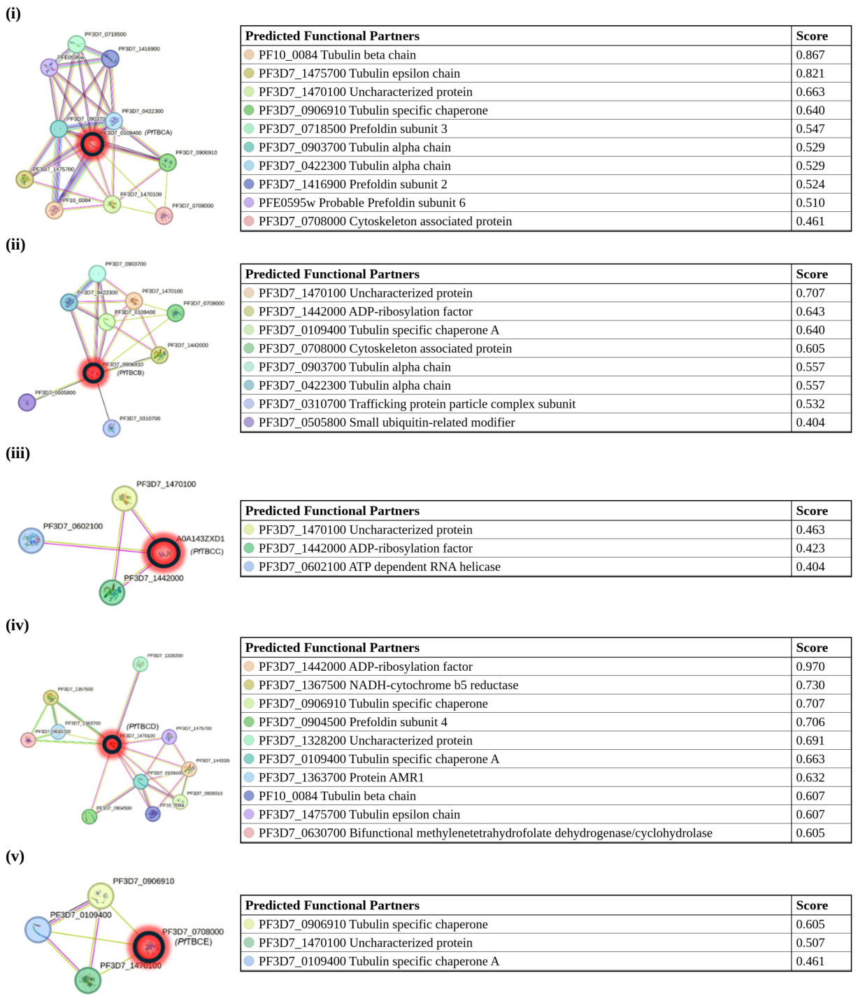
We next explored essentiality profiling of PfTBCs using the PhenoPlasm database [48]. Interestingly, all PfTBCs were annotated as essential for parasite survival. These annotations derive from large-scale mutagenesis screens rather than from targeted disruption of the individual genes, so they are predictions of essentiality and identify the PfTBCs as candidates for targeted functional study. Further we explored the interaction network of PfTBCs using the STRING protein–protein interaction database [49] and found the tubulin alpha chain and beta chain as major hits for PfTBCs (Figure 3i–v). These associations suggest that they may be involved in assisting the assembly and disassembly of tubulin heterodimers. Interestingly, we identified Plasmodium falciparum prefoldin subunits (PfPFDs) as interacting partners of PfTBCs, consistent with the established model that PfPFDs bind newly synthesized PfTBCs and facilitate their transfer to the chaperonin TRiC complex for proper folding (Figure 3i–v).
Figure 3.
Protein–protein interaction networks of the identified PfTBCs. Predicted protein–protein interaction networks generated using the STRING database for the five putative tubulin-binding chaperones: (i) PfTBCA (PF3D7_0109400), (ii) PfTBCB (PF3D7_0906910), (iii) PfTBCC (PF3D7_1015700), (iv) PfTBCD (PF3D7_1470100), and (v) PfTBCE (PF3D7_0708000). The query protein (PfTBCs highlighted by the red circle) is shown at the center of each interaction network. Colored nodes represent predicted interacting proteins while the accompanying tables list the interacting partners together with their STRING confidence scores. PfTBCA, PfTBCB, and PfTBCD exhibit strong predicted interactions with α- and β-tubulin isoforms, consistent with their conserved roles in tubulin heterodimer biogenesis. Notably, several prefoldin (PFD) subunits, including prefoldin subunits 2, 3, 4, 5, and 6, were identified as interacting partners, supporting the existence of a conserved tubulin-folding pathway in P. falciparum.
3.4. In Silico Interaction Interface Analysis of PfTBC-B with Pf α-Tubulin and PfTBC-D with Pf β-Tubulin
Model confidence was assessed before docking. The Pf α- and β-tubulin models were of high confidence (pTM 0.92 and 0.93; mean pLDDT 88.8 and 90.4; mean PAE 4.9 and 4.5 Å), except for the flexible C-terminal tails (pLDDT around 44), as expected for intrinsically disordered tails. The PfTBC-D model (2454 residues) was of low overall confidence (pTM 0.53; mean pLDDT 57.0, with 42% of residues below 50; mean PAE 25.7 Å), and its interface residues had a mean pLDDT of 45.9 (Supplementary Table S1, Supplementary Figure S1). The PfTBC-D/Pf β-tubulin interface, and its residue-level interactions in particular, therefore rests on low-confidence regions of the model and should be regarded as tentative, and the same consideration applies to the structural comparisons in Section 3.6. The docked complexes are accordingly presented as hypotheses to be tested experimentally rather than as mechanistic models.
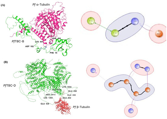
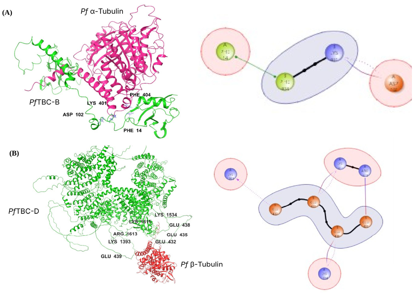
Rigid-body protein–protein docking of PfTBC-B with Pf α-tubulin was performed using PIPER [50], which generated 30 distinct binding poses. The top-ranked pose exhibited PIPER energy −748.314 (score −194.711) from the largest cluster (n = 63) (Table 1). Energy values ranged from −1035.063 to −748.314. Cluster size analysis revealed high-ranking poses derived from clusters containing 19–63 members, indicating reproducibility of the predicted binding orientation. Binding interface analysis identified 67 residue–residue interactions (≤4.0 Å), involving 34 PfTBC-B and 30 α-tubulin residues, which are listed in full in Supplementary Table S4. One prominent salt bridge anchors the PfTBC-B–Pf α-tubulin complex: Asp102–Lys401 (2.2 Å distance, and complementarity 0.51) (Table 2; Figure 4). One pi-stacking interaction provides enhanced aromatic recognition: Phe14–Phe404 (2.7 Å, complementarity 0.87, burial at Phe404: 99.6%) (Table 2; Figure 4). Extensive hydrophobic networks are organized into three regions: N-terminal aromatic cluster (Phe14, Lys17, Trp19 of PfTBC-B engaging Phe404, Trp407, His406 of Pf α-tubulin; mean burial 59.7% PfTBC-B, 87.2% Pf α-tubulin), middle hydrophobic region (His40, Thr39, Tyr38, Tyr99 of PfTBC-B contacting Trp407, His406, Val181, Arg402 of Pf α-tubulin; mean complementarity 0.74, burial 82.7%), and C-terminal region (Ser245, Asn244, Lys187 of PfTBC-B engaging Arg339 of Pf α-tubulin). Surface complementarity analysis revealed mean score 0.69 ± 0.18 with 66.7% of interactions > 0.80. Mean buried SASA: PfTBC-B 47.8%, Pf α-tubulin 61.2%.
Table 1.
PfTBC-B/Pf α-tubulin top 10 docking poses.
Table 2.
Specific interactions (salt bridges, hydrogen bonds, aromatic stacking and steric clashes) at PfTBC-B/Pf α-tubulin interface buried SASA is the percentage of the residue’s solvent accessible surface area that becomes buried on complex formation, and surface complementarity describes how well the exposed molecular surfaces of the two interacting proteins fit together at the interface; both are reported as exported by the Schrödinger interface analysis. Values for every interacting residue pair are given in Supplementary Tables S4 and S5.
Figure 4.
Predicted interaction analysis of PfTBC-B and PfTBC-D with Pf α-tubulin and Pf β-tubulin respectively. (A) Left panel: Structural model of the PfTBC-B–Pf α-tubulin complex. Key interface residues involved in the interaction are depicted. PfTBC-B is shown in green, while Pf α-tubulin is shown in magenta. Right panel: The residue interaction network illustrates the connectivity among interface residues, highlighting central interaction hubs and communication clusters that may contribute to the stability of PfTBC-B–Pf α-tubulin complex. (B) Left panel: Structural model of PfTBC-D–Pf β-tubulin complex. PfTBC-D is shown in green, and Pf β-tubulin is shown in red. Interacting residues at the binding interface are labeled. Right panel: Residue–residue interaction landscape at the binding interface, identifying highly connected interaction hubs and residue communities predicted to facilitate stable association between PfTBC-D and Pf β-tubulin. In the interaction networks, each node is an interface residue labeled with its chain (A, PfTBC; B, tubulin), residue name and number, and node colours denote residue type (green, aromatic; blue, basic; orange, acidic). Black lines join residues adjacent in sequence, the green line marks π-stacking, solid purple lines mark salt bridges and dotted purple arrows mark hydrogen bonds; pink circles enclose PfTBC residues and the grey shaded areas enclose tubulin residues.
For PfTBC-D–Pf β-tubulin docking PIPER generated 30 binding poses. The top-ranked pose exhibited PIPER energy −753.113 (score 11.624) from the largest cluster (n = 72) (Table 3). Energy values ranged from −1001.708 to −709.439. Cluster sizes ranged from 72 to 5 members. The cluster size of 72 indicates that this orientation was sampled repeatedly. Interface analysis identified 63 residue–residue interactions (≤4.0 Å), involving 28 PfTBC-D and 30 β-tubulin residues, which are listed in full in Supplementary Table S5. Three salt bridges anchor the PfTBC-D–Pf β-tubulin complex: Arg1613–Glu439 (1.9 Å, complementarity 0.79, burial 86.4% PfTBC-D/84.8% β-tubulin), Lys1534–Glu438 (2.5 Å, complementarity 0.86, burial 39.0%/53.2%) and Lys1611–Glu435 (2.2 Å, a salt bridge accompanied by a hydrogen bond, complementarity 0.80, burial 62.3%/87.4%) (Table 4; Figure 4). A further hydrogen bond, Lys1393–Glu432 (2.5 Å, complementarity 0.56, burial 24.2%/86.9%), provides supplementary stabilization (Table 4; Figure 4). One van der Waals clash (Gln1618–Tyr425, 1.7 Å, complementarity 0.82, burial 39.5%/93.3%) reflects steric overlap in the rigid-body model. Three functional regions stabilize the interface: acidic anchor cluster (Glu432–Glu444, mean burial 51.7% PfTBC-D, 77.6% Pf β-tubulin, 18 interactions), aromatic hydrophobic secondary region (Phe436, Gln424, Tyr425, Phe260, mean complementarity 0.83, burial 80.4%/72.5%, 20 interactions), and tertiary contact cluster (Pro346, Ile345, Trp344, 8 interactions) (Table 4; Figure 4). Surface complementarity analysis revealed mean score 0.71 ± 0.27 with 69.4% of interactions > 0.80. Mean buried SASA: PfTBC-D 51.4%, Pf β-tubulin 54.8%.
Table 3.
PfTBC-D/Pf β-tubulin top 10 docking poses.
Table 4.
Specific interactions (salt bridges, hydrogen bonds, aromatic stacking and steric clashes) at PfTBC-D/Pf β-tubulin interface buried SASA is the percentage of the residue’s solvent accessible surface area that becomes buried on complex formation, and surface complementarity describes how well the exposed molecular surfaces of the two interacting proteins fit together at the interface; both are reported as exported by the Schrödinger interface analysis. Values for every interacting residue pair are given in Supplementary Tables S4 and S5.
3.5. Molecular Dynamics Simulations of the PfTBC-B/Pf α-Tubulin and PfTBC-D/Pf β-Tubulin Complexes
Both complexes were simulated for 50 ns, saving 1002 frames at 50 ps intervals, with frame 0 taken as the first frame of the production trajectory. For the PfTBC-B–α-tubulin system, the temperature remained at 298.7 ± 0.3 K, the pressure fluctuated around 0.2 bar (standard deviation 31 bar), the box volume changed by about 0.01% over 50 ns and the total energy drifted by about 0.05%. For the PfTBC-D–β-tubulin system the temperature remained at 298.7 ± 0.2 K (excluding the first frame), the pressure fluctuated around 0.8 bar (standard deviation 21 bar), the box volume changed by about 0.002% and the total energy drifted by about 0.04%. Both trajectories were therefore numerically stable. The corresponding energy, pressure, temperature and volume traces are shown in Supplementary Figures S2 and S3.
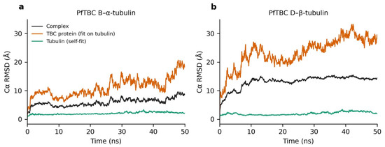
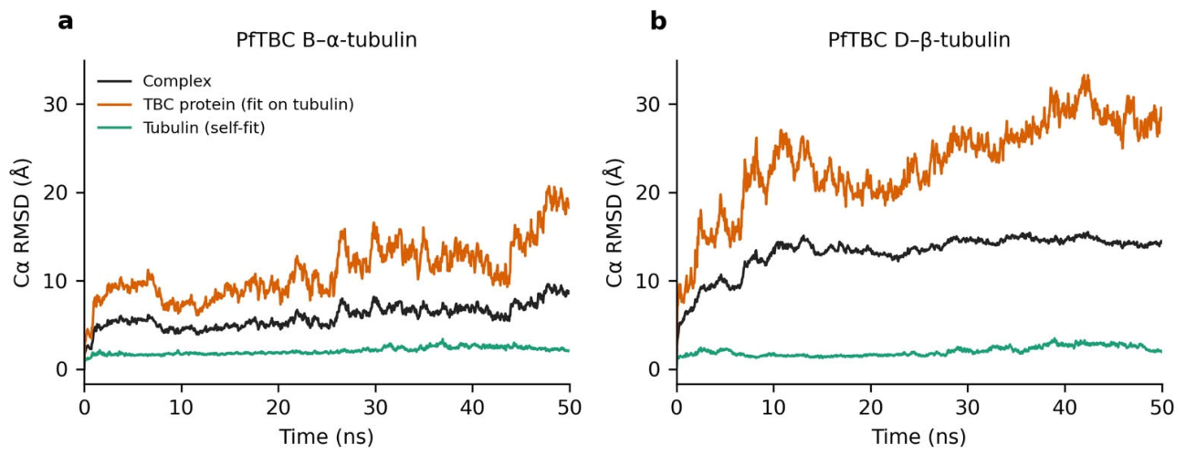
For PfTBC-B–α-tubulin, the Cα RMSD of the complex remained between 4.4 and 5.7 Å during the first 25 ns (window means 4.8 and 5.1 Å), rose to 7.2–7.6 Å at about 30 ns and reached 8.6 Å at 50 ns without a clear plateau (Figure 5a). α-Tubulin was structurally stable (self-fitted RMSD 1.6–2.2 Å), whereas PfTBC-B deviated more strongly (RMSD after superposition on tubulin 8.0 Å over the first 12.5 ns, rising to 14.3 Å over the last 12.5 ns; self-fitted 7.1 to 9.6 Å). The increase therefore reflects mainly reorientation and flexibility of PfTBC-B relative to a rigid tubulin rather than unfolding of tubulin, in agreement with the RMSF profiles: PfTBC-B had a mean Cα RMSF of 5.2 Å (54% of residues above 4 Å) compared with 2.1 Å for α-tubulin, whose flexibility was confined largely to the C-terminal residues 446–453.
Figure 5.
Cα RMSD over the 50 ns simulations of (a) PfTBC-B–α-tubulin and (b) PfTBC-D–β-tubulin. Each panel shows the whole complex, the TBC protein after superposition on the tubulin Cα atoms, and tubulin fitted on itself.
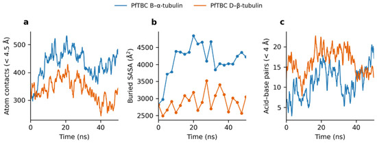
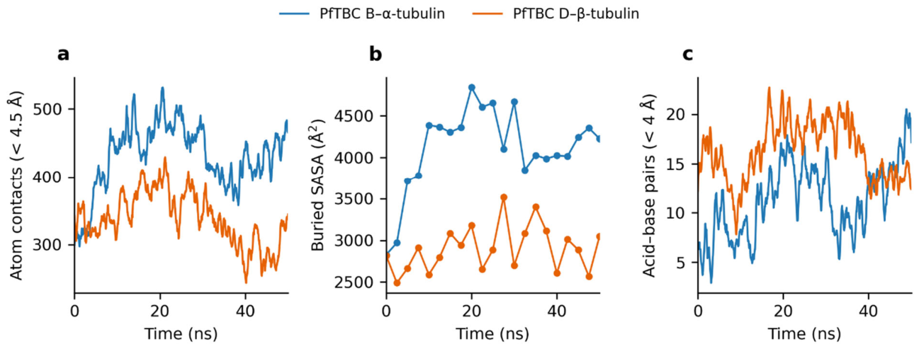
The two proteins stayed in contact throughout the simulation (closest inter-chain heavy atom distance 2.6–2.9 Å, center-of-mass separation 19–25 Å), and the interface expanded over time (Figure 6; Table 5). Inter-chain atom contacts rose from 289 at frame 0 to 390–474 (window means), the interface grew from 31 PfTBC-B and 26 α-tubulin residues to 45 and 39 residues at 50 ns, and the buried surface area increased from 2830 to 4220 Å2 (maximum about 4840 Å2 at 20 ns). N/O pairs increased from 9 to about 25 and acid–base pairs from 3 to 17. The contacts present in more than 90% of frames (Table 6) centered on PfTBC-B residues 38–47 (His40, Thr41, Thr43, Leu44, Asn47) and Ile98/Tyr99/Ile101, which packed against α-tubulin Trp407, Phe404, His406, Arg402, Lys401 and Val181, together with a Lys226–Glu450 salt bridge (92% of frames). Only 26 of the 51 residue-pair contacts present at frame 0 (51%) remained at 50 ns, so the interface was reorganized rather than lost.
Figure 6.
Evolution of the protein–protein interface in the PfTBC-B–α-tubulin and PfTBC-D–β-tubulin simulations: (a) inter-chain heavy-atom contacts within 4.5 Å, (b) buried solvent accessible surface area, and (c) acid–base pairs within 4 Å. Curves in (a,c) are 0.5 ns running averages; points in (b) were sampled every 2.5 ns.
Table 5.
Interface and deviation metrics at the start of the production run (0 ns) and averaged over the last half of the simulation (25–50 ns). ** N/O pairs are inter-chain polar contacts.
Table 6.
Residue pair contacts (heavy atoms within 4.5 Å) present in at least 92% of frames. Residue numbers follow the Desmond input structures.
For PfTBC-D–β-tubulin, the complex Cα RMSD rose rapidly to 9.9 Å within 5 ns and to 13–14 Å by 10–25 ns, then remained stationary (window means 13.4, 14.4 and 14.5 Å from 12.5 ns onwards; Figure 5b). β-Tubulin remained stable (1.5–2.7 Å). The large RMSD arises almost entirely from PfTBC-D, whose self-fitted RMSD was 12.9–17.8 Å and whose RMSD after superposition on tubulin reached 28–29 Å. PfTBC-D is a large, elongated protein (about 100 Å across), and these values indicate a lever-like motion about a localized binding site together with substantial internal flexibility. The RMSF analysis was consistent with this: PfTBC-D had a mean Cα RMSF of 5.1 Å (48% of residues above 4 Å and 18% above 8 Å) compared with 3.2 Å for β-tubulin, in which only the C-terminal residues 437–445 exceeded 6.5 Å.
The chains remained bound for the full 50 ns (closest heavy atom distance 2.6–2.9 Å, center-of-mass separation 49–57 Å, which reflects the size of PfTBC-D). In contrast to the PfTBC-B complex, the interface stayed about the same size (Figure 6; Table 5): 345 contacts at frame 0 and 348 at 50 ns (window means 293–374), buried area 2820 Å2 at the start and 3050 Å2 at the end (range 2490–3520 Å2), N/O pairs 20 at the start and 25 at the end, and acid–base pairs 11 at the start and 14 at the end. A persistent core was formed by the PfTBC-D segment 1611–1619 (Lys1611, Arg1613, Asn1614, Asn1616, Val1617, Gln1618, Asn1619) together with Lys1534, which contacted β-tubulin Glu432, Glu433, Glu435, Glu438, Glu439, Phe436, Trp344 and Gln424 in 90–100% of frames (Table 6). On the tubulin side, 18 of the 24 interface residues present at frame 0 were still at the interface at 50 ns, but on the PfTBC-D side only 11 of 28 and 15 of the 59 starting residue pair contacts (25%) were retained.
Both complexes therefore stayed associated for 50 ns, with buried interfaces of about 3000–4200 Å2 stabilized by pairs between basic residues of the TBC protein and acidic residues in the C-terminal region of tubulin (Glu450 in α-tubulin; Glu432–Glu439 in β-tubulin). The dynamics differed: the PfTBC-B–α-tubulin interface grew progressively while PfTBC-B reoriented, whereas the PfTBC-D–β-tubulin interface kept a constant size around a persistent contact segment while the body of PfTBC-D moved widely. In both systems the large RMSD values, and the retention of only 25–51% of the starting residue contacts, show that the binding region was maintained but the exact docked orientation was not. These results come from a single 50 ns trajectory per complex; replicate simulations and MM-GBSA binding free energies would strengthen the interpretation.
3.6. Comparison of PfTBCs with Human Counterparts
Comparative analysis of the five Plasmodium falciparum TBCs (PfTBCA–PfTBCE) with their corresponding human orthologs was conducted with a perspective of studying their candidature for antimalarial drug targets. Pairwise sequence alignment revealed that PfTBCs exhibit considerable divergence at the primary sequence level, with only 25%, 24.7%, 17.3%, 10.8% and 7.8% sequence identity of PfTBC-A, PfTBC-B, PfTBC-C, PfTBC-D, PfTBC-E respectively with their corresponding human counterparts (Table 7). These data show that PfTBCs represent attractive candidates for antimalarial drug discovery owing to their essential role in microtubule biogenesis and their sequence divergence from the corresponding human proteins that provide opportunities for the rational design of selective inhibitors that can target parasite tubulin-folding machinery while minimizing cross-reactivity with the human homologs.
Table 7.
Sequence identity of Plasmodium falciparum tubulin-specific chaperones (PfTBCs) with their human counterparts.
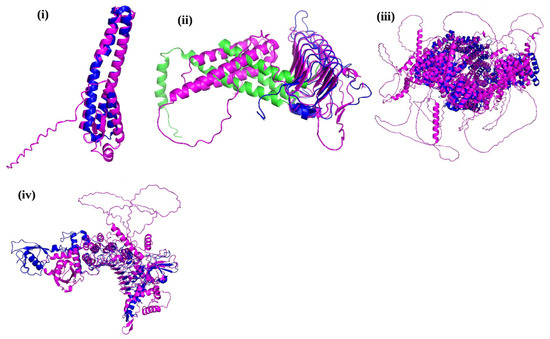
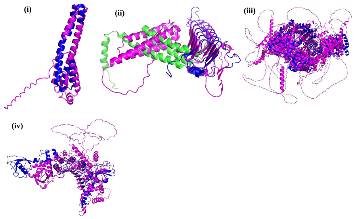
We next performed structural superposition of the PfTBCs with their respective human homologs that revealed conservation of the overall three-dimensional architecture despite pronounced sequence divergence (Figure 7). The superimposed structures depicted significant overlap of the central structural cores, suggesting preservation of the characteristic fold across the orthologous proteins. In contrast, conformational differences were predominantly localized to surface-exposed loops, terminal extensions, and flexible peripheral regions. Although structural superposition demonstrated conservation of the overall fold, PfTBC-C, PfTBC-E exhibited relatively high RMSD values (~15 Å, ~10 Å respectively), indicating pronounced conformational divergence in domain orientations, loop regions and terminal extensions, and such parasite-specific structural differences may contribute to designing selective inhibitors that can preferentially target the Plasmodium proteins while minimizing interactions with their human counterparts. Overall, these findings suggest that the structural framework required for tubulin-folding activity is conserved, whereas parasite-specific structural variations may provide opportunities for selective therapeutic targeting.
Figure 7.
Structural superposition of PfTBCs with their human counterparts. Predicted structures of PfTBC-A, PfTBC-C, PfTBC-D and PfTBC-E ((i–iv) respectively; magenta) were superimposed onto their corresponding human homologs (blue) to assess structural conservation. Green color in (ii) represents human TBC-C overlapping the N terminal region of PfTBC-C whereas magenta represents human TBC-C counterpart overlapping the C terminal region.
3.7. PfTBC-A in Transcriptomic and Proteomic Datasets Associated with Artemisinin Resistance
Several observations from recent omics studies identify expression of TBCs including PfTBCA in transcriptomic and proteomic datasets associated with parasite quiescence and artemisinin-related studies [47,56,57]. TBCA has also been reported in the translatome of quiescent stage V gametocytes [47], indicating active translation during a metabolically restrained developmental stage. This gene has also appeared in transcriptomic datasets associated with investigations of artemisinin resistance [56,57], although it has not yet undergone functional characterization. These observations identify PfTBC-A as a candidate deserving further investigation in the biology of artemisinin resistant parasites.
4. Discussion
Microtubule biogenesis involves several interconnected steps that regulate diverse processes, including mitotic spindle formation, chromosome segregation, host cell invasion, merozoite assembly, and gametogenesis, thereby facilitating survival and transmission of Plasmodium falciparum [13,16,21,22]. The precise folding of α- and β-tubulin into assembly-competent heterodimers is mediated by a conserved tubulin-folding machinery involving the chaperonin TRiC/CCT complex and tubulin-binding chaperones [3,4,5,6,7,8,9,10,11,12]. Although this pathway has been extensively characterized in higher eukaryotes, its composition and functional organization in malaria parasites have remained largely unexplored [4,5,10,11,12]. Here, we attempted to identify all five TBCs (TBCA-E) in Plasmodium falciparum and provide the first comprehensive in silico characterization, expression profiling, and their interaction analysis with their partner proteins, establishing a foundation for understanding their contribution to parasite biology.
Our bioinformatic analysis identified putative orthologs of all five canonical tubulin-binding cofactors in P. falciparum. Conserved domain analysis confirmed the presence of characteristic TBCA, TBCB, TBCC, TBCD, and TBCE domains, suggesting that the parasite harbors the molecular machinery required for tubulin biogenesis. Comparative evolutionary analysis revealed that PfTBCs cluster with their respective orthologs from diverse eukaryotes while remaining evolutionarily distinct. This conservation is consistent with the essential role of tubulin-folding cofactors across eukaryotic organisms [5,34,35,36]. At the same time, the divergence observed suggest specific adaptations that may have evolved to assist the unusual cell division mechanisms of apicomplexan parasites [14,15]. Unlike mammalian cells, P. falciparum undergoes schizogony, a specialized form of endomitosis involving multiple rounds of asynchronous nuclear division prior to cytokinesis [13,16]. Moreover, sexual differentiation involves extensive cytoskeletal remodeling, particularly during male gametogenesis, where rapid microtubule polymerization facilitates the formation of eight flagellated microgametes within minutes [21,22,23]. These unique processes are likely to impose distinct regulatory requirements on the parasite tubulin-folding machinery that may be responsible for evolutionary divergence of PfTBCs from their mammalian counterparts. Arl2 acts together with the TBCs in other eukaryotes [8,12], but no systematic search or validation was performed here for a parasite Arl2.
Transcriptomic analysis revealed expression of PfTBCs during the asexual blood stages, with peak transcript abundance during the trophozoite stage. This developmental stage is depicted by intense metabolic activity, rapid protein synthesis, DNA replication, and preparation for schizogony [58,59], all of which require extensive cytoskeletal remodeling [13,16,21]. Up-regulated expression of PfTBCs during this stage underscores the importance of increased demand for correctly folded tubulin heterodimers necessary for mitotic spindle assembly and daughter merozoite formation. Interestingly we found pronounced up-regulation of PfTBC transcripts during gametocytogenesis, especially in male gametocytes. Male gamete formation represents one of the fastest microtubule assembly events known in eukaryotes, requiring axoneme formation and rapid nuclear division immediately followed by activation within the mosquito midgut [21,22,23]. The up-regulated expression of PfTBCs during this stage strongly suggests that these proteins contribute to the efficient generation of assembly-competent tubulin required for axoneme biogenesis. Functional significance of the PfTBC family was further supported by essentiality profiling using PhenoPlasm, which classified all identified PfTBC genes as essential for parasite survival on the basis of large-scale mutagenesis data. These annotations are predictions rather than experimental essentiality for these genes and require confirmation by targeted gene disruption; if confirmed, inhibition of PfTBC function would be expected to disrupt the tubulin-folding pathway and parasite viability, making these proteins candidate antimalarial drug targets. Importantly, unlike tubulin, which is highly conserved between parasites and humans [60] and therefore poses challenges for selective drug targeting, TBCs exhibit greater sequence divergence while maintaining conserved biological function. Such divergence may provide opportunities for the development of parasite-selective inhibitors with reduced host toxicity. Consequently, PfTBCs may represent attractive multi-stage targeting candidates owing to their role during intra-erythrocytic stages as well as gametocyte developmental stages. This suggests that PfTBCs can be exploited not only for inhibiting asexual parasite proliferation but also for disrupting malaria transmission by impairing gamete development. Moreover, the recurrent detection of PfTBC-A across independent transcriptomic, proteomic, and translatomic studies [47,56,57] suggests that this tubulin-specific chaperone may have a broader role in parasite adaptation. Although no direct functional evidence currently links PfTBC-A to artemisinin resistance, its consistent expression in quiescent parasites and stage V gametocytes [47,56,57] indicates that it remains an active component of the cellular machinery during metabolically restrained states. This indicates that PfTBC-A may contribute to parasite survival, recovery from dormancy, and the emergence or maintenance of artemisinin resistance, suggesting this protein as a potential therapeutic target for overcoming parasite persistence and improving antimalarial efficacy. If tubulin-specific chaperones prove to contribute to parasite survival following artemisinin exposure, the implications would extend beyond understanding resistance mechanisms. Components of the tubulin-folding machinery could represent attractive therapeutic targets capable of sensitizing resistant parasites to existing antimalarial drugs. Pharmacological disruption of tubulin-specific proteostasis, particularly when combined with artemisinin derivatives, might impair recovery from dormancy and reduce parasite survival without directly targeting highly conserved tubulin polymers themselves. Such strategies may offer opportunities to overcome resistance while minimizing selective pressure on existing drug targets.
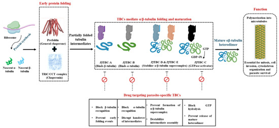
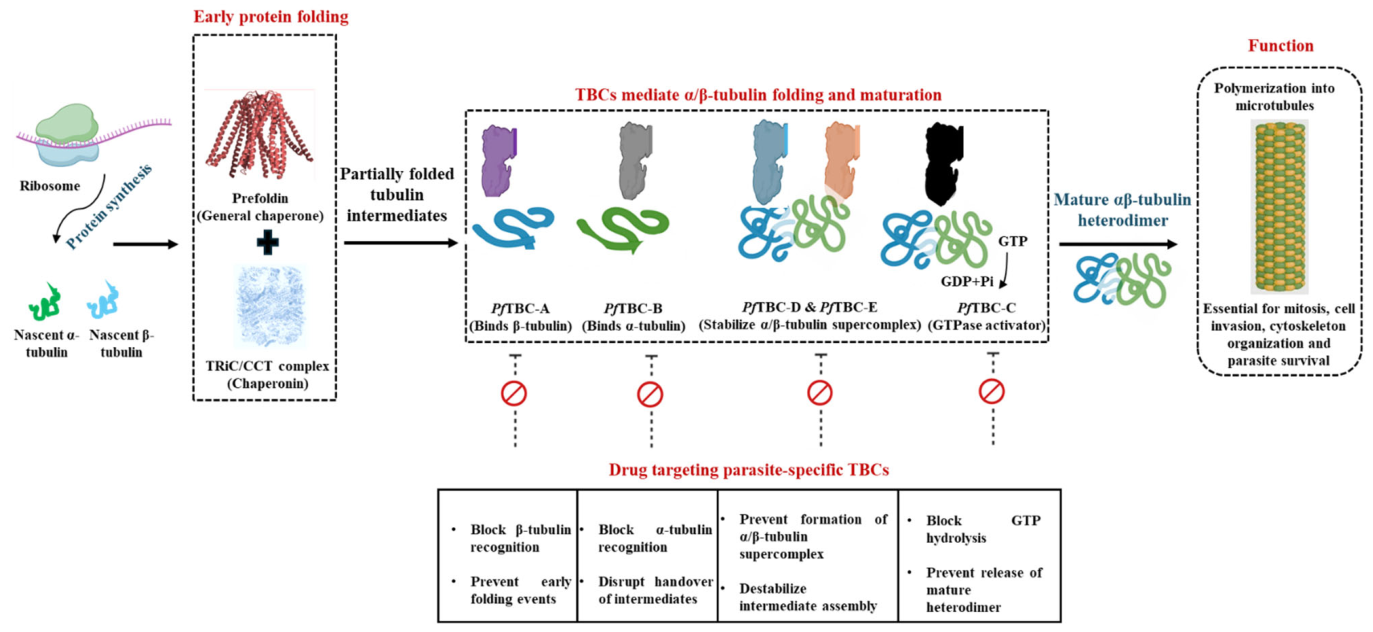
Collectively, these findings significantly expand our understanding of microtubule biogenesis in P. falciparum. While microtubules have long been recognized as attractive antimalarial targets, most therapeutic efforts have focused on directly disrupting tubulin polymerization [60,61,62]. Targeting the upstream tubulin-folding machinery represents an alternative strategy that may compromise microtubule formation before functional polymers are assembled. Figure 8 shows proposed model depicting the mechanism of α/β-tubulin biogenesis pathway. The model highlights the role of PfTBCs and their potential as parasite-specific drug targets in Plasmodium falciparum. Given the essentiality of PfTBCs, their developmental regulation, and their evolutionary divergence from human homologs, these proteins are candidates for experimental evaluation as therapeutic targets. Finally, we emphasize that this study is entirely computational, therefore future studies should focus on biochemical characterization of individual PfTBCs, determination of their three-dimensional structures, identification of interacting partners through experimental proteomics, and functional validation using conditional genetic approaches. Such investigations will provide mechanistic insight into parasite-specific tubulin biogenesis and may facilitate the development of novel antimalarial agents targeting this previously unexplored pathway.
Figure 8.
Proposed model illustrating the role of PfTBCs in α/β-tubulin folding and their potential as antimalarial drug targets. Newly synthesized α- and β-tubulin polypeptides are initially captured by the prefoldin (PFD) complex and subsequently transferred to the chaperonin TRiC/CCT for ATP-dependent folding. Post this, tubulin-specific chaperones (PfTBC-A–PfTBC-E) sequentially facilitate the maturation of α- and β-tubulin through the formation of intermediate complexes, ultimately promoting assembly of the mature αβ-tubulin heterodimer. The correctly folded heterodimer polymerizes into microtubules, which are required for chromosome segregation, mitosis, maintenance of cell morphology, intracellular transport, host cell invasion, and overall parasite survival. The lower panel highlights the proposed therapeutic strategy of selectively targeting parasite-specific TBCs to disrupt tubulin biogenesis. Inhibition of individual TBCs is expected to impair tubulin folding, reduce the availability of functional αβ-tubulin heterodimers, compromise microtubule assembly, and consequently inhibit parasite growth while minimizing effects on host microtubule dynamics due to the divergence of Plasmodium TBCs from their human counterparts.
Supplementary Materials
The following supporting information can be downloaded at: https://www.mdpi.com/article/10.3390/pathogens15111160/s1, Table S1: AlphaFold 3 confidence metrics for the structures used in docking; Table S2: All 30 PIPER poses for the PfTBC-B/Pf α-tubulin docking; Table S3: All 30 PIPER poses for the PfTBC-D/Pf β-tubulin docking; Table S4: All residue–residue interactions at the PfTBC-B/Pf α-tubulin interface; Table S5: All residue–residue interactions at the PfTBC-D/Pf β-tubulin interface; Figure S1: AlphaFold 3 confidence (pLDDT and PAE) for the structures used in docking; Figure S2: Simulation quality for the 50 ns PfTBC-B/Pf α-tubulin trajectory; Figure S3: Simulation quality for the 50 ns PfTBC-D/Pf β-tubulin trajectory.
Author Contributions
A.B.: performed in silico studies, data analysis, conception of idea and manuscript writing. S.G.: in silico studies, data analysis, conception of idea and manuscript writing. H.T.A.A.: performed docking studies. R.S.: figures and manuscript preparation. M.S.: data analysis and manuscript writing. S.S.: data analysis, conception of idea and manuscript writing. All authors have read and agreed to the published version of the manuscript.
Funding
The research was supported by the Technische Universität München (TUM) within the DFG funding program Open Access Publishing.
Institutional Review Board Statement
Not applicable.
Informed Consent Statement
Not applicable.
Data Availability Statement
The original contributions presented in this study are included in the article. Further inquiries can be directed to the corresponding authors.
Acknowledgments
A.B. and S.G. acknowledge Sharda University, Greater Noida, India, for providing the institutional support and research infrastructure necessary for this work.
Conflicts of Interest
The authors declare no conflicts of interest.
References
- Desai, A.; Mitchison, T.J. Microtubule polymerization dynamics. Annu. Rev. Cell Dev. Biol. 1997, 13, 83–117. [Google Scholar] [CrossRef] [Scilit]
- Akhmanova, A.; Steinmetz, M.O. Control of microtubule organization and dynamics. Nat. Rev. Mol. Cell Biol. 2015, 16, 711–726. [Google Scholar] [CrossRef]
- Vainberg, I.E.; Lewis, S.A.; Rommelaere, H.; Ampe, C.; Vandekerckhove, J.; Klein, H.L.; Cowan, N.J. Prefoldin, a chaperone that delivers unfolded proteins to cytosolic chaperonin. Cell 1998, 93, 863–873. [Google Scholar] [CrossRef] [Scilit]
- Lewis, S.A.; Tian, G.; Cowan, N.J. The α- and β-tubulin folding pathways. Trends Cell Biol. 1997, 7, 479–484. [Google Scholar] [CrossRef] [Scilit]
- Tian, G.; Lewis, S.A.; Feierbach, B.; Stearns, T.; Rommelaere, H.; Ampe, C.; Cowan, N.J. Tubulin subunits exist in an activated conformational state generated and maintained by protein cofactors. J. Cell Biol. 1997, 138, 821–832. [Google Scholar] [CrossRef] [Scilit]
- Spiess, C.; Miller, E.J.; McClellan, A.J.; Frydman, J. Identification of the TRiC/CCT substrate binding sites. Mol. Cell 2006, 24, 25–37. [Google Scholar] [CrossRef] [Scilit]
- Gestaut, D.; Roh, S.H.; Ma, B.; Pintilie, G.; Joachimiak, L.A.; Leitner, A.; Walzthoeni, T.; Aebersold, R.; Chiu, W.; Frydman, J. The chaperonin TRiC/CCT associates with tubulin and promotes folding through ATP-driven conformational changes. Cell 2019, 177, 751–765. [Google Scholar]
- Bhamidipati, A.; Lewis, S.A.; Cowan, N.J. ADP ribosylation factor-like protein 2 regulates tubulin folding. J. Cell Biol. 2000, 149, 1087–1096. [Google Scholar] [CrossRef] [Scilit]
- Tian, G.; Bhamidipati, A.; Cowan, N.J.; Lewis, S.A. Tubulin folding cofactors as GTPase-activating proteins. Proc. Natl. Acad. Sci. USA 1999, 96, 10238–10243. [Google Scholar]
- Cowan, N.J.; Lewis, S.A. Type II chaperonins, prefoldin and the tubulin-specific chaperones. Adv. Protein Chem. 2001, 59, 73–104. [Google Scholar] [CrossRef] [Scilit]
- Lopez-Fanarraga, M.; Carranza, G.; Bellido, J.; Kortazar, D.; Villegas, J.C.; Zabala, J.C. Tubulin cofactor B plays a key role in microtubule dynamics. BioEssays 2001, 23, 491–495. [Google Scholar]
- Seong, Y.; Kim, H.; Byun, K.; Park, Y.-W.; Roh, S.-H. Structural dissection of αβ-tubulin heterodimer assembly and disassembly by human tubulin-specific chaperones. Science 2025, 390, eady2708. [Google Scholar] [CrossRef] [Scilit]
- Gerald, N.; Mahajan, B.; Kumar, S. Mitosis in the human malaria parasite Plasmodium falciparum. Eukaryot. Cell 2011, 10, 474–482. [Google Scholar] [CrossRef] [Scilit]
- Morrissette, N.S.; Sibley, L.D. Cytoskeleton of apicomplexan parasites. Microbiol. Mol. Biol. Rev. 2002, 66, 21–38. [Google Scholar] [CrossRef] [Scilit]
- Bertiaux, E.; Balestra, A.; Bournonville, L.; Louvel, V.; Maco, B.; Soldati-Favre, D.; Brochet, M.; Guichard, P.; Hamel, V. Expansion microscopy provides new insights into the cytoskeleton of malaria parasites including the conservation of a conoid. PLoS Biol. 2021, 19, e3001020, Correction in PLoS Biol. 2021, 19, e3001401. [Google Scholar] [CrossRef] [Scilit]
- Francia, M.E.; Striepen, B. Cell division in apicomplexan parasites. Nat. Rev. Microbiol. 2014, 12, 125–136. [Google Scholar] [CrossRef] [Scilit]
- Gubbels, M.J.; Keroack, C.D.; Dangoudoubiyam, S.; Worliczek, H.L.; Paul, A.S.; Bauwens, C.; Elsworth, B.; Engelberg, K.; Howe, D.K.; Coppens, I.; et al. Fussing about fission: Defining variety among mainstream and exotic apicomplexan cell division modes. Front. Cell. Infect. Microbiol. 2020, 10, 269. [Google Scholar] [CrossRef] [Scilit]
- Voß, Y.; Klaus, S.; Guizetti, J.; Ganter, M. Plasmodium schizogony, a chronology of the parasite’s cell cycle in the blood stage. PLoS Pathog. 2023, 19, e1011157. [Google Scholar] [CrossRef] [Scilit]
- Liffner, B.; Cepeda Diaz, A.K.; Blauwkamp, J.; Anaguano, D.; Frolich, S.; Muralidharan, V.; Wilson, D.W.; Dvorin, J.D.; Absalon, S. Atlas of Plasmodium falciparum intraerythrocytic development using expansion microscopy. eLife 2023, 12, e88088. [Google Scholar] [CrossRef]
- Spreng, B.; Fleckenstein, H.; Kübler, P.; Di Biagio, C.; Benz, M.; Patra, P.; Schwarz, U.S.; Cyrklaff, M.; Frischknecht, F. Microtubule number and length determine cellular shape and function in Plasmodium. EMBO J. 2019, 38, e100984. [Google Scholar] [CrossRef] [Scilit]
- Sinden, R.E. Mitosis and meiosis in malaria parasites. Acta Trop. 1991, 60, 19–27. [Google Scholar]
- Zeeshan, M.; Rashpa, R.; Ferguson, D.J.P.; Abel, S.; Chahine, Z.; Brady, D.; Vaughan, S.; Moores, C.A.; Le Roch, K.G.; Brochet, M.; et al. Genome-wide functional analysis reveals key roles for kinesins in the mammalian and mosquito stages of the malaria parasite life cycle. PLoS Biol. 2022, 20, e3001704. [Google Scholar] [CrossRef] [Scilit]
- Zeeshan, M.; Rea, E.; Abel, S.; Vukušić, K.; Markus, R.; Brady, D.; Eze, A.; Rashpa, R.; Balestra, A.C.; Bottrill, A.R.; et al. Plasmodium ARK2 and EB1 drive unconventional spindle dynamics during chromosome segregation in sexual transmission stages. Nat. Commun. 2023, 14, 5652. [Google Scholar] [CrossRef] [Scilit]
- Rashpa, R.; Brochet, M. Expansion microscopy of Plasmodium gametocytes reveals the molecular architecture of a bipartite microtubule organisation centre coordinating mitosis with axoneme assembly. PLoS Pathog. 2022, 18, e1010223. [Google Scholar] [CrossRef] [Scilit]
- Zeeshan, M.; Ferguson, D.J.P.; Abel, S.; Burrrell, A.; Rea, E.; Brady, D.; Daniel, E.; Delves, M.; Vaughan, S.; Holder, A.; et al. Kinesin-8B controls basal body function and flagellum formation and is key to malaria transmission. Life Sci. Alliance 2019, 2, e201900488. [Google Scholar] [CrossRef] [Scilit]
- Fennell, B.J.; Naughton, J.A.; Dempsey, E.; Bell, A. Cellular and molecular actions of dinitroaniline and phosphorothioamidate herbicides on Plasmodium falciparum: Tubulin as a specific antimalarial target. Mol. Biochem. Parasitol. 2006, 145, 226–238. [Google Scholar] [CrossRef] [Scilit]
- Hirst, W.G.; Fachet, D.; Kuropka, B.; Weise, C.; Saliba, K.J.; Reber, S. Purification of functional Plasmodium falciparum tubulin allows for the identification of parasite-specific microtubule inhibitors. Curr. Biol. 2022, 32, 919–926. [Google Scholar] [CrossRef] [Scilit]
- Lu, K.Y.; Quan, B.; Sylvester, K.; Srivastava, T.; Fitzgerald, M.C.; Derbyshire, E.R. Plasmodium chaperonin TRiC/CCT identified as a target of the antihistamine clemastine using parallel chemoproteomic strategy. Proc. Natl. Acad. Sci. USA 2020, 117, 5810–5817. [Google Scholar] [CrossRef] [Scilit]
- Abbaali, I.; Truong, D.A.; Day, S.D.; Haro-Ramirez, N.; Morrissette, N.S. Systematic analysis of clemastine, a candidate apicomplexan parasite-selective tubulin-targeting agent. Int. J. Mol. Sci. 2022, 23, 68. [Google Scholar] [CrossRef] [Scilit]
- Fleming, J.R.; Morgan, R.E.; Fyfe, P.K.; Kelly, S.M.; Hunter, W.N. The architecture of Trypanosoma brucei tubulin-binding cofactor B and implications for function. FEBS J. 2013, 280, 3270–3280. [Google Scholar] [CrossRef] [Scilit]
- André, J.; Harrison, S.; Towers, K.; Qi, X.; Vaughan, S.; McKean, P.G.; Ginger, M.L. The tubulin cofactor C family member TBCCD1 orchestrates cytoskeletal filament formation. J. Cell Sci. 2013, 126, 5350–5356. [Google Scholar] [CrossRef] [Scilit]
- Price, H.P.; Peltan, A.; Stark, M.; Smith, D.F. The small GTPase ARL2 is required for cytokinesis in Trypanosoma brucei. Mol. Biochem. Parasitol. 2010, 173, 123–131. [Google Scholar] [CrossRef] [Scilit]
- Taheri, A.; Wang, Z.; Singal, B.; Guo, F.; Al-Bassam, J. Cryo-EM structures of the tubulin cofactors reveal the molecular basis of α/β-tubulin biogenesis. Nat. Commun. 2025, 17, 1405. [Google Scholar] [CrossRef] [Scilit]
- Steinborn, K.; Maulbetsch, C.; Priester, B.; Trautmann, S.; Pacher, T.; Geiges, B.; Küttner, F.; Lepiniec, L.; Stierhof, Y.-D.; Schwarz, H.; et al. The Arabidopsis PILZ group genes encode tubulin-folding cofactor orthologs required for cell division but not cell growth. Genes Dev. 2002, 16, 959–971. [Google Scholar] [CrossRef] [Scilit]
- Radcliffe, P.A.; Hirata, D.; Childs, D.; Vardy, L.; Toda, T. Identification of novel tubulin-folding cofactors in fission yeast. Mol. Biol. Cell 1999, 10, 2987–3001. [Google Scholar]
- Cowan, N.J.; Lewis, S.A. The role of tubulin cofactors in microtubule biology. Curr. Opin. Cell Biol. 2002, 14, 23–30. [Google Scholar]
- Janke, C. The tubulin code: Molecular components, readout mechanisms, and functions. J. Cell Biol. 2014, 206, 461–472. [Google Scholar] [CrossRef] [Scilit]
- Roll-Mecak, A. The tubulin code in microtubule dynamics and information encoding. Dev. Cell 2020, 54, 7–20. [Google Scholar] [CrossRef] [Scilit]
- Pavithra, S.R.; Kumar, R.; Tatu, U. Systems analysis of chaperone networks in the malarial parasite Plasmodium falciparum. PLoS Comput. Biol. 2007, 3, e168. [Google Scholar] [CrossRef] [Scilit]
- Shoaib, R.; Parveen, N.; Kumar, V.; Behl, A.; Garg, S.; Chaudhary, P.; Arokia Balaya Rex, D.; Saini, M.; Maurya, P.; Jain, R.; et al. Prefoldins are novel regulators of the unfolded protein response in artemisinin resistant Plasmodium falciparum malaria. J. Biol. Chem. 2024, 300, 107496. [Google Scholar] [CrossRef] [Scilit]
- Amos, B.; Aurrecoechea, C.; Barba, M.; Barreto, A.; Basenko, E.Y.; Bazant, W.; Belnap, R.; Blevins, A.S.; Böhme, U.; Brestelli, J.; et al. VEuPathDB: The eukaryotic pathogen, vector and host bioinformatics resource center. Nucleic Acids Res. 2022, 50, D898–D911. [Google Scholar] [CrossRef] [Scilit]
- Lu, S.; Wang, J.; Chitsaz, F.; Derbyshire, M.K.; Geer, R.C.; Gonzales, N.R.; Gwadz, M.; Hurwitz, D.I.; Marchler, G.H.; Song, J.S.; et al. CDD/SPARCLE: The Conserved Domain Database in 2020. Nucleic Acids Res. 2020, 48, D265–D268. [Google Scholar] [CrossRef] [Scilit]
- Jones, P.; Binns, D.; Chang, H.Y.; Fraser, M.; Li, W.; McAnulla, C.; McWilliam, H.; Maslen, J.; Mitchell, A.; Nuka, G.; et al. InterProScan 5: Genome-scale protein function classification. Bioinformatics 2014, 30, 1236–1240. [Google Scholar] [CrossRef] [Scilit]
- Altschul, S.F.; Gish, W.; Miller, W.; Myers, E.W.; Lipman, D.J. Basic Local Alignment Search Tool. J. Mol. Biol. 1990, 215, 403–410. [Google Scholar] [CrossRef] [Scilit]
- Sievers, F.; Higgins, D.G. Clustal Omega for making accurate alignments of many protein sequences. Protein Sci. 2018, 27, 135–145. [Google Scholar] [CrossRef] [Scilit]
- Toenhake, C.G.; Fraschka, S.A.-K.; Vijayabaskar, M.S.; Westhead, D.R.; van Heeringen, S.J.; Bártfai, R. Chromatin accessibility-based characterization of the gene regulatory network underlying Plasmodium falciparum blood-stage development. Cell Host Microbe 2018, 23, 557–569. [Google Scholar] [CrossRef] [Scilit]
- Lasonder, E.; Rijpma, S.R.; van Schaijk, B.C.L.; Hoeijmakers, W.A.M.; Kensche, P.R.; Gresnigt, M.S.; Italiaander, A.; Vos, M.W.; Woestenenk, R.; Bousema, T.; et al. Integrated transcriptomic and proteomic analyses of P. falciparum gametocytes: Molecular insight into sex-specific processes and translational repression. Nucleic Acids Res. 2016, 44, 6087–6101. [Google Scholar] [CrossRef] [Scilit]
- Sanderson, T.; Rayner, J.C. PhenoPlasm: A database of disruption phenotypes for malaria parasite genes. Wellcome Open Res. 2017, 2, 45. [Google Scholar] [CrossRef] [Scilit]
- Szklarczyk, D.; Kirsch, R.; Koutrouli, M.; Nastou, K.; Mehryary, F.; Hachilif, R.; Gable, A.L.; Fang, T.; Doncheva, N.T.; Pyysalo, S.; et al. The STRING database in 2023: Protein–protein association networks and functional enrichment analysis. Nucleic Acids Res. 2023, 51, D638–D646. [Google Scholar] [CrossRef] [Scilit]
- Kozakov, D.; Brenke, R.; Comeau, S.R.; Vajda, S. PIPER: An FFT-based protein docking program with pairwise potentials. Proteins 2006, 65, 392–406. [Google Scholar] [CrossRef] [Scilit]
- Schrödinger LLC. Schrödinger Release 2023-4: Maestro; Schrödinger LLC: New York, NY, USA, 2023. [Google Scholar]
- Abramson, J.; Adler, J.; Dunger, J.; Evans, R.; Green, T.; Pritzel, A.; Ronneberger, O.; Willmore, L.; Ballard, A.J.; Bambrick, J.; et al. Accurate structure prediction of biomolecular interactions with AlphaFold 3. Nature 2024, 630, 493–500. [Google Scholar] [CrossRef] [Scilit]
- PyMOL. The PyMOL Molecular Graphics System, version 2.5; Schrödinger LLC: New York, NY, USA, 2021.
- Bowers, K.J.; Chow, E.; Xu, H.; Dror, R.O.; Eastwood, M.P.; Gregersen, B.A.; Klepeis, J.L.; Kolossvary, I.; Moraes, M.A.; Sacerdoti, F.D.; et al. Scalable algorithms for molecular dynamics simulations on commodity clusters. In Proceedings of the 2006 ACM/IEEE Conference on Supercomputing (SC06), Tampa, FL, USA, 11–17 November 2006. [Google Scholar]
- UniProt Consortium. UniProt: The Universal Protein Knowledgebase in 2023. Nucleic Acids Res. 2023, 51, D523–D531. [Google Scholar] [CrossRef] [Scilit]
- Mok, S.; Ashley, E.A.; Ferreira, P.E.; Zhu, L.; Lin, Z.; Yeo, T.; Chotivanich, K.; Imwong, M.; Pukrittayakamee, S.; Dhorda, M.; et al. Population transcriptomics of human malaria parasites reveals the mechanism of artemisinin resistance. Science 2015, 347, 431–435. [Google Scholar] [CrossRef] [Scilit]
- Mok, S.; Stokes, B.H.; Gnädig, N.F.; Ross, L.S.; Yeo, T.; Amaratunga, C.; Allman, E.; Solyakov, L.; Bottrill, A.R.; Tripathi, J.; et al. Artemisinin-resistant K13 mutations rewire Plasmodium falciparum’s intra-erythrocytic metabolic program to enhance survival. Nat. Commun. 2021, 12, 530. [Google Scholar] [CrossRef] [Scilit]
- Bozdech, Z.; Llinás, M.; Pulliam, B.L.; Wong, E.D.; Zhu, J.; DeRisi, J.L. The transcriptome of the intraerythrocytic developmental cycle of Plasmodium falciparum. PLoS Biol. 2003, 1, e5. [Google Scholar] [CrossRef] [Scilit]
- Otto, T.D.; Wilinski, D.; Assefa, S.; Keane, T.M.; Sarry, L.R.; Böhme, U.; Lemieux, J.; Barrell, B.; Pain, A.; Berriman, M.; et al. New insights into the blood-stage transcriptome of Plasmodium falciparum using RNA-Seq. Mol. Microbiol. 2010, 76, 12–24. [Google Scholar] [CrossRef] [Scilit]
- Jordan, M.A.; Wilson, L. Microtubules as a target for anticancer drugs. Nat. Rev. Cancer 2004, 4, 253–265. [Google Scholar] [CrossRef] [Scilit]
- Derry, W.B.; Wilson, L.; Jordan, M.A. Substoichiometric binding of taxol suppresses microtubule dynamics. Biochemistry 1995, 34, 2203–2211. [Google Scholar] [CrossRef] [Scilit]
- Goodson, H.V.; Jonasson, E.M. Microtubules and microtubule-associated proteins. Cold Spring Harb. Perspect. Biol. 2018, 10, a022608. [Google Scholar] [CrossRef] [Scilit]

Disclaimer/Publisher’s Note: The statements, opinions and data contained in all publications are solely those of the individual author(s) and contributor(s) and not of MDPI and/or the editor(s). MDPI and/or the editor(s) disclaim responsibility for any injury to people or property resulting from any ideas, methods, instructions or products referred to in the content. |
© 2026 by the authors. Licensee MDPI, Basel, Switzerland. This article is an open access article distributed under the terms and conditions of the Creative Commons Attribution (CC BY) license.