


Abstract
Preterm birth (PTB) remains a leading cause of neonatal morbidity and mortality worldwide, with limited tools for early prediction or targeted prevention. Increasing evidence identifies the placenta as a central regulator of gestational duration, and alterations in placental gene regulatory programs have been associated with PTB. This review synthesizes recent advances in placental genomics and epigenomics, emphasizing how cell-type-specific regulatory landscapes shape PTB risk. We describe how DNA methylation, chromatin accessibility, and transcriptional heterogeneity across trophoblasts, Hofbauer cells, endothelial cells, Natural Killer (NK) cells, and mesenchymal stromal cells integrate maternal, fetal, immune, and environmental signals. We further evaluate emerging liquid biopsy approaches, focusing on circulating cell-free placental DNA (cfpDNA) methylation as a minimally invasive strategy to detect placental dysfunction. Single-cell and long-read sequencing technologies have enabled high-resolution mapping of placental regulatory architecture, revealing cell-type-specific pathways underlying inflammation, impaired trophoblast differentiation, and aberrant vascular remodeling. These studies support the investigation of placental epigenetic alterations as potential contributors to PTB-associated biology, although their causal roles and consistency across PTB subtypes remain uncertain. Circulating cfpDNA methylation profiles are being investigated as minimally invasive indicators of placental biology, but their accuracy and clinical utility for PTB prediction require further validation. Together, these findings suggest that epigenomic alterations may be associated with biological pathways implicated in PTB and may also provide measurable molecular signatures in maternal circulation.
1. Introduction
Preterm birth (PTB), defined as delivery before 37 weeks of gestation, remains a leading cause of neonatal morbidity, mortality, and long-term neurodevelopmental impairment [1,2,3]. Despite its clinical significance, current prediction strategies, based on maternal history, cervical length, and biochemical markers, have limited accuracy, and therapeutic options remain inconsistent [4]. These limitations underscore the need for improved mechanistic insight into pathways driving premature parturition.
PTB is a heterogeneous clinical outcome that includes distinct subtypes with potentially different underlying mechanisms [5]. Spontaneous PTB may follow the onset of labor with intact membranes and births following preterm prelabor rupture of membranes (PPROM), in which the membranes rupture before labor begins [6,7]. Medically indicated PTB occurs when delivery is initiated because continuation of pregnancy poses risks to the pregnant individual or fetus, including complications such as preeclampsia or fetal growth restriction (FGR) [8]. Although these categories may involve overlapping biological pathways, findings from one PTB subtype should not automatically be generalized to others [9]. Accordingly, the interpretation of placental epigenetic and molecular biomarker studies requires consideration of the PTB phenotype examined and the clinical circumstances surrounding delivery [10].
The placenta has emerged as a central regulator of gestational duration, integrating maternal, fetal, and environmental signals that shape PTB risk [11]. Disruption of placental endocrine, immune, metabolic, and vascular signaling pathways has been associated with PTB and may contribute to the processes leading to premature parturition [12,13,14]. Epigenetic regulation, including DNA methylation, histone modifications, non-coding RNA regulation, and chromatin accessibility, governs placental cell identity and function. Placental epigenetic remodeling is increasingly implicated in inflammatory activation, impaired trophoblast invasion, vascular dysfunction, and premature activation of parturition pathways [12,15].
Recent advances in single-cell and multi-omic technologies have enabled high-resolution characterization of placental cellular heterogeneity and regulatory programs implicated in PTB [4,16,17,18,19]. In parallel, circulating placental nucleic acid, including cell-free placental DNA (cfpDNA), has emerged as a minimally invasive biomarker capable of capturing placenta-specific epigenetic molecular alterations associated with PTB risk [20,21,22,23]. Together, these advances position the placental epigenome as a unifying framework for understanding PTB. In this review, we synthesize current evidence on placental epigenetic regulation, cellular heterogeneity, and emerging multi-omic and liquid biopsy approaches to better understand, predict, and ultimately prevent PTB.
2. The Placental Epigenome: An Overview
The human placenta is a transient, specialized organ that mediates nutrient exchange, immune tolerance, and endocrine signaling between mother and fetus. It possesses one of the most dynamic and specialized epigenetic landscapes of mammalian tissue. The epigenome integrates DNA methylation, histone modifications, non-coding RNA regulation, and chromatin accessibility to coordinate the rapid cellular differentiation, immune tolerance, and metabolic demands required to sustain pregnancy [24]. These epigenetic mechanisms regulate gene expression in the placenta without altering the underlying DNA sequence and serve as key modulators of trophoblast lineage commitment, nutrient transport, vascular development, and maternal–fetal communication [24,25]. The placenta is exposed simultaneously to maternal physiology, fetal signals, and environmental stressors; its epigenome functions as both a sensor and an effector of intrauterine conditions, making it a target organ for studying the biological interface for underlying pregnancy complications [26] (Figure 1).
Figure 1.
The placental epigenome. The schematic illustrates the complex landscape of the placental epigenome, highlighting its unique regulatory features and sensitivity to external and internal stimuli. (Blue) four primary epigenetic regulatory layers govern placental function: (1) DNA methylation, which mediates promoter silencing and TE regulation; (2) histone modifications, including acetylation and methylation, that dictate chromatin compaction and activation/repression states; (3) non-coding RNAs (miRNAs and lncRNAs), which regulate translation and chromatin structure; and (4) chromatin accessibility, essential for enhancer activation and transcription factor binding. (Purple) distinctive placental epigenetic signatures are characterized by global DNA hypomethylation, genomic imprinting of growth-related genes, and epigenetic plasticity that allows for gestational adaptation and immune tolerance. (Green) the placental epigenome is dynamically shaped by environmental factors (nutrition, toxins, stress), immune signaling (inflammation and infection), and genetic influences (regulatory polymorphisms and variants), which collectively modulate placental health and fetal development. Created in BioRender. Dawning, A. (2026) https://BioRender.com/mjvgby6 (accessed on 19 October 2026). Abbreviations: deoxyribonucleic acid (DNA); long non-coding RNA (lncRNA); microRNA (miRNA); ribonucleic acid (RNA).
2.1. DNA Methylation, Histone Modifications, Non-Coding RNA Regulation, and Chromatin Accessibility
Epigenetic regulation in the human placenta is mediated through DNA methylation and histone modifications, which coordinate gene expression programs required for trophoblast differentiation, placental growth, immune modulation, and maternal–fetal communication throughout gestation [27]. Unlike other tissues, the placental epigenome remains highly dynamic, reflecting rapid cellular turnover and sensitivity to intrauterine environmental cues [24,27]. DNA methylation, the most studied epigenetic mechanism of the placenta, regulates transcriptional activity, genomic imprinting, and suppression of transposable elements. Placental methylation landscapes are characterized by global CpG hypomethylation alongside locus-specific hypermethylation at genes controlling trophoblast lineage specification, immune signaling, and endocrine function [28,29]. In PTB, differential methylation has been associated with abnormal trophoblast invasion, altered inflammatory signaling, vascular remodeling, and hormonal dysregulation [29]. However, most studies remain cross-sectional and limited to delivery. Histone modifications further regulate chromatin accessibility and gene expression; activating marks such as H3K4me3 and H3K27ac are enriched at promoters and enhancers of genes involved in placental development, angiogenesis, and hormone synthesis, whereas repressive marks, including H3K27me3 and H3K9me3, restrict inappropriate lineage programs and excessive immune activation [30,31]. Illustrating this pathological relevance, mapping PTB epigenome dynamics across gestation reveals that severe preeclampsia is characterized by aberrant H3K27ac profiling, displaying distinct subtypes marked by either immature peak patterns or signs of accelerated epigenetic aging [31]. Despite their importance, histone modification landscapes in PTB remain comparatively underexplored.
Non-coding RNAs and chromatin accessibility provide additional regulatory layers that fine-tune placental gene expression and cellular identity [32]. MicroRNAs, long non-coding RNAs, and placenta-enriched clusters such as C19MC regulate trophoblast proliferation, invasion, immune tolerance, and antiviral responses, often through interactions with DNA methylation and histone-modifying complexes [32,33,34]. Altered expression of these RNA species has been associated with PTB, suggesting disruption of post-transcriptional regulation before labor onset, although their functional consequences and cell-type specificity remain incompletely defined [32,34]. Chromatin accessibility governs transcription factor binding to regulatory elements, thereby enabling rapid activation of immune, endocrine, and metabolic gene networks essential for maintaining pregnancy [35,36]. These accessibility landscapes are highly cell-type-specific within the placenta and dynamically regulated across gestation; however, their role in PTB, particularly at single-cell resolution, remains insufficiently characterized [36]. Highlighting the strict requirement for tighter spatial control over these domains, large-scale chromatin reorganization and the erosion of repressive heterochromatin compartments can lead to the aberrant de-repression of placenta-specific genes, such as pregnancy-specific beta-1 glycoproteins, which actively drive cellular senescence and epigenetic instability when misexpressed outside of normal gestational contexts [35]. Together, these interconnected mechanisms confer both adaptability and vulnerability to the placenta, underscoring the need for integrative, cell-type-resolved studies to define their roles in preterm labor and their potential as early biomarkers and therapeutic targets.
2.2. Unique Features of the Placental Epigenome
The placental epigenome is distinguished by global DNA hypomethylation, genomic imprinting, and pronounced epigenetic plasticity, features that collectively support rapid growth and environmental responsiveness. Large partially methylated domains (PMDs), particularly within trophoblast lineages, span megabase-scale regions enriched in gene-poor and repetitive elements, resulting in widespread hypomethylation compared to somatic tissues [37,38]. These PMDs resemble epigenetic landscapes observed in cancer and early development, which are associated with trophoblast invasiveness and cellular plasticity [39], maintaining a transcriptionally permissive state required for placental function [24,29,40]. Hypomethylated regions frequently overlap regulatory elements controlling immune, endocrine, and differentiation pathways, while transposable elements, typically silenced in somatic tissues, can be repurposed as alternative promoters or enhancers to drive placenta-specific gene expression [29,41,42]. In parallel, the placenta exhibits a high density of imprinted genes regulated by allele-specific DNA methylation, including loci such as IGF2/H19 that control fetal growth, nutrient transport, and trophoblast proliferation; disruption of these imprinting patterns has been linked to preeclampsia, fetal growth restriction, and PTB [43,44,45,46,47,48,49]. Underscoring this disease specificity, recent evidence demonstrates that divergent DNA methylation alterations of imprinted loci like KCNQ1 and PEG10 drive the distinct, opposing pathogenesis of preeclampsia and placenta accreta spectrum disorders [48]. Finally, epigenetic plasticity enables dynamic regulation of chromatin accessibility, histone modifications, and enhancer activity during trophoblast differentiation into specialized lineages such as EVTs and STBs, with oxygen-sensitive enhancers driving genes involved in invasion and vascular remodeling [50,51,52]. Expanding on this framework, recent work outlines how the distinct identities of progenitor CTBs, and STBs, and EVTs are tightly governed by the spatial–temporal interplay between transcription factors and distal CREs; any disruption within these cell-type-specific enhancers or silencers can fundamentally derail lineage specification and manifest as severe pregnancy complications [50]. While this plasticity supports adaptation to environmental cues, it may also confer vulnerability, as hypoxia and maternal inflammation can disrupt enhancer activity and contribute to maladaptive transcriptional programs associated with PTB [53,54,55].
2.3. Environmental, Immune, and Genetic Influences on Placental Epigenetic Remodeling
The placenta functions as a dynamic interface between the maternal environment and the developing fetus, where epigenetic mechanisms integrate environmental, immunologic, and genetic signals to regulate placental transcriptional programs. Multiple studies demonstrate that environmental exposures, including cigarette smoke, ambient air pollution, endocrine-disrupting chemicals, nutritional imbalance, and psychosocial stress, are associated with altered DNA methylation, chromatin accessibility, and histone modification patterns in placental tissue [56,57,58,59]. For example, maternal smoking has been consistently associated with differential DNA methylation at loci such as AHRR and CYP1A1, reflecting activation of xenobiotic metabolism pathways, while exposure to ambient air pollution has been linked to methylation changes in genes regulating oxidative stress and mitochondrial function [56,57]. Endocrine-disrupting chemicals, including bisphenol A, have been shown to alter methylation at imprinted growth-related loci such as IGF2, with potential consequences for fetal growth and placental endocrine signaling [58]. Maternal psychosocial stress has been associated with differential methylation of NR3C1, linking stress exposure to epigenetic regulation of placental glucocorticoid signaling [59]. These changes frequently map to genes involved in inflammatory signaling, oxidative stress responses, angiogenesis, and metabolic regulation, which are critical for placental homeostasis and fetal development [57,58,60].
Maternal immune status represents another major determinant of placental epigenetic remodeling. Inflammatory conditions, maternal infection, and dysregulated innate immune signaling have been associated with broad transcriptional and epigenetic alterations in placental cells consistent with activation of cytokine pathways, barrier disruption, and stress-response programs [59,61]. Genetic variation further shapes placental epigenetic landscapes by influencing regulatory element activity and chromatin organization. Single-nucleotide polymorphisms, copy number variants, and structural variants can affect DNA methylation and chromatin accessibility through methylation quantitative trait loci (meQTLs), thereby modulating gene expression programs relevant to placental development, immune signaling, and stress adaptation [62]. Although interest in gene–environment epigenome interactions has grown substantially, direct mechanistic links connecting specific genetic variants or environmental exposures to stable epigenetic remodeling and adverse pregnancy outcomes remain limited. This gap reflects the scarcity of integrative genomic and epigenomic analyses in sufficiently powered diverse pregnancy cohorts.
Collectively, these findings establish DNA methylation, histone modifications, non-coding RNA regulation, and chromatin accessibility as important components of placental development and function. However, the extent to which alterations in each regulatory layer are consistently associated with PTB differs across mechanisms, and evidence of a role in general placental biology does not necessarily establish a PTB-specific association. Interpretation is further complicated by the dynamic nature of the placental epigenome across gestation and by differences in the tissues, cell populations, and clinical outcomes examined. Thus, these mechanisms provide a biological foundation for investigating PTB, but the significance of individual epigenetic findings must be evaluated in the context of the specific study population and outcome.
3. Placental Cellular Heterogeneity and Cell-Type-Specific Epigenetic Programs
The placenta is a highly heterogeneous organ composed of multiple specialized cell types. Each of these populations is governed by distinct epigenetic programs that regulate lineage identity, functional specialization, and adaptive responses throughout gestation [59,62,63]. Advances in single-cell transcriptomic and epigenomic technologies have revealed that placental cell types exhibit unique chromatin architectures, lineage-defining DNA methylation patterns, and dynamic histone modification landscapes that collectively orchestrate placental development, immune homeostasis, and maternal–fetal communication [63].
Importantly, epigenetic dysregulation does not occur uniformly across placental cell types [64]. Instead, selective vulnerabilities emerge within specific cellular compartments, amplifying inflammatory signaling, impairing vascular remodeling, or prematurely activating parturition pathways [27,29,31,64,65,66,67,68,69,70,71] (Table 1). These cell-type-specific disruptions are increasingly recognized as key contributors to PTB, yet remain insufficiently characterized at mechanistic and temporal levels [72].
Table 1.
Cell-type-specific epigenetic programs, regulatory pathways, and functional roles within the human placental interface.
Figure 2 provides a schematic overview of the approximate localization and principal biological characteristics of the major placental cell types discussed in this section.
Figure 2.
Schematic overview of major placental cell types and their principal biological characteristics. The illustration presents the placenta, a magnified view of the chorionic villi, and a schematic representation of the maternal–fetal interface, highlighting selected trophoblast populations, including EVTs, STBs, and CTBs, as well as Hofbauer cells, decidual macrophages, decidual NK cells, fetal endothelial cells, and MSCs. The diagram depicts the approximate localization of these cell populations within the chorionic villi and adjacent maternal decidua and summarizes their principal biological functions, including placental vascularization, nutrient and gas exchange, immune regulation, tissue remodeling, and trophoblast support. Cell shapes, proportions, and spatial relationships are simplified for illustrative purposes and do not represent precise anatomical dimensions or the full complexity of placental tissue architecture. The inclusion of these cell populations reflects their established or proposed roles in placental biology and does not imply that cell-specific epigenetic alterations are established causal mechanisms of PTB. Created in BioRender. Dawning, A. (2026) https://BioRender.com/hasc8sg (accessed on 21 October 2026). Abbreviations: cytotrophoblast (CTB); syncytiotrophoblast (STB); extravillous trophoblast (EVT); Hofbauer cell (HBC); mesenchymal stromal cell (MSC); decidual Natural Killer (NK) cell.
3.1. Trophoblast Lineage: Epigenetic Control of Differentiation and Invasion, and Immune Modulation
Trophoblasts form the primary fetal interface with maternal tissues and comprise CTBs, STBs, and EVTs. These lineages arise through tightly regulated differentiation programs governed by coordinated changes in DNA methylation, histone modifications, and chromatin accessibility [50]. CTBs maintain a progenitor-like epigenetic state characterized by open chromatin at proliferation-associated genes and repression of differentiation pathways, enabling continuous renewal throughout gestation [31,50]. Differentiation of CTBs into STBs requires epigenetic remodeling, including locus-specific demethylation at genes involved in syncytialization, hormone production, and nutrient transport [76]. These transitions are accompanied by activation of enhancer regions enriched for H3K27ac and H3K4me1, facilitating robust transcriptional output essential for endocrine and barrier functions [73,77]. Providing a precise temporal sequence for this transition, genome-wide mapping reveals that the decommissioning and inactivation of progenitor enhancers flanking trophoblast stem cell-active regulators such as TEAD4 and TP63 explicitly precede the activation of dormant enhancers governing mature STB-specific markers such as TBX3 and GCM1, establishing a crucial phased baseline for lineage specification [73]. In contrast, differentiation into EVTs involves the establishment of distinct epigenetic signatures, including permissive chromatin states and hypomethylation at regions associated with invasion-related transcription factor networks involved in cell migration, extracellular matrix degradation, and vascular remodeling [25,77].
Alterations in trophoblast epigenetic programs may be relevant to placental development and function. Methylation patterns at invasion-related loci have been linked to pathways involved in EVT-mediated remodeling of maternal spiral arteries [78]. Altered vascular remodeling, placental hypoxia, oxidative stress, and inflammation have also been implicated in placental dysfunction and pregnancy complications; however, these associations do not by themselves establish a direct causal pathway from methylation changes to PTB [78,79]. Similarly, changes in trophoblast signaling, including altered expression of cytokines, chemokines, and contraction-associated proteins, may be relevant to pathways involved in parturition. However, the extent to which these changes reflect epigenetic remodeling in STBs and contribute directly to PTB remains unclear [79,80].
3.2. Placental Macrophages and Epigenetic Regulation of Immune Activation at the Maternal–Fetal Interface
Placental macrophages represent a critical immunoregulatory axis at the maternal–fetal interface, integrating fetal-derived HBCs and maternally derived decidual macrophages to coordinate immune tolerance, tissue remodeling, and inflammatory signaling during pregnancy. Among these, HBCs are particularly central due to their localization within the villous stroma and their roles in angiogenesis, extracellular matrix remodeling, and immune surveillance [81]. These cells exhibit marked functional plasticity, adopting activation states analogous to pro-inflammatory and tissue-repair macrophages, processes known in other systems to be tightly regulated by epigenetic mechanisms [82,83,84].
In preterm placentas, HBCs display transcriptional signatures consistent with heightened inflammatory activation, including increased expression of cytokines and innate immune pathways [74,85,86]. These changes are associated with fetal inflammatory responses, chorioamnionitis, and parturition-related signaling [87,88]. Emerging single-cell transcriptomic and chromatin accessibility studies further support a role for fetal macrophages in PTB, identifying immune activation states and regulatory element usage consistent with epigenetically primed inflammatory responses [75,89,90]. However, direct evidence linking stable epigenetic remodeling, such as DNA methylation or enhancer reprogramming, to sustained HBC activation remains limited, with most insights inferred from transcriptional or bulk tissue analyses lacking cell-type resolution [75,89].
Decidual macrophages complement these functions by maintaining immune tolerance and regulating trophoblast invasion through cytokine and extracellular matrix signaling [81]. Although epigenetic regulation of macrophage polarization is well established in peripheral systems, its role in decidual macrophages remains poorly defined, with only limited evidence suggesting that DNA methylation and chromatin accessibility influence inflammatory versus tolerogenic states [59,74]. Adding another layer of clinical variability, comparative transcriptomics reveals that maternal gravidity directly shapes macrophage programming; multigravida display an up-regulation of pro-inflammatory cascades in maternal intervillous monocytes alongside an enrichment of cell–cell signaling and differentiation pathways within HBCs [75]. Collectively, these findings highlight placental macrophages, particularly HBCs, as key yet under-characterized mediators of epigenetic immune dysregulation in PTB, and future studies are needed to characterize and validate the specific epigenetic alterations unique to these cell populations in PTB.
3.3. Integrative Epigenetic Regulation Across Placental Vascular, Stromal, and Immune Cell Types
Placental function is sustained by coordinated epigenetic regulation across endothelial, stromal, and immune cell compartments, which together control vascular development, structural integrity, and immune tolerance at the maternal–fetal interface. In endothelial cells, epigenetic mechanisms such as DNA methylation and enhancer-mediated chromatin accessibility regulate key angiogenic pathways, including vascular endothelial growth factor (VEGF) signaling, nitric oxide bioavailability, and integrin-mediated adhesion [91]. In PTB, placental vascular dysfunction, hypoxia, and oxidative stress are frequently observed, with emerging evidence suggesting that endothelial cells adopt stress-associated transcriptional and chromatin states in inflamed or hypoxic environments [92]. Single-cell transcriptomic and chromatin accessibility studies further reveal endothelial heterogeneity, identifying subpopulations enriched for inflammatory, hypoxic, or anti-angiogenic signatures in preterm placentas [69]. However, these datasets are largely cross-sectional and derived at delivery, limiting insight into whether endothelial epigenetic alterations represent initiating events or secondary responses to placental stress.
Mesenchymal stromal cells contribute to the structural and signaling framework of the placenta through epigenetic regulation of extracellular matrix (ECM) organization and paracrine communication [70]. Chromatin accessibility at loci encoding collagen, integrins, and metalloproteinases governs trophoblast anchoring, villous branching, and microvascular stability, while dysregulated DNA methylation or histone acetylation at these regions may impair ECM remodeling and compromise placental integrity [70]. Beyond structural roles, these cells secrete paracrine factors that influence trophoblast invasion, immune activation, and myometrial contractility [93]. Single-cell analysis has identified mesenchymal subpopulations with regulatory landscapes responsive to hypoxia, inflammation, and hormonal cues that are perturbed in preterm placental tissue [94]. Nevertheless, direct evidence linking specific epigenetic configurations in mesenchymal stromal cells to altered signaling or labor initiation remains limited.
Decidual NK cells represent the dominant immune population at the maternal–fetal interface and are critical for spiral artery remodeling, trophoblast interaction, and maintenance of immune tolerance. Their specialized phenotype is thought to be shaped in part by epigenetic regulation of receptor expression and transcriptional responsiveness [71,95]. In PTB, altered NK cell abundance, activation states, and transcriptional profiles have been reported in inflamed decidual tissues, with proposed dysregulation of regulatory regions controlling cytokines, angiogenic mediators, and NK cell receptors [96]. However, direct mapping of epigenetic modification landscapes in decidual NK cells remains sparse. As a result, whether epigenetic remodeling in these cells contributes causally to PTB or reflects secondary immune activation remains unresolved, highlighting a key gap in placental immunoepigenetics.
3.4. Gestational Age as a Consideration in Placental Epigenetic Studies
Gestational age is an important consideration when interpreting placental epigenetic alterations associated with PTB, as the placental epigenome undergoes dynamic changes throughout pregnancy. Genome-wide analyses have identified gestational age-associated differences in placental DNA methylation across pregnancy. Novakovic et al. identified widespread differences in promoter DNA methylation across gestation, with differentially methylated genes enriched for functions related to immune regulation, consistent with dynamic changes in placental epigenetic regulation during pregnancy [97]. More recently, Yuan et al. characterized DNA methylation in isolated trophoblasts, HBCs, endothelial cells, and stromal cells and identified gestational age-associated methylation changes that differed substantially across populations [98]. These findings indicate that placental epigenetic profiles reflect developmental processes that are important to consider when evaluating molecular differences associated with PTB.
Importantly, gestational age-associated methylation differences are not necessarily uniform across the placenta or independent of cellular composition. In a meta-analysis of four cohorts encompassing placentas delivered between 23 and 42 weeks of gestation, Bulka et al. identified associations between gestational age and placental DNA methylation that differed by fetal sex. Adjustment for cell composition substantially reduced the number of significant associations in female placentas, whereas many associations remained in male placentas, indicating that observed associations between gestational age and DNA methylation may vary by fetal sex and cellular composition [99]. Consequently, epigenetic differences detected in bulk placental tissue may reflect changes in methylation within individual cell populations, differences in the relative abundance of those populations, or both. This distinction is particularly relevant to PTB studies, because gestational age at delivery and placental characteristics may differ between preterm and term groups.
These considerations complicate the interpretation of studies comparing placentas from preterm and term deliveries. Because gestational age at delivery differs by definition between these groups, observed epigenetic differences may reflect normal developmental changes, PTB-associated processes, or both. Placental DNA methylation-based epigenetic clocks provide additional evidence of this relationship, as genome-wide methylation patterns can estimate gestational age with high accuracy [100]. These findings do not establish that all differences between preterm and term placentas reflect developmental age; however, they highlight gestational age as an important consideration when interpreting epigenetic signatures associated with PTB.
Several methodological considerations can strengthen the interpretation of placental epigenetic studies. Where feasible, investigators should account for gestational age in study design and statistical analyses, consider fetal sex and relevant maternal or pregnancy characteristics, and assess placental cellular composition when analyzing bulk tissue. Cell-type-specific analyses or validated DNA methylation-based deconvolution methods may help distinguish differences associated with cellular composition from methylation differences observed within specific populations [64,98]. The reliability of deconvolution depends on the suitability of the reference data and the cell populations represented [26,98]. Nevertheless, statistical adjustment alone may not fully resolve the issue when gestational age is strongly associated with delivery outcome or when the study design provides limited overlap in gestational age between comparison groups. Single-cell and other cell-type-resolved approaches can further help distinguish changes in cellular abundance from changes in gene expression or epigenetic regulation within particular populations [26]. However, these approaches also require careful interpretation because findings may be influenced by sampling, gestational age, and the clinical context of delivery. Findings should therefore be interpreted in the context of the sampled population, gestational age at delivery, placental cell composition, and clinical circumstances surrounding delivery.
4. Epigenetic Regulation of Parturition and Its Dysregulation in Preterm Birth
Human parturition is a coordinated, multistage process involving endocrine, immune, and mechanical signaling across maternal, fetal, and placental compartments. Epigenetic mechanisms contribute to the regulation of gene expression programs involved in the transition from uterine quiescence to contractile activation [101]. In PTB, determining whether alterations in these programs reflect premature activation of parturition-related pathways, gestational age-associated changes, or other pregnancy complications remains an important consideration when interpreting placental epigenetic findings. In humans, this process is mediated by functional progesterone withdrawal and progressive chromatin remodeling at labor-associated genes, including those encoding prostaglandin synthesis enzymes, oxytocin receptors, and contraction-associated proteins [102,103,104,105]. For example, the oxytocin receptor gene (OXTR) undergoes epigenetic remodeling during late gestation, with altered DNA methylation and increased chromatin accessibility, enhancing uterine responsiveness to oxytocin and promoting contractile activation [106]. Placental corticotropin-releasing hormone (CRH) serves as a central endocrine regulator, with expression tightly controlled by DNA methylation and histone modifications; hypomethylation and increased histone acetylation at the CRH locus are associated with elevated expression and earlier labor onset [107,108,109,110].
PTB can be conceptualized as the premature activation of these same epigenetic programs. PTB has been associated with epigenetic remodeling involving inflammatory, endocrine, vascular, and extracellular matrix (ECM) pathways. These changes may reflect premature or excessive activation of processes involved in parturition, although their timing and contribution to PTB remain incompletely understood [111,112,113,114]. Epigenomic studies, particularly single-cell and spatial profiling, demonstrate that these alterations are highly structured and cell-type-specific, converging on conserved biological processes including inflammation, oxidative stress, impaired trophoblast differentiation, and vascular dysfunction [10,111,112,115,116]. DNA methylation changes are the most characterized, with both global hypomethylation and locus-specific hypermethylation observed at genes regulating immune signaling, angiogenesis, endocrine function, and ECM organization [29,59,117].
More dynamic regulatory layers, including histone modifications and chromatin accessibility, contribute to PTB-associated epigenetic remodeling. Increased chromatin accessibility at cytokine, prostaglandin synthesis, and contraction-associated gene loci, often accompanied by enrichment of activating histone marks such as H3K27ac, has been observed in preterm placentas [10,103]. Changes in NF-κB-associated enhancer activity and repressive histone marks have been linked to inflammatory signaling and labor-associated pathways in placental studies [103,118]. These changes interact with epigenetic regulation of ECM remodeling in fetal membranes, where altered methylation and enhancer activation at matrix metalloproteinase (MMP) and tissue inhibitors of metalloproteinase (TIMP) loci have been associated with extracellular matrix remodeling, a process relevant to fetal membrane weakening and rupture [65,119,120]. Non-coding RNAs, including microRNAs and long non-coding RNAs, contribute additional regulatory control by modulating inflammatory responses, trophoblast differentiation, and chromatin organization (Figure 3) [110,121].
Figure 3.
Proposed multilevel epigenetic dysregulation framework for preterm birth. This conceptual framework summarizes potential relationships among environmental and psychosocial stressors, placental epigenetic regulation, placental function, and PTB. External influences, including psychosocial stress, infection, pollution, and SDOH, may be associated with changes in placental epigenetic regulation. These changes may involve promoter and enhancer DNA methylation, histone modifications and chromatin accessibility, and non-coding RNA regulation. Such alterations may be associated with changes in gene expression, immune signaling, trophoblast function, and other placental processes relevant to parturition. Some placental-derived molecular signatures may be detectable in maternal circulation and are being investigated as potential non-invasive biomarkers. The arrows represent proposed relationships and do not imply that all pathways have been demonstrated to be causal or specific to PTB. Created in BioRender. Dawning, A. (2026) https://BioRender.com/gt56hjg (accessed on 19 October 2026). Abbreviations: long non-coding RNA (lncRNA); microRNA (miRNA), messenger RNA (mRNA); social determinant of health (SDOH).
A unifying feature of PTB is the integration of these epigenetic alterations across multiple cell types at the maternal–fetal interface, including trophoblasts, immune cells, endothelial cells, and mesenchymal stromal cells [122]. Epigenetic changes in immune cells may be associated with increased accessibility at pro-inflammatory loci and heightened responsiveness to stimuli. These changes could reinforce inflammatory signaling and influence prostaglandin production, ECM degradation, and myometrial activation, although the extent to which these relationships establish a causal pathway to PTB remains uncertain (Figure 3) [59]. Environmental exposures, such as air pollution, may further shape these epigenetic states and contribute to placental dysfunction (Figure 3) [52,123]. Importantly, some of these epigenetic alterations may be stable and detectable in maternal circulation through cell-free placental DNA or RNA, offering potential for early, non-invasive biomarkers of PTB risk [22,124].
Taken together, the literature supports links between placental epigenetic regulation, inflammatory signaling, and pathways involved in parturition, but the strength of evidence for a direct role in PTB varies across mechanisms and studies. Findings from preeclampsia, fetal growth restriction, or other placental complications may provide relevant biological context but should not be treated as direct evidence of PTB-specific mechanisms. Because many studies analyze placental tissue collected at delivery, it is often difficult to determine whether observed epigenetic differences preceded PTB or reflect gestational age, labor, inflammation, or other clinical factors. Prospective studies and independent replication are needed to clarify which changes are reproducibly associated with specific PTB subtypes.
5. Multi-Omics Approaches Transforming Preterm Birth Research
Single-cell multi-omics technologies represent a major inflection point in placental biology, enabling the simultaneous profiling of gene expression, chromatin accessibility, DNA methylation, and protein markers at cellular resolution (Figure 4). In PTB, these approaches have revealed that epigenetic dysregulation is concentrated within specific trophoblast subtypes, immune cell populations, and stromal compartments [72]. Integration of transcriptomic and epigenomic data has uncovered regulatory programs distinguishing adaptive from maladaptive trajectories [63,94]. For example, inflammatory activation in HBCs or stress responses in STBs can now be traced to specific enhancer landscapes and transcription factor networks, reframing PTB as a condition of dysregulated cellular coordination [94]. However, most studies remain cross-sectional and limited to late gestation, restricting causal inference and temporal resolution.
Figure 4.
Multi−omics approaches transforming preterm birth research. The figure illustrates three primary technological pillars transforming the study of the maternal–fetal interface and preterm birth risk. Single-cell and spatial multi-omics approaches (scRNA-seq and scATAC-seq) allow for the identification of cell-type-specific transcriptional and epigenetic states, defining cellular heterogeneity within the placenta. Long-read and methylation platforms (nanopore and SMRT sequencing) enable the detection of native molecules, allele-specific methylation, and isoform resolution, capturing regulatory variation often missed by short-read technologies. Variant-to-function approaches bridge the gap between genetic discovery (GWAS, SNPs, variants) and biological mechanism. This includes gene regulatory mapping through functional annotation, eQTL mapping, and chromatin accessibility to link genetic variants to placental epigenetic remodeling and preterm birth pathogenesis. Created in BioRender. Dawning, A. (2026) https://BioRender.com/2vgo5l7 (accessed on 10 September 2026). Abbreviations: expression quantitative trait loci (eQTL); Natural Killer (NK); red blood cell (RBC); single-cell assay for transposase-accessible chromatin with sequencing (scATAC-seq); single-cell RNA sequencing (scRNA-seq); single-molecule real-time (SMRT); single nucleotide polymorphism (SNP).
Complementing single-cell approaches, long-read sequencing resolves transcript isoforms, structural variants, and DNA methylation without bisulfite conversion, preserving long-range regulatory context [124,125]. These capabilities have been demonstrated across diverse genomic applications, but their utility for PTB remains at an early stage of investigation. Demonstrating its application in PTB research, Linga et al. conducted a dual-tissue long-read DNA methylation study using matched maternal peripheral blood and neonatal cord blood samples from 15 preterm and 7 full-term deliveries [126]. This study identified both tissue-specific and overlapping differentially methylated loci. In maternal blood, reported hypermethylated genes included WNT7B and MED28, while ISG20 and COL4A2 were among the hypermethylated genes reported in neonatal cord blood [126]. The authors also identified overlapping methylation findings at loci including DPPA3 and ABCA1 [126]. These findings provide preliminary evidence that long-read sequencing can characterize DNA methylation patterns across maternal and cord blood compartments in PTB [126]. However, the small sample size, lack of placental tissue profiling, and need for independent replication limit conclusions about the reproducibility, biological significance, and predictive value of the reported signatures. The study also does not establish that long-read sequencing outperforms short-read approaches for PTB-associated methylation analysis [126]. Despite its potential, broader application remains subject to sequencing cost, analytical complexity, and the need for standardized methods and validation in larger, independent cohorts.
Genome-wide association studies identify PTB-associated variants largely in non-coding regions [127]. Variant-to-function approaches integrate genetic, epigenetic, and transcriptional data to define meQTLs, allele-specific enhancers, and transcription factor binding events [62,128,129], linking inherited risk to functional regulatory programs. However, limited placental reference datasets, particularly across diverse populations, constrain interpretation. Expanding multi-omic reference panels will be essential for identifying causal variants and enabling equitable translation.
Overall, single-cell and multi-omic approaches provide a valuable framework for examining how placental cell identity, gene expression, and epigenetic regulation vary in pregnancies affected by PTB. These technologies can help resolve molecular patterns that may be obscured in bulk-tissue analyses, although the reproducibility and PTB specificity of the reported findings remain to be established across larger, independent cohorts. The direct application of long-read sequencing to PTB is particularly preliminary. The available studies discussed demonstrate the feasibility of characterizing methylation patterns across maternal and cord blood compartments, but their small sample size and lack of placental tissue profiling limit conclusions about placental mechanisms or the generalizability of the findings. It also remains unclear whether long-read approaches provide a meaningful advantage over established methods for PTB research. Further studies should establish reproducibility and determine whether these technologies yield additional biological or predictive information beyond existing approaches.
6. Cell-Free Placental DNA (cfpDNA) and Circulating Placental Nucleic Acids as Liquid Biopsy Biomarkers for Preterm Birth
The discovery that placental nucleic acids circulate in maternal blood has provided a noninvasive approach for studying placental biology during pregnancy. Circulating cfpDNA, a placental-derived fraction traditionally termed cell-free fetal DNA (cffDNA), originates primarily from trophoblast turnover and retains molecular features reflective of placental function [22,130] (Table 2). While cfpDNA has been widely implemented in prenatal aneuploidy screening, its broader utility as a biomarker of placental dysfunction and PTB is under investigation. Studies have reported associations between circulating placental nucleic acid measurements and pregnancy outcomes, and some have evaluated their potential to identify pregnancies at increased risk of PTB before delivery [41,131] (Table 2). However, the strength of this evidence varies by marker, study design, and outcome.
6.1. Biological Origins and Mechanisms of cfpDNA Release
cfpDNA is released into maternal circulation through trophoblast apoptosis and necrosis. Under physiological conditions, this process generates low levels of short nucleosome-protected DNA fragments enriched for placental-specific epigenetic signatures [22]. Processes associated with placental injury or altered trophoblast turnover may influence the amount and composition of cfpDNA in maternal circulation, although the contribution of individual stressors can vary by pregnancy condition and has not been established uniformly. Elevated cfpDNA concentrations have been reported in pregnancies complicated by preeclampsia, fetal growth restriction, and PTB, supporting its role as an indicator of placental dysfunction [131,132,133].
A major advantage of cfpDNA is the preservation of DNA methylation patterns that can provide information about tissue origin and epigenetic state. The placental methylome is distinguished by global hypomethylation, placental-specific differentially methylated regions, and imprinted loci, enabling discrimination from maternal cfDNA [22,134]. Methylation-based-tissue-of-origin approaches have leveraged these features to quantify placental contributions to circulating cfDNA and infer placental health. Placenta-associated methylation signatures are therefore of interest as candidate biomarkers. However, their association with an adverse pregnancy outcome should be distinguished from demonstrated predictive performance for PTB. Claims of early prediction require studies in which samples were collected before delivery and the predictive performance of the marker was evaluated [22,134].
6.2. Biomarker Potential and Integration of Molecular Signals
The clinical value of cfpDNA lies in its accessibility and potential for early risk stratification. Studies have investigated circulating total cfDNA measurements in relation to PTB, but evidence of an association does not necessarily demonstrate that a marker can accurately predict a subsequent preterm delivery. For studies that collected samples before delivery, predictive value should be assessed using reported performance measures and interpreted in light of the study design, outcome definition, and sampling time [20,135,136]. For example, van Vilet et al. longitudinally profiled maternal cfDNA methylation across gestation in 10 pregnancies resulting in uncomplicated term deliveries, with usable cfDNA methylation profiles available from 9–10 participants at each sampling time point [135]. The study identified placental methylation markers associated with gestational age but did not evaluate PTB prediction or report predictive-performance metrics, illustrating the distinction between identifying placental methylation signatures and establishing their predictive utility for PTB [135]. Beyond abundance, fragmentomic features, methylation variability, and genomic origin are being investigated as additional sources of information about tissue origin and placental biology [137,138,139].
However, biomarker performance has varied across studies due to differences in assay platforms, gestational timing, cohort composition, and PTB definitions. These challenges suggest that circulating placental nucleic acids may be most informative when interpreted within an integrated molecular framework rather than as a single-analyte biomarker (Table 2). Because circulating nucleic acid changes may occur in more than one pregnancy complication, their specificity for PTB requires careful evaluation. Multimodal approaches incorporating epigenomic, transcriptomic, and clinical data are a potential strategy for investigation, but their added value for PTB prediction needs to be demonstrated.
Table 2.
Placenta Multi-omic Biomarkers Detectable in Maternal Liquid Biopsy and their Translational Potential.
6.3. Complementary Roles of cfDNA and cfRNA
While cfDNA provides information on tissue origin, DNA methylation, and chromatin organization, circulating cfRNA can capture RNA abundance that may reflect biological activity. Circulating cfRNA reflects a mixture of biological processes, including endocrine signaling and immune regulation [143,147,148]. Although these analytes provide different types of molecular information, their relative sensitivity to acute or cumulative placental changes has not been established sufficiently to generalize across pregnancy and PTB studies.
Together, cfDNA and cfRNA provide complementary insights into placental biology. Although integrating these analytes could enable joint assessment of epigenetic and transcript-level features, whether their combination improves PTB prediction beyond either analyte alone remains to be established [149] (Table 2).
The principal advantage of cfDNA is its potential to identify placental tissue contributions and molecular changes that may accompany placental dysfunction [22,136,150]. Placenta-associated DNA methylation patterns can help distinguish placental cfDNA from predominantly maternal sources, while fragmentomic features may provide additional information about DNA release and tissue biology [135,151]. In contrast, circulating RNA can reflect changes in transcript abundance, including altered expression of genes involved in placental development, immune regulation, vascular function, and cellular stress responses [152]. Although cfRNA may provide complementary information about biological activity, its abundance is influenced by RNA stability, release, and clearance and does not directly or comprehensively measure transcription within a particular tissue [148].
Evidence for cfRNA in PTB is promising but remains exploratory. Studies have identified differences in maternal plasma cfRNA profiles between pregnancies ending in spontaneous PTB and those reaching term [142,148,153,154]. For example, Weiner et al. (2021) [142] conducted a prospective case–control study of spontaneous PTB at or before 32 weeks. The discovery group included 10 pregnancies (five PTB cases and five term controls), and four candidate plasma RNA markers were subsequently evaluated in a separate group of 40 pregnancies (20 PTB cases and 20 term controls) [142]. All four markers showed higher expression in the PTB group [142]. In a subsequent first-trimester study, Weiner et al. (2023) [154] evaluated 60 selected nulliparous women, including 40 who subsequently delivered spontaneously at or before 32 weeks and 20 term controls. APOA1 expression alone yielded an AUC of 0.73 (95% CI, 0.60–0.87), while the best model combining APOA1 with maternal and pregnancy characteristics yielded an AUC of 0.79 (95% CI, 0.66–0.91) [154]. These findings support the potential value of cfRNA for studying the molecular pathways associated with PTB and developing early risk-prediction approaches. However, both studies used small, selected case–control populations, limiting generalizability to broader obstetric populations. The 2023 model also included clinical characteristics, so its reported AUC does not reflect RNA-based prediction alone. Neither study establishes cfRNA as a clinically validated PTB screening method or confirms the placental origin or causal role of the identified RNA signatures.
RNA sequencing can also characterize transcript-level features, although their utility for PTB prediction has not been established [155]. Studies of cfRNA in other pregnancy complications provide broader context for pregnancy risk assessment but do not establish the utility of transcript-level features for PTB prediction [148].
Taken together, cfDNA and cfRNA may provide complementary information for investigating PTB heterogeneity. Integrating these measurements with gestational age, clinical characteristics, and relevant maternal exposures may support the development of predictive models; however, the added predictive value of combining cfDNA and cfRNA for PTB remains unestablished.
6.4. From Molecular Signatures to Predictive and Personalized Models
Studies have investigated cfpDNA methylation profiles, fragmentomic features, fetal fraction, and clinical variables as potential inputs for assessing placental dysfunction or PTB risk. However, the features evaluated, study populations, and reported predictive performance vary across studies, and the clinical utility of these approaches remains to be established through independent and prospective validation [41,131,145,146,148].
Despite their promise, most models remain exploratory and require prospective validation across diverse populations. Many existing studies are limited by small sample sizes, homogeneous cohorts, and inadequate representation of PTB heterogeneity. Integrating epigenetic profiles with genetic susceptibility, environmental exposures, immune status, and psychosocial stress may enable biologically informed risk stratification and support precision medicine approaches to pregnancy care [60,156]. However, equitable clinical implementation will require diverse reference datasets and rigorous validation to ensure that molecular prediction tools reduce, rather than reinforce, existing disparities in PTB outcomes.
7. Knowledge Gaps and Future Directions
Despite rapid advances in placental epigenomics and multi-omics technologies, critical gaps remain in defining how epigenetic regulation governs the timing of parturition and how its disruption may cause PTB. Much of the current literature is associative and cross-sectional, limiting the ability to distinguish causal factors from downstream consequences. Longitudinal evidence suggests that many methylation differences observed between preterm and term infants at birth may reflect developmental maturity and largely resolve with age [157,158]. Even so, a subset of loci may retain persistent methylation differences into adulthood, raising the possibility of an enduring “epigenetic legacy” linked to long-term health outcomes. Interpreting these findings remains challenging; however, past studies have been limited to bulk tissue composition, low-resolution methylation arrays, and limited mechanistic insight into causal relationships between methylation changes and later disease susceptibility [158,159].
To overcome these barriers, future studies should adopt temporally resolved, mechanistically grounded, and integrative research frameworks. Longitudinal, gestational-age-resolved epigenetic profiling remains a central focus of study. Most studies rely on placental samples collected at delivery, obscuring early molecular events that precede labor. Circulating biomarkers such as cfDNA and cfRNA offer a promising avenue for capturing these dynamics in vivo, but their integration with gestational-stage-specific placental reference maps remains incomplete [148].
Equally important is functional validation of epigenetic findings. While high-throughput studies have identified candidate regulatory loci and pathways, few have been experimentally tested. Emerging models, including trophoblast stem cells, placental organoids, and ex vivo explants, provide platforms to validate candidate regulatory mechanisms [156,160,161].
Finally, establishing causality will require integrated genetic and epigenetic approaches. Methods such as methylation quantitative trait loci analysis, allele-specific chromatin profiling, and Mendelian randomization can link genetic variation to regulatory function but remain underutilized in placental research [162,163]. Expanding diverse cohorts and multi-omics reference datasets will be essential to ensure both mechanistic insight and equitable clinical translation. Future studies should distinguish spontaneous PTB, PPROM-associated PTB, and medically indicated PTB where possible, because combining these outcomes may obscure subtype-specific molecular associations and limit the generalizability of candidate biomarkers.
8. Conclusions
PTB remains a leading cause of neonatal morbidity and mortality, yet its underlying mechanisms have been difficult to define. Emerging evidence positions the placenta as a dynamic, epigenetically regulated organ whose cellular heterogeneity and plasticity are central to the timing of parturition. Across regulatory layers, including DNA methylation, histone modifications, non-coding RNAs, and chromatin accessibility, epigenetic programs help regulate normal placental function. Alterations in these regulatory processes have been associated with PTB, although the extent to which they contribute to its onset or progression remains to be established.
A key insight is that epigenetic dysregulation in PTB is cell-type-specific rather than uniform. Trophoblasts, placental macrophages, endothelial cells, and stromal populations exhibit distinct vulnerabilities that converge on shared pathways, including inflammation, extracellular matrix remodeling, and vascular dysfunction. A useful framework for understanding PTB is that maladaptive epigenetic remodeling may be one mechanism through which diverse risk factors, including infection, stress, environmental exposures, and genetic variation, are associated with changes in placental function and pathways involved in parturition. However, the relationships among these factors, epigenetic alterations, and PTB likely vary across clinical subtypes, and the proposed mechanistic links require further investigation.
Advances in single-cell and long-read multi-omics now enable resolution of these regulatory networks with unprecedented precision, while circulating biomarkers such as cfpDNA and cfRNA offer a non-invasive window into placental health. However, major challenges remain, including defining temporal dynamics, establishing causality, and validating predictive models across diverse populations.
Together, these advances support a shift toward a placenta-centered, epigenetically informed framework for PTB, one that integrates molecular insight with clinical translation to improve prediction, prevention, and intervention.
Author Contributions
A.C.D. and E.L.J. wrote the manuscript. A.C.D. prepared the figures and compiled tables. R.A.O., M.B.D., J.W.L.J., R.M., F.Y., T.T.J. and E.L.J. edited the manuscript, figures, and tables, and contributed to the structuring and revision of the manuscript. All authors have read and agreed to the published version of the manuscript.
Funding
Research reported in this publication was supported by the National Institute on Minority Health and Health Disparities of the National Institutes of Health under award number R01MD017690. Also, research reported in this publication was supported by the National Institute of General Medical Sciences of the National Institutes of Health under Award Number T32GM152760. This research was funded by the National Institutes of Health (NIH) National Human Genome Research Institute (NHGRI), grant number U54HG013595. The content is solely the responsibility of the authors and does not necessarily represent the official views of the National Institutes of Health.
Institutional Review Board Statement
Not applicable.
Informed Consent Statement
Not applicable.
Data Availability Statement
No new data were created or analyzed in this study. Data sharing is not applicable to this article.
Acknowledgments
The authors acknowledge the collaborative research environment at Morehouse School of Medicine that supported the development of this review.
Conflicts of Interest
The authors declare no conflicts of interest.
Abbreviations
The following abbreviations are used in this manuscript:
| PTB | Preterm Birth |
| DNA | Deoxyribonucleic acid |
| lncRNA | Long non-coding RNA |
| miRNA | microRNA |
| RNA | Ribonucleic acid |
| TE | Transposable Element |
| mRNA | Messenger RNA |
| SDOHs | Social Determinants of Health |
| eQTL | Expression Quantitative Trait Loci |
| NK | Natural Killer |
| RBC | Red blood Cell |
| scATAC-seq | Single-cell Assay for Transposase-Accessible Chromatin with Sequencing |
| scRNA-seq | Single-cell RNA Sequencing |
| SMRT | Single-Molecule Real-Time |
| SNP | Single Nucleotide Polymorphism |
| cfpDNA | Cell-free placental DNA |
| cfDNA | Cell-free DNA |
| cfRNA | Cell-free RNA |
| FGR | Fetal Growth Restriction |
| NIPT | Non-Invasive Prenatal Testing |
| CTB | Cytotrophoblast |
| STB | Syncytiotrophoblast |
| EVT | Extravillous Trophoblast |
| HBC | Hofbauer Cell |
| MSC | Mesenchymal Stromal Cell |
| PPROM | Preterm Prelabor Rupture of Membranes |
| ECM | Extracellular Matrix |
| VEGF | Vascular Endothelial Growth Factor |
| CRH | Corticotropin-Releasing Hormone |
| OXTR | Oxytocin Receptor |
| TIMP | Tissue Inhibitor of Metalloproteinases |
| DNAm | DNA Methylation |
| EMT | Epithelial-Mesenchymal Transition |
| ncRNA | Non-coding RNA |
| PMD | Partially Methylated Domain |
| hCG | Human Chorionic Gonadotropin |
| PSG | Pregnancy-Specific Glycoprotein |
| MMP | Matrix Metalloproteinase |
| HLA-G | Human Leukocyte Antigen G |
| KIR | Killer-cell Immunoglobulin-like Receptor |
| eNOS | Endothelial Nitric Oxide Synthase |
| GWAS | Genome-Wide Association Study |
| meQTL | Methylation Quantitative Trait Locus |
| AUC | Area Under the Curve |
| CI | Confidence Interval |
References
- World Health Organization. Preterm Birth. 2023. Available online: https://www.who.int/news-room/fact-sheets/detail/preterm-birth (accessed on 26 August 2026).
- Ojeda, L.I.; Valencia, C.M.; Cardozo Buitrago, N.; Anumba, D.; Gudu, W.; Bianchi, A.; Jacobsson, B.; FIGO Committee for Preterm Birth. FIGO good practice recommendations on preconception care: A strategy to prevent preterm birth. Int. J. Gynaecol. Obstet. 2025, 171, 574–587. [Google Scholar] [CrossRef] [Scilit] [PubMed]
- U.S. Centers for Disease Control and Prevention. Preterm Birth. 2024. Available online: https://www.cdc.gov/maternal-infant-health/preterm-birth/index.html (accessed on 28 August 2026).
- Kassenova, M.A.; Ryskulova, A.R.; Baimuratova, M.A.; Sokolova, T.M.; Adyrbekova, A.K.; Yesmakhanova, I.S. Novel Technologies in Preterm Birth Prediction: Current Advances and Ethical Challenges. J. Mother Child 2025, 29, 30–38. [Google Scholar] [CrossRef] [PubMed]
- Goldenberg, R.L.; Culhane, J.F.; Iams, J.D.; Romero, R. Epidemiology and causes of preterm birth. Lancet 2008, 371, 75–84. [Google Scholar] [CrossRef] [Scilit] [PubMed]
- Romero, R.; Dey, S.K.; Fisher, S.J. Preterm labor: One syndrome, many causes. Science 2014, 345, 760–765. [Google Scholar] [CrossRef] [Scilit] [PubMed]
- Menon, R.; Richardson, L.S.; Lappas, M. Fetal membrane architecture, aging and inflammation in pregnancy and parturition. Placenta 2019, 79, 40–45. [Google Scholar] [CrossRef] [Scilit] [PubMed]
- Ananth, C.V.; Vintzileos, A.M. Medically indicated preterm birth: Recognizing the importance of the problem. Clin. Perinatol. 2008, 35, 53–67, viii. [Google Scholar] [CrossRef] [Scilit] [PubMed]
- Stevenson, D.K.; Chang, A.L.; Wong, R.J.; Reiss, J.D.; Gaudilliere, B.; Sylvester, K.G.; Ling, X.B.; Angst, M.S.; Shaw, G.M.; Katz, M.; et al. Towards a new taxonomy of preterm birth. J. Perinatol. 2025, 45, 1158–1162. [Google Scholar] [CrossRef] [Scilit] [PubMed]
- Park, B.; Khanam, R.; Vinayachandran, V.; Baqui, A.H.; London, S.J.; Biswal, S. Epigenetic biomarkers and preterm birth. Environ. Epigenet. 2020, 6, dvaa005. [Google Scholar] [CrossRef] [Scilit] [PubMed]
- Aye, I.; Tong, S.; Charnock-Jones, D.S.; Smith, G.C.S. The human placenta and its role in reproductive outcomes revisited. Physiol. Rev. 2025, 105, 2305–2376. [Google Scholar] [CrossRef] [Scilit] [PubMed]
- Knight, A.K.; Smith, A.K. Epigenetic Biomarkers of Preterm Birth and Its Risk Factors. Genes 2016, 7, 15. [Google Scholar] [CrossRef] [Scilit] [PubMed]
- Mead, E.C.; Wang, C.A.; Phung, J.; Fu, J.Y.; Williams, S.M.; Merialdi, M.; Jacobsson, B.; Lye, S.; Menon, R.; Pennell, C.E. The Role of Genetics in Preterm Birth. Reprod. Sci. 2023, 30, 3410–3427. [Google Scholar] [CrossRef] [Scilit] [PubMed]
- Wang, Y.Z.; Zhao, S. Vascular Biology of the Placenta; Morgan & Claypool Life Sciences: San Rafael, CA, USA, 2010. [Google Scholar]
- Puvvula, J.; Braun, J.M.; DeFranco, E.A.; Ho, S.M.; Leung, Y.K.; Huang, S.; Zhang, X.; Vuong, A.M.; Kim, S.S.; Percy, Z.; et al. Epigenetic signatures of maternal-fetal health: Insights from cord blood and placenta. Epigenetics 2025, 20, 2508067. [Google Scholar] [CrossRef] [Scilit] [PubMed]
- Lemoine, L.; Hoelzl, S.; Hasenbein, T.P.; Graf, E.; Andergassen, D. Long-read sequencing disentangles isoform complexity at allele-specific loci. Sci. Rep. 2025, 15, 39389. [Google Scholar] [CrossRef] [Scilit] [PubMed]
- Sinha, S.; Rabea, F.; Ramaswamy, S.; Chekroun, I.; El Naofal, M.; Jain, R.; Alfalasi, R.; Halabi, N.; Yaslam, S.; Sheikh Hassani, M.; et al. Long read sequencing enhances pathogenic and novel variation discovery in patients with rare diseases. Nat. Commun. 2025, 16, 2500. [Google Scholar] [CrossRef] [Scilit] [PubMed]
- Sigurpalsdottir, B.D.; Stefansson, O.A.; Holley, G.; Beyter, D.; Zink, F.; Hardarson, M.Þ.; Sverrisson, S.Þ.; Kristinsdottir, N.; Magnusdottir, D.N.; Magnusson, O.Þ.; et al. A comparison of methods for detecting DNA methylation from long-read sequencing of human genomes. Genome Biol. 2024, 25, 69. [Google Scholar] [CrossRef] [Scilit] [PubMed]
- Wen, L.; Li, G.; Huang, T.; Geng, W.; Pei, H.; Yang, J.; Zhu, M.; Zhang, P.; Hou, R.; Tian, G.; et al. Single-cell technologies: From research to application. Innovation 2022, 3, 100342. [Google Scholar] [CrossRef] [Scilit] [PubMed]
- Van Boeckel, S.R.; Davidson, D.J.; Norman, J.E.; Stock, S.J. Cell-free fetal DNA and spontaneous preterm birth. Reproduction 2018, 155, R137–R145. [Google Scholar] [CrossRef] [Scilit] [PubMed]
- Vornic, I.; Caprariu, R.; Novacescu, D.; Barb, A.C.; Buciu, V.; Băloi, A.; Szekely, D.; Suciu, C.S.; Dumitru, C.; Patrascu, R.; et al. Molecular Insights into Human Placentation: From Villous Morphogenesis to Pathological Pathways and Translational Biomarkers. Int. J. Mol. Sci. 2025, 26, 9483. [Google Scholar] [CrossRef] [Scilit] [PubMed]
- Yuen, N.; Lemaire, M.; Wilson, S.L. Cell-free placental DNA: What do we really know? PLoS Genet. 2024, 20, e1011484. [Google Scholar] [CrossRef] [Scilit] [PubMed]
- Spector, B.L.; Harrell, L.; Sante, D.; Wyckoff, G.J.; Willig, L. The methylome and cell-free DNA: Current applications in medicine and pediatric disease. Pediatr. Res. 2023, 94, 89–95. [Google Scholar] [CrossRef] [Scilit] [PubMed]
- Lesch, B.J. Epigenetic states in the human placenta: A singular epigenome for an exceptional tissue. Dev. Cell 2021, 56, 1211–1212. [Google Scholar] [CrossRef] [Scilit] [PubMed]
- Kohan-Ghadr, H.R.; Kadam, L.; Jain, C.; Armant, D.R.; Drewlo, S. Potential role of epigenetic mechanisms in regulation of trophoblast differentiation, migration, and invasion in the human placenta. Cell Adhes. Migr. 2016, 10, 126–135. [Google Scholar] [CrossRef] [Scilit] [PubMed]
- Dieckmann, L.; Cruceanu, C.; Lahti-Pulkkinen, M.; Lahti, J.; Kvist, T.; Laivuori, H.; Sammallahti, S.; Villa, P.M.; Suomalainen-Konig, S.; Rancourt, R.C.; et al. Reliability of a novel approach for reference-based cell type estimation in human placental DNA methylation studies. Cell. Mol. Life Sci. 2022, 79, 115. [Google Scholar] [CrossRef] [Scilit] [PubMed]
- Nelissen, E.C.; van Montfoort, A.P.; Dumoulin, J.C.; Evers, J.L. Epigenetics and the placenta. Hum. Reprod. Update 2011, 17, 397–417. [Google Scholar] [CrossRef] [Scilit] [PubMed]
- Clark, J.; Martin, E.; Bulka, C.M.; Smeester, L.; Santos, H.P.; O’Shea, T.M.; Fry, R.C. Associations between placental CpG methylation of metastable epialleles and childhood body mass index across ages one, two and ten in the Extremely Low Gestational Age Newborns (ELGAN) cohort. Epigenetics 2019, 14, 1102–1111, Correction in Epigenetics 2021, 16, 1277–1281. [Google Scholar] [CrossRef] [Scilit] [PubMed]
- Robinson, W.P.; Price, E.M. The human placental methylome. Cold Spring Harb. Perspect. Med. 2015, 5, a023044. [Google Scholar] [CrossRef] [Scilit] [PubMed]
- Wang, F.; Wang, W.; Zhang, S.; Wang, Y.; Zhang, R.; An, L.; Tian, J.; Xi, G. Ovulatory Signal-Driven H3K4me3 and H3K27ac Remodeling in Mural Granulosa Cells Orchestrates Oocyte Maturation and Ovulation. Cells 2025, 15, 34. [Google Scholar] [CrossRef] [Scilit] [PubMed]
- Zhang, B.; Kim, M.Y.; Elliot, G.; Zhou, Y.; Zhao, G.; Li, D.; Lowdon, R.F.; Gormley, M.; Kapidzic, M.; Robinson, J.F.; et al. Human placental cytotrophoblast epigenome dynamics over gestation and alterations in placental disease. Dev. Cell 2021, 56, 1238–1252.e5. [Google Scholar] [CrossRef] [Scilit] [PubMed]
- Zarkovic, M.; Hufsky, F.; Markert, U.R.; Marz, M. The Role of Non-Coding RNAs in the Human Placenta. Cells 2022, 11, 1588. [Google Scholar] [CrossRef] [Scilit] [PubMed]
- Xie, L.; Mouillet, J.F.; Chu, T.; Parks, W.T.; Sadovsky, E.; Knofler, M.; Sadovsky, Y. C19MC microRNAs regulate the migration of human trophoblasts. Endocrinology 2014, 155, 4975–4985. [Google Scholar] [CrossRef] [Scilit] [PubMed]
- Hayder, H.; O’Brien, J.; Nadeem, U.; Peng, C. MicroRNAs: Crucial regulators of placental development. Reproduction 2018, 155, R259–R271. [Google Scholar] [CrossRef] [Scilit] [PubMed]
- Liu, Z.; Ji, Q.; Ren, J.; Yan, P.; Wu, Z.; Wang, S.; Sun, L.; Wang, Z.; Li, J.; Sun, G.; et al. Large-scale chromatin reorganization reactivates placenta-specific genes that drive cellular aging. Dev. Cell 2022, 57, 1347–1368.e12. [Google Scholar] [CrossRef] [Scilit] [PubMed]
- Elkin, E.R.; Campbell, K.A.; Lapehn, S.; Harris, S.M.; Padmanabhan, V.; Bakulski, K.M.; Paquette, A.G. Placental single cell transcriptomics: Opportunities for endocrine disrupting chemical toxicology. Mol. Cell. Endocrinol. 2023, 578, 112066. [Google Scholar] [CrossRef] [Scilit] [PubMed]
- Pastor, W.A.; Kwon, S.Y. Distinctive aspects of the placental epigenome and theories as to how they arise. Cell. Mol. Life Sci. 2022, 79, 569. [Google Scholar] [CrossRef] [Scilit] [PubMed]
- Ackerman Iv, W.E.; Rigo, M.M.; DaSilva-Arnold, S.C.; Do, C.; Tariq, M.; Salas, M.; Castano, A.; Zamudio, S.; Tycko, B.; Illsley, N.P. Epigenetic Changes Regulating Epithelial-Mesenchymal Plasticity in Human Trophoblast Differentiation. Cells 2025, 14, 970. [Google Scholar] [CrossRef] [Scilit] [PubMed]
- Serman, A.; Simon, F.; Fabijanovic, D.; Serman, L. Epigenetic control of cell invasion—The trophoblast model. Biomol. Concepts 2012, 3, 487–494. [Google Scholar] [CrossRef] [Scilit] [PubMed][Green Version]
- Chatterjee, A.; Macaulay, E.C.; Rodger, E.J.; Stockwell, P.A.; Parry, M.F.; Roberts, H.E.; Slatter, T.L.; Hung, N.A.; Devenish, C.J.; Morison, I.M. Placental Hypomethylation Is More Pronounced in Genomic Loci Devoid of Retroelements. G3 2016, 6, 1911–1921. [Google Scholar] [CrossRef] [Scilit] [PubMed]
- Meng, Y.; Meng, Y.; Li, L.; Li, Y.; He, J.; Shan, Y. The role of DNA methylation in placental development and its implications for preeclampsia. Front. Cell Dev. Biol. 2024, 12, 1494072. [Google Scholar] [CrossRef] [Scilit] [PubMed]
- Tisato, V.; Silva, J.A.; Scarpellini, F.; Capucci, R.; Marci, R.; Gallo, I.; Salvatori, F.; D’Aversa, E.; Secchiero, P.; Serino, M.L.; et al. Epigenetic role of LINE-1 methylation and key genes in pregnancy maintenance. Sci. Rep. 2024, 14, 3275. [Google Scholar] [CrossRef] [Scilit] [PubMed]
- Monk, D. Genomic imprinting in the human placenta. Am. J. Obstet. Gynecol. 2015, 213, S152–S162. [Google Scholar] [CrossRef] [Scilit] [PubMed]
- Piedrahita, J.A. The role of imprinted genes in fetal growth abnormalities. Birth Defects Res. A Clin. Mol. Teratol. 2011, 91, 682–692. [Google Scholar] [CrossRef] [Scilit] [PubMed]
- Sferruzzi-Perri, A.N.; Lopez-Tello, J.; Salazar-Petres, E. Placental adaptations supporting fetal growth during normal and adverse gestational environments. Exp. Physiol. 2023, 108, 371–397. [Google Scholar] [CrossRef] [Scilit] [PubMed]
- Hanna, C.W. Placental imprinting: Emerging mechanisms and functions. PLoS Genet. 2020, 16, e1008709. [Google Scholar] [CrossRef] [Scilit] [PubMed]
- Canicais, C.; Vasconcelos, S.; Ramalho, C.; Marques, C.J.; Doria, S. Deregulation of imprinted genes expression and epigenetic regulators in placental tissue from intrauterine growth restriction. J. Assist. Reprod. Genet. 2021, 38, 791–801. [Google Scholar] [CrossRef] [Scilit] [PubMed]
- Wang, X.; Liu, Y.; Wu, Y.; Lin, C.; Yang, S.; Yang, Y.; Chen, D.; Yu, B. Methylation alterations of imprinted genes in different placental diseases. Clin. Epigenet. 2024, 16, 132. [Google Scholar] [CrossRef] [Scilit] [PubMed]
- Bourque, D.K.; Avila, L.; Penaherrera, M.; von Dadelszen, P.; Robinson, W.P. Decreased placental methylation at the H19/IGF2 imprinting control region is associated with normotensive intrauterine growth restriction but not preeclampsia. Placenta 2010, 31, 197–202. [Google Scholar] [CrossRef] [Scilit] [PubMed]
- Vcelkova, T.; Latos, P.A. Cis-regulatory elements operating in the trophoblast. Front. Cell Dev. Biol. 2025, 13, 1661952. [Google Scholar] [CrossRef] [Scilit] [PubMed]
- Chang, C.W.; Wakeland, A.K.; Parast, M.M. Trophoblast lineage specification, differentiation and their regulation by oxygen tension. J. Endocrinol. 2018, 236, R43–R56. [Google Scholar] [CrossRef] [Scilit] [PubMed]
- Lapehn, S.; Paquette, A.G. The Placental Epigenome as a Molecular Link Between Prenatal Exposures and Fetal Health Outcomes Through the DOHaD Hypothesis. Curr. Environ. Health Rep. 2022, 9, 490–501. [Google Scholar] [CrossRef] [Scilit] [PubMed]
- Claycombe, K.J.; Brissette, C.A.; Ghribi, O. Epigenetics of inflammation, maternal infection, and nutrition. J. Nutr. 2015, 145, 1109S–1115S. [Google Scholar] [CrossRef] [Scilit] [PubMed]
- Basak, S.; Mallick, R.; Navya Sree, B.; Duttaroy, A.K. Placental Epigenome Impacts Fetal Development: Effects of Maternal Nutrients and Gut Microbiota. Nutrients 2024, 16, 1860. [Google Scholar] [CrossRef] [Scilit] [PubMed]
- Lu, L.; Claud, E.C. Intrauterine Inflammation, Epigenetics, and Microbiome Influences on Preterm Infant Health. Curr. Pathobiol. Rep. 2018, 6, 15–21. [Google Scholar] [CrossRef] [Scilit] [PubMed]
- Breton, C.V.; Landon, R.; Kahn, L.G.; Enlow, M.B.; Peterson, A.K.; Bastain, T.; Braun, J.; Comstock, S.S.; Duarte, C.S.; Hipwell, A.; et al. Exploring the evidence for epigenetic regulation of environmental influences on child health across generations. Commun. Biol. 2021, 4, 769. [Google Scholar] [CrossRef] [Scilit] [PubMed]
- Ghazi, T.; Naidoo, P.; Naidoo, R.N.; Chuturgoon, A.A. Prenatal Air Pollution Exposure and Placental DNA Methylation Changes: Implications on Fetal Development and Future Disease Susceptibility. Cells 2021, 10, 3025. [Google Scholar] [CrossRef] [Scilit] [PubMed]
- Green, B.B.; Marsit, C.J. Select Prenatal Environmental Exposures and Subsequent Alterations of Gene-Specific and Repetitive Element DNA Methylation in Fetal Tissues. Curr. Environ. Health Rep. 2015, 2, 126–136. [Google Scholar] [CrossRef] [Scilit] [PubMed]
- Ma, X.; Chen, X.; Mu, X.; Cao, M.; Zhang, Y. Epigenetics of maternal-fetal interface immune microenvironment and placental related pregnancy complications. Front. Immunol. 2025, 16, 1549839. [Google Scholar] [CrossRef] [Scilit] [PubMed]
- Andrawus, M.; Sharvit, L.; Atzmon, G. Epigenetics and Pregnancy: Conditional Snapshot or Rolling Event. Int. J. Mol. Sci. 2022, 23, 12698. [Google Scholar] [CrossRef] [Scilit] [PubMed]
- Norton, C.; Clarke, D.; Holmstrom, J.; Stirland, I.; Reynolds, P.R.; Jenkins, T.G.; Arroyo, J.A. Altered Epigenetic Profiles in the Placenta of Preeclamptic and Intrauterine Growth Restriction Patients. Cells 2023, 12, 1130. [Google Scholar] [CrossRef] [Scilit] [PubMed]
- Bhattacharya, A.; Freedman, A.N.; Avula, V.; Harris, R.; Liu, W.; Pan, C.; Lusis, A.J.; Joseph, R.M.; Smeester, L.; Hartwell, H.J.; et al. Placental genomics mediates genetic associations with complex health traits and disease. Nat. Commun. 2022, 13, 706. [Google Scholar] [CrossRef] [Scilit] [PubMed]
- Shu, C.; Street, K.; Breton, C.V.; Bastain, T.M.; Wilson, M.L. A review of single-cell transcriptomics and epigenomics studies in maternal and child health. Epigenomics 2024, 16, 775–793. [Google Scholar] [CrossRef] [Scilit] [PubMed]
- Campbell, K.A.; Colacino, J.A.; Dou, J.; Dolinoy, D.C.; Park, S.K.; Loch-Caruso, R.; Padmanabhan, V.; Bakulski, K.M. Placental and immune cell DNA methylation reference panel for bulk tissue cell composition estimation in epidemiological studies. Epigenetics 2024, 19, 2437275. [Google Scholar] [CrossRef] [Scilit] [PubMed]
- Zakar, T.; Paul, J.W. Fetal Membrane Epigenetics. Front. Physiol. 2020, 11, 588539. [Google Scholar] [CrossRef] [Scilit] [PubMed]
- Hemberger, M.; Hanna, C.W.; Dean, W. Mechanisms of early placental development in mouse and humans. Nat. Rev. Genet. 2020, 21, 27–43. [Google Scholar] [CrossRef] [Scilit] [PubMed]
- Bianco-Miotto, T.; Mayne, B.T.; Buckberry, S.; Breen, J.; Rodriguez Lopez, C.M.; Roberts, C.T. Recent progress towards understanding the role of DNA methylation in human placental development. Reproduction 2016, 152, R23–R30. [Google Scholar] [CrossRef] [Scilit] [PubMed]
- Yi, S.; Xiao, J.; Wang, R.; Wang, M.; Le, C.; Yang, G.; Yi, S.; Liu, W.; Wang, H.; Li, X.; et al. Lineage-specific and gestational age-dependent DNA methylation programming orchestrates human placental development. Sci. Adv. 2026, 12, eaee0560. [Google Scholar] [CrossRef] [Scilit] [PubMed]
- Li, H.; Peng, H.; Hong, W.; Wei, Y.; Tian, H.; Huang, X.; Jia, L.; Zheng, J.; Duan, T.; He, Q.; et al. Human Placental Endothelial Cell and Trophoblast Heterogeneity and Differentiation Revealed by Single-Cell RNA Sequencing. Cells 2022, 12, 87. [Google Scholar] [CrossRef] [Scilit] [PubMed]
- Walewska, A.; Janucik, A.; Tynecka, M.; Moniuszko, M.; Eljaszewicz, A. Mesenchymal stem cells under epigenetic control—The role of epigenetic machinery in fate decision and functional properties. Cell Death Dis. 2023, 14, 720. [Google Scholar] [CrossRef] [Scilit] [PubMed]
- Zhang, X.; Wei, H. Role of Decidual Natural Killer Cells in Human Pregnancy and Related Pregnancy Complications. Front. Immunol. 2021, 12, 728291. [Google Scholar] [CrossRef] [Scilit] [PubMed]
- Mani, S.; Ghosh, J.; Lan, Y.; Senapati, S.; Ord, T.; Sapienza, C.; Coutifaris, C.; Mainigi, M. Epigenetic changes in preterm birth placenta suggest a role for ADAMTS genes in spontaneous preterm birth. Hum. Mol. Genet. 2019, 28, 84–95. [Google Scholar] [CrossRef] [Scilit] [PubMed]
- Lee, B.K.; Salamah, J.; Cheeran, E.; Adu-Gyamfi, E.A. Dynamic and distinct histone modifications facilitate human trophoblast lineage differentiation. Sci. Rep. 2024, 14, 4505. [Google Scholar] [CrossRef] [Scilit] [PubMed]
- Kim, S.Y.; Romero, R.; Tarca, A.L.; Bhatti, G.; Kim, C.J.; Lee, J.; Elsey, A.; Than, N.G.; Chaiworapongsa, T.; Hassan, S.S.; et al. Methylome of fetal and maternal monocytes and macrophages at the feto-maternal interface. Am. J. Reprod. Immunol. 2012, 68, 8–27. [Google Scholar] [CrossRef] [Scilit] [PubMed]
- Ozarslan, N.; Robinson, J.F.; Buarpung, S.; Kim, M.Y.; Ansbro, M.R.; Akram, J.; Montoya, D.J.; Kamya, M.R.; Kakuru, A.; Dorsey, G.; et al. Gravidity influences distinct transcriptional profiles of maternal and fetal placental macrophages at term. Front. Immunol. 2024, 15, 1384361. [Google Scholar] [CrossRef] [Scilit] [PubMed]
- Yang, Y.F.; Li, Z.H.; Liu, T.H.; Li, C.; Xie, Y.L.; Ge, L.X.; Tang, J.; Li, F.F.; Luo, X.; Fu, L.J.; et al. Evaluating the role of DNA methyltransferases in trophoblast fusion and preeclampsia development: Insights from methylation-regulated genes. FASEB J. 2024, 38, e23706. [Google Scholar] [CrossRef] [Scilit] [PubMed]
- Milano-Foster, J.; Ray, S.; Home, P.; Ganguly, A.; Bhattacharya, B.; Bajpai, S.; Pal, A.; Mason, C.W.; Paul, S. Regulation of human trophoblast syncytialization by histone demethylase LSD1. J. Biol. Chem. 2019, 294, 17301–17313. [Google Scholar] [CrossRef] [Scilit] [PubMed]
- Ashraf, U.M.; Hall, D.L.; Rawls, A.Z.; Alexander, B.T. Epigenetic processes during preeclampsia and effects on fetal development and chronic health. Clin. Sci. 2021, 135, 2307–2327. [Google Scholar] [CrossRef] [Scilit] [PubMed]
- Zhao, H.; Wong, R.J.; Stevenson, D.K. The Impact of Hypoxia in Early Pregnancy on Placental Cells. Int. J. Mol. Sci. 2021, 22, 9675. [Google Scholar] [CrossRef] [Scilit] [PubMed]
- Ilekis, J.V.; Tsilou, E.; Fisher, S.; Abrahams, V.M.; Soares, M.J.; Cross, J.C.; Zamudio, S.; Illsley, N.P.; Myatt, L.; Colvis, C.; et al. Placental origins of adverse pregnancy outcomes: Potential molecular targets: An Executive Workshop Summary of the Eunice Kennedy Shriver National Institute of Child Health and Human Development. Am. J. Obstet. Gynecol. 2016, 215, S1–S46. [Google Scholar] [CrossRef] [Scilit] [PubMed]
- Duan, H.; Deng, W.; Kzhyshkowska, J.; Chen, D.; Zhang, S. Macrophage at maternal-fetal Interface: Perspective on pregnancy and related disorders. Placenta 2025, 180, 87–99. [Google Scholar] [CrossRef] [Scilit] [PubMed]
- Yan, L.; Wang, J.; Cai, X.; Liou, Y.C.; Shen, H.M.; Hao, J.; Huang, C.; Luo, G.; He, W. Macrophage plasticity: Signaling pathways, tissue repair, and regeneration. MedComm 2024, 5, e658. [Google Scholar] [CrossRef] [Scilit] [PubMed]
- Yin, T.; Li, X.; Li, Y.; Zang, X.; Liu, L.; Du, M. Macrophage plasticity and function in cancer and pregnancy. Front. Immunol. 2023, 14, 1333549. [Google Scholar] [CrossRef] [Scilit] [PubMed]
- Reyes, L.; Golos, T.G. Hofbauer Cells: Their Role in Healthy and Complicated Pregnancy. Front. Immunol. 2018, 9, 2628. [Google Scholar] [CrossRef] [Scilit] [PubMed]
- Fitzgerald, E.; Shen, M.; Yong, H.E.J.; Wang, Z.; Pokhvisneva, I.; Patel, S.; O’Toole, N.; Chan, S.Y.; Chong, Y.S.; Chen, H.; et al. Hofbauer cell function in the term placenta associates with adult cardiovascular and depressive outcomes. Nat. Commun. 2023, 14, 7120. [Google Scholar] [CrossRef] [Scilit] [PubMed]
- Ravi Prakash, G.; Vaidhya, A.; Deepthi, V.; Rathore, L.S.; Panigrahi, M.; Madhu, C.L.; Singh, T.U.; Parida, S. Macrophage Switching in Pregnancy: Regulatory Mechanisms Governing Term Labour and Preterm Birth. Am. J. Reprod. Immunol. 2025, 94, e70123. [Google Scholar] [CrossRef] [Scilit] [PubMed]
- Menon, R. Fetal inflammatory response at the fetomaternal interface: A requirement for labor at term and preterm. Immunol. Rev. 2022, 308, 149–167. [Google Scholar] [CrossRef] [Scilit] [PubMed]
- Levenson, D.; Romero, R.; Miller, D.; Galaz, J.; Garcia-Flores, V.; Neshek, B.; Pique-Regi, R.; Gomez-Lopez, N. The maternal-fetal interface at single-cell resolution: Uncovering the cellular anatomy of the placenta and decidua. Am. J. Obstet. Gynecol. 2025, 232, S55–S79. [Google Scholar] [CrossRef] [Scilit] [PubMed]
- Pique-Regi, R.; Romero, R.; Tarca, A.L.; Sendler, E.D.; Xu, Y.; Garcia-Flores, V.; Leng, Y.; Luca, F.; Hassan, S.S.; Gomez-Lopez, N. Single cell transcriptional signatures of the human placenta in term and preterm parturition. eLife 2019, 8, e52004. [Google Scholar] [CrossRef] [Scilit] [PubMed]
- Toothaker, J.M.; Olaloye, O.; McCourt, B.T.; McCourt, C.C.; Silva, T.N.; Case, R.M.; Liu, P.; Yimlamai, D.; Tseng, G.; Konnikova, L. Immune landscape of human placental villi using single-cell analysis. Development 2022, 149, dev200013. [Google Scholar] [CrossRef] [Scilit] [PubMed]
- Qiu, Y.; Xu, Q.; Xie, P.; He, C.; Li, Q.; Yao, X.; Mao, Y.; Wu, X.; Zhang, T. Epigenetic modifications and emerging therapeutic targets in cardiovascular aging and diseases. Pharmacol. Res. 2025, 211, 107546. [Google Scholar] [CrossRef] [Scilit] [PubMed]
- Amelio, G.S.; Provitera, L.; Raffaeli, G.; Tripodi, M.; Amodeo, I.; Gulden, S.; Cortesi, V.; Manzoni, F.; Cervellini, G.; Tomaselli, A.; et al. Endothelial dysfunction in preterm infants: The hidden legacy of uteroplacental pathologies. Front. Pediatr. 2022, 10, 1041919. [Google Scholar] [CrossRef] [Scilit] [PubMed]
- Zhang, Y.; Zhong, Y.; Zou, L.; Liu, X. Significance of Placental Mesenchymal Stem Cell in Placenta Development and Implications for Preeclampsia. Front. Pharmacol. 2022, 13, 896531. [Google Scholar] [CrossRef] [Scilit] [PubMed]
- Ounadjela, J.R.; Zhang, K.; Kobayashi-Kirschvink, K.J.; Jin, K.; Russell, A.J.C.; Lackner, A.I.; Callahan, C.; Viggiani, F.; Dey, K.K.; Jagadeesh, K.; et al. Spatial multiomic landscape of the human placenta at molecular resolution. Nat. Med. 2024, 30, 3495–3508, Correction in Nat. Med. 2025, 31, 4311. [Google Scholar] [CrossRef] [Scilit] [PubMed]
- Chen, S.; Zhu, H.; Jounaidi, Y. Comprehensive snapshots of natural killer cells functions, signaling, molecular mechanisms and clinical utilization. Signal Transduct. Target. Ther. 2024, 9, 302. [Google Scholar] [CrossRef] [Scilit] [PubMed]
- Lv, M.; Jia, Y.; Dong, J.; Wu, S.; Ying, H. The landscape of decidual immune cells at the maternal–fetal interface in parturition and preterm birth. Inflamm. Res. 2025, 74, 44. [Google Scholar] [CrossRef] [Scilit] [PubMed]
- Novakovic, B.; Yuen, R.K.; Gordon, L.; Penaherrera, M.S.; Sharkey, A.; Moffett, A.; Craig, J.M.; Robinson, W.P.; Saffery, R. Evidence for widespread changes in promoter methylation profile in human placenta in response to increasing gestational age and environmental/stochastic factors. BMC Genom. 2011, 12, 529. [Google Scholar] [CrossRef] [Scilit] [PubMed]
- Yuan, V.; Hui, D.; Yin, Y.; Penaherrera, M.S.; Beristain, A.G.; Robinson, W.P. Cell-specific characterization of the placental methylome. BMC Genom. 2021, 22, 6. [Google Scholar] [CrossRef] [Scilit] [PubMed]
- Bulka, C.M.; Everson, T.M.; Burt, A.A.; Marsit, C.J.; Karagas, M.R.; Boyle, K.E.; Niemiec, S.; Kechris, K.; Davidson, E.J.; Yang, I.V.; et al. Sex-based differences in placental DNA methylation profiles related to gestational age: An NIH ECHO meta-analysis. Epigenetics 2023, 18, 2179726. [Google Scholar] [CrossRef] [Scilit] [PubMed]
- Lee, Y.; Choufani, S.; Weksberg, R.; Wilson, S.L.; Yuan, V.; Burt, A.; Marsit, C.; Lu, A.T.; Ritz, B.; Bohlin, J.; et al. Placental epigenetic clocks: Estimating gestational age using placental DNA methylation levels. Aging 2019, 11, 4238–4253. [Google Scholar] [CrossRef] [Scilit] [PubMed]
- Wu, S.P.S.; Quiroz, E.; Wang, T.; Montague Redecke, S.G.; Xu, X.; Lin, L.; Anderson, M.L.; DeMayo, F.J. Assessment of the histone mark-based epigenomic landscape in human myometrium at term pregnancy. eLife 2025, 13, RP95897. [Google Scholar] [CrossRef] [PubMed]
- Tisato, V.; D’Aversa, E.; Salvatori, F.; Sbracia, M.; Peluso, G.; Scarpellini, F.; Gemmati, D. Epigenetic mechanisms in maternal-fetal crosstalk: Inter- and trans-generational inheritance. Epigenomics 2025, 17, 1303–1322. [Google Scholar] [CrossRef] [Scilit] [PubMed]
- Sun, K.; Wang, N.; Liu, Y.; Gao, L. Regulation of term and preterm labor: Genetics and epigenetics. Front. Genet. 2025, 16, 1594030. [Google Scholar] [CrossRef] [Scilit] [PubMed]
- Khader, N.; Shchuka, V.M.; Shynlova, O.; Mitchell, J.A. Transcriptional control of parturition: Insights from gene regulation studies in the myometrium. Mol. Hum. Reprod. 2021, 27, gaab024. [Google Scholar] [CrossRef] [Scilit] [PubMed]
- Vidal, M.S., Jr.; Lintao, R.C.V.; Severino, M.E.L.; Tantengco, O.A.G.; Menon, R. Spontaneous preterm birth: Involvement of multiple feto-maternal tissues and organ systems, differing mechanisms, and pathways. Front. Endocrinol. 2022, 13, 1015622. [Google Scholar] [CrossRef] [Scilit] [PubMed]
- Erickson, E.N.; Myatt, L.; Danoff, J.S.; Krol, K.M.; Connelly, J.J. Oxytocin receptor DNA methylation is associated with exogenous oxytocin needs during parturition and postpartum hemorrhage. Commun. Med. 2023, 3, 11. [Google Scholar] [CrossRef] [Scilit] [PubMed]
- Herrera, C.L.; Maiti, K.; Smith, R. Preterm Birth and Corticotrophin-Releasing Hormone as a Placental Clock. Endocrinology 2022, 164, bqac206. [Google Scholar] [CrossRef] [Scilit] [PubMed]
- Kassotaki, I.; Valsamakis, G.; Mastorakos, G.; Grammatopoulos, D.K. Placental CRH as a Signal of Pregnancy Adversity and Impact on Fetal Neurodevelopment. Front. Endocrinol. 2021, 12, 714214. [Google Scholar] [CrossRef] [Scilit] [PubMed]
- Blair, J.D.; Yuen, R.K.; Lim, B.K.; McFadden, D.E.; von Dadelszen, P.; Robinson, W.P. Widespread DNA hypomethylation at gene enhancer regions in placentas associated with early-onset pre-eclampsia. Mol. Hum. Reprod. 2013, 19, 697–708. [Google Scholar] [CrossRef] [Scilit] [PubMed]
- Zuccarello, D.; Sorrentino, U.; Brasson, V.; Marin, L.; Piccolo, C.; Capalbo, A.; Andrisani, A.; Cassina, M. Epigenetics of pregnancy: Looking beyond the DNA code. J. Assist. Reprod. Genet. 2022, 39, 801–816. [Google Scholar] [CrossRef] [Scilit] [PubMed]
- Toure, D.M.; ElRayes, W.; Barnes-Josiah, D.; Hartman, T.; Klinkebiel, D.; Baccaglini, L. Epigenetic modifications of human placenta associated with preterm birth: A systematic review. J. Matern. Fetal Neonatal Med. 2018, 31, 530–541. [Google Scholar] [CrossRef] [Scilit] [PubMed]
- Ahmad, F.; Lakshmanan, A.P.; Alabduljabbar, S.; Ahmed, S.H.; Ahmed, A.; Kabeer, B.S.A.; Marr, A.K.; Kino, T.; Brummaier, T.; McGready, R.; et al. Placental and cord blood DNA methylation in preterm birth: Exploring the epigenetic role of maternal dietary protein. npj Sci. Food 2025, 9, 206. [Google Scholar] [CrossRef] [Scilit] [PubMed]
- Nowak, A.L.; Anderson, C.M.; Zhao, Y.; Ford, J.L.; Mackos, A.R.; Ohm, J.; Tan, A.; Saadat, N.; Misra, D.P.; Giurgescu, C. Epigenetic Implications of Neighborhood Disorder and Psychological Distress among Pregnant Black Women. West. J. Nurs. Res. 2023, 45, 780–788. [Google Scholar] [CrossRef] [Scilit] [PubMed]
- Hodge, K.M.; Burt, A.A.; Camerota, M.; Carter, B.S.; Check, J.; Conneely, K.N.; Helderman, J.; Hofheimer, J.A.; Huls, A.; McGowan, E.C.; et al. Epigenetic associations with neonatal age in infants born very preterm, particularly among genes involved in neurodevelopment. Sci. Rep. 2024, 14, 18147. [Google Scholar] [CrossRef] [Scilit] [PubMed]
- Jain, V.G.; Monangi, N.; Zhang, G.; Muglia, L.J. Genetics, epigenetics, and transcriptomics of preterm birth. Am. J. Reprod. Immunol. 2022, 88, e13600. [Google Scholar] [CrossRef] [Scilit] [PubMed]
- Li, C.; Cao, M.; Zhou, X. Role of epigenetics in parturition and preterm birth. Biol. Rev. Camb. Philos. Soc. 2022, 97, 851–873. [Google Scholar] [CrossRef] [Scilit] [PubMed]
- Barcelona, V.; Montalvo-Ortiz, J.L.; Wright, M.L.; Nagamatsu, S.T.; Dreisbach, C.; Crusto, C.A.; Sun, Y.V.; Taylor, J.Y. DNA methylation changes in African American women with a history of preterm birth from the InterGEN study. BMC Genom. Data 2021, 22, 30. [Google Scholar] [CrossRef] [Scilit] [PubMed]
- Pradhan, J.; Mallick, S.; Mishra, N.; Tiwari, A.; Negi, V.D. Pregnancy, infection, and epigenetic regulation: A complex scenario. Biochim. Biophys. Acta Mol. Basis Dis. 2023, 1869, 166768. [Google Scholar] [CrossRef] [Scilit] [PubMed]
- NNikolov, A.; Popovski, N.; Hristova, I. Collagenases MMP-1, MMP-13, and Tissue Inhibitors TIMP-1, TIMP-2: Their Role in Healthy and Complicated Pregnancy and Potential as Preeclampsia Biomarkers—A Brief Review. Appl. Sci. 2020, 10, 7731. [Google Scholar] [CrossRef] [Scilit]
- Wang, H.; Ogawa, M.; Wood, J.R.; Bartolomei, M.S.; Sammel, M.D.; Kusanovic, J.P.; Walsh, S.W.; Romero, R.; Strauss, J.F., 3rd. Genetic and epigenetic mechanisms combine to control MMP1 expression and its association with preterm premature rupture of membranes. Hum. Mol. Genet. 2008, 17, 1087–1096. [Google Scholar] [CrossRef] [Scilit] [PubMed]
- Kamity, R.; Sharma, S.; Hanna, N. MicroRNA-Mediated Control of Inflammation and Tolerance in Pregnancy. Front. Immunol. 2019, 10, 718. [Google Scholar] [CrossRef] [Scilit] [PubMed]
- Garcia-Flores, V.; Romero, R.; Tarca, A.L.; Peyvandipour, A.; Xu, Y.; Galaz, J.; Miller, D.; Chaiworapongsa, T.; Chaemsaithong, P.; Berry, S.M.; et al. Deciphering maternal-fetal cross-talk in the human placenta during parturition using single-cell RNA sequencing. Sci. Transl. Med. 2024, 16, eadh8335. [Google Scholar] [CrossRef] [Scilit] [PubMed]
- Martinez, C.A.; Marteinsdottir, I.; Josefsson, A.; Sydsjo, G.; Theodorsson, E.; Rodriguez-Martinez, H. Epigenetic modifications appear in the human placenta following anxiety and depression during pregnancy. Placenta 2023, 140, 72–79. [Google Scholar] [CrossRef] [Scilit] [PubMed]
- Del Vecchio, G.; Li, Q.; Li, W.; Thamotharan, S.; Tosevska, A.; Morselli, M.; Sung, K.; Janzen, C.; Zhou, X.; Pellegrini, M.; et al. Cell-free DNA Methylation and Transcriptomic Signature Prediction of Pregnancies with Adverse Outcomes. Epigenetics 2021, 16, 642–661. [Google Scholar] [CrossRef] [Scilit] [PubMed]
- Bresnahan, S.T.; Yong, H.; Wu, W.H.; Lopez, S.; Yen Chan, J.K.; White, F.; Jacques, P.E.; Hivert, M.F.; Chan, S.Y.; Love, M.I.; et al. Long-read transcriptome assembly reveals vast isoform diversity in the placenta associated with metabolic and endocrine function. bioRxiv 2025. [Google Scholar] [CrossRef] [Scilit] [PubMed]
- Linga, B.G.; Ibrahim, F.E.; Razzaq, A.; Samara, M.; Roshanuddin, J.; Adi, H.H.; Al-Dewik, A.; Ahmedoglu, A.J.; Hamdan, R.; Ahmed, A.E.I.; et al. Epigenetic profiling of preterm birth: A dual-tissue methylation patterns using long-read sequencing. Front. Epigenet. Epigenom. 2025, 3, 1644521. [Google Scholar] [CrossRef] [Scilit] [PubMed]
- Urakawa, T.; Hattori, A.; Ogiwara, Y.; Masubuchi, H.; Igarashi, M.; Nakamura, S.; Hara-Isono, K.; Ishiwata, K.; Ogata-Kawata, H.; Kamura, H.; et al. A comprehensive long-read sequencing system to assess DNA methylation at differentially methylated regions and imprinting-disorder-related genes. Genome Med. 2025, 17, 144. [Google Scholar] [CrossRef] [Scilit] [PubMed]
- Dolan, S.M.; Christiaens, I. Genome-wide association studies in preterm birth: Implications for the practicing obstetrician-gynaecologist. BMC Pregnancy Childbirth 2013, 13, S4. [Google Scholar] [CrossRef] [Scilit] [PubMed]
- Owen, D.M.; Kwon, M.; Huang, X.; Nagari, A.; Nandu, T.; Kraus, W.L. Genome-wide identification of transcriptional enhancers during human placental development and association with function, differentiation, and diseasedagger. Biol. Reprod. 2023, 109, 965–981. [Google Scholar] [CrossRef] [Scilit] [PubMed]
- Delahaye, F.; Do, C.; Kong, Y.; Ashkar, R.; Salas, M.; Tycko, B.; Wapner, R.; Hughes, F. Genetic variants influence on the placenta regulatory landscape. PLoS Genet. 2018, 14, e1007785, Correction in PLoS Genet. 2019, 15, e1008118. [Google Scholar] [CrossRef] [Scilit] [PubMed]
- Payton, A.; Clark, J.; Eaves, L.; Santos, H.P., Jr.; Smeester, L.; Bangma, J.T.; O’Shea, T.M.; Fry, R.C.; Rager, J.E. Placental genomic and epigenomic signatures associated with infant birth weight highlight mechanisms involved in collagen and growth factor signaling. Reprod. Toxicol. 2020, 96, 221–230. [Google Scholar] [CrossRef] [Scilit] [PubMed]
- Taglauer, E.S.; Wilkins-Haug, L.; Bianchi, D.W. Review: Cell-free fetal DNA in the maternal circulation as an indication of placental health and disease. Placenta 2014, 35, S64–S68. [Google Scholar] [CrossRef] [Scilit] [PubMed]
- Meng, Y.; Yan, Y.; Yue, X.; Li, Y.; Mo, Y. Advances in cfDNA research for pregnancy-related diseases. Front. Cell Dev. Biol. 2025, 13, 1600532. [Google Scholar] [CrossRef] [Scilit] [PubMed]
- Rafaeli-Yehudai, T.; Imterat, M.; Douvdevani, A.; Tirosh, D.; Benshalom-Tirosh, N.; Mastrolia, S.A.; Beer-Weisel, R.; Klaitman, V.; Riff, R.; Greenbaum, S.; et al. Maternal total cell-free DNA in preeclampsia and fetal growth restriction: Evidence of differences in maternal response to abnormal implantation. PLoS ONE 2018, 13, e0200360. [Google Scholar] [CrossRef] [Scilit] [PubMed]
- van Vliet, M.M.; Boers, R.G.; Boers, J.B.; Schaffers, O.J.M.; van der Meeren, L.E.; Gribnau, J.; Schoenmakers, S.; Steegers-Theunissen, R.P.M. Profiling (placental) DNA methylation in cell-free DNA across gestation: The Rotterdam Periconception Cohort. Mol. Hum. Reprod. 2025, 31, gaaf011. [Google Scholar] [CrossRef] [Scilit] [PubMed]
- Guo, Z.; Wang, K.; Huang, X.; Li, K.; Ouyang, G.; Yang, X.; Tan, J.; Shi, H.; Luo, L.; Zhang, M.; et al. Genome-wide nucleosome footprints of plasma cfDNA predict preterm birth: A case-control study. PLoS Med. 2025, 22, e1004571. [Google Scholar] [CrossRef] [Scilit] [PubMed]
- van Vliet, M.M.; Boers, R.G.; Boers, J.B.; Schaffers, O.J.M.; van der Meeren, L.E.; Steegers-Theunissen, R.P.M.; Gribnau, J.; Schoenmakers, S. Genome-wide methylation profiling of maternal cell-free DNA using methylated DNA sequencing (MeD-seq) indicates a placental and immune-cell signature. Eur. J. Clin. Investig. 2025, 55, e14363. [Google Scholar] [CrossRef] [Scilit] [PubMed]
- Court, F.; Tayama, C.; Romanelli, V.; Martin-Trujillo, A.; Iglesias-Platas, I.; Okamura, K.; Sugahara, N.; Simon, C.; Moore, H.; Harness, J.V.; et al. Genome-wide parent-of-origin DNA methylation analysis reveals the intricacies of human imprinting and suggests a germline methylation-independent mechanism of establishment. Genome Res. 2014, 24, 554–569. [Google Scholar] [CrossRef] [Scilit] [PubMed]
- Pilvar, D.; Reiman, M.; Pilvar, A.; Laan, M. Parent-of-origin-specific allelic expression in the human placenta is limited to established imprinted loci and it is stably maintained across pregnancy. Clin. Epigenet. 2019, 11, 94. [Google Scholar] [CrossRef] [Scilit] [PubMed]
- Cowan, A.D.; Rasmussen, M.; Jain, M.; Tribe, R.M. Predicting Preterm Birth Using Cell-Free Ribonucleic Acid. Clin. Perinatol. 2024, 51, 379–389. [Google Scholar] [CrossRef] [Scilit] [PubMed]
- Jin, H.; Zhang, Y.; Fan, Z.; Wang, X.; Rui, C.; Xing, S.; Dong, H.; Wang, Q.; Tao, F.; Zhu, Y. Identification of novel cell-free RNAs in maternal plasma as preterm biomarkers in combination with placental RNA profiles. J. Transl. Med. 2023, 21, 256. [Google Scholar] [CrossRef] [Scilit] [PubMed]
- Weiner, C.P.; Dong, Y.; Zhou, H.; Cuckle, H.; Ramsey, R.; Egerman, R.; Buhimschi, I.; Buhimschi, C. Early pregnancy prediction of spontaneous preterm birth before 32 completed weeks of pregnancy using plasma RNA: Transcriptome discovery and initial validation of an RNA panel of markers. BJOG 2021, 128, 1870–1880. [Google Scholar] [CrossRef] [Scilit] [PubMed]
- Ramzan, F.; Rong, J.; Roberts, C.T.; O’Sullivan, J.M.; Perry, J.K.; Taylor, R.; McCowan, L.; Vickers, M.H. Maternal Plasma miRNAs as Early Biomarkers of Moderate-to-Late-Preterm Birth. Int. J. Mol. Sci. 2024, 25, 9536. [Google Scholar] [CrossRef] [Scilit] [PubMed]
- Illarionov, R.A.; Pachuliia, O.V.; Vashukova, E.S.; Tkachenko, A.A.; Maltseva, A.R.; Postnikova, T.B.; Nasykhova, Y.A.; Bespalova, O.N.; Glotov, A.S. Plasma miRNA Profile in High Risk of Preterm Birth during Early and Mid-Pregnancy. Genes 2022, 13, 2018. [Google Scholar] [CrossRef] [Scilit] [PubMed]
- Li, K.; Guo, Z.; Li, F.; Lu, S.; Zhang, M.; Wei, X.; Sheng, C.; Hao, W.; Yang, X. Maternal plasma cell-free DNA nucleosome footprints can reveal changes in gene expression profiles during pregnancy and pre-eclampsia. BMC Pregnancy Childbirth 2025, 25, 304. [Google Scholar] [CrossRef] [Scilit] [PubMed]
- Ding, S.C.; Lo, Y.M.D. Cell-Free DNA Fragmentomics in Liquid Biopsy. Diagnostics 2022, 12, 978. [Google Scholar] [CrossRef] [Scilit] [PubMed]
- Darghahi, R.; Mobaraki-Asl, N.; Ghavami, Z.; Pourfarzi, F.; Hosseini-Asl, S.; Jalilvand, F. Effect of cell-free fetal DNA on spontaneous preterm labor. J. Adv. Pharm. Technol. Res. 2019, 10, 117–120. [Google Scholar] [CrossRef] [Scilit] [PubMed]
- Moufarrej, M.N.; Wong, R.J.; Shaw, G.M.; Stevenson, D.K.; Quake, S.R. Investigating Pregnancy and Its Complications Using Circulating Cell-Free RNA in Women’s Blood During Gestation. Front. Pediatr. 2020, 8, 605219, Corrigendum in Front. Pediatr. 2021, 9, 680201. [Google Scholar] [CrossRef] [Scilit] [PubMed]
- Jakobsen, T.R.; Clausen, F.B.; Rode, L.; Dziegiel, M.H.; Tabor, A. High levels of fetal DNA are associated with increased risk of spontaneous preterm delivery. Prenat. Diagn. 2012, 32, 840–845. [Google Scholar] [CrossRef] [Scilit] [PubMed]
- Thurik, F.F.; Lamain-de Ruiter, M.; Javadi, A.; Kwee, A.; Woortmeijer, H.; Page-Christiaens, G.C.; Franx, A.; van der Schoot, C.E.; Koster, M.P. Absolute first trimester cell-free DNA levels and their associations with adverse pregnancy outcomes. Prenat. Diagn. 2016, 36, 1104–1111. [Google Scholar] [CrossRef] [Scilit] [PubMed]
- Stanley, K.E.; Lannoo, L.; Souche, E.; Salden, I.; Parijs, I.; Vancoillie, L.; Jatsenko, T.; Van Den Bogaert, K.; Devriendt, K.; Gyselaers, W.; et al. Cell-free DNA fragmentome analysis informs pregnancy outcome in patients with immune-mediated disease. Sci. Transl. Med. 2026, 18, eadz8846. [Google Scholar] [CrossRef] [Scilit] [PubMed]
- Bhatti, G.; Romero, R.; Gomez-Lopez, N.; Pique-Regi, R.; Pacora, P.; Jung, E.; Yeo, L.; Hsu, C.D.; Kavdia, M.; Tarca, A.L. The amniotic fluid cell-free transcriptome in spontaneous preterm labor. Sci. Rep. 2021, 11, 13481. [Google Scholar] [CrossRef] [Scilit] [PubMed]
- Rasmussen, M.; Reddy, M.; Nolan, R.; Camunas-Soler, J.; Khodursky, A.; Scheller, N.M.; Cantonwine, D.E.; Engelbrechtsen, L.; Mi, J.D.; Dutta, A.; et al. RNA profiles reveal signatures of future health and disease in pregnancy. Nature 2022, 601, 422–427. [Google Scholar] [CrossRef] [Scilit] [PubMed]
- Weiner, C.P.; Zhou, H.; Cuckle, H.; Syngelaki, A.; Nicolaides, K.H.; Weiss, M.L.; Dong, Y. Maternal Plasma RNA in First Trimester Nullipara for the Prediction of Spontaneous Preterm Birth ≤ 32 Weeks: Validation Study. Biomedicines 2023, 11, 1149. [Google Scholar] [CrossRef] [Scilit] [PubMed]
- Deng, L.; Han, D. Technologies and opportunities in cell-free RNA analysis for clinical diagnostics. TrAC Trends Anal. Chem. 2026, 196, 118640. [Google Scholar] [CrossRef] [Scilit]
- Malki, Y.; Zhou, Q.; Jiang, P.; Lo, Y.M.D. The comings and goings of cell-free DNA: Biological and clinical implications. Med 2025, 7, 100926. [Google Scholar] [CrossRef] [Scilit] [PubMed]
- Merid, S.K.; Novoloaca, A.; Sharp, G.C.; Kupers, L.K.; Kho, A.T.; Roy, R.; Gao, L.; Annesi-Maesano, I.; Jain, P.; Plusquin, M.; et al. Epigenome-wide meta-analysis of blood DNA methylation in newborns and children identifies numerous loci related to gestational age. Genome Med. 2020, 12, 25. [Google Scholar] [CrossRef] [Scilit] [PubMed]
- Cruickshank, M.N.; Oshlack, A.; Theda, C.; Davis, P.G.; Martino, D.; Sheehan, P.; Dai, Y.; Saffery, R.; Doyle, L.W.; Craig, J.M. Analysis of epigenetic changes in survivors of preterm birth reveals the effect of gestational age and evidence for a long term legacy. Genome Med. 2013, 5, 96. [Google Scholar] [CrossRef] [Scilit] [PubMed]
- Kuula, J.; Czamara, D.; Hauta-Alus, H.; Lahti, J.; Hovi, P.; Miettinen, M.E.; Ronkainen, J.; Eriksson, J.G.; Andersson, S.; Jarvelin, M.R.; et al. Epigenetic signature of very low birth weight in young adult life. Pediatr. Res. 2025, 97, 229–238. [Google Scholar] [CrossRef] [Scilit] [PubMed]
- Mousavi, S.; Shokri, Z.; Bastani, P.; Ghojazadeh, M.; Riahifar, S.; Nateghian, H. Factors affecting low fetal fraction in fetal screening with cell-free DNA in pregnant women: A systematic review and meta-analysis. BMC Pregnancy Childbirth 2022, 22, 918. [Google Scholar] [CrossRef] [Scilit] [PubMed]
- Bashi, A.; Joseph, T.; Schuch, V.; Johnson, E.L. Modeling human placental biology: A review of organoid technologies. Front. Cell Dev. Biol. 2025, 13, 1693923. [Google Scholar] [CrossRef] [Scilit] [PubMed]
- Serman, L.; Dodig, D. Impact of DNA methylation on trophoblast function. Clin. Epigenet. 2011, 3, 7. [Google Scholar] [CrossRef] [Scilit] [PubMed]
- Alvarez-Mejia, D.; Rodas, J.A.; Leon-Rojas, J.E. From Womb to Mind: Prenatal Epigenetic Influences on Mental Health Disorders. Int. J. Mol. Sci. 2025, 26, 6096. [Google Scholar] [CrossRef] [Scilit] [PubMed]
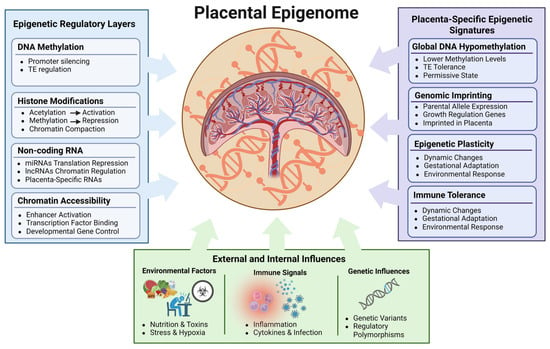
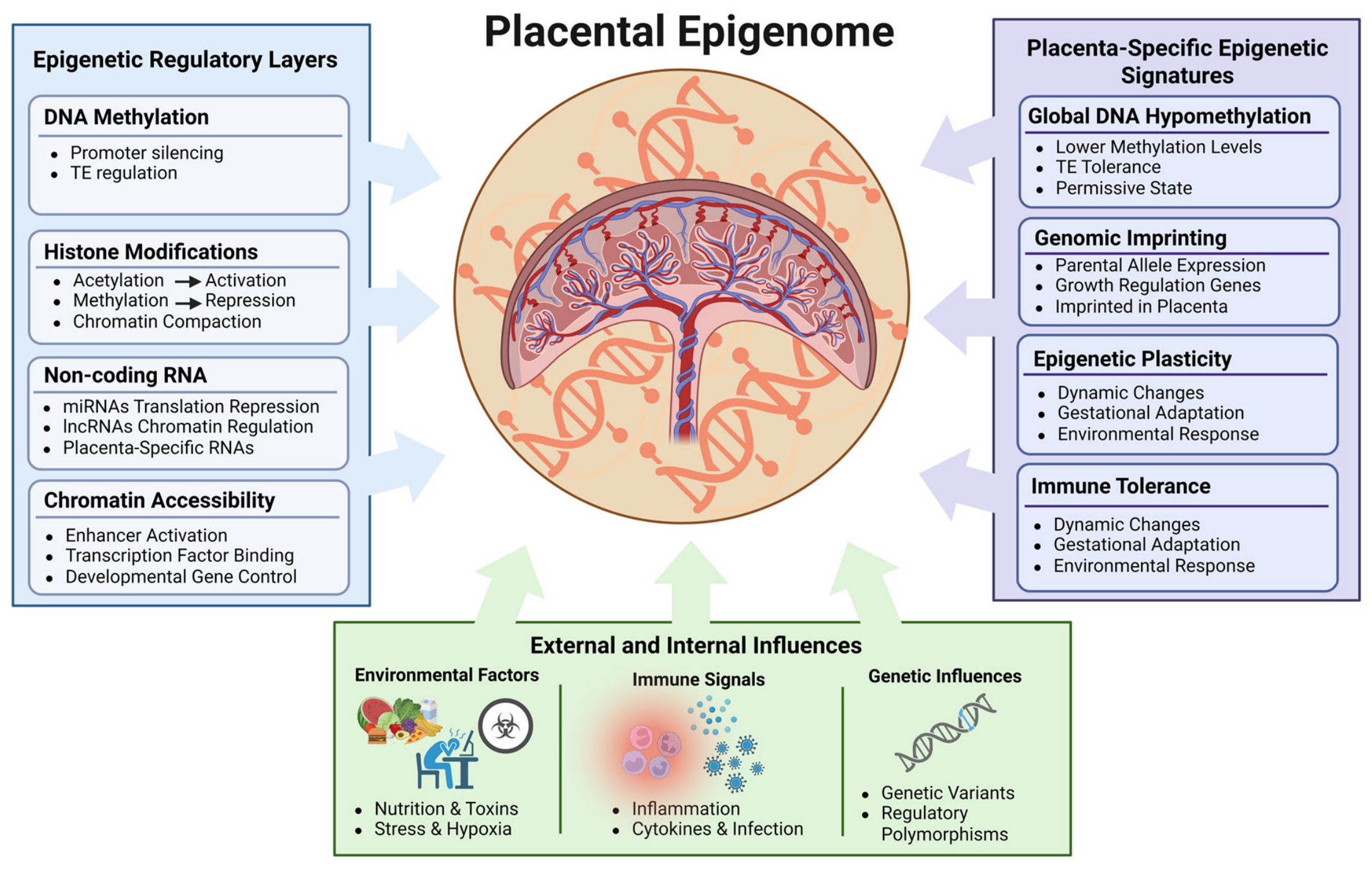
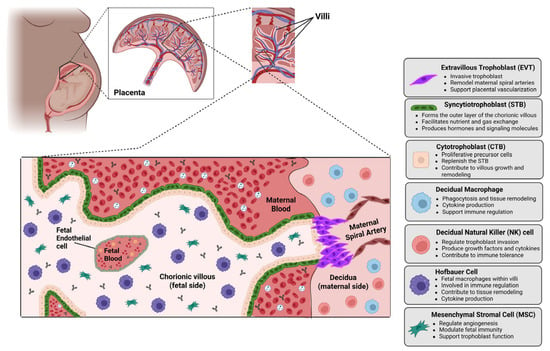
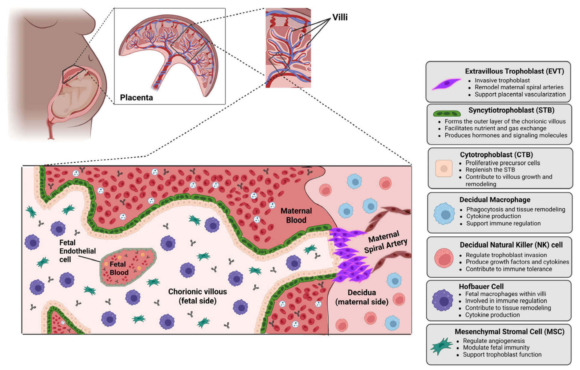
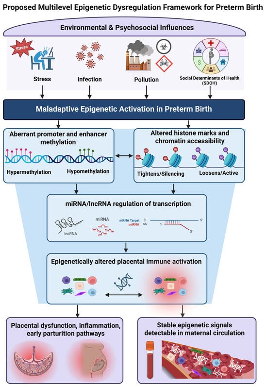
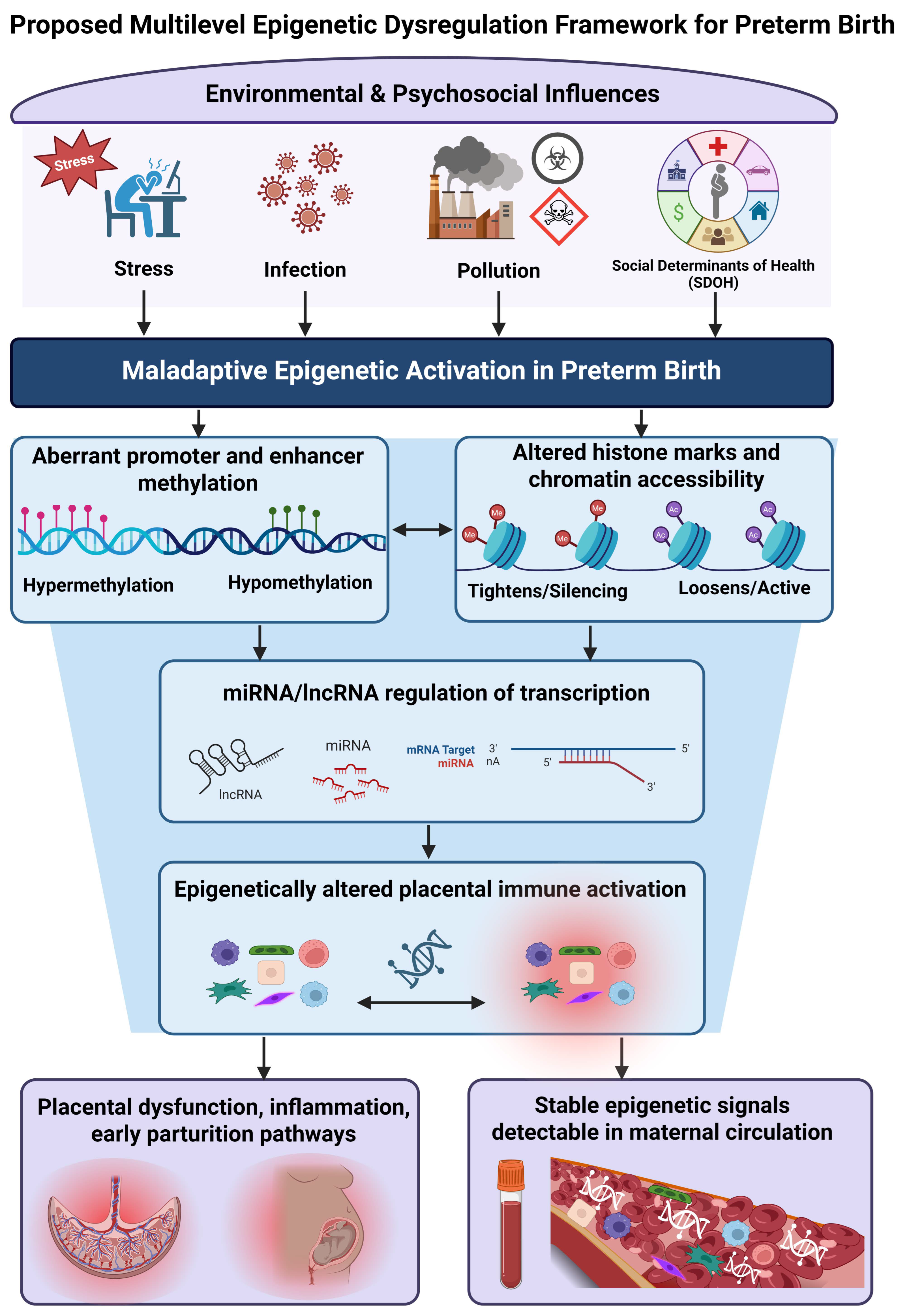
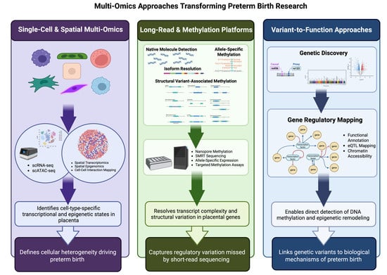
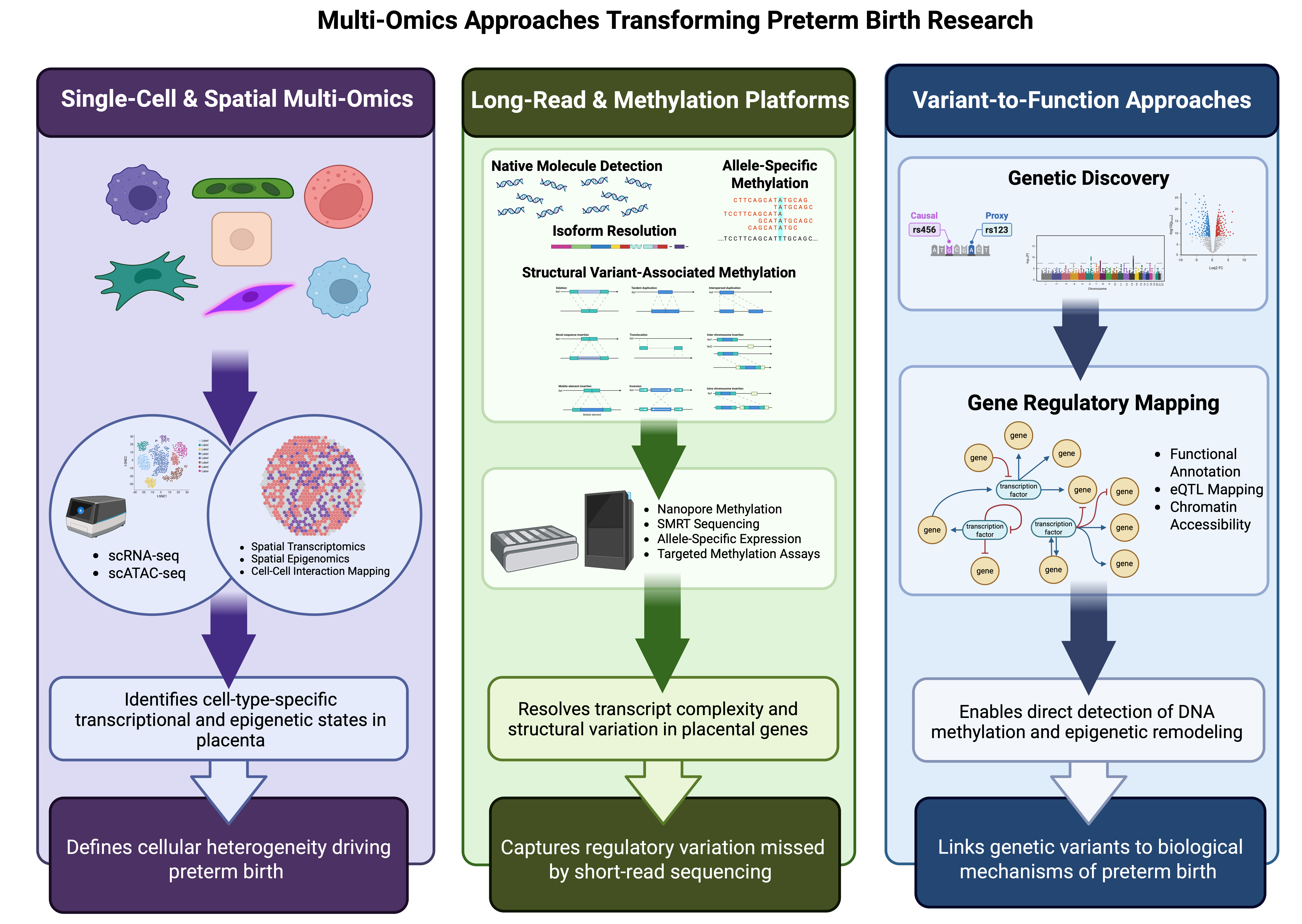

Disclaimer/Publisher’s Note: The statements, opinions and data contained in all publications are solely those of the individual author(s) and contributor(s) and not of MDPI and/or the editor(s). MDPI and/or the editor(s) disclaim responsibility for any injury to people or property resulting from any ideas, methods, instructions or products referred to in the content. |
© 2026 by the authors. Licensee MDPI, Basel, Switzerland. This article is an open access article distributed under the terms and conditions of the Creative Commons Attribution (CC BY) license.