


Abstract
Cardiovascular fibrosis is a dynamic process that involves fibroblast activation, extracellular matrix production, and myocardial remodeling. While it can damage the heart through an unregulated and progressive response, it is also an essential component of the body’s repair mechanisms. Fibroblast activation protein (FAP) targeted positron emission tomography (PET) has emerged as a molecular imaging approach for showing FAP expression in association with activated fibroblasts, potentially providing complementary information to established and contemporary imaging techniques. This narrative review examines the biological basis, tracer development and emerging cardiovascular applications of FAP-targeted PET, with emphasis on the clinical applications and limitations of available evidence. Relevant publications were found through a PubMed/MEDLINE search and selected according to their relevance to cardiovascular FAP-targeted imaging and potential clinical applications. Recent developments include gallium-68-labeled tracers such as 68Ga-FAPI-04, fluorine-18-labeled tracers such as 18F-FAPI-74, and high-affinity ligand platforms such as ONCOFAP. Current evidence consists of case reports, preclinical investigations, and mostly small exploratory clinical studies across many cardiovascular conditions. These studies report associations between cardiovascular FAP tracer uptake and measures of myocardial injury, remodeling and cardiac function, primarily in small exploratory cohorts. However, it is important to note that FAP expression reflects fibroblast activation and does not directly establish active collagen deposition, pathological fibrosis or tissue reversibility. Also, differences in tracer selection and imaging protocol, small study sizes and limited standardization restrict the ability to fully understand the clinical applications of existing findings. Cardiovascular FAP-targeted PET, although promising, remains investigational, and its clinical utility, prognostic value, and superiority over established imaging methods have not been fully demonstrated. More standardized, prospective studies are required to determine its exact role in cardiovascular disease assessment and management.
1. Objective and Approach
This narrative review provides an overview and evaluates the emerging role of fibroblast activation protein-targeted positron emission tomography (FAP-PET) in cardiovascular disease. The review focuses on the biological basis, current radiotracers, current preclinical and clinical evidence, limitations, and potential clinical applications in the future.
Relevant literature was primarily identified through PubMed searches using a combination of terms related to fibroblast activation protein, cardiovascular disease, myocardial fibrosis and infarction, cardiomyopathy, and other relevant cardiovascular conditions. Publications were selected based on their relevance to the present review’s main objectives. Original research articles were prioritized when discussing clinical and experimental findings, while including reviews to provide better background information and context. The final literature search was conducted on 20 October 2025.
Since this paper was written as a narrative review rather than a systematic review or meta-analysis, an exhaustive literature search and risk of bias assessment were not undertaken. The main goal was to provide a coherent and contextual overview of research on the topic at hand. Consequently, when interpreting the findings, acknowledge the possibility of incomplete study identification and selection bias.
2. Background, Biology and FAP
Fibrosis is one of the most fundamental tissue repair mechanisms in response to injury. In the cardiovascular system, it exhibits a unique “friend and foe” dynamic, acutely protective, but chronically harmful. In an acute setting, such as a myocardial infarction (MI), fibrosis is protective and helps maintain the structural integrity of the heart. Fibroblast-driven extracellular matrix (ECM) deposition stabilizes the damaged necrotic myocardium and prevents further mechanical damage in the early post-infarction phase [1,2]. However, when this process is allowed to continue unchecked, fibrosis transitions into a maladaptive state that is characterized by scar formation, myocardial stiffening and a progressive loss of contractility. This can then lead to decreased diastolic and systolic function, increase the risk of arrhythmias and eventually lead to the development of heart failure [3].
Myocardial fibroblasts are present in almost every cardiomyopathic condition, making them a prime target for imaging and therapeutic intervention [3]. However, the key to achieving a beneficial therapeutic effect will be the ability to target inappropriate and excessive fibrosis in the early stages, while allowing the protective fibrotic process to continue. An improved understanding of the timing of the appropriate and inappropriate responses after an MI will be key in preventing the transition to heart failure and sudden cardiac death while also leaving the protective scar untouched [1].
The ability to detect fibrosis non-invasively is ideal in a clinical setting. Current cardiovascular magnetic resonance (CMR) techniques can detect established fibrosis. T1 mapping assesses diffuse and potentially reversible interstitial fibrosis through native T1 values and extracellular volume fraction (ECV%), while late gadolinium enhancement (LGE) identifies irreversible replacement fibrosis [1]. These methods are well established for risk assessment and diagnosis across different cardiac conditions. They are limited to detecting already established fibrosis in the myocardium, without the spatial resolution to assess fibrosis in the valves and plaques in vascular structures [4]. They also cannot determine whether fibrogenesis is ongoing [5]. Molecular positron emission tomography (PET) radiotracers such as FAPI may complement these techniques by visualizing FAP expression associated with fibroblast activation. Its uptake does not directly signify collagen synthesis or distinguish reparative action from pathological fibrogenesis; thus, its use for treatment monitoring will require further validation [6].
Initially, fibrosis serves as a crucial repair system by containing the necrotic tissue. However, in the chronic phase, diffuse interstitial fibrosis can accumulate throughout the myocardium, leading to widespread stiffness and impaired ventricular compliance [7]. While it is possible to reverse a certain degree of fibrosis in early disease states, like hypertension or aortic stenosis, it can progress to an irreversible state if left alone for too long [7]. Notably, fibrotic remodeling is not confined only to the myocardium. In atherosclerosis, fibroblast-driven remodeling of fibrous caps has direct clinical consequences that can trigger a myocardial infarction (MI) [1]. This shows how well fibrosis illustrates the friend-or-foe concept.
2.1. Activated Fibroblasts
Fibroblasts are central mediators of the fibrotic response. Under physiologic conditions, fibroblasts maintain ECM homeostasis by sensing mechanical cues from myocardial contraction-relaxation cycles and replenishing ECM proteins as needed [8]. After myocardial injury, local cardiac fibroblasts activate and proliferate, creating a new extracellular matrix that shapes cardiac remodeling. Lineage tracing experiments in mouse models found that most of the active fibroblasts come from resident populations with epicardial and endothelial origins. There is little evidence that circulating hematopoietic cells or newly transitioned mesenchymal cells play a significant role in the process. Thus, resident cells appear to account for the majority of the fibroblastic response [9].
Activated fibroblasts are the most important cells driving fibrogenesis in the myocardium and they exist in two forms. They first activate into an intermediate form called proto-myofibroblasts, which can infiltrate injury sites and secrete mainly type I and III collagen to cross-link the ECM and promote wound healing [10]. With continued autocrine signaling, proto-myofibroblasts will mature into fully differentiated myofibroblasts [1]. These cells are defined by the expression of α-smooth muscle actin (α-SMA), which integrates into stress fibers that confer enhanced contractile ability and form stable adhesion complexes with ECM proteins [11,12].
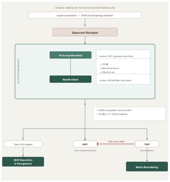
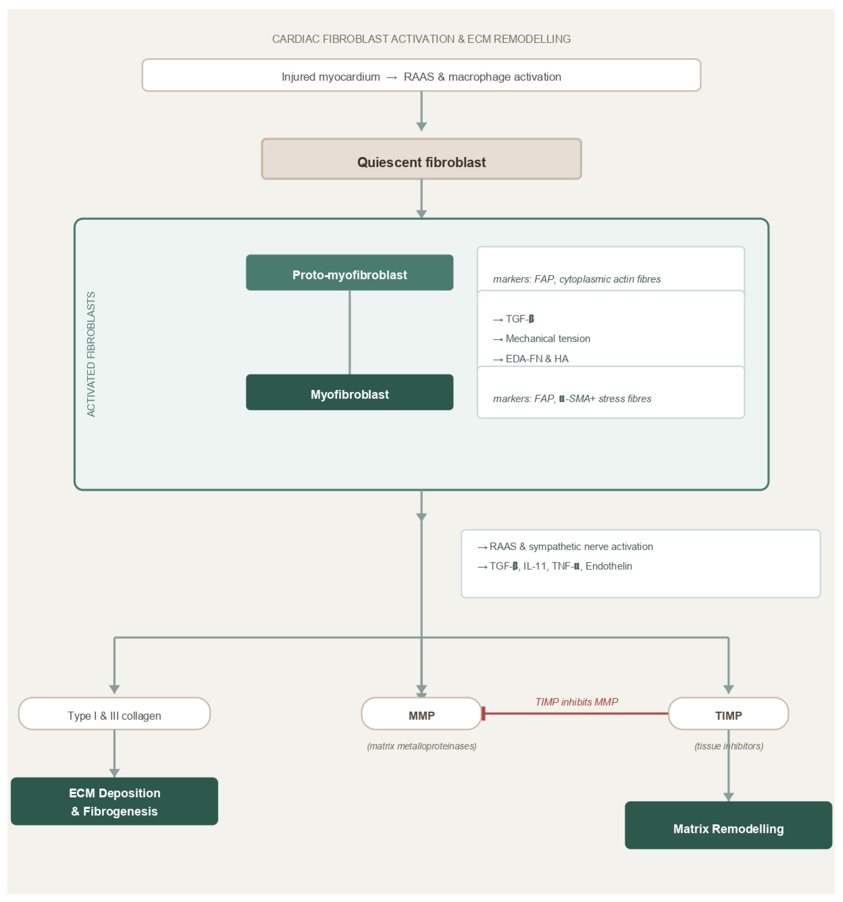
Activated fibroblasts are not just passive collagen-secreting units. They play a key role in matrix remodeling, secreting matrix metalloproteinases (MMPs) that degrade components of the extracellular matrix, as well as their tissue inhibitor metalloproteinases (TIMPs) [1]. The balance between collagen production and the activity of MMPs and TIMPs influences the nature and composition of the extracellular matrix (Figure 1) [1].
Figure 1.
Schematic representation of cardiac fibroblast activation and extracellular matrix (ECM) remodeling following myocardial injury. Following myocardial injury, activation of the renin–angiotensin–aldosterone system (RAAS) and inflammatory macrophages stimulates the transition of quiescent fibroblasts into proto-myofibroblasts and subsequently into activated myofibroblasts. Profibrotic mediators drive this process, including transforming growth factor-β (TGF-β), mechanical stress, extra domain A fibronectin (EDA-FN), and hyaluronic acid (HA). Activated myofibroblasts express fibroblast activation protein (FAP) and α-smooth muscle actin (α-SMA), promote extracellular matrix (ECM) deposition through collagen synthesis, and regulate matrix remodeling via matrix metalloproteinases (MMPs) and their tissue inhibitors (TIMPs). Dysregulation of these pathways contributes to myocardial fibrosis and adverse cardiac remodeling. Abbreviations: IL-11. interleukin-11; MMP, matrix metalloproteinase; RAAS, renin–angiotensin–aldosterone system; TGF-β, transforming growth factor-beta; TIMP, tissue inhibitor of metalloproteinases; TNF-α, tumor necrosis factor-alpha.
2.2. MMP/TIMP Balance
The relationship between MMPs and TIMPs is a central determinant of fibrotic remodeling. MMPs secreted by activated fibroblasts degrade ECM proteins, facilitating cell migration, angiogenesis, and tissue remodeling [1]. TIMPs counteract MMP activity to prevent excessive ECM degradation. In normal wound healing, this balance ensures ECM deposition and degradation proceed together, resulting in stable scar formation and restoration of structural integrity [1].
In cardiovascular disease, overactive MMPs promote pathological ECM breakdown, which, in atherosclerosis, compromises the integrity of fibrous caps and predisposes to rupture and thrombosis [1]. Excessive TIMP activity can lead to disproportionate ECM accumulation and stiffening of the myocardium. Since the MMP/TIMP axis shapes both scar formation and remodeling, it represents another potential target for molecular imaging markers of ECM turnover and antifibrotic therapies.
2.3. Fibroblast Activation Protein as a Selective Target
Fibroblast activation protein (FAP) is a 760-amino acid transmembrane surface glycoprotein that is almost always found only on activated fibroblasts [1]. FAP was first identified in 1986 and then cloned in 1994 and has both dipeptidyl peptidase (DPP) and endopeptidase activity [13]. The specificity of FAP for activated fibroblasts makes it a useful marker of fibroblast activation. FAP expression is minimal in quiescent fibroblasts but is strongly upregulated in activated fibroblasts during wound healing, tissue remodeling, organ fibrosis, and in cancer-associated fibroblasts [1].
Fibroblast activation, active fibrogenesis, and established fibrosis are all related, but they are distinct processes. FAPI uptake reflects tracer binding to FAP and could accompany physiological wound healing, inflammation, or maladaptive remodeling. It neither directly quantifies extracellular matrix deposition nor does it prove that a lesion is irreversible. Interpretation of results requires careful consideration of disease context, timing after injury and complementary structural imaging.
This specific, restricted expression of FAP offers two potential imaging advantages. First, FAP may serve as a marker of fibroblast activation, potentially helping to distinguish ongoing fibroblast-associated tissue remodeling from older, established scar tissue. This is critical for identifying potential reversible disease states and for monitoring the process of remodeling [1]. Secondly, FAP provides an accessible cell-surface target for imaging and treatment. The development of radiolabeled FAPI has enabled PET imaging of fibroblast-associated FAP expression, with studies reporting favorable target-to-background ratios [1]. Together, these properties support the possibility of using FAP as a biomarker of fibroblast activation and a promising potential target for molecular imaging with FAPI PET.
3. Tracers and Methods
Fibroblast activation protein inhibitors (FAPIs) are a relatively new class of quinoline-based tracers designed to target the fibroblast activation protein expressed on activated fibroblasts in tumors and fibrotic tissues [1]. Early developments such as FAPI-02 and FAPI-04 showed high binding affinity and strong selectivity for FAP-positive cells, along with good serum stability and high TBR [1]. These properties initially suggested that they could serve as effective tracers for visualizing fibrotic and tumor-related processes. However, clinical imaging quality was limited due to increased hepatobiliary excretion, which led to unwanted liver uptake [1].
To address this, FAPI-46 was developed, offering improved pharmacokinetics and reduced hepatic elimination, which resulted in clearer imaging and better diagnostic performance [1]. Because fluorine-18 cannot efficiently bind to the 1,4,7,10-tetraazacyclododecane-1,4,7,10-tetraacetic acid (DOTA) chelator used in gallium-based compounds, fluorinated analogs were later designed to enhance both tracer availability and image resolution [1]. Using a smaller chelator, 1,4,7-triazacyclononane-1,4,7-triacetic acid (NOTA), researchers successfully bound aluminum fluoride-18 (18F–AlF), creating FAPI-74—a fluorinated tracer with significant practical advantages [1]. Compared to gallium-68 tracers, fluorine-18 versions yield more doses per production cycle, have a longer half-life (109 min vs. 68 min), and deliver a lower radiation dose [1]. Fluorine-18’s shorter positron range also contributes to sharper image resolution, although gallium-68 tracers remain useful in facilities lacking a cyclotron, as they can be prepared directly from a generator [1].
Another important advancement is the development of ONCOFAP, described as an “ultra–high-affinity FAP ligand” [1]. ONCOFAP displays even higher binding specificity and TBR than earlier analogs and can be labeled with both DOTA and NOTA chelators, making it compatible with isotopes such as 68Ga, 18F–AlF, and 177Lu [1]. This versatility allows ONCOFAP to act as a theranostic agent, a compound used for both diagnostic imaging and targeted radiotherapy, thereby expanding its potential in oncology and fibrosis imaging (Table 1) [1].
Table 1.
Pharmacokinetic and biodistribution characteristics of selected FAPI ligands.
In terms of performance, FAPI tracers undergo rapid renal clearance and show minimal non-specific background activity, enabling high-contrast, well-defined images. Optimal imaging contrast is typically achieved about 60 min after administration [1]. Unlike FDG scans, FAPI imaging does not require patients to fast or follow special dietary rules, simplifying clinical workflow [1]. However, some radioligands show mild physiological uptake in the uterus, kidneys, and pancreas, and others display slight hepatic activity due to partial hepatobiliary excretion, which can make it difficult to distinguish between physiological and pathological uptake in certain regions [1].
Quantitatively, normal tissue uptake of FAPI tracers is low. Myocardial SUVmean values are generally between 0.35 and 1.2 at 1 h post-injection, leading to strong contrast between healthy and diseased tissue [1]. Once FAPI binds to fibroblast activation protein, it is quickly drawn into the fibroblast cells and is retained intracellularly, which helps maintain high tracer specificity, thereby improving image clarity by reducing non-specific signal uptake. These characteristics make FAPI PET particularly valuable for identifying solid tumors and sites of activated fibroblasts across different organs [1].
Despite these promising qualities, limited production capacity, relatively high synthesis costs, and restricted distribution still pose challenges [1]. As automated synthesis methods and multicenter validation studies expand, these obstacles are expected to decrease. With broader access and standardization, FAPI-based tracers, especially fluorinated versions such as FAPI-74 and alternative FAP targeting platforms like ONCOFAP could emerge as strong alternatives or complements to traditional 18F-FDG PET imaging in future clinical practice [1].
4. Clinical Applications
The following disease-specific sections distinguish animal or other preclinical experiments from early human observational findings whenever the study design was explicitly stated. The distinction is essential when interpreting the current evidence supporting FAP-targeted PET imaging in cardiovascular disease. Although preclinical studies may provide valuable insight into the biological mechanisms of fibroblast activation, their findings cannot necessarily be extrapolated to human disease. Associations with imaging or clinical markers should not be interpreted as a validated prognostic, diagnostic or treatment-guiding application.
4.1. MI & Remodelling
Following a myocardial infarction (MI), cardiac remodeling occurs through fibroblast activation in the infarct and surrounding peri-infarct or border zones. Although fibroblast activity is highest early on, effects of collagen secretion and adverse remodeling often progress to an irreversible stage [23,24]. FAP PET may visualize FAP expression associated with activated fibroblasts in these regions. Tracer uptake does not directly count activated cells or quantify collagen deposition. This molecular-level identification allows recognition of fibroblast activity that extends beyond the infarcted region into the border zone. In contrast, LGE function is mainly limited to visualizing established areas of fibrotic tissue. Similarly, perfusion imaging has limited utility, predominantly identifying areas of ischemia [23,25,26].
Therefore, FAP PET has the potential to provide additional information regarding fibroblast remodeling that is actively occurring. This response eventually leads to the formation of scars and stiffening of myocardial tissue. Future utilization of FAP PET may allow for more precise estimation of subsequent ventricular remodeling and heart failure development [26]. A key point is the use of FAP uptake quantification in the border zone as a potential measure of fibroblast activation during tissue remodeling [25]. Furthermore, small exploratory studies have reported associations between early post-MI FAPI uptake, infarct characteristics, subsequent left ventricular remodeling, and ventricular function at follow-up. In one prospective STEMI cohort, baseline 68Ga-DOTA-FAPI-04 uptake volume was associated with left ventricular remodeling at approximately 12 months. These findings suggest potential predictive value for post-MI remodeling, but they do not establish FAP-PET as a validated prognostic biomarker for clinical outcomes such as heart failure [27,28]. Whether FAP uptake can distinguish reparative activity from maladaptive remodeling, or monitor progression and reversal, remains unclear. Furthermore, past necrotic areas seen with LGE lacked FAP uptake, likely due to decreased fibroblast activity as a mature scar formed. This distinction may enable differentiation between recent and previous infarcts in the clinical setting [26].
4.2. HF/HFrEF/HFpEF
The pathogenesis of heart failure (HF) involves an adverse remodeling process with the formation of interstitial fibrosis and extensive fibroblast activity. Heart failure with preserved ejection fraction (HFpEF) is often related to progressive ventricular stiffening, increased interstitial fibrosis, and the replacement of injured myocytes by fibrotic tissue. Similarly, heart failure with reduced ejection fraction (HFrEF) is characterized by reduced left ventricular systolic function. Myocardial fibrosis and adverse remodeling can contribute to both phenotypes [24,29]. One of the major factors inducing heart failure is the remodeling process that consequently occurs following myocardial stress or injury. This transformative process alters the structure and function of cardiac muscle. If persistent, these alterations in the ventricular structure become irreversible, ultimately progressing to HF. If detected early on, fibrosis has been identified as a reversible process that may be treatable [30,31]. Fibroblastic activity within the myocardium plays a vital role in the progression of both reduced (HFrEF) and preserved ejection fraction (HFpEF). FAP PET may reveal fibroblast activation that is not captured by measures of established scar burden, which separates it from LGE. Clinical applications may include monitoring the therapeutic efficacy of antifibrotic medications, in addition to potential risk stratification [32].
4.3. HCM—Risk Association
Hypertrophic Cardiomyopathy (HCM) is a genetic disease involving myocyte hypertrophy and interstitial fibrosis, leading to ventricular dysfunction. Increased production of collagen and fibrotic tissue, both interstitial and perivascular, increases the risk of HF. Additionally, altered interstitial composition promotes conditions conducive to arrhythmias. The utilization of LGE in HCM identifies regions of scarring and is able to stratify risk associated with certain arrhythmias and sudden cardiac death [33].
As previously discussed, FAP-targeted PET is being investigated for its potential to detect FAP expression associated with activated fibroblasts in regions that may extend beyond abnormalities identified by LGE or perfusion imaging. Exploratory studies have also reported FAPI uptake in non-hypertrophied myocardial segments; however, the clinical significance and incremental value of these findings beyond established imaging remain uncertain. This suggests that FAP expression may occur in regions without abnormalities detectable by LGE, but it does not necessarily establish fibrosis. Myocardial uptake of FAP has been linked with markers of both structural and functional remodeling, including increased thickness, expanded extracellular volume, and reduced contractile function. Regions identified by both LGE and FAP-PET exhibit a more significant functional decline. The extent and intensity of FAP uptake correlate positively with elevated risk scores for both atrial fibrillation and sudden cardiac death, and with biomarkers indicative of myocardial injury and maladaptive remodeling. In a preliminary study involving 20 patients with HCM, FAPI uptake was associated with markers of myocardial remodeling and calculated clinical risk scores. The findings in this study suggest a possible link between fibroblast activation and established indicators used today. However, it is important to note that merely observing an association between FAPI uptake and calculated risk scores is far from sufficient. These findings do not establish independent prognostic value for FAPI uptake and therefore do not support its use as a stand-alone predictor of subsequent disease progression or clinical outcomes. They also do not demonstrate that FAPI-PET predicts future clinical events more accurately than established imaging modalities. Larger prospective studies incorporating predefined clinical endpoints and longitudinal follow-up are required to determine whether FAPI-PET provides incremental value beyond existing methods for risk stratification [34,35].
4.4. Cardiotoxicity
Cardiotoxicity is often iatrogenic, occurring as a result of the use of certain agents, particularly medications used in chemotherapy regimens. Some chemotherapeutic agents, like anthracyclines, provoke injury of myocytes, resulting in inflammation, activation of fibroblasts, and eventual fibrosis with functional deterioration [24].
FAP PET can potentially be used as a more sensitive tool for detecting early changes in the cardiotoxic cascade. Comparatively, LGE capabilities are limited to identifying regions of extracellular expansion and fibrosis that have occurred as a late consequence of cardiotoxicity. Therefore, FAP-targeted PET may provide information on fibroblast activation before overt left ventricular functional decline.
With earlier diagnosis, cardioprotective interventions may be initiated sooner, potentially improving long-term patient outcomes [29,36].
4.5. Amyloidosis
Cardiac amyloidosis develops as the extracellular space expands due to progressive deposition of amyloid fibrils. In addition, activation of fibroblasts leads to collagen replacement of the extracellular matrix. The primary pathology of amyloid deposition, in combination with the fibroblast response, eventually induces stiffening of the ventricles [37].
Cardiac MRI using late gadolinium enhancement can detect characteristic myocardial tissue changes associated with amyloid deposition, with transmural enhancement correlating with advanced cardiac involvement. Fibroblast activation is a secondary response in amyloidosis; thus, FAP PET cannot entirely replace LGE in the diagnostic approach. FAP-targeted PET may provide complementary information regarding fibroblast activation in selected cases. However, evidence in this setting remains limited, and its potential role in assessing disease progression, symptom burden, treatment response, or informing antifibrotic therapy remains investigational and requires further prospective validation [29,37].
4.6. Aortic Stenosis and Pressure Overload
Aortic stenosis (AS) occurs due to endothelial dysfunction of the aortic valve, which allows for the infiltration and accumulation of inflammatory cells. This leads to a cascade of inflammatory processes that ultimately activate valvular interstitial cells. These cells differentiate into myofibroblasts and osteoblasts, promoting calcification and stenosis of the aortic valve. As the aortic valve narrows, left ventricular pressure overload occurs, leading to concentric cardiac hypertrophy [38]. With progressive pressure overload, the heart undergoes maladaptive remodeling, with interstitial fibrosis being a key component. Eventually, under sustained fibrotic remodeling, irreversible myocardial dysfunction occurs.
FAP-targeted PET imaging aims to visualize fibroblast activation, potentially identifying active myocardial remodeling before structural changes become apparent on conventional imaging modalities such as LGE-CMR or perfusion imaging. This may provide an opportunity to identify early remodeling processes, although their reversibility and implications for treatment remain to be established [39]. Preclinical studies of aortic stenosis (AS) and pressure overload have demonstrated increased 68Ga-FAPI uptake in hypertrophied myocardial regions, accompanied by elevated FAP expression on immunohistochemical analysis [40]. Furthermore, increased FAPI uptake intensity and volume have been associated with markers of cardiac remodeling and dysfunction, including N-terminal pro-B-type natriuretic peptide (NT-proBNP), left ventricular ejection fraction (LVEF), and global longitudinal strain. In patients with advanced AS undergoing evaluation for transcatheter aortic valve replacement (TAVR), FAP-targeted PET has demonstrated varying degrees of left ventricular fibroblast activation, with tracer uptake patterns that differ from those observed using conventional imaging parameters. These preliminary findings suggest that FAPI-PET may provide complementary information regarding myocardial remodeling and warrant further investigation into its potential role in patient selection and treatment planning. However, its ability to guide TAVR decisions remains unvalidated and requires prospective evaluation [41].
4.7. Pulmonary Hypertension
Pulmonary hypertension (PH) occurs due to pulmonary vascular remodeling involving endothelial dysfunction, smooth muscle proliferation and fibroblast activation. Persistence of such processes leads to increased pulmonary vascular resistance (PVR), right ventricular pressure overload and eventually, right ventricular hypertrophy and failure [42]. In patients with pulmonary arterial hypertension (PAH), increased 68Ga-FAPI was seen in the right ventricular free wall, which was associated with lower tricuspid annular plane systolic excursion (TAPSE). Therefore, suggesting worse right ventricular (RV) systolic function in patients with increased RV uptake of FAPI. Furthermore, increased FAPI uptake also correlated with higher PVR and elevated NT-proBNP, both of which are markers for RV dysfunction and failure [43]. Additionally, in patients with chronic thromboembolic pulmonary hypertension, 68Ga-FAPI positively correlated with RV wall thickness and negatively correlated with RV ejection fraction, suggesting FAP-PET uptake reflects disease severity and maladaptive RV remodeling in pulmonary hypertension [44]. In some exploratory studies, increased FAPI uptake has been observed early in pulmonary hypertension and has been associated with progressive right ventricular remodeling and worsening right ventricular function [45,46].
FAP-PET has shown the potential to detect active fibroblast-driven remodeling of the pulmonary vasculature and right ventricular involvement; LGE and perfusion imaging only identify extensive fibrosis and areas of hypoperfusion, respectively. Although LGE is a well-established marker for disease progression, it reflects only advanced, often irreversible fibrosis [47]. In small exploratory studies, increased FAP-PET uptake has been associated with pulmonary hypertension severity and markers of right ventricular dysfunction and failure; however, its incremental value beyond established imaging modalities such as LGE-CMR or perfusion imaging has not been established.
4.8. AF
Atrial fibrillation (AF) is the most common sustained arrhythmia, involving various mechanisms such as ectopic foci, fibrosis, inflammation and electrical remodeling. In exploratory studies, increased 18F-FAPI uptake has been observed in several atrial regions, particularly the left atrial posterior wall, and has been associated with markers of atrial remodeling and with progression from paroxysmal to persistent atrial fibrillation [48]. In beagles and similarly AF patients, 18F-FAPI activity positively correlated with parameters such as FAP mRNA and protein levels, while BNP levels also correlated with atrial 18F-FAPI activity [49].
4.9. Vasculitis & Atherosclerosis
Atherosclerosis involves chronic endothelial injury and inflammation and lipid accumulation that activate fibroblasts, leading to plaque formation, luminal narrowing, and the development of a fibrous cap and necrotic core [50]. A study using 68Ga-FAP-PET with arterial wall FAPI uptake in major vessel segments was analyzed, where 800 sites demonstrated focal arterial uptake, and of those, 377 had concomitant vessel wall calcification. Only an increased body mass index was identified as a cardiological risk factor that positively correlated with FAPI uptake [51]. Another study found that target-to-background ratio (TBR) was negatively correlated with degree of calcification, and a higher TBR was observed in higher risk patients [52]. In a small exploratory study, FAP-PET signal intensity in vessel walls was positively associated with BMI and negatively associated with the degree of vascular calcification. These findings suggest that FAP expression may increase in less calcified arterial lesions; however, FAP-PET has not been shown to identify early vascular disease or provide incremental information beyond established imaging modalities.
Vasculitis occurs due to immune-mediated endothelial injury leading to eventual chronic vessel wall remodeling and fibrosis, therefore leading to a chronic pathologic process. FAP-PET uptake in inflammatory diseases such as aortitis, irrespective of clinical activity or remission, was significantly increased compared to controls. In the case of patients with remission, increased FAPI uptake was found in vessel walls, while MRI inflammatory indicators were negative, suggesting ongoing fibroblast activity pathology even in remission [53]. Furthermore, FAP-PET may be able to monitor multiple forms of vasculitis, such as Takayasu Arteritis, and demonstrate changes in IgG4-related disease [54,55]. Preliminary studies suggest that FAP-PET may identify additional vascular lesions and provide complementary information on fibroblast activation and vascular remodeling. However, it is too early to establish its superiority over FDG-PET for detecting active vascular inflammation (Table 2) [54].
Table 2.
Potential Clinical applications of FAPI PET imaging in non-oncological cardiovascular diseases.
Although the available studies report associations between FAP expression and cardiovascular remodeling across several conditions, their clinical interpretation still remains limited by tracer selection, differences in imaging protocols, small study populations and a lack of longitudinal outcome data. Thus, reported associations between FAP uptake and structural, biochemical or functional measures of remodeling cannot be interpreted as evidence of clinical utility just yet.
5. Comparators, Limitations and Future
Traditional cardiac imaging methods provide different insights into myocardial muscle structure, tissue composition, metabolism and function. LGE-CMR and T1 mapping/ECV look at problems in the myocardial tissue and extracellular expansion, FDG-PET evaluates glucose metabolism and strain imaging focuses on myocardial deformation. In contrast, FAP-PET shows FAP expression associated with fibroblast activation. These techniques examine different but potentially complementary aspects of cardiovascular remodeling. Their advantages, limitations, biological targets and clinical validation are summarized in Table 3.
Table 3.
Comparison of FAP-targeted PET with established cardiovascular imaging.
Before the development of FAP-PET, several molecular imaging approaches were explored to characterize myocardial fibrogenesis. Among these, radiotracers targeting αvβ3 integrins and proline-based analogs were the most extensively studied, although both provided only indirect insights into fibrotic remodeling [1].
Radiolabeled proline analogs, including 18F-fluoroproline, have been suggested as potential molecular imaging agents for the diagnosis of fibrosis due to prolines endogenous role in collagen biosynthesis. In a preclinical rabbit model of silica-induced lung fibrosis, increased uptake of 18F-fluoroproline was observed by PET in affected animals, with PET findings associated with histological evidence of fibrosis. However, the specificity of tracer uptake for fibrogenesis versus concurrent inflammation remained uncertain, and spatial resolution limited precise localization of fibrotic regions. These findings demonstrate the feasibility of proline-based PET imaging in experimental pulmonary fibrosis, although they do not establish its clinical utility in cardiovascular disease [56].
In contrast, FAP tracers such as 68Ga-FAPI-04 and 18F-AlF-FAPI-74 bind selectively to the serine protease FAP, which is transiently expressed on activated fibroblasts but remains largely absent in quiescent interstitial fibroblasts and healthy myocardium [57]. The molecular targeting characteristics of FAP-targeted PET are associated with favorable image contrast, rapid blood clearance, and low physiological myocardial uptake. By visualizing FAP expression associated with activated fibroblasts, FAP-targeted PET may provide complementary information on fibroblast-associated remodeling. Exploratory studies suggest that increased FAPI uptake may occur before some structural abnormalities become apparent on conventional imaging; however, this has not been established as a consistent temporal advantage over LGE or extracellular volume mapping [58].
Interpretation of FAP PET signals does require some caution. While FAP expression is commonly upregulated in activated fibroblasts during tissue remodeling and fibrotic disease, it may also be transiently upregulated during inflammation and physiological tissue repair [1]. As such, contextual clinical correlation and longitudinal imaging are essential to differentiate transient reparative activity from sustained pathogenic fibrosis.
5.1. Current Limitations of FAP PET Imaging
Despite encouraging preliminary evidence, several methodological and translational barriers must be addressed before FAP PET can be integrated into routine cardiovascular imaging. Most clinical investigations to date have relied on 68Ga-labeled FAP compounds produced on-site, which restricts accessibility due to the short half-life of 68Ga (approximately 68 min) and the limited output of 68Ge/68Ga generators [59]. Developing 18F-labeled analogs with longer half-lives may overcome these logistical constraints by enabling centralized production and wider distribution across imaging centers. Moreover, transient upregulation of FAP during post-infarction healing or systemic inflammatory responses may mimic pathological fibrosis, emphasizing the importance of interpreting imaging results within their biological and temporal context [58].
Quantification presents an additional challenge since thin-walled cardiac structures, such as the atria and right ventricle, are susceptible to partial volume effects, reducing accuracy in small lesions. Intercenter variability in reconstruction algorithms, scanner resolution, and region-of-interest definitions further limits reproducibility [1]. To date, there are no standardized reference ranges or validated normal databases for myocardial FAP uptake, and the optimal quantitative metric, whether SUVmean, SUVmax, or TBR, remains to be established. Furthermore, FAP expression does not necessarily indicate irreversible fibrosis and may instead reflect adaptive or reparative remodeling, particularly during the early stages of myocardial injury. Differentiating transient, reversible activation from maladaptive fibrogenesis will require longitudinal imaging combined with histopathological correlation [58].
Clinical validation of this modality remains limited. Most published studies are small, single-center, and cross-sectional, with heterogeneous endpoints and short follow-up durations. Large-scale, prospective investigations linking FAP PET signal intensity to clinical outcomes such as hospitalization, arrhythmia burden, and mortality are still lacking. Comparative assessments against established modalities, such as LGE MRI, T1 mapping, or strain imaging, are also scarce and multicenter reproducibility has yet to be demonstrated [58]. Finally, FAP PET involves radiation exposure, unlike echocardiography and MRI, and may also be associated with higher costs. Therefore, a clear incremental clinical benefit must be demonstrated [58]. These limitations highlight the need for standardized imaging protocols, multicenter harmonization, biological validation, and longitudinal outcome studies to define the role of FAP-targeted PET in cardiovascular disease and to determine its value as a biomarker of myocardial fibroblast activation and fibroblast-associated remodeling.
5.2. Future Perspectives and Clinical Translation
Emerging evidence suggests that FAP-targeted PET may provide complementary information on fibroblast-associated remodeling, with potential applications in earlier disease characterization, risk assessment, and treatment monitoring that remain investigational. Because FAP expression can occur before mature collagen deposition, FAP-targeted PET is being investigated as a potential method for detecting early fibroblast activation and fibroblast-associated remodeling in patients with preserved ejection fraction, inherited cardiomyopathies, or chemotherapy-induced cardiotoxicity.
Detecting fibroblast activation at such an early stage could enable the initiation of cardioprotective or antifibrotic interventions before irreversible structural remodeling develops [29]. Continuous technological progress is likely to expand the diagnostic reach of this modality. Innovations in detector sensitivity, total body PET systems, and advanced image reconstruction algorithms could enhance both spatial resolution and quantitative accuracy. Consequently, FAP-targeted PET is being investigated across a range of cardiovascular conditions involving fibroblast activation and tissue remodeling, including atrial fibrillation, pulmonary hypertension, and vascular disorders [58].
A study using a mouse model of angiotensin II-induced hypertension found that 68Ga-FAPI-46 uptake increased in the heart and kidneys within one week of inducing hypertension. They confirmed FAP expression in both organs, but observed clear signs of fibrosis only in heart tissue. This suggests that the FAP-targeted tracer may detect fibroblast activation before obvious fibrosis appears on histology. Mice treated with an angiotensin receptor blocker showed less uptake, which connects tracer activity to molecular changes driven by hypertension [60]. However, the clinical implications remain unclear, and further studies are needed to determine the effectiveness of FAPI PET in monitoring treatment response.
Beyond diagnostic imaging, FAP-targeted PET may also have potential as a theranostic platform, although this application remains investigational. Given its selective expression on activated fibroblasts, FAP has emerged as a viable therapeutic target. Preclinical models have explored the efficacy of chimeric antigen receptor (CAR) T-cell therapies and radiolabeled inhibitors directed at FAP to attenuate fibrosis [57,61]. In this context, FAP PET could serve a dual role: as a companion diagnostic to identify suitable candidates for targeted therapy and as a quantitative biomarker to monitor treatment response, effectively bridging molecular imaging with precision therapeutics [58].
Incorporating FAP PET into clinical trial design may also improve the efficiency of antifibrotic drug development. Selecting patients with demonstrable fibroblast activation could enhance statistical power, reduce sample sizes, and enable more meaningful assessment of treatment effects. Serial FAP PET imaging could also serve as a surrogate endpoint, providing an objective measure of therapeutic efficacy and disease stabilization. FAP PET integration with multimodal imaging and artificial intelligence is another compelling avenue. Combining FAP PET with cardiac MRI or CT could allow simultaneous assessment of fibroblast activation, extracellular matrix composition, and structural remodeling, offering a comprehensive picture of myocardial health. When coupled with machine-learning algorithms, these multimodal datasets could refine patient phenotyping and improve prognostic modeling in heterogeneous heart failure populations [58]. The potential applications discussed in this section, including the use of FAP-targeted PET as a surrogate endpoint, its incorporation into clinical trial design, and its integration with artificial intelligence, remain hypothetical. Their clinical value remains unestablished and will require prospective validation before they can be considered for routine clinical use.
Widespread clinical adoption of FAP PET will depend on several key advances, which include expanding access to 18F-labeled tracers to simplify logistics, establishing multicenter standardization of imaging and quantification protocols, and conducting large-scale, outcome-oriented trials to demonstrate clinical utility. As these developments progress, FAP-targeted PET may complement established structural imaging by providing information on FAP expression associated with activated fibroblasts rather than established scar alone. Its potential applications in diagnosis, risk assessment, and treatment monitoring remain investigational and require prospective validation.
6. Conclusions
Fibrosis is a key, dual-acting process in cardiovascular disease, playing an essential role in the acute reparative phase while simultaneously causing progressive tissue degeneration when continuously activated. In many CVDs, such as myocardial infarction, heart failure, cardiomyopathies, valvular heart disease, pulmonary hypertension, and vascular inflammation, fibrotic remodeling through the activation of resident fibroblasts is an important feature driving disease progression. FAP-targeted PET has the potential to serve as a complementary molecular imaging modality in cardiovascular disease by providing a non-invasive assessment of FAP expression associated with activated fibroblasts and insight into fibroblast-associated tissue remodeling in vivo.
Across several cardiovascular conditions, increased FAP uptake has been associated with myocardial injury, adverse remodeling, and impaired cardiac function. These findings suggest potential roles in disease characterization and risk assessment. In this way, FAP PET may complement existing imaging modalities by linking molecular activity with the structural changes seen on conventional imaging. Despite these promising developments, several challenges remain before widespread clinical implementation is possible. These include a lack of standardization in acquisition and quantification methods, variability in tracer availability, and the need for larger, longitudinal studies to confirm clinical endpoints. Additionally, because FAP expression can also be transiently upregulated during tissue repair and inflammatory processes, increased FAPI uptake should not be interpreted as direct evidence of irreversible fibrosis, and careful clinical interpretation remains essential. In its current state, FAP PET imaging should be regarded as an investigational and potentially complementary molecular imaging modality, rather than an established alternative to current cardiovascular imaging methods.
Author Contributions
Conceptualization M.K. and K.H.; Methodology M.K. and K.H.; Investigation J.C., J.J., A.T., S.Y. and R.A.; Data Curation, J.C., J.J., A.T., S.Y. and R.A.; Visualization, J.C., J.J., A.T. and S.Y.; Supervision, M.K. and K.H.; Project administration J.C. and K.H.; Writing—original draft preparation, J.C., J.J., A.T., S.Y., R.A., M.K. and K.H.; Writing—review and editing, J.C., J.J. and K.H. All authors have read and agreed to the published version of the manuscript.
Funding
This research received no external funding.
Institutional Review Board Statement
Not applicable.
Informed Consent Statement
Not applicable.
Data Availability Statement
No new data were created or analyzed in this study. Data sharing is not applicable to this article.
Conflicts of Interest
The authors declare no conflicts of interest.
Abbreviations
| MI | Myocardial infarction |
| ECM | Extracellular matrix |
| CMR | Cardiovascular magnetic resonance |
| ECV% | Extracellular volume fraction |
| LGE | Late gadolinium enhancement |
| PET | Positron emission tomography |
| FAPI | Fibroblast activation protein inhibitor |
| EMT | Epithelial-mesenchymal transformation |
| α-SMA | α-smooth muscle actin |
| MMP | Matrix metalloproteinase |
| TIMP | Tissue inhibitor metalloproteinase |
| FAP | Fibroblast activation protein |
| DPP | Dipeptidyl peptidase |
| TBR | Target-to-background ratio |
| DOTA | 1,4,7,10-tetraazacyclododecane-1,4,7,10-tetraacetic acid |
| NOTA | 1,4,7-Triazacyclononane-1,4,7-triacetic acid |
| HF | Heart failure |
| HFpEF | Heart failure with preserved ejection fraction |
| HFrEF | Heart failure with reduced ejection fraction |
| HCM | Hypertrophic cardiomyopathy |
| AS | Aortic stenosis |
| PH | Pulmonary hypertension |
| PVR | Pulmonary vascular resistance |
| PAH | Pulmonary arterial hypertension |
| TAPSE | Tricuspid annular plane systolic excursion |
| RV | Right ventricle |
| AF | Atrial fibrillation |
| MRI | Magnetic resonance imaging |
| CAR | Chimeric antigen receptor |
References
- Loganath, K.; Craig, N.; Barton, A.; Joshi, S.; Anagnostopoulos, C.; Erba, P.A.; Glaudemans, A.W.J.M.; Saraste, A.; Bucerius, J.; Lubberink, M.; et al. Cardiovascular positron emission tomography imaging of fibroblast activation: A review of the current literature. J. Nucl. Cardiol. 2025, 47, 102106. [Google Scholar] [CrossRef] [Scilit] [PubMed]
- Wynn, T.A. Fibrotic disease and the Th1/Th2 paradigm. Nat. Rev. Immunol. 2004, 4, 583–594. [Google Scholar] [CrossRef] [Scilit] [PubMed]
- Nagaraju, C.K.; Dries, E.; Popovic, N.; Singh, A.A.; Haemers, P.; Roderick, H.L.; Claus, P.; Sipido, K.R.; Driesen, R.B. Global fibroblast activation throughout the left ventricle but localized fibrosis after myocardial infarction. Sci. Rep. 2017, 7, 10801. [Google Scholar] [CrossRef] [Scilit] [PubMed]
- Bing, R.; Dweck, M.R. Myocardial fibrosis: Why image, how to image and clinical implications. Heart 2019, 105, 1832–1840. [Google Scholar] [CrossRef] [Scilit] [PubMed]
- Mewton, N.; Liu, C.Y.; Croisille, P.; Bluemke, D.; Lima, J.A.C. Assessment of myocardial fibrosis with cardiovascular magnetic resonance. J. Am. Coll. Cardiol. 2011, 57, 891–903. [Google Scholar] [CrossRef] [Scilit] [PubMed]
- Mori, Y.; Novruzov, E.; Schmitt, D.; Cardinale, J.; Watabe, T.; Choyke, P.L.; Alavi, A.; Haberkorn, U.; Giesel, F.L. Clinical applications of fibroblast activation protein inhibitor positron emission tomography (FAPI-PET). npj Imaging 2024, 2, 48. [Google Scholar] [CrossRef] [Scilit] [PubMed]
- Travers, J.G.; Kamal, F.A.; Robbins, J.; Yutzey, K.E.; Blaxall, B.C. Cardiac fibrosis. Circ. Res. 2016, 118, 1021–1040. [Google Scholar] [CrossRef] [Scilit] [PubMed]
- van Putten, S.; Shafieyan, Y.; Hinz, B. Mechanical control of cardiac myofibroblasts. J. Mol. Cell. Cardiol. 2016, 93, 133–142. [Google Scholar] [CrossRef] [Scilit] [PubMed]
- Moore-Morris, T.; Guimarães-Camboa, N.; Banerjee, I.; Zambon, A.C.; Kisseleva, T.; Velayoudon, A.; Stallcup, W.B.; Gu, Y.; Dalton, N.D.; Cedenilla, M.; et al. Resident fibroblast lineages mediate pressure overload-induced cardiac fibrosis. J. Clin. Investig. 2014, 124, 2921–2934. [Google Scholar] [CrossRef] [Scilit] [PubMed]
- Singh, S.; Torzewski, M. Fibroblasts and their pathological functions in the fibrosis of aortic valve sclerosis and atherosclerosis. Biomolecules 2019, 9, 472. [Google Scholar] [CrossRef] [Scilit] [PubMed]
- Avagliano, A.; Ruocco, M.R.; Nasso, R.; Aliotta, F.; Sanità, G.; Iaccarino, A.; Bellevicine, C.; Calì, G.; Fiume, G.; Masone, S.; et al. Development of a stromal microenvironment experimental model containing proto-myofibroblast-like cells and analysis of its crosstalk with melanoma cells: A new tool to potentiate and stabilize tumor suppressor phenotype of dermal myofibroblasts. Cells 2019, 8, 1435. [Google Scholar] [CrossRef] [Scilit] [PubMed]
- Tomasek, J.J.; Gabbiani, G.; Hinz, B.; Chaponnier, C.; Brown, R.A. Myofibroblasts and mechano-regulation of connective tissue remodelling. Nat. Rev. Mol. Cell Biol. 2002, 3, 349–363. [Google Scholar] [CrossRef] [Scilit] [PubMed]
- Scanlan, M.J.; Raj, B.K.; Calvo, B.; Garin-Chesa, P.; Sanz-Moncasi, M.P.; Healey, J.H.; Old, L.J.; Rettig, W.J. Molecular cloning of fibroblast activation protein alpha, a member of the serine protease family selectively expressed in stromal fibroblasts of epithelial cancers. Proc. Natl. Acad. Sci. USA 1994, 91, 5657–5661. [Google Scholar] [CrossRef] [Scilit] [PubMed]
- Loktev, A.; Lindner, T.; Mier, W.; Debus, J.; Altmann, A.; Jäger, D.; Giesel, F.; Kratochwil, C.; Barthe, P.; Roumestand, C.; et al. A tumor-imaging method targeting cancer-associated fibroblasts. J. Nucl. Med. 2018, 59, 1423–1429. [Google Scholar] [CrossRef] [Scilit] [PubMed]
- Varasteh, Z.; Mohanta, S.; Robu, S.; Braeuer, M.; Li, Y.; Omidvari, N.; Topping, G.; Sun, T.; Nekolla, S.G.; Richter, A.; et al. Molecular imaging of fibroblast activity after myocardial infarction using a 68Ga-labeled fibroblast activation protein inhibitor, FAPI-04. J. Nucl. Med. 2019, 60, 1743–1749. [Google Scholar] [CrossRef] [Scilit] [PubMed]
- Lindner, T.; Loktev, A.; Altmann, A.; Giesel, F.; Kratochwil, C.; Debus, J.; Jäger, D.; Mier, W.; Haberkorn, U. Development of quinoline-based theranostic ligands for the targeting of fibroblast activation protein. J. Nucl. Med. 2018, 59, 1415–1422. [Google Scholar] [CrossRef] [Scilit] [PubMed]
- Loktev, A.; Lindner, T.; Burger, E.-M.; Altmann, A.; Giesel, F.; Kratochwil, C.; Debus, J.; Marmé, F.; Jäger, D.; Mier, W.; et al. Development of fibroblast activation protein-targeted radiotracers with improved tumor retention. J. Nucl. Med. 2019, 60, 1421–1429. [Google Scholar] [CrossRef] [Scilit] [PubMed]
- Meyer, C.; Dahlbom, M.; Lindner, T.; Vauclin, S.; Mona, C.; Slavik, R.; Czernin, J.; Haberkorn, U.; Calais, J. Radiation dosimetry and biodistribution of 68Ga-FAPI-46 PET imaging in cancer patients. J. Nucl. Med. 2020, 61, 1171–1177. [Google Scholar] [CrossRef] [Scilit] [PubMed]
- Lindner, T.; Altmann, A.; Giesel, F.; Kratochwil, C.; Kleist, C.; Krämer, S.; Mier, W.; Cardinale, J.; Kauczor, H.-U.; Jäger, D.; et al. 18F-labeled tracers targeting fibroblast activation protein. EJNMMI Radiopharm. Chem. 2021, 6, 26. [Google Scholar] [CrossRef] [Scilit] [PubMed]
- Giesel, F.L.; Adeberg, S.; Syed, M.; Lindner, T.; Jiménez-Franco, L.D.; Mavriopoulou, E.; Staudinger, F.; Tonndorf-Martini, E.; Regnery, S.; Rieken, S.; et al. FAPI-74 PET/CT using either 18F-AlF or cold-kit 68Ga labeling: Biodistribution, radiation dosimetry, and tumor delineation in lung cancer patients. J. Nucl. Med. 2021, 62, 201–207. [Google Scholar] [CrossRef] [Scilit] [PubMed]
- Backhaus, P.; Gierse, F.; Burg, M.C.; Büther, F.; Asmus, I.; Dorten, P.; Cufe, J.; Roll, W.; Neri, D.; Cazzamalli, S.; et al. Translational imaging of the fibroblast activation protein (FAP) using the new ligand [68Ga]Ga-OncoFAP-DOTAGA. Eur. J. Nucl. Med. Mol. Imaging 2022, 49, 1822–1832. [Google Scholar] [CrossRef] [Scilit] [PubMed]
- Millul, J.; Bassi, G.; Mock, J.; Elsayed, A.; Pellegrino, C.; Zana, A.; Dakhel Plaza, S.; Nadal, L.; Gloger, A.; Schmidt, E.; et al. An ultra-high-affinity small organic ligand of fibroblast activation protein for tumor-targeting applications. Proc. Natl. Acad. Sci. USA 2021, 118, e2101852118. [Google Scholar] [CrossRef] [Scilit] [PubMed]
- Kupusovic, J.; Kessler, L.; Kazek, S.; Chodyla, M.K.; Umutlu, L.; Zarrad, F.; Nader, M.; Fendler, W.P.; Varasteh, Z.; Hermann, K.; et al. Delayed 68Ga-FAPI-46 PET/MR imaging confirms ongoing fibroblast activation in patients after acute myocardial infarction. Int. J. Cardiol. Heart Vasc. 2024, 50, 101340. [Google Scholar] [CrossRef] [Scilit] [PubMed]
- Siebermair, J.; Köhler, M.I.; Kupusovic, J.; Nekolla, S.G.; Kessler, L.; Ferdinandus, J.; Guberina, N.; Stuschke, M.; Grafe, H.; Siveke, J.T.; et al. Cardiac fibroblast activation detected by Ga-68 FAPI PET imaging as a potential novel biomarker of cardiac injury/remodeling. J. Nucl. Cardiol. 2021, 28, 812–821. [Google Scholar] [CrossRef] [Scilit] [PubMed]
- Notohamiprodjo, S.; Nekolla, S.G.; Robu, S.; Villagran Asiares, A.; Kupatt, C.; Ibrahim, T.; Laugwitz, K.-L.; Makowski, M.R.; Schwaiger, M.; Weber, W.A.; et al. Imaging of cardiac fibroblast activation in a patient after acute myocardial infarction using 68Ga-FAPI-04. J. Nucl. Cardiol. 2022, 29, 2254–2261. [Google Scholar] [CrossRef] [Scilit] [PubMed]
- Barton, A.K.; Craig, N.J.; Loganath, K.; Joshi, S.; Tsampasian, V.; Mahendran, M.; Lenell, J.; Tzolos, E.; Singh, T.; Whittington, B.; et al. Myocardial fibroblast activation after acute myocardial infarction: A positron emission tomography and magnetic resonance study. J. Am. Coll. Cardiol. 2025, 85, 578–591. [Google Scholar] [CrossRef] [Scilit] [PubMed]
- Diekmann, J.; Koenig, T.; Thackeray, J.T.; Derlin, T.; Czerner, C.; Neuser, J.; Ross, T.L.; Schäfer, A.; Tillmanns, J.; Bauersachs, J.; et al. Cardiac fibroblast activation in patients early after acute myocardial infarction: Integration with MR tissue characterization and subsequent functional outcome. J. Nucl. Med. 2022, 63, 1415–1423. [Google Scholar] [CrossRef] [Scilit] [PubMed]
- Zhang, M.; Quan, W.; Zhu, T.; Feng, S.; Huang, X.; Meng, H.; Du, R.; Zhu, Z.; Qu, X.; Li, P.; et al. [68Ga]Ga-DOTA-FAPI-04 PET/MR in patients with acute myocardial infarction: Potential role of predicting left ventricular remodeling. Eur. J. Nucl. Med. Mol. Imaging 2023, 50, 839–848. [Google Scholar] [CrossRef] [Scilit] [PubMed]
- Higuchi, T.; Serfling, S.E.; Leistner, D.M.; Speer, T.; Werner, R.A. FAPI-PET in cardiovascular disease. Semin. Nucl. Med. 2024, 54, 747–752. [Google Scholar] [CrossRef] [Scilit] [PubMed]
- Hosseini, A.; Haj-Yehia, E.; Korste, S.; Kuzay, Y.; Trajkovic-Arsic, M.; Settelmeier, S.; Cantore, M.; Ferenz, K.B.; Siveke, J.T.; Herrmann, K.; et al. How rapidly does the FAPI PET signal reverse following therapy? Assessing the FAPI PET signal in hypertensive cardiac injury and fibrosis in mice. J. Nucl. Med. 2025, 66, 1757–1763. [Google Scholar] [CrossRef] [Scilit] [PubMed]
- Frantz, S.; Hundertmark, M.J.; Schulz-Menger, J.; Bengel, F.M.; Bauersachs, J. Left ventricular remodelling post-myocardial infarction: Pathophysiology, imaging, and novel therapies. Eur. Heart J. 2022, 43, 2549–2561. [Google Scholar] [CrossRef] [Scilit] [PubMed]
- Cui, Y.; Wang, Y.; Wang, S.; Du, B.; Li, X.; Li, Y. Highlighting fibroblasts activation in fibrosis: The state-of-the-art fibroblast activation protein inhibitor PET imaging in cardiovascular diseases. J. Clin. Med. 2023, 12, 6033. [Google Scholar] [CrossRef] [Scilit] [PubMed]
- Green, J.J.; Berger, J.S.; Kramer, C.M.; Salerno, M. Prognostic value of late gadolinium enhancement in clinical outcomes for hypertrophic cardiomyopathy. JACC Cardiovasc. Imaging 2012, 5, 370–377. [Google Scholar] [CrossRef] [Scilit] [PubMed]
- Ding, J.; Zhang, H.; Chen, X.; Wang, H.; Wang, W.; You, Z.; Gao, L.; Zhang, Q.; Zhao, J. Enhanced detection of damaged myocardium and risk stratification in hypertrophic cardiomyopathy using integrated [68Ga]Ga-FAPI-04 PET/CMR imaging. Eur. J. Nucl. Med. Mol. Imaging 2024, 52, 98–108. [Google Scholar] [CrossRef] [Scilit] [PubMed]
- Wang, L.; Wang, Y.; Wang, J.; Xiao, M.; Xi, X.-Y.; Chen, B.-X.; Su, Y.; Zhang, Y.; Xie, B.; Dong, Z.; et al. Myocardial activity at 18F-FAPI PET/CT and risk for sudden cardiac death in hypertrophic cardiomyopathy. Radiology 2023, 306, e221052. [Google Scholar] [CrossRef] [Scilit] [PubMed]
- Wei, Z.; Xu, H.; Chen, B.; Wang, J.; Yang, X.; Yang, M.-F.; Zhao, S. Early detection of anthracycline-induced cardiotoxicity using [68Ga]Ga-FAPI-04 imaging. Eur. J. Nucl. Med. Mol. Imaging 2024, 51, 2204–2215. [Google Scholar] [CrossRef] [Scilit] [PubMed]
- Wang, X.; Guo, Y.; Gao, Y.; Ren, C.; Huang, Z.; Liu, B.; Li, X.; Chang, L.; Shen, K.; Ding, H.; et al. Feasibility of 68Ga-labeled fibroblast activation protein inhibitor PET/CT in light-chain cardiac amyloidosis. JACC Cardiovasc. Imaging 2022, 15, 1960–1970. [Google Scholar] [CrossRef] [Scilit] [PubMed]
- Goody, P.R.; Hosen, M.R.; Christmann, D.; Niepmann, S.T.; Zietzer, A.; Adam, M.; Bönner, F.; Zimmer, S.; Nickenig, G.; Jansen, F. Aortic valve stenosis. Arterioscler. Thromb. Vasc. Biol. 2020, 40, 885–900. [Google Scholar] [CrossRef] [Scilit] [PubMed]
- Bing, R.; Cavalcante, J.L.; Everett, R.J.; Clavel, M.-A.; Newby, D.E.; Dweck, M.R. Imaging and impact of myocardial fibrosis in aortic stenosis. JACC Cardiovasc. Imaging 2019, 12, 283–296. [Google Scholar] [CrossRef] [Scilit] [PubMed]
- Wang, G.; Yang, Q.; Wu, S.; Xu, X.; Li, X.; Liang, S.; Pan, G.; Zuo, C.; Zhao, X.; Cheng, C.; et al. Molecular imaging of fibroblast activity in pressure overload heart failure using [68Ga]Ga-FAPI-04 PET/CT. Eur. J. Nucl. Med. Mol. Imaging 2023, 50, 465–474. [Google Scholar] [CrossRef] [Scilit] [PubMed]
- Diekmann, J.; Neuser, J.; Röhrich, M.; Derlin, T.; Zwadlo, C.; Koenig, T.; Weiberg, D.; Jäckle, F.; Kempf, T.; Ross, T.L.; et al. Molecular imaging of myocardial fibroblast activation in patients with advanced aortic stenosis before transcatheter aortic valve replacement: A pilot study. J. Nucl. Med. 2023, 64, 1279–1286. [Google Scholar] [CrossRef] [Scilit] [PubMed]
- Humbert, M.; Guignabert, C.; Bonnet, S.; Dorfmüller, P.; Klinger, J.R.; Nicolls, M.R.; Olschewski, A.J.; Pullamsetti, S.S.; Schermuly, R.T.; Stenmark, K.R.; et al. Pathology and pathobiology of pulmonary hypertension: State of the art and research perspectives. Eur. Respir. J. 2019, 53, 1801887. [Google Scholar] [CrossRef] [Scilit] [PubMed]
- Gu, Y.; Han, K.; Zhang, Z.; Zhao, Z.; Yan, C.; Wang, L.; Fang, W. 68Ga-FAPI PET/CT for molecular assessment of fibroblast activation in right heart in pulmonary arterial hypertension: A single-center, pilot study. J. Nucl. Cardiol. 2023, 30, 495–503. [Google Scholar] [CrossRef] [Scilit] [PubMed]
- Chen, B.X.; Xing, H.Q.; Gong, J.N.; Guo, X.J.; Xi, X.Y.; Yang, Y.H. Imaging of cardiac fibroblast activation in patients with chronic thromboembolic pulmonary hypertension. Eur. J. Nucl. Med. Mol. Imaging 2022, 49, 1211–1222. [Google Scholar] [CrossRef] [Scilit] [PubMed]
- Hou, P.; Chen, H.; Liang, S.; Guo, W.; Zhao, R.; Pan, H.; Liu, H.; Li, Y.; Lv, J.; Zhong, K.; et al. Targeting fibroblast activation protein for molecular imaging of fibrotic remodeling in pulmonary arterial hypertension. J. Nucl. Med. 2025, 66, 98–103. [Google Scholar] [CrossRef] [Scilit] [PubMed]
- Zeng, X.; Zhao, R.; Wu, Z.; Ma, Z.; Cen, C.; Gao, S.; Hong, W.; Yao, Y.; Wen, K.; Ding, S.; et al. [18F]-FAPI-42 PET/CT assessment of progressive right ventricle fibrosis under pressure overload. Respir. Res. 2023, 24, 270. [Google Scholar] [CrossRef] [Scilit] [PubMed]
- Freed, B.H.; Gomberg-Maitland, M.; Chandra, S.; Mor-Avi, V.; Rich, S.; Archer, S.L.; Jamison, E.B., Jr.; Lang, R.M.; Patel, A.R. Late gadolinium enhancement cardiovascular magnetic resonance predicts clinical worsening in patients with pulmonary hypertension. J. Cardiovasc. Magn. Reson. 2012, 14, 11. [Google Scholar] [CrossRef] [Scilit] [PubMed]
- Zhang, Y.; Chen, Y.; Guo, J.; Xin, F.; Liang, M.; Yin, Z.; Han, J.; Zhao, K.; Zhang, J.; Liu, G.; et al. From paroxysmal to persistent atrial fibrillation: Progressive fibrosis of the left atrial posterior wall. JACC Cardiovasc. Imaging 2025, 18, 1131–1144. [Google Scholar] [CrossRef] [Scilit] [PubMed]
- Li, L.; Gao, J.; Chen, B.-X.; Liu, X.; Shi, L.; Wang, Y.; Wang, L.; Wang, Y.; Su, P.; Yang, M.-F.; et al. Fibroblast activation protein imaging in atrial fibrillation: A proof-of-concept study. J. Nucl. Cardiol. 2023, 30, 2712–2720. [Google Scholar] [CrossRef] [Scilit] [PubMed]
- Jebari-Benslaiman, S.; Galicia-García, U.; Larrea-Sebal, A.; Rekondo Olaetxea, J.; Alloza, I.; Vandenbroeck, K.; Benito-Vicente, A.; Martín, C. Pathophysiology of atherosclerosis. Int. J. Mol. Sci. 2022, 23, 3346. [Google Scholar] [CrossRef] [Scilit] [PubMed]
- Kosmala, A.; Serfling, S.E.; Michalski, K.; Lindner, T.; Schirbel, A.; Higuchi, T.; Hartrampf, P.E.; Derlin, T.; Buck, A.K.; Weich, A.; et al. Molecular imaging of arterial fibroblast activation protein: Association with calcified plaque burden and cardiovascular risk factors. Eur. J. Nucl. Med. Mol. Imaging 2023, 50, 3011–3021. [Google Scholar] [CrossRef] [Scilit] [PubMed]
- Wu, M.; Ning, J.; Li, J.; Lai, Z.; Shi, X.; Xing, H.; Hacker, M.; Liu, B.; Huo, L.; Li, X. Feasibility of in vivo imaging of fibroblast activation protein in human arterial walls. J. Nucl. Med. 2022, 63, 948–951. [Google Scholar] [CrossRef] [Scilit] [PubMed]
- Röhrich, M.; Rosales, J.J.; Hoppner, J.; Kvacskay, P.; Blank, N.; Loi, L.; Paech, D.; Schreckenberger, M.; Giesel, F.; Kauczor, H.U.; et al. Fibroblast activation protein inhibitor-positron emission tomography in aortitis: Fibroblast pathology in active inflammation and remission. Rheumatology 2024, 63, 2473–2483. [Google Scholar] [CrossRef] [Scilit] [PubMed]
- Wu, S.; Pang, Y.; Zhao, L.; Zhao, L.; Chen, H. 68Ga-FAPI PET/CT versus 18F-FDG PET/CT for the evaluation of disease activity in Takayasu arteritis. Clin. Nucl. Med. 2021, 46, 847–849. [Google Scholar] [CrossRef] [Scilit] [PubMed]
- Liu, S.; Pan, Q.; Zhang, H.; Peng, L.; Zhang, W.; Feng, Y.; Wu, D.; Luo, Y. Differences in fibroinflammatory activity shown on 68Ga-FAPI-04 and 18F-FDG PET/CT in the two subtypes of IgG4-related disease. J. Nucl. Med. 2025, 66, 634–640. [Google Scholar] [CrossRef] [Scilit] [PubMed]
- Wallace, W.E.; Gupta, N.C.; Hubbs, A.F.; Mazza, S.M.; Bishop, H.A.; Keane, M.J.; Battelli, L.A.; Ma, J.; Schleiff, P. Cis-4-[(18F)]fluoro-L-proline PET imaging of pulmonary fibrosis in a rabbit model. J. Nucl. Med. 2002, 43, 413–420. [Google Scholar] [PubMed]
- Baum, R.P.; Novruzov, E.; Zhao, T.; Greifenstein, L.; Jakobsson, V.; Perrone, E.; Mishra, A.; Eismant, A.; Ghai, K.; Klein, O.; et al. Radiomolecular theranostics with fibroblast-activation-protein inhibitors and peptides. Semin. Nucl. Med. 2024, 54, 537–556. [Google Scholar] [CrossRef] [Scilit] [PubMed]
- Barton, A.K.; Tzolos, E.; Bing, R.; Singh, T.; Weber, W.; Schwaiger, M.; Varasteh, Z.; Slart, R.H.J.A.; Newby, D.E.; Dweck, M.R. Emerging molecular imaging targets and tools for myocardial fibrosis detection. Eur. Heart J. Cardiovasc. Imaging 2023, 24, 261–275. [Google Scholar] [CrossRef] [Scilit] [PubMed]
- Bailly, M.; Dupont, A.C.; Domain, G.; Darsin-Bettinger, D.; Courtehoux, M.; Metrard, G.; Manrique, A.; Vigne, J. Gallium-Labeled PET Radiopharmaceuticals in Cardiovascular Disease. Pharmaceuticals 2025, 18, 387. [Google Scholar] [CrossRef] [Scilit] [PubMed]
- Byun, J.W.; Paeng, J.C.; Kim, Y.J.; Lee, S.-P.; Lee, Y.-S.; Choi, H.; Kang, K.W.; Cheon, G.J. Evaluation of fibroblast activation protein expression using 68Ga-FAPI46 PET in hypertension-induced tissue changes. J. Nucl. Med. 2024, 65, 1776–1781. [Google Scholar] [CrossRef] [Scilit] [PubMed]
- Aghajanian, H.; Kimura, T.; Rurik, J.G.; Hancock, A.S.; Leibowitz, M.S.; Li, L.; Scholler, J.; Monslow, J.; Lo, A.; Han, W.; et al. Targeting Cardiac Fibrosis with Engineered T Cells. Nature 2019, 573, 430–433, Correction in Nature 2019, 576, E2. [Google Scholar] [CrossRef] [Scilit] [PubMed]

Disclaimer/Publisher’s Note: The statements, opinions and data contained in all publications are solely those of the individual author(s) and contributor(s) and not of MDPI and/or the editor(s). MDPI and/or the editor(s) disclaim responsibility for any injury to people or property resulting from any ideas, methods, instructions or products referred to in the content. |
© 2026 by the authors. Licensee MDPI, Basel, Switzerland. This article is an open access article distributed under the terms and conditions of the Creative Commons Attribution (CC BY) license.