


Abstract
Background: Nasoalveolar molding (NAM) is a presurgical orthopedic treatment used in patients with cleft lip and palate (CLP) to reduce cleft dimensions, reposition the protrusive premaxilla, and improve nasal cartilage shape. Because each appliance is custom-made and adjusted to the individual cleft morphology and response, NAM can be regarded as a personalized, patient-specific intervention. This study aimed to evaluate changes in alveolar and lip gaps, as well as columella and nasal symmetry, following presurgical dentofacial orthopedics with NAM. Methods: This retrospective, single-center study included patients with CLP treated between 2016 and 2022. NAM was performed according to a locally designed protocol. Changes in alveolar and lip gaps, columella and nasal symmetry, and esthetic outcomes were assessed before and after treatment. Results: Nineteen patients who initiated NAM at a median age of 4 weeks were included. Cheiloplasty was performed at a mean age of 18 weeks using the Millard technique, followed by palatoplasty at a median age of 13 months. Significant reductions were observed in lip gap (n = 8), from 1.43 ± 0.81 to 0.63 ± 0.47 cm (p = 0.007), and alveolar gap (n = 14), from 1.47 ± 0.54 to 0.96 ± 0.48 cm (p = 0.001), corresponding to reductions of 56% and 34%, respectively. Nasal asymmetry was observed in 37% of patients, while an asymmetric columella and/or philtrum was identified in 10.5%. Compliance was high, with only one treatment withdrawal. Conclusions: NAM appears to be a feasible approach, with significant reductions in lip and alveolar gaps observed after treatment, and no serious treatment-related adverse events observed in this cohort. Its potential benefits on nasal and columellar morphology could not be separated from the effect of surgery and warrant further investigation in larger prospective studies using objective assessment methods. These findings support NAM as an individualized component of cleft care, in which the indication, timing of nasal stent application and treatment duration should be tailored to each infant and family.
1. Introduction
Cleft lip and palate (CLP) is one of the most common craniofacial congenital malformations, with a prevalence of around 14.5:10,000 live births in Europe. Cleft lip and primary cleft palate arise from an anomaly in the fusion of the frontonasal prominence with the maxillary prominences around intrauterine weeks 4 to 5. Both cleft lip and primary cleft palate can be unilateral or bilateral. In addition, a secondary cleft palate can also occur, resulting from a defect in the fusion between the nasal septum and the primary palate between the 5th and 12th intrauterine weeks [1].
The etiology of CLP is multifactorial with some molecular pathways involved, including the sonic hedgehog, bone morphogenic and fibroblast growth factor pathways. Furthermore, smoking, diabetes mellitus, gestational diabetes and exposure to some drugs are environmental factors shown to play a role in CLP etiology [2]. Frequently, CLP is associated with congenital syndromes, the most common being Pierre Robin sequence, CHARGE, and velocardiofacial and DiGeorge syndromes [3].
This malformation carries a high esthetical and social burden as well as causing dysfunction in feeding, breathing, hearing and speech. Management of CLP patients requires an interdisciplinary team of specialists from birth to adulthood to achieve a normal facial appearance [4]. This team must decide on the timing and optimal sequencing of treatment with a focus on patient-centered care and addressing family needs [5].
The mainstay of treatment is surgery, but several presurgical maxillary orthopedics have been suggested in previous decades which promote the realigning of the maxillary fragments through passive, semi-active or active appliances. The first one was introduced in the 1950s by McNeil, and since then various modifications and appliances have been suggested with heterogeneous results [6]. In the 1990s, Grayson et al. described a nasoalveolar molding (NAM) appliance in which a nasal stent was added with the aim of actively molding the alveolus, lip, nose and columella before surgery in cases of complete unilateral/bilateral cleft [7]. NAM requires a semi-active type of appliance that consists of three main components: lip tapes over hydrocolloid patches joined by orthodontic elastics which approximate the lip and alveolar segments; an intraoral acrylic plate which allows the approximation and realigning of the palatal shelves and other maxillary segments; and nasal stents which elevate and project the depressed nasal cartilage and lengthen the columella [2,6]. NAM should be administered in the first weeks of life to take advantage of the greater plasticity of cartilage resulting from the increased levels of circulating estrogens in the fetal circulation. Moreover, the difficulty immature infants have removed the NAM appliance results in improved adherence [7,8,9,10].
Although most of the literature does not draw consensual conclusions on the effectiveness of NAM, several authors have consistently reported its advantages. Namely, reduction in the cleft promotes less lip tension, which benefits wound healing postoperatively; reduction in tongue interference in the cleft encourages the normal growth of the palatal shelves while shaping a more symmetrical arch. Furthermore, NAM was reported to improve the position of the alar base and to retract the premaxilla into a normal position in bilateral cleft patients [11]. During its application, NAM may improve feeding and nose breathing and decrease nasal regurgitation, which may result in a positive psychological effect on the parents [12]. Additionally, NAM may eliminate the need for a second operative intervention, decreasing total hospitalization time and cost [7,9,13]. Despite all these factors, some complications have also been reported, such as asymmetrical arch form, mucosal ulceration, bleeding and tissue fungal infections [14].
From a personalized medicine perspective, the management of CLP requires treatment planning that considers the individual anatomical presentation of the cleft, its laterality and severity, the infant’s developmental stage, and the feasibility of treatment within the family context. NAM provides an opportunity for repeated clinical assessment and progressive modification of the appliance according to the observed anatomical response. Nevertheless, treatment protocols, outcome measures, and assessment methods vary across clinical settings, and the evidence supporting the long-term benefits of NAM remains uncertain. However, evidence on which patients benefit most from NAM, and under which protocol, remains limited, and both well-designed trials and real-world data from individual centers are needed to inform such personalized decisions.
The Pediatric Surgery Department of the Pediatric Unit of Coimbra Hospital and University Center, in collaboration with the Institute of Orthodontics of the Faculty of Medicine of the University of Coimbra, designed an action protocol for the implementation of presurgical NAM. Therefore, the aim of this study was to determine changes in facial anatomy, namely alveolar and lip gaps, as well as columella and nose symmetry in patients with CLP undergoing presurgical dentofacial orthopedics with NAM.
2. Materials and Methods
2.1. Study Design
This retrospective study was single-center and conducted in accordance with the Declaration of Helsinki. The Ethics Committee of the Faculty of Medicine of the University of Coimbra approved the protocol (CE-063/2023). Informed consent from each participant or their legal representative was obtained.
2.2. Selection Criteria
The study sample was obtained from existing medical records of patients who were followed up at the Pediatric Surgery Department. The selection period included patients with CLP undergoing presurgical dentofacial orthopedics with NAM treatment between 2016 and 2022. Patients were selected according to the following inclusion criteria: (a) healthy individuals of both sexes who initiated NAM treatment in the first weeks of life and underwent cheiloplasty and palatoplasty in the Pediatric Surgery Department; (b) patients with complete unilateral or bilateral cleft lip with or without cleft palate; (c) patients who did not receive any surgical treatment in the region of the cleft. The exclusion criteria used were (a) individuals diagnosed with a genetic syndrome established by a geneticist; (b) individuals who presented rare facial clefts (e.g., oro-orbital, oro-auricular, mandibular cleft); (c) patients and/or caregivers with an inability to comply with the NAM treatment; (d) patients with chronic skin lesions.
Some data for each included individual was retrieved, namely sex, cleft type, laterality of cleft, age at NAM application, timing of nasal stent application and its duration, age at cheiloplasty and palatoplasty, and surgical techniques performed. Primary outcomes were defined as the reductions in alveolar and lip gaps with the use of NAM. Secondary outcomes were the perception of the symmetry of the columella, philtral column and nose.
2.3. NAM Protocol
Firstly, patients with unilateral or bilateral complete cleft lip with or without cleft palate were evaluated by the multidisciplinary team to assess the indication for NAM application.
The CLP treatment protocol considered the type of cleft and the optimal age to intervene. The protocol designed by the center reporting the present study is summarized in Table 1.
Table 1.
Stages of NAM and CLP reconstruction.
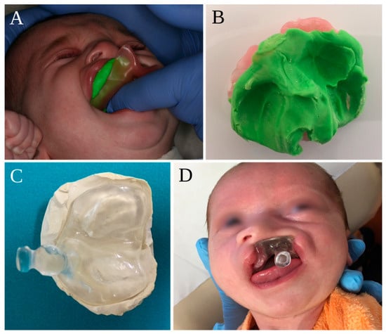
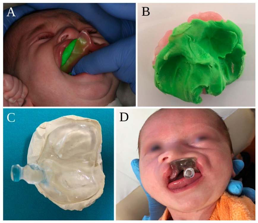
In the first appointment, an intra- and extra-oral physical examination was carried out, as well as an intraoral impression of the maxilla with putty-like addition silicone (soft putty/regular set, AquasilTM, Dentsply DeTrey GmbH, Konstanz, Germany) (Figure 1), using a prefabricated tray adapted to each patient. Subsequently, the NAM appliances were made of 2–4 mm thick self-curing acrylic to provide the necessary rigidity during the orthopedic treatment. An anterior retention button was added to the acrylic plate, with an angle of 40 degrees, to anchor the orthodontic elastics. A 5 mm hole was made in the center of the base, to prevent temporary obstruction of the airways in case of displacement of the device. Finally, and before insertion, each device was meticulously inspected, with the objective of verifying the limits of anatomical coverage to avoid intraoral trauma.
Figure 1.
Sequential stages of manufacturing of the NAM appliance: (A) intraoral impression of the maxilla with silicone; (B) the intraoral impression; (C) the acrylic plate and its anterior retention button; (D) placement of the NAM appliance in the oral cavity.
On the second appointment, the NAM device was placed. The appliance was anchored using two steri-strips (Steri StripTM, 3M, St. Paul, MN, USA) stretched onto the cheeks on either side and joined by orthodontic elastics (diameter 4.8 mm; force ± 185 g, DENTAURUM GmbH & Co.KG, Ispringen, Germany). These elastics were connected to the intraoral acrylic plate through the anterior retention button. The soft tissues were previously protected with a hydrocolloid membrane (DuoDerm®, ConvaTec Limited, Deeside, Flintshire, UK). The traction of each orthodontic elastic generated a force of approximately 100 g, leading to the approximation of the alveolar segments and promoting labial molding and premaxilla retraction. Another steri-strip was placed under tension across the cleft (including the prolabium in cases of bilateral cleft lip). On this appointment, parents were instructed to apply the NAM plate continuously throughout the day and night, removing it only for daily hygiene or in cases of choking.
NAM control was carried out weekly to improve NAM appliance fit and avoid contact ulcers. In this way, adjustments were made to the acrylic plate, less than one millimeter at a time, and the orthodontic force produced by the elastics was verified. When the interalveolar space was 5 mm, a nasal stent was connected to the intraoral acrylic plate. The nasal stent consisted of a stainless-steel wire with a kidney-shaped acrylic-free end to allow adaptation and pressure on the nasal tissues. All through NAM treatment, clinicians checked whether feeding and weight gains were normal.
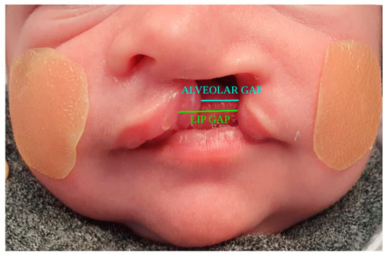
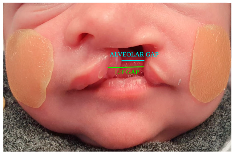
Cheiloplasty was performed from the age of 3 to 4 months. Parents of each patient were informed of the surgical techniques used and informed consent for the realization of surgeries was taken. Lip and alveolar gaps were measured before NAM, at the first appointment, and after NAM, i.e., immediately before cheiloplasty (Figure 2). The lip gap was measured as the distance between the margins of the cleft lip segments, and the alveolar gap as the shortest distance between the alveolar segments. All measurements were performed directly on the infant with a millimeter ruler and recorded to the nearest millimeter. To avoid inter-examiner variability, all measurements were performed by the same surgeon. No formal examiner calibration was performed, the examiner was not blinded to the treatment phase, and each measurement was recorded only once in the clinical records. Therefore, measurement error and intra- and inter-examiner reliability (e.g., intraclass correlation coefficients) could not be assessed retrospectively. During follow-up of the cheiloplasty, esthetic outcomes, including lip, columella, philtral column and nasal symmetry were assessed subjectively by the operating surgeon, without blinding and without standardized or validated assessment criteria. Palatoplasty was accomplished after 12 months of age.
Figure 2.
Periodic measurements of the lip gap and the alveolar gap were performed as depicted.
2.4. Statistical Analysis
Statistical analysis was performed using IBM® SPSS® v26 (IBM™, Armonk, NY, USA). Descriptive statistics are presented as means and standard deviations (SDs), or medians and interquartile ranges (IQRs), as appropriate. Changes in lip and alveolar gap before and after NAM were compared using two-tailed paired-samples t-tests, performed using complete pre- and post-NAM measurements for each outcome. Patients without complete paired measurements for a given outcome were excluded from that specific analysis, and no missing values were imputed. The normality of the paired differences was assessed using the Shapiro–Wilk test. Results are reported as mean differences with 95% confidence intervals (CIs), and effect sizes were calculated manually as Cohen’s d for paired samples (mean difference divided by the standard deviation of the differences). Given the small sample sizes, results were confirmed using the Wilcoxon signed-rank test. Owing to the small number of bilateral cases, the main analysis included unilateral and bilateral clefts together; descriptive statistics by cleft type are also presented, and a sensitivity analysis restricted to unilateral clefts was performed. Lip and alveolar gap reduction were defined a priori as the two primary outcomes; to account for multiple testing, results were also interpreted against a Bonferroni-adjusted significance level (α = 0.025). Analyses by cleft type were considered exploratory. Individual patient changes are presented graphically. Statistical significance was set at p < 0.05.
3. Results
This retrospective study included 19 patients: 13 unilateral cleft patients (68.4%), predominantly on the left side (n = 9), and 6 bilateral cleft patients (31.6%). NAM devices were applied at a median age of 4 weeks (IQR = 3.75–7.25). Figure 3 illustrates the different stages of treatment with NAM (before NAM, during NAM, after NAM and after cheiloplasty) through four exemplary cases.
Figure 3.
NAM therapy: Herein are depicted some examples of patients corresponding to those represented in Table 2. Each patient is shown in different stages of NAM.
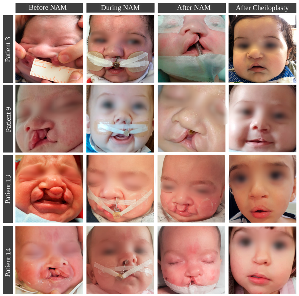
Table 2.
Features of pediatric patients with lip and palate cleft submitted to NAM protocols.
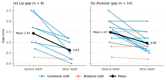
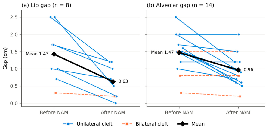
A nasal stent was applied to 10 patients (52.6%) 5.6 weeks (SD = 1.7) after initiating NAM and it was used for a median of 6.3 weeks (SD = 1.2). Paired-samples t-tests showed a significant reduction in both lip and alveolar gap after NAM. Lip gap decreased from 1.43 cm (SD = 0.81) before NAM to 0.63 cm (SD = 0.47) after NAM (n = 8), a mean reduction of 0.80 cm—95% CI [0.30, 1.30], t(7) = 3.76, p = 0.007, d = 1.33. Alveolar gap decreased from 1.47 cm (SD = 0.54) to 0.96 cm (SD = 0.48) (n = 14), a mean reduction of 0.51 cm—95% CI [0.23, 0.78], t(13) = 4.01, p = 0.001, d = 1.07. These correspond to mean reductions of 56% in lip gap and 34% in alveolar gap, respectively, both representing large effect sizes. These findings were confirmed by the Wilcoxon signed-rank test (lip gap: p = 0.008; alveolar gap: p = 0.005), and both remained significant after Bonferroni adjustment for the two primary outcomes. Individual changes are shown in Figure 4: the lip gap decreased in all 8 patients, whereas the alveolar gap decreased in 10 of 14 patients and remained unchanged in 4. The number of infants differed between analyses because missing measurements were attributable to incomplete clinical records or the impossibility of obtaining the measurements in some infants. Specifically, paired lip gap measurements were missing in 11 patients (pre-NAM value only, n = 3; post-NAM value only, n = 1; neither value, n = 7) and paired alveolar gap measurements in 5 patients (pre-NAM value only, n = 1; neither value, n = 4); three patients had no gap measurements recorded.
Figure 4.
Individual changes in (a) lip gap (n = 8) and (b) alveolar gap (n = 14) before and after NAM. Each line represents one patient (solid blue: unilateral cleft; dashed orange: bilateral cleft); the black line shows the mean. Points are slightly offset horizontally to reduce overlap.
Cheiloplasty was performed at a mean age of 18.1 weeks (SD = 4.7) and the Millard technique was used in all patients. Palatoplasty was performed at a median age of 13 months (IQR = 12–14.25). Von Langenbeck palatoplasty with intravelar veloplasty was the most used technique, performed in 15 cases (78.9%), whereas Veau–Wardill–Kilner and intravelar palatoplasty were performed in 3 cases (15.8%). In one case, Bardach palatoplasty was used.
When analyzed by cleft type (Table 3), the reduction remained significant in patients with unilateral clefts for both lip gap (n = 7; mean reduction 0.90 cm, 95% CI [0.37, 1.43], t(6) = 4.15, p = 0.006, d = 1.57) and alveolar gap (n = 11; mean reduction 0.64 cm, 95% CI [0.33, 0.94], t(10) = 4.67, p < 0.001, d = 1.41). In bilateral clefts, paired measurements were available for only one patient for lip gap and three patients for alveolar gap, and smaller changes were observed; no inferential analysis was performed for this subgroup.
Table 3.
Lip and alveolar gaps before and after NAM according to cleft type.
After cheiloplasty, there was satisfactory symmetry of the nose, columella and philtral column, as perceived by the surgeon, in 11 patients (58%), whereas nose asymmetry was observed in 7 patients (37%) and the columella and/or philtrum were asymmetric in 2 patients (10.5%). One patient underwent reintervention with a Z-plasty to repair misalignment of the muco-cutaneous line of the lip.
All cases of contact ulcers were temporary and solved with reshaping of the NAM device and/or temporary suspension, and there was no other reported local complication associated with NAM, namely infection. There was one case of inadaptation of the parents/infant to the NAM plate, which led to definitive NAM suspension in the second week of use, but it was not associated with complications. No cases of airway compromise were reported during NAM utilization.
4. Discussion
CLP is a common problem with life-long manifestations and morbidity. The presence of CLP leads to several problems, namely in feeding, hearing, phonation and dentofacial development, and these manifestations cause morbidity throughout patients’ lives [15]. The first surgery to correct the CLP is the best opportunity to obtain a desirable and lasting esthetic outcome, reducing the stigma of the cleft, and contributing to the correct psychological development and social interaction of affected children. The basis of presurgical orthopedic treatment was supported by studies carried out on auricular cartilage that have demonstrated increased plasticity in newborns. The presence of high estrogen levels in the first 6 weeks of life triggers an increase in hyaluronic acid that alters cartilage and connective tissue elasticity. Subsequently, several studies were carried out advocating the use of NAM in the first weeks of life [16].
NAM allows a non-invasive approximation of the tissues, with the intention of facilitating surgical techniques by reducing tension at the surgical site, potentially decreasing the need for primary complex surgical flaps with resulting visible scars [17]. Furthermore, it may improve function and decrease the number of secondary surgeries [13]. Some authors have already observed the shortening of the gap after NAM [12]. Facial esthetics after NAM and primary rhinoplasty were shown to be very close to those of patients without cleft lip [18,19]. This goes along with the results of the present study, in which lip and alveolar gaps were smaller after NAM than at baseline, which may facilitate surgical approaches. However, in the absence of a control group, this pre–post change cannot be attributed solely to NAM. On the other hand, complications associated with NAM have been reported to be infrequent and generally easy to manage. The commonest are ulcers of the lip and nasal tissues, which are usually resolved by reshaping the acrylic material and/or temporary suspension [7]. In the present study, only transient contact ulcers were observed, and no serious treatment-related adverse events occurred. However, the small sample size and retrospective design do not allow less frequent complications to be excluded.
In functional terms, during NAM treatment, the sealing of oronasal communication by the application of NAM reduces the risk of aspiration and nasal regurgitation, and improves nasal breathing capacity as well as food intake [14]. In the present study, caregivers revealed that the breastfeeding process through a feeding bottle was easier with the NAM plate in the oral cavity. Furthermore, some studies also reported that the optimization of function and lingual positioning can promote improvements in long-term phonetics [14].
Some authors described that the nasal stent allowed projection of the nose tip and promoted nonsurgical elongation of the columella, thus improving nasal convexity and symmetry, with stable long-term results [20,21]. Compared with infants who underwent the same surgical procedures without NAM, those treated with NAM have shown greater nostril height and columellar length in the short and long term, although the certainty of this evidence remains very low [20,21]. In this study, suboptimal nasal tip projection was observed despite the use of a nasal stent during NAM. Several factors may have contributed to this finding. First, the nasal stent was introduced only after the alveolar gap had decreased to approximately 5 mm, at a median age of 11.3 weeks (range, 8–20 weeks). This timing was beyond the first 6 weeks of life, a period associated with greater nasal cartilage plasticity [16], may have limited the potential for presurgical nasal molding. Second, postoperative changes in nasal shape may occur because of cartilage memory and soft-tissue contraction following primary cleft lip and nose repair [22]. Postoperative nasal conformers have been used as adjuncts to help maintain surgical correction, with some centers reporting their use for approximately 4 months after primary lip repair [23]. However, evidence regarding their effectiveness remains inconclusive [22,24]. As postoperative conformer use was not systematically assessed in this cohort, its contribution to nasal tip projection cannot be established. These findings highlight the potential importance of optimizing the timing of presurgical nasal stenting and further investigating the role of postoperative nasal conformers in prospective studies.
Nonetheless, NAM effectiveness is not consensual in the literature, in part because of the difficulty in assessing results due to heterogeneity of surgical timing, the subjectivity of outcomes, and the need for long-term follow-up [7], when concerning hearing, speech and language [25,26]. The treatment of these patients is not complete until adulthood, and only at this stage is it possible to assess the definitive results reported by the patient and caregiver, as well as the economic results. Some studies in the literature demonstrated that therapy with NAM did not improve facial esthetics, while other studies have argued that the treatment had no long-term effects on shape and nasal symmetry until the moment of rhinoplasty [12,27]. However, it is important to point out that the referred studies presented low-quality assessments. In fact, there are very few studies comparing patients submitted to NAM with a control group before surgery, most of them having retrospective or observational designs [17,28]. Published randomized controlled trials are even scarcer but show that NAM treatment is effective in minimizing cleft severity and realigning maxillary segments without compromising maxillary arch growth [29], even showing maxillary arch growth improvement instead [30,31].
Regarding the additional surgeries usually performed in these patients, some authors reported that 60% of the patients treated with NAM therapy and cheiloplasty did not require a secondary bone grafting in the cleft area [32]. This protocol has some long-term advantages from a socioeconomic point of view, namely a decrease in hospitalizations and reductions in more complex orthodontic treatments and the need for orthognathic surgery in adolescence and adulthood [2,13].
NAM therapy demands a multidisciplinary team, with strict coordination between pediatric surgery and orthodontics, as well as strong collaboration with parents, and the economic burden cannot be disregarded. Still, NAM appliances have been shown to have particularly high compliance among children and parents, which is in accordance with the results of this study, with only one case of NAM withdrawal [6,7].
From a personalized medicine perspective, the present findings support viewing NAM as an individualized intervention rather than a uniform protocol. Each appliance was custom-made from the infant’s own impression and adjusted weekly according to the clinical response, and the introduction of the nasal stent was guided by the individual reduction in the alveolar gap. The heterogeneity of individual responses, with a decrease in lip gap in all patients with paired measurements but no change in alveolar gap in four infants, and smaller changes in bilateral clefts, suggests that cleft type and baseline severity may modulate the response to NAM. These characteristics should therefore be considered when selecting candidates and when setting realistic treatment goals with families. Likewise, the late introduction of the nasal stent in some patients suggests that individualized timing, rather than a fixed alveolar gap threshold, might better exploit the period of greater nasal cartilage plasticity. Recent studies have shown that alternative presurgical approaches, such as presurgical lip, alveolus and nose approximation or nasal-only molding devices, may achieve comparable nasolabial outcomes with fewer visits and lower costs in selected patients [33], reinforcing the need to match the type and intensity of presurgical orthopedics to the characteristics and needs of each infant and family. In addition, digital workflows combining intraoral scanning, three-dimensional facial imaging and computer-aided design and manufacturing allow planned segment movements to be simulated and appliances to be tailored to each patient, with potential gains in accuracy, reproducibility and accessibility [34,35]. Integrating such objective, patient-specific data with clinical characteristics and family-reported outcomes may, in the future, support predictive models to identify which infants are most likely to benefit from NAM and which protocol best suits each of them.
The main limitation of this study is the absence of an untreated or alternative-treatment comparison group. Consequently, the reductions in lip and alveolar gaps represent observed pre–post changes rather than evidence of treatment effectiveness, as it is not possible to determine to what extent they are attributable to NAM rather than to spontaneous neonatal growth, maturation, or other components of the treatment protocol, such as lip taping. The sample was also small and heterogeneous in terms of sex and cleft type. The small sample size reduces statistical power and precision, as reflected in the confidence intervals, and may have prevented the detection of smaller effects. Moreover, with only three female patients and few bilateral cases, a stratified analysis by sex was not feasible, and the analysis by cleft type was exploratory, limiting the generalizability of the findings. The availability of complete paired measurements was another limitation. Although 19 patients were included, complete paired measurements were available for only 8 patients for the lip gap and 14 for the alveolar gap. Some measurements were unavailable because of incomplete clinical records or the inability to obtain measurements in some infants. This reduced the number of observations available for analysis and may have introduced bias if missing measurements were associated with patient characteristics or treatment outcomes. Some outcomes, particularly nasal, columellar, and philtral symmetry, were assessed subjectively by the treating surgeon only after cheiloplasty. These assessments were not blinded and may have been influenced by the clinician’s experience and expectations, introducing a risk of observer bias. In the absence of standardized objective measurements and preoperative assessments, it is not possible to distinguish the contribution of NAM from that of surgical repair. Therefore, the findings regarding facial symmetry should be interpreted with caution. Three-dimensional surface-imaging technologies, such as stereophotogrammetry and structured-light facial scanners, were not routinely available or integrated into the clinical workflow during most of the study period. These technologies could provide reproducible quantitative measurements of nasal symmetry, columellar length, nasal width, alar base position, philtral morphology, and three-dimensional facial asymmetry. Similarly, intraoral scans or digitized maxillary casts could improve the reliability of alveolar gap measurements. Future prospective studies should therefore incorporate objective assessments at baseline, after NAM, and after surgery, performed by independent, blinded examiners, with measurement reliability reported. Lip and alveolar gaps were measured directly on infants using a millimeter ruler, a method susceptible to measurement error in young, non-cooperative infants. Although all measurements were performed by the same surgeon, limiting inter-examiner variability, the magnitude of measurement error remains unknown. Nevertheless, the mean reductions observed (0.5–0.8 cm) were larger than the 1 mm resolution of the measurement method, although this does not exclude measurement error.
Unilateral and bilateral clefts were analyzed together because of the small sample size. The analysis by cleft type indicated that the reductions were mainly driven by unilateral clefts, whereas the few bilateral cases with paired data showed minimal changes in gap dimensions. This may reflect anatomical differences between cleft types, as NAM in bilateral clefts is primarily directed at retracting and aligning the premaxilla, which may not be fully captured by gap measurements. However, given the very small number of bilateral cases, no firm conclusions can be drawn for this subgroup. Finally, follow-up was limited to the early postoperative period after cheiloplasty. Long-term outcomes, including the stability of nasal and lip esthetics, nasal tip projection, maxillary growth, dental arch development, hearing, and speech, were not evaluated. Long-term follow-up is therefore needed to assess the stability of treatment outcomes and subsequent craniofacial development. Despite these limitations, the study has several strengths. First, a geneticist participated in the diagnostic assessment, helping to avoid the inclusion of patients with syndromic clefts. Second, pediatric surgery and orthodontics teams were present at all weekly consultations, supporting multidisciplinary care. Third, all data were recorded by medical professionals.
Given the controversy in the literature about NAM therapy, it is recommended to perform more research with standardized methodologies, larger sample sizes, and longer follow-up periods. Quantitative measurement tools should be used, and possible sources of bias must be controlled, such as the randomization procedure, treatment timing, type of surgery performed, and type of cleft.
5. Conclusions
Orthopedic modeling with NAM for CLP in the first weeks of life appears to be feasible and well tolerated, with high compliance among patients and caregivers. Substantial pre–post reductions in lip and alveolar cleft gaps were observed, particularly in unilateral clefts. However, in the absence of a control group, this pre–post change cannot be interpreted as evidence of treatment effectiveness. Similarly, although satisfactory nasal, columellar, and philtral symmetry was observed after cheiloplasty in most patients, the specific contribution of NAM to these outcomes remains uncertain. From a personalized medicine perspective, NAM highlights the importance of individualized treatment planning based on cleft characteristics and clinical assessment. Prospective studies with larger samples, objective and standardized digital assessments, and longer follow-up are needed to clarify the specific effects of NAM and identify patient-specific factors associated with treatment response.
Author Contributions
Conceptualization, V.C. and F.V.; methodology, V.C., I.F., A.P. and F.V.; validation, I.P., L.S. and J.L.; formal analysis, J.L. and I.F.; investigation, A.P., I.F., I.P. and L.S.; resources, V.C. and F.V.; writing—original draft preparation, J.L., L.S. and I.P.; writing—review and editing, I.F., V.C. and F.V.; visualization, I.F.; supervision, F.V. All authors have read and agreed to the published version of the manuscript.
Funding
This research received no external funding.
Institutional Review Board Statement
The study was conducted in accordance with the Declaration of Helsinki, and approved by the Ethics Committee of the Faculty of Medicine of the University of Coimbra (CE-063/2023, 14 June 2023).
Informed Consent Statement
Informed consent was obtained from all subjects involved in the study. Written informed consent has been obtained from the patients’ guardians (parents) to publish this paper.
Data Availability Statement
The datasets generated during and/or analyzed during the current study are available from the corresponding author on request.
Conflicts of Interest
The authors declare no conflicts of interest.
Abbreviations
The following abbreviations are used in this manuscript:
| NAM | Nasoalveolar molding |
| CLP | Cleft lip and palate |
References
- Kirschner, R.E.; LaRossa, D. Cleft lip and palate. Otolaryngol. Clin. N. Am. 2000, 33, 1191–1215. [Google Scholar] [CrossRef] [Scilit] [PubMed]
- Burg, M.L.; Chai, Y.; Yao, C.A.; Magee, W.; Figueiredo, J.C. Epidemiology, Etiology, and Treatment of Isolated Cleft Palate. Front. Physiol. 2016, 7, 67. [Google Scholar] [CrossRef] [Scilit] [PubMed]
- Worley, M.L.; Patel, K.G.; Kilpatrick, L.A. Cleft Lip and Palate. Clin. Perinatol. 2018, 45, 661–678. [Google Scholar] [CrossRef] [Scilit] [PubMed]
- Layuno Matos, M.; Greenlee, G.M.; Sheller, B. Orthodontic Care for Patients with Cleft Lip and Palate. Oral Maxillofac. Surg. Clin. N. Am. 2026, 38, 183–190. [Google Scholar] [CrossRef] [Scilit] [PubMed]
- Mercado, A.M.; Vig, K.W. The orthodontist’s role in a cleft palate-craniofacial team. In Orthodontics: Current Principles & Techniques, 5th ed.; Elsevier/Mosby: St Louis, MO, USA, 2012. [Google Scholar]
- Kuijpers-Jagtman, A.M. The orthodontist, an essential partner in CLP treatment. B-ENT 2006, 2, 57–62. [Google Scholar] [PubMed]
- Grayson, B.H.; Santiago, P.E.; Brecht, L.E.; Cutting, C.B. Presurgical nasoalveolar molding in infants with cleft lip and palate. Cleft Palate Craniofac. J. 1999, 36, 486–498. [Google Scholar] [CrossRef] [Scilit] [PubMed]
- Altuğ, A.T. Presurgical Nasoalveolar Molding of Bilateral Cleft Lip and Palate Infants: An Orthodontist’s Point of View. Turk. J. Orthod. 2017, 30, 118–125, Erratum in Turk. J. Orthod. 2017, 31, 31. [Google Scholar] [CrossRef] [Scilit] [PubMed]
- Nur Yilmaz, R.B.; Germeç Çakan, D. Nasolabial Morphology Following Nasoalveolar Molding in Infants With Unilateral Cleft Lip and Palate. J. Craniofac. Surg. 2018, 29, 1012–1016. [Google Scholar] [CrossRef] [Scilit] [PubMed]
- Kapadia, H.; Olson, D.; Tse, R.; Susarla, S.M. Nasoalveolar Molding for Unilateral and Bilateral Cleft Lip Repair. Oral Maxillofac. Surg. Clin. N. Am. 2020, 32, 197–204. [Google Scholar] [CrossRef] [Scilit] [PubMed]
- Spengler, A.L.; Chavarria, C.; Teichgraeber, J.F.; Gateno, J.; Xia, J.J. Presurgical nasoalveolar molding therapy for the treatment of bilateral cleft lip and palate: A preliminary study. Cleft Palate Craniofac. J. 2006, 43, 321–328. [Google Scholar] [CrossRef] [Scilit] [PubMed]
- Dunworth, K.; Porras Fimbres, D.; Trotta, R.; Hollins, A.; Shammas, R.; Allori, A.C.; Santiago, P.E. Systematic Review and Critical Appraisal of the Evidence Base for Nasoalveolar Molding (NAM). Cleft Palate Craniofac. J. 2024, 61, 654–677. [Google Scholar] [CrossRef] [Scilit] [PubMed]
- Nayak, T.; Bonanthaya, K.; Parmar, R.; Shetty, P.N. Long-Term Comparison of the Aesthetic Outcomes between Nasoalveolar Molding- and Non-Nasoalveolar Molding-Treated Patients with Unilateral Cleft Lip and Palate. Plast. Reconstr. Surg. 2021, 148, 775e–784e. [Google Scholar] [CrossRef] [Scilit] [PubMed]
- Thiruvenkatachari, B.; Vaidhyalingam, T.; Chakkaravarthi, S.; Prathap, M.; Nambiar, K. A survey on nasoalveolar moulding treatment practices at cleft centres across India. Front. Surg. 2025, 12, 1526364. [Google Scholar] [CrossRef] [Scilit] [PubMed]
- Francisco, I.; Antonarakis, G.S.; Caramelo, F.; Fernandes, M.H.; Vale, F. Cleft Orthodontic Care in Europe: A Cross-Sectional Survey. Healthcare 2022, 10, 1555. [Google Scholar] [CrossRef] [Scilit] [PubMed]
- Matsuo, K.; Hirose, T.; Tomono, T.; Iwasawa, M.; Katohda, S.; Takahashi, N.; Koh, B. Nonsurgical correction of congenital auricular deformities in the early neonate: A preliminary report. Plast. Reconstr. Surg. 1984, 73, 38–51. [Google Scholar] [CrossRef] [Scilit] [PubMed]
- Rossell-Perry, P.; Olivencia-Flores, C.; Delgado-Jimenez, M.P.; Ormeño-Aquino, R. Surgical Nasoalveolar Molding: A Rational Treatment for Bilateral Cleft Lip Nose and Systematic Review. Plast. Reconstr. Surg. Glob. Open 2020, 8, e3082. [Google Scholar] [CrossRef] [Scilit] [PubMed]
- Meazzini, M.C.; Parravicini, F.; Donati, V.; Brusati, R.; Biglioli, F.; Autelitano, L. Photometric Evaluation of Adult Patients with Bilateral Cleft Lip and Palate Treated with Nasoalveolar Molding and Primary Columella Lengthening. Cleft Palate Craniofac. J. 2022, 59, 852–858. [Google Scholar] [CrossRef] [Scilit] [PubMed]
- Likitkulthanaporn, A.; Boonpratham, S.; Honglertnapakul, Y.; Saengfai, N.N.; Chaweewannakorn, C.; Jongkhum, N.; Peanchitlertkajorn, S. Effects of Nasoalveolar Molding on Nasolabial Aesthetics in Patients with Cleft Lip and Palate during Pre-Adolescence: A Systematic Review and Meta-Analysis of Retrospective Studies. Orthod. Craniofac. Res. 2024, 27, 350–363. [Google Scholar] [CrossRef] [Scilit] [PubMed]
- Ritschl, L.M.; Narbekovas, K.; Loeffelbein, D.J.; Grabenhorst, A.; Krautkremer, N.; Singer, H.; Grandoch, A.; Kram, H.; Wolff, K.D.; Grill, F.D. Three-dimensional longitudinal assessment of nasal development with and without Nasoalveolar molding therapy in cleft lip and palate patients. Clin. Oral Investig. 2025, 29, 487. [Google Scholar] [CrossRef] [Scilit] [PubMed]
- Souza, T.M.; de Abreu Refaxo, N.; Macari, S.; Abreu, L.G. Nasal Symmetry Outcomes After Nasoalveolar Molding (NAM) Plus Cheiloplasty Treatment in Babies With Cleft Lip/Palate: Systematic Review and Meta-Analysis. Cleft Palate Craniofac. J. 2026, 63, 1688–1700. [Google Scholar] [CrossRef] [Scilit] [PubMed]
- Marston, A.P.; Tollefson, T.T. Is Postoperative Nasal Stenting Necessary After Primary Cleft Lip and Nose Repair? The Laryngoscope 2025, 135, 971–972. [Google Scholar] [CrossRef] [Scilit] [PubMed]
- Hennocq, Q.; Person, H.; Hachani, M.; Bertin, H.; Corre, P.; Gorbonosov, V.; Ivanov, A.; Khonsari, R.H. Quality of life and nasal splints after primary cleft lip and nose repair: Prospective assessment of information and tolerance. J. Craniomaxillofac. Surg. 2018, 46, 1783–1789. [Google Scholar] [CrossRef] [Scilit] [PubMed]
- Rossell-Perry, P.; Romero-Narvaez, C.; Gavino-Gutierrez, A.; Figallo-Hudtwalcker, O. Postoperative Nasal Conformers in Cleft Rhinoplasty: Are They Efficacious? J. Craniofac. Surg. 2023, 34, 1416–1419. [Google Scholar] [CrossRef] [Scilit] [PubMed]
- Masarei, A.G.; Wade, A.; Mars, M.; Sommerlad, B.C.; Sell, D. A randomized control trial investigating the effect of presurgical orthopedics on feeding in infants with cleft lip and/or palate. Cleft Palate Craniofac. J. 2007, 44, 182–193. [Google Scholar] [CrossRef] [Scilit] [PubMed]
- Knösel, M.; Fendel, C.; Jung, K.; Sandoval, P.; Engelke, W.G. Presurgical orthopedics by drink plates does not significantly normalize deglutition in infants with cleft lip and palate. Angle Orthod. 2016, 86, 315–323. [Google Scholar] [CrossRef] [Scilit] [PubMed]
- Thiruvenkatachari, B.; Chakkaravarthi, S.; Bhuvaraghan, A. Effectiveness of Nasoalveolar Molding Treatment for Babies with Nonsyndromic Complete Unilateral Cleft Lip, Alveolus, and Palate: A Systematic Review and Meta-Analysis. Am. J. Orthod. Dentofac. Orthop. 2025, 168, 2–13.e4. [Google Scholar] [CrossRef] [Scilit] [PubMed]
- Hosseini, H.R.; Kaklamanos, E.G.; Athanasiou, A.E. Treatment outcomes of pre-surgical infant orthopedics in patients with non-syndromic cleft lip and/or palate: A systematic review and meta-analysis of randomized controlled trials. PLoS ONE 2017, 12, e0181768. [Google Scholar] [CrossRef] [Scilit] [PubMed]
- Saad, M.S.; Fata, M.; Farouk, A.; Habib, A.M.A.; Gad, M.; Tayel, M.B.; Marei, M.K. Early Progressive Maxillary Changes with Nasoalveolar Molding: Randomized Controlled Clinical Trial. JDR Clin. Trans. Res. 2020, 5, 319–331. [Google Scholar] [CrossRef] [Scilit] [PubMed]
- Shetty, V.; Agrawal, R.K.; Sailer, H.F. Long-term effect of presurgical nasoalveolar molding on growth of maxillary arch in unilateral cleft lip and palate: Randomized controlled trial. Int. J. Oral Maxillofac. Surg. 2017, 46, 977–987. [Google Scholar] [CrossRef] [Scilit] [PubMed]
- Abd El-Ghafour, M.; Aboulhassan, M.A.; Fayed, M.M.S.; El-Beialy, A.R.; Eid, F.H.K.; Hegad, S.E.; El-Gendi, M.; Emara, D. Effectiveness of a Novel 3D-Printed Nasoalveolar Molding Appliance (D-NAM) on Improving the Maxillary Arch Dimensions in Unilateral Cleft Lip and Palate Infants: A Randomized Controlled Trial. Cleft Palate Craniofac. J. 2020, 57, 1370–1381. [Google Scholar] [CrossRef] [Scilit] [PubMed]
- Jahanbin, A.; Alizadeh, F.L.; Bardideh, E.; Sharifi, S.; Nazari, M.S. Does Presurgical Nasoalveolar Molding Reduce the Need for Future Bone Grafting in Cleft Lip and Palate Patients? A Systematic Review and Meta-Analysis. J. Craniofac. Surg. 2022, 33, 2095–2099. [Google Scholar] [CrossRef] [Scilit] [PubMed]
- Perez Rivera, L.R.; Kantar, R.S.; Rrushi, V.; Staffenberg, D.A.; Flores, R.L.; Shetye, P.R. An Institutional Comparative Cost Analysis Between Nasoalveolar Molding and Presurgical Lip, Alveolus, and Nose Approximation. J. Craniofac. Surg. 2026. Online ahead of print. [Google Scholar] [CrossRef] [Scilit] [PubMed]
- Nguyen, B.K.; Caro, C.; Stevens, K.; Garisto, G.A.; Finer, Y. Computer-Aided Design and Computer-Aided Manufacturing Technology for Conducting Nasoalveolar Molding for Infants With Cleft Lip and Palate: A Scoping Review. Cleft Palate Craniofac. J. 2026, 63, 2698–2712. [Google Scholar] [CrossRef] [Scilit] [PubMed]
- Velickovic, A.; Rossouw, P.E.; Javed, F. Effectiveness of digitally- versus conventionally-fabricated nasoalveolar molding appliances for presurgical approximation of complete cleft lip and palate: A systematic review and meta-analysis of randomized controlled trials. J. Stomatol Oral Maxillofac. Surg. 2026, 127, 102796. [Google Scholar] [CrossRef] [Scilit] [PubMed]

Disclaimer/Publisher’s Note: The statements, opinions and data contained in all publications are solely those of the individual author(s) and contributor(s) and not of MDPI and/or the editor(s). MDPI and/or the editor(s) disclaim responsibility for any injury to people or property resulting from any ideas, methods, instructions or products referred to in the content. |
© 2026 by the authors. Licensee MDPI, Basel, Switzerland. This article is an open access article distributed under the terms and conditions of the Creative Commons Attribution (CC BY) license.