


Abstract
Hypertrophic cardiomyopathy (HCM) is a genetically and phenotypically heterogeneous myocardial disease characterized by unexplained left ventricular hypertrophy and a broad spectrum of clinical manifestations, ranging from an asymptomatic phenotype to heart failure, atrial and ventricular arrhythmias, sudden cardiac death, and advanced heart failure. Accurate diagnosis and risk stratification therefore require complex assessment. Multimodality cardiovascular imaging provides complementary structural, functional, and tissue characterization that cannot be obtained using a single imaging technique. Transthoracic echocardiography remains the cornerstone of initial diagnosis, hemodynamic assessment, and longitudinal follow-up, while advanced echocardiographic techniques provide additional information regarding myocardial and left atrial mechanics. Cardiac magnetic resonance (CMR) has become central to phenotypic characterization and tissue assessment. Cardiac computed tomography offers complementary high-resolution anatomical information, particularly for coronary assessment and procedural planning, whereas nuclear imaging, provides insights into coronary microvascular dysfunction, myocardial perfusion, and selected phenocopies such as transthyretin cardiac amyloidosis. Multimodality imaging also facilitates differentiation of HCM from other conditions such as athlete’s heart, hypertensive heart disease, and infiltrative cardiomyopathies. Integration of imaging biomarkers with clinical and genetic information may improve prediction of adverse outcomes. Thus, multimodality imaging should be regarded not as a collection of competing techniques but as an integrated framework for precision diagnosis, risk stratification, therapeutic planning, and longitudinal management of patients with HCM.
1. Introduction
Hypertrophic cardiomyopathy (HCM) is the most common inherited cardiomyopathy and is characterized by unexplained left ventricular (LV) hypertrophy in the absence of abnormal loading conditions sufficient to account for the degree of hypertrophy [1]. The estimated prevalence of HCM is approximately 1:500 in the general population, although contemporary studies incorporating genotype-positive individuals and advanced imaging suggest that the true prevalence may be higher. HCM is associated with substantial clinical heterogeneity, ranging from asymptomatic individuals with normal life expectancy to patients presenting with heart failure, atrial fibrillation, ventricular arrhythmias, sudden cardiac death (SCD), or advanced heart failure requiring transplantation [2,3,4].
The genetic basis of HCM is complex and is mostly related to pathogenic variants affecting sarcomeric proteins involved in cardiac contraction and force generation [5]. The disease exhibits marked genetic and phenotypic variability, with incomplete penetrance, age-dependent expression, and variable clinical manifestations even among individuals carrying the same pathogenic variant [6].
The clinical expression of HCM is highly heterogeneous and reflects complex interactions between genetic background, myocardial remodelling, fibrosis, arrhythmogenic substrate formation, and environmental factors. Morphological patterns vary considerably, including asymmetric septal hypertrophy, reverse curvature septal morphology, concentric hypertrophy, mid-ventricular obstruction, and isolated apical hypertrophy. Beyond increased myocardial thickness, disease progression is characterized by diastolic dysfunction, left atrial remodelling, myocardial fibrosis, microvascular dysfunction, and in advanced stages, LV systolic impairment. These diverse phenotypes contribute to the complexity of diagnosis, prognostication, and therapeutic decision-making [2,3].
Imaging plays a central role throughout the clinical course of HCM, from initial diagnosis and phenotypic characterization to risk stratification and treatment planning. Transthoracic echocardiography remains the first-line imaging technique due to its widespread availability, ability to assess cardiac morphology and function, and unique capacity to evaluate dynamic LV outflow tract (LVOT) obstruction at rest and during physiological provocation [7].
Cardiac magnetic resonance (CMR) has emerged as a cornerstone of contemporary HCM evaluation by providing high-resolution assessment of ventricular morphology, accurate quantification of myocardial mass and volumes, and comprehensive tissue characterization [8]. CMR also improves recognition of atypical phenotypes, including apical HCM, LV aneurysm formation, and myocardial involvement in phenocopies [9].
Although echocardiography and CMR represent the foundation of HCM imaging, other modalities provide complementary information in selected clinical scenarios. Cardiac computed tomography (CT) offers excellent spatial resolution for coronary assessment, anatomical characterization, and procedural planning, particularly in patients undergoing septal reduction therapy or those unable to undergo CMR. Nuclear imaging techniques, particularly positron emission tomography (PET), provide unique insights into myocardial perfusion, coronary microvascular dysfunction, and metabolic activity, expanding the diagnostic spectrum beyond structural assessment [10].
The increasing complexity of HCM phenotyping has shifted the diagnostic paradigm from a single-modality approach toward integrated multimodality imaging. Combining the strengths of echocardiography, CMR, CT, and nuclear imaging allows more accurate differentiation between HCM and phenocopies, improves risk assessment beyond traditional clinical variables, and supports individualized therapeutic strategies. Current guidelines emphasize the complementary role of different imaging modalities in diagnosis, family screening, sudden cardiac death risk assessment, and longitudinal disease monitoring [3,11].
The aim of this review is to provide a contemporary overview of the role of multimodality imaging in patients with HCM, focusing on the contribution of echocardiography, CMR, CT, and nuclear imaging to diagnosis, phenotypic characterization, risk stratification, and clinical management. Furthermore, we discuss emerging imaging biomarkers, artificial intelligence applications, and future perspectives toward precision imaging-based care in HCM.
Literature Search Strategy
Two independent reviewers (N.M. and P.P.) systematically searched PubMed, MEDLINE and SCOPUS. Studies were included in the narrative review according to the following eligibility criteria: (1) studies comprising patients with any form of hypertrophic cardiomyopathy or other conditions that may present as phenocopies, and (2) studies evaluated with non-invasive imaging.
The main search was performed with the following Medical Subject Headings (Mesh) and free-text terms:
(“Cardiomyopathy” OR “Hypertrophic cardiomyopathy” OR “Hypertensive heart”) AND (“Imaging” OR “Multimodality imaging” OR “Echocardiography” OR “Cardiac magnetic resonance” OR “Computed tomography”] OR “Nuclear Imaging” “Positron emission tomography”). We conducted a modified narrative review according to recent recommendations [5]. The search was performed before July 2026, and all relevant data was derived from research articles, reviews, case reports, etc.
2. Imaging Targets in Hypertrophic Cardiomyopathy
The clinical complexity of hypertrophic cardiomyopathy reflects the interaction between myocardial hypertrophy, abnormal ventricular mechanics, tissue remodelling, and progressive structural changes. Contemporary imaging aims not only to confirm the diagnosis but also to characterize disease phenotype, identify prognostically relevant features, and guide therapeutic decision-making (Table 1). Although increased left ventricular wall thickness remains the defining morphological feature of HCM, a comprehensive imaging assessment should evaluate multiple disease substrates, including dynamic obstruction, diastolic dysfunction, mitral valve abnormalities, myocardial fibrosis, microvascular dysfunction, atrial remodelling, apical aneurysm formation, and right ventricular involvement (Figure 1). The integration of these parameters provides a more complete assessment of disease severity and clinical risk than any single imaging marker.
Table 1.
Strengths and limitations of multimodality imaging techniques in hypertrophic cardiomyopathy.
Figure 1.
Non-invasive imaging modalities for the evaluation of patients with hypertrophic cardiomyopathy and main findings. TTE—transthoracic echocardiography; 3D—three dimensional; LV—left ventricular; LA—left atrium; EF—ejection fraction; CT—computed tomography; ASA—alcohol septal ablation; CMR—cardiac magnetic resonance; PET—positron emission tomography; CFR—coronary flow reserve; MRI—magnetic resonance imaging; LVOT—left-ventricular outflow tract; LGE—late gadolinium enhancement; ECV—extracellular volume.
2.1. Left Ventricular Hypertrophy
The hallmark of HCM is unexplained LV hypertrophy, traditionally defined as a maximal LV wall thickness ≥ 15 mm in adults in the absence of other loading conditions capable of producing a similar degree of hypertrophy. In individuals with a pathogenic sarcomeric variant or affected first-degree relatives, a lower threshold of 13–14 mm may support the diagnosis when accompanied by additional features [2,3]. Echocardiography remains the initial modality for identifying hypertrophy patterns and measuring maximal wall thickness; however, cardiac magnetic resonance provides superior spatial resolution and more accurate assessment of LV mass, hypertrophy distribution, and atypical phenotypes [12].
The morphological expression of HCM is heterogeneous, including asymmetric septal hypertrophy, reverse curvature septal morphology, concentric hypertrophy, mid-ventricular hypertrophy, and isolated apical involvement. CMR is particularly valuable for identifying apical HCM and localized hypertrophy involving the anterolateral wall or papillary muscles, which may be underestimated by echocardiography [13]. Beyond wall thickness, assessment of LV geometry, cavity size, and remodelling pattern provides important information regarding disease progression and prognosis. Advanced imaging techniques, including three-dimensional echocardiography and CMR-based feature tracking, allow more comprehensive evaluation of ventricular structure and mechanics.
2.2. Dynamic Left Ventricular Outflow Tract Obstruction
Dynamic LV outflow tract (LVOT) obstruction is a major pathophysiological feature of HCM and results from complex interactions between basal septal hypertrophy, systolic anterior motion (SAM) of the mitral valve, altered ventricular geometry, and abnormal flow dynamics. Resting LVOT obstruction is generally defined as a peak instantaneous gradient ≥30 mmHg, while gradients ≥ 50 mmHg are considered clinically significant when evaluating candidates for septal reduction therapy [2,3].
Transthoracic echocardiography is the primary imaging modality for assessing LVOT obstruction because it enables real-time evaluation of gradients at rest and during physiological provocation, including Valsalva maneuver and exercise. Exercise stress echocardiography is particularly important in symptomatic patients without significant resting obstruction, as latent obstruction may be unmasked during exertion [14]. CMR provides complementary information by accurately characterizing septal morphology, mitral valve anatomy, papillary muscle abnormalities, and the mechanisms contributing to obstruction. Four-dimensional flow CMR is an emerging technique that allows visualization and quantification of abnormal intracardiac flow patterns, although its routine clinical application remains under investigation [15].
2.3. Diastolic Dysfunction
Impaired diastolic function is a fundamental component of HCM and results from myocardial hypertrophy, increased ventricular stiffness, impaired relaxation, and interstitial fibrosis. Elevated LV filling pressures contribute to exercise intolerance, dyspnoea, pulmonary hypertension, and left atrial enlargement [3].
Echocardiography remains central for the evaluation of diastolic function using parameters including mitral inflow velocities, tissue Doppler-derived e′ velocity, E/e′ ratio, left atrial volume, and pulmonary venous flow patterns. Global longitudinal strain and myocardial work analysis provide additional insights into myocardial dysfunction that may precede changes in ejection fraction [16]. CMR contributes through assessment of ventricular volumes, myocardial stiffness surrogates, and tissue characterization. Increased extracellular volume fraction (ECV) and native T1 values reflect diffuse myocardial remodelling and have been associated with impaired ventricular relaxation and adverse clinical outcomes [17].
2.4. Mitral Valve Abnormalities
Mitral valve abnormalities are common in HCM and contribute significantly to symptoms and LVOT obstruction. These abnormalities include systolic anterior motion of the mitral valve, elongated mitral leaflets, abnormal papillary muscle insertion, accessory papillary muscles, and altered mitral-septal geometry [18]. Echocardiography provides the primary assessment of SAM, mitral regurgitation severity, and dynamic interaction between the mitral valve and LVOT. Three-dimensional echocardiography improves anatomical characterization by providing detailed assessment of leaflet morphology and subvalvular structures. CMR and cardiac computed tomography (CT) provide complementary anatomical information, particularly when planning surgical intervention or septal reduction procedures [19]. Recognition of mitral valve abnormalities is essential because obstruction may result from mechanisms beyond septal hypertrophy alone, influencing the choice of therapeutic strategy.
2.5. Myocardial Fibrosis
Myocardial fibrosis represents one of the most important imaging targets in HCM because it reflects disease progression and contributes to electrical instability. Fibrosis creates an arrhythmogenic substrate by disrupting myocardial architecture and promoting conduction heterogeneity. CMR with LGE is the established non-invasive technique for detecting focal replacement fibrosis. LGE in HCM typically demonstrates a patchy mid-wall distribution within hypertrophied segments, although patterns may vary. Extensive LGE has been associated with increased risk of ventricular arrhythmias, sudden cardiac death (SCD), and heart failure progression [9]. Quantitative assessment of LGE burden, particularly values ≥15% of LV mass, has gained importance in contemporary risk assessment strategies [3]. Parametric CMR techniques, including native T1 mapping and extracellular volume quantification, provide assessment of diffuse interstitial fibrosis and may identify myocardial abnormalities before extensive focal fibrosis develops. Elevated native T1 and ECV values have been associated with adverse remodelling and clinical outcomes, although their integration into routine risk algorithms requires further validation [17].
2.6. Coronary Microvascular Dysfunction
Coronary microvascular dysfunction is increasingly recognized as an important component of HCM pathophysiology. Increased myocardial mass, abnormal intramyocardial arteriolar structure, and impaired coronary vasodilatory capacity contribute to myocardial ischemia despite normal epicardial coronary arteries [10]. Positron emission tomography (PET) allows quantitative assessment of myocardial blood flow and coronary flow reserve (CFR). Reduced CFR has been associated with myocardial fibrosis, ventricular dysfunction, heart failure progression, and adverse cardiovascular outcomes [20]. Stress perfusion CMR provides an alternative approach for evaluating myocardial ischemia and microvascular dysfunction, although PET remains the reference method for quantitative assessment of myocardial blood flow. The clinical role of microvascular imaging is evolving and may become increasingly relevant for identifying patients at risk of progressive remodelling.
2.7. Left Atrial Remodelling
Left atrial (LA) enlargement and dysfunction are common consequences of chronic elevation of LV filling pressures in HCM. LA remodelling reflects the cumulative burden of diastolic dysfunction and is strongly associated with atrial fibrillation (AF), stroke, and heart failure symptoms [21]. Echocardiographic assessment of LA volume index remains a widely used marker of chronic diastolic burden. More recently, LA strain analysis has emerged as a sensitive marker of early atrial dysfunction and may provide incremental prognostic information beyond LA size [22,23]. CMR provides highly reproducible measurements of LA volumes and function and allows assessment of atrial fibrosis using late gadolinium enhancement techniques, although atrial tissue characterization remains primarily a research application [24].
2.8. Apical Aneurysm
LV apical aneurysm represents a distinct HCM phenotype characterized by localized apical thinning and dyskinesia, frequently associated with mid-ventricular obstruction. Although relatively uncommon, apical aneurysms are clinically important because they are associated with ventricular arrhythmias, thrombus formation, embolic events, and SCD [25]. CMR is superior to echocardiography for detecting apical aneurysms due to its ability to accurately visualize the LV apex and characterize associated scar tissue using LGE. Identification of apical aneurysm has become an important component of contemporary HCM risk assessment [26].
2.9. Right Ventricular Involvement
Although HCM primarily affects the LV, right ventricular (RV) abnormalities may occur and reflect more advanced myocardial involvement. RV hypertrophy, dilation, systolic dysfunction, and abnormal RV-pulmonary artery coupling have been described in subsets of patients and may contribute to exercise intolerance and adverse outcomes [27]. Echocardiography provides initial assessment of RV size and function, including tricuspid annular plane systolic excursion (TAPSE) and RV strain. CMR remains the reference technique for RV volume and function quantification due to its high reproducibility. RV involvement is not a primary diagnostic criterion for HCM but may provide additional information regarding disease severity and prognosis [28].
3. Echocardiography
Echocardiography remains the cornerstone of imaging evaluation in patients with hypertrophic cardiomyopathy and represents the first-line modality for diagnosis, phenotypic characterization, hemodynamic assessment, and longitudinal follow-up [2,3]. Its central role is based on widespread availability, lack of ionizing radiation, relatively low cost, and the ability to provide real-time assessment of cardiac structure and function. Importantly, echocardiography uniquely enables dynamic evaluation of LVOT obstruction during physiological maneuvers and exercise, which is essential because obstruction severity may vary considerably under resting conditions [14]. Although CMR has become increasingly important for tissue characterization and refined phenotyping, echocardiography remains indispensable in routine clinical practice. A comprehensive echocardiographic examination in HCM should include assessment of LV hypertrophy pattern, maximal wall thickness, LVOT gradients, systolic anterior motion (SAM) of the mitral valve, mitral regurgitation, LV systolic and diastolic function, left atrial size, and right ventricular function [29]. Recent advances in echocardiographic techniques, including three-dimensional echocardiography, speckle-tracking strain imaging, LA strain analysis, and myocardial work assessment, have expanded the ability of echocardiography to detect subtle abnormalities beyond conventional parameters.
3.1. Conventional Transthoracic Echocardiography
Transthoracic echocardiography (TTE) is the initial imaging modality for suspected HCM and remains the primary technique for establishing the morphological diagnosis. The diagnosis is generally based on the presence of unexplained LV hypertrophy, usually defined as maximal LV wall thickness ≥15 mm in adults, or ≥13 mm in individuals with a pathogenic variant or affected first-degree relative [2,3].
TTE provides essential information regarding the distribution and severity of hypertrophy, including asymmetric septal hypertrophy, concentric hypertrophy, reverse curvature morphology, mid-ventricular hypertrophy, and apical involvement. Measurements should be obtained in multiple imaging planes to avoid underestimation or overestimation of wall thickness, particularly in patients with asymmetric or localized forms of disease. Beyond morphology, conventional echocardiography provides critical hemodynamic assessment. Continuous-wave Doppler allows quantification of LVOT gradients, while colour Doppler evaluates associated mitral regurgitation. The detection of SAM of the mitral valve and its interaction with the hypertrophied septum provides important mechanistic information regarding obstruction [30]. Assessment of diastolic function is also a fundamental component of echocardiographic evaluation in HCM. Parameters including mitral inflow velocities, tissue Doppler-derived early diastolic mitral annular velocity (e′), the E/e′ ratio, LA volume index, and pulmonary artery pressure estimates provide indirect measures of LV filling pressures and disease burden [29]. However, conventional echocardiography has limitations. Image quality is dependent on acoustic windows and operator expertise, and certain phenotypes, including apical HCM, anterolateral hypertrophy, and focal hypertrophy, may be incompletely visualized. In such cases, CMR provides complementary or definitive anatomical assessment [31].
3.2. Stress Echocardiography
Dynamic LVOT obstruction is a hallmark of HCM and may be absent at rest despite clinically significant symptoms. Exercise stress echocardiography is therefore an essential component of evaluation in symptomatic patients with no significant resting obstruction. It provides simultaneous assessment of LVOT gradients, exercise-induced mitral regurgitation, blood pressure response, and symptom reproduction [32]. The physiological basis of stress-induced obstruction involves increased contractility, reduced ventricular cavity size, and enhanced Venturi and drag forces contributing to SAM of the mitral valve. A peak LVOT gradient ≥ 50 mmHg during exercise is clinically relevant, particularly when considering septal reduction therapy [3]. Exercise echocardiography has demonstrated clinical value by identifying patients with latent obstruction and explaining symptoms that may otherwise remain unexplained. It also provides prognostic information, as exercise-induced obstruction and abnormal blood pressure response have been associated with increased morbidity in selected patients [33]. Pharmacological stress testing with dobutamine is generally discouraged for routine evaluation of HCM because it may provoke gradients that do not reflect physiological exercise conditions and can produce false-positive results. Exercise-based protocols are preferred when feasible [3].
3.3. Three-Dimensional Echocardiography
Three-dimensional (3D) echocardiography provides volumetric assessment of cardiac chambers and improves anatomical visualization compared with conventional two-dimensional imaging. In HCM, 3D echocardiography may enhance evaluation of LV morphology, hypertrophy distribution, mitral valve anatomy, and papillary muscle abnormalities [34].
One important advantage of 3D echocardiography is improved assessment of LV volumes and ejection fraction, reducing geometric assumptions inherent to two-dimensional methods. This may be particularly relevant in HCM, where abnormal ventricular geometry can limit the accuracy of conventional measurements [35]. Three-dimensional imaging also contributes to procedural planning by providing detailed visualization of the mitral valve apparatus and subvalvular structures. This information may help identify patients in whom LVOT obstruction is driven not only by septal hypertrophy but also by elongated mitral leaflets or abnormal papillary muscle insertion [18]. Despite these advantages, widespread implementation remains limited by lower temporal resolution compared with two-dimensional echocardiography, dependence on image quality, and limited availability of expertise.
3.4. Speckle-Tracking Echocardiography
Speckle-tracking echocardiography (STE) has become an important tool for detecting subtle myocardial dysfunction in HCM patients with preserved LV ejection fraction (LVEF). Global longitudinal strain (GLS), derived from tracking myocardial deformation during the cardiac cycle, identifies impaired longitudinal shortening caused by hypertrophy, fibrosis, and myocardial disarray [36,37]. Reduced GLS is frequently observed in HCM despite preserved LVEF and has been associated with increased myocardial fibrosis, adverse remodelling, heart failure progression, and ventricular arrhythmias [38]. The regional distribution of strain abnormalities may also provide mechanistic information, with reduced deformation commonly observed in hypertrophied segments. STE may have particular value in early disease detection, including genotype-positive individuals without overt hypertrophy, although its role in routine clinical decision-making remains under investigation. Serial assessment of GLS may also provide a sensitive marker of disease progression and response to therapy [38]. Limitations include variability related to vendor-specific software, loading conditions, image quality, and differences in acquisition protocols. Standardization remains an important challenge before strain parameters can be universally incorporated into clinical risk models.
3.5. Left Atrial Strain
LA remodelling is a major consequence of chronic elevation of LV filling pressures in HCM and is strongly associated with atrial fibrillation (AF), stroke, and heart failure symptoms. Conventional assessment relies mainly on LA volume; however, LA strain provides additional information regarding atrial mechanical function and reservoir properties [39]. LA strain analysis evaluates the three phases of atrial function: reservoir, conduit, and contractile phases. Reduced LA reservoir strain reflects impaired atrial compliance and increased ventricular filling pressures and may precede measurable LA enlargement [40]. Several studies have demonstrated associations between reduced LA strain and incident AF, exercise intolerance, diastolic dysfunction severity, and adverse outcomes in HCM [41]. Because LA strain integrates the cumulative effects of LV stiffness and myocardial remodelling, it may represent an early marker of disease progression. Nevertheless, clinical adoption remains limited by differences in methodology, reference values, and software platforms. Further prospective studies are needed to establish its incremental value over conventional LA parameters.
3.6. Myocardial Work Analysis
Myocardial work analysis is an emerging echocardiographic technique that combines LV strain measurements with estimated LV pressure curves to provide load-adjusted indices of myocardial performance. Unlike GLS alone, myocardial work incorporates afterload and ventricular pressure generation, potentially providing a more physiologically meaningful assessment of myocardial function in various clinical scenarios [42,43]. In HCM, myocardial work analysis has demonstrated abnormalities in patients with preserved LVEF and may provide additional information regarding mechanical inefficiency, dyssynchrony, and disease severity [44,45]. Alterations in myocardial work indices have been associated with hypertrophy burden, fibrosis markers, and functional limitation. The clinical relevance of myocardial work in HCM remains investigational, and its role in routine risk stratification requires validation in larger longitudinal cohorts.
3.7. Strengths and Limitations of Echocardiography in HCM
The major strengths of echocardiography are its accessibility, portability, absence of radiation exposure, excellent temporal resolution, and unique ability to assess dynamic hemodynamics. It remains the most practical modality for initial diagnosis, family screening, serial follow-up, and assessment of LVOT obstruction.
However, echocardiography has limitations, particularly regarding tissue characterization and detection of complex phenotypes. It cannot directly quantify myocardial fibrosis, and image quality may limit diagnostic accuracy in some patients. Furthermore, advanced echocardiographic parameters such as GLS, LA strain, and myocardial work require further standardization before they can be universally integrated into clinical algorithms. Therefore, echocardiography should be considered the foundation of HCM imaging, complemented by CMR and other modalities when additional anatomical, tissue, or prognostic information is required. The optimal approach is not replacement of echocardiography by advanced imaging, but integration of echocardiographic findings within a broader multimodality framework.
4. Cardiac Magnetic Resonance
Cardiac magnetic resonance has become an essential component of contemporary hypertrophic cardiomyopathy evaluation because it provides a comprehensive assessment of cardiac morphology, ventricular function, tissue composition, and myocardial mechanics within a single examination. While echocardiography remains the first-line imaging modality due to its accessibility and ability to assess dynamic hemodynamics, CMR offers unique diagnostic and prognostic information through superior spatial resolution and advanced tissue characterization techniques. Current guidelines recommend CMR in patients with suspected or established HCM when echocardiographic findings are inconclusive, when additional phenotypic characterization is required, and for sudden cardiac death (SCD) risk assessment [2,3].
The increasing importance of CMR in HCM reflects a shift from a purely morphological definition of disease toward a more comprehensive understanding of myocardial remodelling. Beyond quantifying hypertrophy, CMR can identify focal and diffuse fibrosis, detect atypical phenotypes, characterize disease progression, and provide imaging biomarkers associated with adverse outcomes. The integration of late gadolinium enhancement, parametric mapping, strain analysis, and emerging flow-based techniques has positioned CMR as a central tool in precision phenotyping and individualized risk assessment.
4.1. Morphological Phenotyping
Accurate characterization of LV morphology is fundamental to the diagnosis and classification of HCM. CMR provides highly reproducible measurements of ventricular volumes, myocardial mass, wall thickness, and regional hypertrophy distribution, overcoming some limitations of echocardiography related to acoustic windows and complex ventricular geometry [46]. HCM exhibits substantial morphological heterogeneity, including asymmetric septal hypertrophy, reverse curvature septal morphology, concentric hypertrophy, mid-ventricular obstruction, and isolated apical hypertrophy. CMR is particularly valuable for detecting atypical forms, such as apical HCM, which may be underestimated by transthoracic echocardiography [13]. In addition, CMR enables detailed assessment of structural features associated with disease severity, including apical aneurysm formation, LV cavity obliteration, papillary muscle hypertrophy or displacement, abnormal papillary muscle insertion, myocardial crypts, and right ventricular involvement.
Myocardial crypts, particularly in the basal inferoseptal region, have been described in individuals with sarcomeric mutations and may represent early markers of genotype-positive disease before overt hypertrophy develops [47]. However, their specificity remains limited, and they should be interpreted within the clinical and genetic context. CMR can also provide an accurate assessment of left ventricular (LV) systolic function, which may be particularly useful for patients with a poor acoustic window or suboptimal results from a transthoracic echocardiogram [31].
4.2. Late Gadolinium Enhancement
Late gadolinium enhancement is the most established CMR biomarker in HCM and reflects focal replacement fibrosis caused by expansion of the extracellular space following myocardial injury and collagen deposition. In HCM, LGE typically demonstrates a patchy mid-wall distribution within hypertrophied segments, although heterogeneous patterns involving the right ventricular insertion points, septum, and areas of maximal hypertrophy may occur [8].
The presence and extent of LGE provide important prognostic information. Multiple studies have demonstrated an association between increased LGE burden and adverse outcomes, including ventricular arrhythmias, SCD, heart failure progression, and cardiovascular mortality [9,48]. The prognostic impact appears to be related not only to the presence of fibrosis but also to the quantitative extent of scar burden. Chan et al. demonstrated that extensive LGE was associated with increased risk of SCD events in HCM, supporting its incorporation into contemporary risk assessment strategies [9]. Current guidelines recognize extensive LGE (commonly defined as ≥15% of LV mass) as an additional risk marker that may influence decisions regarding primary prevention implantable cardioverter–defibrillator (ICD) implantation, particularly in patients with otherwise-intermediate risk profiles [2,3].
Despite strong prognostic associations, LGE assessment has limitations. Quantification methods vary between centres, thresholding techniques are not fully standardized, and fibrosis burden should not be interpreted in isolation but integrated with clinical risk factors, genetic findings, ventricular function, and other imaging markers.
4.3. Native T1 Mapping
Native T1 mapping provides quantitative assessment of myocardial tissue characteristics without the need for contrast administration. In HCM, elevated native T1 values reflect diffuse myocardial abnormalities, including interstitial expansion, fibrosis, inflammation, and potentially changes in myocardial composition [17]. Unlike LGE, which identifies focal replacement fibrosis, native T1 mapping detects diffuse alterations that may occur earlier in the disease process. Studies have demonstrated increased native T1 values in HCM patients, including individuals with preserved ejection fraction and limited or absent LGE [49]. Native T1 mapping may be particularly useful in identifying early myocardial involvement, differentiating HCM from phenocopies, monitoring disease progression, and improving understanding of genotype-positive/phenotype-negative states [17]. Importantly, native T1 values vary according to scanner platform, magnetic field strength, and acquisition sequence, limiting widespread clinical implementation. Standardization of acquisition protocols and establishment of reference ranges remain necessary before routine incorporation into risk models.
4.4. Extracellular Volume Quantification
Extracellular volume fraction (ECV) is calculated using pre- and post-contrast T1 measurements combined with haematocrit correction and provides an estimate of diffuse extracellular matrix expansion. In HCM, increased ECV reflects diffuse interstitial fibrosis and adverse myocardial remodelling [50]. Compared with LGE, which identifies focal scar, ECV provides a more quantitative assessment of diffuse fibrosis. Elevated ECV has been associated with increased LV stiffness, impaired diastolic function, reduced myocardial performance, and adverse clinical outcomes [51]. ECV may have particular value in early disease stages, where diffuse fibrosis precedes the development of extensive replacement scar. However, similar to native T1 mapping, limitations include technical variability and incomplete validation of clinically meaningful thresholds.
4.5. T2 Mapping
T2 mapping is a quantitative CMR technique that reflects myocardial water content and is primarily used for detection of edema and active myocardial injury. Although inflammation and edema are not dominant features of typical sarcomeric HCM, increased T2 values may identify patients with active myocardial remodelling or superimposed inflammatory processes [52]. Potential applications of T2 mapping in HCM include the identification of active myocardial injury, differentiation of HCM from inflammatory cardiomyopathies, and assessment of disease activity in selected phenotypes [53]. At present, the clinical role of T2 mapping in routine HCM evaluation remains limited, and further studies are required to determine whether it provides incremental prognostic value beyond established markers such as LGE and fibrosis quantification.
4.6. Feature-Tracking Strain Analysis
CMR feature-tracking (CMR-FT) enables assessment of myocardial deformation using routinely acquired cine images, without requiring dedicated tagging sequences. Similar to echocardiographic speckle-tracking, CMR strain analysis provides sensitive detection of impaired myocardial mechanics despite preserved LVEF [54]. Reduced global longitudinal strain (GLS) has been demonstrated in HCM patients and correlates with hypertrophy severity, myocardial fibrosis, and functional limitation [38]. Because deformation abnormalities may precede overt systolic dysfunction, CMR-FT may provide early markers of disease progression. CMR-derived strain has several advantages, including excellent reproducibility and independence from acoustic window limitations [54]. However, variability between software platforms and limited standardization currently restrict widespread clinical application.
4.7. Four-Dimensional Flow MRI
Four-dimensional (4D) flow MRI is an emerging technique that enables three-dimensional visualization and quantification of intracardiac blood flow throughout the cardiac cycle [15]. In HCM, abnormal flow patterns related to LVOT obstruction, altered ventricular geometry, and mitral valve abnormalities may provide additional mechanistic insights.
Potential applications include the quantification of abnormal flow jets, assessment of LVOT obstruction, evaluation of kinetic energy and viscous energy loss, characterization of vortex formation, and improved understanding of exercise intolerance. Although 4D flow MRI remains primarily a research tool, future applications may include improved phenotyping of obstructive HCM and assessment of response to septal reduction therapies.
5. Cardiac Computed Tomography
Cardiac computed tomography (CT) occupies a complementary role within the multimodality imaging evaluation of hypertrophic cardiomyopathy (HCM). Although echocardiography and CMR remain the primary imaging modalities for diagnosis, phenotyping, and risk stratification, cardiac CT provides unique advantages related to its high spatial resolution, rapid acquisition, and excellent visualization of cardiac and extracardiac anatomy [55]. In contemporary practice, CT is primarily used in selected clinical scenarios, including patients with contraindications or limitations to CMR, evaluation of concomitant coronary artery disease, pre-procedural planning for septal reduction therapies, and detailed assessment of complex mitral valve and subvalvular anatomy [7]. Unlike CMR, CT does not provide comprehensive myocardial tissue characterization and involves exposure to ionizing radiation and iodinated contrast. Therefore, it should not be considered a replacement for CMR but rather an imaging complement that addresses specific anatomical and procedural questions. With recent technological developments, including photon-counting CT, the potential applications of CT in HCM are expanding.
5.1. Cardiac CT in Patients Unable to Undergo CMR
CMR is considered the reference standard for morphological characterization and myocardial tissue assessment in HCM; however, a proportion of patients cannot undergo CMR because of contraindications, limited availability, severe claustrophobia, inability to cooperate with breath-holding, or the presence of certain implanted devices [56]. In these situations, cardiac CT provides an alternative method for accurate anatomical evaluation. Modern cardiac CT scanners provide excellent spatial resolution and high-quality three-dimensional reconstruction of cardiac structures, allowing assessment of LV hypertrophy distribution, apical morphology, ventricular aneurysm formation, papillary muscle abnormalities, mitral valve anatomy and coronary anatomy. CT may be particularly useful in patients with complex hypertrophic phenotypes where echocardiographic windows are suboptimal and CMR is not feasible. However, the inability of conventional CT to reliably characterize myocardial fibrosis remains a major limitation compared with CMR-based LGE and parametric mapping techniques.
5.2. Coronary Artery Evaluation
Although HCM is primarily a myocardial disease, evaluation of the coronary arteries may be clinically relevant because concomitant coronary artery disease (CAD) can contribute to symptoms, myocardial ischemia, and adverse outcomes, particularly in older patients or those with cardiovascular risk factors [57]. Coronary CT angiography (CCTA) provides a non-invasive assessment of coronary anatomy with high diagnostic accuracy for the detection of obstructive coronary disease [58]. In HCM patients presenting with chest pain, exertional symptoms, abnormal stress testing, or unclear ischemic mechanisms, CCTA may help distinguish epicardial coronary disease from HCM-related mechanisms such as microvascular dysfunction. CT may also identify anatomical features relevant to treatment planning, including coronary artery course and the relationship with the hypertrophied myocardium, septal perforator anatomy before alcohol septal ablation, and coronary plaque distribution.
However, it is important to distinguish epicardial coronary disease from the microvascular abnormalities commonly observed in HCM. Coronary microvascular dysfunction, which is associated with myocardial ischemia and adverse remodelling, is better assessed using functional techniques such as positron emission tomography (PET) or stress perfusion CMR [10].
5.3. Surgical and Septal Reduction Therapy Planning
In patients with obstructive HCM who remain symptomatic despite medical therapy, septal reduction therapy, including surgical septal myectomy or alcohol septal ablation, may be indicated [3]. Detailed anatomical characterization is essential for selecting the appropriate intervention and optimizing procedural outcomes. Cardiac CT provides high-resolution visualization of the interventricular septum, LVOT anatomy, coronary arteries, and surrounding structures. Three-dimensional CT reconstructions can assist in defining the extent and location of septal hypertrophy, identifying abnormal papillary muscle insertion, assessing accessory papillary muscles, evaluating the relationship between the septum and mitral valve apparatus, and planning alcohol septal ablation by delineating septal perforator anatomy. This information may be particularly valuable in patients with complex obstruction mechanisms in whom LVOT narrowing is not caused solely by basal septal hypertrophy. CT findings can complement echocardiography and CMR by providing a detailed anatomical roadmap for intervention [59].
5.4. Assessment of Mitral Valve Anatomy and Subvalvular Abnormalities
Mitral valve abnormalities play an important role in the pathophysiology of obstructive HCM [60]. In addition to septal hypertrophy, obstruction may result from elongated mitral valve leaflets, abnormal papillary muscle position, accessory papillary muscles, and altered mitral-septal geometry. Three-dimensional cardiac CT offers excellent spatial resolution and allows detailed evaluation of mitral leaflet morphology, leaflet length, papillary muscle configuration, subvalvular apparatus, and mitral annular geometry. This information may be particularly useful in patients undergoing surgical myectomy, where concomitant mitral valve intervention may be required. CT can complement echocardiographic assessment by providing a more complete anatomical understanding of the mechanisms contributing to LVOT obstruction.
5.5. Emerging Role of Photon-Counting CT
Photon-counting CT (PCCT) represents a major technological advancement in cardiovascular CT imaging. Unlike conventional energy-integrating detectors, photon-counting detectors directly convert X-ray photons into electrical signals, enabling improved spatial resolution, reduced electronic noise, and enhanced spectral imaging capabilities [61]. Potential advantages of PCCT in HCM include improved anatomical characterization. Higher spatial resolution may allow more accurate assessment of myocardial thickness, small anatomical structures, papillary muscle abnormalities, and mitral valve morphology. Enhanced spatial resolution may improve visualization of coronary arteries and small septal perforators, facilitating procedural planning. The clinical role of PCCT in HCM remains investigational, and prospective studies are needed to determine whether it provides incremental diagnostic or prognostic value compared with established imaging modalities.
6. Nuclear Imaging
Nuclear imaging techniques provide complementary functional and molecular information that cannot be obtained from conventional structural imaging modalities. While echocardiography and cardiac magnetic resonance (CMR) remain the primary imaging techniques for diagnosis and phenotyping of hypertrophic cardiomyopathy (HCM), nuclear imaging contributes unique insights into myocardial perfusion, coronary microvascular function, metabolism, and tissue-specific processes [62]. The current clinical role of nuclear imaging in HCM is selective rather than routine, but advances in positron emission tomography (PET) technology and the development of novel radiotracers may expand its role in disease characterization and personalized risk assessment. The most established application of nuclear imaging in HCM is the assessment of coronary microvascular dysfunction (CMD) using quantitative myocardial perfusion PET. Additional applications include evaluation of myocardial perfusion abnormalities, differentiation of HCM from phenocopies such as cardiac amyloidosis using bone-avid tracers, and emerging molecular imaging approaches targeting fibrosis, inflammation, and myocardial metabolism.
6.1. PET Assessment of Coronary Microvascular Dysfunction
Coronary microvascular dysfunction is an important pathophysiological component of HCM and contributes to myocardial ischemia, fibrosis development, ventricular remodelling, and progression toward heart failure. In HCM, increased myocardial mass, abnormal intramyocardial arteriolar architecture, impaired vasodilatory reserve, and increased extravascular compressive forces may reduce myocardial blood flow despite angiographically normal epicardial coronary arteries [63]. Positron emission tomography (PET) provides a non-invasive technique for quantitative assessment of myocardial blood flow (MBF) and coronary flow reserve (CFR) [64]. The technique allows measurement of absolute resting and stress myocardial perfusion, enabling detection of impaired coronary vasodilatory capacity [65].
Early studies demonstrated that patients with HCM frequently exhibit reduced CFR, particularly in hypertrophied myocardial segments, even in the absence of significant epicardial coronary artery disease. Cecchi et al. demonstrated that impaired coronary microvascular function was associated with increased risk of clinical deterioration and adverse outcomes, establishing CMD as an important contributor to disease progression [66].
Reduced CFR has subsequently been associated with increased myocardial fibrosis, greater hypertrophy burden, ventricular dysfunction, heart failure progression, and adverse cardiovascular outcomes [65,66]. The combination of PET-derived perfusion parameters with CMR markers of fibrosis may provide complementary information regarding the relationship between ischemic injury, myocardial remodelling, and arrhythmogenic risk. Patients with preserved ejection fraction but marked CMD may represent a subgroup at increased risk of progressive disease. Despite these findings, routine PET evaluation of CMD is not currently recommended for all HCM patients, because of limited availability, cost, radiation exposure, and the need for specialized expertise [3]. Its role is primarily reserved for selected patients with unexplained symptoms, suspected ischemic mechanisms, discordant findings between modalities, or research evaluation.
6.2. Myocardial Perfusion Imaging
Assessment of myocardial perfusion provides insight into the functional consequences of HCM-related microvascular abnormalities. Conventional single-photon emission computed tomography (SPECT) and PET perfusion imaging have demonstrated that myocardial ischemia may occur in HCM despite normal epicardial coronary arteries. Perfusion abnormalities in HCM are thought to result from inadequate capillary density relative to hypertrophied myocardium, increased myocardial oxygen demand, impaired coronary vasodilatory reserve, and elevated LV end-diastolic pressure causing microvascular compression [63]. Stress perfusion imaging may help explain exertional chest pain and dyspnoea in patients without obstructive coronary artery disease. In addition, abnormalities in perfusion may correlate with areas of myocardial fibrosis identified by CMR, supporting the concept that repeated ischemic injury contributes to adverse remodelling [65]. PET has advantages over SPECT because it provides an absolute quantification of myocardial blood flow, measurement of coronary flow reserve, higher spatial and temporal resolution, and improved diagnostic accuracy for microvascular dysfunction. However, SPECT remains more widely available and may still be used in selected clinical settings, although its role in HCM-specific risk assessment is limited.
6.3. Nuclear Imaging in Differential Diagnosis of Hypertrophic Phenotypes
One of the expanding roles of nuclear imaging in patients with LV hypertrophy is differentiation between HCM and infiltrative cardiomyopathies, particularly transthyretin cardiac amyloidosis (ATTR-CM) [67]. Bone-seeking radiotracers have demonstrated high diagnostic accuracy for ATTR-CM when interpreted together with appropriate exclusion of monoclonal protein disorders [68]. This application is particularly relevant because ATTR-CM may mimic HCM by presenting with increased LV wall thickness, diastolic dysfunction, and heart failure symptoms. Multimodality assessment combining echocardiography, CMR tissue characterization, and bone scintigraphy enables accurate differentiation between sarcomeric HCM and infiltrative disease.
7. Imaging in Differential Diagnosis of Left Ventricular Hypertrophy
The identification of increased LV wall thickness represents one of the most frequent diagnostic challenges in cardiovascular imaging. Although HCM is the most common inherited cause of pathological LV hypertrophy (LVH), increased wall thickness may also result from physiological adaptation, pressure overload, infiltrative disorders, metabolic diseases, and systemic genetic syndromes. Accurate differentiation between these entities is essential because prognosis, genetic implications, therapeutic strategies, and family screening approaches differ substantially (Table 2).
Table 2.
Differential diagnosis of left ventricular hypertrophy using multimodality imaging.
A multimodality imaging approach combining echocardiography, CMR, CT, and nuclear imaging allow integration of morphological, functional, tissue, and molecular information. Echocardiography provides initial assessment of hypertrophy pattern and hemodynamics, while CMR plays a central role through tissue characterization. CT and nuclear imaging provide complementary information in selected cases, particularly when assessing coronary anatomy, procedural planning, or infiltrative disease.
7.1. Athlete’s Heart
Physiological cardiac remodelling associated with intensive athletic training may mimic HCM, particularly in highly trained endurance athletes who develop increased LV wall thickness. Differentiating athlete’s heart from early HCM can be challenging, especially when wall thickness falls within the overlap zone (approximately 13–15 mm) [69]. Echocardiography typically demonstrates symmetrical LV hypertrophy with symmetrically enlarged ventricular cavity size, normal or enhanced diastolic function, normal left atrial size, and absence of significant LVOT obstruction. In contrast, HCM more commonly demonstrates asymmetric hypertrophy, reduced LV cavity size, abnormal diastolic function, and impaired myocardial deformation.
CMR provides additional discriminatory information through assessment of morphology and tissue characteristics. Athlete’s heart is generally associated with absence of significant late gadolinium enhancement, normal native T1 values, normal ECV, and increased LV mass accompanied by increased chamber volume. Conversely, HCM frequently demonstrates focal fibrosis, increased T1 values, and reduced myocardial deformation [70]. Exercise-induced remodelling may regress following detraining, and serial imaging after a period of reduced training intensity may assist diagnosis in selected cases. However, detraining studies are not always practical, and interpretation should incorporate clinical history, family history, genetic findings, and comprehensive imaging assessment.
7.2. Hypertensive Heart Disease
Chronic systemic hypertension is one of the most common acquired causes of LVH and may overlap phenotypically with HCM. Hypertensive heart disease (HHD) typically produces concentric LV remodelling due to increased afterload, although asymmetric patterns may occasionally occur. Echocardiographic features favouring HHD include concentric LV hypertrophy, increased relative wall thickness, increased LV mass, and impaired LV relaxation in the absence of marked LVOT obstruction [71]. CMR provides important differentiation through tissue characterization. Compared with HCM, hypertensive LVH usually demonstrates lower fibrosis burden, less frequent LGE, and smaller increases in native T1 and ECV [72]. However, advanced hypertensive disease may produce substantial fibrosis and overlap with HCM phenotypes [73]. Clinical context remains essential. Older age, longstanding hypertension, metabolic risk factors, and regression of hypertrophy after blood pressure control support HHD, whereas family history, marked hypertrophy disproportionate to blood pressure elevation, and sarcomeric variants favour HCM.
7.3. Cardiac Amyloidosis
Cardiac amyloidosis represents one of the most important HCM phenocopies because both conditions may present with increased LV wall thickness, diastolic dysfunction, and heart failure symptoms. Differentiation is clinically critical because disease-specific therapies are available for amyloid cardiomyopathy [74]. Echocardiography may demonstrate concentric LV thickening, increased ventricular wall brightness (“sparkling” appearance, although nonspecific), biatrial enlargement, restrictive filling pattern, and reduced global longitudinal strain with relative apical sparing and the “cherry on top” global longitudinal strain pattern [75].
CMR provides characteristic findings of diffuse subendocardial or transmural LGE, abnormal gadolinium kinetics, markedly elevated native T1, and increased ECV. These findings contrast with the patchy mid-wall fibrosis pattern more typical of HCM [76]. Nuclear imaging has transformed the diagnosis of transthyretin cardiac amyloidosis (ATTR-CM). Bone-avid tracers such as 99mTc-PYP, 99mTc-DPD, and 99mTc-HMDP demonstrate myocardial uptake in ATTR-CM and may permit non-invasive diagnosis when plasma cell dyscrasia has been excluded [68]. Multimodality integration is essential because ATTR-CM can coexist with other causes of LVH, particularly in elderly patients.
7.4. Fabry Disease
Fabry disease is an X-linked lysosomal storage disorder caused by deficiency of α-galactosidase A, resulting in intracellular accumulation of globotriaosylceramide. Cardiac involvement frequently manifests as LVH and may closely mimic HCM [77]. Echocardiography typically demonstrates concentric LVH with increased papillary muscle size, and LV diastolic dysfunction. A distinctive CMR feature is reduced native T1, reflecting intracellular lipid accumulation, particularly in early disease stages. As disease progresses, focal fibrosis may develop, typically involving the inferolateral basal wall with mid-wall or subepicardial LGE [78]. The combination of unexplained LVH, low native T1 values, inferolateral fibrosis, and systemic features (renal disease, neuropathy, angiokeratomas) should prompt evaluation for Fabry disease. Early diagnosis is important because enzyme replacement therapy and chaperone therapy may alter disease progression.
7.5. Glycogen Storage Diseases
Several glycogen storage disorders can produce severe LVH, particularly in children and young adults. These include Pompe disease and PRKAG2 syndrome. Pompe disease results from lysosomal acid α-glucosidase deficiency and causes glycogen accumulation within cardiomyocytes [79]. Imaging findings include marked concentric LVH, increased LV mass, and variable systolic dysfunction. CMR findings are variable and may include increased native T1 and patchy fibrosis in advanced disease [11]. Clinical features such as skeletal muscle weakness, respiratory involvement, and elevated muscle enzymes assist diagnosis.
PRKAG2 syndrome is characterized by abnormal glycogen metabolism due to mutations affecting AMP-activated protein kinase signalling. Characteristic features include LVH resembling HCM, ventricular pre-excitation, conduction abnormalities, and atrioventricular block [80]. CMR generally demonstrates hypertrophy without the extensive fibrosis typical of sarcomeric HCM. Recognition of this phenotype is important because management differs substantially from conventional HCM.
7.6. Danon Disease
Danon disease is an X-linked lysosomal storage disorder caused by mutations in the LAMP2 gene [81]. It frequently presents with severe LV hypertrophy that develops rapidly, resulting in wall thicknesses exceeding 30 mm, particularly in young males. Imaging features include severe concentric or asymmetric left ventricular hypertrophy, rapid progression to systolic dysfunction and LV dilation in advanced disease. CMR may demonstrate extensive late gadolinium enhancement, increased myocardial fibrosis, and abnormal myocardial mechanics [82]. Furthermore, clinical features of Danon disease include skeletal myopathy, cognitive impairment and elevated creatine kinase levels, as well as early disease onset. Recognizing Danon disease is essential because the prognosis is often more severe than that of sarcomeric HCM and advanced heart failure therapies may need to be considered earlier [83].
7.7. Mitochondrial Cardiomyopathies
Mitochondrial disorders represent a heterogeneous group of diseases affecting oxidative phosphorylation and myocardial energy metabolism [84]. Cardiac involvement may present with hypertrophy, dilation, arrhythmias, or mixed phenotypes. Imaging findings are variable with concentric LVH, impaired systolic function, abnormal strain patterns, and increased myocardial fibrosis in advanced stages. CMR may demonstrate patchy non-ischemic LGE, altered native T1 and ECV, and evidence of metabolic remodelling. The presence of extracardiac manifestations, including neurological abnormalities, ophthalmological disease, diabetes, or skeletal muscle involvement, is often crucial for diagnosis.
7.8. Cardiac Sarcoidosis
Cardiac sarcoidosis is an inflammatory cardiomyopathy that may mimic HCM through regional hypertrophy, wall thickening, ventricular dysfunction, and arrhythmias [85]. Recognition is important because immunosuppressive therapy may modify disease progression. Echocardiographic findings may include regional wall motion abnormalities, ventricular aneurysms, reduced LV function and basal septal thinning. CMR is central for diagnosis and typically demonstrates patchy mid-wall or subepicardial LGE, involvement of the basal septum and lateral wall, and myocardial edema on T2 mapping [86]. FDG-PET provides complementary information by identifying active inflammatory myocardial metabolism [87].
The combination of CMR fibrosis assessment and FDG-PET inflammatory assessment allows differentiation between active inflammation requiring treatment and chronic scar substrate associated with arrhythmic risk.
8. Imaging-Based Risk Stratification in Hypertrophic Cardiomyopathy
Risk stratification in HCM has evolved from a predominantly clinical approach toward a multimodality imaging-based strategy integrating markers of myocardial remodelling, fibrosis, hemodynamic burden, and electrical instability [88]. Traditional risk models based on clinical characteristics such as family history of sudden cardiac death (SCD), unexplained syncope, maximal wall thickness, LVOT obstruction, age, and ventricular function remain important; however, they do not fully capture the biological heterogeneity of HCM [2,3].
Advanced imaging has substantially improved the ability to identify patients at increased risk of adverse outcomes (Figure 2). Echocardiography provides essential information regarding hypertrophy, obstruction, diastolic dysfunction, and chamber remodelling. CMR contributes unique tissue characterization through LGE, native T1 mapping, ECV, and strain analysis (Table 3). Cardiac CT provides complementary anatomical information, particularly for coronary anatomy and procedural planning, whereas nuclear imaging, especially PET, provides insights into coronary microvascular dysfunction, inflammation, and myocardial metabolism. A clinically useful approach is to organize imaging biomarkers according to the outcome they predict rather than according to imaging modality. This framework better reflects contemporary precision cardiology and highlights the complementary contribution of each imaging technique.
Figure 2.
Multimodality imaging algorithm for patients with suspected or confirmed hypertrophic cardiomyopathy. HCM—hypertrophic cardiomyopathy; LV—left ventricular; ECG—electrocardiogram; LVOT—left ventricular outflow tract; SAM—systolic anterior motion; RV—right ventricle; LGE—late gadolinium enhancement; TTE—transthoracic echocardiography; CCTA—cardiac computed tomography; PET—positron emission tomography.
Table 3.
Imaging biomarkers and their association with clinical outcomes in hypertrophic cardiomyopathy.
Sudden cardiac death remains one of the most feared complications of HCM and is primarily related to ventricular tachyarrhythmias arising from myocardial disorganization, fibrosis, and electrical instability. Although absolute SCD risk remains low in many patients, accurate identification of individuals who may benefit from implantable cardioverter–defibrillator (ICD) therapy remains challenging. Echocardiography remains fundamental for initial SCD risk assessment. Established echocardiographic markers include maximal LV wall thickness, LVOT obstruction, LV systolic dysfunction, left ventricular apical aneurysm, and abnormal global longitudinal strain. Marked hypertrophy (commonly ≥30 mm) is associated with increased SCD risk and remains an important risk modifier in current guidelines [3]. LVOT obstruction reflects increased myocardial stress and has been associated with adverse outcomes, although obstruction alone is insufficient to define high arrhythmic risk [89]. Reduced GLS may identify early myocardial dysfunction despite preserved ejection fraction. Several studies have demonstrated that impaired longitudinal deformation correlates with fibrosis burden and arrhythmic risk, suggesting potential incremental value beyond conventional echocardiographic parameters [36].
CMR has become one of the most important imaging tools for refinement of SCD risk assessment because of its ability to characterize myocardial fibrosis. LGE represents replacement fibrosis and is one of the strongest imaging biomarkers associated with ventricular arrhythmias and SCD. Extensive LGE (typically ≥15% of LV mass) is associated with increased risk of malignant ventricular arrhythmias and has been incorporated into contemporary guideline-based risk assessment [90]. Chan et al. demonstrated that quantitative LGE burden provided incremental prognostic information beyond conventional risk markers. Subsequent studies confirmed that fibrosis burden correlates with ventricular arrhythmia risk and adverse outcomes [9]. Other CMR features associated with increased SCD risk include LV apical aneurysm, LVEF <50%, extensive hypertrophy, increased native T1 and ECV, and abnormal myocardial strain. Apical aneurysms represent areas of regional myocardial scarring and are associated with ventricular arrhythmias, thromboembolism, and progressive heart failure [25].
9. Imaging-Guided Management in Hypertrophic Cardiomyopathy
The management of HCM has evolved from a symptom-driven approach toward a more individualized strategy integrating clinical characteristics, genetics, electrophysiology, and multimodality imaging (Table 4). Imaging is no longer limited to diagnosis and surveillance but has become central to therapeutic decision-making, including SCD prevention, selection of septal reduction strategies, assessment of response to medical therapy, and longitudinal risk reassessment [3]. Contemporary guidelines emphasize shared decision-making and management within experienced HCM centres, particularly when considering invasive therapies, ICD implantation, or complex phenotypes.
Table 4.
Clinical scenarios and suggested imaging modalities.
9.1. Imaging-Guided ICD Implantation
Prevention of SCD is one of the most important management goals in HCM. Implantable cardioverter–defibrillator (ICD) therapy is effective in terminating malignant ventricular arrhythmias; however, because many HCM patients have a low absolute risk of SCD, accurate identification of individuals likely to benefit remains challenging [91]. Traditional clinical risk markers include previous cardiac arrest or sustained ventricular tachycardia, family history of HCM-related SCD, unexplained syncope, massive LV hypertrophy, LV systolic dysfunction, LV apical aneurysm, and significant LVOT obstruction. Imaging has substantially refined risk assessment by identifying the myocardial substrate responsible for arrhythmogenesis [92].
Echocardiography provides several established markers relevant to ICD consideration. Massive hypertrophy (typically ≥30 mm) is associated with increased SCD risk and remains an important risk modifier in contemporary guidelines. LVEF <50% represents an advanced HCM phenotype and is associated with increased risk of ventricular arrhythmias and heart failure events. Echocardiography can detect apical aneurysms, although CMR is more sensitive. These aneurysms represent areas of fibrosis and electrical instability and are associated with ventricular tachycardia and thromboembolic complications.
CMR has become the most important imaging tool for refining ICD decisions because it identifies the arrhythmogenic substrate directly [93]. LGE reflects replacement fibrosis and is strongly associated with ventricular arrhythmias. Extensive LGE, commonly defined as ≥15% of LV mass, is recognized as an important modifier of SCD risk and may influence ICD decisions, particularly in patients with intermediate clinical risk. Additional CMR findings supporting ICD consideration include extensive myocardial fibrosis, LV apical aneurysm, LVEF reduction, progressive adverse remodelling, and increased extracellular volume fraction.
CMR therefore provides a biological assessment of arrhythmic risk rather than simply measuring anatomical severity.
9.2. Imaging-Guided Septal Reduction Therapy
Approximately 20–30% of patients with HCM develop significant LVOT obstruction, which contributes to exertional dyspnea, chest pain, syncope, and heart failure symptoms. When symptoms persist despite optimal medical therapy, septal reduction therapy (SRT) may be considered [94]. Available approaches include surgical septal myectomy and alcohol septal ablation (ASA). Both strategies aim to reduce LVOT obstruction but require detailed anatomical assessment to determine the optimal approach.
9.2.1. Alcohol Septal Ablation
Alcohol septal ablation involves selective injection of ethanol into septal perforator arteries supplying the hypertrophied basal septum, producing a controlled infarction and subsequent thinning of the obstructing myocardium [95]. Echocardiography is central before, during, and after ASA. Pre-procedural assessment includes resting LVOT gradient, provoked obstruction, SAM of the mitral valve, mitral regurgitation, and septal morphology. Intraprocedural echocardiography confirms reduction in LVOT gradient and evaluates complications. Cardiac CT has an expanding role in ASA planning because it provides detailed visualization of septal perforator coronary anatomy, the relationship between septal branches and the hypertrophied myocardium, coronary artery disease, and alternative obstruction mechanisms. CT may help identify patients in whom ASA is anatomically feasible and reduce procedural uncertainty. CMR can characterize procedural success by demonstrating infarct location, scar formation, reduction in septal thickness, and ventricular remodelling. The extent and location of ablation-related scar may also provide prognostic information, although routine post-procedure CMR is not universally required.
9.2.2. Surgical Septal Myectomy
Surgical septal myectomy remains the reference standard of septal reduction therapy, particularly for patients with complex LVOT obstruction, severe hypertrophy, or associated mitral/subvalvular abnormalities [96]. The procedure involves removal of the hypertrophied septal myocardium to enlarge the LVOT and reduce SAM-related obstruction. Echocardiography provides assessment of obstruction mechanism, septal thickness, SAM severity, mitral regurgitation, and exercise-induced gradients. Three-dimensional echocardiography may improve visualization of mitral valve and papillary muscle abnormalities. Furthermore, CMR provides detailed anatomical characterization of hypertrophy distribution, papillary muscle abnormalities, anomalous muscle bundles, and myocardial fibrosis. This is particularly important because obstruction is not always caused by septal hypertrophy alone. High-resolution CT may provide additional anatomical information regarding coronary arteries and septal morphology.
9.3. Cardiac Myosin Inhibitors and Imaging-Guided Therapy
The introduction of cardiac myosin inhibitors represents a major therapeutic advance in obstructive HCM [97,98]. These agents reduce excessive actin–myosin cross-bridge formation, decreasing hypercontractility and LVOT obstruction. Mavacamten is currently the best-established agent in this class and has demonstrated improvements in symptoms, LVOT gradients, and functional capacity in patients with obstructive HCM. Imaging plays a central role in patient selection and monitoring [99]. Before initiating therapy, echocardiography evaluates maximal law thickness, LVOT gradient, LVEF, mitral valve function, and ventricular geometry. Because excessive reduction in contractility may cause systolic dysfunction, baseline and serial assessment of LVEF are mandatory. Serial echocardiography evaluates the reduction in LVOT gradient, improvement in symptoms, changes in mitral regurgitation, and maintenance of systolic function. CMR may provide additional information regarding remodelling, although its routine role during myosin inhibitor therapy remains under investigation. Exercise echocardiography may be repeated when symptoms change or when latent obstruction is suspected.
10. Emerging Technologies
Artificial intelligence (AI), machine learning (ML), and radiomics are emerging as potentially transformative tools in the imaging-based assessment of HCM. Their principal value lies not in replacing established imaging modalities, but in extracting reproducible quantitative information from large and complex datasets and integrating imaging findings with clinical, electrocardiographic, laboratory, and genetic information [100]. In HCM, where phenotypic expression is highly heterogeneous and clinically relevant information is distributed across multiple imaging parameters, AI-based approaches may facilitate automated phenotyping, improve diagnostic consistency, and support individualized risk assessment. Nevertheless, most currently available AI applications remain investigational, and prospective validation demonstrating incremental clinical utility is required before widespread implementation.
One of the most utilized applications of AI is the automated detection and quantification of LV hypertrophy. Deep learning algorithms applied to echocardiographic and CMR images can potentially identify abnormal myocardial thickness, characterize the distribution and geometry of hypertrophy, and distinguish focal from diffuse or atypical patterns [101]. Automated wall-thickness measurements may reduce interobserver variability and facilitate detection of subtle or regional hypertrophy, particularly in anatomically challenging regions such as the LV apex. Such approaches may be particularly useful in large-scale screening or longitudinal assessment, although their performance depends strongly on image quality, acquisition protocols, segmentation accuracy, and the populations used for algorithm development [102].
Automated chamber segmentation and volumetric analysis represent another important application. AI-based segmentation can rapidly delineate the LV, right ventricle, atria, myocardium, and blood pool on echocardiographic and CMR datasets, enabling automated calculation of ventricular volumes, ejection fraction, LV mass, atrial volume, and other geometric parameters. These capabilities could facilitate high-throughput analysis and longitudinal monitoring, although automated outputs still require appropriate quality control and, in many clinical settings, expert verification.
Radiomics provides a complementary approach by converting medical images into large numbers of quantitative features describing intensity, texture, spatial distribution, shape, and tissue heterogeneity [103]. In HCM, radiomic analysis of CMR may identify subtle patterns of myocardial heterogeneity that are not apparent on visual assessment and may complement conventional LGE quantification and parametric mapping. Radiomic features derived from cine, LGE, T1-, T2-, or extracellular-volume maps could potentially characterize different patterns of myocardial remodelling and fibrosis and identify imaging phenotypes associated with adverse outcomes. However, radiomic signatures are highly dependent on image acquisition, reconstruction, segmentation, preprocessing, and feature-extraction methods. Consequently, apparently promising signatures require rigorous standardization and independent validation before they can be considered clinically robust biomarkers.
Another important area is machine learning-based prediction of clinical outcomes [104]. Conventional HCM risk assessment relies on a combination of clinical, family-history, electrocardiographic, genetic, and imaging variables. ML algorithms may identify nonlinear relationships and interactions among these variables that are difficult to capture using conventional statistical models. Imaging-derived features could therefore be incorporated into models predicting SCD, ventricular arrhythmias, and HF progression. Such models may eventually provide individualized estimates of risk rather than relying solely on categorical risk markers. AI may also contribute to the differentiation of HCM from phenocopies. AI algorithms integrating morphology, tissue characterization, strain, perfusion, and clinical or laboratory variables may identify combinations of features characteristic of specific disease entities. In particular, multimodal models could potentially recognize phenotypes that are difficult to distinguish and direct patients toward targeted biochemical, genetic, or nuclear imaging investigations.
Overall, AI, ML, and radiomics have the potential to transform HCM imaging from predominantly qualitative or manually derived assessment toward automated, quantitative, multimodal phenotyping and individualized risk prediction. However, despite these opportunities, several important limitations currently restrict the clinical translation of AI in HCM. These technologies should currently be considered complementary and predominantly investigational. Their widespread clinical implementation should require robust prospective external validation, reproducibility across institutions and imaging platforms, appropriate explainability, a demonstration of incremental value over established clinical and imaging approaches, and—most importantly—evidence of clinical utility and improved patient care.
11. Conclusions
Multimodality imaging has transformed the evaluation and management of hypertrophic cardiomyopathy, shifting the clinical approach from a purely morphological diagnosis toward comprehensive phenotypic, functional, and biological characterization. Echocardiography remains the cornerstone of initial diagnosis, hemodynamic assessment, and longitudinal surveillance because of its accessibility and unique ability to evaluate dynamic left ventricular outflow tract obstruction. However, the complex and heterogeneous nature of HCM requires complementary information that can only be obtained through advanced imaging techniques.
The future of HCM imaging will depend on integration rather than replacement of individual modalities. Combining echocardiographic hemodynamics, CMR tissue biomarkers, CT anatomical modelling, and PET molecular information has the potential to create individualized imaging profiles that more accurately predict sudden cardiac death, heart failure progression, arrhythmias, and thromboembolic complications. Emerging technologies, including artificial intelligence, radiomics, machine learning, and multiparametric imaging approaches, may further enhance diagnostic precision and enable earlier detection of disease before overt clinical manifestations.
Author Contributions
N.M.: Conceptualization, Literature Search, Writing—Original Draft Preparation. P.P.: Writing—Review and Editing, Literature Search, Figure Design. All authors have read and agreed to the published version of the manuscript.
Funding
This research received no external funding.
Institutional Review Board Statement
Not applicable.
Informed Consent Statement
Not applicable.
Data Availability Statement
No new data were created or analyzed in this study.
Acknowledgments
During the preparation of this manuscript, the authors used ChatGPT-5.6 Luna (OpenAI, San Francisco, CA, USA) for language refinement, vocabulary and grammar revision. All AI-assisted content was critically reviewed, edited, and verified by the authors. The authors are fully responsible for the content of the manuscript and for the accuracy and integrity of the final submitted version.
Conflicts of Interest
N.M. reports receiving speaker fees from Abbott, TEVA, Gedeon Richter, Egis, Novartis and Berlin Chemie. The remaining author declares that the research was conducted in the absence of any commercial or financial rela-tionships that could be construed as a potential conflict of interest.
References
- Maron, B.J.; Gardin, J.M.; Flack, J.M.; Gidding, S.S.; Kurosaki, T.T.; Bild, D.E. Prevalence of hypertrophic cardiomyopathy in a general population of young adults. Echocardiographic analysis of 4111 subjects in the CARDIA Study. Coronary Artery Risk Development in (Young) Adults. Circulation 1995, 92, 785–789. [Google Scholar] [CrossRef] [Scilit] [PubMed]
- Ommen, S.R.; Mital, S.; Burke, M.A.; Day, S.M.; Deswal, A.; Elliott, P.; Evanovich, L.L.; Hung, J.; Joglar, J.A.; Kantor, P.; et al. 2020 AHA/ACC Guideline for the Diagnosis and Treatment of Patients With Hypertrophic Cardiomyopathy: A Report of the American College of Cardiology/American Heart Association Joint Committee on Clinical Practice Guidelines. Circulation 2020, 142, e633, Erratum in Circulation 2020, 142, e558–e631. https://doi.org/10.1161/CIR.0000000000000937. [Google Scholar] [CrossRef] [Scilit] [PubMed]
- Arbelo, E.; Protonotarios, A.; Gimeno, J.R.; Arbustini, E.; Barriales-Villa, R.; Basso, C.; Bezzina, C.R.; Biagini, E.; Blom, N.A.; de Boer, R.A.; et al. 2023 ESC Guidelines for the management of cardiomyopathies. Eur. Heart J. 2023, 44, 3503–3626. [Google Scholar] [CrossRef] [Scilit] [PubMed]
- Ommen, S.R.; Ho, C.Y.; Asif, I.M.; Balaji, S.; Burke, M.A.; Day, S.M.; Dearani, J.A.; Epps, K.C.; Evanovich, L.; Ferrari, V.A.; et al. 2024 AHA/ACC/AMSSM/HRS/PACES/SCMR Guideline for the Management of Hypertrophic Cardiomyopathy: A Report of the American Heart Association/American College of Cardiology Joint Committee on Clinical Practice Guidelines. Circulation 2024, 150, e198, Erratum in Circulation 2024, 149, e1239–e1311. https://doi.org/10.1161/CIR.0000000000001250. [Google Scholar] [CrossRef] [Scilit] [PubMed]
- Seidman, J.G.; Seidman, C. The genetic basis for cardiomyopathy: From mutation identification to mechanistic paradigms. Cell 2001, 104, 557–567. [Google Scholar] [CrossRef] [Scilit] [PubMed]
- Maron, B.J.; Rowin, E.J.; Casey, S.A.; Link, M.S.; Lesser, J.R.; Chan, R.H.; Garberich, R.F.; Udelson, J.E.; Maron, M.S. Hypertrophic Cardiomyopathy in Adulthood Associated With Low Cardiovascular Mortality With Contemporary Management Strategies. J. Am. Coll. Cardiol. 2015, 65, 1915–1928. [Google Scholar] [CrossRef] [Scilit] [PubMed]
- Nagueh, S.F.; Phelan, D.; Abraham, T.; Armour, A.; Desai, M.Y.; Dragulescu, A.; Gilliland, Y.; Lester, S.J.; Maldonado, Y.; Mohiddin, S.; et al. Recommendations for multimodality cardiovascular imaging of patients with hypertrophic cardiomyopathy: An update from the American Society of Echocardiography, in collaboration with the American Society of Nuclear Cardiology, the Society for Cardiovascular Magnetic Resonance, and the Society of Cardiovascular Computed Tomography. J. Am. Soc. Echocardiogr. 2022, 35, 533–569. [Google Scholar] [CrossRef] [Scilit] [PubMed]
- Moon, J.C.; Reed, E.; Sheppard, M.N.; Elkington, A.G.; Ho, S.Y.; Burke, M.; Petrou, M.; Pennell, D.J. The histologic basis of late gadolinium enhancement cardiovascular magnetic resonance in hypertrophic cardiomyopathy. J. Am. Coll. Cardiol. 2004, 43, 2260–2264. [Google Scholar] [CrossRef] [Scilit] [PubMed]
- Chan, R.H.; Maron, B.J.; Olivotto, I.; Pencina, M.J.; Assenza, G.E.; Haas, T.; Lesser, J.R.; Gruner, C.; Crean, A.M.; Rakowski, H.; et al. Prognostic value of quantitative contrast-enhanced cardiovascular magnetic resonance for the evaluation of sudden death risk in patients with hypertrophic cardiomyopathy. Circulation 2014, 130, 484–495. [Google Scholar] [CrossRef] [Scilit] [PubMed]
- Cecchi, F.; Olivotto, I.; Gistri, R.; Lorenzoni, R.; Chiriatti, G.; Camici, P.G. Coronary microvascular dysfunction and prognosis in hypertrophic cardiomyopathy. N. Engl. J. Med. 2003, 349, 1027–1035. [Google Scholar] [CrossRef] [Scilit] [PubMed]
- Escribano, D.; Díaz-Peláez, E.; Delgado, V. Multimodality Imaging in Hypertrophic Cardiomyopathy. Card. Fail. Rev. 2026, 12, e12. [Google Scholar] [CrossRef] [Scilit] [PubMed]
- Maron, M.S.; Appelbaum, E.; Harrigan, C.J.; Buros, J.; Gibson, C.M.; Hanna, C.; Lesser, J.R.; Udelson, J.E.; Manning, W.J.; Maron, B.J. Clinical profile and significance of delayed enhancement in hypertrophic cardiomyopathy. Circ. Heart Fail. 2008, 1, 184–191. [Google Scholar] [CrossRef] [Scilit] [PubMed]
- Moon, J.C.; Fisher, N.G.; McKenna, W.J.; Pennell, D.J. Detection of apical hypertrophic cardiomyopathy by cardiovascular magnetic resonance in patients with non-diagnostic echocardiography. Heart 2004, 90, 645–649. [Google Scholar] [CrossRef] [Scilit] [PubMed]
- Suzuki, K.; Akashi, Y.J. Exercise stress echocardiography in hypertrophic cardiomyopathy. J. Echocardiogr. 2017, 15, 110–117. [Google Scholar] [CrossRef] [Scilit] [PubMed]
- Dyverfeldt, P.; Bissell, M.; Barker, A.J.; Bolger, A.F.; Carlhäll, C.J.; Ebbers, T.; Francios, C.J.; Frydrychowicz, A.; Geiger, J.; Giese, D.; et al. 4D flow cardiovascular magnetic resonance consensus statement. J. Cardiovasc. Magn. Reson. 2015, 17, 72. [Google Scholar] [CrossRef] [Scilit] [PubMed]
- Kalam, K.; Otahal, P.; Marwick, T.H. Prognostic implications of global LV dysfunction: A systematic review and meta-analysis of global longitudinal strain and ejection fraction. Heart 2014, 100, 1673–1680. [Google Scholar] [CrossRef] [Scilit] [PubMed]
- Puntmann, V.O.; Voigt, T.; Chen, Z.; Mayr, M.; Karim, R.; Rhode, K.; Pastor, A.; Carr-White, G.; Razavi, R.; Schaeffter, T.; et al. Native T1 mapping in differentiation of normal myocardium from diffuse disease in hypertrophic and dilated cardiomyopathy. JACC Cardiovasc. Imaging 2013, 6, 475–484. [Google Scholar] [CrossRef] [Scilit] [PubMed]
- Sherrid, M.V.; Balaram, S.; Kim, B.; Axel, L.; Swistel, D.G. The mitral valve in obstructive hypertrophic cardiomyopathy: A test in context. J. Am. Coll. Cardiol. 2016, 67, 1846–1858. [Google Scholar] [CrossRef] [Scilit] [PubMed]
- Harrigan, C.J.; Appelbaum, E.; Maron, B.J.; Buros, J.L.; Gibson, C.M.; Lesser, J.R.; Udelson, J.E.; Manning, W.J.; Maron, M.S. Significance of papillary muscle abnormalities identified by cardiovascular magnetic resonance in hypertrophic cardiomyopathy. Am. J. Cardiol. 2008, 101, 668–673. [Google Scholar] [CrossRef] [Scilit] [PubMed]
- Johnson, N.P.; Gould, K.L. PET imaging for cardiomyopathy challenges simplistic notions of ischemia and viability. Heart Fail. Clin. 2025, 21, 191–200. [Google Scholar] [CrossRef] [Scilit] [PubMed]
- Saura, D.; Marín, F.; Climent, V.; González, J.; Roldán, V.; Hernández-Romero, D.; Oliva, M.J.; Sabater, M.; De La Morena, G.; Lip, G.; et al. Left atrial remodelling in hypertrophic cardiomyopathy: Relation with exercise capacity and biochemical markers of tissue strain and remodelling. Int. J. Clin. Pract. 2009, 63, 1465–1471. [Google Scholar] [CrossRef] [Scilit] [PubMed]
- Jia, F.; Chen, A.; Zhang, D.; Fang, L.; Chen, W. Prognostic value of left atrial strain in heart failure: A systematic review and meta-analysis. Front. Cardiovasc. Med. 2022, 9, 935103. [Google Scholar] [CrossRef] [Scilit] [PubMed]
- Modin, D.; Biering-Sørensen, S.R.; Møgelvang, R.; Alhakak, A.S.; Jensen, J.S.; Biering-Sørensen, T. Prognostic value of left atrial strain in predicting cardiovascular morbidity and mortality in the general population. Eur. Heart J.-Cardiovasc. Imaging 2019, 20, 804–815. [Google Scholar] [CrossRef] [Scilit] [PubMed]
- Krittayaphong, R.; Jirataiporn, K.; Yindeengam, A.; Songsangjinda, T. Cardiac magnetic resonance left atrial strain in the prediction of death, ischemic stroke, and heart failure. J. Am. Heart Assoc. 2024, 13, e034336. [Google Scholar] [CrossRef] [Scilit] [PubMed]
- Rowin, E.J.; Maron, B.J.; Haas, T.S.; Garberich, R.F.; Wang, W.; Link, M.S.; Maron, M.S. Hypertrophic cardiomyopathy with left ventricular apical aneurysm: Implications for risk stratification and management. J. Am. Coll. Cardiol. 2017, 69, 761–773. [Google Scholar] [PubMed]
- Lee, D.Z.; Montazeri, M.; Bataiosu, R.; Hoss, S.; Adler, A.; Nguyen, E.T.; Rakowski, H.; Chan, R.H. Clinical characteristics and prognostic importance of left ventricular apical aneurysms in hypertrophic cardiomyopathy. JACC Cardiovasc. Imaging 2022, 15, 1696–1711. [Google Scholar] [CrossRef] [Scilit] [PubMed]
- Maron, M.S.; Hauser, T.H.; Dubrow, E.; Horst, T.A.; Kissinger, K.V.; Udelson, J.E.; Manning, W.J. Right ventricular involvement in hypertrophic cardiomyopathy. Am. J. Cardiol. 2007, 100, 1293–1298. [Google Scholar] [CrossRef] [Scilit] [PubMed]
- Figliozzi, S.; Masci, P.-G.; Monti, L.; Stankowski, K.; Tondi, L.; Aivalioti, E.; Mavraganis, G.; Francone, M.; Condorelli, G.; Olivotto, I.; et al. Prognostic value of right ventricular involvement in hypertrophic cardiomyopathy: A systematic review and meta-analysis. Int. J. Cardiol. 2024, 413, 132390. [Google Scholar] [CrossRef] [Scilit] [PubMed]
- Nagueh, S.F.; Appleton, C.P.; Gillebert, T.C.; Marino, P.N.; Oh, J.K.; Smiseth, O.A.; Waggoner, A.D.; Flachskampf, F.A.; Pellikka, P.A.; Evangelisa, A. Recommendations for the evaluation of left ventricular diastolic function by echocardiography. Eur. J. Echocardiogr. 2009, 10, 165–193. [Google Scholar] [CrossRef] [Scilit] [PubMed]
- Guigui, S.A.; Torres, C.; Escolar, E.; Mihos, C.G. Systolic anterior motion of the mitral valve in hypertrophic cardiomyopathy: A narrative review. J. Thorac. Dis. 2022, 14, 2309–2325. [Google Scholar] [CrossRef] [Scilit] [PubMed]
- Cardim, N.; Haugaa, K.; Mohiddin, S.A.; Hinojar, R.; Hirsch, A.; Szabo, L.; Podlesnikar, T.; Dall’Armellina, E.; Cameli, M.; Mandoli, G.E.; et al. Role of multi-modality cardiac imaging in the management of patients with hypertrophic cardiomyopathy in 2025. A Clinical Consensus Statement of the European Association of Cardiovascular Imaging (EACVI) of the ESC. Eur. Heart J.-Cardiovasc. Imaging 2026, 27, 369–399. [Google Scholar] [CrossRef] [Scilit] [PubMed]
- Abbasi, M.; Ong, K.C.; Newman, D.B.; Dearani, J.A.; Schaff, H.V.; Geske, J.B. Obstruction in hypertrophic cardiomyopathy: Many faces. J. Am. Soc. Echocardiogr. 2024, 37, 613–625. [Google Scholar] [CrossRef] [Scilit] [PubMed]
- Maron, M.S.; Olivotto, I.; Zenovich, A.G.; Link, M.S.; Pandian, N.G.; Kuvin, J.T.; Nistri, S.; Cecchi, F.; Udelson, J.E.; Maron, B.J. Hypertrophic cardiomyopathy is predominantly a disease of left ventricular outflow tract obstruction. Circulation 2006, 114, 2232–2239. [Google Scholar] [CrossRef] [Scilit] [PubMed]
- Inciardi, R.M.; Galderisi, M.; Nistri, S.; Santoro, C.; Cicoira, M.; Rossi, A. Echocardiographic advances in hypertrophic cardiomyopathy: Three-dimensional and strain imaging echocardiography. Echocardiography 2018, 35, 716–726. [Google Scholar] [CrossRef] [Scilit] [PubMed]
- Williams, L.; Frenneaux, M.; Steeds, R. Echocardiography in hypertrophic cardiomyopathy diagnosis, prognosis, and role in management. Eur. J. Echocardiogr. 2009, 10, iii9–iii14. [Google Scholar] [CrossRef] [Scilit] [PubMed]
- Tower-Rader, A.; Mohananey, D.; To, A.; Lever, H.M.; Popovic, Z.B.; Desai, M.Y. Prognostic value of global longitudinal strain in hypertrophic cardiomyopathy: A systematic review of existing literature. JACC Cardiovasc. Imaging 2019, 12, 1930–1942. [Google Scholar] [PubMed]
- Mileva, N.; Vassilev, D.; Panayotov, P.; Tsonev, S.; Golebiewski, S.; Rigatelli, G.; Gil, R.J. Dynamic assessment of global longitudinal strain after isometric exercise to predict functionally significant coronary lesion. Eur. Heart J.-Imaging Methods Pract. 2026, 4, qyag007. [Google Scholar] [CrossRef] [Scilit] [PubMed]
- Reant, P.; Mirabel, M.; Lloyd, G.; Peyrou, J.; Lopez Ayala, J.-M.; Dickie, S.; Bulluck, H.; Captur, G.; Rosmini, S.; Guttmann, O.; et al. Global longitudinal strain is associated with heart failure outcomes in hypertrophic cardiomyopathy. Heart 2016, 102, 741–747. [Google Scholar] [CrossRef] [Scilit] [PubMed]
- Bandera, F.; Mollo, A.; Frigelli, M.; Guglielmi, G.; Ventrella, N.; Pastore, M.C.; Cameli, M.; Guazzi, M. Cardiac imaging for the assessment of left atrial mechanics across heart failure stages. Front. Cardiovasc. Med. 2022, 8, 750139. [Google Scholar] [CrossRef] [Scilit] [PubMed]
- Raafs, A.G.; Verdonschot, J.; Knackstedt, C.; Nijveldt, R.; Heymans, S. Clinical and prognostic impact of left atrial strain in non-ischemic cardiomyopathies. Int. J. Cardiovasc. Imaging 2026, 42, 175–193. [Google Scholar] [CrossRef] [Scilit] [PubMed]
- Mangia, M.; D’Andrea, E.; Cecchetto, A.; Beccari, R.; Mele, D.; Nistri, S. Current and clinically relevant echocardiographic parameters to analyze left atrial function. J. Cardiovasc. Dev. Dis. 2024, 11, 241. [Google Scholar] [CrossRef] [Scilit] [PubMed]
- Banovic, M.; Mileva, N.; Moya, A.; Paolisso, P.; Beles, M.; Boskovic, N.; Jovanovic, M.; Nedeljkovic, I.; Radunovic, A.; Radjenovic, M.; et al. Myocardial Work Predicts Outcome in Asymptomatic Severe Aortic Stenosis: Subanalysis of the Randomized AVATAR Trial. JACC Cardiovasc. Imaging 2023, 16, 708–710. [Google Scholar] [CrossRef] [Scilit] [PubMed]
- Paolisso, P.; Gallinoro, E.; Mileva, N.; Moya, A.; Fabbricatore, D.; Esposito, G.; De Colle, C.; Beles, M.; Spapen, J.; Heggermont, W.; et al. Performance of non-invasive myocardial work to predict the first hospitalization for de novo heart failure with preserved ejection fraction. ESC Heart Fail. 2022, 9, 373–384. [Google Scholar] [CrossRef] [Scilit] [PubMed]
- Russell, K.; Eriksen, M.; Aaberge, L.; Wilhelmsen, N.; Skulstad, H.; Remme, E.W.; Haugaa, K.H.; Opdahl, A.; Fjeld, J.G.; Gjesdal, O.; et al. A novel clinical method for quantification of regional left ventricular pressure-strain loop area: A non-invasive index of myocardial work. Eur. Heart J. 2012, 33, 724–733. [Google Scholar] [CrossRef] [Scilit] [PubMed]
- Batzner, A.; Seggewiss, H.; Hahn, P.; Sahiti, F.; Maack, C.; Gerull, B.; Stoerk, S.; Morbach, C. Myocardial work in hypertrophic cardiomyopathy—A new non-invasive parameter for segmental myocardial function? Eur. Heart J.-Cardiovasc. Imaging 2022, 23, jeab289.276. [Google Scholar] [CrossRef] [Scilit]
- Rickers, C.; Wilke, N.M.; Jerosch-Herold, M.; Casey, S.A.; Panse, P.; Panse, N.; Weil, J.; Zenovich, A.G.; Maron, B.J. Utility of cardiac magnetic resonance imaging in the diagnosis of hypertrophic cardiomyopathy. Circulation 2005, 112, 855–861. [Google Scholar] [CrossRef] [Scilit] [PubMed]
- Maron, M.S.; Rowin, E.J.; Lin, D.; Appelbaum, E.; Chan, R.H.; Gibson, C.M.; Lesser, J.R.; Lindberg, J.; Haas, T.S.; Udelson, J.E.; et al. Prevalence and clinical profile of myocardial crypts in hypertrophic cardiomyopathy. Circ. Cardiovasc. Imaging 2012, 5, 441–447. [Google Scholar] [CrossRef] [Scilit] [PubMed]
- Mentias, A.; Raeisi-Giglou, P.; Smedira, N.G.; Feng, K.; Sato, K.; Wazni, O.; Kanj, M.; Flamm, S.D.; Thamilarasan, M.; Popovic, Z.B.; et al. Late gadolinium enhancement in patients with hypertrophic cardiomyopathy and preserved systolic function. J. Am. Coll. Cardiol. 2018, 72, 857–870, Erratum in J. Am. Coll. Cardiol. 2018, 72, 1761. https://doi.org/10.1016/j.jacc.2018.08.2142. [Google Scholar] [CrossRef] [Scilit] [PubMed]
- Xu, J.; Zhuang, B.; Sirajuddin, A.; Li, S.; Huang, J.; Yin, G.; Song, L.; Jiang, Y.; Zhao, S.; Lu, M. MRI T1 mapping in hypertrophic cardiomyopathy: Evaluation in patients without late gadolinium enhancement and hemodynamic obstruction. Radiology 2020, 294, 275–286. [Google Scholar] [CrossRef] [Scilit] [PubMed]
- Roller, F.; Harth, S.; Schneider, C.; Krombach, G. T1, T2 mapping and extracellular volume fraction (ECV): Application, value and further perspectives in myocardial inflammation and cardiomyopathies. Rofo 2015, 187, 760–770. [Google Scholar] [CrossRef] [Scilit] [PubMed]
- Ho, C.Y.; López, B.; Coelho-Filho, O.R.; Lakdawala, N.K.; Cirino, A.L.; Jarolim, P.; Kwong, R.; Gonzalez, A.; Colan, S.D.; Seidman, J.; et al. Myocardial fibrosis as an early manifestation of hypertrophic cardiomyopathy. N. Engl. J. Med. 2010, 363, 552–563. [Google Scholar] [CrossRef] [Scilit] [PubMed]
- Ferreira, V.M.; Schulz-Menger, J.; Holmvang, G.; Kramer, C.M.; Carbone, I.; Sechtem, U.; Kindermann, I.; Gutberlet, M.; Cooper, L.T.; Liu, P.; et al. Cardiovascular magnetic resonance in nonischemic myocardial inflammation: Expert recommendations. J. Am. Coll. Cardiol. 2018, 72, 3158–3176. [Google Scholar] [CrossRef] [Scilit] [PubMed]
- O’Brien, A.T.; Gil, K.E.; Varghese, J.; Simonetti, O.P.; Zareba, K.M. T2 mapping in myocardial disease: A comprehensive review. J. Cardiovasc. Magn. Reson. 2022, 24, 33. [Google Scholar] [CrossRef] [Scilit] [PubMed]
- Xu, J.; Yang, W.; Zhao, S.; Lu, M. State-of-the-art myocardial strain by CMR feature tracking: Clinical applications and future perspectives. Eur. Radiol. 2022, 32, 5424–5435. [Google Scholar] [CrossRef] [Scilit] [PubMed]
- Crean, A.M.; Small, G.R.; Saleem, Z.; Maharajh, G.; Ruel, M.; Chow, B.J. Application of cardiovascular computed tomography to the assessment of patients with hypertrophic cardiomyopathy. Am. J. Cardiol. 2023, 205, 481–492. [Google Scholar] [CrossRef] [Scilit] [PubMed]
- Oliveira, D.C.L.D.; Assunção, F.B.; Santos, A.A.S.M.D.D.; Nacif, M.S. Cardiac magnetic resonance and computed tomography in hypertrophic cardiomyopathy: An update. Arq. Bras. Cardiol. 2016, 107, 163–172. [Google Scholar] [CrossRef] [Scilit] [PubMed]
- Sorajja, P.; Ommen, S.R.; Nishimura, R.A.; Gersh, B.J.; Berger, P.B.; Tajik, A.J. Adverse prognosis of patients with hypertrophic cardiomyopathy who have epicardial coronary artery disease. Circulation 2003, 108, 2342–2348. [Google Scholar] [CrossRef] [Scilit] [PubMed]
- Budoff, M.J.; Dowe, D.; Jollis, J.G.; Gitter, M.; Sutherland, J.; Halamert, E.; Scherer, M.; Bellinger, R.; Martin, A.; Benton, R.; et al. Diagnostic performance of 64-multidetector row coronary computed tomographic angiography for evaluation of coronary artery stenosis in individuals without known coronary artery disease: Results from the prospective multicenter ACCURACY (Assessment by Coronary Computed Tomographic Angiography of Individuals Undergoing Invasive Coronary Angiography) trial. J. Am. Coll. Cardiol. 2008, 52, 1724–1732. [Google Scholar] [PubMed]
- Zhang, Y.; Zhang, X.; Zhao, Z. Emerging technologies for septal reduction therapy in hypertrophic cardiomyopathy. Chin. Med. J. 2025, 138, 2511–2513. [Google Scholar] [CrossRef] [Scilit] [PubMed]
- Kaple, R.K.; Murphy, R.T.; DiPaola, L.M.; Houghtaling, P.L.; Lever, H.M.; Lytle, B.W.; Blackstone, E.H.; Smedira, N.G. Mitral valve abnormalities in hypertrophic cardiomyopathy: Echocardiographic features and surgical outcomes. Ann. Thorac. Surg. 2008, 85, 1527–1535.e2. [Google Scholar] [CrossRef] [Scilit] [PubMed]
- Flohr, T.; Schmidt, B.; Ulzheimer, S.; Alkadhi, H. Cardiac imaging with photon counting CT. Br. J. Radiol. 2023, 96, 20230407. [Google Scholar] [CrossRef] [Scilit] [PubMed]
- Pradeep Kundur, S.; Malik, A.; Sivalokanathan, S. The Utility of Nuclear Imaging in Hypertrophic Cardiomyopathy: A Narrative Review. J. Clin. Med. 2025, 14, 2183. [Google Scholar] [CrossRef] [Scilit] [PubMed]
- Camici, P.G.; Crea, F. Coronary microvascular dysfunction. N. Engl. J. Med. 2007, 356, 830–840. [Google Scholar] [CrossRef] [Scilit] [PubMed]
- Murthy, V.L.; Naya, M.; Foster, C.R.; Hainer, J.; Gaber, M.; Di Carli, G.; Blankstein, R.; Dorbala, S.; Sitek, A.; Pencina, M.J.; et al. Improved cardiac risk assessment with noninvasive measures of coronary flow reserve. Circulation 2011, 124, 2215–2224. [Google Scholar] [CrossRef] [Scilit] [PubMed]
- Olivotto, I.; Cecchi, F.; Gistri, R.; Lorenzoni, R.; Chiriatti, G.; Girolami, F.; Torricelli, F.; Camici, P.G. Relevance of coronary microvascular flow impairment to long-term remodeling and systolic dysfunction in hypertrophic cardiomyopathy. J. Am. Coll. Cardiol. 2006, 47, 1043–1048. [Google Scholar] [CrossRef] [Scilit] [PubMed]
- Aguiar Rosa, S.; Rocha Lopes, L.; Fiarresga, A.; Ferreira, R.C.; Mota Carmo, M. Coronary microvascular dysfunction in hypertrophic cardiomyopathy: Pathophysiology, assessment, and clinical impact. Microcirculation 2021, 28, e12656. [Google Scholar] [CrossRef] [Scilit] [PubMed]
- Paeng, J.C.; Choi, J.Y. Nuclear imaging for cardiac amyloidosis: Bone scan, SPECT/CT, and amyloid-targeting PET. Nucl. Med. Mol. Imaging 2021, 55, 61–70. [Google Scholar] [CrossRef] [Scilit] [PubMed]
- Gillmore, J.D.; Maurer, M.S.; Falk, R.H.; Merlini, G.; Damy, T.; Dispenzieri, A.; Wechalekar, A.D.; Berk, J.L.; Quarta, C.C.; Grogan, M.; et al. Nonbiopsy diagnosis of cardiac transthyretin amyloidosis. Circulation 2016, 133, 2404–2412. [Google Scholar] [CrossRef] [Scilit] [PubMed]
- Pelliccia, A.; Di Gioia, G.; Squeo, M.R. Expanding our understandings of the exercise-induced cardiac remodelling: The ‘recreational athlete’s heart’. Eur. J. Prev. Cardiol. 2026, 33, 1389–1391. [Google Scholar] [CrossRef] [Scilit] [PubMed]
- Rowin, E.J.; Schulz, A. Role of cardiac magnetic resonance imaging in the management of hypertrophic cardiomyopathy. Indian J. Thorac. Cardiovasc. Surg. 2026, 42, 168–180. [Google Scholar] [CrossRef] [Scilit] [PubMed]
- Sanver, T.; Masci, P.; Young, A. Basal septal curvature as a transformative imaging-based biomarker in hypertensive and metabolic heart diseases. Eur. Heart J.-Digit. Health 2026, 7, ztaf143.142. [Google Scholar] [CrossRef] [Scilit]
- Lertsiripatarajit, P.; Wanitchung, K.; Prechawuttidech, S.; Tongsai, S.; Kaolawanich, Y. Cardiac magnetic resonance assessment of left ventricular phenotypes and prognostic implications in hypertensive heart disease. Clin. Hypertens. 2026, 32, e14. [Google Scholar] [CrossRef] [Scilit] [PubMed]
- Okema, J.N.; Chukwuocha, C.; Danvictor, E.; Uche, C.N.; Ekengwu, C.C.; Egharevba, J.E.; Ebunoluwa, E.O.; Bongomin, F. Apical Hypertrophic Cardiomyopathy Misdiagnosed as Hypertensive Heart Disease Due to Anchoring Bias in an African Man: A Case Report. Int. Med. Case Rep. J. 2026, 19, 605653. [Google Scholar] [CrossRef] [Scilit] [PubMed]
- Spencer-Bonilla, G.; Fan, J.; Cheng, P.; Din, N.; Rodriguez, F.; Davies, M.; Papas, M.A.; Huang, J.; Venditto, J.; Witteles, R.M.; et al. Timeliness of transthyretin cardiac amyloidosis diagnosis in the medicare population. JAMA Cardiol. 2026, 11, 524–533. [Google Scholar] [CrossRef] [Scilit] [PubMed]
- Ioannou, A.; Khouri, M.G.; Kitai, T.; Vemulapalli, S.; Hung, C.-L.; Lim, S.C.; Frost, M.; Tee, W.W.; Mansell, J.; Sheikh, A.; et al. Diagnosis of cardiac amyloidosis on echocardiography using artificial intelligence. Circ. Cardiovasc. Imaging 2026, 19, e018991. [Google Scholar] [CrossRef] [Scilit] [PubMed]
- Unger, L.; Gessner, R.; Hagendorff, A.; Tiepolt, S.; Sabri, O.; Denecke, T.; Ebel, S.; Laufs, U.; Lavall, D. Cardiac magnetic resonance imaging for the diagnosis of cardiac amyloidosis in patients with heart failure. Eur. Heart J.-Cardiovasc. Imaging 2026, 27, jeaf367.426. [Google Scholar] [CrossRef] [Scilit]
- Graziani, F.; Biagini, E.; Argirò, A.; Citro, R.; Iaccarino, G.; Lanzillo, C.; Monte, I.; Pieruzzi, F.; Zuchi, C.; Pieroni, M. Cardiological aspects of Fabry disease: From diagnosis to therapeutic efficacy assessment. Orphanet J. Rare Dis. 2026. [Google Scholar] [CrossRef] [Scilit] [PubMed]
- Camporeale, A.; De Angeli, G.; Guida, G.; Pieruzzi, F.; Econimo, L.; Lusardi, P.; Motta, I.; Mignani, R.; Spada, M.; Pieroni, M.; et al. Impact of cardiac magnetic resonance on the clinical management of patients with Fabry disease: Data from the CMR Italian research and CLinical nEtwork for Fabry disease (CIRCLE-FD). Int. J. Cardiol. 2026, 444, 134011. [Google Scholar] [CrossRef] [Scilit] [PubMed]
- Sharshakova, A.; Fattakhova, A.; Solovyeva, V.; Sufianov, A.; Sufianova, G.; Kutovoi, G.; Rizvanov, A. Pompe Disease: Pathogenesis, Molecular Mechanisms, Neurological Aspects, Diagnostics and Modern Therapeutic Approaches. Int. J. Mol. Sci. 2026, 27, 3703. [Google Scholar] [CrossRef] [Scilit] [PubMed]
- Crea, P.; Moncada, A.; Catanzariti, F.; Agnelli, G.; Navarra, M.; Rubino, C.; Scimè, I.; Teresi, L.; Cusmà Piccione, M.; Colarusso, L.; et al. Non-Lysosomal Glycogen Storage Cardiomyopathy with Hypertrophic Phenotype Due to PRKAG2 c.905G>A (p.Arg302Gln): Case Report and Narrative Review. Cardiogenetics 2026, 16, 2. [Google Scholar] [CrossRef] [Scilit]
- Sugie, K.; Yamamoto, A.; Murayama, K.; Oh, S.; Takahashi, M.; Mora, M.; Riggs, J.; Colomer, J.; Iturriaga, C.; Meloni, A.; et al. Clinicopathological features of genetically confirmed Danon disease. Neurology 2002, 58, 1773–1778. [Google Scholar] [CrossRef] [Scilit] [PubMed]
- He, J.; Xu, J.; Chen, L.; Ji, K.; Fan, X.; Zhao, S.; Lu, M. Clinical features and cardiovascular magnetic resonance characteristics in Danon disease. Clin. Radiol. 2020, 75, 712.e1–712.e11. [Google Scholar] [CrossRef] [Scilit] [PubMed]
- D’souza, R.S.; Levandowski, C.; Slavov, D.; Graw, S.L.; Allen, L.A.; Adler, E.; Mestroni, L.; Taylor, M.R. Danon disease: Clinical features, evaluation, and management. Circ. Heart Fail. 2014, 7, 843–849. [Google Scholar] [PubMed]
- Meyers, D.E.; Basha, H.I.; Koenig, M.K. Mitochondrial cardiomyopathy: Pathophysiology, diagnosis, and management. Tex. Heart Inst. J. 2013, 40, 385–394. [Google Scholar] [PubMed]
- Hulten, E.; Aslam, S.; Osborne, M.; Abbasi, S.; Bittencourt, M.S.; Blankstein, R. Cardiac sarcoidosis—State of the art review. Cardiovasc. Diagn. Ther. 2016, 6, 50–63. [Google Scholar] [CrossRef] [Scilit] [PubMed]
- Schatka, I.; Bengel, F.M. Advanced imaging of cardiac sarcoidosis. J. Nucl. Med. 2014, 55, 99–106. [Google Scholar] [CrossRef] [Scilit] [PubMed]
- Birnie, D.H.; Nery, P.B.; Ha, A.C.; Beanlands, R.S. Cardiac sarcoidosis. J. Am. Coll. Cardiol. 2016, 68, 411–421. [Google Scholar] [CrossRef] [Scilit] [PubMed]
- Kalenderoğlu, K.; Hayıroğlu, M.; Pay, L.; Çetin, T.; Yumurtaş, A.; Dereli, Ş.; Kadıoğlu, H.; Çınar, T.; Gürkan, K. The Long-Term Mortality Predictors in Hypertrophic Cardiomyopathy Patients with Low Risk of Sudden Cardiac Death. Turk. Kardiyol. Dern. Ars. 2025, 53, 312–318. [Google Scholar] [CrossRef] [Scilit] [PubMed]
- Santoro, F.; Mango, F.; Mallardi, A.; D’Alessandro, D.; Casavecchia, G.; Gravina, M.; Correale, M.; Brunetti, N.D. Arrhythmic risk stratification among patients with hypertrophic cardiomyopathy. J. Clin. Med. 2023, 12, 3397. [Google Scholar] [CrossRef] [Scilit] [PubMed]
- AlJaroudi, W.A.; Flamm, S.D.; Saliba, W.; Wilkoff, B.L.; Kwon, D. Role of CMR imaging in risk stratification for sudden cardiac death. JACC Cardiovasc. Imaging 2013, 6, 392–406. [Google Scholar] [CrossRef] [Scilit] [PubMed]
- Zeppenfeld, K.; Tfelt-Hansen, J.; de Riva, M.; Winkel, B.G.; Behr, E.R.; Blom, N.A.; Charron, P.; Corrado, D.; Dagres, N.; de Chillou, C.; et al. 2022 ESC Guidelines for the management of patients with ventricular arrhythmias and the prevention of sudden cardiac death. Eur. Heart J. 2022, 43, 3997–4126. [Google Scholar] [CrossRef] [Scilit] [PubMed]
- Rijnierse, M.T.; Knaapen, P. Imaging to guide ICD therapy: Beware what lies beneath. Neth. Heart J. 2014, 22, 446–448. [Google Scholar] [CrossRef] [Scilit] [PubMed][Green Version]
- Selvanayagam, J.B.; Cleland, J.G.F.; Hillis, G.S.; Pasupathy, S.; Billot, L.; George, K.E.; Atherton, J.J.; Berry, C.; Tonchev, I.; Gianacas, C.; et al. Cardiovascular Magnetic Resonance to Guide Defibrillator Implantation for LVEF of 36% to 50%: The CMR GUIDE Randomized Clinical Trial. JAMA 2026. [Google Scholar] [CrossRef] [Scilit] [PubMed]
- Cui, H.; Lai, Y.; Schaff, H.V. Septal reduction therapies for obstructive hypertrophic cardiomyopathy: Current strategies and evolving innovations. Trends Cardiovasc. Med. 2026, 36, 412–421. [Google Scholar] [CrossRef] [Scilit] [PubMed]
- Mateo, J.J.S.; Gimeno, J.R. Alcohol septal ablation in hypertrophic cardiomyopathy. Glob. Cardiol. Sci. Pract. 2018, 2018, 30. [Google Scholar] [CrossRef] [Scilit] [PubMed]
- Kindi, H.N.A.; Yacoub, M.H. Surgical myectomy: Rationale and personalized technique. Glob. Cardiol. Sci. Pract. 2018, 2018, 35. [Google Scholar] [CrossRef] [Scilit] [PubMed]
- Ogieuhi, I.J.; Ajekiigbe, V.O.; Adeyemi, B.A.; Nwatamole, B.; Ayodeji, K.B.; Ugiomoh, O.M.; Odeniyi, T.A.; Akingbola, A.; Iyawe, E.P.; Oladejo, O.O.; et al. Cardiac myosin inhibitors: Efficacy, safety and future directions of aficamten in hypertrophic obstructive cardiomyopathy. Egypt. Heart J. 2025, 77, 61. [Google Scholar] [CrossRef] [Scilit] [PubMed]
- Masri, A.; Olivotto, I. Cardiac Myosin Inhibitors as a Novel Treatment Option for Obstructive Hypertrophic Cardiomyopathy: Addressing the Core of the Matter. J. Am. Heart Assoc. 2022, 11, e024656. [Google Scholar] [CrossRef] [Scilit] [PubMed]
- Lim, J.; Kim, H.K. Cardiac myosin inhibitors in hypertrophic cardiomyopathy. J. Cardiovasc. Imaging 2025, 33, 7. [Google Scholar] [CrossRef] [Scilit] [PubMed]
- Zhou, J.; Du, M.; Chang, S.; Chen, Z. Artificial intelligence in echocardiography: Detection, functional evaluation, and disease diagnosis. Cardiovasc. Ultrasound 2021, 19, 29. [Google Scholar] [CrossRef] [Scilit] [PubMed]
- Myhre, P.L.; Grenne, B.; Asch, F.M.; Delgado, V.; Khera, R.; Lafitte, S.; Lang, R.M.; Pellikka, P.A.; Sengupta, P.P.; Vemulapalli, S.; et al. Artificial intelligence-enhanced echocardiography in cardiovascular disease management. Nat. Rev. Cardiol. 2026, 23, 164–182. [Google Scholar] [CrossRef] [Scilit] [PubMed]
- Aromiwura, A.A.; Cavalcante, J.L.; Kwong, R.Y.; Ghazipour, A.; Amini, A.; Bax, J.; Raman, S.; Pontone, G.; Kalra, D.K. The role of artificial intelligence in cardiovascular magnetic resonance imaging. Prog. Cardiovasc. Dis. 2024, 86, 13–25. [Google Scholar] [CrossRef] [Scilit] [PubMed]
- Polidori, T.; De Santis, D.; Rucci, C.; Tremamunno, G.; Piccinni, G.; Pugliese, L.; Zerunian, M.; Guido, G.; Pucciarelli, F.; Bracci, B.; et al. Radiomics applications in cardiac imaging: A comprehensive review. Radiol. Med. 2023, 128, 922–933. [Google Scholar] [CrossRef] [Scilit] [PubMed]
- Phillips, S.P.; Han, H. Future promise, current clinical ambiguity: A systematic review of machine learning algorithm outputs predicting risk of cardiovascular disease. Open Heart 2026, 13, e004228. [Google Scholar] [CrossRef] [Scilit] [PubMed]
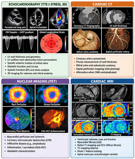
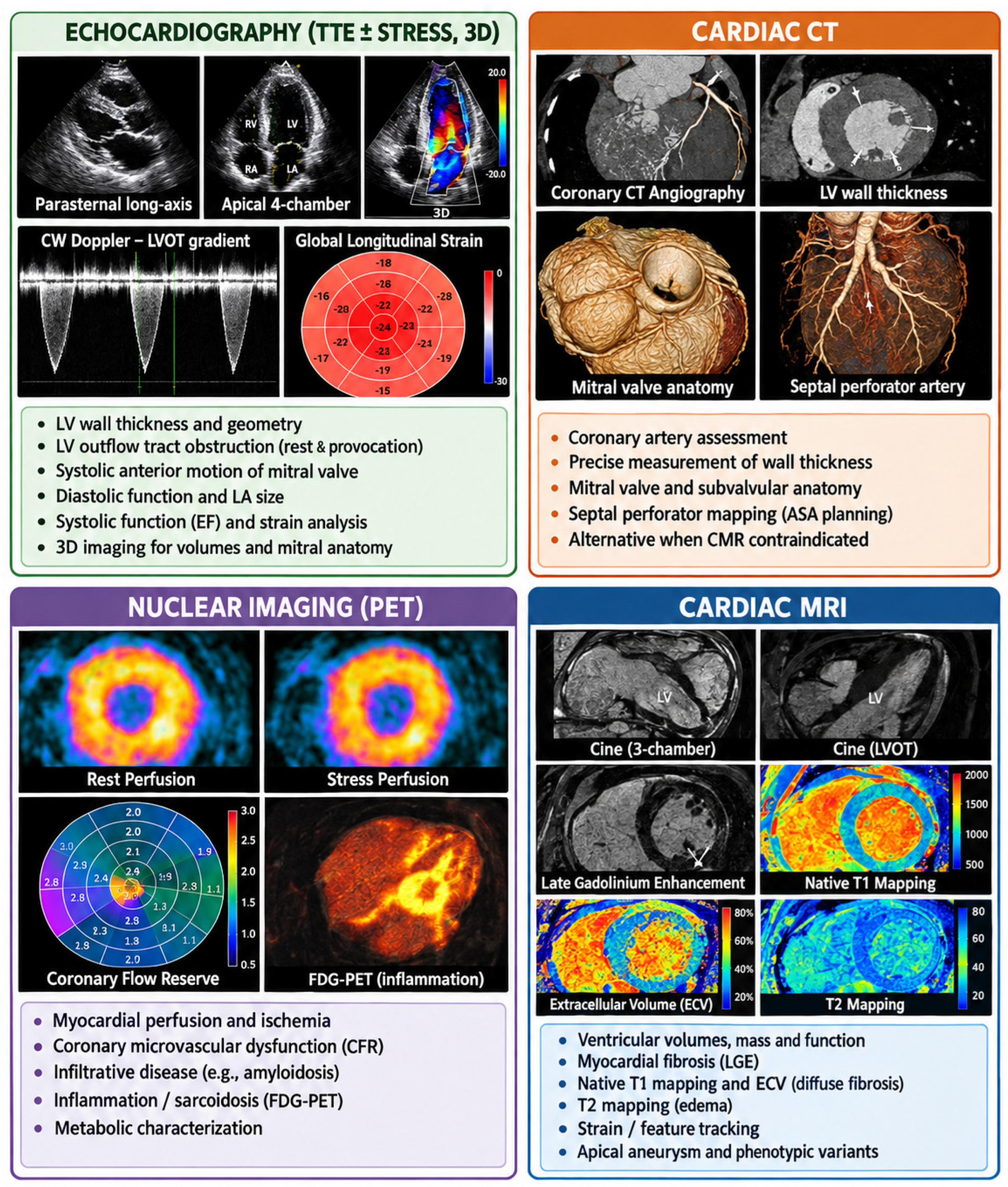
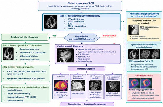
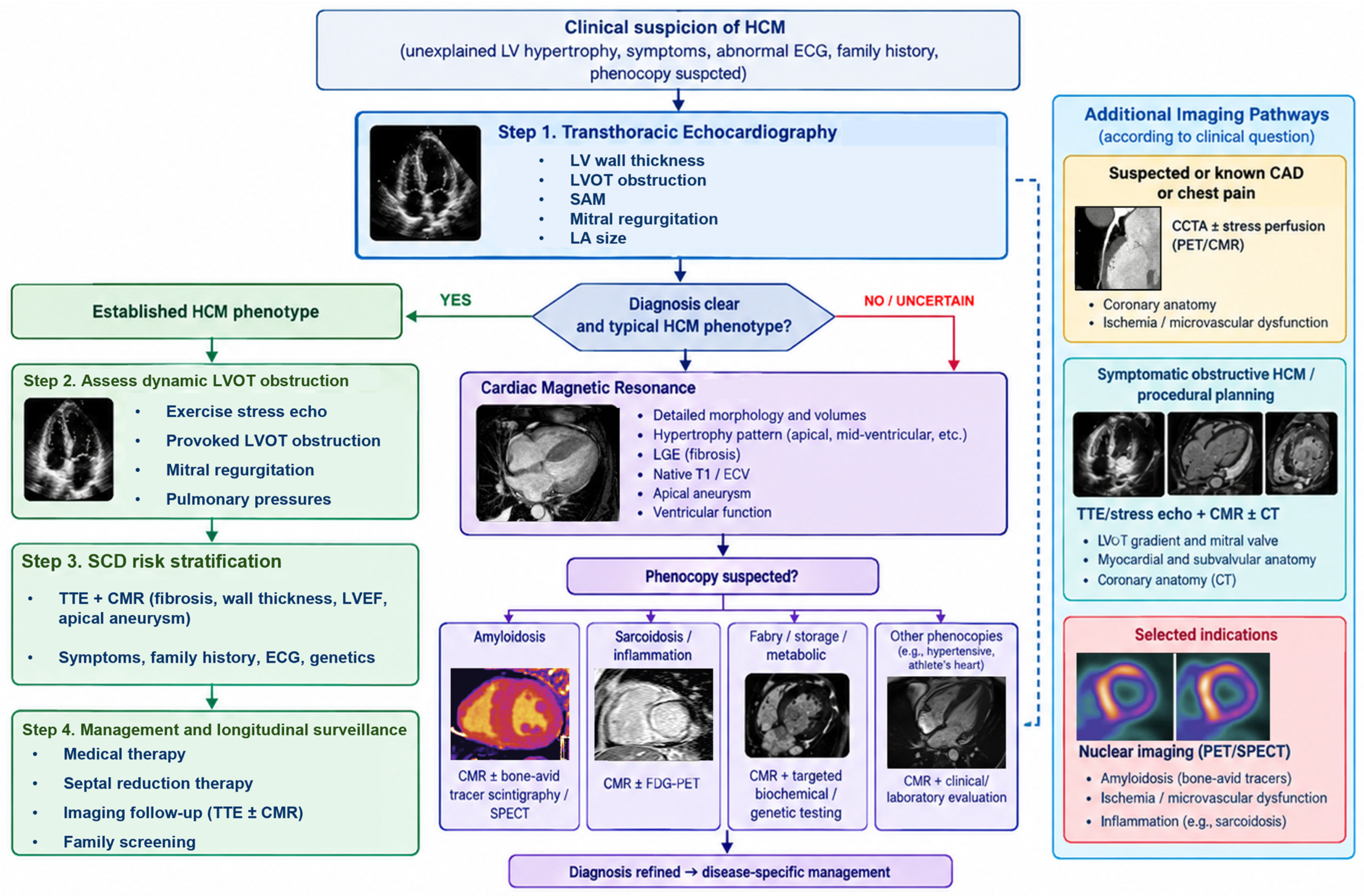

Disclaimer/Publisher’s Note: The statements, opinions and data contained in all publications are solely those of the individual author(s) and contributor(s) and not of MDPI and/or the editor(s). MDPI and/or the editor(s) disclaim responsibility for any injury to people or property resulting from any ideas, methods, instructions or products referred to in the content. |
© 2026 by the authors. Licensee MDPI, Basel, Switzerland. This article is an open access article distributed under the terms and conditions of the Creative Commons Attribution (CC BY) license.