


Abstract
The article examines alpha-rhythm spindles. The importance of studying alpha rhythm spindles for improving the reliability of nervous system diagnostics is substantiated. The analysis employs a mathematical framework based on the Hilbert transform as well as spectral analysis based on the classical Fourier transform. Using the Hilbert transform, the instantaneous frequency of the spindles is determined. It is shown that a spindle is not an amplitude-modulated oscillation and a spindle can be described using the beating method. Two frequencies are obtained that determine the frequency composition of the spindles. It is shown that the beating method is more accurate compared to the Fourier transform.
MSC:
42A38; 92-10
1. Introduction
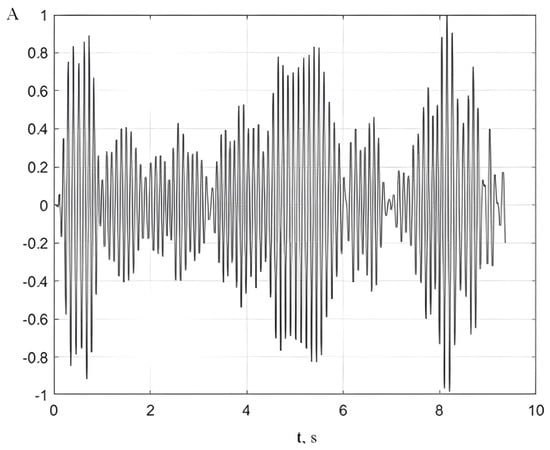
Alpha activity in the electroencephalogram (EEG) is one of the key forms of rhythmic neural activity in the brain. It is characterized by oscillations in the 8–13 Hz range and is most pronounced in the occipital–parietal regions of the cortex in a resting state (see Figure 1). In modern neuroscience, the alpha rhythm is no longer regarded as a marker of a passive state but is interpreted as an active mechanism for regulating cognitive processes. Recent studies show that alpha activity plays an important role in selective attention, sensory inhibition, and the control of information flow in the cerebral cortex [1,2].
Figure 1.
Alpha rhythmof the awake adult.
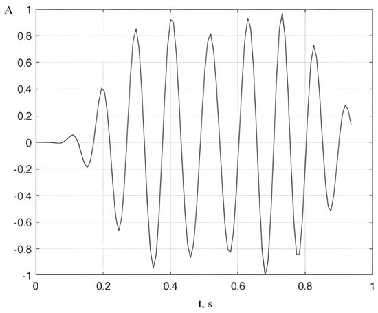
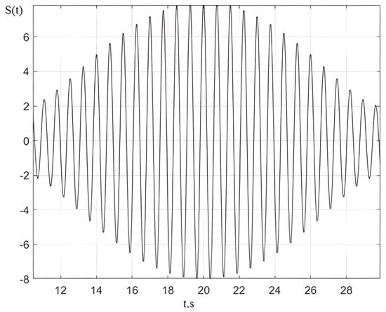
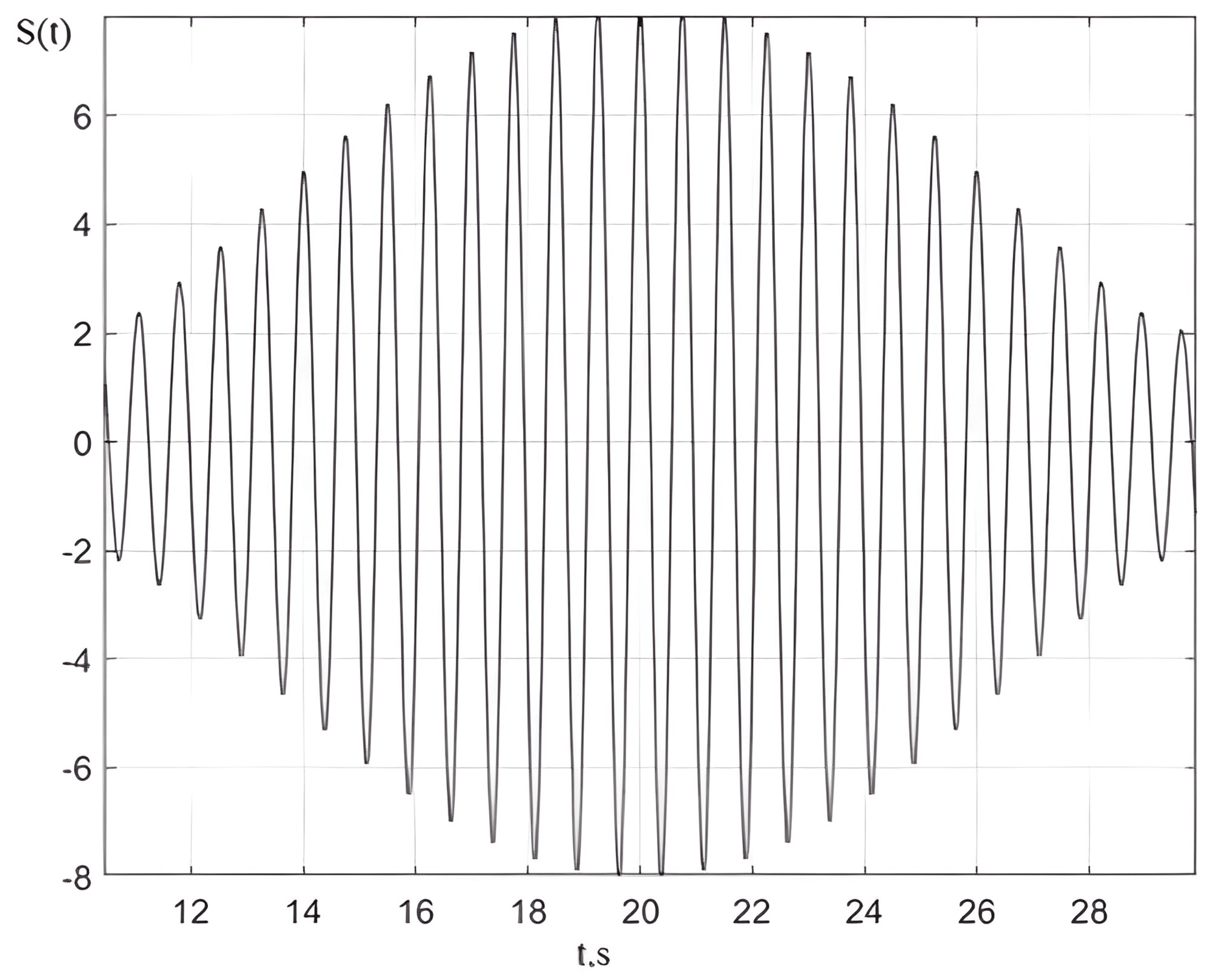
In this context, important microstructural elements of the alpha rhythm that reflect the dynamic organization of neural activity are alpha spindles. In contrast to earlier views of the alpha rhythm as a stationary process, modern evidence indicates its pronounced episodic structure, manifested in the form of short bursts—alpha spindles (see Figure 2). These events typically last from 0.5 to 2 s and are characterized by a gradual increase and decrease in amplitude, indicating the temporal synchronization of large neuronal populations [3,4,5].
Figure 2.
Alpha rhythm spindle.
The mechanisms underlying the generation of alpha spindles are primarily associated with the interaction between the thalamus and the cerebral cortex. According to modern models, a key role is played by thalamocortical loops, in which rhythmic activity is formed through the interaction of excitatory thalamocortical neurons and inhibitory neurons of the thalamic reticular nucleus [6,7].
These structures create conditions for the emergence of oscillations in the alpha range through feedback mechanisms and rhythmic membrane hyperpolarization.
Thus, alpha spindles may arise as a result of a transient increase in synchronization within these networks, when a large number of neurons simultaneously enter an oscillatory mode [8,9,10].
At the same time, in addition to the thalamic contribution, local cortical networks also play a significant role in the generation of alpha spindles. Studies show that alpha activity can arise from intracortical processes, in particular, from interactions between excitatory pyramidal neurons and inhibitory interneurons. In this context, alpha spindles are considered an indicator of the balance between excitation and inhibition in the cortex, allowing the brain to rapidly switch between active information processing and its temporary suppression [2].
From the perspective of their functional nature, alpha spindles are closely associated with mechanisms of inhibition and selective attention. Their occurrence is often interpreted as a marker of the temporary suppression of information processing in specific sensory or associative regions of the cortex. For example, during cognitive task performance, an increased probability of alpha spindle occurrence in the visual cortex may indicate suppression of visual information when attention is focused on other modalities. Thus, alpha spindles act as a kind of “filter” that regulates the access of information to further processing [6,9].
From the standpoint of quantitative analysis, alpha spindles are characterized by several parameters, among which the key ones are amplitude, frequency, duration, and periodicity of occurrence. An important feature is their variability—both between different individuals and within the same subject depending on the functional state. In particular, under increased cognitive load or fatigue, both the frequency of spindle occurrence and their duration may change, reflecting adaptive mechanisms of the brain [11].
In this regard, an important aspect of studying alpha activity is its rhythmic organization, in particular the stability of frequency, the regularity of alpha spindle occurrence, and the coherence of oscillations over time. In the clinical context, disturbances in the rhythmicity of alpha activity—manifested as disorganization, fragmentation, or reduced rhythm stability—are considered indicators of dysfunction in neural networks, primarily the thalamocortical system. Such changes may reflect impairments in neural synchronization mechanisms underlying cognitive processes and the regulation of states of consciousness [11,12].
A reduction in the rhythmicity of alpha activity is often observed in various neurological and psychiatric conditions. In particular, in cognitive impairments and neurodegenerative diseases such as Alzheimer’s disease, a slowing of the alpha rhythm, a decrease in its regularity, and reduced spatial organization are observed. This is associated with the degradation of thalamocortical connections and impaired synchronization of neural activity [9,13,14,15].
Similar changes can also be observed in attention disorders, depression, and anxiety states, where the disorganization of alpha activity reflects impairments in inhibitory mechanisms and the regulation of cognitive processes [2].
Particular importance is attributed to disruptions in the microstructure of alpha activity, specifically changes in the characteristics of alpha spindles. A decrease in their regularity, the appearance of chaotic or atypical spindles, as well as a reduction in their occurrence frequency may indicate instability of neural networks and reduced efficiency of information processing. Such changes are often associated with states of decreased alertness, cognitive fatigue, or disorders of consciousness [11].
In addition, disturbances in the rhythmicity of alpha activity may manifest as increased variability in frequency and amplitude, indicating a reduced ability of the brain to maintain stable functional states. Modern studies emphasize that such variability is not random but reflects an imbalance between excitatory and inhibitory processes in the cerebral cortex (see [16]). In this context, the analysis of the rhythmicity and stability of alpha activity is considered a promising approach for assessing the functional state of the brain.
From a clinical perspective, the assessment of alpha activity rhythmicity has diagnostic and prognostic significance. It can be used for the early detection of cognitive impairments, monitoring disease progression, and evaluating the effectiveness of therapeutic interventions. In particular, the normalization of alpha rhythm rhythmicity is considered a positive marker of the restoration of brain functional state and is used in neurorehabilitation and neurofeedback therapy [17].
Therefore, disturbances in the rhythmicity of alpha activity represent an important neurophysiological indicator reflecting dysfunction of neural networks and have significant clinical relevance. In this context, the analysis of both macro- and microstructural characteristics of the alpha rhythm (including alpha spindles) opens up opportunities for more accurate diagnosis and a deeper understanding of the pathophysiological mechanisms of various central nervous system disorders.
Further development of this field is associated with the improvement of methods for analyzing alpha spindles, which have undergone significant evolution in recent years. Previously, alpha activity was assessed mainly by the average power of the signal, whereas nowadays event-based analysis methods are becoming increasingly widespread, allowing the identification of individual spindles in the time signal. Such methods include threshold algorithms, time–frequency analysis (in particular, wavelet transform), as well as modern approaches based on machine learning, which provide more accurate and automated detection. Of particular importance is single-trial analysis, which makes it possible to account for the instability of neural rhythms and to obtain more detailed information about their dynamics [3].
In addition, the individual alpha frequency (IAF), which determines the specific features of alpha spindle generation in a particular individual, is being actively studied. It has been established that this characteristic is associated with cognitive abilities, information processing speed, and attention levels, highlighting the functional significance of alpha spindles as a neurophysiological marker [18].
The significance of alpha spindles extends beyond fundamental research and has practical applications. They are used as indicators of the functional state of the brain in studies of attention, fatigue, drowsiness, and cognitive load. In particular, an increase in their occurrence frequency is associated with reduced alertness, which has practical importance for monitoring the condition of operators, drivers, and other professionals whose work requires prolonged concentration. Moreover, alpha spindles are considered a promising tool in neurofeedback technologies, where their parameters can be used to train self-regulation of brain activity [17].
An additional important aspect of alpha spindle analysis is the examination of their internal dynamics, in particular the relationship between amplitude and frequency characteristics within a single episode. Traditionally, most studies have focused on the amplitude or overall power of alpha activity; however, modern approaches indicate that the frequency component is a more sensitive and dynamic indicator of the brain’s functional state. In particular, it has been established that the instantaneous frequency of alpha oscillations can change within a single spindle much faster than its amplitude, reflecting rapid reconfigurations of neural synchronization [16,18].
Such rapid frequency changes are associated with the variability of thalamocortical interactions and the dynamic balance between excitatory and inhibitory processes. Unlike amplitude, which reflects the degree of involvement of neuronal populations, the oscillation frequency is more directly related to the temporal characteristics of neural communication and the speed of information processing [8,13].
In this context, the analysis of the frequency structure of alpha spindles makes it possible to detect subtle changes in the functional state of the brain that may remain unnoticed when using only amplitude-based measures. From a clinical perspective, increased variability of frequency or disruption of its stability may serve as earlier markers of neural network dysfunction than amplitude changes. Studies show that in cognitive impairments, neurodegenerative processes, and states of reduced alertness, it is primarily the destabilization of the frequency characteristics of alpha activity that is observed, whereas amplitude changes may appear later or be less specifi [9,11].
In modern neurophysiology, the alpha rhythm spindle is traditionally interpreted as a type of amplitude-modulated (AM) oscillation. This view is supported by a number of publications in which changes in the amplitude of the alpha rhythm are considered a form of modulation capable of generating slow evoked responses or reflecting neuromodulatory mechanisms in the brain. In particular, such an interpretation is found in the work of Mazaheri & Jensen (see [19]), where the contribution of symmetric amplitude modulation of the alpha rhythm to the formation of slow evoked potentials is discussed, and in the study by Zarubin et al. (see [20]), where amplitude variations in alpha oscillations are treated as the central mechanism of their modulation.
Taken together, these studies have led to the widespread assumption that variation in amplitude within an alpha rhythm spindle constitutes amplitude modulation. However, a change in amplitude by itself does not necessarily constitute amplitude modulation in the strict sense used in radio engineering.
Therefore, before classifying alpha spindles as amplitude-modulated processes, it is necessary to rigorously define which features truly characterize a signal as an AM oscillation and which do not. To investigate this issue, let us consider the formal definition of modulation and the specific features of amplitude modulation from the perspective of classical radio engineering and signal theory [21,22,23].
2. Materials and Methods
2.1. The Concept of Modulation—Amplitude Modulation
To transmit information over radio channels, a high-frequency oscillation called a carrier wave is used. One of the parameters of this oscillation changes over time in accordance with the message being transmitted. The carrier wave typically has the following form (see [24]):
Any oscillation parameter can be modulated, that is, the amplitude, frequency, or initial phase. Regarding this, a distinction is made between amplitude modulation (AM) and angle modulation (AM). Angular modulation, in turn, can be frequency modulation (FM) or phase modulation (PM).
As the name suggests, with AM, the amplitude of the carrier oscillation changes according to the law (see [25]):
The simplest case of tonal AM is when
From here we get
The parameter
is called the amplitude modulation coefficient. Taking this into account, a tone-modulated signal can be written as follows:
Here is the physical envelope of a signal with tonal AM. Only when , does the physical envelope of an AM signal contain undistorted information of the modulated signal. If , the condition of linear dependence on the envelope is not met. This leads to the appearance of linear distortion (overmodulation). In amplitude modulation, information is conveyed by varying the signal’s amplitude, while the carrier frequency remains constant. Consequently, if any variability in the instantaneous frequency is detected, this contradicts the model of pure amplitude modulation and indicates the presence of a frequency or phase modulation component [21,22,23].
2.2. Determination of the Instantaneous Frequency of the Alpha Rhythm
Signal processing apparatus is well developed in radio engineering. Of great importance is the Fourier Transform (FT).
The FT is a fundamental mathematical tool that decomposes a time domain signal. Classical Fourier analysis implicitly assumes that the signal under study is stationary, meaning its statistical properties do not vary significantly over time. Because the FT produces a global frequency representation—integrating information over the entire time axis—it provides an averaged description of the spectral content rather than a time localized one.
Traditional Fourier analysis presumes signals to be infinite in time or periodic, and thus performs poorly when analyzing signals whose frequency characteristics evolve over time. This limitation motivates the development of time–frequency methods [22,26].
Moreover, the uncertainty principle of Fourier analysis highlights a fundamental limitation: the FT cannot simultaneously provide high resolution in both time and frequency. It smears together contributions from all time instants, making it insufficient for signals with rapid or localized temporal changes.
A fundamental limitation of the classical Fourier Transform is that it does not provide instantaneous frequency information. The FT reveals what frequencies are present, but not when they occur. Its output reflects a time averaged spectrum and cannot capture temporal variations. In non-stationary contexts, the FT therefore fails to display frequency evolution over time, which is why specialized methods are necessary for adequate analysis.
There are few methods for determining instantaneous frequency [22]. One method is the Hilbert transform (HT). It is highly accurate, but its use requires a narrowband. In our case, we are using a narrow frequency band—the alpha rhythm. Therefore, we can use this method.
The HT is a fundamental mathematical operation used in modern signal processing to extract phase related information from real valued signals. Formally, it is defined as a singular integral that produces a new function by convolving the original signal with the Cauchy kernel , evaluated in the sense of the Cauchy principal value.
In practical terms, the Hilbert transform separates the original real signal into two orthogonal components, enabling a quadrature representation.
A narrowband signal possesses a notable property: it can be represented with high precision in the following form [21,22,27]:
where
- and are slowly varying low-frequency functions of time;
- is full phase;
- is physical envelope of narrowband signal.
The instantaneous frequency of a narrowband signal is defined as follows
The envelope of a narrowband signal, along with its complete phase, can be obtained through the application of the Hilbert transform [21,22,23]:
Here is a new function that is related to the function by the relations
Relations (11) and (12) are referred to as the Hilbert transform. The function is known as the Hilbert conjugate of . The Hilbert transform plays a crucial role in the theory of narrowband signals as well as in the design of radio engineering devices and systems [21,22,23]. It is a linear operator. The analysis of electrical circuits subjected to electrical oscillations becomes substantially simpler when the oscillation is represented in the form of a complex signal [22]:
where is the complex amplitude.
The analytic signal has the form
It follows that
An analytic signal is defined as the complex-valued representation associated with a real-valued signal .
By examining the analytic signal expression together with (9) and (10), it follows that the envelope of the analytic signal is given by
and the overall phase is expressed as
Thus, the analytic signal takes the form
By extending the phase relation of a harmonic signal, the phase of a narrowband signal takes the form
where is the reference frequency of the narrowband signal; is a low-frequency function of time that excludes any linearly time-dependent components.
The low frequency, slowly varying time function is given by
and is known as the signal’s complex envelope.
2.3. Beating Method
In [28], the beating method is examined and the alpha spindle pattern is shown to be consistent with this method.
The beating method represents a fundamental analytical approach for studying harmonic and quasi-harmonic signals based on the interference of two oscillations with closely spaced frequencies. Consider two harmonic components:
where and denote nearly identical frequencies. Their linear superposition produces a resulting signal whose amplitude is modulated at the beating frequency . This interference phenomenon, known as beating, manifests as periodic amplitude fluctuations and provides a sensitive indicator of small frequency deviations.
From a spectral perspective, beats can be interpreted as the interaction between two narrowband spectral components that lie in close proximity to one another. This property makes the beating method highly suitable for precise frequency comparison and the quantification of small frequency offsets.
In the analysis of non-stationary processes, the beating method serves as an efficient tool for localizing instantaneous frequency variations. When the frequency of a signal changes over time, the beat envelope reflects the relative spacing of local spectral components.
The average frequency acts as an effective carrier, while the difference frequency plays the role of a modulation component.
Overall, the beating method constitutes a robust and computationally efficient framework for analyzing signals containing closely spaced frequency components or narrowband structures. Its high sensitivity to minute frequency changes, combined with conceptual simplicity, ensures continued relevance in radio engineering. The method offers a rigorous yet practical means for identifying subtle frequency variations and elucidating the structure of complex oscillatory processes.
Accordingly, the signal in this case is defined as
In the case when the amplitudes are equal, i.e., , we obtain
The resulting expression shows that the resulting signal is a high-frequency oscillation with a frequency , the amplitude of which is modulated by a low-frequency envelope with a frequency .
However, the frequency of the maximum/minimum amplitude, i.e., the “beat frequency”, is equal to
Since the envelope varies as , the maximum amplitude is reached twice during the envelope period.
If the amplitudes are not equal, then the instantaneous frequency is determined as [22]
It is important to note that, as follows from the expression for instantaneous frequency, its value depends not only on the frequencies of the interfering oscillations but also on their amplitudes. This reflects a fundamental difference between an amplitude-modulated oscillation and a signal formed as a result of beating.
Applying this approach to the analysis of electroencephalographic signals makes it possible to interpret alpha spindles not as homogeneous oscillations, but as the result of the superposition of several generators of alpha activity with slightly different frequency characteristics. In the neurophysiological context, the beating phenomenon may reflect the interaction of different sources of the alpha rhythm, in particular thalamic and cortical generators, as well as spatially separated cortical networks. The presence of several oscillators with close frequencies leads to the formation of a complex temporal signal structure, in which amplitude fluctuations arise as a result of frequency inhomogeneity. Thus, the characteristic shape of an alpha spindle—with gradual increase and decrease in amplitude—can be partially explained by the beating effect between synchronized but not fully identical neural rhythms.
An important implication of this approach is a reconsideration of the role of amplitude: it is viewed not only as an indicator of the strength of neural activity, but also as a derivative of the interaction between frequency components. This is consistent with modern views that the instantaneous frequency of neural oscillations is a more labile characteristic, capable of changing faster than amplitude, which is formed as an integral result of synchronization [16]. Accordingly, beating analysis makes it possible to indirectly investigate the microdynamics of frequency changes within a single alpha spindle.
From a clinical perspective, the analysis of alpha spindle beating may be of particular value, as it allows the detection of disrupted coordination between different generators of alpha activity. Imbalance or desynchronization between such generators may manifest as changes in the beating structure, in particular in the form of irregular amplitude fluctuations or instability of frequency components. This, in turn, may serve as an early indicator of impaired thalamocortical interaction observed in cognitive disorders, fatigue states, or other functional brain disturbances [9,11].
3. Results and Discussion
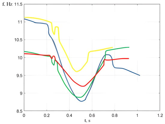
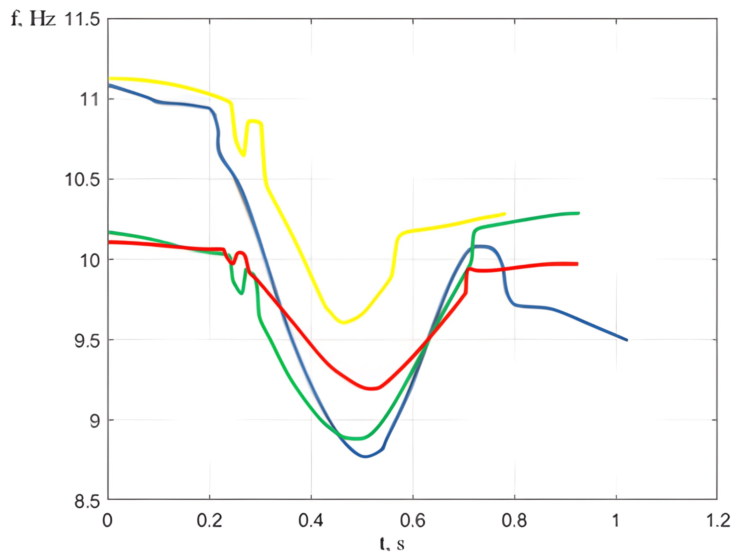
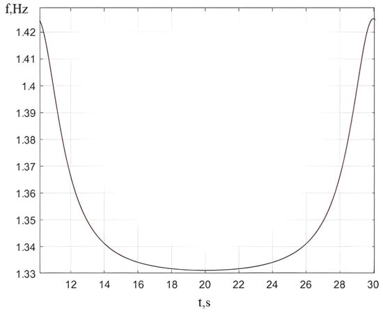
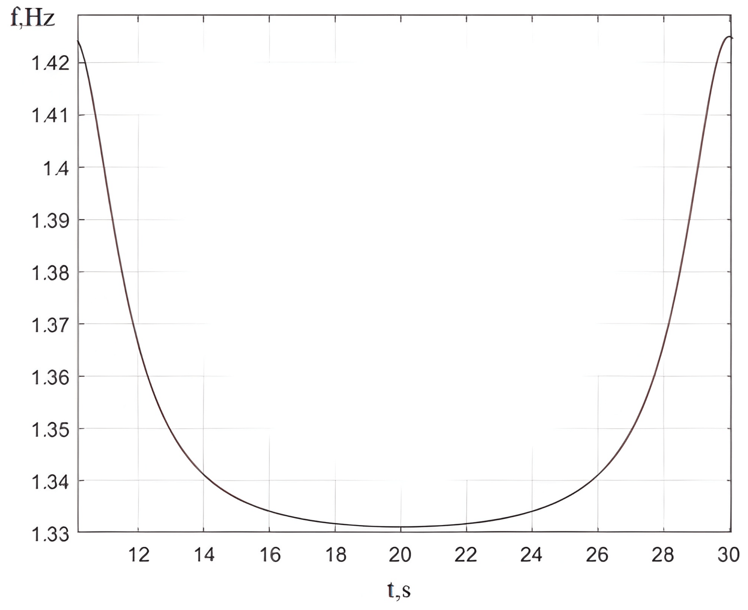
We calculate the instantaneous frequency of several spindles using the Hilbert transform (see [29]). The result is shown in Figure 3.
Figure 3.
Instantaneous frequencies of several spindles calculated using the Hilbert transform. Different colours correspond to different spindles and are used for visual clarity only.
Comparison of changes in instantaneous frequency and amplitude shows that these parameters change in antiphase. As a result, the greatest amplitude is observed when the instantaneous frequency reaches its minimum. Thus, the minima and maxima of the instantaneous frequency can be determined by analyzing the signal amplitude.
It can be seen that the instantaneous frequency is not constant in each case. As shown in the figure, the instantaneous frequency exhibits a local minimum at 1.33 Hz and reaches a maximum of 1.42 Hz. However, with amplitude modulation, the frequency must remain constant, so spindles are not amplitude-modulated oscillations.
Let us consider a signal that is the sum of two monochromatic oscillations with close frequencies and arbitrary amplitudes—that is, a beat. Figure 4 illustrates the timing diagram of such a signal. Using (27), we determine the instantaneous frequency of this signal. Figure 5 shows the calculation results.
Figure 4.
Time dependence of the signal .
Figure 5.
Instantaneous frequency of the signal .
Let us compare Figure 3 and Figure 5. We can conclude that the spindles are beats, not amplitude modulation. From the spindle graph, we determine the frequencies that determine the harmonic sum of the beating method. Model parameters were selected algorithmically in the MATLAB R2022a environment using an exhaustive search (brute-force) method. The frequency values and , as well as the amplitude values and were varied systematically within specified ranges. For each set of parameters, a simulated signal was generated, after which the variance of the difference between the experimental EEG and the model output was calculated:
The root-mean-square approximation error averaged 12.3 ± 4.1% of the root-mean-square value of the original signal. In total, approximately 100 alpha rhythm spindles, extracted from 13 healthy adult subjects, were analyzed.
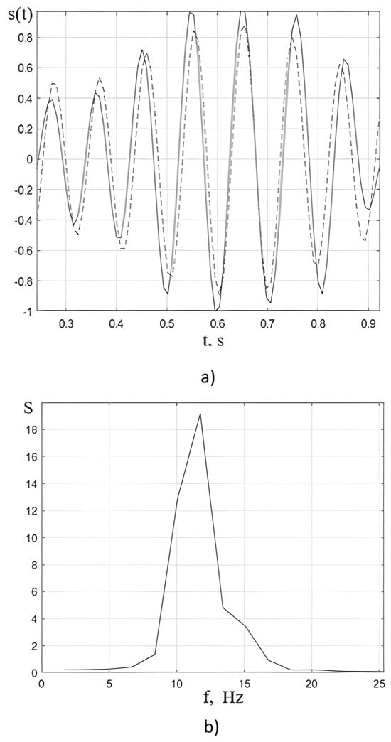
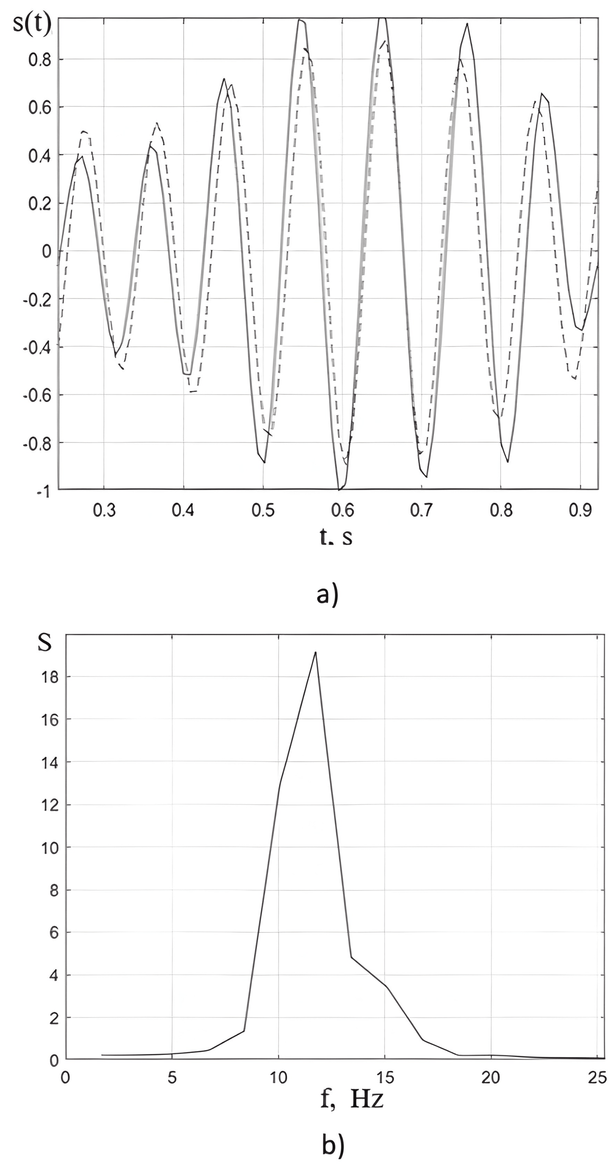
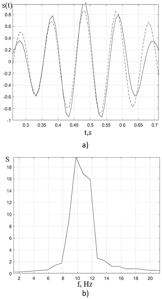
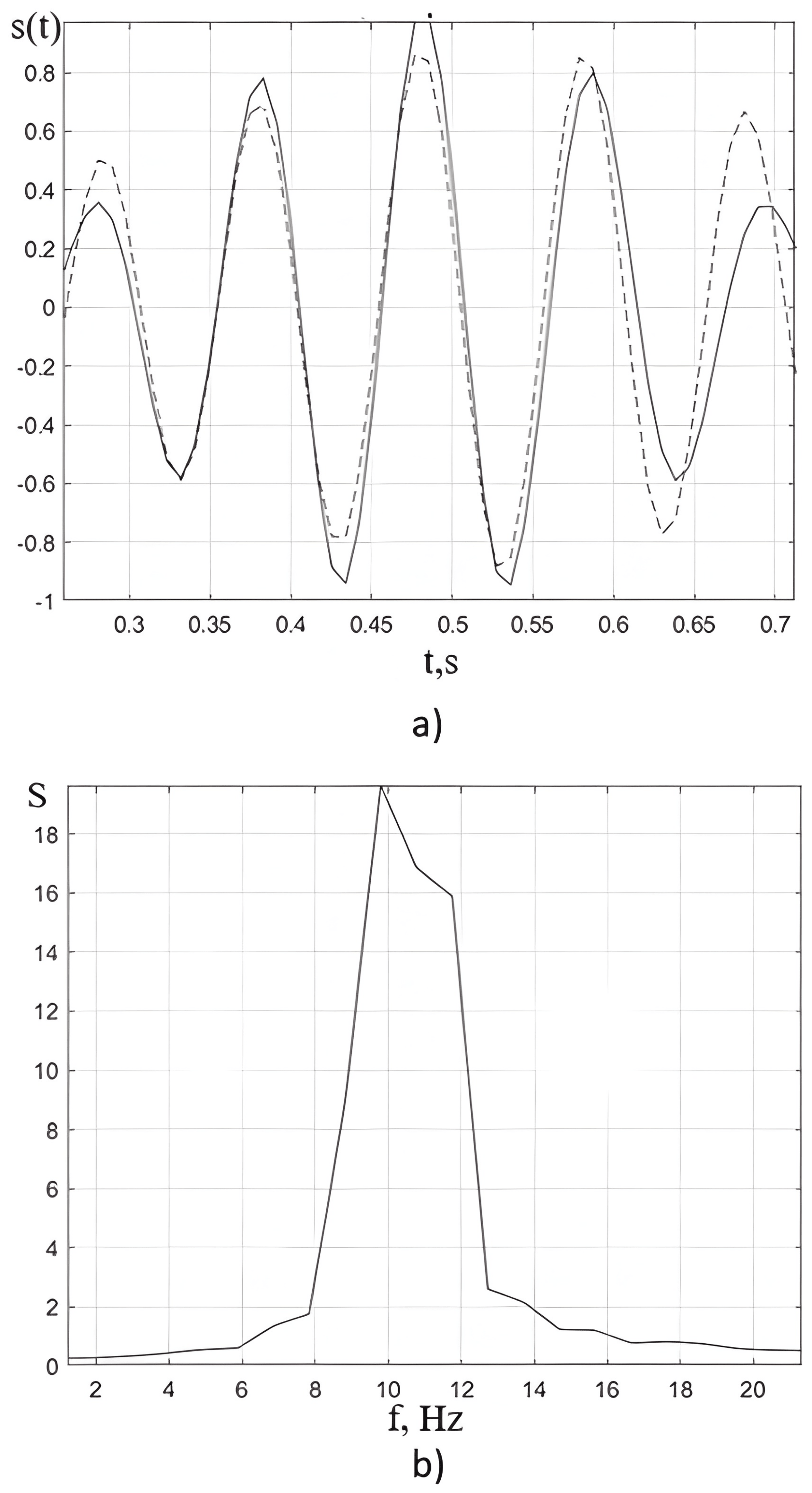
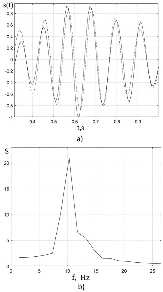
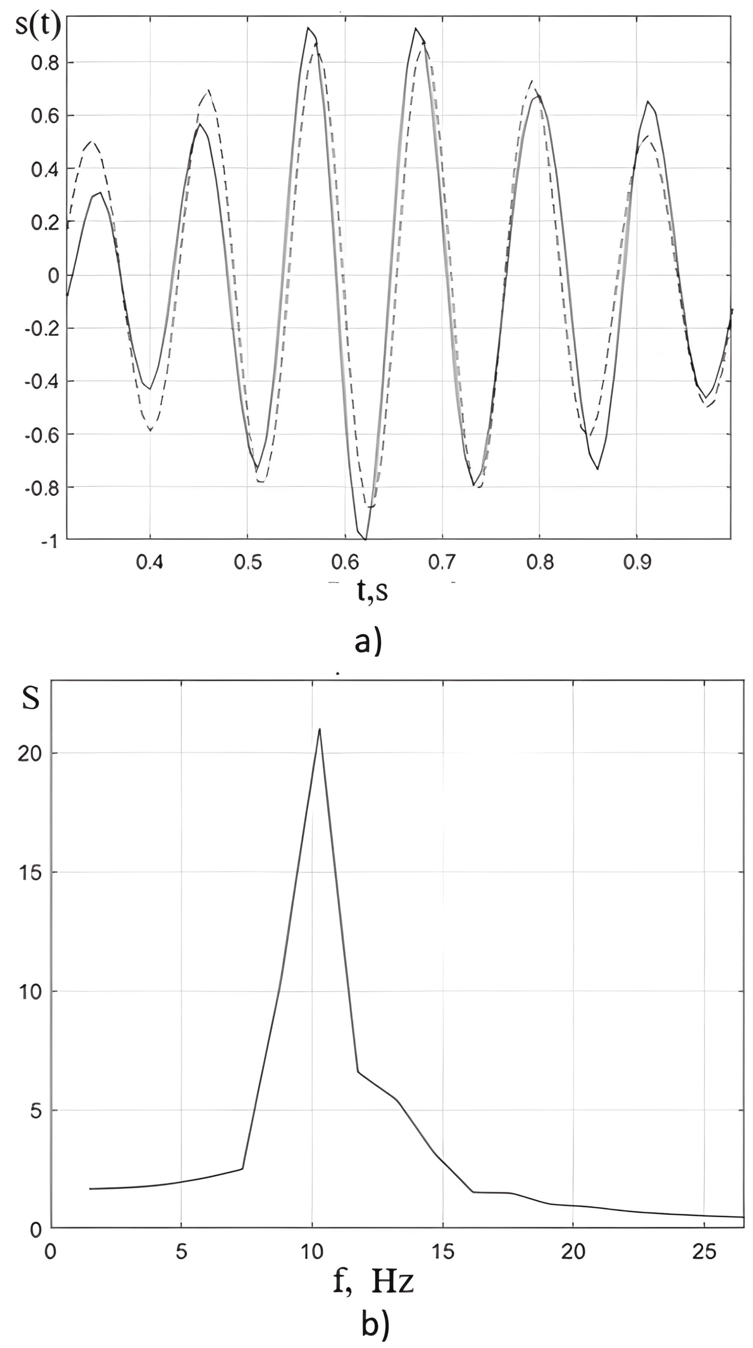
Figure 6, Figure 7 and Figure 8 show an approximation of the spindles as the sum of two harmonic signals and the spindle’s FT. The calculated frequency results are presented in Table 1.
Figure 6.
Approximation of spindle 1: (a) by the beating method (spindle signal—solid line, and its approximation—dashed line), and (b) by FT.
Figure 7.
Approximation of spindle 2: (a) by the beating method (spindle signal—solid line, and its approximation—dashed line), and (b) by FT.
Figure 8.
Approximation of spindle 3: (a) by the beating method (spindle signal—solid line, and its approximation—dashed line), and (b) by FT.
Table 1.
Shows the spindle frequencies obtained by the beating method () and the frequency obtained by ().
Comparing the frequencies obtained by these two methods and based on Figure 6, Figure 7 and Figure 8, we can draw the following conclusions:
- The spindle is described by the beating method and is not amplitude modulation, as is commonly believed;
- Applying the FTto the spindle is inappropriate. This is explained by the fact that in the case of a short signal, the resolution is low [22]. The graphs also show that the PF selects one frequency, not two.
4. Conclusions
In the present study, alpha rhythm spindles were investigated using two complementary approaches: the Hilbert transform and the classical Fourier transform. The use of the Hilbert transform is motivated by the fact that this method allows one to obtain instantaneous characteristics of narrowband signals, including instantaneous frequency and phase, provided that the narrowband condition is satisfied—a fundamental limitation described in the theory of analytic signals and the Hilbert transform.
The analysis showed that the instantaneous frequency of alpha spindles is not constant but exhibits pronounced fluctuations within a single spindle. This behavior contradicts the model of an amplitude-modulated signal, in which the carrier frequency remains fixed while only the amplitude envelope changes. Therefore, alpha spindles cannot be adequately described by the classical amplitude modulation model.
The obtained time–frequency dependencies indicate that the structure of spindles corresponds to a beating mechanism of formation. This mechanism suggests interference of two harmonic components with close frequencies, leading to periodic increases and decreases in amplitude due to beating. The application of the beating method made it possible to approximate the spindle waveform, identify pairs of close frequencies forming the observed envelope, and thus propose a more accurate model of the origin of spindles in the alpha range.
To verify the reliability of the obtained results, an analysis based on the Fourier transform was also applied. However, the classical Fourier transform, as is well known, provides a global spectral distribution averaged over the entire duration of the signal and is therefore optimal for stationary processes, but not for structures with rapidly changing temporal characteristics. As follows from the theory of time–frequency analysis, the Fourier transform does not allow one to capture local frequency changes, including instantaneous frequency and its dynamics within a single spindle. In accordance with this limitation, it was established that the resolution of the Fourier transform is insufficient to reveal the time–frequency structures detected using the beating method and the Hilbert transform.
Thus, the analysis demonstrates that
- Alpha spindles are not amplitude-modulated oscillations, since their instantaneous frequency is not constant.
- The Hilbert transform is an adequate tool for studying these signals, allowing their true dynamics to be revealed.
- The beating model provides a correct interpretation of the nature of alpha spindles and makes it possible to recover the frequencies forming the observed structure.
In this case, the Fourier transform has limited applicability and does not reproduce the spectral features identified using beating analysis.
These results collectively confirm that alpha spindles are complex nonstationary structures requiring analysis methods focused on instantaneous characteristics of narrowband signals. The application of the beating method to EEG spindle signal processing has promising prospects for future research.
A comprehensive analysis of alpha activity, including its macro- and microstructure, parameters of alpha spindles, and their frequency dynamics, represents a promising direction in modern neurophysiology. It opens new opportunities for a deeper understanding of brain function mechanisms and for improving diagnostic approaches.
The beating model provides a signal-level interpretation of alpha rhythm spindles as the superposition of two harmonic components with close frequencies. Their interference produces periodic amplitude fluctuations, which may account for the spindle-like waveform without requiring amplitude modulation of a single carrier frequency. From a neurophysiological perspective, this description may be relevant to the study of interactions between oscillatory processes within thalamocortical loops and local cortical networks, both of which have been implicated in the generation and regulation of alpha activity. However, the presence of two frequency components in an EEG signal does not necessarily imply the existence of two anatomically distinct neural generators. The components may reflect interacting neuronal populations, different dynamical modes of a distributed network, or other processes contributing to the recorded signal. Consequently, the beating model, in its current form, should be considered a phenomenological description of alpha spindledynamics rather than a definitive mechanistic explanation of their generation. Establishing a direct relationship between the identified frequency components and specific thalamic or cortical sources requires further investigation using spatially resolved EEG analysis and appropriate physiological or computational models.
The proposed interpretation may also have implications for the quantitative assessment of alpha activity. In addition to conventional measures such as amplitude, spectral power, and dominant frequency, the beating approach makes it possible to characterize the frequency components contributing to individual spindles and their relationship to the observed temporal envelope. Combined with instantaneous-frequency analysis based on the Hilbert transform, these measures may provide complementary information about the temporal organization and variability of alpha activity. Such parameters could potentially be investigated as additional markers of changes in neural synchronization associated with vigilance, cognitive load, fatigue, and neurological dysfunction.
Nevertheless, the potential clinical value of these measures should be interpreted cautiously. The present signal analysis does not establish their sensitivity, specificity, or prognostic value for any particular neurological or psychiatric condition. Further studies involving larger and clinically characterized cohorts are needed to determine whether beating-related parameters provide reproducible information beyond conventional EEG measures. In particular, future research should examine their test–retest reliability, association with clinical and behavioral measures, and ability to distinguish physiological variability from pathological alterations.
Thus, the principal contribution of the beating model is its alternative mathematical description of alpha spindlewaveform structure and frequency composition. Its potential to improve neurophysiological interpretation and clinical assessment represents a promising research direction that requires independent experimental validation.
Author Contributions
Conceptualization, O.K., V.C. and L.Z.; methodology, O.K., V.C., Z.K. and L.Z.; formal analysis, O.K., V.C., Z.K., L.Z. and V.A.; writing—original draft preparation, O.K., Z.K. and V.A.; writing—review and editing, O.K., Z.K. and V.A.; visualization, O.K., V.C. and V.A. All authors have read and agreed to the published version of the manuscript.
Funding
This research received no external funding
Institutional Review Board Statement
Not applicable.
Informed Consent Statement
Not applicable.
Data Availability Statement
The original contributions presented in this study are included in the article. Further inquiries can be directed to the corresponding author.
Conflicts of Interest
The authors declare no conflicts of interest.
References
- Klimesch, W.; Sauseng, P.; Hanslmayr, S. EEG alpha oscillations: The inhibition-timing hypothesis. Brain Res. Rev. 2007, 53, 63–88. [Google Scholar] [CrossRef] [Scilit] [PubMed]
- Foxe, J.J.; Snyder, A.C. The role of alpha-band brain oscillations as a sensory suppression mechanism during selective attention. Front. Psychol. 2011, 2, 154. [Google Scholar] [CrossRef] [Scilit] [PubMed]
- Kosciessa, J.Q.; Grandy, T.H.; Garrett, D.D.; Werkle-Bergner, M. Single-trial characterization of neural rhythms: Potential and challenges. NeuroImage 2020, 206, 116331. [Google Scholar] [CrossRef] [Scilit] [PubMed]
- Samaha, J.; Iemi, L.; Postle, B.R. Prestimulus alpha-band power biases perception and confidence. Conscious. Cogn. 2020, 54, 47–55. [Google Scholar]
- Ippolito, G.; Bertaccini, R.; Tarasi, L.; Di Gregorio, F.; Trajkovic, J.; Battaglia, S.; Romei, V. The Role of Alpha Oscillations among the Main Neuropsychiatric Disorders in the Adult and Developing Human Brain: Evidence from the Last 10 Years of Research. Biomedicines 2022, 10, 3189. [Google Scholar] [CrossRef] [Scilit] [PubMed]
- Tabarelli, D.; Brancaccio, A.; Zrenner, C.; Belardinelli, P. Functional Connectivity States of Alpha Rhythm Sources in the Human Cortex at Rest: Implications for Real-Time Brain State Dependent EEG-TMS. Brain Sci. 2022, 12, 348. [Google Scholar] [CrossRef] [Scilit] [PubMed]
- Yang, X.; Liu, K.; Liu, L.; Du, Y.; Yu, H.; Yao, Y.; Sun, Y.; Li, C. The Differential and Interactive Effects of Aging and Mental Fatigue on Alpha Oscillations: A Resting-State Electroencephalography Study. Brain Sci. 2025, 15, 546. [Google Scholar] [CrossRef] [Scilit] [PubMed]
- Jensen, O.; Mazaheri, A. Shaping functional architecture by oscillatory alpha activity. Front. Hum. Neurosci. 2010, 4, 186. [Google Scholar] [CrossRef] [Scilit] [PubMed]
- Van Diepen, R.M.; Foxe, J.J.; Mazaheri, A. The functional role of alpha-band activity in attentional processing: The current zeitgeist and future outlook. Curr. Opin. Psychol. 2019, 29, 229–238. [Google Scholar] [CrossRef] [Scilit] [PubMed]
- Zhang, Y.; Li, Z.; Xu, H.; Song, Z.; Xie, P.; Wei, P.; Zhao, G. Neural Mass Modeling in the Cortical Motor Area and the Mechanism of Alpha Rhythm Changes. Sensors 2025, 25, 56. [Google Scholar] [CrossRef] [Scilit] [PubMed]
- Lendner, J.D.; Helfrich, R.F.; Mander, B.A.; Romundstad, L.; Lin, J.J.; Walker, M.P.; Larsson, P.G.; Knight, R.T. An electrophysiological marker of arousal level in humans. eLife 2020, 9, e55092. [Google Scholar] [CrossRef] [Scilit] [PubMed]
- Buzsáki, G.; Draguhn, A. Neuronal oscillations in cortical networks. Science 2004, 304, 1926–1929. [Google Scholar] [CrossRef] [Scilit] [PubMed]
- Klimesch, W. α-band oscillations, attention, and controlled access to stored information. Trends Cogn. Sci. 2012, 16, 606–617. [Google Scholar] [CrossRef] [Scilit] [PubMed]
- Li, X.; Yang, X.; Sun, Z. Alpha rhythm slowing in a modified thalamo-cortico-thalamic model related with Alzheimer’s disease. PLoS ONE 2020, 15, E0229950. [Google Scholar] [CrossRef] [Scilit] [PubMed]
- Sharma, R.; Nadkarni, S. New Research, Neuronal Excitability Biophysical Basis of Alpha Rhythm Disruption in Alzheimer’s Disease. eNeuro 2020, 7, ENEURO.0293-19.2020. [Google Scholar] [CrossRef] [Scilit] [PubMed]
- Kosciessa, J.Q.; Lindenberger, U.; Garrett, D.D. Thalamocortical Excitability Modulation Guides Human Perception under Uncertainty. Nat. Commun. 2021, 12, 2430. [Google Scholar] [CrossRef] [Scilit] [PubMed]
- Enriquez-Geppert, S.; Huster, R.J.; Herrmann, C.S. EEG-neurofeedback as a tool to modulate cognition and behavior: A review tutorial. Front. Hum. Neurosci. 2017, 11, 51. [Google Scholar] [CrossRef] [Scilit] [PubMed]
- Samaha, J.; Iemi, L.; Haegens, S.; Busch, N.A. Spontaneous brain oscillations and perceptual decision-making. Trends Cogn. Sci. 2020, 24, 639–653. [Google Scholar] [CrossRef] [Scilit] [PubMed]
- Mazaheri, A.; Jensen, O. Asymmetric amplitude modulations of brain oscillations generate slow evoked responses. J. Neurosci. 2008, 28, 7781–7787. [Google Scholar] [CrossRef] [Scilit] [PubMed]
- Zarubin, G.; Gundlach, C.; Nikulin, V.; Villringer, A.; Bogdan, M. Transient amplitude modulation of alpha-band oscillations by short-time intermittent closed-loop tACS. Front. Hum. Neurosci. 2020, 14, 366. [Google Scholar] [CrossRef] [Scilit] [PubMed]
- Siebert, W. Circuits, Signals, and Systems; MIT Press: Cambridge, MA, USA, 1986. [Google Scholar]
- Voloshchuk, Y.I. Signals and Processes Radio Technic; OO “Company Smith”: Milwaukee, WI, USA, 2005. (In Ukrainian) [Google Scholar]
- Baskakov, S.I. Radio Technical Circuits and Signals; Vysshaya Shkola: Moscow, Russia, 1983. (In Russian) [Google Scholar]
- Badillo, I.; Portilla, J. Experimental Study of AM and PM Noise in Cascaded Amplifiers. Electronics 2022, 11, 470. [Google Scholar] [CrossRef] [Scilit]
- Shi, Y.; Li, Z.; Liu, R.; Zhang, X.; Wang, M.; Ma, R.; Yin, T.; Zhou, X.; Liu, Z. A Multi-Channel AM-TMAS Driving System Based on Amplitude-Modulated Sine Waves. Bioengineering 2026, 13, 405. [Google Scholar] [CrossRef] [Scilit] [PubMed]
- Zhou, Y.; Ma, Z.; Fu, L. A Review of Key Signal Processing Techniques for Structural Health Monitoring: Highlighting Non-Parametric Time-Frequency Analysis, Adaptive Decomposition, and Deconvolution. Algorithms 2025, 18, 318. [Google Scholar] [CrossRef] [Scilit]
- Waszczuk-Młyńska, A.; Gałęzia, A.; Stanisław, R. Fault Identification in Membrane Structures Using the Hilbert Transforms. Sensors 2022, 22, 6224. [Google Scholar] [CrossRef] [Scilit] [PubMed]
- Kharchenko, O.; Kovacheva, Z.; Chumakov, V.; Zabrodina, L. Application of the beating method to define the electroencephalogram algorithm frequency content. TEM J. 2024, 13, 2667–2675. [Google Scholar] [CrossRef] [Scilit]
- Kharchenko, O. Definition of the Instantaneous Frequency of an Electroencephalogram Using the Hilbert Transform. Adv. Biosci. Bioeng. 2016, 4, 43–50. [Google Scholar] [CrossRef] [Scilit]

Disclaimer/Publisher’s Note: The statements, opinions and data contained in all publications are solely those of the individual author(s) and contributor(s) and not of MDPI and/or the editor(s). MDPI and/or the editor(s) disclaim responsibility for any injury to people or property resulting from any ideas, methods, instructions or products referred to in the content. |
© 2026 by the authors. Licensee MDPI, Basel, Switzerland. This article is an open access article distributed under the terms and conditions of the Creative Commons Attribution (CC BY) license.