


Abstract
Plastic pollution is an escalating issue, and exposure to it can cause various negative reactions in different organisms. In response, bio-based plastics have been developed as a safer alternative for the environment, organisms, and humans. This study represents the first attempt to assess the impact of bio-based polylactic acid (PLA) and two conventional polymers: polyethylene (PE) and polystyrene (PS) on the growth, survival, and coloration of red swamp crayfish Procambarus clarkii, an invasive and aquaculture species of great economic importance and bamboo shrimp Atyopsis moluccensis, the popular species in aquarium industry. Juvenile crayfish were fed diets based on two types of commercial feed—insect meal and fish meal. The adult shrimp fed commercial crustacean food. The results showed that supply of polyethylene microplastic with food reduced the growth and coloration in P. clarkii. Polystyrene microplastic affected the carapace coloration in A. moluccensis, while no significant effect on growth was observed. Furthermore, the ingestion of bio-based PLA was associated with alterations in coloration in both crayfish and shrimp when compared to the control group. The observed changes in coloration may indicate a deterioration in body condition due to oxidative stress, potentially induced by exposure to stress factors, such as microplastic exposure. An increased abundance of Staphylococcus spp. was observed in the treatment water of crayfish-exposed groups. The addition of both bioplastic and conventional microplastics facilitates bacterial growth and resulted in an increased presence of Gram-positive bacteria. Our study provides the first evidence of coloration changes in decapods in response to microplastics, potentially linked to microorganisms that may contribute to an inflammatory response.
1. Introduction
Plastic, once a revolutionary material owing to its durability, low production cost, and water resistance, has become a global environmental threat due to its ubiquitous distribution. As much as 79% of plastic waste ends up in landfills and wastewater [1]. Under environmental conditions such as UV radiation, abrasion, and interactions with living organisms, plastic degrades into small particles commonly known as mesoplastics (typically defined as 5–25 mm in size), microplastics (0.1 µm to 5 mm), and nanoplastics (<0.1 µm) [2,3,4].
Exposure to plastic particles across various living organisms, from invertebrates to birds and mammals, has been widely documented in the literature, revealing varying degrees of damage, toxicity, behavioral disorders, and even increased mortality [5,6]. Early research on microplastics primarily focused on marine organisms, particularly fish, poly-chaetes, and bivalves [7,8,9,10,11,12,13,14]. Over time, marine and freshwater crustaceans began to be studied. One of the first crustacean species observed to be ingesting microplastics was the amphipod Orchestia gammarellus [15], followed by the shrimp Crangon crangon [16], the Norway lobster Nephrops norvegicus [17,18], the amphipod Talitrus saltator [19], and the freshwater Amazon river prawn Macrobrachium amazonicum [20]. Due to the open circulatory system of hemocoel crustaceans, there is an increased risk of microplastic transfer between organs. Microplastics ingested with food can be transferred to the hemolymph and organs, mainly the stomach, hepatopancreas, ovaries, and gills [21]. In the Chinese mitten crab Eriocheir sinensis, exposure to microplastics was found to cause damage and induce oxidative stress in the hepatopancreas [22].
The negative effects of microplastic exposure on living organisms are associated with not only physical impacts, such as mechanical damage, inflammatory changes, and obstruction of the gastrointestinal and respiratory systems, but also chemical contaminants, such as polychlorinated biphenols, dichlorodiphenyl trichloroethane, nonylphenol, and polyaromatic and aliphatic hydrocarbons, which are absorbed and transported by micro-plastics [23,24]. Studies indicate that plastic pollution will pose a serious future threat to not only living organisms but also humans who consume them. Microplastic particles have already been recorded in human blood and placenta [25,26]. Microplastics can cause oxidative stress, cytotoxicity, neurotoxicity, and immune system disruption and are potentially carcinogenic to humans [27,28].
An alternative to these petroleum-based materials is bioplastics, a term encompassing chemically diverse polymers of biological origin (e.g., materials derived from starch, lignin, or cellulose, as well as bio-based polymers, such as polyhydroxyalkanoate, polylactic acid (PLA), and polybutylene adipate terephthalate [29]. Bioplastics, which share similar physical properties with synthetic plastics, are characterized by their biodegradability and renewability and can be recycled through anaerobic fermentation and composting. Over the past two decades, interest in bio-based plastics has increased. Their global production in 2020 amounted to 1.23 million tons, and by 2025, the market is expected to increase by 50% [29]. However, bioplastics do not readily degrade in nature, especially in aquatic environments [30].
The ingestion of bio-based microplastics is not considered inert to living organisms. Bioplastics were found to cause behavioral changes and biochemical dysfunctions (e.g., gastrointestinal damage) in zebrafish (Danio rerio) and reported to inhibit diatom cell population [31,32]. There have also been reports of the negative effect of bioplastics on the reproduction of soil organisms [33]. In contrast, PLA microplastics were found to have no significant immuno- or neurotoxic effect on blue mussel Mylilus edulis [34]. Then, the question arises whether bioplastics are safer than conventional plastics for living organisms? Ali et al. [30] reported that, under aquatic conditions, potentially bio-based PLA disintegrates into microplastics faster than typical plastic polymers and may pose severe threats to the biota.
Given the concerns about the safety of bioplastics, especially the widely used PLA, we aimed to study their effects on freshwater red swamp crayfish Procambarus clarkii (Decapoda: Cambaridae) and bamboo shrimp Atyopsis moluccensis (Decapoda: Atyidae) compared to petroleum-based plastics. We also conducted an additional baseline study of the widespread microorganisms, including Staphylococcus spp., in relation to diet and the presence of PLA and polyethylene microplastics in the red swamp crayfish experiment. The study decapods, which differ in their feeding habits—collector–gatherers versus filter feeders—occur across a wide range of habitats. Additionally, P. clarkii is an aquaculture species of great economic importance [35,36]. We examined the effects of incorporating polyethylene and bio-based PLA microplastics in diets consisting of either insect meal or fish meal baits served to juvenile crayfish and polystyrene and bio-based PLA microplastics in a diet consisting of commercial crustacean food served to adult bamboo shrimp. Packaging materials and commercial fish baits can be introduced by humans into the aquatic habitats. We analyzed the survival, growth rate, and coloration of the carapace, which is one of the features indicating body condition in crustaceans. Their coloration is associated with exoskeletal toughness and may depend on diet composition and temperature. Color changes have been known to occur under stressful conditions, such as for thermoregulation or predator avoidance [37]. Toleration of hypoxia, starvation, or an ability to withstand toxins can also be associated with coloration [38]. Increased presence of bacteria in the environment, including pathogenic ones, may also be a cause. To our knowledge, changes in the coloration of crustaceans in response to microplastics have not been reported in the previous literature.
2. Materials and Methods
2.1. The Study Animals and Laboratory Experiment Set-Up
The first laboratory experiment was conducted on 36 juveniles of the red swamp crayfish species Procambarus clarkii. The crayfish were single-aged siblings from one pair of parents. At the start of the experiment, the crayfish of the same carapace coloration were 25 days old, with an average body length of 21.5 mm. The parents of the crayfish used in the experiment were obtained through captures conducted in the “Dębina II” ecological site in Poznań, Poland (52°21′53.4″ N 16°54′47.6″ E) in July 2023. These captures were a part of a campaign to prevent the spread and control of invasive alien species (IGO). The polypropylene treatment containers (210 mm × 130 mm × 140 mm) were prepared 7 days before the start of the experiment, each of which was filled with 2 L of tap water. To accelerate the nitrogen cycle and ensure proper microbiology, 100 mL of water taken from the aquarium in which the crayfish selected for the experiment were kept was added to each container. As for substrate in each container, 100 mL of gravel with a grain size of 1–3 mm was used. To provide hiding places for the crayfish, 100 mL of the liverwort Monosolenium tenerum was added to the containers. Each container was supplied with a water aeration system. The crayfish were placed in the experimental containers after one week of stabilization at the temperature 22.2 °C ± 0.8. Due to high inter-individual aggression, each crayfish was placed in a separate container for the duration of the experiment.
The second laboratory experiment was conducted on 50 adult bamboo shrimp Atyopsis moluccensis. At the start of the experiment, the shrimp were of the same carapace coloration, with an average body length of 59.2 mm. Additionally, to examine the effects of background brightness and plastic containers on the decapod’s coloration, five experimental treatments were established. Four treatments (A, B, D, and E) each consisted of ten glass aquaria (300 mm × 200 mm × 200 mm), whereas treatment C included ten polypropylene containers (300 mm × 220 mm × 220 mm) with lower light permeability (transmission) compared to the others. Each treatment constituted a separate trial that allowed for isolating the effect of the study factor (microplastics added to the food) and eliminating potential confounding variables, which can influence the intensity of study decapod coloration, particularly plastic containers, background color and the intensity of light penetrating the aquaria and containers. Control animals received food with no added microplastics. The treatment aquaria and containers were prepared 7 days before the start of the experiment, each of which was filled with 10 L of water. In order to improve the quality of tap water, 1 L of distilled water was added to 9 L of tap water in each case. To provide hiding places for the shrimp, 100 mL of the Java moss Taxiphyllum barbieri was added to each aquarium and container. The remaining conditions (nitrogen cycle, substrate, temperature) were similar to those in the experiment with crayfish.
2.2. Experimental Treatments and Measurements of Crayfish
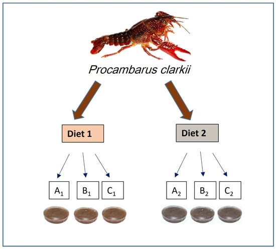
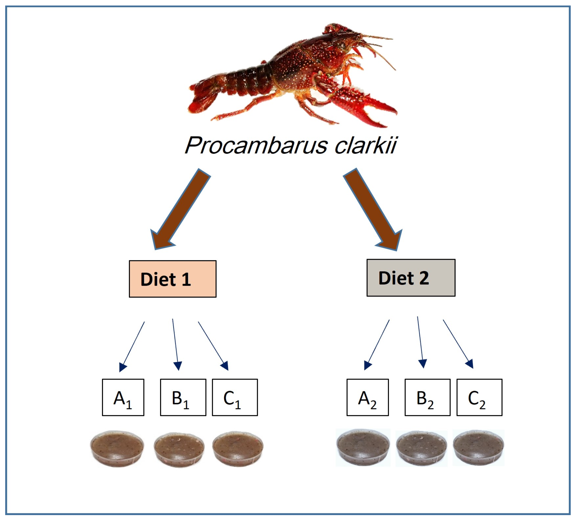
We investigated the effect of polyethylene and bio-based PLA on the growth rate, survival rate, and condition of crayfish in the most intensive phase of individual growth. Polyethylene is one of the most common types of plastic used in everyday life. Its environmentally safer alternative is bioplastics. Fragments of the selected plastics were cut with scissors into pieces measuring 1 mm × 3 mm (microplastics). These were carefully mixed with two types of diets that imitated the food naturally consumed by crayfish: Diet 1—commercial insect meal (BIB 055; Stil, Międzyrzecz, Poland), which has gained increasing popularity recently and been approved as a food additive and for preparing fish bait—Diet 2—fish meal (PL3004107p; Rol-Pasz, Piaski, Poland), intended for an animal feed additive. Insect meal and fish meal, each weighing 42 g and placed into separate beakers, were mixed with 20 mL of distilled water. Each mixture was then spread into three separate Petri dishes. Pieces of polyethylene film and bio-based PLA film from disposable bags were added to the two dishes. A third dish containing only mixed insect meal or fish meal served as control (Figure 1).
Figure 1.
Schematic diagram summarizing the crayfish experimental design. Diet 1 is based on insect meal, Diet 2 is based on fish meal. The experimental treatments: A1—Diet 1, food without plastic (control treatment); B1—Diet 1, food with bio-based PLA microplastics; C1—Diet 1, food with polyethylene (PE) microplastics; A2—Diet 2, food without plastic (control treatment); B2—Diet 2, food with bio-based PLA microplastics; C2—Diet 2, food with polyethylene (PE) microplastics.
The prepared food was stored in a freezer at −20 °C and thawed before feeding to the crayfish. Food was served (75 mg) three times a week in excess, each portion of food in the microplastic treatments contained microparticles (~7 mg). The leftovers were removed before feeding the next portion of food. The actual ingestion of microplastic was noted though the quantity was not specified. Once a week, a leaf of steamed nettle Urtica dioica was added to the crayfish containers as an additional source of plant food to supplement essential minerals. Once a week, 30% of the water was replaced.
In total, the experiment involved six treatments: (A1) Diet 1, insect meal control, (B1) Diet 1, insect meal with bio-based PLA foil, (C1) Diet 1, insect meal with polyethylene foil, (A2) Diet 2, fish meal control, (B2) Diet 2, fish meal with bio-based PLA foil, (C2) Diet 2, fish meal with polyethylene foil (Figure 1). Each treatment had six replicates. During the experiment, survival, growth rate, and coloration intensity were studied. Total length was measured to the nearest 1 mm in accordance with the applicable principle of measuring the crayfish from the rostrum to the end of the telson. Coloration intensity indicates the overall condition of the crustaceans. In crayfish, such measurements can be made in juveniles when the carapace has not yet fully matured. After 60 days, at the end of the experiment, the crayfish were photographed using the same settings on the digital camera. The images were then processed using MS Paint software (22H2, Windows 10) on the RGB (red, green, blue) scale—a program designed to create and edit raster graphics in Windows systems. MS Paint software does not automatically analyze an entire image in terms of average brightness or histogram distribution. However, it enables precise measurement of individual pixel parameters. This process is carried out manually by selecting a specific point on the image. The measurement was performed at the same location on every specimen. The brightness, color saturation and the values of red, green, and blue colors were studied at three points—on the cephalothorax, the abdomen—as well as the background of the photographs.
2.3. Microbial Analyses in the Crayfish Experiment
Basic microbiological analyses were conducted to obtain a more comprehensive understanding of the conditions in the water under different experimental treatments. Water samples from each treatment were frozen at −20 °C until analysis. Three independent replicates per variant were used for all microbial analyses. Bacteria from thawed water samples were inoculated using the serial dilution method onto the following selected media: (1) R2A for estimating the total number of heterotrophic bacteria (CFU—colony-forming units) per mL of water, (2) Mannitol Salt Agar (CHAPMAN) to determine the quantity of Staphylococcus spp., and (3) MacConkey Agar (for Gram-negative bacteria). At least 10 technical replicates for each dilution of 10 μL suspension of the bacteria were inoculated per agar plate. Microorganisms [except for sulfate-reducing bacteria (SRB)] were cultured for six days at 25 °C ± 1 °C, and the single bacterial colonies were counted. The results are presented in CFU/mL.
To estimate the number of SRB, 10 mL of inoculum was mixed with 90 mL of mineral Postgate C medium [39], which also contained sodium thioglycolate. Quanti-Tray 2000 trays and IDEXX Sealer PLUS (IDEXX Laboratories, Inc., Westbrook, Maine, USA) were used for the analysis. The SRB were cultured at 25 °C ± 1 °C for up to 32 days. Positive growth was indicated by blackening of the medium at the bottom of the cells due to iron sulfide formation. All blackened cells were counted as potential SRB. Results for the most probable number (MPN) of SRB in 100 mL from days 8, 15, and 32 were used for statistical analyses. The aim was to determine whether these hydrogen sulfide-producing bacteria could have an effect on crayfish, depending on the duration of incubation.
2.4. Experimental Treatments and Measurements of Shrimp
We investigated the effect of different polymers: polystyrene and biodegradable PLA, on the growth rate, survival rate, and condition of shrimp in the less intensive phase of individual growth (adult individuals). Since bamboo shrimp are filter feeders, the plastic was broken down into smaller fragments than in the case of crayfish. Polystyrene was abraded with sandpaper into 100–500 µm fragments. In the case of the bio-plastic, liquid nitrogen and then a mixer were used to obtain fragments 100–900 µm in size. These were carefully mixed with “Shrimp Sticks” (Tropical, Poland) powdered food for crustaceans in a 9:1 ratio (food:plastic). The prepared food was stored in a freezer at −20 °C and thawed before feeding to the shrimp. Food was served (200 mg) three times a week in excess. Each portion of food in the treated treatments contained microplastics (~20 mg). The leftovers were removed before feeding the next portion of food.
The experiment involved five treatments: (A) control aquaria, food without microplastic, (B) control aquaria with light-colored bottoms and side walls, food without microplastic, (C) polypropylene control containers with light-colored bottoms and side walls, food without microplastic, (D) treatment aquaria, food with polylactic acid, (E) treatment aquaria, food with polystyrene. Each treatment had ten replicates.
The shrimp were measured and photographed at the beginning and at the end of the experiment (after 60 days). Total length was measured to the nearest 1 mm in accordance with the applicable principle of measuring the shrimp from the rostrum to the end of the telson. The images were then processed using MS Paint software on the RGB scale. The brightness, color saturation and the values of red, green, and blue colors were studied at three points—on the cephalothorax, the abdomen—as well as the background of the photographs.
2.5. Statistical Analyses
The Shapiro–Wilk test did not confirm normal distribution in both experiments. Thus, non-parametric tests—the Kruskal–Wallis test and the Mann–Whitney test—were conducted to analyze the differences in growth rate, survival percentage, and coloration intensity of the carapace of crayfish and shrimp that consumed bio-based PLA and conventional microplastics compared to control ones. The differences in Staphylococcus spp. (Gram-positive), Gram-negative, and SRB between the treatments were also analyzed in the experiment with crayfish. A significance level of α = 0.05 was used for the statistical analyses, all of which were performed using Statistica 12 software (StatSoft, Inc., Tulsa, OK, USA).
3. Results
3.1. Growth and Survival of the Studied Decapods
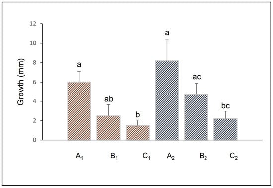
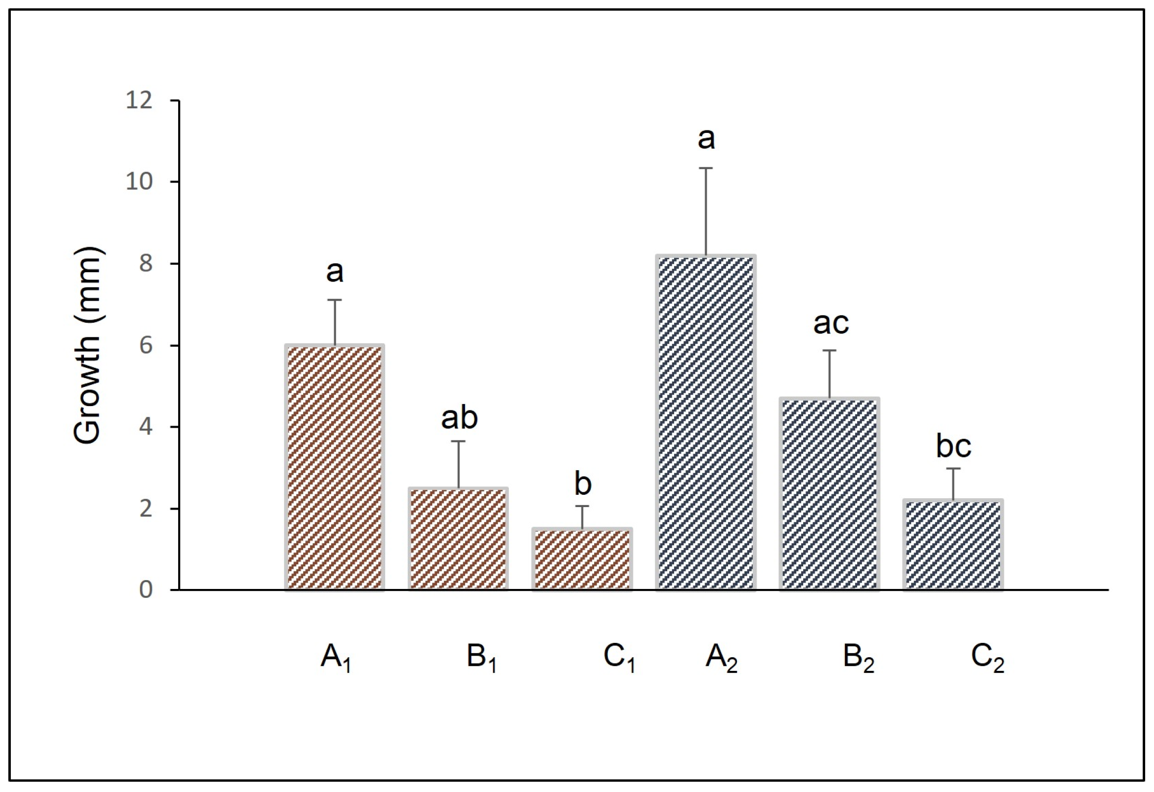
In an experiment involving crayfish, statistically significant differences in the growth patterns were observed (Kruskal–Wallis test, H5,36 = 14.4, p = 0.01), which were related to the addition of polyethylene microplastics to the food, both in Diets 1 and 2 (Mann–Whitney test, p = 0.007 and p = 0.03, respectively), which indicated that microplastics reduced the growth of the crayfish (Figure 2). For the crayfish fed Diet 1, the difference was higher. In contrast, no significant negative impact of bio-based microplastics was noted, although a trend was observed, particularly in the crayfish fed Diet 1 (Figure 2). Crayfish fed Diet 1 exhibited 100% survival in the control treatment, while survival decreased to 50% ± 22 in the treatments with bio-based and polyethylene microplastics. Crayfish fed Diet 2 showed 100% survival in both the control and bio-based microplastic treatments, while survival was 83% ± 17 in the polyethylene microplastic treatment. In terms of growth, Diet 1 was less beneficial for the juvenile P. clarkii than Diet 2. Despite the observed trends, the nonparametric tests did not show significant differences in the survival of the studied crayfish (p = 0.06).
Figure 2.
Average changes in body length of Procambarus clarkii, depending on the treatment, where the crayfish were fed: A1—Diet 1, food without plastic (control treatment); B1—Diet 1, food with bio-based PLA microplastics; C1—Diet 1, food with polyethylene microplastics; A2—Diet 2, food without plastic (control treatment); B2—Diet 2, food with bio-based PLA microplastics; C2—Diet 2, food with polyethylene microplastics. Diet 1 is based on insect meal, and Diet 2 is based on fish meal. Bars sharing the same letter are not significantly different (Mann–Whitney test; p < 0.05). Error bars represent ±1 SE.
In the shrimp experiment, no statistically significant differences in growth and survival were observed. Molting shrimp were observed but the average increase in length was minimal (~2 mm) and not significant (Kruskal–Wallis test, H4,45 = 1.5; p > 0.05). Shrimp showed an identical survival rate of 90% in both the control groups (A, B, C) and the groups exposed to microplastics (D, E).
3.2. Coloration Intensity of the Carapace
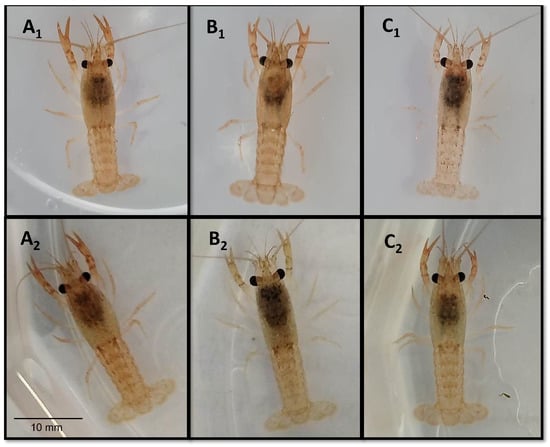
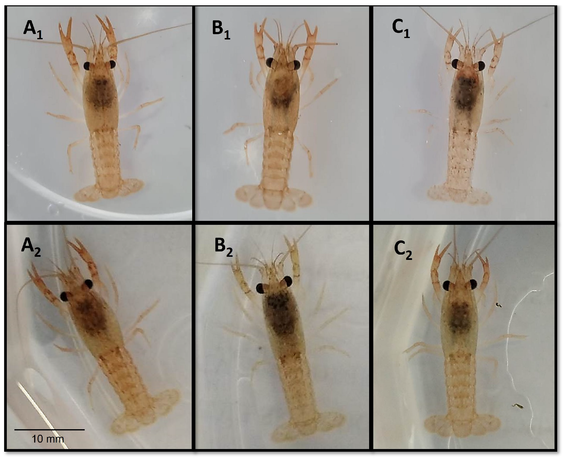
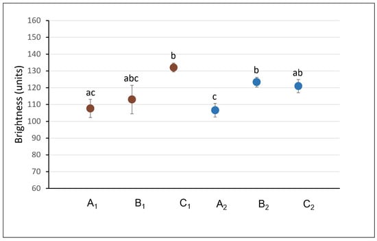
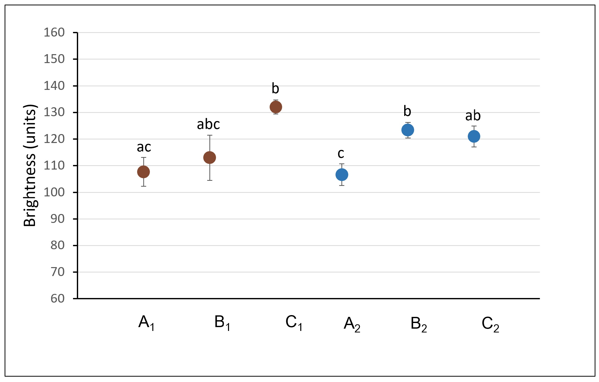
Significant differences in the coloration of the studied crayfish were observed, specifically in the brightness of the carapace at abdomen (Kruskal–Wallis test, H5,29 = 14, p = 0.01). The crayfish fed with bio-based microplastics and those fed with polyethylene microplastics had lighter carapaces compared to the control group (Figure 3). These differences were observed in crayfish fed Diet 1 (p = 0.03 for polyethylene microplastic treatments) and crayfish fed Diet 2 (p = 0.03 for polyethylene microplastics, p = 0.02 for bio-based microplastics, respectively; Figure 4). In the case of the cephalothorax, differences in color intensity by the treatment were not observed. This is probably due to differences in the thickness of the exoskeleton, which in decapod crustaceans on the cephalothorax is thicker than on the abdomen [40].
Figure 3.
Coloration intensity of Procambarus clarkii carapace depending on the treatment, in which the crayfish was fed: A1—Diet 1, food without plastic (control treatment); B1—Diet 1, food with bio-based PLA microplastics; C1—Diet 1, food with polyethylene microplastics; A2—Diet 2, food without plastic (control treatment); B2—Diet 2, food with bio-based PLA microplastics; C2—Diet 2, food with polyethylene microplastics. Diet 1 is based on insect meal, and Diet 2 is based on fish meal.
Figure 4.
Average brightness of the Procambarus clarkii carapace measured from pictures of the dorsal side of the abdomen at the end of the experiment. A1—Diet 1, food without plastic (control treatment); B1—Diet 1, food with bio-based PLA microplastics; C1—Diet 1, food with polyethylene microplastics; A2—Diet 2, food without plastic (control treatment); B2—Diet 2, food with bio-based PLA microplastics; C2—Diet 2, food with polyethylene microplastics. Diet 1 is based on insect meal, and Diet 2 is based on fish meal. Dot plots sharing the same letter are not significantly different (Mann–Whitney test; p < 0.05). Error bars represent ±1 SE.
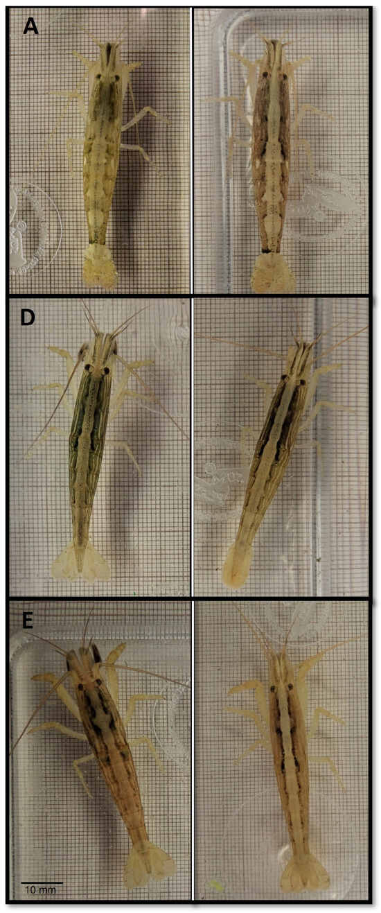
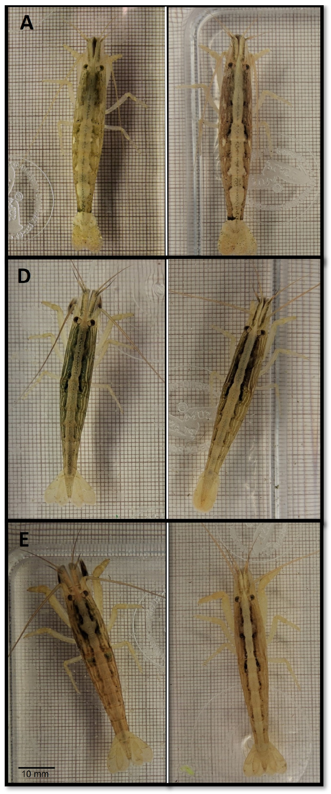
At the beginning of the experiment, no statistically significant differences were found among the treatments regarding any of the shrimp color parameters examined (Kruskal–Wallis test, H4,50 = 5.0; p > 0.05). In turn, significant changes were observed in the coloration of the carapace of shrimp at the end of the experiment (Figure 5).
Figure 5.
Coloration intensity of Atyopsis moluccensis carapace at the beginning (left) and at the end (right) of the experiment. A—food without plastic (control aquaria); D—food with bio-based PLA microplastics; E—food with polystyrene microplastics. The photo for the experimental treatment shows the same individual at the beginning and at the end of the experiment.
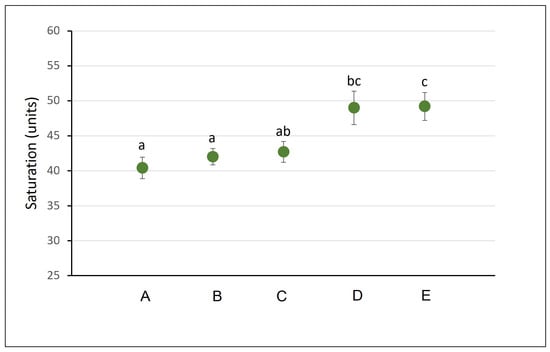
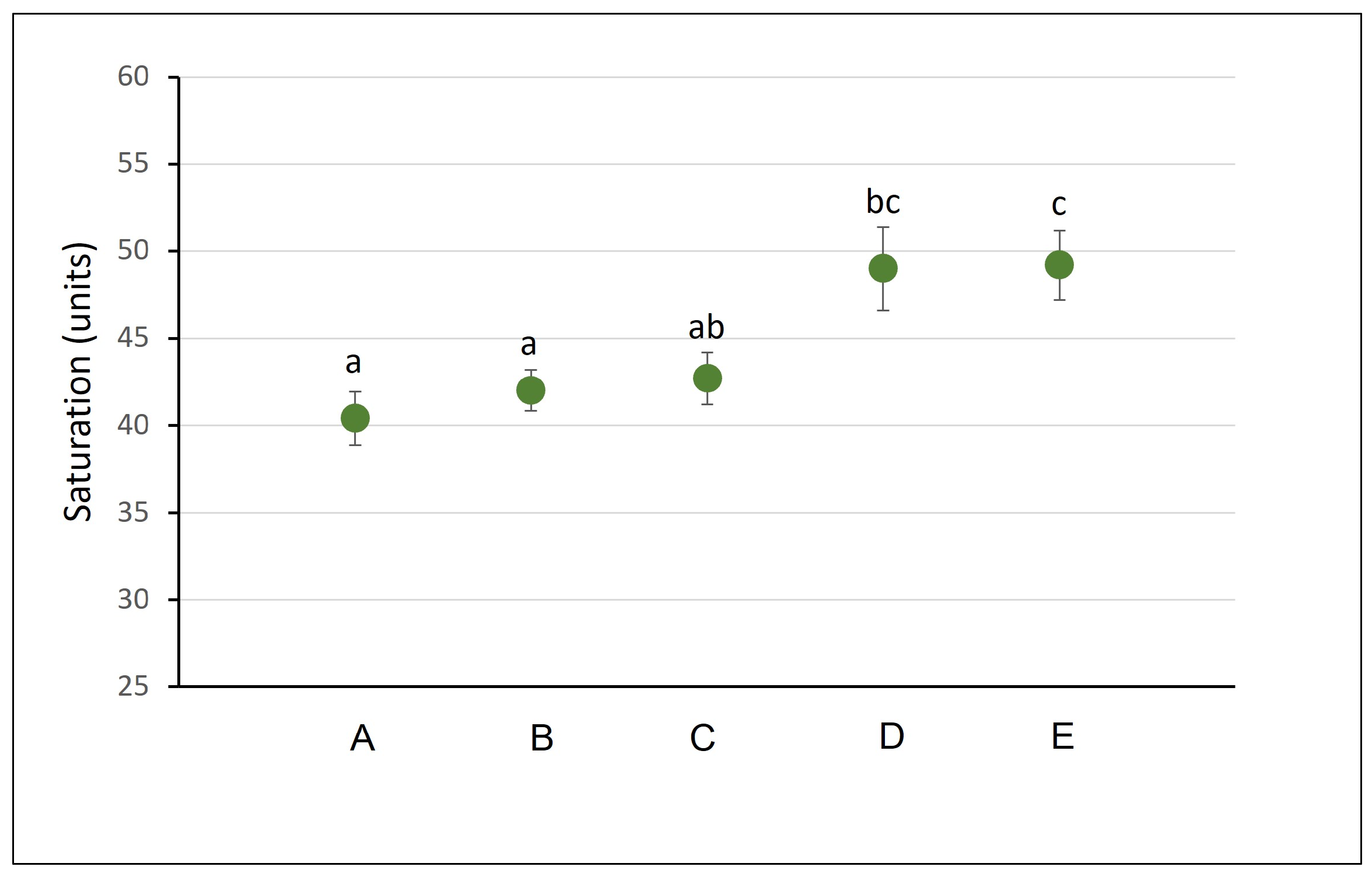
We noticed a significant difference in abdomen color saturation of shrimp between experimental groups (Kruskal–Wallis test, H4,45 = 15.6; p = 0.004). The changes were observed in shrimp fed diets containing either bio-microplastic (D) or conventional microplastics (E), which showed less shadowed coloration compared to the control (A, B and C) groups (Figure 6).
Figure 6.
Average color saturation of the Atyopsis moluccensis carapace measured from pictures of the dorsal side of the abdomen at the end of the experiment. A—control aquaria, food without microplastic, B—control aquaria with light-colored bottoms and side walls, food without microplastic, C—polypropylene control containers with light-colored bottoms and side walls, food without microplastic, D—treatment aquaria, food with polylactic acid, E—treatment aquaria, food with polystyrene. Dot plots sharing the same letter are not significantly different (Mann–Whitney test; p < 0.05). Error bars represent ±1 SE.
The shrimp in the control treatments B (aquaria with light-colored bottoms and side walls) and C (polypropylene containers with light-colored bottoms and side walls) did not differ statistically in coloration compared to the A control group (p > 0.05). This result indicates that neither the background color nor the plastic experimental containers affected the shrimp’s coloration.
There were no significant differences in the background brightness of the images on which the measurements were performed in either the crayfish or the shrimp experiment (Kruskal–Wallis test, H2,53 = 0.8, p > 0.05; H4,50 = 2.7, p > 0.05, respectively)—the crayfish and shrimp were photographed under the same lighting conditions. There was also no correlation between the brightness of the carapace and the size of the crayfish (Spearman correlation, rs = −0.3, p > 0.05; rs = −0.4, p > 0.05 for Diet 1 and Diet 2, respectively). This indicates that the differences in coloration intensity of the carapace were not due to differences in the size of the studied individuals but were, in fact, the result of the addition of conventional microplastics and PLA in the diet.
3.3. Presence of Microorganisms
The microbial analysis indicated significant differences in Staphylococcus spp. (Gram-positive) bacteria abundance between treatments (Kruskal–Wallis test: H5,18 =14.3, p = 0.01). We observed a significantly higher amount of these bacteria in treatments with Diet 1 (CFU = 159 ± 31.8) than Diet 2 (CFU = 28 ± 8.6) fed to the crayfish (Mann–Whitney test, p = 0.0006). This is consistent with the weaker coloration of P. clarkii fed Diet 1 and suggests that the insect meal is less beneficial to crayfish. The addition of bioplastic or polyethylene plastic to Diet 1 did not have a significant effect on the amount of Gram-positive bacteria. However, in the case of Diet 2, significant differences were observed (Kruskal–Wallis test: H2,9 = 6.5, p = 0.04). In the treatment with polyethylene microplastic added, the highest amount of Gram-positive bacteria was detected (CFU = 53.3 ± 5.8) compared to the treatment with bioplastic added (CFU = 30 ± 12) and the control (CFU = 0).
No statistically significant differences (p > 0.05) were observed between the experimental treatments in terms of total heterotrophs (R2A agar), Gram-negative bacteria (Mc Conkey Agar), and SRB (irrespective of days of culture).
4. Discussion
The negative impact of conventional microplastics was confirmed by observing significantly lower growth in exposed Procambarus clarkii individuals. Another indicator of the adverse effects of adding microplastics to the diet was a change in exoskeleton coloration, observed in both the crayfish and the shrimp studied. These primarily involved changes in brightness and saturation. The literature provides data on the impact of microplastics on crustaceans in natural environments and the overall microplastic content in the organisms of Procambarus clarkii and Pontastacus (Astacus) leptodactylus [41,42]. Microplastics accumulate much more readily in the gut of crayfish than in their gills, indicating the potential ingestion of plastic fragments with food. The accumulation of polyethylene and polystyrene microplastics can lead to changes in the lipid metabolism, intestinal microbiome dysbiosis, and oxidative stress in crayfish and crabs [22,42]. An increased abundance of intestinal pathogens and reduced amounts of probiotics, such as Lactobacillus, can impair immune responses, thereby increasing the probability of diseases, which can result in slower growth and development of P. clarkii [42]. The juvenile redclaw crayfish Cherax quadricarinatus showed disturbed lipid metabolism in the hepatopancreas and reduced growth due to exposure to polystyrene microplastics [43]. Similarly, growth disturbances have also been observed in the brine shrimp Artemia parthenogenetica, where exposure to polystyrene resulted in approximately 15% lower body length and that to polyethylene resulted in approximately 18% lower body length [44]. In juvenile horseshoe crab Tachypleus tridentatus and the freshwater shrimp Macrobrachium rosenbergii, polyethylene terephthalate (PET), polystyrene, and polyethylene microplastics were found to have a negative effect on growth [45,46].
In contrast to conventional plastics, the effect of bioplastics on crustaceans is not yet well understood. Our results indicated a negative impact of microplastics from both bio-based PLA and polyethylene microplastics on the lighter coloration of the carapace of crayfish, especially evident in the telson of individuals fed Diet 1 based on insect meal. This type of food was less favorable to crayfish than the fish meal served in Diet 2, which may be because insects are not the natural food of P. clarkii. Emerging studies on bio-based microplastics have indicated their harmful effects on living organisms. The fish Lates calcarifer can ingest microplastic particles from biodegradable plastics, which causes changes in the expression of proteins responsible for reducing oxidative stress in the liver [47]. Bio-based PLA and polybutylene succinate (PBS) particles ingested by Artemia franciscana lead to a reduction in the hatching percentage and an increase in mortality rate [48]. PLA microplastics have also been noted to disrupt the structure of the digestive gland in the eastern mudsnail (Ilyanassa obsoleta). At higher concentrations, they additionally increase the number of hemocytes and reduce fecundity [49].
To the best of our knowledge, no study has yet investigated the direct effect of microplastics on the coloration of the crayfish and shrimp carapace. The changes in color of the decapod exoskeleton can be a result of growth, physiological response to stressful conditions or camouflage strategy including background matching [37,50]. The possibility of color changes resulting from background matching was ruled out in our experiment with the A. moluccensis shrimp. The changes in color of the carapace as response to stressful conditions have been documented in the sand fiddler crab Leptuca pugilator; the animal’s carapace lightened within 15 min of being exposed to warm temperatures [51]. These changes can also be attributed to internal factors, such as oxidative stress, which may be the result of changes in the bacterial flora inside the intestine or changes in the hepatopancreas of crayfish. This organ plays an important role in digestion, immunity, growth, and reproduction of crustaceans [43,52]. As a result of exposure to microplastics, P. clarkii exhibited encapsulation of hepatopancreatic tubules and a reduction in the richness of intestinal microflora [42]. These changes can cause oxidative stress in cells and tissues. A coloration change related to the response to stressogenic factors was observed in the Pacific white shrimp Litopenaeus vannamei, whose body color changed after being infected with the Taura syndrome virus [53]. In the same shrimp species, the effect of intestinal microflora on carapace color was observed. The development of the white feces syndrome disease caused reduced food consumption, which resulted in changes in carapace coloration [54].
In our study, neither bio-based nor conventional microplastics had a significant effect on P. clarkii and A. moluccensis mortality during the experiments. Other studies conducted on P. clarkii indicated that, despite the occurrence of oxidative stress and metabolic disorders, polyethylene plastics did not cause significant changes in their mortality [42]. Red swamp crayfish is an invasive species resistant to adverse environmental conditions, including exposure to microplastics. There are no known studies examining a longer exposure duration than our experiment. Therefore, further research is needed, as the negative effects of polyethylene microplastics may become lethal with long-term exposure. Studies have indicated that polyethylene microplastics can increase mortality, as observed in, for example, the Pacific white shrimp Litopenaeus vannamei. Microplastics were found to damage the hepatopancreas, causing oxidative stress, which contributed to the death of the animals [55]. In a study of the effect of microplastic fibers on freshwater cladoceran water flea Daphnia magna, after 48 h of exposure to PET microfibers, the mortality significantly increased compared to the control group [56]. Increased mortality after exposure to polypropylene microfibers was noted in the Pacific mole crab Emerita analogue [57]. In contrast, 24 h and 48 h exposure of the larval stages of the barnacle Amphibalanus amphitrite and the brine shrimp Artemia franciscana to polystyrene microplastics, despite inducing oxidative stress, did not result in increased mortality [58]. These contradictory results indicate that the lethal effect of microplastics on some organisms may be apparent in long-term experiments.
Microplastics are considered a suitable substrate for the growth of microbial communities known as plastisphere. Their growth is facilitated by the rough surface of microplastics and the provided nutrients and is associated with the generation of biofilms [59]. Gram-positive bacteria, such as Staphylococcus, are known to form a stable biofilm on the surface [60]. Staphylococcus aureus isolates from P. clarkii crayfish have already been reported to be closely related to clinical isolates with virulence potential [61]. We detected higher abundances of Staphylococcus spp. in water from insect meal treatments and in treatments with the addition of bioplastics, especially polyethylene plastic, to the fish meal diet. Interestingly, water from the control treatment with fish meal-based diet did not contain Staphylococcus spp. The presence of Gram-positive bacteria can be a stressor factor for the studied crayfish. Zhang et al. [42] reported that polyethylene exposure induces transcription factors and antimicrobial peptides, disrupting the balance of intestinal microbiota in P. clarkii and consequently increasing the risk of disease. In our study, we observed reduced growth of P. clarkii in polyethylene treatments and lighter carapace coloration in both bio-based and polyethylene treatments, particularly in crayfish fed an insect-based diet. Our research demonstrated the effect within a limited, experimental scope. It would be valuable to study the combined effect of multiple polymers in a long-term experiment, which would provide a more comprehensive look at microplastic pollution risk in the natural environment.
According to the literature, bio-based plastic PLA as a polymer substitute is not safe for natural environments and may have negative effects on aquatic organisms as a stressogenic factor. Note that not all biodegradable plastics decompose at the same rate; the rate of decomposition depends on many factors, such as the type of polymer, the temperature and type of climatic conditions, and the group of microorganisms in the environment [62]. Due to this variability and, almost importantly, the limited number of studies, it is not yet possible to unequivocally confirm or deny the harmfulness of bioplastics. However, existing data indicate that not all types of bio-based plastic are safe.
Author Contributions
Conceptualization, A.K. and K.K.; methodology, M.W., A.C. and J.J.; formal analysis, M.Ż.-B.; investigation, A.C., R.M., M.W. and J.J.; writing—original draft, A.K.; writing—review and editing, K.K. and J.R.; funding acquisition, J.R. All authors have read and agreed to the published version of the manuscript.
Funding
This research was funded by the Ministry of Science and Higher Education, and the statutory grants of Cardinal Stefan Wyszynski University in Warsaw PBBNS-2/23.
Institutional Review Board Statement
Studies involving Procambarus clarkii were conducted based on the permit issued by the Polish General Director of Environmental Protection, with the reference number DZP-WG.672.78.2022.EP.
Data Availability Statement
The data presented in this study are available in https://doi.org/10.6084/m9.figshare.34166403.
Acknowledgments
We would like to thank anonymous reviewers for their insightful comments and suggestions.
Conflicts of Interest
The authors declare no conflicts of interest. The funders had no role in the design of the study; in the collection, analyses, or interpretation of data; in the writing of the manuscript; or in the decision to publish the results.
References
- Geyer, R.; Jambeck, J.R.; Law, K.L. Production, use, and fate of all plastics ever made. Sci. Adv. 2017, 3, e1700782. [Google Scholar] [CrossRef] [Scilit] [PubMed]
- Young, A.M.; Elliott, J.A. Characterization of microplastic and mesoplastic debris in sediments from Kamilo Beach and Kahuku Beach, Hawaii. Mar. Pollut. Bull. 2016, 113, 477–482. [Google Scholar] [CrossRef] [Scilit] [PubMed]
- Kihara, S.; Köper, I.; Mata, J.P.; McGillivray, D.J. Reviewing nanoplastic toxicology: It’s an interface problem. Adv. Colloid Interface Sci. 2021, 288, 102337. [Google Scholar] [CrossRef] [Scilit] [PubMed]
- García-Regalado, A.; Herrera, A.; Almeda, R. Microplastic and mesoplastic pollution in surface waters and beaches of the Canary Islands: A review. Mar. Pollut. Bull. 2024, 201, 116230. [Google Scholar] [CrossRef] [Scilit] [PubMed]
- Güven, O.; Gökdağ, K.; Jovanović, B.; Kıdeyş, A.E. Microplastic Litter Composition of the Turkish Territorial Waters of the Mediterranean Sea, and its Occurrence in the Gastrointestinal Tract of Fish. Environ. Pollut. 2017, 223, 286–294. [Google Scholar] [CrossRef] [Scilit] [PubMed]
- Mallik, A.; Xavier, K.M.; Naidu, B.C.; Nayak, B.B. Ecotoxicological and Physiological Risks of Microplastics on Fish and Their Possible Mitigation Measures. Sci. Total Environ. 2021, 779, 146433. [Google Scholar] [CrossRef] [Scilit] [PubMed]
- Carpenter, E.J.; Anderson, S.J.; Harvey, G.R.; Miklas, H.P.; Peck, B.B. Polystyrene spherules in coastal waters. Science 1972, 178, 749–750. [Google Scholar] [CrossRef] [Scilit] [PubMed]
- Colton, J.B.; Knapp, F.D.; Burns, B.R. Plastic particles in surface waters of the northwestern Atlantic. Science 1974, 185, 491–497. [Google Scholar] [CrossRef] [Scilit] [PubMed]
- Hoss, D.E.; Settle, L.R. Ingestion of plastics by teleost fishes. In Proceedings of the Second International Conference on Marine Debris; Shomura, R.S., Codfrey, H.L., Eds.; NOAA Tech. Memo. NMFS, NOAA-TM-NMFS-SWFSC-154; U.S. Department of Commerce: Miami, FL, USA, 1990; pp. 693–709. [Google Scholar]
- Ward, J.E.; Levinton, J.S.; Shumway, S.E. Influence of diet on pre-ingestive particle processing in bivalves I: Transport velocities on the ctenidium. J. Exp. Mar. Biol. Ecol. 2003, 293, 129–149. [Google Scholar] [CrossRef] [Scilit]
- Foekema, E.M.; De Gruijter, C.; Mergia, M.T.; van Franeker, J.A.; Murk, A.J.; Koelmans, A.A. Plastic in north sea fish. Environ. Sci. Technol. 2013, 47, 8818–8824. [Google Scholar] [CrossRef] [Scilit] [PubMed]
- Lusher, A.L.; McHugh, M.; Thompson, R.C. Occurrence of microplastics in the gastrointestinal tract of pelagic and demersal fish from the English Channel. Mar. Pollut. Bull. 2013, 67, 94–99. [Google Scholar] [CrossRef] [Scilit] [PubMed]
- De Witte, B.; Devriese, L.; Bekaert, K.; Hoffman, S.; Vandermeersch, G.; Cooreman, K.; Robbens, J. Quality assessment of the blue mussel (Mytilus edulis): Comparison between commercial and wild types. Pollut. Bull. 2014, 85, 146–155. [Google Scholar] [CrossRef] [Scilit] [PubMed]
- Van Cauwenberghe, L.; Janssen, C.R. Microplastics in bivalves cultured for human consumption. Environ. Pollut. 2014, 193, 65–70. [Google Scholar] [CrossRef] [Scilit] [PubMed]
- Thompson, R.C. Microplastics in the marine environment: Sources, consequences and solutions. In Marine Anthropogenic Litter; Bergmann, M., Gutow, L., Klages, M., Eds.; Springer: Cham, Switzerland, 2015; pp. 185–200. [Google Scholar] [CrossRef] [Scilit]
- Devriese, L.I.; Van der Meulen, M.D.; Maes, T.; Bekaert, K.; Paul-Pont, I.; Frère, L.; Vethaak, A.D. Microplastic contamination in brown shrimp (Crangon crangon, Linnaeus 1758) from coastal waters of the Southern North Sea and Channel area. Mar. Pollut. Bull. 2015, 98, 179–187. [Google Scholar] [CrossRef] [Scilit] [PubMed]
- Murray, F.; Cowie, P.R. Plastic contamination in the decapod crustacean Nephrops norvegicus (Linnaeus, 1758). Mar. Pollut. Bull. 2011, 62, 1207–1217. [Google Scholar] [CrossRef] [Scilit] [PubMed]
- Hara, J.; Frias, J.; Nash, R. Quantification of microplastic ingestion by the decapod crustacean Nephrops norvegicus from Irish waters. Mar. Pollut. Bull. 2020, 152, 110905. [Google Scholar] [CrossRef] [Scilit] [PubMed]
- Iannilli, V.; Di Gennaro, A.; Lecce, F.; Sighicelli, M.; Falconieri, M.; Pietrelli, L.; Battisti, C. Microplastics in Talitrus saltator (Crustacea, Amphipoda): New evidence of ingestion from natural contexts. Environ. Sci. Pollut. Res. 2018, 25, 28725–28729. [Google Scholar] [CrossRef] [Scilit] [PubMed]
- dos Anjos Guimarães, G.; de Moraes, B.R.; Ando, R.A.; Sant’Anna, B.S.; Perotti, G.F.; Hattori, G.Y. Microplastic contamination in the freshwater shrimp Macrobrachium amazonicum in Itacoatiara, Amazonas, Brazil. Environ. Monit. Assess. 2023, 195, 434. [Google Scholar] [CrossRef] [Scilit] [PubMed]
- Farrell, P.; Nelson, K. Trophic level transfer of microplastic: Mytilus edulis (L.) to Carcinus maenas (L.). Environ. Pollut. 2013, 177, 1–3. [Google Scholar] [CrossRef] [Scilit] [PubMed]
- Yu, P.; Liu, Z.; Wu, D.; Chen, M.; Lv, W.; Zhao, Y. Accumulation of polystyrene microplastics in juvenile Eriocheir sinensis and oxidative stress effects in the liver. Aquat. Toxicol. 2018, 200, 28–36. [Google Scholar] [CrossRef] [Scilit] [PubMed]
- Rios, L.M.; Moore, C.; Jones, P.R. Persistent organic pollutants carried by synthetic polymers in the ocean environment. Mar. Pollut. Bull. 2007, 54, 1230–1237. [Google Scholar] [CrossRef] [Scilit] [PubMed]
- Teuten, E.L.; Rowland, S.J.; Galloway, T.S.; Thompson, R.C. Potential for plastics to transport hydrophobic contaminants. Environ. Sci. Technol. 2007, 41, 7759–7764. [Google Scholar] [CrossRef] [Scilit] [PubMed]
- Ragusa, A.; Svelato, A.; Santacroce, C.; Catalano, P.; Notarstefano, V.; Carnevali, O.; Giorgini, E. Plasticenta: First evidence of microplastics in human placenta. Environ. Int. 2021, 146, 106274. [Google Scholar] [CrossRef] [Scilit] [PubMed]
- Leslie, H.A.; Van Velzen, M.J.; Brandsma, S.H.; Vethaak, A.D.; Garcia-Vallejo, J.J.; Lamoree, M.H. Discovery and quantification of plastic particle pollution in human blood. Environ. Int. 2022, 163, 107199. [Google Scholar] [CrossRef] [Scilit] [PubMed]
- Talsness, C.E.; Andrade, A.J.; Kuriyama, S.N.; Taylor, J.A.; Vom Saal, F.S. Components of plastic: Experimental studies in animals and relevance for human health. Philos. Trans. R. Soc. Lond. B Biol. Sci. 2009, 364, 2079–2096. [Google Scholar] [CrossRef] [Scilit] [PubMed]
- Bhuyan, M.S. Effects of microplastics on fish and in human health. Front. Environ. Sci. 2022, 10, 827289. [Google Scholar] [CrossRef] [Scilit]
- Ribba, L.; Lopretti, M.; de Oca-Vásquez, G.M.; Batista, D.; Goyanes, S.; Vega-Baudrit, J.R. Biodegradable plastics in aquatic ecosystems: Latest findings, research gaps, and recommendations. Environ. Res. Lett. 2022, 17, 033003. [Google Scholar] [CrossRef] [Scilit]
- Ali, W.; Ali, H.; Gillani, S.; Zinck, P.; Souissi, S. Polylactic acid synthesis, biodegradability, conversion to microplastics and toxicity: A review. Environ. Chem. Lett. 2023, 21, 1761–1786. [Google Scholar] [CrossRef] [Scilit]
- Janik, H.; Sienkiewicz, M.; Przybytek, A.; Guzman, A.; Kucinska-Lipka, J.; Kosakowska, A. Novel biodegradable potato starch-based compositions as candidates in packaging industry, safe for marine environment. Fibers Polym. 2018, 19, 1166–1174. [Google Scholar] [CrossRef] [Scilit]
- Duan, Z.; Cheng, H.; Duan, X.; Zhang, H.; Wang, Y.; Gong, Z.; Wang, L. Diet preference of zebrafish (Danio rerio) for bio-based polylactic acid microplastics and induced intestinal damage and microbiota dysbiosis. J. Hazard. Mater. 2022, 429, 128332. [Google Scholar] [CrossRef] [Scilit] [PubMed]
- Liwarska-Bizukojc, E. Evaluation of the impact of polylactide microparticles on soil biota. Environ. Sci. Pollut. Res. 2025, 32, 21187–21197. [Google Scholar] [CrossRef] [Scilit] [PubMed]
- Khalid, A.; Zalouk-Vergnoux, A.; Benali, S.; Mincheva, R.; Raquez, J.M.; Bertrand, S.; Poirier, L. Are bio-based and biodegradable microplastics impacting for blue mussel (Mytilus edulis)? Mar. Pollut. Bull. 2021, 167, 112295. [Google Scholar] [CrossRef] [Scilit] [PubMed]
- Food and Agricultural Organization (FAO) of the United Nations. FAO Yearbook 2018; FAO: Rome, Italy, 2018. [Google Scholar]
- Jin, S.; Jacquin, L.; Xiong, M.; Li, R.; Lek, S.; Li, W.; Zhang, T. Reproductive pattern and population dynamics of commercial red swamp crayfish (Procambarus clarkii) from China: Implications for sustainable aquaculture management. PeerJ 2019, 7, e6214. [Google Scholar] [CrossRef] [Scilit] [PubMed]
- Takeshita, F. Color changes of fiddler crab between seasons and under stressful conditions: Patterns of changes in lightness differ between carapace and claw. J. Exp. Mar. Biol. Ecol. 2019, 511, 113–119. [Google Scholar] [CrossRef] [Scilit]
- Caro, T.; Morgan, V.M. Correlates of color polymorphism in coconut crabs Birgus latro. Zoology 2018, 129, 1–8. [Google Scholar] [CrossRef] [Scilit] [PubMed]
- Postgate, J.R. The Sulphate-Reducing Bacteria, 2nd ed.; Cambridge University Press: Cambridge, UK, 1984; p. 208. [Google Scholar]
- Joffe, I.; Hepburn, H.R.; Nelson, K.J.; Green, N. Mechanical properties of a crustacean exoskeleton. Comp. Biochem. Physiol. A Physiol. 1975, 50, 545–549. [Google Scholar] [CrossRef] [Scilit]
- Gholamhosseini, A.; Banaee, M.; Sureda, A.; Timar, N.; Zeidi, A.; Faggio, C. Physiological response of freshwater crayfish, Astacus leptodactylus exposed to polyethylene microplastics at different temperature. Comp. Biochem. Physiol. C Toxicol. Pharmacol. 2023, 267, 109581. [Google Scholar] [CrossRef] [Scilit] [PubMed]
- Zhang, X.; Jin, Z.; Shen, M.; Chang, Z.; Yu, G.; Wang, L.; Xia, X. Accumulation of polyethylene microplastics induces oxidative stress, microbiome dysbiosis and immunoregulation in crayfish. Fish Shellfish Immunol. 2022, 125, 276–284. [Google Scholar] [CrossRef] [Scilit] [PubMed]
- Chen, Q.; Lv, W.; Jiao, Y.; Liu, Z.; Li, Y.; Cai, M.; Zhao, Y. Effects of exposure to waterborne polystyrene microspheres on lipid metabolism in the hepatopancreas of juvenile redclaw crayfish, Cherax quadricarinatus. Aquat. Toxicol. 2020, 224, 105497. [Google Scholar] [CrossRef] [Scilit] [PubMed]
- Li, H.; Chen, H.; Wang, J.; Li, J.; Liu, S.; Tu, J.; Liu, X. Influence of microplastics on the growth and the intestinal microbiota composition of brine shrimp. Front. Microbiol. 2021, 12, 717272. [Google Scholar] [CrossRef] [Scilit] [PubMed]
- Jaikumar, I.M.; Periyakali, S.B.; Rajendran, U.; Joen-Rong, S.; Thanasekaran, J.; Tsorng-Harn, F. Effects of microplastics, polystyrene, and polyethylene on antioxidants, metabolic enzymes, HSP-70, and myostatin expressions in the giant river prawn Macrobrachium rosenbergii: Impact on survival and growth. Arch. Environ. Contam. Toxicol. 2021, 80, 645–658. [Google Scholar] [CrossRef] [Scilit] [PubMed]
- Fang, J.K.H.; Tse, T.W.; Maboloc, E.A.; Leung, R.K.L.; Leung, M.M.L.; Wong, M.W.T.; Cheung, S.G. Adverse impacts of high-density microplastics on juvenile growth and behaviour of the endangered tri-spine horseshoe crab Tachypleus tridentatus. Mar. Pollut. Bull. 2023, 187, 114535. [Google Scholar] [CrossRef] [Scilit] [PubMed]
- Xie, M.; Xu, P.; Zhou, W.; Xu, X.; Li, H.; He, W.; Suo, A. Impacts of conventional and biodegradable microplastics on juvenile Lates calcarifer: Bioaccumulation, antioxidant response, microbiome, and proteome alteration. Mar. Pollut. Bull. 2022, 179, 113744. [Google Scholar] [CrossRef] [Scilit] [PubMed]
- Charoeythornkhajhornchai, P.; Kunjiek, T.; Chaipayang, S.; Phosri, S. Toxicity assessment of bioplastics on brine shrimp (Artemia franciscana) and cell lines. Emerg. Contam. 2023, 9, 100253. [Google Scholar] [CrossRef] [Scilit]
- Hewins, B.; Gibson, G. Petroleum-based and biodegradable microplastics alter tissue structure and fecundity in the eastern mudsnail (Ilyanassa obsoleta). Can. J. Zool. 2022, 100, 776–788. [Google Scholar] [CrossRef] [Scilit]
- Kronstadt, S.M.; Darnell, M.Z.; Munguia, P. Background and temperature effects on Uca panacea color change. Mar. Biol. 2013, 160, 1373–1381. [Google Scholar] [CrossRef] [Scilit]
- Silbiger, N.; Munguia, P. Carapace color change in Uca pugilator as a response to temperature. J. Exp. Mar. Biol. Ecol. 2008, 355, 41–46. [Google Scholar] [CrossRef] [Scilit]
- Sastry, A.N. Ecological aspects of reproduction. In The Biology of Crustacea; Vernberg, F.J., Vernberg, W.B., Eds.; Academic Press: New York, NY, USA, 1983; pp. 179–254. [Google Scholar]
- Song, Y.L.; Yu, C.I.; Lien, T.W.; Huang, C.C.; Lin, M.N. Haemolymph parameters of Pacific white shrimp (Litopenaeus vannamei) infected with Taura syndrome virus. Fish Shellfish Immunol. 2003, 14, 317–331. [Google Scholar] [CrossRef] [Scilit] [PubMed]
- Mastan, S.A. Incidences of white feces syndrome (WFS) in farm-reared shrimp, Litopenaeus vannamei, Andhra Pradesh. Indo Am. J. Pharm. Res. 2015, 5, 3044–3047. [Google Scholar]
- Niemcharoen, S.; Haetrakul, T.; Palić, D.; Chansue, N. Microplastic-Contaminated Feed Interferes with Antioxidant Enzyme and Lysozyme Gene Expression of Pacific White Shrimp (Litopenaeus vannamei) Leading to Hepatopancreas Damage and Increased Mortality. Animals 2022, 12, 3308. [Google Scholar] [CrossRef] [Scilit] [PubMed]
- Jemec, A.; Horvat, P.; Kunej, U.; Bele, M.; Kržan, A. Uptake and effects of microplastic textile fibers on freshwater crustacean Daphnia magna. Environ. Pollut. 2016, 219, 201–209. [Google Scholar] [CrossRef] [Scilit] [PubMed]
- Horn, D.A.; Granek, E.F.; Steele, C.L. Effects of environmentally relevant concentrations of microplastic fibers on Pacific mole crab (Emerita analoga) mortality and reproduction. Limnol. Oceanogr. Lett. 2020, 5, 74–83. [Google Scholar] [CrossRef] [Scilit]
- Gambardella, C.; Morgana, S.; Ferrando, S.; Bramini, M.; Piazza, V.; Costa, E.; Faimali, M. Effects of polystyrene microbeads in marine planktonic crustaceans. Ecotoxicol. Environ. Saf. 2017, 145, 250–257. [Google Scholar] [CrossRef] [Scilit] [PubMed]
- Yang, W.; Li, Y.; Boraschi, D. Association between Microorganisms and Microplastics: How Does It Change the Host–Pathogen Interaction and Subsequent Immune Response? Int. J. Mol. Sci. 2023, 24, 4065. [Google Scholar] [CrossRef] [Scilit] [PubMed]
- Mack, D.; Becker, P.; Chatterjee, I.; Dobinsky, S.; Knobloch, J.K.; Peters, G.; Rohde, H.; Herrmann, M. Mechanisms of biofilm formation in Staphylococcus epidermidis and Staphylococcus aureus: Functional molecules, regulatory circuits, and adaptive responses. Int. J. Med. Microbiol. 2024, 294, 203–212. [Google Scholar]
- Dong, X.; Wang, X.; Chen, X.; Yan, Z.; Cheng, J.; Gao, L.; Li, J. Genetic diversity and virulence potential of Staphylococcus aureus isolated from crayfish (Procambarus clarkii). Curr. Microbiol. 2017, 74, 28–33. [Google Scholar] [CrossRef] [Scilit] [PubMed]
- Lott, C.; Eich, A.; Makarow, D.; Unger, B.; Van Eekert, M.; Schuman, E.; Weber, M. Half-life of biodegradable plastics in the marine environment depends on material, habitat, and climate zone. Front. Mar. Sci. 2021, 8, 662074. [Google Scholar] [CrossRef] [Scilit]
Disclaimer/Publisher’s Note: The statements, opinions and data contained in all publications are solely those of the individual author(s) and contributor(s) and not of MDPI and/or the editor(s). MDPI and/or the editor(s) disclaim responsibility for any injury to people or property resulting from any ideas, methods, instructions or products referred to in the content. |
© 2026 by the authors. Licensee MDPI, Basel, Switzerland. This article is an open access article distributed under the terms and conditions of the Creative Commons Attribution (CC BY) license.