


Highlights
- Polystyrene microplastic inhibited growth, increased mortality, and impaired feeding of loach larvae.
- Taurine supplementation (≥0.8%) protected loach larvae from PS-MP toxicity without reducing microplastic ingestion.
- Taurine rescued microplastic-disrupted antioxidant defence and alleviated lipid peroxidation.
- Activation of the Nrf2/Keap1 pathway underpins the antioxidant protection of taurine against PS-MP stress.
Abstract
Microplastics, ubiquitous emerging contaminants in diverse aquatic environments, can be ingested by aquatic organisms and trigger oxidative stress injury, suppressing growth and development. Taurine is a common functional substance with excellent anti-oxidative stress properties. This study sought to investigate whether dietary taurine could mitigate oxidative stress injury triggered by polystyrene microplastics and to analyse its underlying mechanisms in loach (Paramisgurnus dabryanus) larvae. Loach larvae were exposed to 1000 μg/L polystyrene microplastics for 28 days, with graded dietary taurine supplementation (0.2%, 0.4%, 0.8%, and 1.6%), to investigate taurine’s protective effects and regulatory mechanisms. The growth status, feeding ability, mortality, antioxidant enzyme activities (SOD, CAT, GSH-PX), MDA content, and expression of Nrf2/Keap1 pathway-related genes were determined. Following 28 days of PS-MP exposure, the MP group exhibited a cumulative mortality of 42.9%, with its average body weight reduced to 83.51% relative to the control. Dietary taurine supplementation (≥0.8%) effectively reversed these adverse phenotypes. Notably, taurine did not reduce microplastic accumulation in larvae, indicating its protection is independent of microplastic ingestion. Furthermore, taurine enhanced antioxidant enzyme activities, reduced MDA accumulation, and relieved oxidative stress. Mechanistically, taurine activated the Nrf2/Keap1 signaling pathway to upregulate downstream antioxidant gene expression, eliminate excess ROS, and maintain the stability of the antioxidant system under microplastic stress. This study provides a nutritional mitigation strategy against microplastic toxicity and advances our understanding of the ecological risks posed by microplastics to benthic fish in freshwater aquaculture ecosystems.
1. Introduction
Plastic products are widely used in daily life and industrial production due to their high durability, good portability and low cost [1]. Polystyrene ranks among the most widely manufactured plastic polymers globally, due to its favorable electrical insulating properties, high transparency and convenient machinability [2]. It is commonly manufactured into disposable food containers, shock-absorbing buffers for transportation, housings for household appliances and many other commodities. Nevertheless, it is a typical non-biodegradable material [3]. Discarded plastic items gradually break down under physical or chemical conditions to form microplastics (MPs). The resulting pollution has become increasingly severe and evolved into a global environmental concern [4]. Microplastics are defined as plastic particles or fragments with a diameter ranging from 1 micrometer to 5 mm [5]. These microplastics enter aquatic environments via various industrial and agricultural wastewater and domestic sewage pathways, including surface runoff, rivers and lakes [6]. Within aquaculture environments, microplastics may harm farmed aquatic organisms and accumulate through food chain transfer, which creates potential risks to human health [7].
Previous reports have indicated that microplastics are more abundant in freshwater than in marine environments, giving rise to more complex ecotoxicity [2]. In freshwater ecosystems, microplastics are generally widely dispersed across aquatic compartments, both in the water column and sediments, and can be ingested by filter-feeding and benthic organisms [8,9]. Due to their constant interaction with the aquatic environment, fish are particularly susceptible to microplastics [10]. Multiple exposure routes enable microplastic uptake in fish, presenting considerable risks to organism health [11]. Multiple exposure routes enable microplastic uptake in fish, presenting considerable risks to organism health [12,13]. Accumulating evidence demonstrates that microplastics can alter the behaviour of aquatic organisms and disrupt ecosystem functions [14]. Microplastic particles exert adverse effects on feeding activity, individual behaviour and development of diverse planktonic and benthic species [15]. Ingested microplastics may block the digestive tract, reduce energy reserves and suppress growth performance [16,17]. Moreover, microplastics can penetrate tissues and cells of organisms, trigger massive accumulation of reactive oxygen species, disrupt the in vivo redox balance, and induce oxidative-stress damage [18,19]. Such damage further causes cellular lipid peroxidation, disturbance of enzyme systems and aberrant gene expression, and consequently impairs the growth, development, reproduction and survival of aquatic organisms [20,21,22].
Taurine (2-aminoethanesulfonic acid) is a sulfur-containing amino acid that ubiquitously exists across animal tissues, including muscle and liver. It plays vital physiological roles in the growth, antioxidant capacity, stress resistance, and immunomodulation of aquatic animals [23]. Taurine can effectively enhance the endogenous antioxidant capacity of fish, shrimp, and crabs, and alleviate oxidative damage induced by various environmental stressors such as heavy metals, ammonia nitrogen, and high temperature [24,25,26]. Nevertheless, it remains unknown whether taurine could alleviate the growth inhibition and oxidative stress triggered by PS-MPs.
Loach (Paramisgurnus dabryanus) is a typical benthic fish, and its aquaculture production in China has increased remarkably in recent years [27]. In natural and aquaculture water bodies, loaches are directly exposed to contaminants suspended in the water column as well as those deposited at the bottom [28]. As omnivorous species, loaches tend to ingest far greater amounts of pollutants compared with herbivorous and carnivorous fish [29]. For this reason, loaches have been adopted as experimental animals for the assessment of environmental contaminants [30]. Fish larvae are small and physiologically immature, making them more susceptible to environmental pollutants. The development and survival of these larvae govern the physiological characteristics of mature fish and population viability [16]. Therefore, loach larvae were selected as experimental subjects in this study to better verify the protective effects of taurine on fish larvae under PS-MPs stress.
The present study aimed to evaluate the protective effects of taurine on MP-ingested loach larvae, and further investigate whether dietary taurine could alleviate PS-MP-induced growth inhibition and oxidative stress in loach larvae. We evaluated larval growth and feeding performance, antioxidant enzyme activities, and the changes in oxidative stress-related gene expression under PS-MP exposure with graded taurine supplementation. This study provides a nutritional tactic to lower microplastic risks in larval loach farming and sheds light on mitigating microplastic pollution in freshwater benthic systems.
2. Materials and Methods
2.1. Chemicals and Experimental Fish
Green fluorescent polystyrene microplastics (PS-MPs) with a diameter of 5 μm were purchased from Da’e Technology Co., Ltd., Tianjin, China, with an excitation wavelength of 488 nm and an emission wavelength of 518 nm. Taurine was purchased from Shanghai Macklin Biochemical Technology Co., Ltd., Shanghai, China. Dechlorinated tap water used for aquaculture met the following conditions: pH 7.2–7.5, water temperature 26 ± 1 °C, and dissolved oxygen >6.0 mg/L. Fertilized loach embryos were obtained from a fish farm in Zhongshan City (Guangdong, China). The fertilized embryos were incubated in dechlorinated tap water. Unfertilized embryos were removed 24 h post-fertilization, and the culture water was renewed regularly to guarantee healthy embryonic development. The grouping experiment was initiated when loach larvae became free-swimming at 3 days post-fertilization. The loach used in this study was approved by the Institutional Animal Care and Use Committee of South China Agricultural University.
2.2. Experimental Diets
Diets containing graded taurine concentrations (0%, 0.2%, 0.4%, 0.8%, and 1.6%) were prepared, and the formulation of experimental diets is presented in Table 1. This taurine gradient was established with reference to previous studies on juvenile turbot (Scophthalmus maximus L.) [26] and larval largemouth bass (Micropterus salmoides) [31], to investigate whether dietary taurine could alleviate the toxic effects induced by PS-MPs in loach larvae. All diets were stored in sealed food-grade glass jars at −20 °C in the dark prior to use.
Table 1.
Dietary Formulation and proximate composition [31].
2.3. Experimental Design
Based on environmentally relevant PS-MP concentrations and commonly adopted experimental concentrations in freshwater fish [1,11], 1000 μg/L was chosen as the exposure concentration in this study. PS-MPs were suspended in ultrapure water and dispersed by ultrasonication to prepare a stock suspension at 1000 μg/mL. One milliliter of the stock suspension was added to each liter of culture water to obtain the final exposure concentration of 1000 μg/L. Before each use, the stock suspension was homogenized by ultrasonication to achieve uniform dispersion.
Loach larvae with initial feeding activity were randomly allocated to eighteen glass containers (20 × 14 × 16 cm). Each container was filled with 2 L of culture water and stocked with 80 loach larvae. The experimental individuals were divided into 6 groups with 3 replicates per group according to Table 2.
Table 2.
Experimental group division and the addition amounts of PS-MPs and taurine in each group.
During the experiment, larvae in each aquarium were fed to satiation with the corresponding diet at 08:00 and 18:00 every day. Two hours post each feeding, uneaten diet and fecal wastes were siphoned out. One-third of the rearing water was exchanged every two days to sustain suitable water quality. For the microplastic exposure groups, newly prepared polystyrene microplastic solution was supplemented to maintain the target experimental concentration. The exposure trial lasted for 28 days, and sampling and measurements were performed on Day 7 and Day 28.
2.4. Growth Performance Measurement
At the termination of the 28-day exposure experiment, 10 larvae were randomly sampled from each of the three replicate aquariums per treatment group. After anesthetization with MS222, the larvae were photographed under a stereomicroscope. The body length, body height and body thickness of each larva were quantified using ImageJ software 1.52. Given the extremely low individual weight of larvae, 20 loach larvae were sampled from each aquarium; 10 larvae were pooled per tube for weighing to calculate the average body weight, yielding a total of six replicates per treatment.
2.5. Survival Performance, Satiation Rate and Fasting Rate
Larval mortalities were recorded before feeding each day over the 28-day exposure period, and dead larvae were immediately taken out of every container. The Kaplan–Meier method was adopted to plot survival curves [32]. On Day 7 and Day 28 of the experiment, two hours after feeding, the numbers of loach larvae with fully filled intestines and those with empty intestines in each aquarium were recorded, respectively.
Satiation rate = (Number of loach larvae with fully filled intestines/Total number of surviving loach larvae in the aquarium) × 100%
Fasting rate = (Number of loach larvae with empty intestines/Total number of surviving loach larvae in the aquarium) × 100%
2.6. Microplastic Distribution and Residue Analysis
Loach larvae were randomly sampled before feeding and anesthetized in water containing MS222 for the observation of microplastic distribution. Prior to observation, the loach larvae were rinsed three times with ultrapure water to remove surface contaminants, followed by fixation in 4% paraformaldehyde. After 24 h, the samples were transferred into 70% ethanol solution for preservation. Subsequently, fluorescence microscopy was applied to observe and record fluorescent signals of microplastics as well as their distribution across different tissues.
Microplastic burdens in loach larvae were quantified by KOH digestion combined with fluorescence microscopy counting [33]. Ten loach larvae were randomly selected from each aquarium; each treatment group contained 30 loach larvae. All glassware were thoroughly cleaned prior to use to reduce potential microplastic contamination. Larvae from the CK group (without PS-MPs exposure) were processed in parallel as controls following the same digestion, filtration and observation procedures. Particles on the filter membranes were identified under fluorescence microscopy based on morphology and fluorescence features. The selected larvae were rinsed with ultrapure water, and five loach larvae were placed into each 2 mL glass vial as one biological replicate, with six biological replicates per treatment. One milliliter of 10% (w/v) potassium hydroxide (KOH) solution was added to each vial. Samples were incubated in a shaking water bath at 60 °C for 24 h until no visible tissue fragments remained. After digestion, all digested samples underwent vacuum filtration with 0.22 μm membranes. The membrane was then mounted onto a glass slide for microscopic observation. Five fields of view were selected using the five-point sampling method for PS-MPs counting. The average PS-MPs abundance per larva (particles/larva) was calculated using the following formula:
Average PS-MP abundance (particles/larva) = (mean particle count of five fields of view/area of a single microscopic field) × effective filtration area of the membrane/number of pooled larvae.
The abundance of PS-MP fragments was quantified following the same formula.
2.7. Enzymatic Activity Analysis
On Day 7 and Day 28 of the experiment, 10 loach larvae were randomly sampled from each replicate aquarium and pooled into one centrifuge tube to constitute one biological replicate. Three biological replicates were established for each treatment group. The sampled larvae were rinsed with sterile PBS, blotted dry, and weighed collectively with an analytical balance. According to the total body weight of the larvae, pre-chilled PBS was added at a mass ratio of 1:9 to prepare tissue homogenates via low-temperature grinding. After centrifugation (6000× g, 10 min, 4 °C) to discard precipitates, the supernatants were retained for enzyme activity measurement. The levels of malondialdehyde (MDA) and the activities of catalase (CAT), glutathione peroxidase (GSH-PX), superoxide dismutase (SOD) and total protein (TP) were determined using commercial assay kits (Nanjing Jiancheng Bioengineering Institute, Nanjing, China). Kit catalog numbers: MDA (A003-2, TBA method); CAT (A007-1, ammonium molybdate method); GSH-PX (A005-1, colorimetric method); SOD (A001-3, WST-1 method); TP (A045-4, BCA method). Enzyme activities were normalized to total protein content and expressed as U/mg protein. All detection procedures were strictly performed in accordance with the manufacturer’s instructions. The absorbance was detected using a multi-mode microplate reader (Synergy HTX, BioTek, Winooski, VT, USA) at the corresponding wavelengths: MDA at 532 nm, CAT at 405 nm, GSH-PX at 412 nm, SOD at 450 nm, and TP at 562 nm.
2.8. qPCR Analysis
To further evaluate the effects of microplastics and taurine on oxidative stress-related enzymes and the expression of their upstream regulatory genes, three loach larvae were randomly collected from each replicate aquarium on Day 7 and Day 28 of the exposure experiment. For each biological replicate, three loach larvae were randomly selected from one glass container and pooled into a single RNase-free EP tube. Three biological replicates were included for each treatment group. Total RNA was extracted using 1 mL RNAiso Plus (Takara, Beijing, China). After thorough homogenization, the supernatant was collected, and 1/5 volume of chloroform was added. The mixture was centrifuged, and the upper aqueous phase was carefully transferred. RNA was precipitated with an equal volume of isopropanol. The RNA pellet was washed twice with pre-cooled 70% ethanol, air-dried, and dissolved in RNase-free water. The integrity of total RNA was verified by agarose gel electrophoresis, and RNA concentration was determined by ultraviolet spectrophotometry. Qualified RNA was reverse-transcribed into cDNA using the PrimeScript™ RT reagent Kit with gDNA Eraser (Takara, Beijing, China). TB Green Advantage qPCR Premix (Takara, Beijing, China) was applied for quantitative real-time PCR (qPCR) amplification.
The qPCR thermal cycling program was set as follows: initial denaturation at 95 °C for 30 s, followed by 40 amplification cycles of 95 °C for 5 s, 60 °C for 20 s, and 72 °C for 15 s. The primer sequences for target genes, including cat, sod, gsh-px, keap1, nrf2, and maf, are listed in Table 3. β-actin was selected as the internal reference gene for normalization. The relative expression levels of all target genes were calculated via the 2−∆∆CT method.
Table 3.
The primers used in the experiments.
2.9. Statistical Analysis
Experimental data were expressed as mean ± standard error of the mean (SEM). The Shapiro–Wilk test was used to verify the normality of data distribution; one-way ANOVA followed by Tukey’s post hoc test was used to compare differences among groups, and p < 0.05 was considered statistically significant. Statistical analyses were performed using SPSS statistical package version 19.0. Graph plotting was performed using GraphPad Prism 10.6.0 software.
3. Results
3.1. Growth Performance
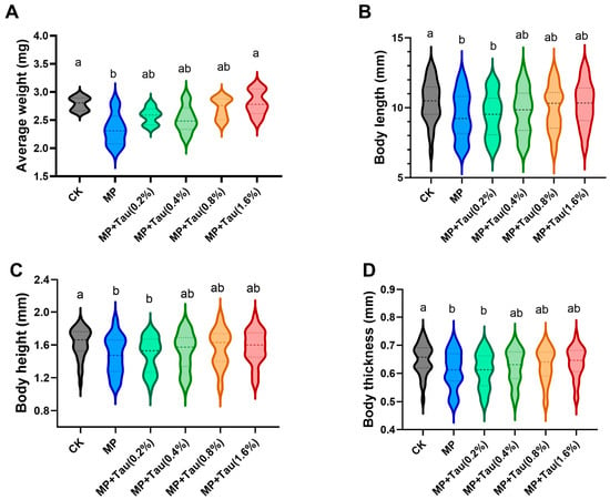
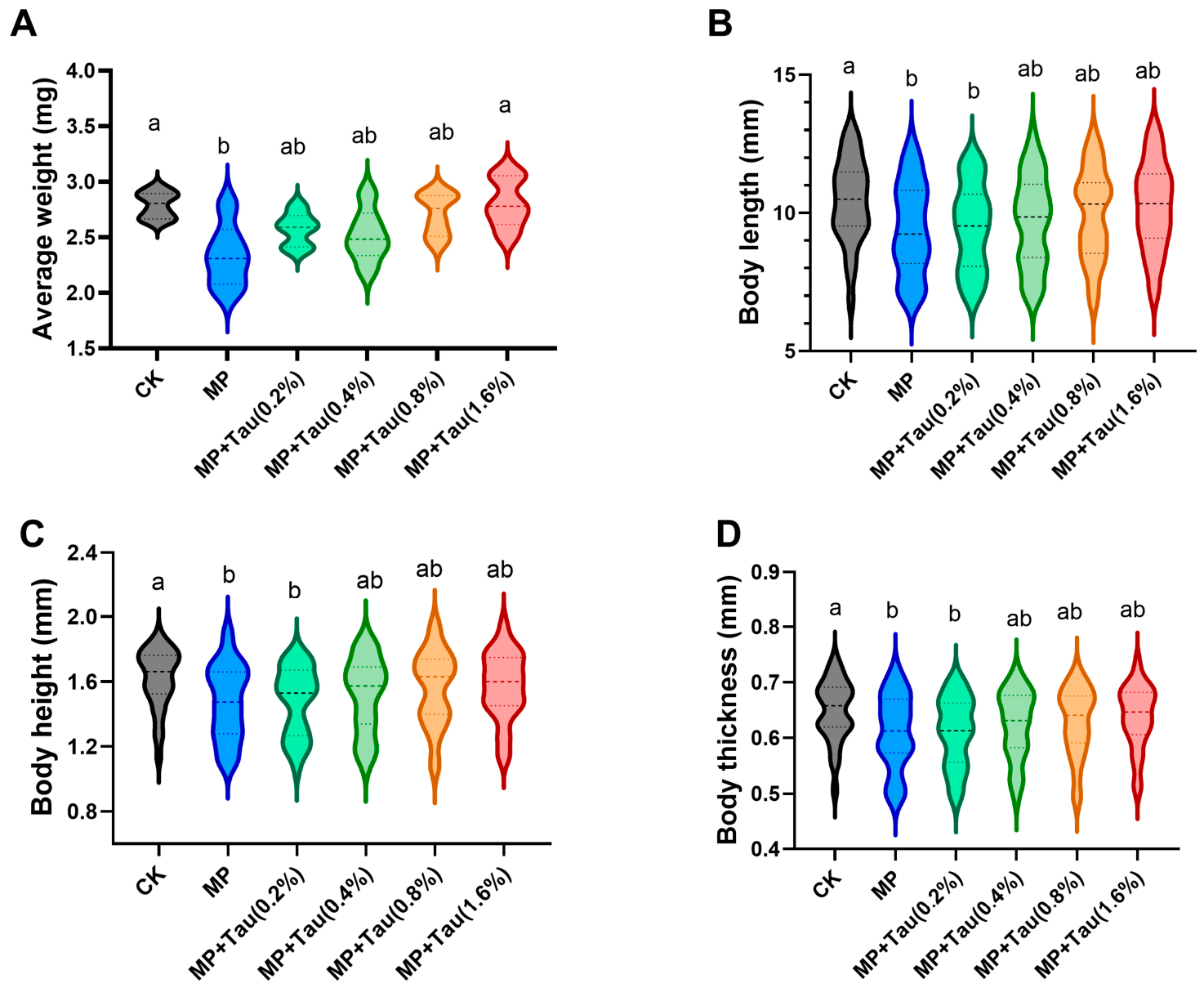
Following the 28-day exposure experiment, the average body weight of loach larvae across the six treatment groups was determined, and the results are shown in Figure 1. The average body weight of the CK group was 2.79 mg per larva at day 28, whereas that of the MP group was only 2.33 mg per larva, accounting for 83.51% of the CK group; the average body weight in the MP + Tau (0.8%) group recovered to 2.71 mg per larva. Compared with the CK group, microplastic exposure induced a significant reduction in average body weight (p < 0.05). Taurine supplementation ameliorated microplastic-triggered body weight loss in a dose-dependent fashion (Figure 1A). To further elucidate the mechanisms underlying body weight reduction, body length, body height and body thickness were measured for 30 loach larvae. Microplastic exposure significantly lowered the mean values of body length, body height and body thickness (p < 0.05), while no notable differences were detected in maximum values. The decreased mean morphological indices were primarily ascribed to an elevated proportion of smaller-sized individuals (Figure 1B–D). Taurine supplementation at concentrations higher than 0.4% could partially rescue these detrimental effects.
Figure 1.
Growth performance of larvae from 6 groups. (A) Average body weight calculated by weighing 10 loach larvae per replicate, with six replicates per treatment. (B) Body length; (C) Body height; (D) Body thickness. Thirty loach larvae were measured for each treatment. Treatments marked with different superscript letters are significantly different (p < 0.05).
3.2. Survival
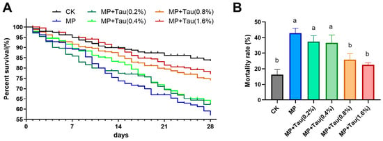
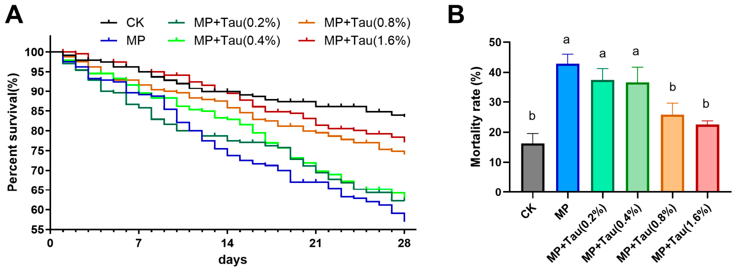
Mortality was monitored on a daily basis throughout the 28-day trial. Dead larvae were promptly siphoned out to avoid water quality degradation and subsequent confounding effects on experimental outcomes. The survival curves of larvae in different treatment groups are presented in Figure 2. Survival percentages of loach larvae in all treatments decreased progressively as the exposure duration extended. After 28 days, the survival rates of the six groups were 83.6% (CK), 57.1% (MP), 62.5% (MP + Tau [0.2%]), 63.3% (MP + Tau [0.4%]), 74.2% (MP + Tau [0.8%]), and 77.5% (MP + Tau [1.6%]), respectively. Kaplan–Meier survival analysis demonstrated significant divergence in survival curves between the MP group and the CK group (χ2 = 36.61, df = 1, p < 0.05). Comparisons among taurine-supplemented groups showed no significant differences between the MP group and MP + Tau (0.2%) (χ2 = 0.60, df = 1, p > 0.05), or MP + Tau (0.4%) (χ2 = 2.07, df = 1, p > 0.05). In contrast, significant differences were observed between the MP group and MP + Tau (0.8%) (χ2 = 14.25, df = 1, p < 0.05), as well as MP + Tau (1.6%) (χ2 = 23.39, df = 1, p < 0.05). Meanwhile, no significant difference was detected between the MP + Tau (1.6%) group and the CK group (χ2 = 2.26, df = 1, p > 0.05) (Figure 2A). The cumulative mortality at day 28 showed that in the MP group, MP + Tau (0.2%) group and MP + Tau (0.4%) group was significantly greater than that in the CK group (p < 0.05). By contrast, no significant difference in cumulative mortality was observed between the MP + Tau (0.8%), MP + Tau (1.6%) groups and the CK group (Figure 2B).
Figure 2.
Survival curves and mortality. (A) Survival curves of loach larvae across six experimental groups. The y-axis represents the percentage of surviving fish, and the x-axis indicates days post-exposure. (B) Cumulative mortality at day 28 of loach larvae under six treatment groups. Each treatment contained three replicate aquaria. Data are presented as mean ± SEM. Different superscript letters indicate significant differences between treatments in pairwise comparisons (p < 0.05).
3.3. Satiation Rate and Fasting Rate
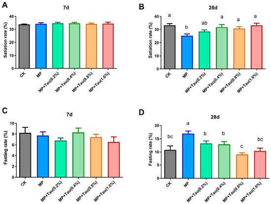
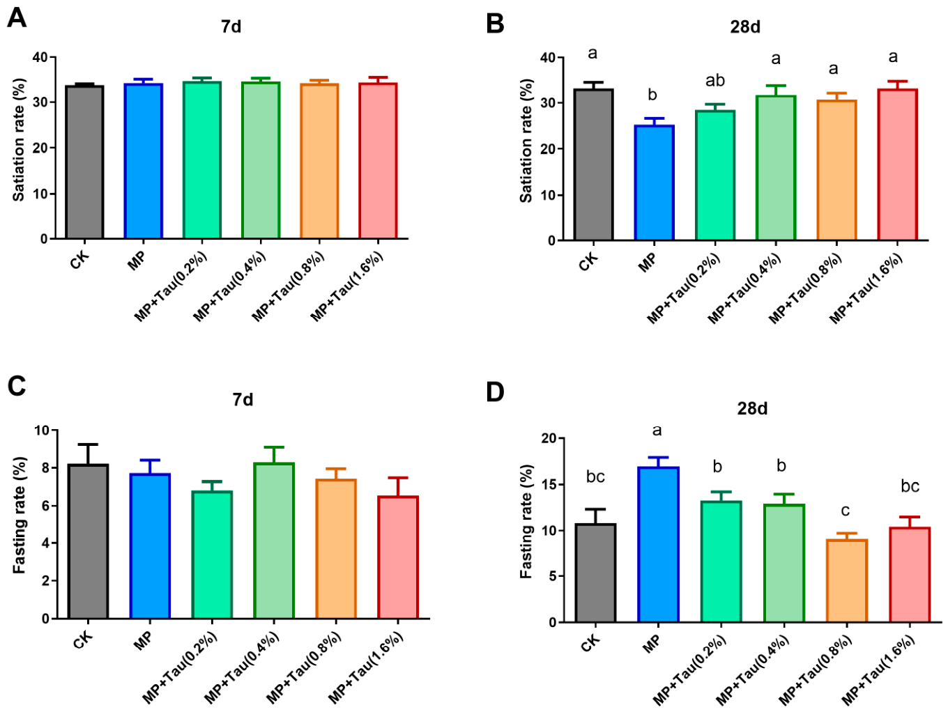
To analyze whether the reduction in body weight induced by microplastic exposure was attributed to feeding disturbance in loach larvae, the satiation rate and fasting rate were determined on Day 7 and Day 28 of the experiment. Three gut conditions of loach larvae were observed at 2 h post-feeding: gut fully filled with food, partially filled gut, and empty gut. Statistical analysis of the satiation rate showed no significant intergroup differences at day 7 (Figure 3A). On Day 28, the satiation rate of the MP group was 25.28%, which was significantly lower than that of the CK group (33.27%) (p < 0.05) (Figure 3B). Similarly, no significant difference in the fasting rate was detected among all groups on Day 7, with values ranging from 6.5% to 8.3% (Figure 3C). On Day 28, the fasting rate in the MP group increased markedly to 16.93%, which was significantly higher than those in the CK group and all four taurine supplementation groups. The fasting rates of the MP + Tau (0.8%) and MP + Tau (1.6%) groups were lower than those of the MP + Tau (0.2%) and MP + Tau (0.4%) groups, while no significant differences were detected between these taurine treatment groups and the CK group (Figure 3D).
Figure 3.
Satiation rate and fasting rate of loach larvae from six treatment groups on Day 7 and Day 28 of exposure. Each treatment had three replicate aquaria. (a) Satiation rate on Day 7; (b) satiation rate on Day 28; (c) fasting rate on Day 7; (d) fasting rate on Day 28. All results are presented as mean ± SEM (n = 3). Treatments marked with different superscript letters are significantly different (p < 0.05); superscript letters are omitted where no significant difference was detected.
3.4. Microplastic Distribution
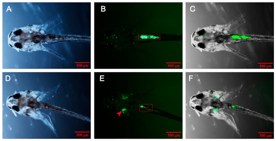
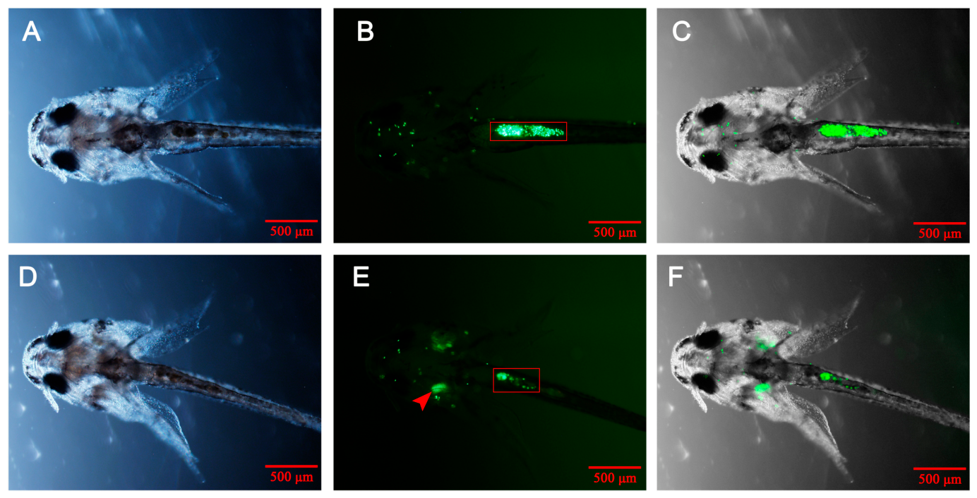
Fluorescence microscopy revealed that fluorescent PS-MPs were mainly distributed in the intestine, oral cavity, pharynx and gills, while a small amount adhered to the body surface skin and fins (Figure 4B). Comparison of merged bright-field and fluorescence images demonstrated that the location of fluorescent PS-MPs in the digestive tract highly overlapped with chyme. Interestingly, aggregates of fluorescent PS-MPs were observed in some larvae, attaching to the gill tissue (Figure 4E).
Figure 4.
Distribution of fluorescent PS-MPs in loach larvae. (A,D): bright field; (B,E): fluorescence; (C,F): merged images. Scale bar = 500 μm. The red rectangular boxes indicate the aggregation of fluorescent PS-MPs in the intestine, and red arrows denote aggregated fluorescent PS-MPs on the gills.
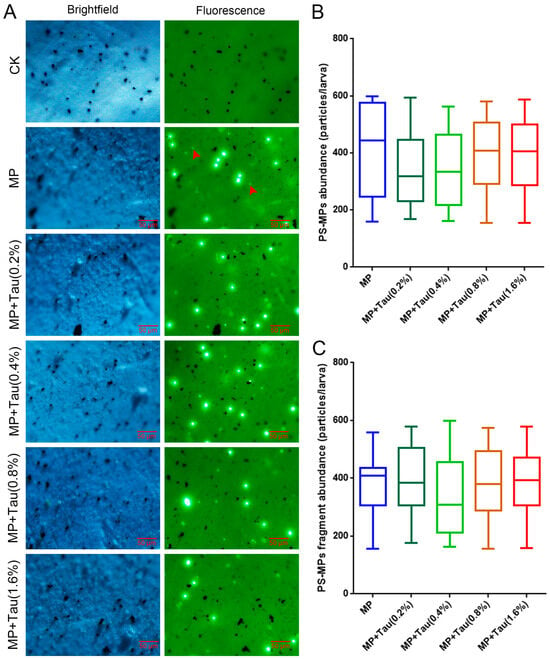
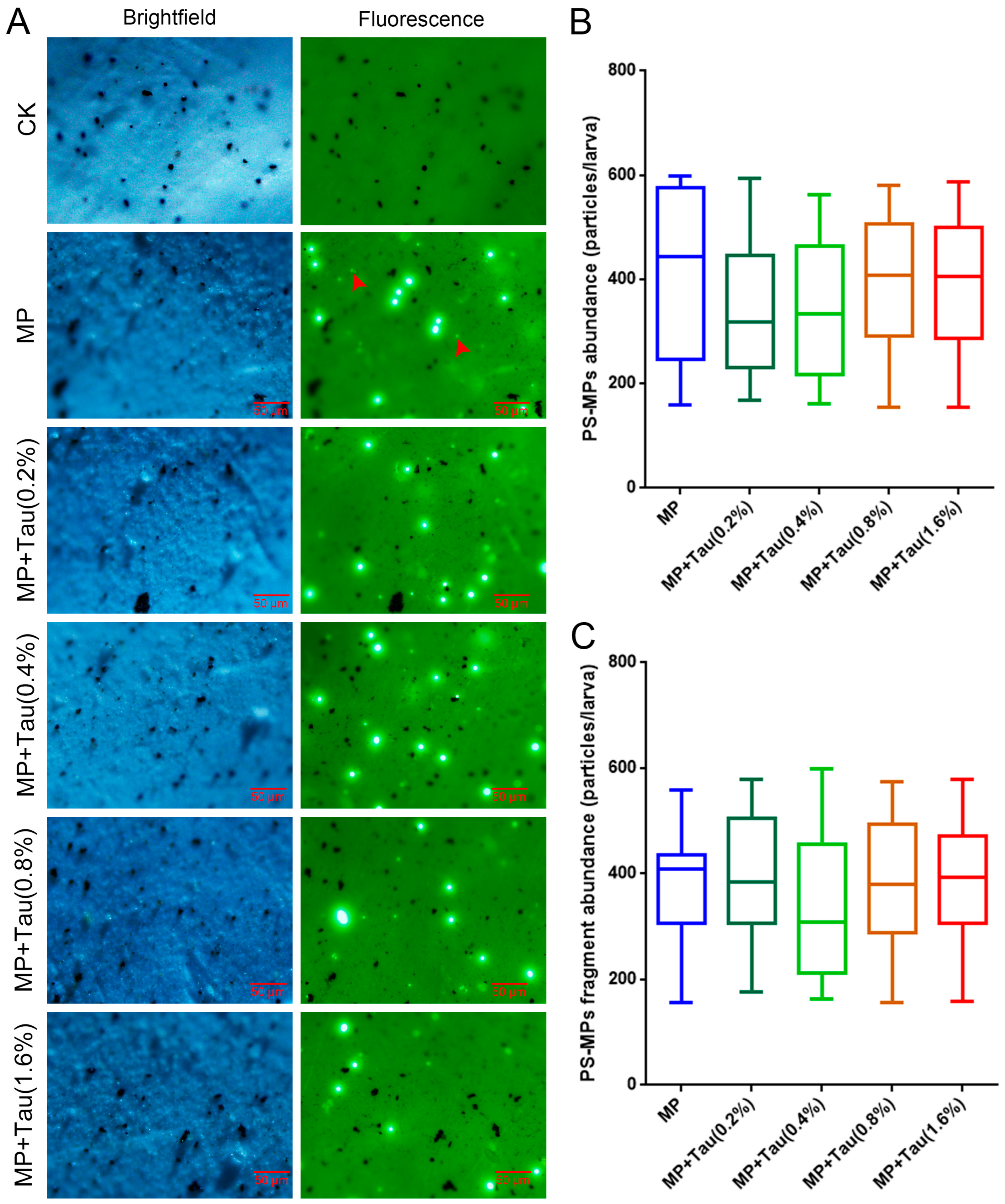
Loach larvae sampled at Day 28 of exposure were digested with potassium hydroxide (KOH) and filtered through 0.22 μm membranes. Green fluorescently labeled PS-MPs were visualized under fluorescence microscopy in all PS-MP exposure groups. Apart from intact spherical PS-MP particles, small fragments were also detected on the filter membranes. No fluorescent microplastics were observed in samples from the CK group. (Figure 5A). Quantification of fluorescent PS-MPs in larvae revealed no significant difference in PS-MPs abundance between the MP group and the four MP-supplemented taurine groups (Figure 5B). Furthermore, the number of PS-MP fragments was quantified, and no notable divergence was found between the MP group and the taurine-amended MP treatments. (Figure 5C).
Figure 5.
Fluorescent PS-MPs recovered from loach larvae after 28 days of exposure. Fluorescence microscopy revealed fragmented debris derived from damaged fluorescent PS-MPs in all treatment groups. Red arrows indicate PS-MP fragments (A). Quantification of total fluorescent PS-MPs per larva showed no significant difference between the MP group and the four MP groups supplemented with graded taurine (B). The number of PS-MP fragments was also quantified, and no significant difference was observed between the MP group and taurine-supplemented MP treatments (C). Each biological replicate contained five loach larvae in a 2 mL glass vial, with six biological replicates per treatment.
3.5. Enzymatic Activity
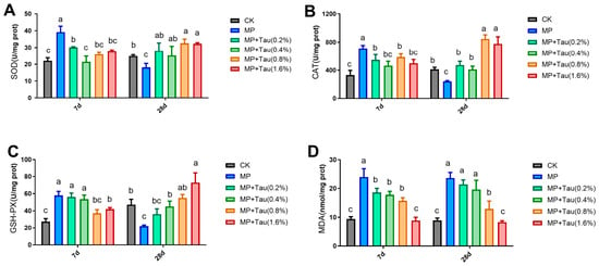
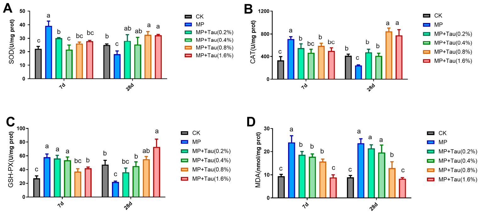
The activities of SOD, CAT and GSH-PX as well as MDA levels were analyzed on Day 7 and Day 28 post-exposure. The antioxidant enzyme activities showed that the SOD, CAT and GSH-PX activities in the MP group increased significantly after 7 days of exposure. The SOD and CAT activities in the MP + Tau (0.2%) and MP + Tau (0.4%) groups were lower than those in the MP group, whereas no significant difference in GSH-PX activity was found between the MP group and the MP + Tau (0.2%) and MP + Tau (0.4%) groups. Dietary taurine supplementation at concentrations above 0.8% inhibited the PS-MP-induced elevation of SOD, CAT and GSH-PX activities. Specifically, the SOD, CAT and GSH-PX activities in the MP + Tau (0.8%) and MP + Tau (1.6%) groups were markedly lower than those in the MP group but still moderately higher than those in the CK group. After 28 days of PS-MP exposure, the MP group displayed marked reductions in SOD, CAT and GSH-PX activities relative to CK. Conversely, higher activities of these enzymes were detected in the MP + Tau (0.8%) and MP + Tau (1.6%) groups. Exposure to fluorescent PS-MPs significantly increased MDA content (p < 0.05), and taurine supplementation attenuated this MDA accumulation in an obvious dose-dependent manner (Figure 6D).
Figure 6.
Antioxidant enzyme activities and MDA content in loach larvae measured on Day 7 and Day 28 of exposure. (A) SOD; (B) CAT; (C) GSH-PX; (D) MDA. All results are presented as mean ± SEM (n = 3). Treatments marked with different superscript letters are significantly different (p < 0.05).
3.6. Antioxidant-Related Gene Expression
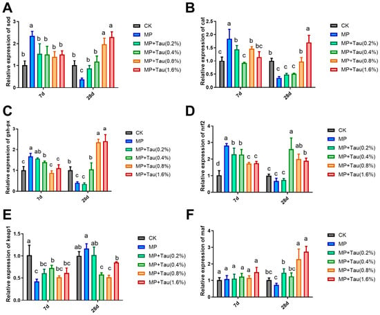
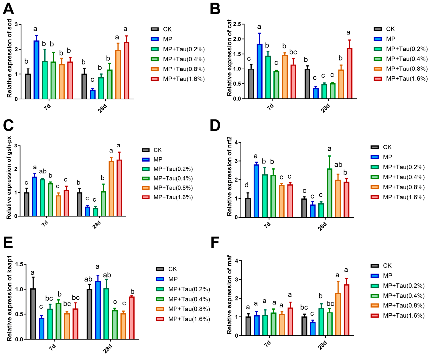
qPCR analysis of antioxidant enzyme genes revealed that the relative expression of the sod gene in the MP group on Day 7 was 2.35-fold higher than that in the CK group, showing a significant upregulation (p < 0.05). Following taurine supplementation, the relative expression of sod was approximately 1.5-fold of the CK group, with no significant difference compared with the CK group (p > 0.05). The relative expression level of sod was significantly downregulated in the MP group versus CK on day 28 (p < 0.05). The relative expression levels in the MP + Tau (0.2%) and MP + Tau (0.4%) groups were significantly higher than those in the MP group (p < 0.05) and did not differ significantly from the CK group. Moreover, the MP + Tau (0.8%) and MP + Tau (1.6%) groups exhibited significantly elevated sod expression (p < 0.05) (Figure 7A).
Figure 7.
Relative mRNA expression levels of antioxidant enzyme genes and Nrf2/Keap1 pathway-related genes in loach larvae on Day 7 and Day 28 post-exposure. (A) sod; (B) cat; (C) gsh-px; (D) nrf2; (E) keap1; (F) maf. All data are shown as mean ± SEM (n = 3). Treatments marked with different superscript letters are significantly different (p < 0.05).
The expression patterns of the cat and gsh-px genes were similar to that of sod. Briefly, the relative expression levels of cat and gsh-px increased in the MP group on Day 7 and decreased on Day 28. Taurine supplementation significantly upregulated the relative expression of the cat gene in the MP + Tau (1.6%) group (Figure 7B). The MP + Tau (0.8%) and MP + Tau (1.6%) treatments exhibited significantly elevated gsh-px expression relative to the CK group at day 28 (p < 0.05) (Figure 7C).
To further explore the upstream regulatory pathway of antioxidant enzyme genes, the expression of nrf2, keap1 and maf genes was determined. On Day 7, the nrf2 expression in the MP group was significantly higher than that in the CK group, and taurine supplementation alleviated this sharp increase in nrf2 expression (Figure 7D). On Day 28, no significant difference in nrf2 expression was observed among the MP group, MP + Tau (0.2%) group and CK group. Treatments with taurine supplementation above 0.4% maintained high nrf2 expression.
The keap1 expression in the MP group was significantly downregulated on Day 7 compared with the CK group (p < 0.05). Dietary taurine addition partially recovered keap1 transcript levels compared with the MP group, even though these values were still suppressed relative to the CK group. A slight increase in keap1 expression was detected in the MP group on Day 28 (Figure 7E). No significant intergroup difference in maf expression was found on Day 7. On Day 28, the maf expression levels in the MP + Tau (0.8%) and MP + Tau (1.6%) groups were significantly higher than those in the other treatment groups (p < 0.05) (Figure 7F).
4. Discussion
Mortality serves as a critical biomarker for evaluating the toxic effects of environmental pollutants [34,35]. In the present study, microplastic exposure significantly increased the mortality of loach larvae, while taurine supplementation effectively alleviated microplastic-induced mortality elevation, indicating the protective role of taurine against microplastic toxicity in loach larvae. During the observation of PS-MPs distribution in loach larvae, aggregated MP clumps were found attached to the gills, and these individuals appeared more emaciated. Gill accumulation of microplastics may contribute to mortality independently of gastrointestinal ingestion, possibly by disrupting gas exchange and gill tissue function. Previous studies have demonstrated that microplastic ingestion inhibits the growth of various aquatic organisms, including fish, crustaceans, and shellfish [1,36]. Such growth inhibition is primarily attributed to the impaired development of bone and muscle tissues [37]. For instance, oxidized polyamide microplastics have been reported to significantly suppress the musculoskeletal development of zebrafish larvae [38]. Consistent with these findings, the present study observed remarkable reductions in the body length, head length, and body width of loach larvae in the microplastic-exposed group. This evidence suggests that microplastic exposure exerts prominent inhibitory effects on skeletal and muscle growth in loach larvae, and dietary taurine supplementation can effectively reverse these adverse impacts.
On day 28 of the exposure experiment, the MP group exhibited a decreased satiation rate and an elevated fasting rate. This phenomenon implies that exposure to high concentrations of microplastics or microplastic-contaminated diets may impair the feeding selectivity of fish, resulting in passive microplastic ingestion and further reducing predation capacity and feeding efficiency [39]. In addition, accidental microplastic intake can induce false satiety in aquatic organisms, which causes gastrointestinal blockage and appetite depression and is recognized as a dominant factor underlying feeding disorders [40,41,42]. Notably, no significant changes in the satiation rate and fasting rate were observed in loach larvae at the early exposure stage. This may be attributed to the energy supply from the yolk sac during early larval development, which compensates for the physiological stress induced by microplastic exposure.
Although dietary taurine supplementation alleviated microplastic-induced growth inhibition in loach larvae, it remains unclear whether this protective effect is achieved by promoting larval growth or reducing microplastic ingestion. To clarify this mechanism, the abundance of microplastics in loach larvae was determined, and the results confirmed that taurine supplementation did not reduce microplastic intake by larvae. This finding rules out reduced MP ingestion as the main protective route of taurine, and directs our focus toward antioxidant defense. Dietary taurine is capable of enhancing intestinal protein digestion and amino acid absorption, facilitating muscle protein deposition and muscle fiber development, and improving immune capacity and skeletal growth in fish [31]. Nevertheless, excessive dietary taurine can weaken its growth-promoting effects [26]. According to previous research on largemouth bass (Micropterus salmoides), the optimal dietary taurine concentration for maximizing growth rate is 0.288% under normal culture conditions [31]. Notably, a higher taurine dosage (>0.8%) exhibited superior protective effects against microplastic stress in the present study, indicating that aquatic organisms require higher taurine levels to resist adverse environmental stressors. From an aquaculture perspective, our results show that dietary taurine supplementation at 0.8% and 1.6% exerted obvious protective effects against PS-MPs stress in loach larvae. Previous studies have demonstrated that dietary taurine at 2.0% does not produce toxic effects in Scophthalmus maximus and Megalobrama amblycephala [24,26]. Nevertheless, considering economic cost, Under the challenge of environmentally relevant PS-MPs pollution, we tentatively recommend a dietary taurine addition level of 0.8% for loach larviculture, balancing protective efficacy and feed cost.
Once microplastics invade fish, the antioxidant system is activated initially, triggering massive production of reactive oxygen species (ROS) and disrupting the redox balance [19]. Fish are equipped with a sophisticated antioxidant network that counteracts the toxic impacts of reactive oxygen species (ROS) [43]. As core endogenous antioxidant enzymes, SOD and CAT constitute the primary defensive barrier against peroxide radicals [44]. SOD facilitates the dismutation of superoxide anions into hydrogen peroxide, which CAT subsequently breaks down into water and oxygen [45]. In addition, GSH-PX is an essential ubiquitous peroxidase that eliminates excess hydrogen peroxide accumulated upon ROS overproduction and effectively prevents ROS buildup [4,46]. This enzyme has been widely adopted as a reliable biomarker for identifying oxidative stress in aquatic toxicology [44].
Previous studies have demonstrated that high-concentration microplastic exposure suppresses the activities of SOD, CAT, and GSH-PX in aquatic organisms [47]. Consistent with the dual-phase oxidative stress response, the present study showed that SOD and CAT activities were significantly elevated in the MP group on Day 7 but markedly inhibited on Day 28 compared with the control group. The early enzymatic activation can be explained by the initial defensive response of loach larvae to microplastic stress. Microplastic intrusion induces excessive ROS generation, and the increased SOD activity accelerates the conversion of superoxide anions into hydrogen peroxide, further triggering the activation of CAT and GSH-PX to eliminate accumulated H2O2. However, sustained ROS overproduction leads to excessive H2O2 accumulation, which ultimately exerts inhibitory effects on SOD and CAT activities [48]. Long-term microplastic exposure causes progressive physiological damage, impairs the stability of the antioxidant system, and eventually suppresses antioxidant enzyme capacity [49,50]. Certain antioxidant food additives are capable of conferring moderate protection from microplastic-elicited oxidative damage. Dietary supplements such as lycopene and citric acid can alleviate microplastic-triggered oxidative injury. These additives offset the harmful effects of dietary microplastics and recovered hematological parameters in African catfish (Clarias gariepinus) [51]. Astaxanthin relieves microplastic accumulation and oxidative stress in juvenile European seabass (Dicentrarchus labrax) fed microplastic-contaminated diets [52]. Our findings indicated that taurine supplementation attenuated the fluctuation amplitudes of the antioxidant stress-related enzyme activities of SOD, CAT and GSH-PX. Previous studies have confirmed that taurine confers cytoprotection via direct scavenging of diverse free radicals and upregulation of endogenous antioxidant enzymes including SOD and CAT [24]. Dietary taurine supplementation can boost the activities of antioxidant enzymes and reduce ROS production in multiple aquatic species [53]. Furthermore, taurine may redirect cysteine toward glutathione (GSH) biosynthesis, thereby elevating intracellular GSH levels. As a crucial endogenous antioxidant, GSH further protects cells from ROS-induced oxidative injury [26].
MDA is a canonical end-product of lipid peroxidation, and its cellular level serves as a reliable indirect indicator of ROS-induced lipid peroxidation and tissue oxidative damage [13,54]. Previous studies have documented significantly elevated MDA accumulation in fish and shrimp following PS-MPs exposure, exhibiting a positive concentration-dependent correlation with microplastic stress [4,30]. In accordance with these findings, the present study observed a significant increase in MDA content in the MP-exposed loach larvae starting from Day 7, and this elevated status persisted throughout the entire experimental period. These results confirm that PS-MPs induce persistent lipid peroxidation, thereby causing tissue damage and cytotoxicity in loach larvae. Importantly, dietary taurine supplementation effectively reversed MP-induced MDA accumulation and alleviated lipid peroxidation injury.
The Keap1-Nrf2 signaling cascade represents a pivotal pathway for regulating cellular antioxidant responses [55]. Nuclear factor erythroid 2-related factor 2 (Nrf2) is known to bind antioxidant response elements (AREs) in the nucleus and confer cellular protection against exogenous adverse stimuli [56]. Under physiological homeostasis, Nrf2 forms a complex with Kelch-like ECH-associated protein 1 (Keap1), and this binding restrains Nrf2 function. Upon exogenous oxidative stress and ROS overloading, Nrf2 dissociates from Keap1 [57]. Subsequently, Nrf2 translocates into the nucleus, forms heterodimers with small Maf proteins, and binds to AREs to initiate the transcription of downstream antioxidant enzyme genes, including sod, cat, and gsh-px [58]. Prior evidence has confirmed that antioxidant astaxanthin modulates antioxidant capacity via the Nrf2 signaling pathway in loach [58], and dietary taurine can activate the Nrf2/Keap1 pathway to alleviate high-temperature-induced impairment of intestinal barrier function in Megalobrama amblycephala [24]. In the present study, dietary taurine supplementation at concentrations above 0.8% maintained the upregulated expression of nrf2 and downregulated keap1 at both 7 d and 28 d, sustaining the activation of endogenous antioxidant enzyme expression via the Keap1-Nrf2 signaling pathway. Before ROS accumulation exceeds the scavenging threshold of the intrinsic antioxidant defense system, taurine triggers this cellular antioxidant signaling cascade and promotes the transcription of sod, cat and gsh-px, thereby strengthening the resistance of loach larvae to oxidative damage caused by PS-MPs stress.
5. Conclusions
This study demonstrated that environmentally relevant concentrations of polystyrene microplastics significantly inhibited growth performance, increased mortality, and disrupted feeding behavior by reducing the satiation rate of loach larvae. Microplastic exposure also induced severe oxidative stress damage, characterized by disordered antioxidant enzyme activities and excessive accumulation of MDA. Dietary taurine supplementation effectively ameliorated microplastic-induced growth retardation, feeding disorders and oxidative injury. Notably, the protective effects of taurine were independent of microplastic ingestion reduction in loach larvae, and this protective effect was observed at dietary taurine levels of 0.8% and above, indicating that taurine exerts its protective function via an endogenous antioxidant mechanism. Taurine activated the Nrf2/Keap1 signaling pathway, upregulated the expression of downstream antioxidant genes, improved the activities of SOD, CAT and GSH-PX, and reduced MDA accumulation. These responses efficiently eliminated excess reactive oxygen species and restored redox homeostasis, thereby alleviating microplastic-triggered oxidative damage. Collectively, appropriate taurine supplementation (≥0.8%) can serve as an effective dietary additive to resist microplastic stress. The present findings provide a theoretical basis for the healthy culture of loach seedlings and ecological risk prevention and control of microplastic pollution in aquaculture ecosystems.
Author Contributions
Conceptualization, Y.F. and S.X.; methodology, Y.F. and C.S.; validation, C.S., Y.D. and M.J.; formal analysis, Y.F. and C.S.; resources, Y.F. and S.X.; data curation, C.S., Y.D. and S.X.; writing—original draft preparation, Y.F. and C.S.; writing—review and editing, S.X.; visualization, Y.D. and M.J.; supervision, S.X.; project administration, Y.F.; funding acquisition, Y.F. and S.X. All authors have read and agreed to the published version of the manuscript.
Funding
This research was funded by Doctoral Scientific Research Startup Fund of Weifang University (grant number: 44121015).
Institutional Review Board Statement
Loach used in this study was approved by the Institutional Animal Care and Use Committee of South China Agricultural University (ETHICS CODE Permit NO. 2021G005).
Data Availability Statement
The original contributions presented in this study are included in the article. Further inquiries can be directed to the corresponding authors.
Acknowledgments
During the preparation of this manuscript, the authors used Doubao2.15.9 for the purposes of language translation and grammatical checking. The authors have reviewed and edited the output and take full responsibility for the content of this publication.
Conflicts of Interest
The authors declare no conflicts of interest.
References
- Xia, R.; Meng, X.; Zhang, Q.; Wang, B.; Tian, X.; Xu, X.; Zhang, S. Characteristics, drivers, and risks of microplastic pollution in fish from diverse water bodies in North China. J. Environ. Sci. 2026, 105656. [Google Scholar] [CrossRef] [Scilit]
- Azarm-Karnagh, S.; Sattari, M.; Banaee, M.; Shirkavand Hadavand, B.; Falco, F. Effects of Polystyrene Nanoplastics on Oxidative Stress, Blood Biochemistry, and Digestive Enzyme Activity in Goldfish (Carassius auratus). Toxics 2025, 13, 336. [Google Scholar] [CrossRef] [Scilit] [PubMed]
- Gao, C.; Xu, B.; Li, Z.; Wang, Z.; Huang, S.; Jiang, Z.; Gong, X.; Yang, H. From plankton to fish: The multifaceted threat of microplastics in freshwater environments. Aquat. Toxicol. 2025, 279, 107242. [Google Scholar] [CrossRef] [Scilit] [PubMed]
- Zhou, Q.; Wang, Y.; Wei, J.; Jiao, T.; Xu, S.; Hong, K.; Su, Q.; Liufu, B.; Mai, Z.; Huang, Y.; et al. Chronic exposure to polystyrene microplastics at environmentally relevant concentration induced growth retardation in Macrobrachium rosenbergii via multi-pathway toxicity: Oxidative stress, microbial dysbiosis, and biodistribution. Environ. Pollut. 2026, 396, 127836. [Google Scholar] [CrossRef] [Scilit] [PubMed]
- Thompson, R.C.; Olsen, Y.; Mitchell, R.P.; Davis, A.; Rowland, S.J.; John, A.W.G.; McGonigle, D.; Russell, A.E. Lost at Sea: Where Is All the Plastic? Science 2004, 304, 838. [Google Scholar] [CrossRef] [Scilit] [PubMed]
- Jambeck, J.R.; Geyer, R.; Wilcox, C.; Siegler, T.R.; Perryman, M.; Andrady, A.; Narayan, R.; Law, K.L. Plastic waste inputs from land into the ocean. Science 2015, 347, 768–771. [Google Scholar] [CrossRef] [Scilit] [PubMed]
- Parvin, F.; Jannat, S.; Tareq, S.M. Abundance, characteristics and variation of microplastics in different freshwater fish species from Bangladesh. Sci. Total Environ. 2021, 784, 147137. [Google Scholar] [CrossRef] [Scilit] [PubMed]
- Gambino, G.; Falleni, A.; Nigro, M.; Salvetti, A.; Cecchettini, A.; Ippolito, C.; Guidi, P.; Rossi, L. Dynamics of interaction and effects of microplastics on planarian tissue regeneration and cellular homeostasis. Aquat. Toxicol. 2020, 218, 105354. [Google Scholar] [CrossRef] [Scilit] [PubMed]
- Carbery, M.; O’Connor, W.; Palanisami, T. Trophic transfer of microplastics and mixed contaminants in the marine food web and implications for human health. Environ. Int. 2018, 115, 400–409. [Google Scholar] [CrossRef] [Scilit] [PubMed]
- Yu, Y.; Jo, A.; Choi, C.Y.; Kang, J.; Kim, J. Toxic effects of microplastic (polyethylene) exposure: Stress, immune responses and neurotoxicity in crucian carp, Carassius carassius. Fish Shellfish Immunol. 2026, 170, 111139. [Google Scholar] [CrossRef] [Scilit] [PubMed]
- Liu, M.; Liu, X.; Xu, J. Influence of microplastic pollution on the toxicity of potamodromous fish grass carp (Ctenopharyngodon idellus) and its swimming capacity. J. Environ. Chem. Eng. 2024, 12, 113520. [Google Scholar] [CrossRef] [Scilit]
- Eze, C.G.; Nwankwo, C.E.; Dey, S.; Sundaramurthy, S.; Okeke, E.S. Food chain microplastics contamination and impact on human health: A review. Environ. Chem. Lett. 2024, 22, 1889–1927. [Google Scholar] [CrossRef] [Scilit]
- Zhang, Q.R.; Zhou, X.; Sun, Y.; Deng, Q.F.; Wu, Q.; Wen, Z.R.; Chen, H.G. Harmful effects of microplastics on respiratory system of aquatic animals: A systematic review and meta-analysis. Aquat. Toxicol. 2024, 273, 107003. [Google Scholar] [CrossRef] [Scilit] [PubMed]
- Bhat, R.; Sidiq, M.J.; Altinok, I. Impact of microplastics and nanoplastics on fish health and reproduction. Aquaculture 2024, 590, 741037. [Google Scholar] [CrossRef] [Scilit]
- Oyovwi, M.O.; Ugwuishi, E.W.; Udi, O.A.; Atere, A.D.; Rotu, A.R.; Odokuma, E.I.; Emojevwe, V.O.; Olowe, G.T.; Nwangwa, E.K.; Ben-Azu, B. Epigallocatechin-gallate ameliorates polystyrene microplastics-induced oxido-inflammation and mitochondria-mediated apoptosis in testicular cells via modulation of Nrf2/HO-1, /mTOR/Atg-7, and Cx-43/NOX-1 levels. Eur. J. Med. Chem. Rep. 2025, 13, 100243. [Google Scholar] [CrossRef] [Scilit]
- Jiang, Q.; Chen, X.; Jiang, H.; Wang, M.; Zhang, T.; Zhang, W. Effects of Acute Exposure to Polystyrene Nanoplastics on the Channel Catfish Larvae: Insights from Energy Metabolism and Transcriptomic Analysis. Front. Physiol. 2022, 13, 923278. [Google Scholar] [CrossRef] [Scilit] [PubMed]
- Li, X.; Huang, Y.; Li, W.; Deng, C.; Cao, W.; Yao, Y. Effects of Polystyrene Microplastic Exposure on Liver Cell Damage, Oxidative Stress, and Gene Expression in Juvenile Crucian Carp (Carassius auratus). Toxics 2025, 13, 53. [Google Scholar] [CrossRef] [Scilit] [PubMed]
- Cheng, H.D.; Zou, Y.; Lu, B.; Wang, J.Z.; Magnuson, J.T.; Xu, B.T.; Qiu, W.H.; Xuan, R.R. Immunotoxic response of bio-based plastic on early life stage zebrafish (Danio rerio): A safe alternative to petroleum-based plastics? J. Hazard. Mater. 2024, 480, 135846. [Google Scholar] [CrossRef] [Scilit] [PubMed]
- Yedier, S.; Yalçınkaya, S.K.; Bostancı, D. Exposure to polypropylene microplastics via diet and water induces oxidative stress in Cyprinus carpio. Aquat. Toxicol. 2023, 259, 106540. [Google Scholar] [CrossRef] [Scilit] [PubMed]
- Fernândez-Míguez, M.; Puvanendran, V.; Burgerhout, E.; Presa, P.; Tveiten, H.; Vorkamp, K.; Hansen, O.J.; Johansson, G.S.; Bogevik, A.S. Effects of weathered polyethylene microplastic ingestion on sexual maturation, fecundity and egg quality in maturing broodstock Atlantic cod Gadus morhua. Environ. Pollut. 2023, 320, 121053. [Google Scholar] [CrossRef] [Scilit] [PubMed]
- Zhang, Y.Y.; Zhang, S.Y.; Sun, K.; Zhang, Q.H.; Wangkahart, E.; Wang, Z.S.; Sui, Y.M.; Qi, Z.T. Effects of Microplastics on the Activity of Digestive-, Antioxidative- and Immune-related Enzymes in Soiny Mullet (Liza haematocheila Temminck & Schlegel, 1845) Larvae. Turk. J. Fish. Aquat. Sci. 2024, 24, TRJFAS24718. [Google Scholar] [CrossRef] [Scilit]
- Hou, M.M.; Zou, X.H.; Su, L.X.; Xu, C.S.; Xia, Z.J.; Wang, Q.; Zhao, X.J.; He, Y.F.; Wang, C.L.; Wang, J.W. Effects of environmentally relevant polystyrene microplastics and cadmium on the development and reproduction of rare minnow (Gobiocypris rarus). J. Environ. Chem. Eng. 2024, 12, 111886. [Google Scholar] [CrossRef] [Scilit]
- Souto, C.N.; de Menezes, L.L.C.; Proença, D.C.; Bueno, G.W.; Guimarães, I.G. Pirarucu requires taurine to maximize growth and antioxidant status when fed diets high in plant-based ingredients. Aquaculture 2024, 590, 741022. [Google Scholar] [CrossRef] [Scilit]
- Chen, K.; Chen, L.; He, Z.; Xie, P.; Liu, H.; Gao, Z.; Wang, H. Dietary taurine supplementation ameliorates growth performance, intestinal health, and environmental stress tolerance in summer-reared Megalobrama amblycephala. Anim. Nutr. 2026, 27, 264–278. [Google Scholar] [CrossRef] [Scilit] [PubMed]
- Peter, N.; Pradhan, C.; Dileep, N.; Mohanta, K.N. Effect of taurine supplementation on growth, nutrient utilization, antioxidant status, digestive enzyme activity, and metabolic enzyme activity in Striped Catfish Pangasianodon hypophthalmus fed fish-meal- and plant-protein-based diets. N. Am. J. Aquac. 2025, 87, 15–27. [Google Scholar] [CrossRef] [Scilit]
- Zhang, Y.; Wei, Z.; Yang, M.; Liu, D.; Pan, M.; Wu, C.; Zhang, W.; Mai, K. Dietary taurine modulates hepatic oxidative status, ER stress and inflammation in juvenile turbot (Scophthalmus maximus L.) fed high carbohydrate diets. Fish Shellfish Immunol. 2021, 109, 1–11. [Google Scholar] [CrossRef] [Scilit] [PubMed]
- Xia, X.H.; Ma, X.Y.; Liang, N.; Qin, L.; Huo, W.R.; Li, Y. Effective ways and related mechanisms of Leuconostoc mesenteroides DH purifying PE-MPs in loach (Paramisgurnus dabryanus) to exert protective effects. Aquaculture 2024, 593, 741265. [Google Scholar] [CrossRef] [Scilit]
- Yin, L.; Chen, B.; Xia, B.; Shi, X.; Qu, K. Polystyrene microplastics alter the behavior, energy reserve and nutritional composition of marine jacopever (Sebastes schlegelii). J. Hazard. Mater. 2018, 360, 97–105. [Google Scholar] [CrossRef] [Scilit] [PubMed]
- Zhang, C.; Wang, J.; Zhou, A.; Ye, Q.; Feng, Y.; Wang, Z.; Wang, S.; Xu, G.; Zou, J. Species-specific effect of microplastics on fish embryos and observation of toxicity kinetics in larvae. J. Hazard. Mater. 2021, 403, 123948. [Google Scholar] [CrossRef] [Scilit] [PubMed]
- Wang, X.Q.; Jian, S.Q.; Zhang, S.S.; Wu, D.; Wang, J.H.; Gao, M.; Sheng, J.Q.; Hong, Y.J. Enrichment of polystyrene microplastics induces histological damage, oxidative stress, Keap1-Nrf2 signaling pathway-related gene expression in loach juveniles (Paramisgurnus dabryanus). Ecotoxicol. Environ. Saf. 2022, 237, 113540. [Google Scholar] [CrossRef] [Scilit] [PubMed]
- Zhou, Q.; Hu, S.; Yu, J.; Li, C.; Liu, X.; Zhou, C.; Zhao, C.; Luo, K.; Gao, W.; Wu, C. Dietary taurine supplementation can improve the growth, protein metabolism, immunity and muscle development of larval largemouth bass (Micropterus salmoides). Aquac. Rep. 2025, 42, 102817. [Google Scholar] [CrossRef] [Scilit]
- Jager, K.J.; van Dijk, P.C.; Zoccali, C.; Dekker, F.W. The analysis of survival data: The Kaplan–Meier method. Kidney Int. 2008, 74, 560–565. [Google Scholar] [CrossRef] [Scilit] [PubMed]
- Kim, J.A.; Lee, J.Y.; Hwang, S.D.; Kim, J.; Choi, C.Y. Microplastic and Vibrio harveyi co-exposure induces oxidative stress in big-belly seahorse Hippocampus abdominalis. Comp. Biochem. Physiol. Part B Biochem. Mol. Biol. 2025, 279, 111124. [Google Scholar] [CrossRef] [Scilit] [PubMed]
- Ge, Q.; Zheng, T.; Ding, P.; Li, Z.; Lin, X.; Li, X.; He, M.; Hu, G. Aged microplastics-induced growth inhibition via DNA damage, GH/IGF-1 and HPT axes disruption in zebrafish larvae. Sci. Total Environ. 2025, 975, 179215. [Google Scholar] [CrossRef] [Scilit] [PubMed]
- Sun, S.; Jin, Y.; Luo, P.; Shi, X. Polystyrene microplastics induced male reproductive toxicity and transgenerational effects in freshwater prawn. Sci. Total Environ. 2022, 842, 156820. [Google Scholar] [CrossRef] [Scilit] [PubMed]
- Wang, J.; Wu, F.; Dong, S.; Wang, X.; Ai, S.; Liu, Z.; Wang, X.; Abarghouei, S. Meta-analysis of the effects of microplastic on fish: Insights into growth, survival, reproduction, oxidative stress, and gut microbiota diversity. Water Res. 2024, 267, 122493. [Google Scholar] [CrossRef] [Scilit] [PubMed]
- Ghosh, T. Microplastics bioaccumulation in fish: Its potential toxic effects on hematology, immune response, neurotoxicity, oxidative stress, growth, and reproductive dysfunction. Toxicol. Rep. 2025, 14, 101854. [Google Scholar] [CrossRef] [Scilit] [PubMed]
- Zou, W.; Xia, M.; Jiang, K.; Cao, Z.; Zhang, X.; Hu, X. Photo-Oxidative Degradation Mitigated the Developmental Toxicity of Polyamide Microplastics to Zebrafish Larvae by Modulating Macrophage-Triggered Proinflammatory Responses and Apoptosis. Environ. Sci. Technol. 2020, 54, 13888–13898. [Google Scholar] [CrossRef] [Scilit] [PubMed]
- de Sá, L.C.; Luís, L.G.; Guilhermino, L. Effects of microplastics on juveniles of the common goby (Pomatoschistus microps): Confusion with prey, reduction of the predatory performance and efficiency, and possible influence of developmental conditions. Environ. Pollut. 2015, 196, 359–362. [Google Scholar] [CrossRef] [Scilit] [PubMed]
- Li, Y.; Ye, Y.; Rihan, N.; Zhu, B.; Jiang, Q.; Liu, X.; Zhao, Y.; Che, X. Polystyrene nanoplastics induce lipid metabolism disorder and alter fatty acid composition in the hepatopancreas of Pacific whiteleg shrimp (Litopenaeus vannamei). Sci. Total Environ. 2024, 906, 167616. [Google Scholar] [CrossRef] [Scilit] [PubMed]
- König Kardgar, A.; Ghosh, D.; Sturve, J.; Agarwal, S.; Carney Almroth, B. Chronic poly(l-lactide) (PLA)- microplastic ingestion affects social behavior of juvenile European perch (Perca fluviatilis). Sci. Total Environ. 2023, 881, 163425. [Google Scholar] [CrossRef] [Scilit] [PubMed]
- Banaee, M.; Multisanti, C.R.; Impellitteri, F.; Piccione, G.; Faggio, C. Environmental toxicology of microplastic particles on fish: A review. Comp. Biochem. Physiol. Part C Toxicol. Pharmacol. 2025, 287, 110042. [Google Scholar] [CrossRef] [Scilit] [PubMed]
- Cui, J.; Zhu, M.; Sun, X.; Yang, J.; Guo, M. Microplastics induced endoplasmic reticulum stress to format an inflammation and cell death in hepatocytes of carp (Cyprinus carpio). Aquat. Toxicol. 2024, 269, 106870. [Google Scholar] [CrossRef] [Scilit] [PubMed]
- Xia, X.; Sun, M.; Zhou, M.; Chang, Z.; Li, L. Polyvinyl chloride microplastics induce growth inhibition and oxidative stress in Cyprinus carpio var. larvae. Sci. Total Environ. 2020, 716, 136479. [Google Scholar] [CrossRef] [Scilit] [PubMed]
- Kim, J.; Yu, Y.; Choi, J. Toxic effects on bioaccumulation, hematological parameters, oxidative stress, immune responses and neurotoxicity in fish exposed to microplastics: A review. J. Hazard. Mater. 2021, 413, 125423. [Google Scholar] [CrossRef] [Scilit] [PubMed]
- Jia, Y.; Cao, L.; Li, T.; Zhao, H.; Chen, G.; Xiong, Z.; Rong, W.; Liu, Q.; Wang, W.; Song, J.; et al. Synergistic effects of polystyrene microplastics and 17α-methyltestosterone on immune and oxidative stress responses in the gill and liver of Gobiocypris rarus. Fish Shellfish Immunol. 2026, 171, 111143. [Google Scholar] [CrossRef] [Scilit] [PubMed]
- Yu, P.; Liu, Z.; Wu, D.; Chen, M.; Lv, W.; Zhao, Y. Accumulation of polystyrene microplastics in juvenile Eriocheir sinensis and oxidative stress effects in the liver. Aquat. Toxicol. 2018, 200, 28–36. [Google Scholar] [CrossRef] [Scilit] [PubMed]
- Jeong, C.; Won, E.; Kang, H.; Lee, M.; Hwang, D.; Hwang, U.; Zhou, B.; Souissi, S.; Lee, S.; Lee, J. Microplastic Size-Dependent Toxicity, Oxidative Stress Induction, and p-JNK and p-p38 Activation in the Monogonont Rotifer (Brachionus koreanus). Environ. Sci. Technol. 2016, 50, 8849–8857. [Google Scholar] [CrossRef] [Scilit] [PubMed]
- Xiao, Q.; Xiang, H.; Tian, Y.; Huang, J.; Li, M.; Wang, P.; Lian, K.; Yu, P.; Xu, M.; Zhang, R.; et al. Polystyrene microplastics alleviate the developmental toxicity of silver nanoparticles in embryo-larval zebrafish (Danio rerio) at the transcriptomic level. Sci. Total Environ. 2024, 954, 176485. [Google Scholar] [CrossRef] [Scilit] [PubMed]
- Singh, P.K.; Singh, N.; Kumar, I.; Kumar, U.; Dwivedi, A.; Singh, P.; Mishra, S.; Pal, K.; Prajapati, A.; Sharma, R.K. Mapping of microplastic toxicity: Uptake, trophic transfer, and ecotoxicological risks in marine ecosystems. Aquat. Toxicol. 2026, 298, 107921. [Google Scholar] [CrossRef] [Scilit] [PubMed]
- Sayed, A.E.H.; Hana, M.N.; Hamed, M.; Abdel-Latif, H.M.R.; Lee, J.; Soliman, H.A.M. Protective efficacy of dietary natural antioxidants on microplastic particles-induced histopathological lesions in African catfish (Clarias gariepinus). Environ. Sci. Pollut. Res. 2023, 30, 24424–24440. [Google Scholar] [CrossRef] [Scilit] [PubMed]
- Zarantoniello, M.; Cattaneo, N.; Conti, F.; Carrino, M.; Cardinaletti, G.; Ener, D.; Olivotto, I. Mitigating Dietary Microplastic Accumulation and Oxidative Stress Response in European Seabass (Dicentrarchus labrax) Juveniles Using a Natural Microencapsulated Antioxidant. Antioxidants 2024, 13, 812. [Google Scholar] [CrossRef] [Scilit] [PubMed]
- Cai, M.L.; Chu, W.Y.; Wang, J.; Shao, C.; Hu, Y.J.; Che, C.B.; Zhang, J.Z.; Dai, J.H.; Hu, Y. Intervention of taurine on fatty acid profiles, oxidative injury and autophagy status in the muscle of rice field eel (Monopterus albus) fed oxidized fish oil. Aquaculture 2022, 551, 737904. [Google Scholar] [CrossRef] [Scilit]
- Prado, C.C.A.; Queiroz, L.G.; de Paiva, T.C.B.; Pompêo, M.; Ando, R.; Rani-Borges, B. Oxidative stress dynamics in Hyalella azteca under sub-chronic exposure to naturally aged polypropylene microplastics. Aquat. Toxicol. 2025, 281, 107303. [Google Scholar] [CrossRef] [Scilit] [PubMed]
- Deng, L.; Li, M.; Jiang, Z.; Xiang, G.; He, S.; Zhang, H.; Deng, A.; Wang, Y. Cobalt nanoparticles attenuate microplastic-induced vascular endothelial injury via Nrf2 pathway activation. Sci. Total Environ. 2024, 951, 175711. [Google Scholar] [CrossRef] [Scilit] [PubMed]
- Dong, T.; Gao, H.; Zhao, B.; Wang, Y.; Tu, T.; Zhang, H.; Yao, B.; Luo, H.; Liu, X. Novel cottonseed-derived antioxidant peptides with dual mechanisms: Direct free radical scavenging and Nrf2/Keap1 activation for alleviating oxidative stress. J. Funct. Foods 2026, 141, 107306. [Google Scholar] [CrossRef] [Scilit]
- Zhang, D.; Guo, Z.; Zhao, Y.; Wang, Q.; Gao, Y.; Yu, T.; Chen, Y.; Chen, X.; Wang, G. L-carnitine regulated Nrf2/Keap1 activation in vitro and in vivo and protected oxidized fish oil-induced inflammation response by inhibiting the NF-κB signaling pathway in Rhynchocypris lagowski Dybowski. Fish Shellfish Immunol. 2019, 93, 1100–1110. [Google Scholar] [CrossRef] [Scilit] [PubMed]
- Chen, X.M.; Gao, C.S.; Du, X.Y.; Yao, J.M.; He, F.F.; Niu, X.T.; Wang, G.Q.; Zhang, D.M. Effects of dietary astaxanthin on the growth, innate immunity and antioxidant defence system of Paramisgurnus dabryanus. Aquac. Nutr. 2020, 26, 1453–1462. [Google Scholar] [CrossRef] [Scilit]

Disclaimer/Publisher’s Note: The statements, opinions and data contained in all publications are solely those of the individual author(s) and contributor(s) and not of MDPI and/or the editor(s). MDPI and/or the editor(s) disclaim responsibility for any injury to people or property resulting from any ideas, methods, instructions or products referred to in the content. |
© 2026 by the authors. Licensee MDPI, Basel, Switzerland. This article is an open access article distributed under the terms and conditions of the Creative Commons Attribution (CC BY) license.