


Highlights
What are the main findings?
- Nanoparticles induce breast cancer cell death through seven distinct regulated pathways including pyroptosis, ferroptosis, cuproptosis, and disulfidptosis via specific molecular mechanisms.
- Nanoparticle composition and surface functionalization determine the activation of specific death modalities, with mechanisms demonstrating universality across various solid tumor types.
What is the implication of the main finding?
- Nanoparticle-mediated non-apoptotic cell death offers critical bypass strategies to overcome apoptotic resistance that limits current clinical breast cancer treatments.
- Rational nanoplatform design based on molecular subtypes and tumor microenvironment characteristics enables personalized breast cancer therapy and facilitates clinical translation.
Abstract
Breast cancer is among the most common forms of cancer in women worldwide and continues to be a major challenge in medical science and clinical care because of the complexity of its biological nature and common resistance to treatment. Nanoparticles for treating breast cancer are becoming a new-generation approach to induce the death of breast cancer cells because of their favorable physicochemical properties and excellent targeting ability. Recent studies have shown that nanoparticles can significantly increase anticancer activity by activating several cell death mechanisms, such as pyroptosis, apoptosis, necroptosis, autophagy, ferroptosis, cuproptosis, and disulfidptosis. The present review focuses on the molecular processes that lead to cell death in breast cancer models due to nanoparticle exposure. Such mechanisms have also been documented in other solid tumors, suggesting the possible universality of the cell death induced by nanoparticles. In the current review, we systematically summarize the molecular mechanisms underlying the various forms of cell death caused by nanoparticles in breast cancer cells to provide a theoretical background for the translational use of nanotechnology in precise breast cancer treatment and a cross-validation model for future mechanistic research on various types of cancer.
1. Introduction
Breast cancer remains among the most prevalent malignancies affecting women globally [1]. The pathogenesis of this disease is highly complex and involves interactions among genetic, lifestyle, and environmental factors [2]. Although multimodal therapies such as surgery, chemotherapy, radiotherapy, and endocrine therapy have significantly improved patient survival rates, major challenges persist, particularly drug resistance and severe treatment-related adverse effects [3]. These limitations not only compromise the efficacy of traditional treatments but also diminish patient quality of life, underscoring the urgent need to develop innovative, safer, and more efficient therapeutic strategies [4]. Breast cancer is characterized by distinct molecular subtypes, such as luminal, HER2+, and triple-negative breast cancer (TNBC), which exhibit marked differences in metabolic flux and immune landscapes [5]. For instance, a subset of TNBC cells display glutamine and cystine addiction with high expression of SLC7A11/xCT, making them vulnerable to redox-disrupting therapies such as xCT inhibition (e.g., sulfasalazine) or GLUT inhibition that induces disulfidptosis in SLC7A11-high cells [6,7,8]. Conversely, the HER2+ and luminal subtypes rely on different survival signaling pathways, which influence their threshold for undergoing apoptosis or autophagy [5]. Understanding these subtype-specific vulnerabilities is crucial for the translational design of nanoparticle-based interventions [9].
Rapid advancements in nanotechnology have ushered in a new era in breast cancer management. Owing to their unique physicochemical properties, such as tunable particle size, versatile surface functionalization, and enhanced targeting capabilities, nanoparticles have demonstrated a broad utility in drug delivery and cancer therapy [10,11]. Various nanocarriers, including polymeric nanoparticles, mesoporous silica nanoparticles, gold nanoparticles, and solid lipid nanoparticles (SLNs), have been extensively investigated as anticancer drug delivery systems. At present, nanocarriers can be roughly classified into two types: organic nanoparticles (such as polymer nanoparticles, solid lipid nanoparticles and liposomes) and inorganic nanoparticles (such as mesoporous silica, gold and metal oxide nanoparticles), which are widely studied and applied in anticancer drug delivery systems. Organic platforms are favored for their excellent biocompatibility and programmable drug release characteristics, but in recent years, inorganic nanoparticles have received increasing attention because of their unique physicochemical properties (such as photothermal conversion and inherent catalytic activity) [12,13]. These platforms have the potential to increase drug bioavailability, minimize systemic toxicity, and mitigate multidrug resistance through controlled release, increased accumulation in tumors, and improved drug stability and intracellular retention [14,15]. For instance, SLNs can encapsulate both hydrophobic and hydrophilic agents to enable controlled release and targeted delivery, thereby improving tolerability and reducing adverse effects in breast cancer treatment [16]. Furthermore, polymeric nanoparticles can be surface modified to achieve tumor-specific targeting, which further increases the therapeutic index of anticancer agents [17,18].
Inducing various forms of cell death, such as apoptosis, autophagy, and ferroptosis, is a critical strategy in the treatment of breast cancer [19]. Nanoparticles can selectively activate these cell death pathways. They can be used to directly induce tumor cell death by delivering chemotherapeutic drugs or photothermal agents, which sensitize tumors to treatment by interfering with intracellular signaling pathways, potentially delaying or reversing the onset of drug resistance [20,21]. Moreover, nanoparticles can facilitate the activation of immune cells and potentiate antitumor immune responses, representing a promising vehicle for combination immunotherapy [22].
Importantly, a critical methodological distinction must be noted when evaluating the current nanomedicine literature. While many mechanistic claims regarding nanoparticle-induced cell death remain primarily descriptive and rely on phenotypic observations or biomarker changes (e.g., ROS accumulation or altered protein expression), definitive causation requires rigorous pathway validation. Therefore, throughout this review, we critically examine representative studies to distinguish those using gold-standard pharmacological inhibitors or genetic approaches (e.g., CRISPR-Cas9 or siRNA) for confirming specific regulated cell death modalities from those drawing conclusions predominantly on the basis of biomarker profiling.
Owing to their distinct structural and functional properties, nanoparticles offer a potential solution to the challenges of drug resistance and treatment-induced toxicity associated with traditional breast cancer therapies. However, it is crucial to note that specific TME metabolic features, which include localized hypoxia, extracellular acidity, and aberrant redox homeostasis driven by elevated glutathione (GSH), represent a biological paradox. Whereas they act as primary biological barriers limiting nanoparticle delivery, they can also be harnessed as responsive triggers to modulate local ROS balance, influence immune activation, and ultimately define the sensitivity of breast cancer cells to regulated cell death [23]. Capitalizing on this duality, the integration of nanomedicine with tumor biology is paving the way for more precise and personalized breast cancer treatments through the regulation of diverse cell death processes [24]. This review systematically summarizes the molecular mechanisms through which nanoparticles trigger different modes of cell death in breast cancer cells and discusses their applicability across various tumor subtypes. It aims to provide a theoretical framework to guide future research on nanomedicine in breast cancer treatment and facilitate the clinical translation of these novel therapeutic approaches (Figure 1).
Figure 1.
Nanoparticle-based drug delivery system for breast cancer cells. There are various types of nanocarriers, including solid lipid nanoparticles, liposomes, gold nanoparticles, magnetic nanoparticles, polymeric micelles, dendrimers, and mesoporous silica nanoparticles, which are designed to improve drug solubility, stability, bioavailability, and targeted delivery efficiency. The key drug resistance mechanisms and potential therapeutic targets of multidrug resistant breast cancer cells include drug efflux pumps, anti-apoptotic protein Bcl-2, p53 mutations, dynamic changes in tubulin, autophagy activation, gene mutations and DNA abnormalities, down-regulation of topoisomerase IIα, surface and intracellular targets, and DNA repair enhancement. Nanotech-based delivery strategies can overcome multidrug resistance and induce tumor cell death in breast cancer by achieving targeted drug delivery, increasing intracellular drug accumulation and acting on multiple pathways. Grey spheres represent breast cancer cells, while other colored spheres represent various infiltrating immune cells (e.g., lymphocytes and macrophages). Red solid circles represent therapeutic drugs or nano-formulations. Black arrows indicate biological processes. Red cross signs (×) denote the inhibition of specific pathways or the failure of drug-target interactions.
2. Pyroptosis
2.1. Biological Characteristics of Pyroptosis and Its Role in Breast Cancer
Pyroptosis is a type of Regulated Cell Death involving the gasdermin protein family that is evolutionarily conserved from prokaryotes to mammals [25]. The term pyroptosis is derived from the Greek words “pyro” (fire) and “ptosis” (falling), implying that this form of cell death is inherently inflammatory [26]. Its initiation typically requires the cleavage of gasdermin proteins by specific proteases to release the N-terminal pore-forming domain. This domain oligomerizes within the cell membrane to form pores, disrupting membrane integrity. This disruption leads to the dissipation of ion gradients and a massive influx of water driven by osmotic pressure, ultimately resulting in cell swelling and lysis [25]. Morphologically, pyroptosis is characterized by rapid cell swelling, membrane rupture, and the release of cytosolic contents. Concurrently, mature proinflammatory cytokines, specifically IL-1β and IL-18, are secreted into the extracellular space via gasdermin pores, thereby amplifying local and potentially systemic inflammatory responses. While noncanonical pathways exist, pyroptosis is canonically mediated by inflammasomes that are activated upon the sensing of endogenous or exogenous stimuli, such as pathogen-associated molecular patterns (PAMPs) and damage-associated molecular patterns (DAMPs) [27]. Key morphological hallmarks include cellular swelling (ballooning), increased membrane permeability, and lactate dehydrogenase (LDH) release [28]. This cell death mechanism not only eliminates infected or damaged cells but also recruits immune cells via the release of inflammatory mediators. Although it serves as a crucial component of innate immunity against infection, aberrant pyroptosis is implicated in sepsis, autoimmune diseases, and neuroinflammation. Conversely, its capacity to potentiate antitumor immunity highlights its potential as a promising therapeutic target [29].
In breast cancer, pyroptosis is increasingly recognized to exert dual, context-dependent effects. It functions as a tumor suppressor by directly inhibiting tumor growth. For instance, tumor necrosis factor-α (TNF-α) induces mitochondrial dysfunction and increases mitochondrial reactive oxygen species (ROS), thereby activating the NLRP3–caspase-1–gasdermin D (GSDMD) pathway to trigger pyroptosis and suppress proliferation in MCF-7 breast cancer cells (a representative luminal A subtype model) [30]. Conversely, inflammatory mediators released during pyroptosis can activate immune cells within the tumor microenvironment (TME), contributing to the regulation of antitumor immunity. Emerging evidence suggests that NLRP3 inflammasome-mediated pyroptosis influences tumor progression and metastasis by modulating the immunosuppressive microenvironment in breast cancer [31]. This strategy is particularly critical for aggressive subtypes such as triple-negative breast cancer (TNBC), which typically exhibit a highly immunosuppressive tumor microenvironment (TME) characterized by insufficient tumor-infiltrating immune cells and immunosuppressive cytokines. Intentional induction of pyroptosis via engineered nanoparticles results in the release of abundant damage-associated molecular patterns (DAMPs), which effectively disrupt local immune tolerance and actively stimulate the maturation of dendritic cells alongside the infiltration of cytotoxic T lymphocytes. This conversion from an immunologically ‘cold’ to a ‘hot’ TME achieves robust immune activation, thereby priming the tumor for synergistic immunotherapeutic approaches [32].
At the transcriptome level, numerous bioinformatics analyses have revealed the dysregulated expression of pyroptosis-related genes in breast cancer, which is significantly associated with patient prognosis. These genes have been used to construct prognostic models that robustly predict clinical outcomes and immune status in patients with breast cancer [33,34]. These findings suggest that pyroptosis-related genes not only contribute to tumor cell death but also influence breast cancer progression by reshaping the immune microenvironment. Specifically, genes such as KLRB1, CHI3L1, PAK7, and FREM1 have been implicated in breast cancer pathogenesis and therapeutic responses via the modulation of the immune microenvironment.
2.2. Molecular Mechanisms of Pyroptosis Activation by Nanoparticles
In breast cancer cells, nanoparticles trigger canonical and noncanonical pyroptosis through an interplay of oxidative events. Direct mitochondrial impairment and concurrent glutathione depletion force the accumulation of reactive oxygen species. This unchecked oxidative stress compromises the membrane, leading to pore formation and cellular demise [35].
The molecular mechanisms underlying nanoparticle-induced pyroptosis in tumor cells are generally categorized into two primary pathways. The canonical pathway involves the activation of the NLRP3 inflammasome, which subsequently triggers caspase-1 activation and the cleavage of gasdermin D (GSDMD) to form membrane pores. In contrast, the noncanonical pathway is typically initiated by DNA damage or mitochondrial dysfunction, leading to caspase-3 activation and the subsequent cleavage of gasdermin E (GSDME). Nanoparticles can modulate these pathways through distinct mechanisms: they may directly activate signaling cascades via their intrinsic physicochemical properties, function as delivery vehicles for pyroptosis inducers, or promote the accumulation of reactive oxygen species (ROS) as precursors to the pyroptotic program. Furthermore, pyroptosis facilitates the release of damage-associated molecular patterns (DAMPs), thereby increasing tumor immunogenicity and eliciting potent antitumor immune responses [35] (Figure 2).
Figure 2.
The molecular mechanism of nanoparticle-induced pyroptosis of breast cancer cells. Nanoparticles enter breast cancer cells through endocytosis or membrane damage, triggering ROS bursts, lysosomal damage or directly providing signals such as lipopolysaccharide (LPS), which then activate the classical LRR- and pyrin domain-containing protein 3 (NLRP3)-Caspase-1 pathway or the non-classical Caspase-4/5/11 pathway. These activated Caspases cleave the key executor of pyroptosis, gasdermin D (GSDMD), and its N-terminal fragment forms pores on the cell membrane, leading to the release of cell contents and inflammatory factors interleukin-1 Beta (IL-1β) and interleukin-18 (IL-18), thereby activating the immune response and forming an antitumor positive feedback loop. The downward-pointing orange arrow indicates the reduction in ATP production. Thin red arrows highlight the key underlying mechanisms and critical regulatory steps. Black arrows indicate biological processes.
Pyroptosis can be triggered by various nanoplatforms, including metal-based nanoparticles and drug-loaded systems. With respect to the canonical pathway, metal-based nanoplatforms such as ACS-Z-P and BSM have been shown to increase intracellular ROS levels and activate the NLRP3 inflammasome. This activation leads to caspase-1-mediated GSDMD cleavage, resulting in membrane pore formation and the release of IL-1β upon near-infrared (NIR) or ultrasound stimulation, respectively. Notably, these therapeutic strategies have demonstrated the ability to increase CD8+ T-cell infiltration and suppress distant metastasis in a 4T1 breast cancer model (a widely used triple-negative breast cancer model) [36,37]. CS-HAPATO, a drug-loaded nanocarrier, induces pyroptosis by encapsulating atorvastatin (ATO) within CS-modified HAP and targeting tumor cells via CD44. Under acidic conditions, it releases Ca2+ and ATO, activates the NLRP3 inflammasome–caspase-1 axis, and promotes GSDMD cleavage. Notably, while these mechanisms are highly suggestive, the activation of the NLRP3–caspase-1 axis in this model was characterized primarily by descriptive biomarker profiling, such as GSDMD cleavage and cytokine release, rather than definitive rescue using gold-standard pharmacological inhibitors [38]. In the noncanonical pathway, TiO–F spindles generate ROS under low-intensity ultrasonic cavitation and activate caspase-3 in an inflammasome-independent manner. Caspase-3 then cleaves GSDME, which leads to pore formation and pyroptosis-associated immune activation. In the 4T1 model (a widely used triple-negative breast cancer model), this strategy synergizes with immune checkpoint blockade and markedly reduces both the primary tumor burden and the rechallenge tumor burden, indicating that GSDME-mediated pyroptosis can facilitate the remodeling of the immune microenvironment in breast cancer [39]. PLGA biomimetic granules coated with a breast cancer cell membrane were coloaded with photothermal ICG and the demethylating agent DAC, which upregulated GSDME expression in tumor cells. Upon near-infrared irradiation, ICG-mediated heating promoted cytochrome c release and activated caspase-3, leading to GSDME cleavage and noncanonical pyroptosis in 4T1 cells (a widely used triple-negative breast cancer model). This strategy also induced the release of DAMPs and systemic antitumor immunity [40]. The release of DAC in neutrophil-disguised nanomedicines increases the expression of GSDME in tumor cells, whereas IR820 generates a photothermal effect upon near-infrared irradiation. This activation leads to the activation of caspase-3, which leads to the cleavage of GSDME and pore formation on the plasma membrane, which results in pyroptosis in breast cancer cells [41]. In summary, the nanoparticle composition and design can influence the ability to induce pyroptosis as well as the specific pathway used by breast cancer cells, thus determining antitumor efficacy.
Another significant group of nanoplatforms comprises carbon-based nanomaterials that have demonstrated the potential to trigger pyroptosis. For instance, graphene oxide can be internalized into cells through phagocytosis and activate NADPH oxidase, which leads to lipid peroxidation in the plasma membrane. Subsequently, phospholipase C activation, intracellular Ca2+ release, and ROS formation in mitochondria occur. This cascade causes NLRP3 inflammasome formation and caspase-1 activation, ultimately resulting in the pyroptosis of Kupffer cells (KCs) through the cleavage of GSDMD [42]. Carbon-based nanoparticles may be cytotoxic because they induce lysosome dysfunction and trigger pyroptosis-related inflammasome pathways [43]. Thus, the surface chemistry and structure-related properties of carbon-based nanomaterials are critical determinants of pyroptosis induction. Overall, the effects of various nanoparticles on pyroptosis activation depend on the particle size, morphology, surface chemistry, and intracellular location. For example, biomimetic NPs may induce intracellular Ca2+ buildup facilitated by cell membrane coating, which causes mitochondrial injury, caspase-3 activation, and, subsequently, GSDME-mediated pyroptosis. Complex polymeric nanoparticles responsive to ROS can release glucose oxidase upon exposure to high-ROS conditions enabled by thioacetal-crosslinked shells, thereby inducing pyroptosis due to oxidative stress and glucose deprivation [35]. Moreover, pH-sensitive or enzyme-sensitive nanoparticles can deliver drugs or active ions to acidic tumors or lysosomes to achieve more precise activation of signaling cascades associated with pyroptosis [44,45]. For example, pH-responsive CaNMs trigger a burst release of Ca2+ under acidic conditions, causing mitochondrial Ca2+ overload and activating caspase-3, which cleaves GSDME to induce pyroptosis. MCPP, which is degraded more rapidly in a mildly acidic microenvironment, amplifies ROS upon light stimulation and similarly induces pyroptosis via the GSDME pathway. GOx-Mn/HA dienzymatic nanoparticles consume glucose to generate H2O2, whereas Mn nanozymes catalyze ROS production to activate the NLRP3–caspase-1–GSDMD pathway, ultimately leading to pyroptosis [46].
Mitochondrial damage is also a critical driver of nanoparticle-induced pyroptosis. CS-HAPATO nanoparticles release Ca2+ and atorvastatin in the tumor microenvironment, synergistically causing mitochondrial Ca2+ overload and dysfunction. Consequently, a surge of mitochondrial ROS facilitates the production and cytosolic release of oxidized mitochondrial DNA fragments, which are sensed by the NLRP3 inflammasome, leading to caspase-1 activation and GSDMD cleavage. This process culminates in pyroptosis followed by the release of inflammatory mediators [38]. Photosensitizers can be delivered to mitochondria via anhydrase-responsive nanocarriers, causing mitochondrial ROS generation and inducing pyroptosis along with its associated immune response [47]. CSEPP nanoparticles release Ca2+ and H2S to synergistically induce mitochondrial dysfunction and intracellular oxidative stress, thereby activating caspase-1, promoting GSDMD cleavage, and inducing pyroptosis in tumor cells [48]. Nanomotors achieve mitochondrial accumulation via surface-modified triphenylphosphine groups. They catalyze nitric oxide (NO) production inside mitochondria, reduce the mitochondrial membrane potential, and facilitate cytochrome c release upon ROS stimulation. This triggers caspase-3 activation, which cleaves GSDME to generate membrane pores, eventually leading to cell swelling, membrane rupture, and the release of inflammatory mediators that are characteristic of pyroptosis [49].
In addition to causing mitochondrial damage, nanoparticles may also increase oxidative stress by lowering the level of intracellular glutathione (GSH) and increasing the level of ROS. MCPP NPs are dual-responsive (ROS/GSH) nanoplatforms capable of delivering chemotherapeutic drugs and photosensitizers, thereby triggering caspase-3 activation and GSDME cleavage and initiating tumor cell pyroptosis under ROS burst conditions [50]. HPPH-ss-NPs are responsive to GSH; they deliver DEC and reduce the level of GSH upon 660 nm irradiation while simultaneously inducing ROS saturation and mitochondrial dysfunction. This leads to cascades of caspase-3 activation, GSDME cleavage, and rapid pyroptosis induction in breast cancer cells [51].
Nanoparticles can also be used to synergize with photothermal therapy and photodynamic therapy. These modalities can activate the caspase-3/GSDME pathway to induce pyroptosis, leading to immunogenic cell death, the release of inflammatory mediators and antigens, and robust antitumor immune responses that improve therapeutic efficacy in patients with breast cancer [52,53]. The combination of BRD4-PROTAC nanoparticles with photosensitizers has been used to achieve light-induced pyroptosis and potent suppression of lung metastasis in breast cancer models [54]. Collectively, these studies offer mechanistic insights and potential therapeutic targets for precision breast cancer treatment.
The activation of pyroptosis by diverse nanoparticles may also interact with other types of Regulated Cell Death initiated by the same platforms. For instance, ROS-sensitive PIC nanoreactors induce GSH depletion and inhibit glutathione peroxidase 4 (GPX4) activity. This inhibition is driven by the severe oxidative stress and reactive oxygen species (ROS) generated during GOx-catalyzed glucose oxidation. Concurrently, they stimulate caspase-3 to cleave GSDME and initiate pyroptosis while simultaneously activating ferroptosis, thus initiating two distinct types of controlled regulated cell death (RCD) [35]. Other nanoparticles induce pyroptosis and immunogenic cell death, which increases the immune response in the tumor microenvironment and can serve as a rationale for combined immunotherapy approaches [46].
Distinct mechanisms exist through which various classes of nanoparticles can activate pyroptosis, and these nanoplatforms have been used in a variety of tumor models. To induce pyroptosis more precisely, several regulatory interventions may be combined to increase therapeutic efficacy. For instance, HMCe6HPBCS-5, a nanoplatform, integrates drug loading, membrane coating, enzyme responsiveness, GSH depletion, and redox modulation. It generates ROS via the photodynamic activity of Ce6 and synergizes with CS-5 to inhibit GPX4, thereby amplifying oxidative stress, caspase-3 activation, and GSDME cleavage. This induces the formation of membrane pores and morphological alterations characteristic of pyroptosis in 4T1 breast cancer cells (a widely used triple-negative breast cancer model). The immunogenic cell death signals associated with pyroptosis promote dendritic cell maturation and CD8+ T-cell infiltration, which significantly inhibit primary tumors, distant metastases, and lung metastasis in the 4T1 model [55]. However, importantly, while the immunocompetent 4T1 model is highly valuable for evaluating these immune responses, its intrinsic immune environment may not fully recapitulate the profound and complex immunosuppressive nature of human breast cancer, suggesting that these robust therapeutic outcomes should be interpreted with cautious optimism. Accordingly, the next generation of nanoparticles must prioritize their physicochemical characteristics, targeting efficacy, and intracellular release behavior to optimize the clinical translation of pyroptosis-inducing strategies and advance the treatment of breast cancer.
3. Apoptosis and Necroptosis
3.1. Differentiation Between Apoptosis and Necrotizing Apoptosis and Its Significance in the Treatment of Breast Cancer
Apoptosis involves noninflammatory, tightly controlled mechanisms of Regulated Cell Death. The apoptotic process is mediated by signaling cascades that initiate effectors such as caspases, which promote membrane blebbing, chromatin condensation, cellular shrinkage, DNA fragmentation into nucleosomal fragments, and the formation of apoptotic bodies. It is particularly critical that membrane integrity is not significantly compromised, which facilitates the maintenance of tissue homeostasis and physiological cell turnover [56]. Inducing apoptosis in tumor cells is a fundamental therapeutic mechanism used in breast cancer therapy (e.g., chemotherapy and radiotherapy). Nevertheless, it has been suggested that cancer cells may develop resistance to drugs by blocking apoptotic signaling, thereby compromising the efficacy of therapy [57].
Unlike apoptosis, necroptosis is a type of programmed necrosis involving receptor-interacting protein kinase 1 (RIPK1), receptor-interacting protein kinase 3 (RIPK3), and mixed lineage kinase domain-like protein (MLKL). Necroptosis is characterized by cell swelling, membrane disruption, and leakage of intracellular contents. It is highly proinflammatory and may also activate the immune system, thereby eliciting inflammatory processes [58]. These proinflammatory properties contribute to the significant role of necroptosis in the context of antitumor immunity. Notably, when tumor cells escape apoptosis and acquire treatment resistance, necroptosis offers an alternative cell death pathway to eliminate cancer cells [59,60].
In the treatment of breast cancer, necroptosis has been proposed as an alternative cell death program when apoptosis fails. Apoptosis reduces tumor burden via the noninflammatory elimination of dying cells and represents one of the primary modes of cell death triggered by conventional chemotherapeutic agents, including doxorubicin [61]. However, the resistance of tumor cells to apoptosis limits the efficacy of treatments that rely solely on apoptosis induction. Necroptosis triggers the release of damage-associated molecular patterns (DAMPs) and elicits an antitumor immune response, potentially leading to enhanced immunotherapy outcomes [62].
Apoptosis has long been regarded as a noninflammatory process that involves maintaining tissue homeostasis and eliminating tumor cells. In certain instances, apoptosis may engage the immune system by releasing DAMPs and accelerating dendritic cell maturation and T-cell responses. In contrast, necroptosis is typically highly inflammatory, as membrane rupture and DAMP release can trigger inflammation and augment antitumor immunity. However, the beneficial effects of necroptosis on immune responses in tumors such as breast cancer depend on the tumor microenvironment and are context specific; indeed, necroptosis has also been implicated in immunosuppression and metastasis. Thus, the immune-activating properties of necroptosis should be harnessed and controlled with caution in a context-dependent manner in breast cancer treatment [63,64]. Both apoptosis and necroptosis have distinct mechanisms and therapeutic potentials, suggesting that future approaches should consider the coordinated regulation of both (particularly in conjunction with nanotechnology) to achieve effective, precise, and immunoactivated breast cancer therapy. For instance, photothermal therapy using nanoparticles or the combination of nanocarriers delivering chemotherapeutic agents with immunomodulators can induce apoptosis and necroptosis simultaneously, facilitating the elimination of a greater tumor burden and activation of the immune system [65].
To avoid conceptual redundancy in evaluating these therapies, clearly delineating the mechanistic boundaries among highly inflammatory cell death, such as pyroptosis, necroptosis, and ferroptosis, is imperative. While all these pathways culminate in membrane rupture and the release of damage-associated molecular patterns [27,35,62], their executioner mechanisms are distinct. Pyroptosis is uniquely driven by the pore-forming activity of the gasdermin protein family [25,28,29]. Necroptosis is executed exclusively by MLKL oligomerization and membrane translocation [35,66]. It typically functions as a failsafe alternative when apoptosis is blocked [59,60,61]. Ferroptosis involves no pore-forming proteins but is defined by metabolic collapse in which iron-catalyzed lipid peroxidation overwhelms the antioxidant axis [67,68,69].
3.2. Signaling Pathways for Nanoparticle-Induced Apoptosis
Nanoparticles hijack both the intrinsic mitochondrial and extrinsic death receptor pathways to strip tumor cells of their apoptotic resistance. They compromise membrane permeability and unleash reactive oxygen species; this abrupt stress stalls the cell cycle, ultimately leading to cell death [70,71].
Nanoparticle-induced apoptosis in breast cancer cells involves primarily two canonical pathways: the mitochondrial (intrinsic) pathway and the death receptor (extrinsic) pathway. In the intrinsic pathway, disruption of the mitochondrial membrane potential and alterations in the balance between pro- and antiapoptotic proteins promote the release of cytochrome c (cyt c) into the cytosol, which activates downstream caspase-9 and caspase-3 [70]. For instance, paclitaxel–triphenylphosphine (PTX–TPP) prodrugs target mitochondria, disrupt membrane potential, and increase outer membrane permeability, thereby promoting cytochrome c release and activating intrinsic caspase cascades. This approach markedly increases mitochondria-mediated apoptosis in MCF-7 breast cancer cells (a classic luminal A model) [72]. Because luminal breast cancers are typically treated with endocrine therapies but often develop resistance through the activation of compensatory survival pathways [73], the use of nanomedicines to directly trigger intrinsic apoptosis represents a critical precision strategy to overcome such resistance in this specific subtype [74]. ZnO nanofluids induce apoptosis in breast cancer stem-like cells primarily by inhibiting the JAK/STAT signaling pathway and downregulating the expression of antiapoptotic proteins such as Mcl-1 and Bcl-XL [75]. Infrared laser–excited conjugated polymer nanoparticles modulate TRPA1 channels to inhibit Ca2+–calmodulin complex formation, suppress the antiapoptotic protein Mcl-1, and promote ROS-mediated apoptosis, thereby accelerating cancer cell death [71]. In contrast, the extrinsic pathway is initiated by ligand–receptor engagement (e.g., via TRAIL signaling), which activates caspase-8 and subsequently caspase-3 to execute apoptosis [74,76]. Nanoparticle carriers upregulate TRAIL expression and act synergistically to induce caspase-8 cleavage, thereby bypassing resistance to TRAIL-induced apoptosis and increasing therapeutic efficacy [74]. Furthermore, mPEG-PCL-DDAB nanoparticles loaded with siRNA can be surface modified to simultaneously silence insulin-like growth factor-1 receptor and integrin αvβ3, leading to cell cycle arrest and apoptosis in breast cancer cells and showing multitarget synergy [77,78]. In general, nanoparticles may activate the intrinsic apoptotic cascade through the mitochondrial pathway, along with promoting extrinsic apoptosis by enhancing death receptor signaling, highlighting their multifunctional proapoptotic effects in breast cancer cells.
In breast cancer, nanoparticles can facilitate ROS production, perturb membrane permeability, and induce cell cycle arrest. Treatment with SLNPs can significantly increase intracellular ROS. As a signaling molecule, ROS can trigger the JNK and p38 MAPK pathways, initiating apoptosis [79]. ZnO nanoparticles may stimulate intrinsic apoptosis signaling via oxidative stress, followed by alterations in cell cycle regulatory protein expression that lead to cell cycle arrest [80]. Silver nanoparticles (AgNPs) disrupt membrane integrity by generating ROS and lipid peroxidation, leading to increased membrane permeability and the occurrence of apoptosis and necrosis [81]. Other nanoparticles may damage membrane structure directly and synergize with ROS to induce apoptosis via mechanical injury. Specifically, tBTOma-NPs can spontaneously assemble within the acidic tumor microenvironment. Once excited via ultrasound, the assembled structures produce increased levels of ROS compared with their monodisperse analogs and form submicron structures surrounding 4T1 cells (a widely used triple-negative breast cancer model), thus mechanically damaging them and inducing apoptosis [82]. Another mechanism of growth inhibition is cell cycle arrest; for instance, composite nanoparticles containing paclitaxel can arrest cell division in breast cancer cells [58]. Similarly, mPEG-PCL-DDAB nanoparticles loaded with siRNA targeting IGF-1R can induce cell cycle arrest in MCF-7 cells (a representative luminal A subtype model) [77]. Overall, nanoparticles can suppress breast cancer cell proliferation and promote cell death by inducing ROS accumulation, driving oxidative stress and membrane dysfunction, and promoting cell cycle arrest, which together amplify pro-apoptotic signaling.
Emerging evidence suggests that when combined with immunostimulatory therapy, chemotherapy exerts a synergistic effect on tumor cell apoptosis. Specifically, IONP-DOX-PolyIC-EBP nanoparticles target endoglin—which is overexpressed in triple-negative breast cancer—via an endoglin-binding peptide. In the acidic tumor microenvironment, both doxorubicin (DOX) and polyinosinic acid (PolyIC) are liberated. While DOX induces DNA damage and immunogenic cell death (ICD), PolyIC activates the TLR3 pathway, leading to increased dendritic cell maturation and CD8+ T-cell activation. These components act synergistically to promote apoptosis in tumor cells, a conclusion supported by descriptive molecular changes, although further causal validation with specific inhibitors remains a subject for future investigation [83]. Similarly, the combination of chemotherapy and photodynamic therapy synergistically induces apoptosis. Biomimetic nanoplatforms facilitate dual targeting of the tumor vasculature and malignant cells through cell membrane camouflage, concurrently delivering photosensitizers and chemotherapeutics. Upon laser irradiation, these platforms generate reactive oxygen species (ROS) and accelerate drug release, thereby inducing apoptosis, damaging the vascular endothelium, and preventing the relapse of triple-negative breast cancer [84]. A recent study reported a multifunctional nanomedicine system comprising oxidative treatments and immune stimulation, which exhibited superior efficacy in increasing apoptosis and inducing ICD. IR783 nanoparticles are internalized by tumor cells, where NQO1 catalyzes β-lapachone to produce ROS. Concurrently, CUDC-101 inhibits HDAC and EGFR, further exacerbating DNA damage. ROS-driven breakdown of IR783 facilitates drug release and elicits hallmarks of ICD, including calreticulin exposure, HMGB1 release, and ATP depletion [85].
Collectively, nanoparticles induce apoptosis in breast cancer cells through multiple mechanisms, including single signaling pathway modulation and synergistic strategies that integrate chemotherapy, phototherapy, and immune stimulation. Particularly for hormone receptor-positive (luminal) breast cancers, nanomedicine-enhanced apoptosis has great potential for overcoming acquired resistance to standard therapies. Notably, approaches based on immunogenic cell death (ICD) represent promising strategies for mitigating recurrence and metastasis in refractory triple-negative breast cancer subtypes. Future research should prioritize the optimization of nanoplatform composition and delivery efficiency by maximizing the remodeling of the tumor immune microenvironment and apoptosis induction to facilitate therapeutic efficacy and prevent relapse.
3.3. Mechanism of Nanoparticle-Induced Necroptosis
Unlike the classic receptor–ligand binding mode, the induction of necroptosis by nanomaterials seems to rely more on their inherent physicochemical properties that trigger this process by activating the RIPK1–RIPK3–MLKL signaling axis. Once inside cells, these nanoparticles disrupt energy metabolism and redox homeostasis, leading to the rapid accumulation of reactive oxygen species. When metabolic disorders and oxidative stress act in concert, MLKL is ultimately activated and perforates the cell membrane, destroying membrane integrity and thereby initiating immunogenic cell death [35,86].
Nanotechnology has offered novel strategies for the induction of necroptosis. Nanoparticles facilitate the enhancement of drug targeting precision and bioavailability; under optimized design parameters, they can elicit necroptosis in tumor cells, presenting a promising avenue for treating apoptosis-resistant malignancies [86]. By inducing immunogenic cell death (ICD), these nanoplatforms prime host immune responses, facilitate the circumvention of chemotherapy resistance, and increase the efficacy of cancer immunotherapy [87].
Nanoparticles have been demonstrated to stimulate necroptosis in breast cancer cells through various signaling pathways, among which the RIPK1–RIPK3–MLKL axis has emerged as a primary signaling axis [35]. Mechanistically, following intracellular transport, nanoparticles activate upstream sensors such as TNF receptor 1 (TNFR1/Fas), TLR3/4, or the nucleic acid receptor Z-DNA-binding protein 1 (ZBP1). These signals trigger the recruitment of RIPK1, which forms the necrosome by binding with RIPK3 via the RHIM domain. Activated RIPK3 subsequently phosphorylates MLKL, precipitating MLKL oligomerization, membrane insertion, and pore formation, which ultimately leads to the loss of membrane integrity, cell rupture, and DAMP release. Caspase-8 negatively regulates this pathway by cleaving RIPK1 or RIPK3; thus, necroptosis is predominantly favored under conditions of caspase-8 inhibition or the absence of cellular inhibitors of apoptosis proteins (cIAPs) [35]. Concurrently, nanoparticles may further promote necroptosis and enhance antitumor properties by modulating oxidative stress [35].
The RIPK1, RIPK3, and MLKL proteins constitute the core signaling axis in necroptosis. In breast cancer cells undergoing nanoparticle-mediated necroptosis, this axis is frequently robustly activated. Specifically, in triple-negative breast cancer models, silver-chitosan nanoparticles with incorporated shikonin exert synergistic effects in upregulating RIPK3 expression while concurrently increasing the phosphorylation of both RIPK3 and MLKL, thereby initiating necroptosis. This event is characterized by concomitant immunogenic cell death (ICD) and enhanced antitumor immune responses. In this study, this induction of necroptosis was rigorously validated through both pharmacological inhibition and genetic approaches, which confirmed the essential role of the RIPK3–MLKL axis in nanoparticle-mediated cell death [66]. Upon the codelivery of MET and DOX via FCA nanocarriers, tumor cells exhibited elevated MLKL expression, along with the simultaneous activation of the pyroptosis effector GSDMD and the apoptosis-related protease caspase-7. Collectively, these alterations indicate the occurrence of PANoptosis, a coordinated engagement of pyroptosis, apoptosis, and necroptosis [88]. Critically, the crosstalk among these pathways is not a coincidental overlap but rather a highly coordinated network rooted in shared regulatory hubs, such as those involved in redox homeostasis. For instance, nanoparticle-mediated depletion of intracellular glutathione disables the redox buffering capacity of cells. This disruption simultaneously induces lipid peroxidation to drive ferroptosis [89] and amplifies oxidative stress to activate the multimeric PANoptosome scaffold [90]. Such engineered crosstalk enables nanomedicines to engage compensatory death mechanisms. Consequently, if a highly heterogeneous cancer subclone silences classical apoptotic cascades to evade therapy, the death signal is executed via alternative inflammatory modalities, such as MLKL-dependent necroptosis or GSDMD-dependent pyroptosis. Ultimately, engaging these interconnected nodes effectively eliminates compensatory escape routes, offering a robust strategy to overcome multidrug resistance. Notably, this mechanism may be particularly applicable to heterogeneous breast cancers, where subclonal diversity frequently drives therapeutic evasion [91]. Similarly, PLGA nanoparticles loaded with Brassica napus extract (BNE-PNPs) can induce programmed necrosis in human MCF-7 breast cancer cells (a representative luminal A subtype model), accompanied by the upregulated expression of necroptosis-related genes such as tumor necrosis factor-α (TNF-α) and p53 [92]. In addition, PLGA nanoparticles loaded with docetaxel (PLGA-Dtx) increased RIPK1 and RIPK3 expression and promoted necroptosis in a non-small cell lung cancer model, supporting the ability of drug-loaded nanosystems to modulate the RIPK1–RIPK3–MLKL axis [93]. mRNA-protective nanocage technology has also been utilized for the delivery of RIPK3 mRNA to increase its expression at tumor sites, which further highlights the pivotal role of RIPK3 in nanoparticle-induced necroptosis [94]. Taken together, these findings suggest that the activation of the RIPK1–RIPK3–MLKL axis is central to nanoparticle-induced necroptosis in breast cancer, a process that is frequently integrated with inflammatory and immune activation.
Nanoparticles not only can directly initiate necroptotic signaling but may also indirectly facilitate it by altering cellular energy metabolism and redox homeostasis. Evidence indicates that nanoparticles can significantly increase the level of intracellular ROS and that high levels of ROS can facilitate the potentiation of necroptotic signaling. Specifically, FePdNZ can be incorporated into functional lipid nanoparticles (FPS-LNPs) by its combination with shikonin. In this system, nanozyme activity results in robust generation of ROS, which augments the shikonin-induced necroptosis activity, exerts a potent inhibitory effect on tumor growth, and activates host immune responses [95]. Nanoparticle-induced ROS not only can potentiate the process of necroptosis but may also engage in crosstalk with the activity of other types of programmed regulated cell death (RCD) to yield superior antitumor effects. For example, shikonin, when combined with Fe3+, can form Fe(III)-shikonin nanoparticles (FSSNs), which release Fe3+ intracellularly and elicit necroptosis through Fenton-type processes; this mechanism potentially synergizes with other regulated cell death (RCD) mechanisms to increase antitumor efficacy [60]. Similarly, nanoparticles of magnesium-doped piezoelectric hydroxyapatite (MHMO) can simultaneously liberate Mg2+ ions and ROS/Ca2+ when stimulated by ultrasound. The combination of inflammation induced by necroapoptosis and TCR conformational remodeling mediated by Mg2+ can activate the NF-κB pathway, resulting in macrophage polarization toward the M1 phenotype. Concurrently, the signaling of ROS/Ca2+ also increases the expression of death receptor 5 (DR5), necroapoptosis in tumor cells, and immunogenic cell death and immunogenic activity in the microenvironment [96]. Taken together, these findings suggest that nanoparticles can potentiate necroptotic signaling by increasing ROS levels and integrate other modes of Regulated Cell Death and immunogenic cell death to orchestrate multimodal antitumor activity. However, future research should focus on developing stimuli-responsive nanosystems that precisely modulate ROS levels to regulate necroptosis and elucidate interactions with other modes of Regulated Cell Death and the immune response. These developments could increase the efficacy of cancer immunotherapy and circumvent drug resistance in tumors more efficiently (Figure 3).
Figure 3.
The dual programmed mechanisms of nanoparticle-induced cell death in breast cancer cells: apoptosis and necroptosis. (a) Apoptosis mechanism: After nanoparticles enter the cells through endocytosis, they mainly activate three parallel and crosstalk existing classical apoptotic pathways: (1) Mitochondrial (intrinsic) pathway: triggered by reactive oxygen species (ROS) or DNA damage, resulting in the release of cytochrome C and the formation of apoptotic bodies, activating Caspase-9; (2) Death receptor (extrinsic) pathway: formed by ligand-receptor interaction, activating Caspase-8; (3) Endoplasmic reticulum stress pathway. These pathways eventually converge on the executor Caspase-3/7 for activation, cleaving key substrates, and causing DNA fragmentation and nuclear condensation, thereby clearing the cells in a highly ordered manner. (b) Necroptosis mechanism: When nanoparticles inhibit the key executors of apoptosis (such as Caspase-8), the cell fate will shift to programmed necrosis. After the initiation of signals such as tumor necrosis factor-alpha/tumor necrosis factor receptor 1 (TNF-α/TNFR1), the inhibited apoptotic pathways cause receptor-interacting protein kinase 1 (RIPK1) to remain continuously activated due to its inability to be cleaved. Activated RIPK1 recruits and phosphorylates receptor-interacting protein kinase 3 (RIPK3), forming a necrosome. Subsequently, RIPK3 phosphorylates the terminal effector protein mixed lineage kinase domain-like protein (MLKL), leading to its oligomerization and translocation to the cell membrane to form channels, disrupting membrane integrity and triggering cell swelling, content release, and inflammatory death. Upward-pointing orange arrows represent the up-regulation of pro-apoptotic factors (e.g., Bax), while downward-pointing orange arrows denote the downregulation of anti-apoptotic proteins (e.g., Bcl-2). Thin red arrows highlight the key underlying mechanisms and critical regulatory steps. Black arrows indicate biological processes. Red cross signs (×) indicate the blockage or inhibition of specific signaling cascades by the therapeutic agents.
4. Autophagy
4.1. Biological Characteristics of Autophagy and Its Dual Roles in Breast Cancer
The self-degradation process known as autophagy is an evolutionarily preserved mechanism that degrades defective cellular organelles and abnormal proteins through the autophagosome–lysosome pathway, thus sustaining cellular homeostasis and energy metabolism [97]. Autophagy exhibits marked, context-dependent dualism in breast cancer. Under physiological conditions, autophagy facilitates mitigation of the development of tumors by clearing damaged cellular components. Conversely, when tumor microenvironment stressors (nutrient deprivation, hypoxia, or therapy-induced stress) are present, autophagy may transition to a cytoprotective mode to promote cancer cell survival, drug resistance, and metastasis [97,98,99]. Because breast cancer cells develop an addiction to TME-driven autophagic adaptation to survive chronic hypoxia and nutrient scarcity, they exhibit marked vulnerability to targeted autophagic interference. Nanoparticles strategically exploit this dependence by either blocking protective autophagic flux or inducing autophagy-dependent immunogenic cell death, thereby disrupting the metabolic resilience of tumors within the hostile microenvironment and attenuating the barrier functions of cancer associated fibroblasts [100]. Consequently, current therapeutic strategies are generally categorized into two distinct types: suppression of cytoprotective autophagy to increase therapeutic efficacy and stimulation of cytotoxic (lethal) autophagy to eradicate tumor cells directly.
4.2. Mechanisms of Nanoparticle-Induced Autophagy in Breast Cancer Cells
For the regulation of autophagy, nanoparticles function by inhibiting the mTOR signaling pathway, inducing excessive production of reactive oxygen species, and modulating the expression of specific autophagy-related genes. These combined mechanisms disrupt cytoprotective homeostasis within tumor cells, ultimately converting pro-survival signals into lethal autophagic cell death [97,101].
Autophagy is frequently exploited by tumor cells to resist treatment pressure. Studies have demonstrated that the chemoresistance of triple-negative breast cancers is closely associated with autophagy activation, but inhibition of autophagy can significantly increase chemosensitivity. It should be mentioned that this causal relationship was rigorously validated through the use of LC3 siRNA-mediated genetic knockdown, which explicitly confirmed the functional role of autophagy in treatment resistance [102]. Similarly, cytoprotective autophagy induced by photothermal therapy (PTT) and photodynamic therapy (PDT) may be detrimental to therapeutic efficacy. The solution to this limitation is the Tf-Te/HCQ system, which uses transferrin (Tf) to specifically recognize tumor cells and hydroxychloroquine (HCQ) to alkalinize lysosomes. It also inhibits autophagic flux, along with Fe2+-mediated lysosomal injury, resulting in the accumulation of LC3-II and p62 and hence sensitizing tumors to phototherapy [103]. Additionally, autophagy has been demonstrated to maintain the stemness of cancer stem cells (CSCs) and establish an immunosuppressive environment, which facilitates immune evasion and promotes disease progression [101,104]. Building upon these findings, there are nanoplatforms such as reduction-sensitive RNA interference mechanisms that can suppress autophagy to alleviate cytoprotection in tumor cells and increase ROS stress to elicit immunogenic cell death (ICD) and potentiate antitumor immune responses [105].
Excessive autophagy activation can eliminate tumor cells in certain scenarios with direct death due to metabolic collapse or fatal signaling pathways. Specifically, stimulation of autophagy can increase endoplasmic reticulum (ER) stress, induce immunogenic cell death (ICD), increase the rate of SNAI1 decay to prevent epithelial–mesenchymal transition (EMT), and collectively impede tumor growth and metastasis [106]. PTEN loss can constitute autophagy activation as a critical juncture of immune priming in tumors. Lipid nanoparticles (PepLNPs) that target programmed death ligand 1 (PD-L1) elicit high levels of autophagic stress, which is mediated by the re-expression of PTEN and inhibition of the PI3K–AKT–mTOR pathway. This is associated with calreticulin (CRT) exposure and ATP and HMGB1 release, which promote dendritic cell (DC) maturation and CD8+ T-cell infiltration, ultimately resulting in the abrogation of immune tolerance and effective suppression of tumor progression and metastasis [107].
Autophagy is regulated in breast cancer cells by nanoparticles in several ways, primarily via the modulation of mammalian target of rapamycin (mTOR) signaling pathway and the expression of autophagy-related genes. mTOR has been identified as one of the main negative regulators of autophagy, and inhibiting mTOR signaling stimulates the initiation of autophagy [101,108]. Autophagy can be elicited by nanoparticles that modulate the AMPK pathway or by direct inhibition of the mTOR pathway. Rapamycin is an established mTOR inhibitor, and rapamycin-encapsulating nanosystems can effectively activate autophagy and increase the chemosensitivity of breast cancer cells [109]. Similarly, nanoparticle delivery systems encapsulating curcumin (CUR) inhibit the PI3K–Akt–mTOR pathway, thereby alleviating autophagy suppression and activating autophagy in breast cancer cells [110]. In addition, nanoparticles can modulate autophagy-related regulators such as Beclin-1 and LC3 at the transcriptional or translational level, thereby remodeling autophagic flux and disrupting the cytoprotective autophagy balance in tumor cells [111]. In the nano-CUR system, the delivery of CUR to breast cancer cells activates the ROS–AMPK–ULK1 axis and suppresses PI3K–Akt–mTOR signaling, promoting the dissociation of Bcl-2 from Beclin-1 and increasing ATG-mediated LC3-II formation, which potentiates autophagy. Autophagic surcharge subsequently downregulates the expression of breast cancer stem cell (BCSC) markers and antiapoptotic proteins, induces type II Regulated Cell Death, reduces Treg populations, and increases the activity of cytotoxic T lymphocytes and natural killer cells, thereby exerting synergistic autophagy–immune anticancer effects [110]. Independent of mTOR-mediated regulation, small-molecule complexes such as NMK-T-057 induce autophagic death in 4T1 cells (a widely used triple-negative breast cancer model) by targeting the γ-secretase complex, inhibiting the release of the Notch intracellular domain (NICD), and downregulating Hes1 expression [112]. In parallel, nanoplatforms such as N1-ABT-NPs block Notch1 receptors, sensitize cells to ABT-737, target Bcl-2 family proteins, and induce cell death in triple-negative breast cancer (TNBC) cells [112].
Nanoparticle-induced autophagy is also closely associated with ROS signaling. Constructed Ce6-MnO2-BSA (CMB) nanoparticles deplete GSH in the tumor Nanoparticle-induced autophagy is also closely associated with ROS signaling. Constructed Ce6-MnO2-BSA (CMB) nanoparticles deplete GSH in the tumor microenvironment (TME) and catalyze the decomposition of H2O2 to generate substantial quantities of hydroxyl radicals (·OH) and O2, resulting in ROS bursts. Excessive ROS serve as a critical signal to trigger pro-death autophagic flux, characterized by pronounced autophagosome accumulation, enhanced LC3-I to LC3-II conversion, and reduced p62 expression, indicating increased autophagic activity rather than flux blockade [113]. Numerous metal-based nanoparticles increase intracellular ROS levels and subsequently activate autophagic pathways through oxidative stress induction [114,115]. For example, gold nanoparticles loaded with Xanmaolian and adriamycin act on breast cancer stem cells (BCSCs) to target the transferrin receptor and trigger NCOA4-mediated ferritinophagy, resulting in elevated concentrations of Fe2+ and increased ROS production due to the Fenton reaction. This process blocks mTORC1, activates AMPK, releases ULK1 inhibition, and induces autophagy, thus creating a positive feedback loop between ferroptosis and autophagy and successfully eliminating breast cancer stem cells [116]. ROS-induced autophagy is an important adaptive response of tumor cells to microenvironmental stress, and its biological consequences are highly dose dependent. Excessive ROS accumulation is characterized by an overabundance of autophagosomes and autophagic cell death, and moderate ROS elevation has been shown to elicit cytoprotective autophagy via AMPK-associated signaling, facilitating tumor cells to resist the stress of therapy. Such bidirectional regulation forms the basis of the dual role of ROS-mediated autophagy in cancer treatment, which could be identified as an anti-ROS toxicity mechanism and cell viability maintenance along with a potential sensitization target [117].
Additionally, other nanoparticles can enhance biocompatibility through surface functionalization, specifically ligand conjugation or charge modulation, which facilitates the identification of breast cancer cells with greater specificity and precisely regulates the process of autophagy [118,119]. In conclusion, nanoparticles can activate autophagy in breast cancer cells by modulating mTOR signaling, inducing the production of ROS and modifying autophagy-associated gene expression. Drug resistance in breast cancer is anticipated to be circumvented by the precise modulation of autophagic processes by nanoparticles and emerge as a feasible treatment approach [97,120]. Future research needs to clarify how autophagy-inducing mechanisms vary among different nanoparticles and how they impact different breast cancer subtypes, which will provide a theoretical framework for the design of more specific and safer nanomedicines.
5. Ferroptosis
5.1. Biological Characteristics of Ferroptosis
Ferroptosis was first defined by Brent R. Stockwell in 2012 [67]. It is a regulated form of cell death driven by iron-dependent lipid peroxidation [68]. Its execution depends on an imbalance between lipid oxidative damage and cellular antioxidant defense systems [121,122,123]. Acyl-CoA synthetase long-chain family member 4 (ACSL4) and lysophosphatidylcholine acyltransferase 3 (LPCAT3) facilitate the incorporation of polyunsaturated fatty acids into membrane phospholipids, thereby supplying substrates for lipid peroxidation [124]. Intracellular iron catalyzes lipid peroxide generation through the Fenton reaction and lipoxygenase activity [68]. Cysteine uptake via the cystine/glutamate antiporter system supports glutathione synthesis and sustains GPX4 antioxidant activity [67,125]. In addition, ferroptosis inhibitory protein 1 (FSP1) cooperates with GPX4-independent pathways, such as those involving dihydroorotate dehydrogenase (DHODH), to preserve membrane lipid homeostasis [126,127,128]. When iron homeostasis is perturbed or GPX4 function is compromised, excessive accumulation of lipid peroxides disrupts membrane integrity and ultimately triggers ferroptotic cell death [69,129].
5.2. Mechanisms of Nanoparticle-Induced Ferroptosis in Breast Cancer Cells
Nanoparticles can augment the intracellular iron pool via the release of iron ions, thereby depleting glutathione and suppressing GPX4 expression. They facilitate extensive lipid peroxidation through the Fenton reaction. Certain nanoparticles additionally release iron via mechanical disruption of the lysosomal membrane. These concerted mechanisms collectively disrupt intracellular redox homeostasis [130,131].
Nanoparticles have shown significant potential to induce ferroptosis because of their favorable physicochemical and cellular targeting capabilities. Nanoparticles can facilitate the process of ferroptosis by modulating the level of intracellular iron homeostasis and ROS. However, the mechanisms underlying the induction of ferroptosis differ fundamentally between organic and inorganic nanoplatforms. Inorganic nanoparticles function as active biochemical participants by directly providing transition metal ions to increase the intracellular labile iron pool and catalyze lipid peroxidation via the Fenton reaction. In contrast, organic nanoparticles typically serve as multifunctional carrier platforms that do not directly catalyze reactive oxygen species generation; instead, they compromise cellular antioxidant defense systems by delivering specific effector molecules (e.g., GPX4 inhibitors or glutathione-depleting agents), thereby indirectly triggering ferroptosis [132,133,134]. In the TME context, breast cancer cells frequently increase GSH and antioxidant defense systems, which provides a formidable barrier against lipid peroxidation. Smart nanoparticles circumvent this resistance by actively depleting the aberrantly high GSH pool and exploiting endogenous H2O2 to fuel Fenton-like reactions. This active intervention disrupts redox homeostasis within the TME, effectively sensitizing breast cancer cells to ferroptotic cell death which further aids in repolarizing tumor associated macrophages toward an anti tumor phenotype [134].
However, the susceptibility to ferroptosis significantly varies among breast cancer subtypes [135]. TNBC cells in particular often overexpress the cystine/glutamate antiporter (SLC7A11) to manage high basal oxidative stress [136]. Consequently, they exhibit heightened dependency on GSH maintenance, creating a metabolic ‘Achilles heel’ that nanoparticles can exploit through targeted GSH depletion [135,137]. In contrast, luminal subtypes may exhibit relative resistance to ferroptosis because of decreased basal oxidative stress or alternative antioxidant pathways (e.g., the thioredoxin system), necessitating subtype-specific therapeutic strategies [138,139]. Iron-based nanoparticles can liberate Fe2+ ions, expand the intracellular labile iron pool, and catalyze lipid peroxidation. For example, ssP-tHBFe polymer micelles, which are enriched with disulfide bonds and loaded with high-valence Fe3+, have been demonstrated to promote ferroptosis [137]. In terms of the HER2-positive subtype, targeted nanoplatforms have distinct advantages; for example, the PPAPH nanoplatform can markedly elicit ferroptosis in HER2-positive breast cancer cells through synergistic chemo-photothermal therapy, primarily through increased ROS levels, disrupted lipid metabolism, and redox homeostasis imbalance [140]. Nanoparticles can further potentiate lipid peroxidation and ferroptosis by inhibiting GPX4 expression, depleting glutathione, and promoting ROS generation [130]. Functional iron-based nanoparticles (FHA NPs) constructed from hyaluronic acid and iron ions selectively accumulate in tumor cells via CD44-mediated endocytosis. These nanoparticles induce ROS generation and lipid peroxidation through the Fenton reaction, downregulate GPX4 expression, and trigger ferroptosis while being minimally cytotoxic toward normal cells. In this study, the induction of ferroptosis in this model was conclusively confirmed via rescue experiments using gold-standard inhibitors and genetic validation, providing robust causal evidence for the reported mechanism [131]. Ferroptosis induced by gold nanoparticles (AuNPs) is characterized by intracellular iron overload, GSH depletion, reduced GPX4 activity, ROS accumulation, and lipid peroxidation, culminating in mitochondrial dysfunction and membrane rupture [141]. Cu-Pb nanoparticles attenuate GPX4 antioxidant activity through a combination of synergistic depletion of intracellular GSH and sustained elevation in ROS levels and thus promote the accumulation of lipid peroxides until the threshold of membrane integrity loss and robust activation of ferroptosis in triple-negative breast cancer (TNBC) [142]. IP-ss-FRT is a nanoparticle system derived from ferritin that, in response to heat and hyperthermia as well as the effects of glutathione, precipitates the release of Fe2+ and facilitates ferroptosis upon photothermal therapy (PTT). This system results in high levels of intracellular GSH depletion and weakened antioxidant defenses and facilitates the efficient eradication of drug-resistant breast cancer cells [143]. Additionally, new studies have extended the potential applications of nanoparticles, showing that nanoparticles can not only trigger ferroptosis in tumor cells but also prevent the death of immune cells due to ferroptosis. For example, nanoparticles coated with human serum albumin (LHS NPs) specifically induce ferroptosis in tumor cells and suppress ferroptosis in CD8+ T cells, thus increasing the antitumor effects of the immune system and circumventing the limitations of conventional treatments, causing immunosuppression in patients with tumors [144].
Nanoparticles can also increase ferroptosis by increasing lysosomal membrane permeability (LMP). NBTXR3 nanoparticles induce lysosomal membrane permeabilization in tumor cells, promote lipid peroxide accumulation, accelerate ferroptosis, and enhance radiotherapy efficacy upon radiation activation [145]. In recent years, nanomaterial-derived mechanical forces have been utilized to regulate LMP and amplify ferroptosis [146]. Studies have shown that T7 peptide-modified magnetic nanoparticles (T7-MNTs) target transferrin receptors on breast cancer cells and accumulate within lysosomes. Under a low-frequency rotating magnetic field, they assemble into rod-like clusters that generate ~36.4 pN of torque, mechanically disrupting lysosomal membranes and triggering massive Fe2+ release and lipid peroxidation. This process converts lysosomes into ferroptosis-initiating platforms and provides a low-toxicity, long-acting, and programmable physicochemical sensitization strategy for deep-seated tumors [146]. In addition, studies in TNBC (a highly aggressive TNBC model) have demonstrated that ionizing radiation can induce a nonapoptotic, nonclassical ferroptotic form of cell death. Ionizing radiation increases lysosomal membrane permeability, leading to iron leakage from lysosomes. Released iron reacts with cytosolic H2O2 to generate hydroxyl radicals and trigger ROS bursts. Increased ROS levels are accompanied by autophagy activation, which promotes ferritin degradation and further iron release, continuously amplifying oxidative stress and ultimately driving cell death [147]. Owing to this nonclassical death mechanism, several important questions arise. How should lysosome-specific nanoparticles be engineered to specifically increase LMP-induced iron release upon irradiation with ionizing radiation? What is the methodology for quantifying the ROS level needed to engage autophagy? How to predict that TNBC may be sensitive to both ionizing radiation and nanoparticle therapies? Moreover, research in colorectal cancer has also indicated that one lysosome-targeted photosensitive agent (TLA) can induce lysosomal lysis and inhibit autophagy under photodynamic therapy to further potentiate ferroptosis, initiate immunogenic cell death, remodel the immune microenvironment, and combine with PD-L1 antibodies to eradicate drug-refractory malignancies [148]. Similarly, colorectal cancer shares characteristics with TNBC, as it is characterized as an immune-cold type of tumor, and the mono-therapeutic use of immune checkpoint inhibitors does not result in substantial clinical efficacy [149,150]. These findings suggest that combination therapy with ferroptosis-inducing strategies directed at lysosomes plus immunotherapy might be used to treat breast cancer. The most significant unsolved question is whether the breast cancer molecular subtypes vary in their lysosomal iron storage capacities and ferroptosis sensitivities.
Collectively, these findings indicate that ferroptosis is primarily driven by iron-induced lipid oxidation. Nanoparticles may significantly increase ferroptosis through the regulation of intracellular iron accumulation, as well as through reactive oxygen species and antioxidant defense mechanisms. Specifically, for the HER2-overexpressing subtype, the use of targeted nanocarriers to induce specific lipid metabolism collapse and redox imbalance represents a highly promising translational strategy.
Importantly, the susceptibility of breast cancer cells to nanoparticle-induced ferroptosis is deeply intertwined with their metabolic reprogramming [134,151], and the robust glutamine metabolism and heightened de novo lipogenesis required for rapid tumor proliferation inherently create a PUFA-enriched and high-ROS environment, rendering the disruption of redox and iron homeostasis a lethal metabolic vulnerability upon targeted interference [139,152]. Future detailed research on nanoparticle-induced ferroptosis mechanisms will aid in the development of new strategies and provide a robust theoretical foundation for the precision therapy of breast cancer and other types of cancers.
6. Cuproptosis
6.1. Biological Characteristics of Cuproptosis and Its Roles in Tumor Cells
Cuproptosis is a novel cell death mechanism that was characterized in 2022. It consists of copper ionophores that introduce Cu2+ into mitochondria, where ferric redox protein (FDX1) reduces Cu2+ to Cu+ and facilitates copper binding to oligomerizing lipoylated tricarboxylic acid (TCA) cycle enzymes. As a result of this process, iron–sulfur cluster biogenesis is disrupted, proteotoxic stress is induced, and ultimately, cell death ensues [153]. Given that aggressive breast cancer cells still fundamentally rely on mitochondrial oxidative phosphorylation (OXPHOS) for metabolic plasticity and macromolecule synthesis [154], this copper-induced aggregation of lipoylated TCA cycle enzymes acts as a targeted metabolic poison, directly paralyzing the mitochondrial hub and precipitating an irreversible bioenergetic crisis [153]. Notably cuproptosis may be reversed by copper chelators and exhibits crosstalk with ferroptosis and apoptosis, pyroptosis and other regulated cell death processes. These interactions suggest that cuproptosis has the potential to not only initiate tumor cell death but also trigger antitumor immune responses, making it a promising therapeutic approach for cancer treatment [155]. High YAP expression has been identified as a primary determinant of cuproptosis susceptibility [156]. High YAP levels are frequently associated with aggressive features of TNBC, in which they upregulate the expression of copper-binding proteins such as ATOX1. This molecular signature suggests that cuproptosis-inducing nanoparticles may be particularly effective for these high-YAP-expressing cohorts, offering a potential companion diagnostic marker for clinical stratification [156].
The potentiating effect of ferroptosis on cuproptosis is synergistic. Copper ion retention due to the administration of T-TCu attenuates ATP synthesis during ferroptosis induction, inhibits the activity of ATP7A/ATP7B indirectly, and facilitates long-term intracellular copper sequestration. The accumulated copper ions catalyze hydroxyl radical (·OH) production through Fenton-type oxidation and increase ROS production and lipid peroxide accumulation, which synergistically potentiate downregulation of GPX4 expression. This precipitates a cascade of excessive copper load, depleted ATP, and ROS amplification that ultimately leads to increased tumor cell lethality [157]. This synergy involves the precise regulation of intracellular levels of copper and its association with metabolic enzymes, thus making it a potential vulnerability for therapeutic targeting in tumor cells.
6.2. Mechanisms of Nanoparticle-Induced Cuproptosis in Breast Cancer Cells
Nanoparticles induce copper-induced cell death through specific metabolic pathways. By functioning as targeted delivery vehicles, they increase intracellular copper accumulation, which subsequently depletes glutathione to compromise tumor defense mechanisms. Furthermore, nanoparticles catalyze the generation of reactive oxygen species via hydrogen peroxide decomposition. Collectively, these actions promote the lethal aggregation of lipidated proteins, thereby disrupting the tricarboxylic acid cycle and ultimately leading to cell death [158,159].
The regulation of cuproptosis is also significantly mediated by nanoparticles. Inorganic nanoparticles based on copper can serve as direct donors rather than mere carriers of copper ions, which selectively penetrate tumor tissue, increase the intracellular accumulation of copper, and subsequently induce cuproptosis. In particular, inorganic copper-based nanoplatforms such as CuS and CuO-2 have been shown to release copper ions in the tumor microenvironment, promote the aggregation of lipoylated proteins and the depletion of GSH, increase oxidative stress and ultimately induce cell death [158,159]. Cu(I)-BSA monoatomic nanozymes use a catalytic process to convert H2O2 into reactive oxygen species, reduce glutathione, lower ATP7A levels, and maintain Cu(I) within cells without oxidation into paramagnetic Cu(II) in tumors. Together, these processes initiate cuproptosis and intensify T-weighted MRI signals, enabling self-validating theranostics [160]. The hyaluronate-modified CuO-DOX clusters containing Cu2+ and H2O2 are internalized into the cell, liberating Cu2+ and converting H2O into ·OH and O2, increasing oxidative stress and alleviating hypoxia. DOX also potentiates the formation of endogenous H2O2 and increases the levels of intracellular ROS. The disulfide bonds in the carrier synergize with Cu2+ to deplete GSH, increase copper and ROS toxicity, and act synergistically to elicit cuproptosis [159]. Copper ions are released together with hydrogen peroxide by Cu-ZnO PDA in acidic conditions, resulting in oxidative stress and mitochondrial injury. Activation of the cGAS–STING pathway leads to dendritic cell maturation and T-cell infiltration, augments antitumor immunity, increases the expression of PD-L1, transforms cold tumors into hot tumors, and potentiates immune checkpoint inhibitor therapy [161]. Cuproptosis can therefore also be identified as a mechanism underlying the interaction between cellular metabolic activity and antitumor immune responses. Tumor targeting can be facilitated by the use of platelet membrane-coated PCB nanoparticles, with CuP releasing copper and inducing H2O2 catalysis to produce free radicals. The binding of dihydrolipoamide acetyltransferase (DLAT) to copper ions is mediated by FDX1 to form protein aggregates, which disrupt the tricarboxylic acid cycle and induce cuproptosis. Moreover, this system suppresses GSH levels, potentiates cuproptosis, and elicits antitumor immune responses, a mechanism further substantiated by molecular or genetic validation to confirm the specificity of copper-dependent cell death [162].
Notably, the induction of cuproptosis is not without limitations. Copper homeostasis can be maintained in tumor cells through upregulation of the expression of copper efflux transporters that attenuate the efficacy of induction of cuproptosis [163]. However, nanoparticles can enhance cuproptosis by inhibiting those efflux proteins and thus circumvent tumor tolerance and improve therapeutic outcomes [158], especially after eliciting ICD and activating antitumor immunity [163]. The CussOMEp nanoplatform impedes primary tumor progression, facilitates the maturation of DCs and promotes CD4+ and CD8+ T-cell infiltration. In combination with a PD-1 antibody, it exhibits a superior efficacy in suppressing remote tumors and lung metastasis and eliciting systemic antitumor immunity [163]. Although high GSH concentrations and hypoxia in the tumor microenvironment profoundly attenuate cuproptosis, CussOMEp generates ·OH, depletes GSH, relieves hypoxia and, through loading of the ATP7A inhibitor omeprazole, inhibits copper efflux, leading to robust accumulation of copper intracellularly [163].
As a result of further research developments, the application of nanoparticles has facilitated the integration of various therapeutic approaches. One example is where synergy among photothermal therapy (PTT), chemodynamic therapy (CDT), photodynamic therapy (PDT), and immunotherapeutic therapy yields synergistic antitumor benefits. Multifunctional nanoplatforms based on copper and containing photosensitizers, enzymes or immunomodulators have been developed to concurrently induce cuproptosis and alternative cell death pathways, stimulate immune activation, and inhibit tumor growth and metastasis [164,165,166]. In particular, mild photothermal heating increases copper ion-mediated Fenton-like reactions, induces increased ROS formation and accelerates ferroptosis. Cuproptosis decreases ATP concentrations and represses copper efflux proteins, thus increasing the retention of copper and the induction of cuproptosis. This interaction relieves the restrictions on the copper loading of mitochondria because of GSH scavenger activity and efflux transporters [157]. During the PTT process, CuS nanoparticles readily convert light into heat and induce irreversible thermal damage to tumor cells. Moreover, this photothermal action combines synergistically with cuproptosis, immune microenvironment remodeling and blocking of the C5a–C5aR pathway to potentiate therapeutic efficacy in breast cancer [158]. PTT increases the localized temperature of Cu-ZnO PDA, potentiates the production of copper ions and H2O2, and stimulates ROS generation through the Fenton reaction, which induce mitochondrial destruction and the release of mtDNA. Once released, mtDNA interacts synergistically with zinc ions to trigger cGAS–STING pathway activation, maturation of dendritic cells and T-cell infiltration, and an increase in cuproptosis-related immune cascades [161]. Overall, this multimodal treatment approach is highly efficacious in overcoming the major drawbacks of single cuproptosis therapy, such as a lack of sufficient copper supply, hypoxia in tumors, and high levels of glutathione.
Biocompatibility and the ability to target tumors constitute critical determinants when nanoparticles are designed. Strategies involving surface modification, such as platelet membrane coating, tumor cell membrane camouflage or ligand functionalization, yield nanoparticles with long circulation times, targeted in vivo accumulation, increased accumulation of copper in tumors, and minimized nonspecific toxicity [167,168]. To implement this strategy, the PCB nanoplatform was combined with metabolism intervention and immune regulation. It exhibited active tumor enrichment following systemic administration via intravenous administration facilitated by a PM coating mediated by P-selectin recognition and CD44. In the tumor microenvironment, CuP released Cu2+ continuously at high H2O2 levels, which was reduced with FDX1 to Cu+, which then bound to lipoylated DLAT, causing aberrant aggregation, which disrupted the tricarboxylic acid cycle and ultimately led to cuproptosis [162]. In addition, leveraging tumor microenvironment-responsive cues, such as acidic pH and enzymatic activity, to achieve controlled copper ion release and activity regulation has emerged as a pivotal focus in nanoparticle design [159,169]. DHCC-CuTH is a hollow calcium carbonate-based nanodelivery system that releases copper ions and disulfiram under acidic tumor conditions and generates CuET complexes in situ. CuET inhibits the ubiquitin–proteasome system and induces ER stress and calcium redistribution. Copper ions further induce DLAT aggregation and GSH depletion, potentiate oxidative stress, induce mitochondrial damage, and activate multiple cell death pathways. This system achieves targeted delivery to CD44-high breast cancer cells through hyaluronic acid modification and exhibits potent antitumor activity in breast cancer models, demonstrating remarkable antitumor efficacy in these models [170].
Therefore, cuproptosis—defined as a copper-dependent form of Regulated Cell Death characterized by the aberrant aggregation of mitochondrial lipoylated proteins and the depletion of iron–sulfur cluster proteins—presents a novel therapeutic paradigm for the treatment of breast cancer [155]. Nanosystems can reprogram copper metabolism, amplify death-related enzymatic activity, and cooperate with multimodal interventions to achieve lesion-selective activation, thereby advancing cuproptosis from the proof-of-concept stage to the preclinical translational stage. However, multiple challenges remain before large-scale clinical translation can be realized. Given the narrow therapeutic window of copper ions, how can overdose-induced vascular copper toxicity, particularly in cardiac tissues, be avoided? How can safety thresholds be defined to prevent systemic inflammatory autophagy without compromising cuproptosis-mediated antitumor efficacy?
Therefore, although research on cuproptosis is rapidly expanding, successful clinical translation will require the construction of cuproptosis sensitivity prediction models based on multiomics data and AI, the development of subtype-selective nanodelivery systems, and the validation of combined cuproptosis–immunotherapy strategies in patient-derived organoid–microenvironment coculture models, ultimately enabling truly precise copper homeostasis-based therapy (Figure 4).
Figure 4.
Schematic illustration of the mechanism by which nanoparticles treat breast cancer through induction of autophagy, ferroptosis, and copyloptosis. (a) Autophagy pathway: Nanoparticles can cause intracellular stress such as endoplasmic reticulum stress or mitochondrial damage, which in turn activates autophagy signaling. This pathway is initiated by ULK1 complex, Beclin-1 participates in the formation of autophagosomes, and through the key steps such as microtubule-associated protein 1 light chain 3 (LC3)-II lipidation, the damaged organelles are wrapped to form autophagosomes. Subsequently, autophagosomes fuse with lysosomes to form autolysosomes for degradation. Nanoparticle-induced excessive autophagy or disordered autophagic flow eventually leads to cell fate toward autophagy-dependent death. (b) Ferroptosis pathway: Iron ions (Fe3+) in autolysosomes produce ROS through Fenton reaction, which induces the peroxidation of polyunsaturated fatty acids (PUFA), leads to lipid peroxide (LPO) accumulation and membrane damage, and finally causes iron-dependent cell death. (c) Cuproptosis pathway: Nanoparticles interfere with the copper ion (Cu+) efflux protein adenosine triphosphatase copper transporting alpha (ATP7A), resulting in intracellular copper accumulation, inhibition of mitochondrial respiratory chain, proteotoxicity and abnormal mitochondrial fusion, thereby inducing copper-dependent cell death. Thin red arrows highlight the key underlying mechanisms and critical regulatory steps. Black arrows indicate biological processes. Red cross signs (×) designate inhibition, blockage, or pathway failure. Downward-pointing arrows indicating suppression or depletion, and upward-pointing arrows indicating up-regulation.
7. Disulfidptosis
7.1. Biological Characteristics of Disulfidptosis and Its Roles in Breast Cancer
Disulfide bonds are critical covalent linkages in proteins primarily formed through the oxidation of thiol groups from two cysteine residues, that thereby maintain protein conformational stability and functional integrity [171]. In breast cancer cells, disruption of disulfide bonds results in protein conformational alterations and functional loss, thereby triggering Regulated Cell Death [172]. Specifically, alterations in the intracellular redox state are key determinants of disulfide bond stability [173]. Nanoparticles can effectively mediate disulfide bond disruption by modulating the intracellular redox environment, thereby inducing cancer cell death [174].
7.2. Mechanisms of Nanoparticle-Induced Disulfidptosis in Breast Cancer Cells
Nanoparticles deplete intracellular glutathione to alter redox homeostasis while concurrently inhibiting glucose uptake, thereby inducing NADPH exhaustion and facilitating disulfide bond formation. This metabolic dysregulation triggers severe disulfide stress, which promotes aberrant actin crosslinking and cytoskeletal disintegration, ultimately suppressing tumor metastasis [175,176].
Disulfide bonds present in nanoparticles can function as reduction-sensitive linkers because when exposed to the highly reducing intracellular microenvironment of a breast cancer cell, bond cleavage triggers the release of therapeutic payloads, eliciting a cytotoxic response. The highly reducing nature of the TME enables responsive nanoparticles to achieve precise spatial–temporal control over drug release. Ultimately, these nanoplatforms translate the specific physicochemical cues of the TME into lethal ROS imbalance and severe disulfide stress, achieving highly specific tumor eradication while sparing normal tissues [173,177]. Paclitaxel prodrug nanoparticles incorporating glutathione-responsive disulfide linkages are cleaved by elevated concentrations of intracellular GSH, which releases paclitaxel along with the antimetastatic agent 30-hydroxypterostilbene (30-HPT). This release elicits drug-specific cytotoxic effects in breast cancer cells and effectively suppresses tumor metastasis by regulating metastasis-related protein expression and inhibiting epithelial–mesenchymal transition [178]. Similarly, paclitaxel can be linked to maleimide via disulfide bonds to construct prodrug nanoparticles (PSSMALs). After entering the bloodstream, PSSMALs bind to albumin and accumulate at tumor sites, where disulfide bond cleavage in the reducing environment releases active paclitaxel and induces cell death [179]. Similarly, IrssQu nanoparticles use disulfide bonds as reduction-sensitive bridges that are cleaved in the highly reducing intracellular environment, triggering irinotecan release from quinine, inhibiting P-glycoprotein (Pgp)-mediated efflux, increasing cytotoxicity, and reversing drug resistance [180]. In addition, SN38 (7-ethyl-10-hydroxycamptothecin) was linked via disulfide bonds to form d-SN38, which was further coassembled into d-SN38NPs with the photosensitizer Ce6 or BR-FFVLK-PEG complexes. Assisted by iRGD peptides, these nanoparticles efficiently accumulated in and penetrated tumors and were subsequently internalized by 4T1 cells (a widely used triple-negative breast cancer model). Elevated levels of intracellular GSH reduce and cleave disulfide bonds, leading to SN38 release, topoisomerase I inhibition, and apoptosis induction [181].
Nanoparticles can increase intracellular oxidative stress by depleting GSH or promoting ROS generation, thereby disrupting the redox balance between disulfide bonds and sulfhydryl groups and affecting protein disulfide bond formation and cleavage. Leveraging the highly reducing, GSH-rich environment of tumor cells, nanoparticles induce disulfide bond scission and the release of active drugs and, at the same time, deplete GSH and consequently increase oxidative stress [177]. Disulfide bond-containing metal–organic nanoparticles or gold nanocarriers used as drug-loading systems are capable of targeted drug delivery upon disulfide bond cleavage by GSH, which increases ROS production in tumor cells and ultimately leads to cell death [182,183]. In combination with nanoparticles, photodynamic therapy generates ROS through laser irradiation by Ce6-loaded nanocarriers, with Ce6 acting as a photosensitizer, activating the oxidative stress response synergistically with SN38 chemotherapy and exacerbating damage to tumor cells [181]. PEI-SS-VES nanoparticles are constructed by conjugating polyethylene imine (PEI) to vitamin E succinate through disulfide bonds that allow them to target both CD44 and αvβ3 receptors. They are cleaved and released into the GSH-rich tumor milieu as plasmids. Activation of GAVPO with blue light initiates diphtheria toxin A chain (DTA) expression, which inhibits eukaryotic elongation factor 2 (EF2) activity, which prevents the synthesis of proteins that lead to apoptosis in 4T1 cells (a widely used triple-negative breast cancer model) longation factor 2 (EF2), blocks protein synthesis, and induces apoptosis in 4T1 cells (a widely used triple-negative breast cancer model) [184].
Disruption of disulfide bonds affects not only the release of drugs but also the structure and function of pivotal proteins. The protein disulfide isomerase (PDI) family members ERp44 and AGR2 are key enzymes involved in the regulation of protein disulfide bond formation and rearrangement. Their inhibition or dysregulation leads to protein misfolding and facilitates the oligomerization of death receptors 4 and 5 through disulfide bonds, triggering apoptotic signaling pathways [185]. This mechanism implies that nanoparticles might indirectly elicit the eradication of breast cancer cells by modifying the activity of disulfide bond-related enzymes.
Disulfidptosis is a novel form of regulated cell death characterized by the aberrant accumulation of intracellular disulfide bonds, resulting in abnormal disulfide cross-linking of actin cytoskeletal proteins, cytoskeleton collapse and, subsequently, the inhibition of tumor cell migration and metastasis [175]. Following this approach, nanoparticle preparations incorporating disulfide bonds were developed to induce intracellular disulfide stress and demonstrated considerable antimetastatic properties [186]. Disulfide stress may also synergize with various regulated cell death pathways to maximize anticancer effects. The composition of CCDRF includes celastrol (Cel), dihydroartemisinin (DHA), and copper cations. With this nanoplatform, a marked reduction in the expression of glucose transporter (GLUT1), the concentration of intracellular glucose, the level of NADPH, a pronounced increase in the amount of cystine, and the constriction of F-actin were observed. Interestingly, this research revealed that the mechanism of lethality was twofold, consisting of disulfidptosis and cuproptosis. Cel downregulated GSH synthase expression through the inhibition of nuclear factor-κB (NF-κB), thereby inhibiting GSH, and intracellular Cu2+ was reduced by GSH, thus exhausting GSH and potentiating cuproptosis. The loss of reducing capacity precipitated increased disulfide stress, which facilitated increased damage to F-actin cross-linking. Therefore, the combination of GSH depletion due to Cel-Cu-mediated metabolic blockade with GSH depletion due to DHA-induced metabolic blockade ensured rapid tumor cell lethality through two distinct mechanisms, namely, cytoskeletal disruption and disruption of the GSH redox system [176]. FeOOHFe-ApAuNSs trigger both disulfide stress-induced lethality and ferroptosis in the tumor microenvironment. AuNDs mimic the activity of glucose oxidase to promote excessive downstream glucose uptake and substantially inhibit the production of NADPH and the conversion of cystine into cysteine mediated by SLC7A11. Exorbitant accumulation of cystine triggers actin disulfide cross-linking and initiates disulfidptosis. Simultaneously, the deficiency in cysteine and GSH downregulates expression of GPX4, which compromises the process of lipid peroxide elimination. This process further liberates Fe2+ and furnishes endogenous hydrogen peroxide, thereby accelerating the kinetics of Fenton-type reactive oxygen species (ROS) formation, which ultimately precipitates the induction of ferroptosis [187]. This study was conducted utilizing an ovarian cancer model; however, a systematic glucose deprivation strategy can be extrapolated to breast cancer, especially a TNBC model. TNBC cells exhibit robust expression of GLUT1 [188] and SLC7A11 [189], utilize the pentose phosphate pathway (PPP) as a substrate for NADPH production [190], and are highly sensitive to the processes of GSH synthesis [191]. These unique subtype-specific metabolic vulnerabilities render TNBC cells inherently more susceptible to disulfidptosis induced by targeted nanomaterials. A key clinical question is whether synergistic nanoplatforms, such as FeOOHFe-ApAuNSs, can induce comparable glucose deprivation in TNBC by targeting GLUT1 and promoting GOx-like consumption. As a functional implementation of this strategy, the biomimetic nanoplatform CYBC NPs utilizes the GLUT1 inhibitor BAY-876 and a homologous tumor membrane coating to increase targeting. By exploiting the metabolic heterogeneity of TNBC, CYBC NPs induce intense disulfide stress through glucose uptake inhibition and exogenous cystine supply, triggering irreversible F-actin cytoskeletal collapse. This subtype-stratified approach not only eradicates tumor cells directly but also initiates immunogenic cell death. The subsequent release of damage-associated molecular patterns promotes M1 macrophage polarization and activates CD4+ and CD8+T cells, thereby restructuring the immunosuppressive microenvironment and establishing long-term immune memory. Notably, this metabolic cell death pathway has been rigorously validated using both pharmacological inhibitors and genetic tools, unequivocally confirming the induction of disulfidptosis and the subsequent remodeling of the tumor microenvironment. This transforms disulfidptosis from a theoretical concept into a precision therapeutic strategy for the most refractory forms of breast cancer [192].
In summary, nanoparticles may induce different types of regulated cell death, such as apoptosis, immunogenic cell death, and the recently discovered disulfide stress-mediated cell death, by altering the intracellular redox status, inducing disulfide bond disruption and modulating protein structure and activity. These pathways offer a key strategy and theoretical basis underlying specific therapeutic strategies for treating breast cancer. Synergistic mechanisms of cell death based on nanotechnology will be subject to further development in the future. In particular, the unique metabolic vulnerabilities of the triple-negative breast cancer (TNBC) subtype, such as the high expression of GLUT1 and SLC7A11 and the heavy reliance on the pentose phosphate pathway for GSH and NADPH production, make it an optimal candidate for nanomedicines inducing disulfidptosis [192,193]. Exploiting these specific metabolic bottlenecks could facilitate more effective and precise therapeutic outcomes for this highly aggressive subtype.
8. Biosafety and Translation
In our view, relying on basic in vitro viability evaluations, such as MTT or CCK-8 assays, creates a false sense of security regarding the biosafety of metal-based nanoparticles. Upon intravenous administration, metallic nanoplatforms rapidly acquire a highly unpredictable biomolecular corona [194,195]. The subsequent “reticuloendothelial system (RES) hijacking” by hepatic Kupffer cells and splenic macrophages indiscriminately clears the vast majority of the injected dose [196,197]. We consistently struggle to predict this anatomical redistribution; it pulls the material far away from the intended breast tumor site, driving inevitable, yet erratically distributed, off-target accumulation.
This unpredictable distribution unmasks specific, element-dependent toxicities that static cell models fail to capture. Despite the translational enthusiasm surrounding iron-induced ferroptosis, the systemic reality is perplexing. Introducing superparamagnetic iron oxide nanoparticles (SPIONs) or ultrasmall superparamagnetic iron oxide nanoparticles (USPIOs) often leads to massive hepatic sequestration [198,199]. The biological dilemma lies in the threshold: at what exact local concentration does physiological iron storage transition into a pathological Fenton-mediated ROS burst? This tipping point appears highly heterogeneous in vivo, frequently resulting in unexpected glutathione (GSH) depletion in non-target hepatocytes. Clinically, this uncontrolled lipid peroxidation translates to sudden, dose-limiting AST and ALT spikes that routinely stall early-phase trials [200,201,202,203], leaving researchers questioning the actual safety margins in diverse patient populations.
Copper interventions face an arguably more immediate, tightly constrained hurdle: a notoriously narrow therapeutic window. As highlighted by current preclinical challenges, it remains unclear how overdose-induced vascular copper toxicity, particularly in cardiac tissues, can be reliably avoided. While intratumoral copper delivery triggers precise cuproptosis, systemic leakage disrupts delicate baseline homeostasis. Even at sub-lethal doses, prematurely released Cu2+ ions can rapidly induce hydropic degeneration in periportal hepatocytes or systemic hemolysis [204,205,206]. Defining a universal safety threshold to prevent systemic inflammatory responses without compromising cuproptosis-mediated antitumor efficacy currently seems out of reach.
The regulatory landscape for silver nanoparticles demonstrates the highest level of caution, driven by lingering scientific unknowns. Silver’s primary anti-tumor mechanism, disrupting membrane integrity, is inherently a double-edged sword. We still lack a definitive understanding of why certain sub-20 nm silver nanoparticles inadvertently cross stringent biological boundaries, such as the blood–brain barrier (BBB) [207,208,209], while others do not. This erratic tissue retention and slow clearance precipitate potential systemic toxicity concerns. Consequently, regulatory bodies increasingly demand exhaustive, Good Laboratory Practice (GLP)-compliant multi-organ clearance data (>90 days) before approving Investigational New Drug (IND) applications, as demonstrated by OECD Test Guideline 411 studies [210,211,212,213], effectively pausing many promising platforms.
Bridging the gap between a successful murine xenograft and clinical application requires a fundamental shift in preclinical study design. Future nanomedicine research must move beyond superficial safety claims and confront the uncomfortable uncertainties of longitudinal in vivo pharmacokinetic tracking and long-term elemental clearance.
9. From Bench to Bedside
Although nanotechnology has profoundly impacted breast cancer therapy, the current clinical landscape is largely dominated by first-generation nanomedicines, such as PEGylated liposomal doxorubicin (Doxil) and albumin-bound paclitaxel (Abraxane) [214,215]. While these FDA-approved platforms successfully improve pharmacokinetics and reduce systemic toxicity [216], their fundamental mechanism relies on inducing conventional apoptosis. Given that breast cancer cells frequently develop adaptive resistance to apoptotic signaling, the long-term efficacy of these traditional nanodrugs is often compromised. For the broader research community, the seven non-apoptotic regulated cell death pathways (e.g., ferroptosis, cuproptosis, and disulfidptosis) systematically summarized in this review represent far more than isolated preclinical phenomena, they offer critical bypass strategies to overcome the apoptotic resistance inherently limiting current clinical treatments.
Translating these microenvironment-dependent mechanisms into the clinic remains a formidable challenge, yet specific pathways are now yielding valuable patient data. For autophagy, ongoing trials pair inhibitors like chloroquine with albumin-bound paclitaxel to sensitize refractory breast tumors (NCT01446016) [97]. A major practical hurdle here is delivery; achieving true clinical efficacy requires both agents to reliably co-localize within the same hypoxic zones. A distinctly different metabolic strategy exploits iron. Ferumoxytol nanoparticles originally developed for iron deficiency is now being repurposed in metastatic breast cancer cohorts [217,218]. The clinical intent is to use this platform to drive macrophage polarization [217,219] while triggering ferroptosis-driven immune responses within the TME [218,220]. A critical biological caveat remains, however: pushing cells past the ferroptotic threshold assumes the local tumor microenvironment possesses a sufficient oxidative baseline to sustain the Fenton reaction. These early trials provide essential proof-of-concept that non-apoptotic cell death can be therapeutically induced in vivo. At the same time, the biological variability inherent to these single-target interventions highlights a clear clinical need. Overcoming adaptive tumor resistance will likely require next-generation nanoplatforms engineered to engage multiple death cascades simultaneously.
To advance these nanoplatforms toward clinical application, we outline a translational roadmap (Figure 5). A major priority is transitioning from conventional murine models to more clinically relevant systems. While the immunocompetent 4T1 model is widely used for initial immunogenic cell death (ICD) evaluations, its specific macrophage polarization dynamics, pronounced local hypoxia, and altered redox baseline might inadvertently overestimate the efficacy of ROS- or GSH-driven therapies [221,222,223,224,225,226]. Therefore, future validations would benefit from incorporating humanized mice, patient-derived xenografts (PDXs), or 3D patient-derived organoids (PDOs) co-cultured with autologous immune cells, although it remains an open question whether even these advanced systems can fully recapitulate the complex immune microenvironment of human breast tumors. Beyond disease models, clinical translation relies heavily on addressing manufacturing and biosafety challenges. For complex nanocarriers containing transition metals (e.g., copper, iron), meeting strict Chemistry, Manufacturing, and Controls (CMC) criteria is essential but often practically challenging. This includes the ongoing difficulty of establishing careful therapeutic windows to limit the risk of off-target metal toxicity and systemic inflammatory response syndrome (SIRS). Rather than anticipating broad efficacy across all cohorts, the feasibility of these interventions in future trials will likely depend on strict patient stratification guided by specific molecular biomarkers. The deployment of cuproptosis-inducing nanomedicines, for instance, should be guided by specific YAP expression profiles [156], while disulfidptosis-targeted platforms necessitate the profiling of GLUT1 and metabolic dependencies [192]. By adhering to this roadmap, breast cancer nanotherapy can transcend empirical trial-and-error approaches and advance toward personalized clinical practice.
Figure 5.
Translational roadmap for next-generation non-apoptotic nanomedicines in breast cancer. (A) First-generation nanomedicines primarily rely on conventional apoptosis, which is frequently limited by adaptive tumor resistance. Next-generation platforms exploit non-apoptotic regulated cell death pathways as bypass strategies. (B) Advancing these platforms require shifting from conventional murine models to highly clinically relevant systems, including humanized mice, PDXs, and immune-co-cultured 3D PDOs. (C) Successful clinical translation is contingent upon fulfilling strict CMC criteria and establishing careful therapeutic windows to avoid off-target metal toxicity and SIRS. (D) Future clinical deployment relies on patient stratification driven by specific molecular biomarkers (e.g., YAP for cuproptosis, GLUT1 for disulfidptosis) to achieve personalized nanotherapy. Colored human icons categorize the heterogenous patient cohort into distinct subsets. Black curved arrows trace the clinical decision paths from patient cohorts, through biomarker profiling, leading to customized treatment plans using specific nanomedicine platforms tailored to each subset.
10. Conclusions
As a functional position of nanoparticles in breast cancer treatment, it has been evolved beyond the role of mere a drug carrier to complex systems used to orchestrate cell death. This present review elucidates systematically the crosstalk between the apoptosis pathway, autophagy pathway and ferroptosis pathway, and highlights that although nanomaterials can induce synergistic effects by signaling interactions, they can also trigger adaptive resistance in tumor cells. Such dual nature does not only provide a theoretical rationale to the reversal of clinical multidrug resistance but also requires greater precision concerning therapeutic strategies.
Currently, three main bottlenecks impede further development of this field. Firstly, mechanistic studies have been conducted in a fragmented manner and most studies have been confined to the phenotypic validation of individual or paired pathways without any systematic elucidation of the dynamic equilibrium between the death programs; hence, simple inhibition of one node can often trigger therapeutic rebound due to inadvertent pro-survival signals. Secondly, there are significant gaps in biosafety evaluation systems, especially regarding the off-target toxicity and long-term biocompatibility of multi-pathway synergistic activation approaches. Lastly, there remains the challenge of limited translational potential, where existing designs tend to overlook the high levels of molecular heterogeneity of breast cancer. While emerging nanomedicines have begun to target specific receptors or metabolic traits, a systematic integration of distinct molecular subtypes (Luminal, HER2+, and TNBC) into the rational design of regulated cell death remains insufficient. The lack of tailored strategies based on patient stratification continues to hinder clinical translation.
Furthermore, the mechanistic studies and in vivo efficacy evaluations reviewed here rely predominantly on the murine 4T1 breast cancer model (a widely used triple-negative breast cancer model). Although this immunocompetent model is highly valuable for preliminary assessment of immunogenic cell death (ICD) and tumor immune microenvironment remodeling, recognizing its inherent immunological and metabolic limitations relative to human breast cancer is crucial when interpreting the findings discussed herein.
The 4T1 model remains a cornerstone of preclinical breast cancer research, yet its immunological and metabolic fidelity to clinical disease is increasingly debated. Human breast tumors typically present a profoundly immunosuppressive and heterogeneous landscape. In contrast, the robust T-cell responses and distinct macrophage polarization often observed in murine hosts may not adequately capture the translational hurdles of human disease. Metabolic differences further complicate these evaluations. The exceptionally rapid proliferation of 4T1 tumors tends to drive severe local hypoxia and inherently elevated baseline reactive oxygen species (ROS). For nanoparticles designed to trigger ROS bursts, deplete glutathione (GSH), or exploit hypoxia, this hypermetabolic environment can act as a confounding variable. The murine baseline might inadvertently exaggerate the apparent therapeutic response, making it difficult to separate the effects of the nanoparticle design from the model’s pre-existing metabolic bias. Extrapolating such in vivo results to clinical settings generally requires caution. Integrating more clinically reflective systems including humanized mice, patient-derived xenografts (PDXs), and immune-competent 3D organoids (PDOs) can provide a more robust evaluation framework. While no single model is flawless, cross-validating nanoparticle efficacy across these advanced platforms remains a critical step in addressing the complexities of human breast cancer.
Future studies should adopt a mechanism-driven paradigm for rational nanoplatform design. This will require systematic mapping of dynamic cell death regulatory networks through integration of CRISPR screening and single-cell multi-omics, together with the construction of intelligent, microenvironment-responsive systems aided by AI-based nanostructure prediction models, enabling spatiotemporally controlled activation of multiple death pathways. We posit that the future development of organic-inorganic hybrid nano-platforms may transcend the conventional boundaries of material classification. Such intelligent systems can integrate the exceptional biocompatibility and programmable targeting capabilities of organic carriers with the robust catalytic and energy conversion properties of inorganic cores, thereby enabling more precise activation of Regulated Cell Death pathways. It should also be noted that clinical translation of these nanomedicines requires a paradigm shift from solely targeting breast cancer cells to actively remodeling TME. Future nanoplatforms warrant rational design to capitalize on TME hallmarks such as dense stroma, hypoxia, and redox imbalance, thereby dismantling delivery barriers while reactivating immune surveillance and priming tumors for regulated cell death. Pursuing this direction may allow breast cancer nanotherapy to transcend empirical trial-and-error approaches and advance toward a new era of individualized medicine centered on precise decision-making (Table 1).
Table 1.
Characteristics and mechanistic pathways of nanoparticle-induced regulated cell death (RCD) in breast cancer and cross-tumor models.
Author Contributions
Y.L. and Y.G. were involved in the conception of the study. S.W., C.C., R.X., L.W., B.G. and Y.Y. were involved in writing the article. All authors have read and agreed to the published version of the manuscript.
Funding
The work was supported by the Xinglin Scholar Discipline Talent Research Enhancement Program of Chengdu University of Traditional Chinese Medicine (ZYTS2024012).
Data Availability Statement
No new data were created or analyzed in this study.
Conflicts of Interest
The authors declare no conflicts of interest.
Abbreviations
30-HPT: 30-hydroxypterostilbene; AKT: Protein kinase B; AMPK: AMP-activated protein kinase; ATG: Autophagy-related gene/protein; ATO: Atorvastatin; ATP: Adenosine triphosphate; ATP7A: ATPase copper transporting alpha; AuNPs: Gold nanoparticles; BAY-876: A selective GLUT1 inhibitor; Bcl-2: B-cell lymphoma 2; Bcl-XL: B-cell lymphoma-extra large; BSA: Bovine serum albumin; Casp-1/3/7/8/9: Caspase-1/3/7/8/9; CD44: Cluster of differentiation 44; Ce6: Chlorin e6; cGAS: Cyclic GMP-AMP synthase; CRT: Calreticulin; CuET: Copper diethyldithiocarbamate; CUR: Curcumin; cyt c: Cytochrome c; DAC: Decitabine; DC: Dendritic cell; DLAT: Dihydrolipoamide S-acetyltransferase; DOX: Doxorubicin; DR/DR5: Death receptor/Death receptor 5; DTA: Diphtheria toxin A chain; Dtx: Docetaxel; EF2: Eukaryotic elongation factor 2; EGFR: Epidermal growth factor receptor; ER: Endoplasmic reticulum; F-actin: Filamentous actin; FDX1: Ferredoxin 1; FePdNZ: Iron-palladium nanozyme; GLUT1: Glucose transporter 1; GO: Graphene oxide; GOx: Glucose oxidase; GPX4: Glutathione peroxidase 4; GSDMD: Gasdermin D; GSDME: Gasdermin E; GSH: Glutathione; HA: Hyaluronic acid; HCQ: Hydroxychloroquine; HDAC: Histone deacetylase; Hes1: Hes family bHLH transcription factor 1; ICD: Immunogenic cell death; ICG: Indocyanine green; IGF-1R: Insulin-like growth factor 1 receptor; IL-1β: Interleukin-1 beta; IONP: Iron oxide nanoparticles; iRGD: Internalizing RGD peptide; JAK/STAT: Janus kinase/signal transducers and activators of transcription; JNK: c-Jun N-terminal kinase; KCs: Kupffer cells; LC3: Microtubule-associated protein 1A/1B-light chain 3; LMP: Lysosomal membrane permeabilization; LNPs: Lipid nanoparticles; MAPK: Mitogen-activated protein kinase; MCF-7: Michigan Cancer Foundation-7 (Breast cancer cell line); Mcl-1: Myeloid cell leukemia 1; MET: Metformin; Mito: Mitochondria; MLKL: Mixed lineage kinase domain-like protein; MRI: Magnetic resonance imaging; mRNA: Messenger RNA; mtDNA: Mitochondrial DNA; mTOR: Mammalian target of rapamycin; mTORC1: Mammalian target of rapamycin complex 1; mtROS: Mitochondrial reactive oxygen species; NADPH: Nicotinamide adenine dinucleotide phosphate; NCOA4: Nuclear receptor coactivator 4; NF-κB: Nuclear factor kappa B; NICD: Notch intracellular domain; NIR: Near-infrared; NLRP3: NLR family pyrin domain containing 3; NO: Nitric oxide; NPs: Nanoparticles; NQO1: NAD(P)H quinone dehydrogenase 1; NSCLC: Non-small cell lung cancer; p53: Tumor protein p53; PANoptosis: Pyroptosis, Apoptosis, and Necroptosis; PD-L1: Programmed death-ligand 1; PDT: Photodynamic therapy; P-gp: P-glycoprotein; PI3K: Phosphoinositide 3-kinase; PLC: Phospholipase C; PLGA: Poly (lactic-co-glycolic acid); PolyIC: Polyinosinic:polycytidylic acid; PTEN: Phosphatase and tensin homolog; PTT: Photothermal therapy; PTX: Paclitaxel; RIPK1: Receptor-interacting protein kinase 1; RIPK3: Receptor-interacting protein kinase 3; ROS: Reactive oxygen species; siRNA: Small interfering RNA; SLC7A11: Solute carrier family 7 member 11; SLNPs: Solid lipid nanoparticles; SN38: 7-ethyl-10-hydroxycamptothecin; STING: Stimulator of interferon genes; TCA: Tricarboxylic acid; TCR: T-cell receptor; Tf: Transferrin; TLR3: Toll-like receptor 3; TME: Tumor microenvironment; TNBC: Triple-negative breast cancer; TNF-α: Tumor necrosis factor-alpha; topo I: Topoisomerase I; TPP: Triphenylphosphonium; TRAIL: Tumor necrosis factor-related apoptosis-inducing ligand; TRPA1: Transient receptor potential ankyrin 1; ULK1: Unc-51 like autophagy activating kinase 1.
References
- Sung, H.; Ferlay, J.; Siegel, R.L.; Laversanne, M.; Soerjomataram, I.; Jemal, A.; Bray, F. Global Cancer Statistics 2020: GLOBOCAN Estimates of Incidence and Mortality Worldwide for 36 Cancers in 185 Countries. CA Cancer J. Clin. 2021, 71, 209–249. [Google Scholar] [CrossRef] [Scilit] [PubMed]
- Nicolis, O.; De Los Angeles, D.; Taramasco, C. A contemporary review of breast cancer risk factors and the role of artificial intelligence. Front. Oncol. 2024, 14, 1356014. [Google Scholar] [CrossRef] [Scilit] [PubMed]
- Zafar, A.; Khatoon, S.; Khan, M.J.; Abu, J.; Naeem, A. Advancements and limitations in traditional anti-cancer therapies: A comprehensive review of surgery, chemotherapy, radiation therapy, and hormonal therapy. Discov. Oncol. 2025, 16, 607. [Google Scholar] [CrossRef] [Scilit]
- Haghmorad, D.; Razavi, F.T.; Eivazzadeh, Y.; Yazdanpanah, E.; Orooji, N. Therapeutic challenges in breast cancer: Navigating the impact of oxidative stress on treatment efficacy and toxicity. Biomed. Pharmacother. 2025, 190, 118364. [Google Scholar] [CrossRef] [Scilit] [PubMed]
- Harbeck, N.; Penault-Llorca, F.; Cortes, J.; Gnant, M.; Houssami, N.; Poortmans, P.; Ruddy, K.; Tsang, J.; Cardoso, F. Breast cancer. Nat. Rev. Dis. Primers 2019, 5, 66. [Google Scholar] [CrossRef] [Scilit]
- Timmerman, L.A.; Holton, T.; Yuneva, M.; Louie, R.J.; Padró, M.; Daemen, A.; Hu, M.; Chan, D.A.; Ethier, S.P.; van ‘t Veer, L.J.; et al. Glutamine sensitivity analysis identifies the xCT antiporter as a common triple-negative breast tumor therapeutic target. Cancer Cell 2013, 24, 450–465. [Google Scholar] [CrossRef] [Scilit]
- Liu, X.; Nie, L.; Zhang, Y.; Yan, Y.; Wang, C.; Colic, M.; Olszewski, K.; Horbath, A.; Chen, X.; Lei, G.; et al. Actin cytoskeleton vulnerability to disulfide stress mediates disulfidptosis. Nat. Cell Biol. 2023, 25, 404–414. [Google Scholar] [CrossRef] [Scilit]
- Hangauer, M.J.; Viswanathan, V.S.; Ryan, M.J.; Bole, D.; Eaton, J.K.; Matov, A.; Galeas, J.; Dhruv, H.D.; Berens, M.E.; Schreiber, S.L.; et al. Drug-tolerant persister cancer cells are vulnerable to GPX4 inhibition. Nature 2017, 551, 247–250. [Google Scholar] [CrossRef] [Scilit]
- Mundekkad, D.; Cho, W.C. Nanoparticles in Clinical Translation for Cancer Therapy. Int. J. Mol. Sci. 2022, 23, 1685. [Google Scholar] [CrossRef] [Scilit]
- Oehler, J.B.; Rajapaksha, W.; Albrecht, H. Emerging Applications of Nanoparticles in the Diagnosis and Treatment of Breast Cancer. J. Pers. Med. 2024, 14, 723. [Google Scholar] [CrossRef] [Scilit]
- Chaudhari, R.; Patel, V.; Kumar, A. Cutting-edge approaches for targeted drug delivery in breast cancer: Beyond conventional therapies. Nanoscale Adv. 2024, 6, 2270–2286. [Google Scholar] [CrossRef] [Scilit]
- Lee, D.; Shin, J.; Son, H.; Cheon, S.Y.; Lee, Y.; Park, J.; Koo, H. Organic and inorganic nanomedicine for combination cancer therapies. Nanoscale Adv. 2023, 5, 1600–1610. [Google Scholar] [CrossRef] [Scilit] [PubMed]
- Jahan, S.; Karim, M.E.; Chowdhury, E.H. Nanoparticles Targeting Receptors on Breast Cancer for Efficient Delivery of Chemotherapeutics. Biomedicines 2021, 9, 114. [Google Scholar] [CrossRef] [Scilit] [PubMed]
- Elmowafy, M.; Shalaby, K.; Elkomy, M.H.; Alsaidan, O.A.; Gomaa, H.A.M.; Abdelgawad, M.A.; Mostafa, E.M. Polymeric Nanoparticles for Delivery of Natural Bioactive Agents: Recent Advances and Challenges. Polymers 2023, 15, 1123. [Google Scholar] [CrossRef] [Scilit] [PubMed]
- Park, S.; Lu, G.L.; Zheng, Y.C.; Davison, E.K.; Li, Y. Nanoparticle-Based Delivery Strategies for Combating Drug Resistance in Cancer Therapeutics. Cancers 2025, 17, 2628. [Google Scholar] [CrossRef] [Scilit]
- Marwah, H.; Dewangan, H.K. Advancements in Solid Lipid Nanoparticles and Nanostructured Lipid Carriers for Breast Cancer Therapy. Curr. Pharm. Des. 2024, 30, 2922–2936. [Google Scholar] [CrossRef] [Scilit]
- Sartaj, A.; Qamar, Z.; Qizilbash, F.F.; Annu; Md, S.; Alhakamy, N.A.; Baboota, S.; Ali, J. Polymeric Nanoparticles: Exploring the Current Drug Development and Therapeutic Insight of Breast Cancer Treatment and Recommendations. Polymers 2021, 13, 4400. [Google Scholar] [CrossRef] [Scilit]
- Kumar, V.; Jha, S.K.; Gupta, G.; Sahebkar, A.; Kesharwani, P. Unveiling the power of polymeric nanoparticles: Pioneering advances in breast cancer treatment. Adv. Colloid. Interface Sci. 2025, 344, 103606. [Google Scholar] [CrossRef] [Scilit]
- Chen, C.; Liu, J.; Lin, X.; Xiang, A.; Ye, Q.; Guo, J.; Rui, T.; Xu, J.; Hu, S. Crosstalk between cancer-associated fibroblasts and regulated cell death in tumors: Insights into apoptosis, autophagy, ferroptosis, and pyroptosis. Cell Death Discov. 2024, 10, 189. [Google Scholar] [CrossRef] [Scilit]
- Cai, Z.; Huan, M.L.; Zhang, Y.W.; Zhao, T.T.; Han, T.Y.; He, W.; Zhou, S.Y.; Zhang, B.L. Tumor targeted combination therapeutic system for the effective treatment of drug resistant triple negative breast cancer. Int. J. Pharm. 2023, 636, 122821. [Google Scholar] [CrossRef] [Scilit]
- Mokoena, D.; George, B.P.; Abrahamse, H. Conjugation of Hypericin to Gold Nanoparticles for Enhancement of Photodynamic Therapy in MCF-7 Breast Cancer Cells. Pharmaceutics 2022, 14, 2212. [Google Scholar] [CrossRef] [Scilit] [PubMed]
- Rashid, H.; Acharya, S.; Muhammad, H.; Aziz, M.A. A comprehensive review of using nanomaterials in cancer immunotherapy: Pros and Cons of clinical usage. 3 Biotech 2025, 15, 205. [Google Scholar] [CrossRef] [Scilit] [PubMed]
- Zhao, X.; Liu, D.; Li, G.; Xu, W.; Wu, G. Nanomedicines Reshape the Tumor Microenvironment: Multidimensional Strategies from Modulating “Barriers” to Metabolic Intervention. Int. J. Nanomed. 2026, 21, 1–24. [Google Scholar] [CrossRef] [Scilit] [PubMed]
- Adzavon, K.P.; Zhao, W.; He, X.; Sheng, W. Ferroptosis resistance in cancer cells: Nanoparticles for combination therapy as a solution. Front. Pharmacol. 2024, 15, 1416382. [Google Scholar] [CrossRef] [Scilit]
- Johnson, A.G.; Mayer, M.L.; Schaefer, S.L.; McNamara-Bordewick, N.K.; Hummer, G.; Kranzusch, P.J. Structure and assembly of a bacterial gasdermin pore. Nature 2024, 628, 657–663. [Google Scholar] [CrossRef] [Scilit]
- Wang, H.; Zhou, X.; Li, C.; Yan, S.; Feng, C.; He, J.; Li, Z.; Tu, C. The emerging role of pyroptosis in pediatric cancers: From mechanism to therapy. J. Hematol. Oncol. 2022, 15, 140. [Google Scholar] [CrossRef] [Scilit]
- Zhou, S.; Liu, J.; Wan, A.; Zhang, Y.; Qi, X. Epigenetic regulation of diverse cell death modalities in cancer: A focus on pyroptosis, ferroptosis, cuproptosis, and disulfidptosis. J. Hematol. Oncol. 2024, 17, 22. [Google Scholar] [CrossRef] [Scilit]
- Deng, W.; Bai, Y.; Deng, F.; Pan, Y.; Mei, S.; Zheng, Z.; Min, R.; Wu, Z.; Li, W.; Miao, R.; et al. Streptococcal pyrogenic exotoxin B cleaves GSDMA and triggers pyroptosis. Nature 2022, 602, 496–502. [Google Scholar] [CrossRef] [Scilit]
- Johnson, A.G.; Wein, T.; Mayer, M.L.; Duncan-Lowey, B.; Yirmiya, E.; Oppenheimer-Shaanan, Y.; Amitai, G.; Sorek, R.; Kranzusch, P.J. Bacterial gasdermins reveal an ancient mechanism of cell death. Science 2022, 375, 221–225. [Google Scholar] [CrossRef] [Scilit]
- Gao, K.; Liu, Y.; Sun, C.; Wang, Y.; Bao, H.; Liu, G.; Ou, J.; Sun, P. TNF-ɑ induces mitochondrial dysfunction to drive NLRP3/Caspase-1/GSDMD-mediated pyroptosis in MCF-7 cells. Sci. Rep. 2024, 14, 25880. [Google Scholar] [CrossRef] [Scilit]
- Faria, S.S.; Costantini, S.; de Lima, V.C.C.; de Andrade, V.P.; Rialland, M.; Cedric, R.; Budillon, A.; Magalhães, K.G. NLRP3 inflammasome-mediated cytokine production and pyroptosis cell death in breast cancer. J. Biomed. Sci. 2021, 28, 26. [Google Scholar] [CrossRef] [Scilit]
- Li, F.; Zhang, X.Q.; Ho, W.; Tang, M.; Li, Z.; Bu, L.; Xu, X. mRNA lipid nanoparticle-mediated pyroptosis sensitizes immunologically cold tumors to checkpoint immunotherapy. Nat. Commun. 2023, 14, 4223. [Google Scholar] [CrossRef] [Scilit]
- Chen, H.; Luo, H.; Wang, J.; Li, J.; Jiang, Y. Identification of a pyroptosis-related prognostic signature in breast cancer. BMC Cancer 2022, 22, 429. [Google Scholar] [CrossRef] [Scilit]
- Wang, C.; Zhang, L.; Ren, L.; Zhang, G.; Wan, A.; Xiong, S.; Tian, H.; Peng, Z.; Zhao, T.; Gao, P.; et al. A novel pyroptosis-related indicator of immune infiltration features and prognosis in breast cancer. Front. Oncol. 2022, 12, 961500. [Google Scholar] [CrossRef] [Scilit]
- Tong, X.; Tang, R.; Xiao, M.; Xu, J.; Wang, W.; Zhang, B.; Liu, J.; Yu, X.; Shi, S. Targeting cell death pathways for cancer therapy: Recent developments in necroptosis, pyroptosis, ferroptosis, and cuproptosis research. J. Hematol. Oncol. 2022, 15, 174. [Google Scholar] [CrossRef] [Scilit] [PubMed]
- Yan, X.; Chen, C.; Ren, Y.; Su, T.; Chen, H.; Yu, D.; Huang, Y.; Chao, M.; Wu, G.; Jiang, G.; et al. A dual-pathway pyroptosis inducer based on Au-Cu2−xSe@ZIF-8 enhances tumor immunotherapy by disrupting the zinc ion homeostasis. Acta Biomater. 2024, 188, 329–343. [Google Scholar] [CrossRef] [Scilit] [PubMed]
- Chen, G.; Yang, Z.; Du, J.; He, Z.; Zhang, Y.; Zheng, K.; Cai, S.; Chen, M.; Li, Y.; Zheng, L.; et al. Topological Regulating Bismuth Nano-Semiconductor for Immunogenic Cell Death-Mediated Sonocatalytic Hyperthermia Therapy. Small 2023, 19, e2304032. [Google Scholar] [CrossRef] [Scilit] [PubMed]
- Yang, Y.; Yang, J.; Zhu, N.; Qiu, H.; Feng, W.; Chen, Y.; Chen, X.; Chen, Y.; Zheng, W.; Liang, M.; et al. Tumor-targeting hydroxyapatite nanoparticles for remodeling tumor immune microenvironment (TIME) by activating mitoDNA-pyroptosis pathway in cancer. J. Nanobiotechnol. 2023, 21, 470. [Google Scholar] [CrossRef] [Scilit]
- Sun, S.; Huang, X.; Yang, N.; Lei, H.; Pei, Z.; Han, Z.; Liu, L.; Gong, F.; Yu, Q.; Li, J.; et al. Fluorinated Titanium Oxide (TiO2−xFx) Nanospindles as Ultrasound-Triggered Pyroptosis Inducers to Boost Sonodynamic Immunotherapy. ACS Nano 2024, 18, 19756–19770. [Google Scholar] [CrossRef] [Scilit]
- Zhao, P.; Wang, M.; Chen, M.; Chen, Z.; Peng, X.; Zhou, F.; Song, J.; Qu, J. Programming cell pyroptosis with biomimetic nanoparticles for solid tumor immunotherapy. Biomaterials 2020, 254, 120142. [Google Scholar] [CrossRef] [Scilit]
- Yu, X.; Xing, G.; Sheng, S.; Jin, L.; Zhang, Y.; Zhu, D.; Mei, L.; Dong, X.; Lv, F. Neutrophil Camouflaged Stealth Nanovehicle for Photothermal-Induced Tumor Immunotherapy by Triggering Pyroptosis. Adv. Sci. 2023, 10, e2207456. [Google Scholar] [CrossRef] [Scilit] [PubMed]
- Li, J.; Wang, X.; Mei, K.C.; Chang, C.H.; Jiang, J.; Liu, X.; Liu, Q.; Guiney, L.M.; Hersam, M.C.; Liao, Y.P.; et al. Lateral size of graphene oxide determines differential cellular uptake and cell death pathways in Kupffer cells, LSECs, and hepatocytes. Nano Today 2021, 37, 101061. [Google Scholar] [CrossRef] [Scilit] [PubMed]
- Arezki, Y.; Rapp, M.; Lebeau, L.; Ronzani, C.; Pons, F. Cationic Carbon Nanoparticles Induce Inflammasome-Dependent Pyroptosis in Macrophages via Lysosomal Dysfunction. Front. Toxicol. 2022, 4, 925399. [Google Scholar] [CrossRef] [Scilit] [PubMed]
- Huang, C.; Li, J.; Wu, R.; Li, Y.; Zhang, C. Targeting pyroptosis for cancer immunotherapy: Mechanistic insights and clinical perspectives. Mol. Cancer 2025, 24, 131. [Google Scholar] [CrossRef] [Scilit]
- Liang, Y.; Lei, P.; An, R.; Du, P.; Liu, S.; Wei, Y.; Zhang, H. Biodegradable Monometallic Aluminum as a Biotuner for Tumor Pyroptosis. Angew. Chem. Int. Ed. Engl. 2024, 63, e202317304. [Google Scholar] [CrossRef] [Scilit]
- Deng, W.; Shang, H.; Tong, Y.; Liu, X.; Huang, Q.; He, Y.; Wu, J.; Ba, X.; Chen, Z.; Chen, Y.; et al. The application of nanoparticles-based ferroptosis, pyroptosis and autophagy in cancer immunotherapy. J. Nanobiotechnol. 2024, 22, 97. [Google Scholar] [CrossRef] [Scilit]
- Liu, J.; Yan, Y.; Zhang, Y.; Pan, X.; Xia, H.; Zhou, J.; Wan, F.; Huang, X.; Zhang, W.; Zhang, Q.; et al. Lysosome-Mitochondria Cascade Targeting Nanoparticle Drives Robust Pyroptosis for Cancer Immunotherapy. J. Am. Chem. Soc. 2024, 146, 34568–34582. [Google Scholar] [CrossRef] [Scilit]
- Liu, L.; Shi, J.; Wang, J.; He, L.; Gao, Y.; Lin, P.; Han, Y.; Ma, P.; Lin, J.; Zhang, Y. Biodegradable Persistent Luminescence Nanoparticles as Pyroptosis Inducer for High-Efficiency Tumor Immunotherapy. Adv. Sci. 2024, 11, e2406340. [Google Scholar] [CrossRef] [Scilit]
- Sun, M.; van Oss, L.; Wan, C.; Wilson, D.A. Communicative Nanomotors Reprogram Cancer Cell Death via Pyroptosis. Angew. Chem. Int. Ed. Engl. 2025, 64, e202510014. [Google Scholar] [CrossRef] [Scilit]
- Xiao, Y.; Zhang, T.; Ma, X.; Yang, Q.C.; Yang, L.L.; Yang, S.C.; Liang, M.; Xu, Z.; Sun, Z.J. Microenvironment-Responsive Prodrug-Induced Pyroptosis Boosts Cancer Immunotherapy. Adv. Sci. 2021, 8, e2101840. [Google Scholar] [CrossRef] [Scilit]
- Yu, H.; Chen, Y.; Yin, J.; Yuan, Z.; Feng, S.; Duan, Y.; Yan, P.; Liu, S.; Zhu, W. Light-controlled pyroptosis via redox-responsive microneedles enhances photodynamic-epigenetic immunotherapy in breast cancer. Mater. Today Bio 2025, 34, 102158. [Google Scholar] [CrossRef] [Scilit]
- Yang, J.; Guo, W.; Huang, R.; Bian, J.; Zhang, S.; Wei, T.; He, C.; Hu, Z.; Li, J.; Zhou, C.; et al. Self-assembled albumin nanoparticles induce pyroptosis for photodynamic/photothermal/immuno synergistic therapies in triple-negative breast cancer. Front. Immunol. 2023, 14, 1173487. [Google Scholar] [CrossRef] [Scilit]
- Huang, Y.; Wang, C.; Chen, Y.; Wang, D.; Yao, D. Nanomedicine-induced pyroptosis for anti-tumor immunotherapy: Mechanism analysis and application prospects. Acta Pharm. Sin. B 2025, 15, 3487–3510. [Google Scholar] [CrossRef] [Scilit] [PubMed]
- Huang, D.; Zou, Y.; Huang, H.; Yin, J.; Long, S.; Sun, W.; Du, J.; Fan, J.; Chen, X.; Peng, X. A PROTAC Augmenter for Photo-Driven Pyroptosis in Breast Cancer. Adv. Mater. 2024, 36, e2313460. [Google Scholar] [CrossRef] [Scilit] [PubMed]
- Chen, S.; Fan, J.; Xie, Q.; Qin, Y.; Xie, H.; Xiao, C.; Wang, W.; Liu, B. Bufotalin loaded biomimetic nanodrug for combined chemo/photodynamic therapy of cancer. Mater. Today Bio 2025, 32, 101684. [Google Scholar] [CrossRef] [Scilit] [PubMed]
- Park, M.Y.; Ha, S.E.; Vetrivel, P.; Kim, H.H.; Bhosale, P.B.; Abusaliya, A.; Kim, G.S. Differences of Key Proteins between Apoptosis and Necroptosis. Biomed. Res. Int. 2021, 2021, 3420168. [Google Scholar] [CrossRef] [Scilit]
- Thakur, B.; Verma, R.; Bhatia, A. Mutations in Necroptosis-Related Genes Reported in Breast Cancer: A Cosmic and Uniport Database-Based Study. Clin. Breast Cancer 2025, 25, e341–e359. [Google Scholar] [CrossRef] [Scilit]
- Shah, N.; Natesan, G.; Gupta, R. Uncovering Necroptosis in Alzheimer’s Disease: A Systematic Review of Evidence Across Experimental Models. Cell Mol. Neurobiol. 2025, 45, 83. [Google Scholar] [CrossRef] [Scilit]
- Xie, L.; Xia, L.; Klaiber, U.; Sachsenmaier, M.; Hinz, U.; Bergmann, F.; Strobel, O.; Büchler, M.W.; Neoptolemos, J.P.; Fortunato, F.; et al. Effects of neoadjuvant FOLFIRINOX and gemcitabine-based chemotherapy on cancer cell survival and death in patients with pancreatic ductal adenocarcinoma. Oncotarget 2019, 10, 7276–7287. [Google Scholar] [CrossRef] [Scilit]
- Fu, B.; Lou, Y.; Wu, P.; Lu, X.; Xu, C. Emerging role of necroptosis, pyroptosis, and ferroptosis in breast cancer: New dawn for overcoming therapy resistance. Neoplasia 2024, 55, 101017. [Google Scholar] [CrossRef] [Scilit]
- Yu, R.; Wang, L.; Ji, X.; Mao, C. SBP-0636457, a Novel Smac Mimetic, Cooperates with Doxorubicin to Induce Necroptosis in Breast Cancer Cells during Apoptosis Blockage. J. Oncol. 2022, 2022, 2390078. [Google Scholar] [CrossRef] [Scilit] [PubMed]
- Zhang, Z.; Zhang, F.; Xie, W.; Niu, Y.; Wang, H.; Li, G.; Zhao, L.; Wang, X.; Xie, W. Induced Necroptosis and Its Role in Cancer Immunotherapy. Int. J. Mol. Sci. 2024, 25, 10760. [Google Scholar] [CrossRef] [Scilit] [PubMed]
- Sprooten, J.; De Wijngaert, P.; Vanmeerbeerk, I.; Martin, S.; Vangheluwe, P.; Schlenner, S.; Krysko, D.V.; Parys, J.B.; Bultynck, G.; Vandenabeele, P.; et al. Necroptosis in Immuno-Oncology and Cancer Immunotherapy. Cells 2020, 9, 1823. [Google Scholar] [CrossRef] [Scilit] [PubMed]
- Hou, G.; Chen, Y.; Lei, H.; Lu, Y.; Liu, L.; Han, Z.; Sun, S.; Li, J.; Cheng, L. Bimetallic peroxide nanoparticles induce PANoptosis by disrupting ion homeostasis for enhanced immunotherapy. Sci. Adv. 2024, 10, eadp7160. [Google Scholar] [CrossRef] [Scilit]
- Duan, X.; Chan, C.; Lin, W. Nanoparticle-Mediated Immunogenic Cell Death Enables and Potentiates Cancer Immunotherapy. Angew. Chem. Int. Ed. Engl. 2019, 58, 670–680. [Google Scholar] [CrossRef] [Scilit]
- Liang, J.; Tian, X.; Zhou, M.; Yan, F.; Fan, J.; Qin, Y.; Chen, B.; Huo, X.; Yu, Z.; Tian, Y.; et al. Shikonin and chitosan-silver nanoparticles synergize against triple-negative breast cancer through RIPK3-triggered necroptotic immunogenic cell death. Biomaterials 2024, 309, 122608. [Google Scholar] [CrossRef] [Scilit]
- Dixon, S.J.; Lemberg, K.M.; Lamprecht, M.R.; Skouta, R.; Zaitsev, E.M.; Gleason, C.E.; Patel, D.N.; Bauer, A.J.; Cantley, A.M.; Yang, W.S.; et al. Ferroptosis: An iron-dependent form of nonapoptotic cell death. Cell 2012, 149, 1060–1072. [Google Scholar] [CrossRef] [Scilit]
- Stockwell, B.R. Ferroptosis turns 10: Emerging mechanisms, physiological functions, and therapeutic applications. Cell 2022, 185, 2401–2421. [Google Scholar] [CrossRef] [Scilit]
- Tang, D.; Chen, X.; Kang, R.; Kroemer, G. Ferroptosis: Molecular mechanisms and health implications. Cell Res. 2021, 31, 107–125. [Google Scholar] [CrossRef] [Scilit]
- Kouri, M.A.; Tsaroucha, A.; Axakali, T.-M.; Varelas, P.; Kouloulias, V.; Platoni, K.; Efstathopoulos, E.P. Targeting Cancer Cell Fate: Apoptosis, Autophagy, and Gold Nanoparticles in Treatment Strategies. Curr. Issues Mol. Biol. 2025, 47, 460. [Google Scholar] [CrossRef] [Scilit]
- Li, B.; Ren, S.; Gao, D.; Li, N.; Wu, M.; Yuan, H.; Zhou, M.; Xing, C. Photothermal Conjugated Polymer Nanoparticles for Suppressing Breast Tumor Growth by Regulating TRPA1 Ion Channels. Adv. Healthc. Mater. 2022, 11, e2102506. [Google Scholar] [CrossRef] [Scilit] [PubMed]
- Esfandyari-Manesh, M.; Morshedi, B.; Joolaie, P.; Dinarvand, R. Self-assembled nanoparticles of alginate and paclitaxel-triphenylphosphonium for mitochondrial apoptosis targeting. Med. Oncol. 2024, 41, 299. [Google Scholar] [CrossRef] [Scilit] [PubMed]
- Hanker, A.B.; Sudhan, D.R.; Arteaga, C.L. Overcoming Endocrine Resistance in Breast Cancer. Cancer Cell 2020, 37, 496–513. [Google Scholar] [CrossRef] [Scilit]
- Li, N.; Gao, D.; Li, C.; Wang, B.; Li, B.; Bao, B.; Wu, M.; Li, M.; Xing, C. Polymer Nanoparticles Overcome Drug Resistance by a Dual-Targeting Apoptotic Signaling Pathway in Breast Cancer. ACS Appl. Mater. Interfaces 2022, 14, 23117–23128. [Google Scholar] [CrossRef] [Scilit]
- Fakhroueian, Z.; Rajabi, S.; Salehi, N.; Tavirani, M.R.; Noori, S.; Nourbakhsh, M. Anticancer properties of novel zinc oxide quantum dot nanoparticles against breast cancer stem-like cells. Anticancer. Drugs 2022, 33, e311–e326. [Google Scholar] [CrossRef] [Scilit] [PubMed]
- Lan, M.; Kong, Z.; Liu, F.; Zou, T.; Li, L.; Cai, T.; Tian, H.; Cai, Y. Activating caspase-8/Bid/ROS signaling to promote apoptosis of breast cancer cells by folate-modified albumin baicalin-loaded nanoparticles. Nanotechnology 2022, 33, 435101. [Google Scholar] [CrossRef] [Scilit]
- Mennati, A.; Rostamizadeh, K.; Manjili, H.K.; Fathi, M.; Danafar, H. Co-delivery of siRNA and lycopene encapsulated hybrid lipid nanoparticles for dual silencing of insulin-like growth factor 1 receptor in MCF-7 breast cancer cell line. Int. J. Biol. Macromol. 2022, 200, 335–349. [Google Scholar] [CrossRef] [Scilit]
- Mennati, A.; Rostamizadeh, K.; Fathi, M. Dual silencing of integrin αvβ3 receptor and insulin-like growth factor 1 receptor using mPEG-PCL/DDAB hybrid nanoparticle loaded siRNA in breast cancer therapy: An in vitro study on MCF-7 cells. Int. J. Biol. Macromol. 2025, 294, 139334. [Google Scholar] [CrossRef] [Scilit]
- Singh, S.B.; Kuniyal, K.; Rawat, A.; Bisht, A.; Shah, V.; Daverey, A. Sophorolipids as anticancer agents: Progress and challenges. Discov. Oncol. 2025, 16, 507. [Google Scholar] [CrossRef] [Scilit]
- Alsagaby, S.A. Transcriptomics-Based Investigation of Molecular Mechanisms Underlying Apoptosis Induced by ZnO Nanoparticles in Human Diffuse Large B-Cell Lymphoma. Int. J. Nanomed. 2022, 17, 2261–2281. [Google Scholar] [CrossRef] [Scilit]
- Čekuolytė, K.; Šapaitė, D.; Žemgulytė, E.; Gudiukaitė, R.; Lastauskienė, E. Induction of Apoptosis with Silver Nanoparticles Obtained Using Thermophilic Bacteria. J. Funct. Biomater. 2024, 15, 142. [Google Scholar] [CrossRef] [Scilit] [PubMed]
- Xiang, Z.; Xu, L.; Shan, Y.; Cui, X.; Shi, B.; Xi, Y.; Ren, P.; Zheng, X.; Zhao, C.; Luo, D.; et al. Tumor microenviroment-responsive self-assembly of barium titanate nanoparticles with enhanced piezoelectric catalysis capabilities for efficient tumor therapy. Bioact. Mater. 2024, 33, 251–261. [Google Scholar] [CrossRef] [Scilit] [PubMed]
- Mu, Q.G.; Lin, G.; Jeon, M.; Wang, H.; Chang, F.C.; Revia, R.A.; Yu, J.; Zhang, M. Iron oxide nanoparticle targeted chemo-immunotherapy for triple negative breast cancer. Mater. Today 2021, 50, 149–169. [Google Scholar] [CrossRef] [Scilit] [PubMed]
- Zhang, T.; Liu, H.; Li, L.; Guo, Z.; Song, J.; Yang, X.; Wan, G.; Li, R.; Wang, Y. Leukocyte/platelet hybrid membrane-camouflaged dendritic large pore mesoporous silica nanoparticles co-loaded with photo/chemotherapeutic agents for triple negative breast cancer combination treatment. Bioact. Mater. 2021, 6, 3865–3878. [Google Scholar] [CrossRef] [Scilit]
- Liu, J.; Wu, M.; Lyu, Q.; Yang, C.; Fan, N.; Chen, K.; Wang, W. IR783-Stabilized Nanodrugs Enhance Anticancer Immune Response by Synergizing Oxidation Therapy and Epigenetic Modulation. Adv. Sci. 2025, 12, e2415684. [Google Scholar] [CrossRef] [Scilit]
- Dong, J.; Zhang, J.; Yao, K.; Xu, X.; Zhou, Y.; Zhang, L.; Qin, C. Exploring necroptosis: Mechanistic analysis and antitumor potential of nanomaterials. Cell Death Discov. 2025, 11, 211. [Google Scholar] [CrossRef] [Scilit]
- Mei, T.; Ye, T.; Huang, D.; Xie, Y.; Xue, Y.; Zhou, D.; Wang, W.; Chen, J. Triggering immunogenic death of cancer cells by nanoparticles overcomes immunotherapy resistance. Cell Oncol. 2024, 47, 2049–2071. [Google Scholar] [CrossRef] [Scilit]
- Song, M.; Xia, W.; Tao, Z.; Zhu, B.; Zhang, W.; Liu, C.; Chen, S. Self-assembled polymeric nanocarrier-mediated co-delivery of metformin and doxorubicin for melanoma therapy. Drug Deliv. 2021, 28, 594–606. [Google Scholar] [CrossRef] [Scilit]
- Shi, J.; Cui, G.; Jin, Y.; Mi, B.; Liu, K.; Zhao, L.; Bao, K.; Lu, Z.; Liu, J.; Wang, Y.; et al. Glutathione-Depleted Photodynamic Nanoadjuvant for Triggering Nonferrous Ferroptosis to Amplify Radiotherapy of Breast Cancer. Adv. Healthc. Mater. 2024, 13, e2402474. [Google Scholar] [CrossRef] [Scilit]
- Lin, J.; Chen, J.; Li, M.; Li, D.; Zhang, Z.; Chen, P.; Zhang, W. Redox-engineered gold single-atom nanozymes orchestrate mitochondria-driven PANoptosis for energy-independent cancer catalytic therapy. J. Colloid. Interface Sci. 2026, 703, 139108. [Google Scholar] [CrossRef] [Scilit]
- Hou, G.; Chen, Y.; Lei, H.; Lu, S.; Cheng, L. Nanomaterials-Induced PANoptosis: A Promising Anti-Tumor Strategy. Angew. Chem. Int. Ed. Engl. 2025, 64, e202419649. [Google Scholar] [CrossRef] [Scilit]
- Shabestarian, H.; Tabrizi, M.H.; Es-Haghi, A.; Khadem, F. The Brassica Napus Extract (BNE)-Loaded PLGA Nanoparticles as an Early Necroptosis and Late Apoptosis Inducer in Human MCF-7 Breast Cancer Cells. Nutr. Cancer 2022, 74, 2540–2549. [Google Scholar] [CrossRef] [Scilit] [PubMed]
- Gupta, P.; Singh, A.; Verma, A.K.; Kant, S.; Pandey, A.K.; Khare, P.; Prakash, V. The Anti-Tumor and Immunomodulatory Effects of PLGA-Based Docetaxel Nanoparticles in Lung Cancer: The Potential Involvement of Necroptotic Cell Death through Reactive Oxygen Species and Calcium Build-Up. Vaccines 2022, 10, 1801. [Google Scholar] [CrossRef] [Scilit] [PubMed]
- Kim, S.; Kim, S.; Kim, S.; Lee, N.E.; Lee, S.H.; Kim, H.; Lee, H. Improvement of Therapeutic Effect via Inducing Non-Apoptotic Cell Death Using mRNA-Protection Nanocage. Adv. Healthc. Mater. 2024, 13, e2400240. [Google Scholar] [CrossRef] [Scilit] [PubMed]
- Xie, W.; Li, Y.; Guo, Z.; Lu, J.; Li, G.; Zhang, Z.; Zhang, F.; Wei, Y.; Wang, X.; Zhao, L. FePd Nanozyme- and SKN-Encapsulated Functional Lipid Nanoparticles for Cancer Nanotherapy via ROS-Boosting Necroptosis. ACS Appl. Mater. Interfaces 2024, 16, 18411–18421. [Google Scholar] [CrossRef] [Scilit]
- Yang, J.; Du, Y.; Yao, Y.; Liao, Y.; Wang, B.; Yu, X.; Yuan, K.; Zhang, Y.; He, F.; Yang, P. Employing Piezoelectric Mg2+-Doped Hydroxyapatite to Target Death Receptor-Mediated Necroptosis: A Strategy for Amplifying Immune Activation. Adv. Sci. 2024, 11, e2307130. [Google Scholar] [CrossRef] [Scilit]
- Gharoonpour, A.; Simiyari, D.; Yousefzadeh, A.; Badragheh, F.; Rahmati, M. Autophagy modulation in breast cancer utilizing nanomaterials and nanoparticles. Front. Oncol. 2023, 13, 1150492. [Google Scholar] [CrossRef] [Scilit]
- Yu, T.; Rui, L.; Jiumei, Z.; Ziwei, L.; Ying, H. Advances in the study of autophagy in breast cancer. Breast Cancer 2024, 31, 195–204. [Google Scholar] [CrossRef] [Scilit]
- Hashemi, M.; Paskeh, M.D.A.; Orouei, S.; Abbasi, P.; Khorrami, R.; Dehghanpour, A.; Esmaeili, N.; Ghahremanzade, A.; Zandieh, M.A.; Peymani, M.; et al. Towards dual function of autophagy in breast cancer: A potent regulator of tumor progression and therapy response. Biomed. Pharmacother. 2023, 161, 114546. [Google Scholar] [CrossRef] [Scilit]
- Sabit, H.; Rashwan, S.; Albrahim, Y.; Wadan, A.S.; Radwan, F.; Alqosaibi, A.I.; Abdel-Ghany, S.; Arneth, B. Targeting Resistance Pathways in Breast Cancer Through Precision Oncology: Nanotechnology and Immune Modulation Approaches. Biomedicines 2025, 13, 1691. [Google Scholar] [CrossRef] [Scilit]
- Prabhu, K.S.; Mariyam, Z.; Rahman, S.A.; Kuttikrishnan, S.; Ahmad, F.; Habeeba, U.; Khan, A.Q.; Raza, A.; Dermime, S.; Gehani, S.A.; et al. Targeting mTOR and Its Associated Signaling to Induce Cell Death in Breast Cancer Stem Cells. Cell Biol. Int. 2025, 49, 1493–1506. [Google Scholar] [CrossRef] [Scilit]
- Walweel, N.; Cinar, V.; Mersin, O.; Macit, S.; Yildiz, U.; Demirel, E.; Tunç, C.U.; Ulutabanca, H.; Hamurcu, Z.; Yuksel Durmaz, Y.; et al. Enhanced In Vitro and In Vivo Autophagy Suppression via LC3 siRNA-Loaded “Smart” Nanoparticles and Doxorubicin Combination Therapy in Triple Negative Breast Cancer. ACS Appl. Bio Mater. 2025, 8, 2938–2953. [Google Scholar] [CrossRef] [Scilit]
- Zhang, H.; Xuan, X.; Wang, Y.; Qi, Z.; Cao, K.; Tian, Y.; Wang, C.; Chang, J.; Zhang, Z.; Hou, L. In situ autophagy regulation in synergy with phototherapy for breast cancer treatment. Acta Pharm. Sin. B 2024, 14, 2317–2332. [Google Scholar] [CrossRef] [Scilit] [PubMed]
- Garg, S.; Rai, G.; Singh, S.; Gauba, P.; Ali, J.; Dang, S. An insight into the role of innate immune cells in breast tumor microenvironment. Breast Cancer 2025, 32, 79–100. [Google Scholar] [CrossRef] [Scilit] [PubMed]
- Fang, J.; Xu, R.; Cao, Y.; Zhao, Z.; Li, W.; Lin, L.; Hou, J.; Xu, X.; Saw, P.E. Reduction-responsive RNAi nanoplatform for enhanced cancer sonoimmunotherapy via dual inhibition of mitophagy and Nrf2 pathways. Theranostics 2025, 15, 7973–7989. [Google Scholar] [CrossRef] [Scilit] [PubMed]
- Shen, X.; Deng, Y.; Chen, L.; Liu, C.; Li, L.; Huang, Y. Modulation of Autophagy Direction to Enhance Antitumor Effect of Endoplasmic-Reticulum-Targeted Therapy: Left or Right? Adv. Sci. 2023, 10, e2301434. [Google Scholar] [CrossRef] [Scilit]
- Kim, Y.; Choi, J.; Kim, E.H.; Park, W.; Jang, H.; Jang, Y.; Chi, S.G.; Kweon, D.H.; Lee, K.; Kim, S.H.; et al. Design of PD-L1-Targeted Lipid Nanoparticles to Turn on PTEN for Efficient Cancer Therapy. Adv. Sci. 2024, 11, 2309917. [Google Scholar] [CrossRef] [Scilit]
- Elimam, H.; El-Say, K.M.; Cybulsky, A.V.; Khalil, H. Regulation of Autophagy Progress via Lysosomal Depletion by Fluvastatin Nanoparticle Treatment in Breast Cancer Cells. ACS Omega 2020, 5, 15476–15486. [Google Scholar] [CrossRef] [Scilit]
- Yao, H.; Qiao, P.; Zhu, Z.; Sun, F.; Zhou, H.; Geng, M.; Du, B. Multiple Strikes Achieve Remarkable Tumor-Inhibition Efficiency via Multi-mechanism Combination. ACS Biomater. Sci. Eng. 2022, 8, 4413–4427. [Google Scholar] [CrossRef] [Scilit]
- Zhou, Y.; Gong, J.; Deng, X.; Shen, L.; Wu, S.; Fan, H.; Liu, L. Curcumin and nanodelivery systems: New directions for targeted therapy and diagnosis of breast cancer. Biomed. Pharmacother. 2024, 180, 117404. [Google Scholar] [CrossRef] [Scilit]
- Zhang, X.; Gao, H.; Wei, D.; Pei, X.; Zhang, Y.; Wang, J.; Ding, D.; Chang, J.; Wu, X. ROS Responsive Nanoparticles Encapsulated with Natural Medicine Remodel Autophagy Homeostasis in Breast Cancer. ACS Appl. Mater. Interfaces 2023, 15, 29827–29840. [Google Scholar] [CrossRef] [Scilit]
- Pandey, P.; Khan, F.; Choi, M.; Singh, S.K.; Kang, H.N.; Park, M.N.; Ko, S.G.; Sahu, S.K.; Mazumder, R.; Kim, B. Review deciphering potent therapeutic approaches targeting Notch signaling pathway in breast cancer. Biomed. Pharmacother. 2023, 164, 114938. [Google Scholar] [CrossRef] [Scilit] [PubMed]
- Zhang, J.; Li, H.; Ye, L.; Leng, Y.; Wang, X.; Yang, Y.; Jiang, Q.; Feng, L.; Li, L.; Li, Y.; et al. Ferroptosis boosting system based on a sonodynamic therapy cascade-augmented strategy for triple-negative breast cancer therapy. Regen. Biomater. 2025, 12, rbaf042. [Google Scholar] [CrossRef] [Scilit] [PubMed]
- Lewińska, A.; Radoń, A.; Gil, K.; Błoniarz, D.; Ciuraszkiewicz, A.; Kubacki, J.; Kądziołka-Gaweł, M.; Łukowiec, D.; Gębara, P.; Krogul-Sobczak, A.; et al. Carbon-Coated Iron Oxide Nanoparticles Promote Reductive Stress-Mediated Cytotoxic Autophagy in Drug-Induced Senescent Breast Cancer Cells. ACS Appl. Mater. Interfaces 2024, 16, 15457–15478. [Google Scholar] [CrossRef] [Scilit] [PubMed]
- Skalska, J.; Dąbrowska-Bouta, B.; Frontczak-Baniewicz, M.; Sulkowski, G.; Strużyńska, L. A Low Dose of Nanoparticulate Silver Induces Mitochondrial Dysfunction and Autophagy in Adult Rat Brain. Neurotox. Res. 2020, 38, 650–664. [Google Scholar] [CrossRef] [Scilit]
- Zhang, C.; Xu, S.; Yin, C.; Hu, S.; Liu, P. The role of the mTOR pathway in breast cancer stem cells (BCSCs): Mechanisms and therapeutic potentials. Stem Cell Res. Ther. 2025, 16, 156. [Google Scholar] [CrossRef] [Scilit]
- Yu, T.J.; Shiau, J.P.; Tang, J.Y.; Yen, C.H.; Hou, M.F.; Cheng, Y.B.; Shu, C.W.; Chang, H.W. Physapruin A Induces Reactive Oxygen Species to Trigger Cytoprotective Autophagy of Breast Cancer Cells. Antioxidants 2022, 11, 1352. [Google Scholar] [CrossRef] [Scilit]
- Unal, O.; Akkoc, Y.; Kocak, M.; Nalbat, E.; Dogan-Ekici, A.I.; Yagci Acar, H.; Gozuacik, D. Treatment of breast cancer with autophagy inhibitory microRNAs carried by AGO2-conjugated nanoparticles. J. Nanobiotechnol. 2020, 18, 65. [Google Scholar] [CrossRef] [Scilit]
- Zhang, H.; Xue, Q.; Zhou, Z.; He, N.; Li, S.; Zhao, C. Co-delivery of doxorubicin and hydroxychloroquine via chitosan/alginate nanoparticles for blocking autophagy and enhancing chemotherapy in breast cancer therapy. Front. Pharmacol. 2023, 14, 1176232. [Google Scholar] [CrossRef] [Scilit]
- A, H.N.; Rai, A.; Bhatia, D. Nanoparticle-Mediated Modulation of Bulk and Selective Autophagy: From Mechanistic Activation to Clinical Perspectives. Small 2025, 21, e07517. [Google Scholar] [CrossRef] [Scilit]
- Wei, X.; Jiang, Y.; Chenwu, F.; Li, Z.; Wan, J.; Li, Z.; Zhang, L.; Wang, J.; Song, M. Synergistic Ferroptosis-Immunotherapy Nanoplatforms: Multidimensional Engineering for Tumor Microenvironment Remodeling and Therapeutic Optimization. Nanomicro Lett. 2025, 18, 56. [Google Scholar] [CrossRef] [Scilit] [PubMed]
- Deng, Q.; Ji, Y.; Liu, J.; Wen, T. Lipid reprogramming and ferroptosis crosstalk in clear cell renal cell carcinoma: Metabolic vulnerabilities and therapeutic targeting. Mol. Cancer 2025, 24, 236. [Google Scholar] [CrossRef] [Scilit] [PubMed]
- Jing, Z.; Huang, W.; Mei, J.; Bhushan, S.; Wu, X.; Yan, C.; Zheng, H.; Yang, Y. Advances in novel cell death mechanisms in breast cancer: Intersecting perspectives on ferroptosis, cuproptosis, disulfidptosis, and pyroptosis. Mol. Cancer 2025, 24, 224. [Google Scholar] [CrossRef] [Scilit] [PubMed]
- Lei, G.; Zhuang, L.; Gan, B. Targeting ferroptosis as a vulnerability in cancer. Nat. Rev. Cancer 2022, 22, 381–396. [Google Scholar] [CrossRef] [Scilit]
- Yang, W.S.; SriRamaratnam, R.; Welsch, M.E.; Shimada, K.; Skouta, R.; Viswanathan, V.S.; Cheah, J.H.; Clemons, P.A.; Shamji, A.F.; Clish, C.B.; et al. Regulation of ferroptotic cancer cell death by GPX4. Cell 2014, 156, 317–331. [Google Scholar] [CrossRef] [Scilit]
- Doll, S.; Freitas, F.P.; Shah, R.; Aldrovandi, M.; da Silva, M.C.; Ingold, I.; Goya Grocin, A.; Xavier da Silva, T.N.; Panzilius, E.; Scheel, C.H.; et al. FSP1 is a glutathione-independent ferroptosis suppressor. Nature 2019, 575, 693–698. [Google Scholar] [CrossRef] [Scilit]
- Bersuker, K.; Hendricks, J.M.; Li, Z.; Magtanong, L.; Ford, B.; Tang, P.H.; Roberts, M.A.; Tong, B.; Maimone, T.J.; Zoncu, R.; et al. The CoQ oxidoreductase FSP1 acts parallel to GPX4 to inhibit ferroptosis. Nature 2019, 575, 688–692. [Google Scholar] [CrossRef] [Scilit]
- Mao, C.; Liu, X.; Zhang, Y.; Lei, G.; Yan, Y.; Lee, H.; Koppula, P.; Wu, S.; Zhuang, L.; Fang, B.; et al. DHODH-mediated ferroptosis defence is a targetable vulnerability in cancer. Nature 2021, 593, 586–590. [Google Scholar] [CrossRef] [Scilit]
- Xie, L.H.; Fefelova, N.; Pamarthi, S.H.; Gwathmey, J.K. Molecular Mechanisms of Ferroptosis and Relevance to Cardiovascular Disease. Cells 2022, 11, 2726. [Google Scholar] [CrossRef] [Scilit]
- Liu, Y.; Liu, Y.; Li, X.; Li, S.; Zhang, X.; Si, L.; Jiang, S.; Hu, J.; Chen, J. Versatile Nanomaterials That Interfere with Ferroptosis in the Tumor Microenvironment. Int. J. Nanomed. 2025, 20, 2461–2473. [Google Scholar] [CrossRef] [Scilit]
- Bae, C.; Kim, H.; Kook, Y.M.; Lee, C.; Kim, C.; Yang, C.; Park, M.H.; Piao, Y.; Koh, W.G.; Lee, K. Induction of ferroptosis using functionalized iron-based nanoparticles for anti-cancer therapy. Mater. Today Bio 2022, 17, 100457. [Google Scholar] [CrossRef] [Scilit] [PubMed]
- Mohapatra, A.; Mohanty, A.; Park, I.K. Inorganic Nanomedicine-Mediated Ferroptosis: A Synergistic Approach to Combined Cancer Therapies and Immunotherapy. Cancers 2024, 16, 3210. [Google Scholar] [CrossRef] [Scilit] [PubMed]
- Szwed, M.; Poczta-Krawczyk, A.; Bukowski, K.; Marczak, A. Nanoparticle-Mediated Ferroptosis for Cancer Therapy: Mechanisms and Therapeutic Strategies. Nanotechnol. Sci. Appl. 2025, 18, 445–470. [Google Scholar] [CrossRef] [Scilit] [PubMed]
- Gao, J.; Ma, N.; Ni, S.; Han, X. Intersection of ferroptosis and nanomaterials brings benefits to breast cancer. Cell Biol. Toxicol. 2025, 41, 119. [Google Scholar] [CrossRef] [Scilit]
- Szulc, A.; Woźniak, M. Targeting Ferroptosis as the Achilles’ Heel of Breast Cancer: Mechanisms and Therapeutic Opportunities from a Comprehensive Systematic Review. Int. J. Mol. Sci. 2025, 26, 9902. [Google Scholar] [CrossRef] [Scilit]
- Azizi, R.; Ahmed, H.H.; Abdul Kareem, R.; Taher Waam, W.M.; Alwan, M.; Jawad, M.J.; Hamad, A.K.; Darzi, S. SLC7A11 Inhibitors Represent a Promising Therapeutic Target by Facilitating the Induction of Ferroptosis in Breast Cancer. Int. J. Mol. Cell Med. 2025, 14, 496–516. [Google Scholar] [CrossRef] [Scilit]
- Pei, M.; Guan, X.; Hou, X.; Niu, Z.; Lyu, Q.; Wang, K.; Wang, S.; Zhang, J.; Ke, Y.; Zhuang, S.; et al. A GSH-consuming polymeric nanoparticles drives ferroptosis amplification and combines chemotherapy to amplify breast cancer treatment. J. Nanobiotechnol. 2025, 23, 497. [Google Scholar] [CrossRef] [Scilit]
- Cao, J.; Zhou, T.; Wu, T.; Lin, R.; Huang, J.; Shi, D.; Yu, J.; Ren, Y.; Qian, C.; He, L.; et al. Targeting estrogen-regulated system x(c)(-) promotes ferroptosis and endocrine sensitivity of ER+ breast cancer. Cell Death Dis. 2025, 16, 30. [Google Scholar] [CrossRef] [Scilit]
- Glibetic, N.; Weichhaus, M. Metabolic Regulation of Ferroptosis in Breast Cancer. Int. J. Mol. Sci. 2025, 26, 9686. [Google Scholar] [CrossRef] [Scilit]
- Zhao, L.; Chang, F.; Tong, Y.; Yin, J.; Xu, J.; Li, H.; Du, L.; Jiang, Y. A Multifunctional Bimetallic Nanoplatform for Synergic Local Hyperthermia and Chemotherapy Targeting HER2-Positive Breast Cancer. Adv. Sci. 2024, 11, 2308316. [Google Scholar] [CrossRef] [Scilit]
- Kwun, M.S.; Lee, D.G. Ferroptosis-Like Death Induction in Saccharomyces cerevisiae by Gold Nanoparticles. J. Microbiol. Biotechnol. 2025, 35, e2501029. [Google Scholar] [CrossRef] [Scilit] [PubMed]
- Wang, Y.; Chu, Z.; Wang, P.; Li, T.; Jin, Y.; Wu, S.; Song, X.; Zhang, W.; Yang, M.; Zha, Z.; et al. Dual-ferroptosis induction-based microneedle patches for enhanced chemodynamic/photothermal combination therapy against triple-negative breast cancer. Acta Pharm. Sin. B 2025, 15, 4210–4224. [Google Scholar] [CrossRef] [Scilit] [PubMed]
- Chen, Y.; Li, X.; Luo, K.; Wang, T.; Liu, T.; Lu, E.; Wang, R.; Luo, Y.; Sha, X. Hyperthermia/glutathione-triggered ferritin nanoparticles amplify the ferroptosis for synergistic tumor therapy. Mater. Today Bio 2024, 26, 101085. [Google Scholar] [CrossRef] [Scilit] [PubMed]
- Yang, T.; Liu, Z.; Fu, Z.; Zhang, X.; Cao, Y.; Liang, Q.; Miao, J.; Yang, H.; Zhang, T.; Hei, J.; et al. Albumin nanoassembly bi-directionally manipulated ferroptosis in tumor and CD8+ T cells for triple-negative breast cancer therapy. Asian J. Pharm. Sci. 2025, 20, 100970. [Google Scholar] [CrossRef] [Scilit]
- Da Silva, J.; Bienassis, C.; Schmitt, P.; Berjaud, C.; Guedj, M.; Paris, S. Radiotherapy-activated NBTXR3 nanoparticles promote ferroptosis through induction of lysosomal membrane permeabilization. J. Exp. Clin. Cancer Res. 2024, 43, 11. [Google Scholar] [CrossRef] [Scilit]
- Wei, X.; Li, Y.; Chen, H.; Gao, R.; Ning, P.; Wang, Y.; Huang, W.; Chen, E.; Fang, L.; Guo, X.; et al. A Lysosome-Targeted Magnetic Nanotorquer Mechanically Triggers Ferroptosis for Breast Cancer Treatment. Adv. Sci. 2024, 11, 2302093. [Google Scholar] [CrossRef] [Scilit]
- Ma, S.; Fu, X.; Liu, L.; Liu, Y.; Feng, H.; Jiang, H.; Liu, X.; Liu, R.; Liang, Z.; Li, M.; et al. Iron-Dependent Autophagic Cell Death Induced by Radiation in MDA-MB-231 Breast Cancer Cells. Front. Cell Dev. Biol. 2021, 9, 723801. [Google Scholar] [CrossRef] [Scilit]
- Chen, Z.; Wang, Y.; Li, Z.; Chen, M.; Li, Y.; Lu, C.; Lin, Z.; Zheng, H.; Chen, L.; Zhang, Q. Improving ferroptosis-mediated immunotherapy for colorectal cancer through lysosome-targeted photodynamic therapy. Mater. Today Bio 2025, 31, 101552. [Google Scholar] [CrossRef] [Scilit]
- Zhao, J.; Huang, J. Breast cancer immunology and immunotherapy: Targeting the programmed cell death protein-1/programmed cell death protein ligand-1. Chin. Med. J. 2020, 133, 853–862. [Google Scholar] [CrossRef] [Scilit]
- Dvir, K.; Giordano, S.; Leone, J.P. Immunotherapy in Breast Cancer. Int. J. Mol. Sci. 2024, 25, 7517. [Google Scholar] [CrossRef] [Scilit]
- Li, Y.; Li, M.; Liu, L.; Xue, C.; Fei, Y.; Wang, X.; Zhang, Y.; Cai, K.; Zhao, Y.; Luo, Z. Cell-Specific Metabolic Reprogramming of Tumors for Bioactivatable Ferroptosis Therapy. ACS Nano 2022, 16, 3965–3984. [Google Scholar] [CrossRef] [Scilit]
- Zhu, K.; Cai, Y.; Lan, L.; Luo, N. Tumor Metabolic Reprogramming and Ferroptosis: The Impact of Glucose, Protein, and Lipid Metabolism. Int. J. Mol. Sci. 2024, 25, 13413. [Google Scholar] [CrossRef] [Scilit]
- Tsvetkov, P.; Coy, S.; Petrova, B.; Dreishpoon, M.; Verma, A.; Abdusamad, M.; Rossen, J.; Joesch-Cohen, L.; Humeidi, R.; Spangler, R.D.; et al. Copper induces cell death by targeting lipoylated TCA cycle proteins. Science 2022, 375, 1254–1261. [Google Scholar] [CrossRef] [Scilit] [PubMed]
- Vasan, K.; Werner, M.; Chandel, N.S. Mitochondrial Metabolism as a Target for Cancer Therapy. Cell Metab. 2020, 32, 341–352. [Google Scholar] [CrossRef] [Scilit] [PubMed]
- Wang, J.; Tian, Q.; Liu, Y.; Cai, C.Y.; Fu, S.; Li, J.; Guan, Y.; Liao, X.; Su, D.; Sun, T.; et al. Targeting metalloptosis in tumor therapy: From molecular mechanisms to application of metal nanoparticles. Mol. Cancer 2025, 24, 260. [Google Scholar] [CrossRef] [Scilit] [PubMed]
- Liu, Z.W.; Chu, C.Y.; Chen, Y.L.; Chung, C.H.; Mi, F.L.; Ho, M.H.; Hsu, W.J.; Hsieh, M.Y.; Chiang, M.C.; Huang, C.J.; et al. YAP Expression Confers Therapeutic Vulnerability to Cuproptosis in Breast Cancer Cells by Regulating Copper Homeostasis. Adv. Healthc. Mater. 2026, 15, e02769. [Google Scholar] [CrossRef] [Scilit]
- Liang, X.; Fang, S.; Xin, Y.; Lei, J.; Wang, W.; Wei, Y.; Li, W.; Li, C.; Tang, H.; Wei, X.; et al. Cascade-targeting copper homeostasis nano-regulators for mild-photothermal boosted cuproptosis/ferroptosis mediated breast cancer therapy. J. Nanobiotechnol. 2025, 23, 651. [Google Scholar] [CrossRef] [Scilit]
- Yang, H.; Deng, B.; Han, X.; Wang, L.; Zhao, J.; Zhao, Y.; Sun, Z.; Wang, S.; Liu, G.; Wu, Y.; et al. C5a/C5aR pathway blocking promoted CuS-mediated cancer therapy effect by inhibiting cuproptosis resistance. J. Immunother. Cancer 2025, 13, e011472. [Google Scholar] [CrossRef] [Scilit]
- Qian, J.; Aldai, A.J.M.; Xu, W.; Wang, T.; Zhao, K.; Wang, Y.; Fan, J.; Suo, A. Hyaluronan-decorated CuO2-doxorubicin nanodot clusters for targetedly sensitizing cuproptosis in breast cancer via a three-pronged strategy. Carbohydr. Polym. 2025, 352, 123201. [Google Scholar] [CrossRef] [Scilit]
- Wang, H.; Yang, W.; Xu, Y.; Zeng, W.; Liu, D.; Cui, T.; Yun, Y.; Liu, C.; Wu, Z.; Zhang, B. Bioinspired Ultrastable Cu(I)-Bovine Serum Albumin Single-Atom Nanozymes Enable MRI-Trackable Synergistic Catalytic Therapy and Cuproptosis for Tumors. ACS Nano 2025, 19, 34368–34383. [Google Scholar] [CrossRef] [Scilit]
- Zhou, B.; Chen, M.; Hao, Z.; Li, L.; Zhang, Y.; Fang, B.; Shao, M.; Ren, G.; Wang, K.; Liu, H.; et al. Zinc-copper bimetallic nanoplatforms trigger photothermal-amplified cuproptosis and cGAS-STING activation for enhancing triple-negative breast cancer immunotherapy. J. Nanobiotechnol. 2025, 23, 137. [Google Scholar] [CrossRef] [Scilit] [PubMed]
- Zhang, N.; Ping, W.; Rao, K.; Zhang, Z.; Huang, R.; Zhu, D.; Li, G.; Ning, S. Biomimetic copper-doped polypyrrole nanoparticles induce glutamine metabolism inhibition to enhance breast cancer cuproptosis and immunotherapy. J. Control Release 2024, 371, 204–215. [Google Scholar] [CrossRef] [Scilit] [PubMed]
- Gu, L.; Sun, Y.; Bai, T.; Shao, S.; Tang, S.; Xue, P.; Cai, W.; Qin, X.; Zeng, X.; Yan, S. Functional nanozyme system for synergistic tumor immunotherapy via cuproptosis and ferroptosis activation. J. Nanobiotechnol. 2025, 23, 212. [Google Scholar] [CrossRef] [Scilit] [PubMed]
- Li, Y.; Liu, J.; Chen, Y.; Weichselbaum, R.R.; Lin, W. Nanoparticles Synergize Ferroptosis and Cuproptosis to Potentiate Cancer Immunotherapy. Adv. Sci. 2024, 11, e2310309. [Google Scholar] [CrossRef] [Scilit]
- Li, Z.; Cheng, L.; Xu, X.; Jia, R.; Zhu, S.; Zhang, Q.; Cheng, G.; Wu, B.; Liu, Z.; Tong, X.; et al. Cuproptosis-based layer-by-layer silk fibroin nanoplatform-loaded PD-L1 siRNA combining photothermal and chemodynamic therapy against metastatic breast cancer. Mater. Today Bio 2024, 29, 101298. [Google Scholar] [CrossRef] [Scilit]
- Wen, Z.; Zhao, R.R.; Wu, X.; Liu, C.L.; Liu, C.Z. H2O2 self-supplying cascade catalytic nanoreactors amplify oxidative stress for augmented cuproptosis-driven multimodal synergistic therapy of breast cancer. Colloids Surf. B Biointerfaces 2025, 254, 114802. [Google Scholar] [CrossRef] [Scilit]
- Ning, S.; Lyu, M.; Zhu, D.; Lam, J.W.Y.; Huang, Q.; Zhang, T.; Tang, B.Z. Type-I AIE Photosensitizer Loaded Biomimetic System Boosting Cuproptosis to Inhibit Breast Cancer Metastasis and Rechallenge. ACS Nano 2023, 17, 10206–10217. [Google Scholar] [CrossRef] [Scilit]
- Chen, C.; Xie, B.; Sun, S.; Guo, S.; Yang, Z.; Yang, L.; Zhang, Y.; Li, S.A.; Sun, W.; Wang, Z.; et al. Bovine serum albumin-bound homologous targeted nanoparticles for breast cancer combinatorial therapy. Int. J. Biol. Macromol. 2024, 281, 136090. [Google Scholar] [CrossRef] [Scilit]
- Zhang, J.; Zhang, A.; Guo, Y.; Miao, G.; Liang, S.; Wang, J.; Wang, J. Nanoparticle-Mediated Cuproptosis and Photodynamic Synergistic Strategy: A Novel Horizon for Cancer Therapy. Cancer Med. 2025, 14, e70599. [Google Scholar] [CrossRef] [Scilit]
- Xu, W.; Suo, A.; Aldai, A.J.M.; Wang, Y.; Fan, J.; Xia, Y.; Xu, J.; Chen, Z.; Zhao, H.; Zhang, M.; et al. Hollow Calcium/Copper Bimetallic Amplifier for Cuproptosis/Paraptosis/Apoptosis Cancer Therapy via Cascade Reinforcement of Endoplasmic Reticulum Stress and Mitochondrial Dysfunction. ACS Nano 2024, 18, 30053–30068. [Google Scholar] [CrossRef] [Scilit]
- Robinson, P.J.; Bulleid, N.J. Mechanisms of Disulfide Bond Formation in Nascent Polypeptides Entering the Secretory Pathway. Cells 2020, 9, 1994. [Google Scholar] [CrossRef] [Scilit]
- Yang, S.; Jackson, C.; Karapetyan, E.; Dutta, P.; Kermah, D.; Wu, Y.; Wu, Y.; Schloss, J.; Vadgama, J.V. Roles of Protein Disulfide Isomerase in Breast Cancer. Cancers 2022, 14, 745. [Google Scholar] [CrossRef] [Scilit] [PubMed]
- Wu, X.; Zhou, Z.; Li, K.; Liu, S. Nanomaterials-Induced Redox Imbalance: Challenged and Opportunities for Nanomaterials in Cancer Therapy. Adv. Sci. 2024, 11, e2308632. [Google Scholar] [CrossRef] [Scilit] [PubMed]
- Zhen, W.; Zhao, T.; Chen, X.; Zhang, J. Unlocking the Potential of Disulfidptosis: Nanotechnology-Driven Strategies for Advanced Cancer Therapy. Small 2025, 21, e2500880. [Google Scholar] [CrossRef] [Scilit] [PubMed]
- Li, T.; Song, Y.; Wei, L.; Song, X.; Duan, R. Disulfidptosis: A novel cell death modality induced by actin cytoskeleton collapse and a promising target for cancer therapeutics. Cell Commun. Signal 2024, 22, 491. [Google Scholar] [CrossRef] [Scilit]
- Cao, Y.; Zhao, X.; Miao, Y.; Liu, X.; Liu, X.; Yue, Z.; Ruan, X.; Tu, Q.; Wang, X.; Zhou, G.; et al. Bilayer self-assembly encapsulated by engineered vesicle disrupts glutathione to induce Disulfidptosis-enhanced cuproptosis for tumor immunotherapy. J. Control Release 2025, 387, 114232. [Google Scholar] [CrossRef] [Scilit]
- Meng, X.; Shen, Y.; Zhao, H.; Lu, X.; Wang, Z.; Zhao, Y. Redox-manipulating nanocarriers for anticancer drug delivery: A systematic review. J. Nanobiotechnol. 2024, 22, 587. [Google Scholar] [CrossRef] [Scilit]
- Chen, Y.; Chen, Y.; Xu, H.; Liu, J.; Wang, Y.; Zeng, Y.; Chen, H.; Cao, Y.; Sun, C.; Ge, X.; et al. GSH-Responsive Heterodimeric Dual-Targeted Nanomedicine Modulates EMT to Conquer Paclitaxel-Induced Invasive Breast Cancer Metastasis. Bioconjugate Chem. 2025, 36, 1098–1112. [Google Scholar] [CrossRef] [Scilit]
- Lou, X.; Zhang, D.; Ling, H.; He, Z.; Sun, J.; Sun, M.; Liu, D. Pure redox-sensitive paclitaxel-maleimide prodrug nanoparticles: Endogenous albumin-induced size switching and improved antitumor efficiency. Acta Pharm. Sin. B 2021, 11, 2048–2058. [Google Scholar] [CrossRef] [Scilit]
- Huang, P.; Wang, G.; Su, Y.; Zhou, Y.; Huang, W.; Zhang, R.; Yan, D. Stimuli-responsive nanodrug self-assembled from amphiphilic drug-inhibitor conjugate for overcoming multidrug resistance in cancer treatment. Theranostics 2019, 9, 5755–5768. [Google Scholar] [CrossRef] [Scilit]
- Lin, C.; Tong, F.; Liu, R.; Xie, R.; Lei, T.; Chen, Y.; Yang, Z.; Gao, H.; Yu, X. GSH-responsive SN38 dimer-loaded shape-transformable nanoparticles with iRGD for enhancing chemo-photodynamic therapy. Acta Pharm. Sin. B 2020, 10, 2348–2361. [Google Scholar] [CrossRef] [Scilit] [PubMed]
- Hao, D.; Meng, Q.; Li, C.; Lu, S.; Xiang, X.; Pei, Q.; Jing, X.; Xie, Z. A Paclitaxel Prodrug with Copper Depletion for Combined Therapy toward Triple-Negative Breast Cancer. ACS Nano 2023, 17, 12383–12393. [Google Scholar] [CrossRef] [Scilit] [PubMed]
- Hou, Y.; Sun, B.; Li, R.; Meng, W.; Zhang, W.; Jia, N.; Chen, M.; Chen, J.; Tang, X. GSH-activatable camptothecin prodrug-loaded gold nanostars coated with hyaluronic acid for targeted breast cancer therapy via multiple radiosensitization strategies. J. Mater. Chem. B 2023, 11, 9894–9911. [Google Scholar] [CrossRef] [Scilit] [PubMed]
- Hou, X.; Shou, C.; He, M.; Xu, J.; Cheng, Y.; Yuan, Z.; Lan, M.; Zhao, Y.; Yang, Y.; Chen, X.; et al. A combination of LightOn gene expression system and tumor microenvironment-responsive nanoparticle delivery system for targeted breast cancer therapy. Acta Pharm. Sin. B 2020, 10, 1741–1753. [Google Scholar] [CrossRef] [Scilit]
- Law, M.E.; Yaaghubi, E.; Ghilardi, A.F.; Davis, B.J.; Ferreira, R.B.; Koh, J.; Chen, S.; DePeter, S.F.; Schilson, C.M.; Chiang, C.W.; et al. Inhibitors of ERp44, PDIA1, and AGR2 induce disulfide-mediated oligomerization of Death Receptors 4 and 5 and cancer cell death. Cancer Lett. 2022, 534, 215604. [Google Scholar] [CrossRef] [Scilit]
- Hu, X.; Guo, H.; Wang, G.; Liao, X.; Zhang, S. Poly(sodium lipoate) Particles with Nitroimidazole Modification for Disulfide Stress-Mediated Antitumor Metastasis. ACS Appl. Mater. Interfaces 2025, 17, 35304–35315. [Google Scholar] [CrossRef] [Scilit]
- Zhang, M.; Zheng, H.; Zhu, X.; Liu, S.; Jin, H.; Chen, Y.; Wan, L.; Zhang, S.; Zhang, H. Synchronously Evoking Disulfidptosis and Ferroptosis via Systematical Glucose Deprivation Targeting SLC7A11/GSH/GPX4 Antioxidant Axis. ACS Nano 2025, 19, 14233–14248. [Google Scholar] [CrossRef] [Scilit]
- Oh, S.; Kim, H.; Nam, K.; Shin, I. Glut1 promotes cell proliferation, migration and invasion by regulating epidermal growth factor receptor and integrin signaling in triple-negative breast cancer cells. BMB Rep. 2017, 50, 132–137. [Google Scholar] [CrossRef] [Scilit]
- Xu, J.; Bai, X.; Dong, K.; Du, Q.; Ma, P.; Zhang, Z.; Yang, J. GluOC Induced SLC7A11 and SLC38A1 to Activate Redox Processes and Resist Ferroptosis in TNBC. Cancers 2025, 17, 739. [Google Scholar] [CrossRef] [Scilit]
- Winter, M.; Nait Eldjoudi, A.; Guette, C.; Hondermarck, H.; Bourette, R.P.; Fovez, Q.; Laine, W.; Ghesquiere, B.; Adriaenssens, E.; Kluza, J.; et al. Mitochondrial adaptation decreases drug sensitivity of persistent triple negative breast cancer cells surviving combinatory and sequential chemotherapy. Neoplasia 2023, 46, 100949. [Google Scholar] [CrossRef] [Scilit]
- Beatty, A.; Fink, L.S.; Singh, T.; Strigun, A.; Peter, E.; Ferrer, C.M.; Nicolas, E.; Cai, K.Q.; Moran, T.P.; Reginato, M.J.; et al. Metabolite Profiling Reveals the Glutathione Biosynthetic Pathway as a Therapeutic Target in Triple-Negative Breast Cancer. Mol. Cancer Ther. 2018, 17, 264–275. [Google Scholar] [CrossRef] [Scilit] [PubMed]
- Xu, Y.; Ming, X.; Qi, J.; Huang, Z.; Zhu, H.; Wu, M.; Feng, S.; Wan, Y. Disulfidptosis Nanoinducer Interrupts Tumor Metabolic Privilege to Boost Sustained Immunotherapy. ACS Nano 2025, 19, 30303–30321. [Google Scholar] [CrossRef] [Scilit] [PubMed]
- Dong, Z.X.; Ou-Yang, Y.; Fang, L.; Song, X.Q. Intratumoral disulfidptosis heterogeneity in triple-negative breast cancer, a multiomics integration analysis. Transl. Cancer Res. 2025, 14, 6653–6666. [Google Scholar] [CrossRef] [Scilit] [PubMed]
- Simon, J.; Kuhn, G.; Fichter, M.; Gehring, S.; Landfester, K.; Mailänder, V. Unraveling the In Vivo Protein Corona. Cells 2021, 10, 132. [Google Scholar] [CrossRef] [Scilit]
- García-Álvarez, R.; Hadjidemetriou, M.; Sánchez-Iglesias, A.; Liz-Marzán, L.M.; Kostarelos, K. In vivo formation of protein corona on gold nanoparticles. The effect of their size and shape. Nanoscale 2018, 10, 1256–1264. [Google Scholar] [CrossRef] [Scilit]
- Ouyang, B.; Poon, W.; Zhang, Y.N.; Lin, Z.P.; Kingston, B.R.; Tavares, A.J.; Zhang, Y.; Chen, J.; Valic, M.S.; Syed, A.M.; et al. The dose threshold for nanoparticle tumour delivery. Nat. Mater. 2020, 19, 1362–1371. [Google Scholar] [CrossRef] [Scilit]
- Tsoi, K.M.; MacParland, S.A.; Ma, X.Z.; Spetzler, V.N.; Echeverri, J.; Ouyang, B.; Fadel, S.M.; Sykes, E.A.; Goldaracena, N.; Kaths, J.M.; et al. Mechanism of hard-nanomaterial clearance by the liver. Nat. Mater. 2016, 15, 1212–1221. [Google Scholar] [CrossRef] [Scilit]
- Arami, H.; Khandhar, A.; Liggitt, D.; Krishnan, K.M. In vivo delivery, pharmacokinetics, biodistribution and toxicity of iron oxide nanoparticles. Chem. Soc. Rev. 2015, 44, 8576–8607. [Google Scholar] [CrossRef] [Scilit]
- Daldrup-Link, H.E. Ten Things You Might Not Know about Iron Oxide Nanoparticles. Radiology 2017, 284, 616–629. [Google Scholar] [CrossRef] [Scilit]
- Das, S.K.; Sen, K.; Ghosh, B.; Ghosh, N.; Sinha, K.; Sil, P.C. Molecular mechanism of nanomaterials induced liver injury: A review. World J. Hepatol. 2024, 16, 566–600. [Google Scholar] [CrossRef] [Scilit]
- Zu, D.; Yao, J.; Li, X.; Huang, Y.; Hu, C.; Deng, M.; Bao, Q.; Shi, Y.; Ye, Z.; Cheng, X. Dual-Targeted Biomimetic Nanoparticles for Enhanced Delivery of Polyphyllin B Synergistically Induce Ferroptosis and Immunogenic Cell Death in Gastric Cancer. ACS Appl. Mater. Interfaces 2025, 17, 61676–61694. [Google Scholar] [CrossRef] [Scilit]
- Althubyani, S.A. The protective effects of Saudi propolis against hepatic injury induced by gold nanoparticles in adult male albino rats. Vet. World 2025, 18, 252–262. [Google Scholar] [CrossRef] [Scilit] [PubMed]
- Wang, J.; Guo, D.; Jiang, S.; Wu, W.; Gao, X. Targeting ferroptosis in cancer: From mechanistic insights to therapeutic approaches. Mol. Biomed. 2026, 7, 20. [Google Scholar] [CrossRef] [Scilit] [PubMed]
- Noh, D.; Lee, H.; Lee, S.; Sun, I.C.; Yoon, H.Y. Copper-Based Nanomedicines for Cuproptosis-Mediated Effective Cancer Treatment. Biomater. Res. 2024, 28, 0094. [Google Scholar] [CrossRef] [Scilit] [PubMed]
- Panhwar, F.H.; Ahsan, M.Z.; Jia, X.; Ye, X.; Chen, R.; Li, L.; Zhu, J. Selenium Supplementation Mitigates Copper-Induced Systemic Toxicity via Transcriptomic Reprogramming and Redox Homeostasis in Mice. Foods 2025, 14, 3528. [Google Scholar] [CrossRef] [Scilit]
- Ghareeb, O.A. Hematotoxicity Induced by Copper Oxide Nanoparticles and the Attenuating Role of Giloy In Vivo. Cureus 2023, 15, e46577. [Google Scholar] [CrossRef] [Scilit]
- Sati, A.; Mali, S.N.; Ranade, T.N.; Yadav, S.; Pratap, A. Silver Nanoparticles (AgNPs) as a Double-Edged Sword: Synthesis, Factors Affecting, Mechanisms of Toxicity and Anticancer Potentials-An Updated Review till March 2025. Biol. Trace Elem. Res. 2026, 204, 401–452. [Google Scholar] [CrossRef] [Scilit]
- Pirutin, S.; Chaikovskii, D.; Shank, M.; Chivarzin, M.; Jia, S.; Yusipovich, A.; Suvorov, O.; Zhao, Y.; Bezryadnov, D.; Rubin, A. Investigation of Cell Damage Induced by Silver Nanoparticles in a Model Cell System. Pharmaceutics 2025, 17, 398. [Google Scholar] [CrossRef] [Scilit]
- Liao, C.; Li, Y.; Tjong, S.C. Bactericidal and Cytotoxic Properties of Silver Nanoparticles. Int. J. Mol. Sci. 2019, 20, 449. [Google Scholar] [CrossRef] [Scilit]
- He, Y.; Wang, Y.; Wang, L.; Jiang, W.; Wilhelm, S. Understanding nanoparticle-liver interactions in nanomedicine. Expert Opin. Drug Deliv. 2024, 21, 829–843. [Google Scholar] [CrossRef] [Scilit]
- Manke, A.; Wang, L.; Rojanasakul, Y. Mechanisms of nanoparticle-induced oxidative stress and toxicity. Biomed. Res. Int. 2013, 2013, 942916. [Google Scholar] [CrossRef] [Scilit] [PubMed]
- Csóka, I.; Ismail, R.; Jójárt-Laczkovich, O.; Pallagi, E. Regulatory Considerations, Challenges and Risk-based Approach in Nanomedicine Development. Curr. Med. Chem. 2021, 28, 7461–7476. [Google Scholar] [CrossRef] [Scilit] [PubMed]
- Ryu, H.J.; Seo, M.Y.; Jung, S.K.; Maeng, E.H.; Lee, S.Y.; Jang, D.H.; Lee, T.J.; Jo, K.Y.; Kim, Y.R.; Cho, K.B.; et al. Zinc oxide nanoparticles: A 90-day repeated-dose dermal toxicity study in rats. Int. J. Nanomed. 2014, 9, 137–144. [Google Scholar] [CrossRef] [Scilit] [PubMed]
- Barenholz, Y. Doxil®--the first FDA-approved nano-drug: Lessons learned. J. Control Release 2012, 160, 117–134. [Google Scholar] [CrossRef] [Scilit]
- Gradishar, W.J.; Tjulandin, S.; Davidson, N.; Shaw, H.; Desai, N.; Bhar, P.; Hawkins, M.; O’Shaughnessy, J. Phase III trial of nanoparticle albumin-bound paclitaxel compared with polyethylated castor oil-based paclitaxel in women with breast cancer. J. Clin. Oncol. 2005, 23, 7794–7803. [Google Scholar] [CrossRef] [Scilit]
- Bobo, D.; Robinson, K.J.; Islam, J.; Thurecht, K.J.; Corrie, S.R. Nanoparticle-Based Medicines: A Review of FDA-Approved Materials and Clinical Trials to Date. Pharm. Res. 2016, 33, 2373–2387. [Google Scholar] [CrossRef] [Scilit]
- Zanganeh, S.; Hutter, G.; Spitler, R.; Lenkov, O.; Mahmoudi, M.; Shaw, A.; Pajarinen, J.S.; Nejadnik, H.; Goodman, S.; Moseley, M.; et al. Iron oxide nanoparticles inhibit tumour growth by inducing pro-inflammatory macrophage polarization in tumour tissues. Nat. Nanotechnol. 2016, 11, 986–994. [Google Scholar] [CrossRef] [Scilit]
- Daldrup-Link, H.E. Pretherapy Ferumoxytol-enhanced MRI for Metastatic Breast Cancer: A New Approach for Predicting Tumor Delivery of Macromolecular Therapeutics? Radiol. Imaging Cancer 2023, 5, e220183. [Google Scholar] [CrossRef] [Scilit]
- Korangath, P.; Jin, L.; Yang, C.T.; Healy, S.; Guo, X.; Ke, S.; Grüttner, C.; Hu, C.; Gabrielson, K.; Foote, J.; et al. Iron Oxide Nanoparticles Inhibit Tumor Progression and Suppress Lung Metastases in Mouse Models of Breast Cancer. ACS Nano 2024, 18, 10509–10526. [Google Scholar] [CrossRef] [Scilit]
- Yang, H.; Li, G.; Zhang, J.; Zhao, J.; Zhao, Y.; Wu, Y.; Sun, Z.; Song, S.; Zou, Y.; Zou, Z.; et al. A novel hollow iron nanoparticle system loading PEG-Fe(3)O(4) with C5a receptor antagonist for breast cancer treatment. Front. Immunol. 2024, 15, 1466180. [Google Scholar] [CrossRef] [Scilit]
- Holen, I.; Speirs, V.; Morrissey, B.; Blyth, K. In vivo models in breast cancer research: Progress, challenges and future directions. Dis. Model. Mech. 2017, 10, 359–371. [Google Scholar] [CrossRef] [Scilit]
- Forman, H.J.; Zhang, H. Targeting oxidative stress in disease: Promise and limitations of antioxidant therapy. Nat. Rev. Drug Discov. 2021, 20, 689–709. [Google Scholar] [CrossRef] [Scilit]
- Chaiswing, L.; St Clair, W.H.; St Clair, D.K. Redox Paradox: A Novel Approach to Therapeutics-Resistant Cancer. Antioxid. Redox Signal 2018, 29, 1237–1272. [Google Scholar] [CrossRef] [Scilit]
- Tripathi, C.; Tewari, B.N.; Kanchan, R.K.; Baghel, K.S.; Nautiyal, N.; Shrivastava, R.; Kaur, H.; Bhatt, M.L.; Bhadauria, S. Macrophages are recruited to hypoxic tumor areas and acquire a pro-angiogenic M2-polarized phenotype via hypoxic cancer cell derived cytokines Oncostatin M and Eotaxin. Oncotarget 2014, 5, 5350–5368. [Google Scholar] [CrossRef] [Scilit]
- Griess, B.; Mir, S.; Datta, K.; Teoh-Fitzgerald, M. Scavenging reactive oxygen species selectively inhibits M2 macrophage polarization and their pro-tumorigenic function in part, via Stat3 suppression. Free Radic. Biol. Med. 2020, 147, 48–60. [Google Scholar] [CrossRef] [Scilit]
- Arroyo-Crespo, J.J.; Armiñán, A.; Charbonnier, D.; Deladriere, C.; Palomino-Schätzlein, M.; Lamas-Domingo, R.; Forteza, J.; Pineda-Lucena, A.; Vicent, M.J. Characterization of triple-negative breast cancer preclinical models provides functional evidence of metastatic progression. Int. J. Cancer 2019, 145, 2267–2281. [Google Scholar] [CrossRef] [Scilit]
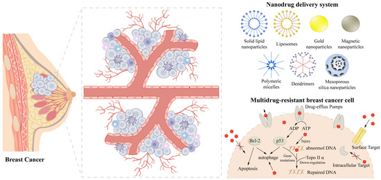
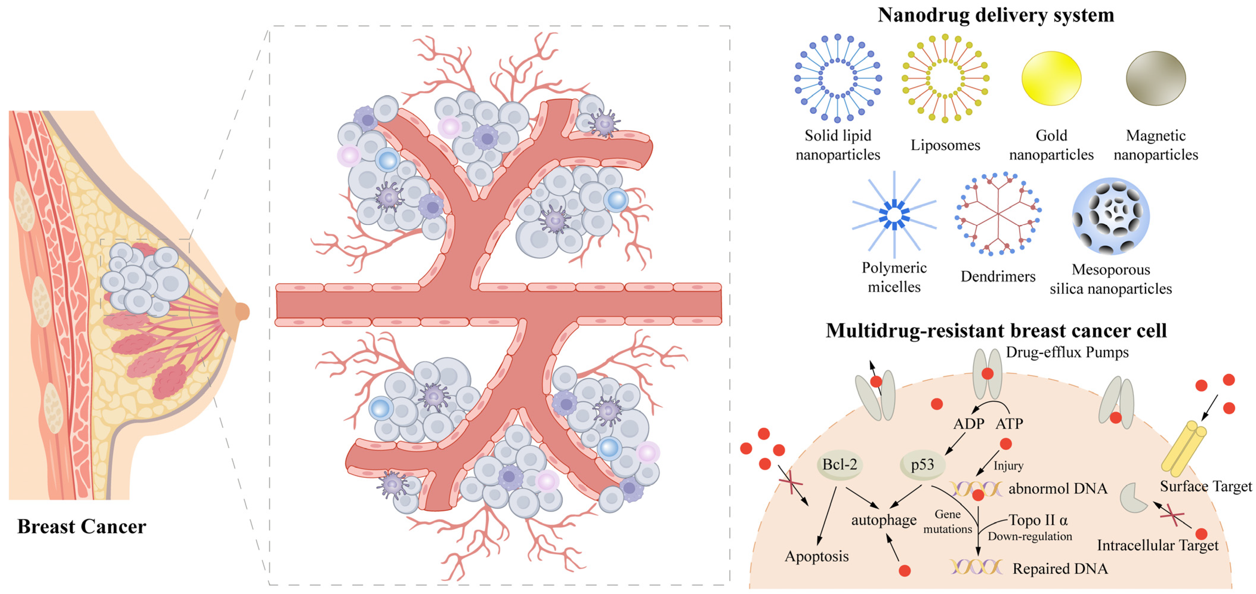
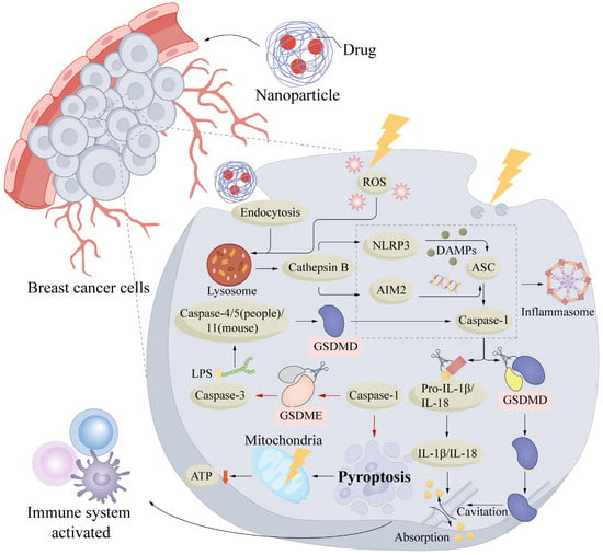
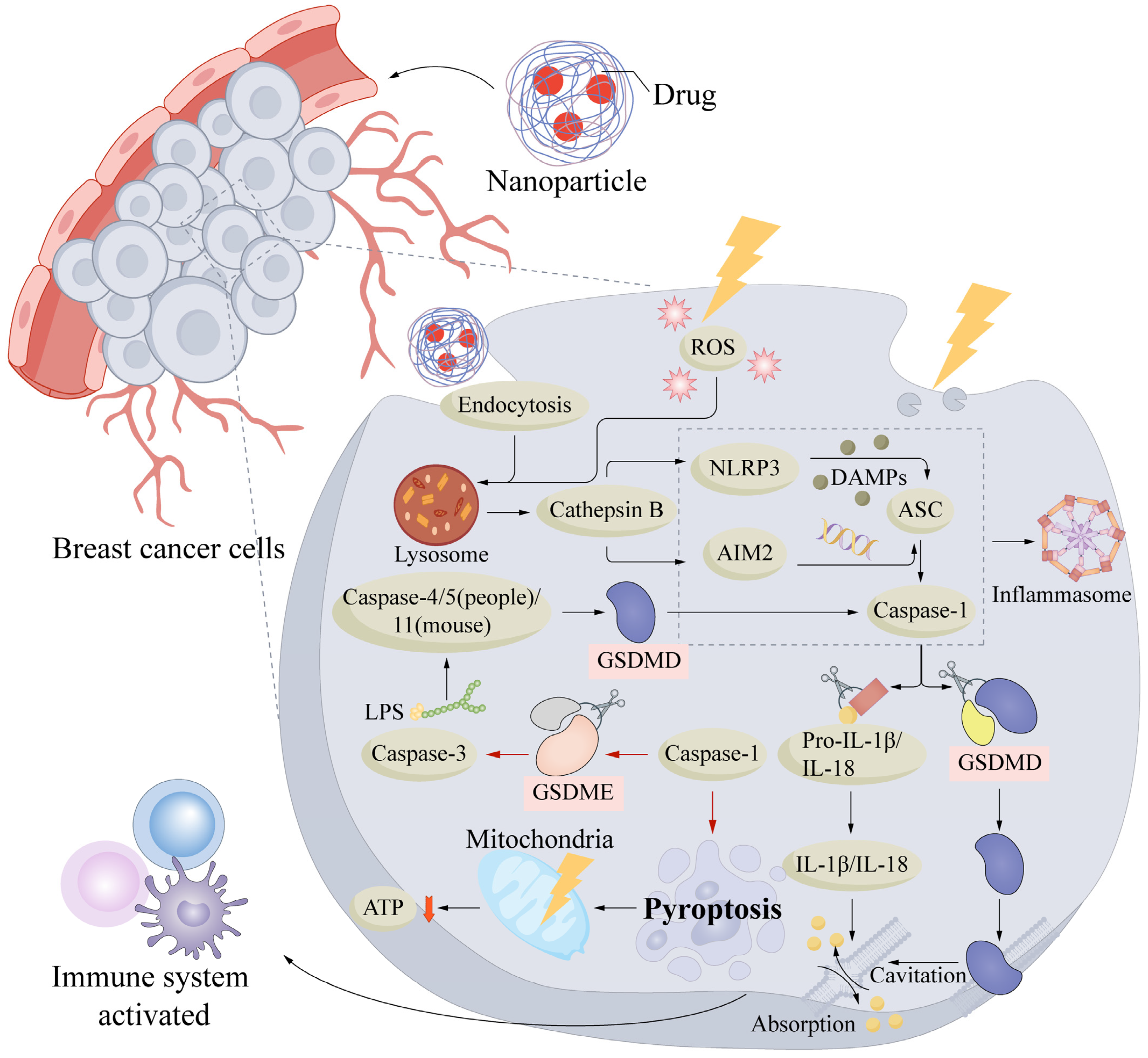
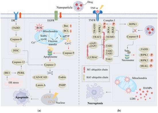
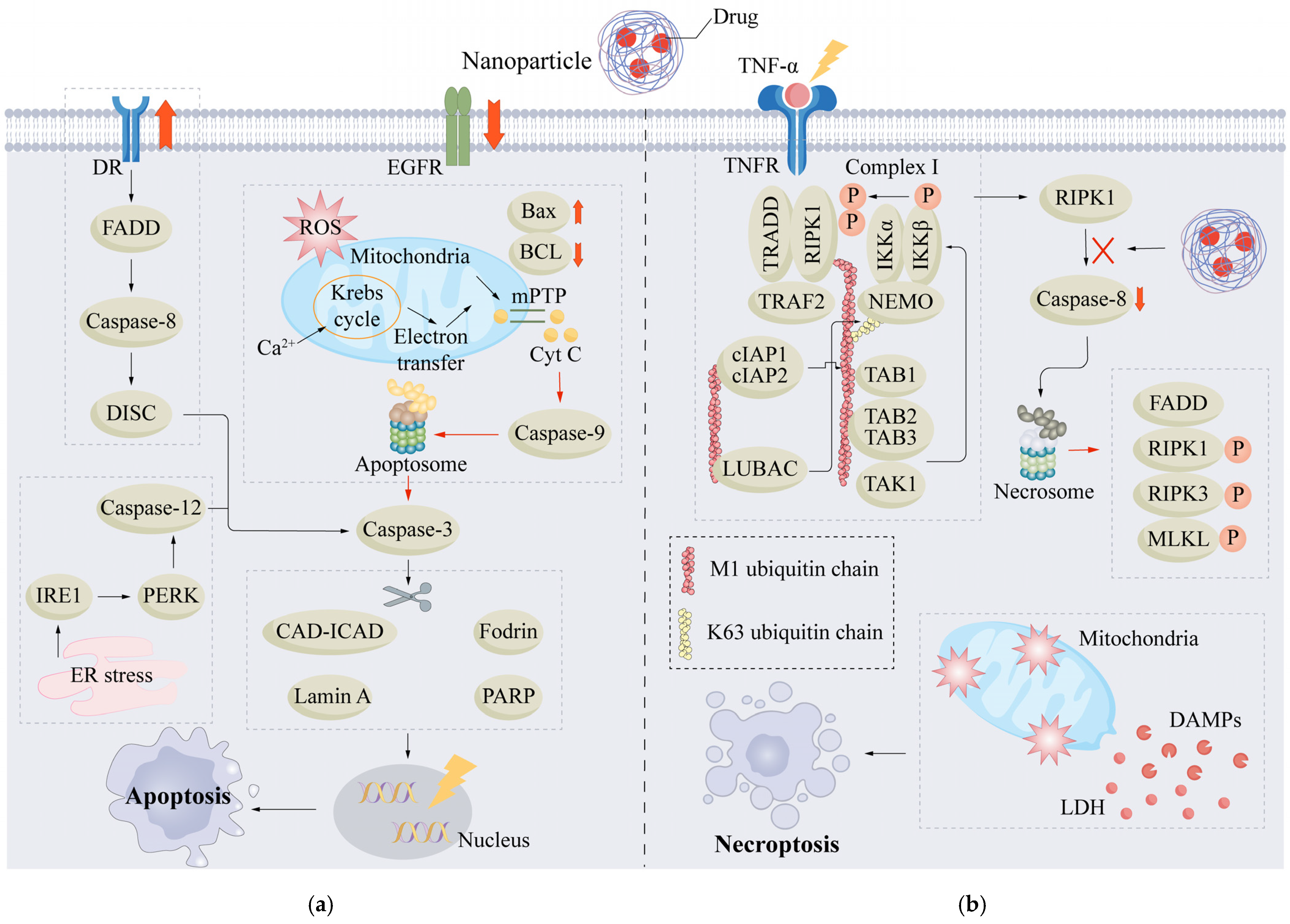
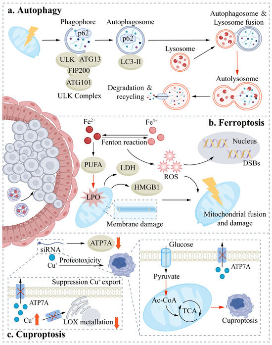
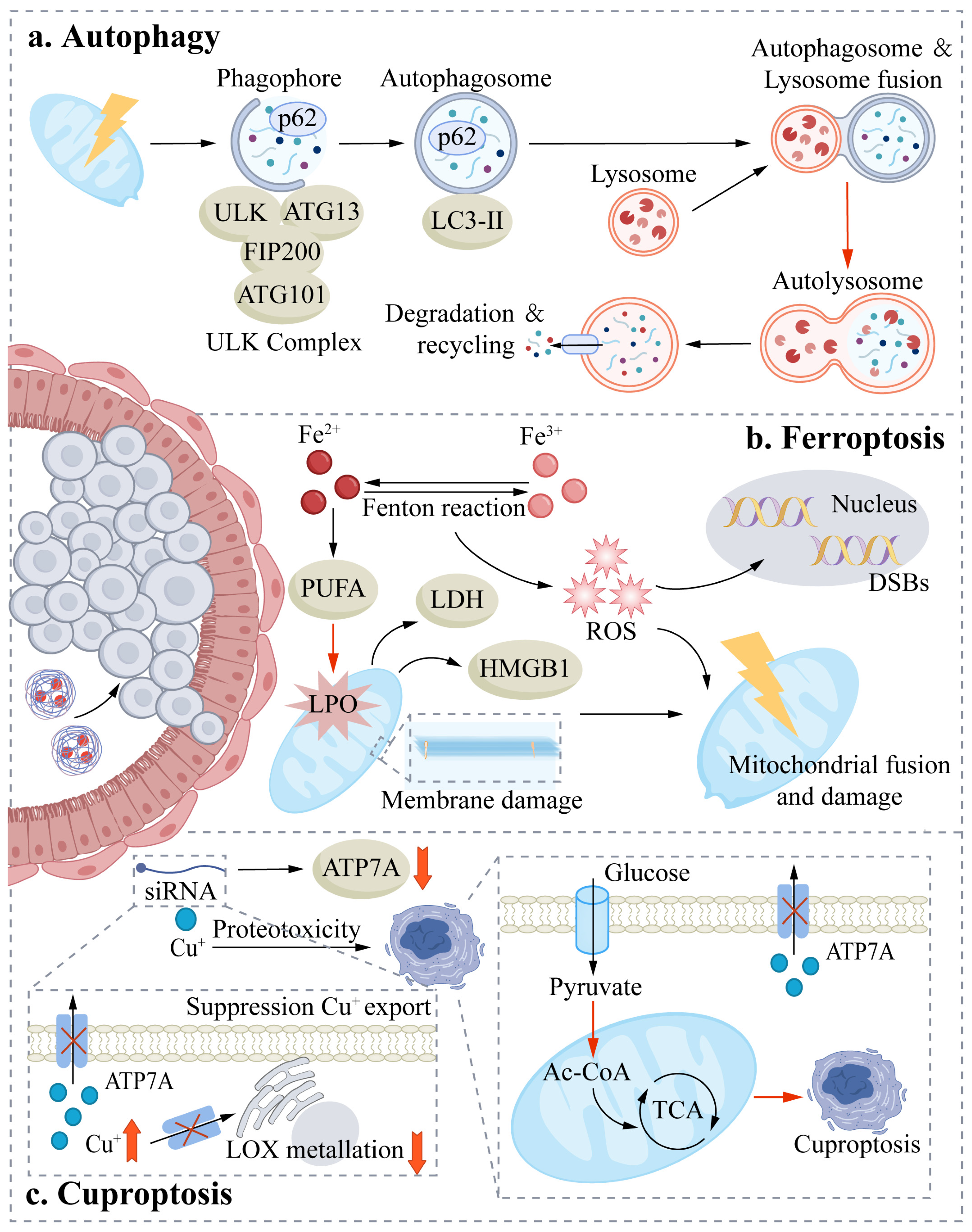
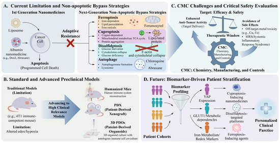
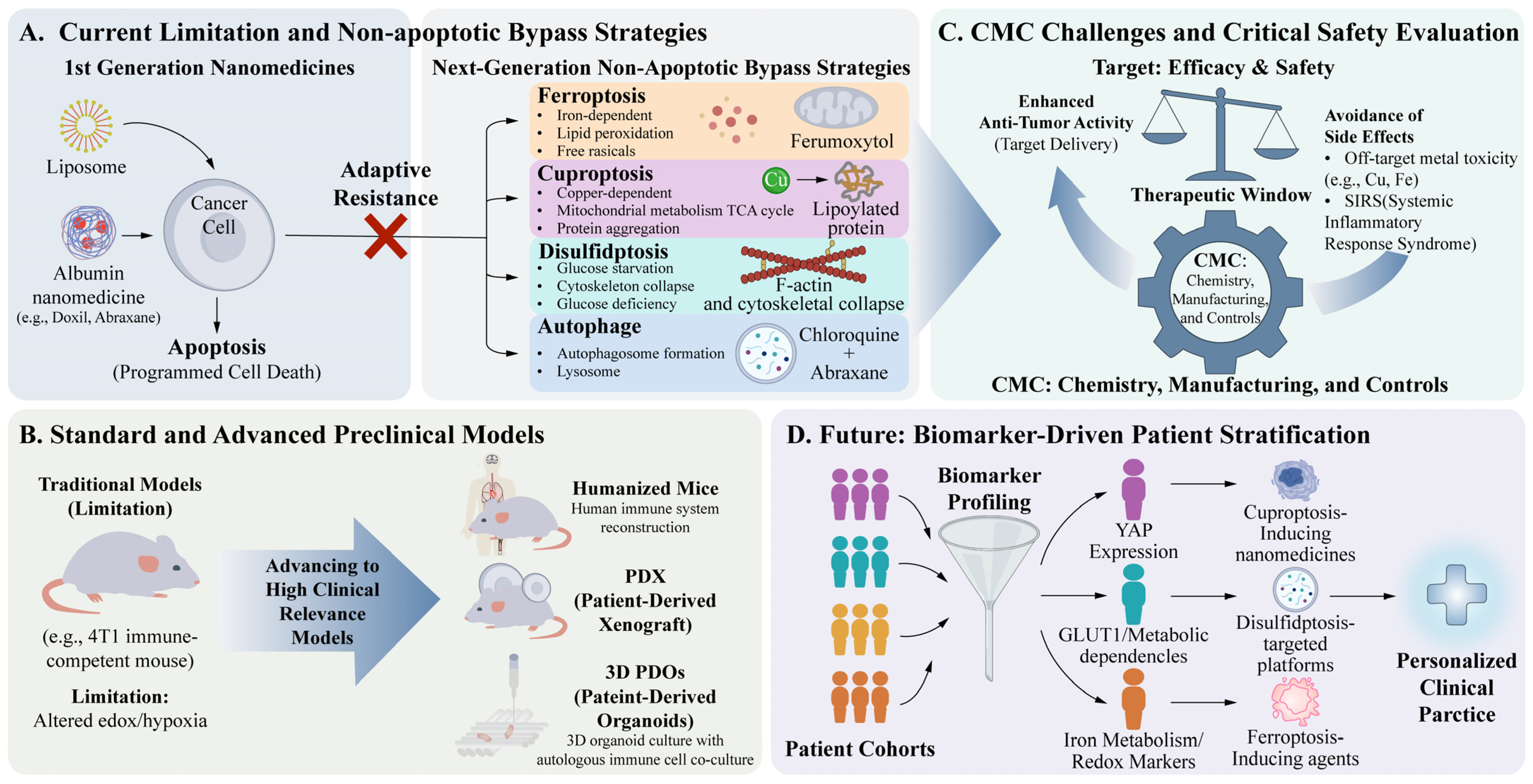

Disclaimer/Publisher’s Note: The statements, opinions and data contained in all publications are solely those of the individual author(s) and contributor(s) and not of MDPI and/or the editor(s). MDPI and/or the editor(s) disclaim responsibility for any injury to people or property resulting from any ideas, methods, instructions or products referred to in the content. |
© 2026 by the authors. Licensee MDPI, Basel, Switzerland. This article is an open access article distributed under the terms and conditions of the Creative Commons Attribution (CC BY) license.