


Abstract
Background: Osteosarcopenic obesity (OSO) syndrome, also defined as osteosarcopenic adiposity (OSA), is characterized by the concurrent loss of bone and muscle mass, accompanied by excess fat, leading to reduced functionality and metabolic imbalances. Recent studies have highlighted the role of microRNAs (miRNAs) in the pathophysiology of OSO/OSA, showing differential expression in individuals with osteosarcopenia and obesity. However, a thorough investigation in this area has been limited. Methods: A comprehensive search of international bibliographic databases, including Embase, PubMed and Scopus, was conducted. Results: From an initial search yielding 1311 records, 19 studies met the eligibility criteria for final evaluation. These findings highlight how physical exercise and nutritional factors can influence miRNA expression, emphasizing their role in promoting better health outcomes in aging populations. Furthermore, the critical role of miRNAs as indicators of muscle atrophy and the biological processes associated with aging and sarcopenia have been documented in various animal studies. Conclusions: Despite the limitations of this review, the findings indicate that miRNAs could serve as promising biomarkers and therapeutic targets for managing OSO/OSA. These results suggest that targeted interventions, such as resistance training and lifelong exercise, may effectively influence miRNA expression, potentially alleviating the impacts of OSO/OSA.
1. Introduction
Osteosarcopenic obesity (OSO) was first defined in 2014 to describe the intricate cellular communication between fat, bone and muscle [1], leading to a syndrome characterized by the co-occurrence of overweight/obesity, osteopenia/osteoporosis and sarcopenia [2,3]. Recent developments have also led to its definition as osteosarcopenic adiposity (OSA), highlighting the complex interplay between adipose tissue and the musculoskeletal system in this condition [4].
Overweight/obesity, osteopenia/osteoporosis and sarcopenia develop concurrently, contributing to the complex pathophysiology of OSO/OSA, which poses significant health risks, particularly in aging populations [5]. Advances in diagnostic techniques have allowed for a more precise identification of OSO/OSA, highlighting the importance of recognizing this syndrome as a public health concern as the global population ages. Chronic inflammation, insulin resistance and altered cellular metabolism are key factors associated with the syndrome, making it increasingly prevalent, as age-related changes promote fat redistribution in muscle and bone [6,7,8,9]. Given this background, the urgency of identifying biomarkers for the early detection of OSO/OSA has risen, enabling healthcare providers to implement targeted interventions that can reduce the related morbidity and healthcare costs.
Recent studies have shown that microRNAs (miRNAs), which are non-coding RNAs that regulate different gene pathways, may play a critical role in the development and progression of OSO/OSA, negatively affecting muscle fiber quantity and quality [10]. In fact, specific miRNAs, known as myomiRNAs, are critical regulators for both pro-inflammatory cytokines and skeletal muscle function. The most studied are miR-1, miR-133, miR-206, miR-208, miR-486, miR431 and miR-499, which modulate myogenic factors such as serum response factor (SRF), myocyte enhancer factor-2 (MEF2) and myostatin [11]. The influence of obesity on miRNA profiles in the elderly remains poorly understood [12,13]. In general, age-related and obesity-related alterations in silent information regulator sirtuin 1 (SIRT1) expression, a member of a class III histone deacetylase family proteins (HDACs) dependent on nicotinamide adenine dinucleotide (NAD+), have been linked to changes in miRNA expression [14,15]. For instance, there are metabolic consequences to miR-34a’s control of SIRT1 expression; in the liver, activated Farnesoid X Receptor (FXR) stimulates the transcription of Small Heterodimer Partner (SHP), which suppresses p53 transcriptional activity [16]. As a result, miR-34a levels fall and SIRT1 expression is positively regulated [17]. SIRT1 deacetylates and stimulates FXR transcriptional activity, which in turn activates its own expression, resulting in a positive feedback loop of the FXR/miR-34a/SIRT1 pathway [18,19]. miR-34a is increased in nonalcoholic fatty livers in humans [20] and the fatty livers of diet-induced obese mice [21]. The liver of elderly rats showed increased expression of miR-34a and miR-93, which are both involved in SIRT1’s decreased expression [22]. Still, more investigations are needed to fully examine this issue.
Adipose-derived miRNAs have been identified in rodent models as potent mediators of metabolic functions, capable of impacting muscle, liver and immune cell health, underscoring their potential involvement in OSO/OSA etiology [23,24,25,26].
Importantly, regular physical activity emerges as a vital intervention for combating the components of OSO/OSA. Exercise not only improves muscle mass, enhances bone density and reduces excess body fat, but it also mitigates inflammation, improves insulin sensitivity and modulates miRNA expression. In fact, some miRNAs appear to be involved in the response to different types of exercise, thus playing a relevant role in the modulation of myofiber gene expression in the muscle tissue of adults and elderly people. The literature data have shown that after 3 h of one acute exercise, miR-1, miR-133a, miR-133-b and miR181a expression levels were increased [27,28,29], whereas miR-9, miR23a, miR-23b and miR-31 were decreased [30,31]. In addition, Spakova and colleagues also identified miRNA206 as a predictor of response to strength training. High levels of miRNA206 and the resulting improved functional adaptation may be explained by the increased proliferative activity of satellite cells, through the TGF-β/SMAD2-3/MyoD signaling pathway [32]. For instance, it has demonstrated that a resistance training program significantly impacted miRNA expression and osteoporosis markers in elderly women with OSO/OSA [33]. Likewise, low-intensity, lifelong exercise routines could alter miRNA expression and prevent OSO/OSA by mitigating inflammatory processes [34]. These findings highlight the importance of exercise as a strategic approach to alter the disease trajectory of OSO/OSA, emphasizing its beneficial role in addressing the multifaceted aspects of this syndrome.
As society increasingly recognizes the therapeutic potential of miRNAs [35]—previously explored as therapeutic alternatives in conditions such as cancer and cardiovascular diseases—this review seeks to provide an overview of the literature surrounding the role of miRNAs in the etiology of OSO/OSA. By advancing our understanding of these mechanisms, we hope to contribute to the development of effective interventions that leverage the benefits of physical activity and miRNA modulation in addressing this challenging syndrome.
2. Materials and Methods
2.1. Information Sources and Search Strategy
A scoping review was performed to identify all studies reporting outcomes of miRNA in OSO/OSA. This study was registered on the PROSPERO database [36], number CRD42025642098. Three different databases were searched for the studies: Embase, Pubmed and Scopus. The following keywords were mixed in different combinations for each data bank: “microRNA” or “miRNA” and “Osteosarcopenic Obesity” or “Osteosarcopenic Adiposity” or “Osteosarcopenia”. Boolean operators “AND” and “OR” were employed in our search plan. The research in databases ended on 22 March 2025. To assess the eligibility of each article, each PICO (Population, Intervention, Comparison, Outcome) [37] element was identified as follows: Population (P): human and animal subjects; Intervention (I): miRNA measurement; Comparison (C): case–control; Outcome (O): role of miRNAs. The various topics analyzed in this scoping review are summarized in a checklist according to the Preferred Reporting Items for Systematic reviews and Meta-Analyses extension for Scoping Reviews (PRISMA-ScR) [38,39]. The PRISMA-ScR checklist is reported in Supplementary File S1.
2.2. Eligibility Criteria
One author searched the combination of keywords using the Boolean operators AND and OR across the three different databases. Afterwards, the author combined the three spreadsheets into a single spreadsheet, eliminating duplicates (Supplementary File S1). Then, independently, three authors screened each remaining article based on the following inclusion criteria: (i) English language; (ii) studies that evaluate the role of miRNA in OSO/OSA.
Articles were considered to be not eligible considering the following specific exclusion criteria: (i) articles missing one or more keywords; (ii) conference abstract, paper or meeting; Editorial, letter, note or short survey; (iii) articles irrelevant to the main subject; (iv) no English language; (v) retracted; or (vi) review, systematic review or meta-analysis.
The reason for the exclusion of each record is reported in Supplementary File S2. The title and abstract of all studies found in the search were independently examined by two reviewers, who applied the eligibility criteria. In the case of disagreement between the reviewers, a third reviewer was consulted.
2.3. Data Extraction and Quality Process
Following selection, data extraction was carried out by manual curation. The data were extracted by 3 authors, who then independently summarized each article’s findings.
3. Results
3.1. Literature Research
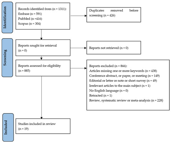
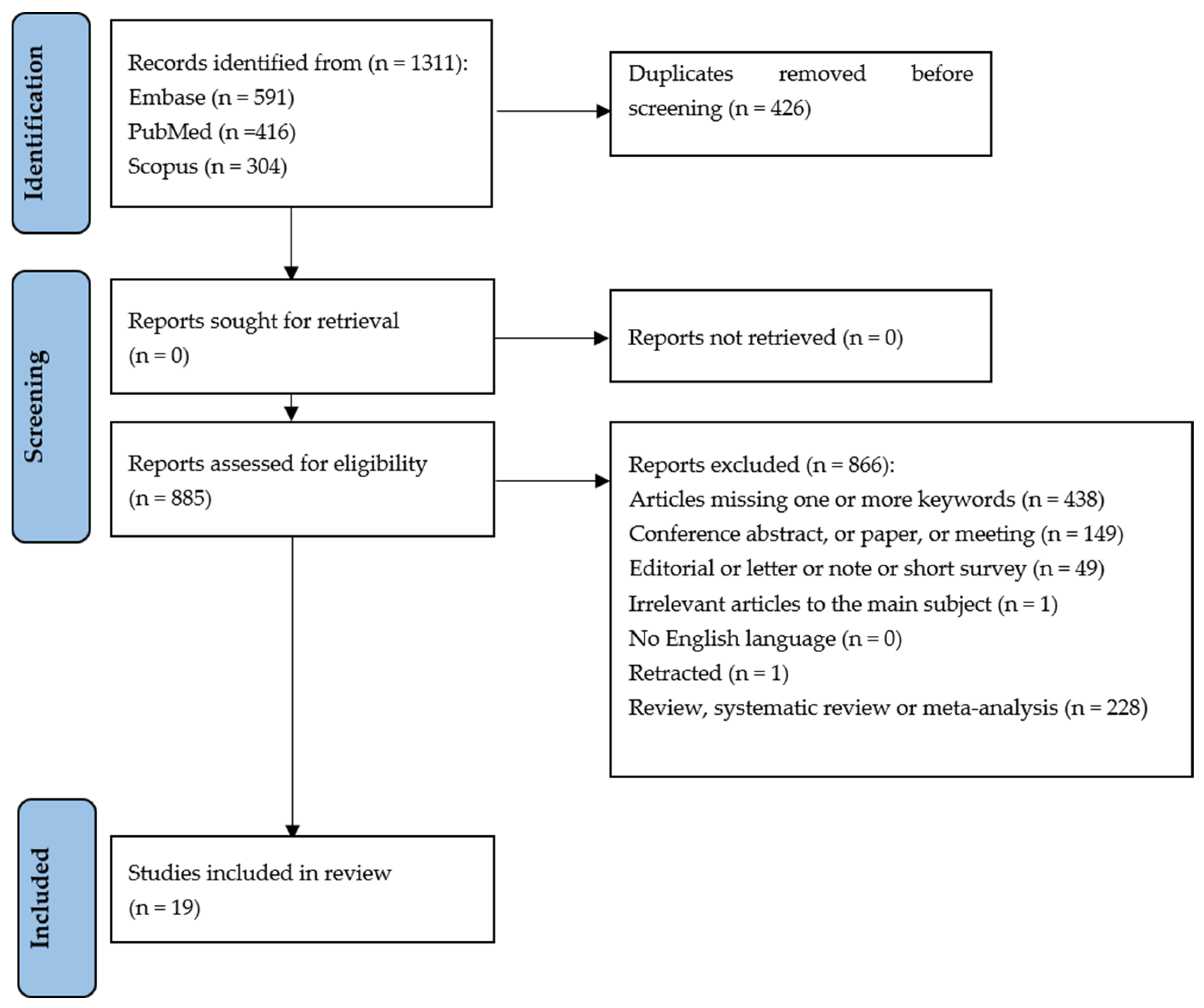
The flowchart provides a clear illustration of the process for literature searching (Figure 1). A total of 1311 articles were obtained by searching Pubmed, Embase and Scopus based on the following keywords: “microRNA” or “miRNA” and “Ostosarcopenic Obesity”.
Figure 1.
PRISMA-ScR flow diagram showing research strategy [38,39].
After discarding duplicates, 885 articles were assessed according to the eligibility criteria. After completing the screening procedure, 19 articles were included in this scoping review in order to elucidate the role of miRNA in OSO/OSA.
3.2. Study Characteristics
The main characteristics of the 19 included studies are reported in Table 1. The included articles were published between 2010 and 2024. Eight articles were conducted on humans, seven on animal models and four on both. The range of participants included was from 21 to 534, with a total of 1011 participants. Of these, 302 were women and 302 were men. The sex of the remaining individuals was not specified. Regarding the animal model, the range varied from 12 to 80 animals included, with a total of 255 animals. Of these, 143 were female and 62 were male. A total of 128 mice and 173 rats were also used. As for the methodologies, 13 articles used only PCR methods (qPCR, ddPCR, RT-PCR), 4 used the array and 7 articles employed sequencing methodology. Finally, these studies were conducted on 13 samples from peripheral blood (blood, serum and plasma) and 6 on tissue. Based on the outcomes of each study, we analyzed the role of miRNAs in OSO/OSA by distinguishing them into results obtained on human subjects and animal subjects.
Table 1.
Key features of the studies included in our scoping review.
3.3. Clinical Outcome
3.3.1. Human Studies
The articles by Banitalebi E et al. [33] and Huang L et al. [40] investigate the expression of microRNAs (miRNAs) following physical exercise (Table 2). The study by Banitalebi E et al. [33] focuses on miR-133 and miR-206 in Iranian women with osteopenia/osteoporosis and examines their correlations with various health indicators after 12 weeks of resistance training using elastic bands. The results showed significant correlations between miR-133 levels and FRAX score (r = −0.845, p < 0.001), vitamin D (r = −0.551, p = 0.025) and ALP (r = 0.620, p = 0.012). Similarly, miR-206 showed analogous correlations with FRAX score (r = −0.847, p < 0.001), vitamin D (r = −0.500, p = 0.041) and ALP (r = 0.662, p = 0.007). Meanwhile, the study by Huang et al. [40] examines the expression of miR-126, miR-146a and miR-222 in community-dwelling older adults after an 8-week cycling training program. They found that exercise increased the expression of these miRNAs for up to 16 weeks post-training, suggesting their involvement in improvements in body composition and cardiorespiratory fitness. Additionally, miR-21 was suggested as a potential mediator of the effects of exercise on strength and muscle health. Both studies highlight the potential role of exercise in modulating miRNA expression and its relevance to health outcomes.
Table 2.
Key features of the human studies included in our scoping review.
Similar to the study conducted by Banitalebi E et al. [33], the research by Faraldi M et al. [41] also focuses on post-menopausal women, specifically aiming to identify circulating miRNAs as potential biomarkers for muscle mass wasting in this population (Table 2). The cohort was divided into tertiles based on appendicular skeletal muscle mass index (ASMMI) to better highlight the differences in skeletal muscle mass: the first tertile comprised nine participants with an ASMMI of 4.88 ± 0.40 kg·m−2; the second tertile included ten participants with an ASMMI of 5.73 ± 0.23 kg·m−2; and the third tertile had nine participants with an ASMMI of 6.40 ± 0.22 kg·m−2. The study found that five miRNAs (miR-221-3p, miR-374b-5p, miR-146a-5p, miR-126-5p and miR-425-5p) were downregulated, while two miRNAs (miR-145-5p and miR-25-3p) were upregulated in the first tertile (relatively low ASMMI) compared to the third tertile (relatively high ASMMI).
The two articles by He N et al. [42] and Qaisar R et al. [43] focus on the interaction between miRNAs, sarcopenia and cardiovascular issues, providing important insights into the health of the elderly (Table 2). In the study by He N et al. [42], the relationship between sarcopenia and cardiovascular risk factors (CVRF) in elderly Chinese individuals is investigated. The authors highlight how the presence of CVRF is associated with a higher prevalence of sarcopenia in the elderly population, underscoring the crucial role of factors such as hypertension and dyslipidemia in predisposing individuals to sarcopenia. Furthermore, the results show a significant correlation between the levels of the miRNA miR-29b and appendicular muscle mass, suggesting that this miRNA could serve as a potential biomarker for sarcopenia, which is also useful in assessing cardiovascular risk. Similarly, Qiasar et al. [43] explore the correlation between circulating levels of specific miRNAs and indices of sarcopenia in patients with congestive heart failure (CHF). The results indicate that patients with CHF exhibit reduced muscle strength and physical capacity compared to healthy controls. In addition, miR-133a shows the strongest correlation with handgrip strength, an important indicator of sarcopenia, while other miRNAs such as miR-434-3p demonstrate strong diagnostic potential for CHF. Changes in the levels of these miRNAs are associated with signs of inflammation and oxidative stress, suggesting a significant connection between sarcopenia and cardiovascular complications. Both articles emphasize the importance of understanding miRNA levels as indicators in the diagnosis and management of sarcopenia, particularly in vulnerable populations such as the elderly with cardiovascular conditions.
Iannone F et al. [44] conducted a study focused on the association between myomiRs, specifically miR-133b, and sarcopenia, investigating the importance of nutrition as a mediating factor in this relationship (Table 2). The results revealed that reduced levels of miR-133b were significantly correlated with the presence of sarcopenia. This suggests that poor nutrition could contribute to the lowering of miR-133b, with negative repercussions on muscle mass. The research therefore highlights the importance of nutrition in managing sarcopenia and the need to consider myomiRs as potential biomarkers to monitor and address this health issue, especially in the elderly.
La Rosa F et al. [45] examined whether rehabilitation reduces inflammation in sarcopenic patients and explored the biological factors associated with this effect (Table 2). They found a significant increase in the levels of miR-335-3p, a post-transcriptional regulator of IL-37 production, as a result of the rehabilitation program.
Finally, Millet M et al. [46] aimed to identify a microRNA signature linked to sarcopenia among older adults living in the community from the SarcoPhAge cohort (Table 2). Their findings revealed that in individuals with sarcopenia, the expression levels of serum miRNA-133a-3p and miRNA-200a-3p were decreased. This reduction aligns with the role of these microRNAs in regulating the proliferation and differentiation of muscle cells, suggesting they may play a significant role in the development of sarcopenia.
3.3.2. Animal Studies
All the articles focus on miRNAs as key indicators of muscle atrophy and the biological processes involved in aging and sarcopenia conditions in animal models, suggesting potential therapies and interventions for managing age-related muscle issues.
Gao H et al. [47] and Pedraza-Vázquez G et al. [34] evaluate the expression of miRNAs following physical exercise, highlighting their regulatory roles in skeletal muscle growth and aging (Table 3). In the study by Gao H et al. [47], the authors focused on the effects of lifelong moderate-intensity continuous training (MICT) using a treadmill on age-related changes in muscle miRNA expression profiles as well as on muscle atrophy, apoptosis and mitochondrial dysfunction. They found that adult rats engaged in MICT experienced notable improvements in their muscle miRNA profiles and a reduction in age-related muscle deterioration, suggesting that an increased expression of miR-486 may be linked to the beneficial effects observed. Similarly, the research conducted by Pedraza-Vázquez G et al. [34] involved a comparison between rats subjected to low-intensity running exercise and a sedentary control group. The study revealed significant alterations in miRNA expression in the gastrocnemius muscle across different age ranges. They discovered that several miRNAs were differentially expressed before and after physical activity, indicating that exercise can modulate miRNA levels. This modulation was associated with inflammation and muscle aging, with a clear relationship between miRNA expression profiles and the inflammatory status of the exercised versus sedentary rats.
Table 3.
Key features of the animal studies included in our scoping review.
Hamrick M et al. [48] investigated the changes in miRNA expression linked to muscle mass loss and myofiber size reduction due to aging in mice. Their research revealed that aging is associated with significant alterations in the expression of 57 specific miRNAs within the mouse skeletal muscle, many of which correlate with age-related muscle atrophy. To assess the therapeutic potential of leptin, the researchers administered recombinant leptin to aged mice. The treatment resulted in a noteworthy increase in hindlimb muscle mass and the size of the extensor digitorum longus fibers in these aged subjects. These findings underscore the intricate relationships between miRNA regulation, muscle mass and potential interventions for combating muscle atrophy related to aging (Table 3).
In another study, Jung H et al. [49] pointed out that while circulating miRNA levels change with age, their potential as noninvasive biomarkers for muscle atrophy remains largely uncharted (Table 3). They conducted a comprehensive miRNA-seq analysis in the tibialis anterior muscle and serum of a mouse model designed to mimic disuse-induced atrophy, analogous to the acute atrophy observed after prolonged bed rest. The comparative analysis revealed that miR-455-3p was significantly reduced in both the atrophy model and older mice. Additionally, miR-434-3p levels were lower in the serum and muscle of aged mice compared to younger counterparts. These results suggest that the deregulation of miR-455-3p may play a vital role in the mechanisms underlying muscle atrophy, and miR-434-3p might serve as a potential serum biomarker for muscle aging.
To delve deeper into the genome-wide changes in miRNA and mRNA expressions associated with muscle aging, Kim J et al. [50] performed sequencing on samples from the gastrocnemius muscles of mice at two different ages (6 and 24 months). Their analysis identified 34 differentially expressed miRNAs (15 upregulated and 19 downregulated) with age. Among them, miR-206 and miR-434 showed significant changes that were corroborated by previous studies. Intriguingly, eight miRNAs from a clustered region at the Dlk1-Dio3 locus on chromosome 12 exhibited coordinated downregulation. Furthermore, the research unveiled 16 novel miRNAs. The integrative analysis brought to light that these miRNAs might impact muscle aging through their positive regulation of transcription, metabolic processes and kinase activity. A number of age-related miRNAs identified in this study have also been implicated in human muscular diseases, pointing to the broader relevance of these findings (Table 3).
Additionally, Lee H et al. [51]. confirmed that muscle mass and functional decline, along with mitochondrial dysfunction, occur in the quadriceps of ovariectomized (OVX) mice. Their research highlighted that miR-141-3p is upregulated in OVX models, with perturbed mitochondrial function through the inhibition of Fkbp5 and Fibin. This suggests that targeting miR-141-3p might represent a promising therapeutic strategy for addressing obesogenic sarcopenia (Table 3).
Lastly, the study of Pardo P et al. [52] also established a connection between the aging process and the alterations in miRNA expressions in skeletal muscles, noting the downregulation of miR-434-3p, which they identified as an anti-apoptotic miRNA. As such, miR-434-3p may have therapeutic potential in treating muscle atrophy across various pathophysiological contexts, including sarcopenia (Table 3).
3.3.3. Studies on Both Animals and Humans
In a series of studies assessing the role of specific microRNAs (miRNAs) in muscle health and pathology, several key findings emerged from both human and mouse models. Rivas D et al. [53] conducted a randomized controlled trial and identified six differentially expressed miRNAs (miR-1-3p, miR-19b-3p, miR-92a, miR-126, miR-133a-3p and miR-133b) between gainers and losers in older adults following a 6-month progressive resistance exercise training intervention. Notably, miR-19b-3p was strongly associated with increased fat-free mass and exhibited higher levels in young mice compared to aged counterparts, correlating positively with muscle mass and grip strength. Similarly, Yang S et al. [54]. found elevated serum levels of miR-193b in individuals with type 2 diabetes, which negatively correlated with muscle mass and was linked to muscle loss in both healthy and db/db mice. In a related study, Itokazu M et al. [55] demonstrated that the transplantation of perimuscular adipose tissue to young mice inhibited muscular stem cell proliferation via Let-7d-3p, highlighting the inhibitory role of this miRNA on crucial stem cell transcription factors. Finally, Okamura T et al. [56]’s microarray analysis revealed that let-7e-5p was significantly downregulated in orchiectomized mice, suggesting a critical involvement in muscle degradation associated with androgen depletion, as its suppression led to enhanced muscle-specific protein expression and improved mitochondrial function. Collectively, these findings underscore the complex interplay of miRNAs in muscle mass regulation, atrophy and the physiological adaptations to exercise and other metabolic conditions (Table 4).
Table 4.
Key features of the studies involving both humans and animals included in our scoping review.
4. Discussion
The purpose of the present study was to provide a broad and recent overview of which miRNAs are involved in modulating the mechanisms that control skeletal muscle development and maintenance. Since the aging process is accompanied by muscle decay and the concomitant accumulation of adipose tissue, the correlation between the modulation of expression levels of miRNAs involved in the regulation of skeletal muscle status and the onset of OSO/OSA syndrome was also investigated. For this purpose, studies conducted both in the human model, using postmenopausal women and older adult men as study populations, and in the animal model were analyzed. It was first found that there are several miRNAs related to the processes of muscle development, differentiation and regeneration. Indeed, Faraldi and colleagues [41] identified miR-146a as a molecule involved in the regulation of muscle quality and mass; this miRNA, along with others, was indeed found to be differentially expressed in postmenopausal women characterized by high and low ASMMI [41]. In agreement, Millet et al. [46] identified miRNAs 133a-3p and 200a-3p as potentially involved in the modulation of muscle cell proliferation and differentiation processes, as decreased expression of these miRNAs was observed in sarcopenic patients, suggesting their role in the mechanisms of sarcopenia onset [46]. The study by Hamrick and colleagues [48] conducted in a mouse model further confirms these findings, as this study showed that aging is associated with significant alterations in the expression of 57 miRNAs specific to the skeletal muscle in mice, many of which are related to age-related muscle atrophy [48]. The correlation between age-dependent muscle decay and modulation of the expression of certain miRNAs is also highlighted by the study by Kim and colleagues [50], in which genome-wide changes in the expression of miRNAs and mRNAs associated with muscle aging were investigated. In fact, the analysis conducted identified 34 differentially expressed miRNAs in 6- and 24-month-old mice, including miR-206 and miR-434, that might have an impact on muscle aging [50]. miR-434-3p could also represent a potential biomarker of musculoskeletal status, as it was observed to be downregulated in aged mice by Pardo and colleagues, further suggesting a link between the aging process and alterations in miRNA expression in skeletal muscles [52].
To date, miRNAs have emerged as promising early biomarkers for a variety of pathologies due to their stability in biological fluids and crucial roles in gene regulation. For instance, miR-134-5p is associated with acute myocardial infarction [57]. Similarly, miR-125a-3p is recognized for its involvement in cancer and autoimmune diseases [58,59], suggesting its utility in early diagnostics. Other miRNAs, such as miR-122-5p, have been studied for their roles in metabolic disorders and liver health [60]. Moreover, miR-146a-5p plays a role in inflammation, indicating chronic inflammatory states [61]. Some of these diseases are known to be accompanied by concomitant muscle decay. In agreement, Qiasar and colleagues have indeed shown that there is a correlation between the expression of certain miRNAs, such as miR-133a, and muscle atrophy typical of sarcopenia in patients with CHF [43]. In contrast, He et al. identified miR-29b as a potential biomarker for sarcopenia associated with cardiovascular issues, since its expression levels were found to correlate with Appendicular Muscle Mass in a sarcopenic population characterized by high cardiovascular risk [42]. Moreover, Yang et al. found that elevated serum levels of miR-193b in subjects with type II diabetes correlated negatively with muscle mass [54]. Therefore, monitoring the expression levels of these miRNAs could facilitate the early diagnosis of various diseases, thereby improving intervention strategies and health outcomes. This understanding is crucial not only for early detection but also for elucidating the pleiotropic functions of miRNAs in general, as they often regulate multiple target genes involved in diverse biological processes [30]. As research continues, these miRNAs stand out as valuable tools for enhancing our understanding of disease mechanisms and developing effective clinical applications.
Several studies have also strongly demonstrated that the performance of exercise is able to modulate the expression of several miRNAs, thus exerting an effect on muscle mass and muscle response to the physical activity performed. Indeed, the work of Huang et al. highlights that 8-week cycling training was able to modulate the expression of several miRNAs involved in muscle health, and in particular in modulating body composition and cardiorespiratory capacity [40]. In agreement, Gao and colleagues [47] also highlighted the role of exercise (continuous high intensity training) in modulating the expression of miRNAs that have regulatory roles in skeletal muscle development and maintenance. Indeed, they found that adult rats subjected to this type of training showed significant improvements in the expression pattern of muscle miRNAs, and a concomitant reduction in age-related muscle deterioration; specifically, the increased expression of miR-486 may be related to the observed beneficial effects [47]. Therefore, it appears that modulating the expression of miRNAs involved in the regulation of muscle quality status in high-intensity exercise, as also shown in the study by Rivas and colleagues, who identified miRNAs miR-1-3p, miR-19b-3p, miR-92a, miR-126, miR-133a-3p, and miR-133b differentially expressed in groups of older adults who had shown loss or gain of muscle mass after 6 months of progressive resistance training [53].
Unfortunately, to date there are only two studies in the literature analyzing the modulation of muscle miRNA expression levels in response to the performance of exercise in humans and animals characterized by OSO/OSA syndrome. In detail, Banitalebi et al. [33] found that a 12-week elastic band resistance training program significantly improved myomiR (miR-133 and miR-206) expression and markers of osteoporosis in elderly women with OSO/OSA. Both miR-133 and miR-206 have been shown to be members of a family of muscle-specific miRNAs expressed predominantly in skeletal and cardiac muscle tissues, playing a crucial role in muscle development, differentiation and regeneration [62,63,64,65,66]. In fact, these miRNAs target several key genes involved in muscle biology, including Mef2c (a transcription factor crucial for muscle differentiation) [67,68] and Hdac4 (Histone deacetylase that represses muscle gene expression) [67,68], SRF and Cx43 (which participate in skeletal muscle differentiation) [69]. As myomiRs are responsible for intercellular communication, they may also be essential for the one health improvements provided by exercise programs [70], supporting the data in our scoping review. This aligns with the findings of a study by Yang et al. (2023) [71], which also reported that resistance training notably enhanced muscular strength and bone mineral density in older adults, indicating a positive impact on the components of OSO/OSA. Furthermore, these improvements in myomiRs, which are crucial in muscle regeneration and adaptation, suggest potential biomarker utility for assessing the efficacy of exercise interventions, as supported by evidence from Spakova et al. (2020) [32], who emphasized the role of myomiRs in muscle physiology and pathology.
In contrast, Pedraza-Vázquez et al. [34] demonstrated that a low-intensity lifelong exercise routine could modulate miRNA expression and reduce inflammation, ultimately preventing OSO/OSA. Most of the DEmiRs found in rats are involved in both osteogenesis and metabolic syndromes. Among them, the following miRNAs were identified: miR-125a-3p [72,73,74,75,76], miR-134-5p [77,78], miR-23a-5p [78,79,80,81,82], miR-30e-5p [83,84], miR-494-3p [85,86,87,88], miR-122-5p [89,90,91,92], miR-152-3p [93,94] and miR-20a-5p [95,96,97]. Some of these are involved mainly in osteogenesis, such as miR-194-5p [98,99], miR-497-5p [100], miR-127-3p [101] and miR-672-5p [102], while miR-32-3p [103,104] is associated with both functions. In contrast, miR-10b-5p appears to be involved in both osteogenesis [105,106,107,108] and atrophy [109], and miR-146a-5p regulates bone mass and bone remodeling [110,111]. Also identified was miR-3075, specifically associated with obesity [112], and miR-296-3p [113,114]. Finally, unlike the previous ones, miR-678, miR-1839-5p and miR-6332 appear to be unknown in the main mechanisms involved in OSO/OSA, so deeper investigations would be interesting to carry out in this context.
It is interesting that the authors demonstrate how exercise can alter the epigenetic factor, represented by miRNAs, as people age, decreasing muscle atrophy and significantly affecting how the inflammatory component is regulated [115,116]. According to the study, two miRNAs linked to inflammation in the 8–12 month SED and LRER groups are miR-134-5p and miR-23a-5p. miR-134-5p may control inflammation and the loss of muscle tissue in SED rats by inducing apoptosis and inflammation in fibroblasts and epithelial cells [104]. The expression of miR-134-5p may be inhibited by the LRER exercise regimen. However, miR-23a-5p may be used to trigger compensatory processes including myoblast differentiation and proliferation. LRER (12–18 months) has high expressions of four miRNAs (miR-10b-5p, miR-494-3p, miR-127-3p, and miR-122-5p) linked to pro-inflammatory processes [108,117,118,119], indicating their involvement in metabolic disorders linked to the buildup of lipid tissue in response to inflaming or injury. At least three miRNAs shared with the LRER group, such as miR-30e-5p, cause a protective inflammatory response in SED rats 12–18 months of age [120,121,122]. When miRNA regulation is examined in the later stages of life (18–24 months), in the aging process, four miRNAs (miR146a-5p, miR-152-3p, miR-296-3p and miR-20a-5p) are upregulated in LRER and linked to inflammation suppression [123,124,125,126,127,128,129]. When combined, these findings lend more credence to the idea that miRNAs are linked to inflammation, one of the major factors that causes OSO/OSA. The authors demonstrated through the LRER exercise program that lower body fat also lowers inflammation, and that this process is linked to the differentiating expression of different miRNAs as people age. Additionally, the authors affirm that long-term low-intensity exercise is protective to muscle function [130,131]; by contrast, sedentarism is linked to a stronger pro-inflammatory profile in older adults [132]. By allowing adipocytes to infiltrate the muscle tissue, this regulatory mechanism may reduce the inflammatory profile.
Their findings are consistent with research by Fan et al. [11], which highlighted that regular physical activity leads to favorable changes in inflammatory markers and enhances metabolic health in older adults. This synergy between exercise and inflammatory regulation is critical, as chronic inflammation is a known contributor to OSO/OSA and other age-related diseases. Moreover, the focus on low-intensity exercise reflects a growing body of literature emphasizing its benefits for older adults, such as the study by Ultimo et al. [31], which reported significant health improvements with moderate exercise, making it feasible for broader populations.
Both the studies by Banitalebi et al. [33] and Pedraza-Vázquez et al. [34] provide significant insights into the relationship between exercise, miRNA expression and the management of OSO/OSA; however, they face several weaknesses and criticisms. Banitalebi et al.’s [16] randomized controlled trial is limited by its relatively small sample size, which may restrict the generalizability of the findings to broader populations. Additionally, the study focuses exclusively on elderly Iranian women, potentially overlooking the effects of resistance training in other demographics, such as men or younger individuals. The intervention duration of 12 weeks may also be insufficient to ascertain the long-term sustainability of the observed changes in myomiRs and osteoporosis markers, raising questions about the lasting impact of the training regimen.
Conversely, the observational design of Pedraza-Vázquez et al.’s study [34] limits the ability to draw causal inferences regarding the changes in miRNA expression and inflammation resulting from the low-intensity lifelong exercise routine. The study’s reliance on subjective measures of exercise intensity could result in variability in adherence and outcomes, further complicating the interpretation of results. Moreover, while the research identifies important relationships between exercise and various health markers, it does not delve deeply into the underlying mechanisms by which exercise modifies miRNA profiles and inflammation, leaving gaps in understanding. Both studies would benefit from larger, more diverse cohorts and more rigorous methodologies to enhance the robustness of their findings and their applicability to the broader population.
Such improvements could lead to a more comprehensive understanding of the effects of physical activity on miRNA expression and its implications for managing OSO/OSA, ultimately informing the development of effective public health strategies and clinical interventions aimed at improving health outcomes for aging populations.
To our knowledge, this is the first scoping review that specifically discusses the effects of miRNA expression on patients with OSO/OSA. Nevertheless, the number of studies included in this analysis was very limited, with only two articles, which may have had some influence on the reliability of the outcomes, similar to that found by the group of Yang et al. [133] in discussing the effects of exercise in patients with OSO/OSA. Despite this, both the existing literature, as previously described, and a clear and well-defined methodological protocol (PROSPERO) [36] solidly support the data obtained by the two research groups, lending significant scientific relevance to our scoping review although the discussion is limited to these two papers.
5. Conclusions
In conclusion, the emerging relationship between exercise, miRNA expression and osteoporosis underscores the potential of physical activity as a pivotal intervention for combating OSO/OSA. Continued research in this domain is essential for unraveling the biological mechanisms involved and for translating these findings into effective preventative and therapeutic strategies.
Supplementary Materials
The following supporting information can be downloaded at https://www.mdpi.com/article/10.3390/cells14110802/s1, Supplementary File S1: PRISMA Checklist; Supplementary File S2: List of studies not included and exclusion reasons.
Author Contributions
Conceptualization, A.B. and M.B.; methodology, A.B.; investigation, A.B.; re-sources, M.B.; data curation, A.B., M.B. and V.V.V.; writing—original draft preparation, A.B., M.B., G.C. and C.G.; writing—review and editing, M.B., V.V.V., G.C. and C.G.; visualization, M.B., M.C.T. and C.G.; supervision, G.T., C.G. and U.T.; project administration, G.T., M.C.T. and C.G.; funding acquisition, U.T. All authors have read and agreed to the published version of the manuscript.
Funding
This research received no external funding.
Institutional Review Board Statement
Not applicable.
Informed Consent Statement
Not applicable.
Data Availability Statement
All data generated or analyzed during this study are included in this published article and its Supplementary Files.
Conflicts of Interest
The authors declare no conflicts of interest.
Abbreviations
The following abbreviations are used in this manuscript:
| ALP | Alkaline Phosphatase; |
| AMSTAR 2 | A MeaSurement Tool to Assess systematic Reviews 2 |
| DEmiRs | Differential expression microRNAs |
| FRAX® score | Fracture Risk Assessment Tool Score |
| LRER | Low-Intensity Running Exercise Routine |
| miRNAs | microRNAs |
| OSA | Osteosarcopenic Adiposity |
| OSO | Osteosarcopenic Obesity |
| PRISMA | Preferred Reporting Items for Systematic Reviews and Meta-Analyses |
| RT-PCR | Reverse transcription polymerase chain reaction |
References
- Ilich, J.Z.; Kelly, O.J.; Inglis, J.E.; Panton, L.B.; Duque, G.; Ormsbee, M.J. Interrelationship among muscle, fat, and bone: Connecting the dots on cellular, hormonal, and whole body levels. Ageing Res. Rev. 2014, 15, 51–60. [Google Scholar] [CrossRef] [Scilit] [PubMed]
- Ilich, J.Z.; Kelly, O.J.; Inglis, J.E. Osteosarcopenic Obesity Syndrome: What Is It and How Can It Be Identified and Diagnosed? Curr. Gerontol. Geriatr. Res. 2016, 2016, 7325973. [Google Scholar] [CrossRef] [Scilit]
- Nasabian, P.J.; Inglis, J.; Kelly, O.J.; Ilich, J.Z. Osteosarcopenic obesity in women: Impact, prevalence, and management challenges. Int. J. Women’s Health 2017, 9, 33–42. [Google Scholar] [CrossRef] [Scilit]
- Ilich, J.Z.; Pokimica, B.; Ristić-Medić, D.; Petrović, S.; Arsić, A.; Vasiljević, N.; Vučić, V.; Kelly, O.J. Osteosarcopenic adiposity (OSA) phenotype and its connection with cardiometabolic disorders: Is there a cause-and-effect? Ageing Res. Rev. 2024, 98, 102326. [Google Scholar] [CrossRef] [Scilit]
- Vaidya, R. Obesity, sarcopenia and postmenopausal osteoporosis: An interlinked triad! J. Midlife Health 2014, 5, 1–2. [Google Scholar] [CrossRef] [Scilit] [PubMed]
- Pang, B.W.J.; Wee, S.-L.; Chen, K.K.; Lau, L.K.; Jabbar, K.A.; Seah, W.T.; Ng, D.H.M.; Tan, Q.L.L.; Jagadish, M.U.; Ng, T.P. Coexistence of osteoporosis, sarcopenia and obesity in community-dwelling adults—The Yishun Study. Osteoporos Sarcopenia 2021, 7, 17–23. [Google Scholar] [CrossRef] [Scilit] [PubMed]
- Chung, J.H.; Hwang, H.J.; Shin, H.-Y.; Han, C.H. Association between Sarcopenic Obesity and Bone Mineral Density in Middle-Aged and Elderly Korean. Ann. Nutr. Metab. 2016, 68, 77–84. [Google Scholar] [CrossRef] [Scilit]
- Chen, X.; Kong, C.; Yu, H.; Gong, J.; Lan, L.; Zhou, L.; Gong, J.; Liu, P.; Xu, L.; Deng, Q. Association between osteosarcopenic obesity and hypertension among four minority populations in China: A cross-sectional study. BMJ Open 2019, 9, e026818. [Google Scholar] [CrossRef] [Scilit]
- Abidin, N.Z.; Mitra, S.R. Determination of Cutoff Values for the Screening of Osteosarcopenia in Obese Postmenopausal Women. Curr. Gerontol. Geriatr. Res. 2021, 2021, 6634474. [Google Scholar] [CrossRef] [Scilit]
- Selbach, M.; Schwanhäusser, B.; Thierfelder, N.; Fang, Z.; Khanin, R.; Rajewsky, N. Widespread changes in protein synthesis induced by microRNAs. Nature 2008, 455, 58–63. [Google Scholar] [CrossRef] [Scilit]
- Fan, J.; Kou, X.; Yang, Y.; Chen, N. MicroRNA-Regulated Proinflammatory Cytokines in Sarcopenia. Mediat. Inflamm. 2016, 2016, 1438686. [Google Scholar] [CrossRef] [Scilit] [PubMed]
- Sannicandro, A.J.; Soriano-Arroquia, A.; Goljanek-Whysall, K. Micro(RNA)-managing muscle wasting. J. Appl. Physiol. 2019, 127, 619–632. [Google Scholar] [CrossRef] [Scilit]
- Drummond, M.J.; McCarthy, J.J.; Sinha, M.; Spratt, H.M.; Volpi, E.; Esser, K.A.; Rasmussen, B.B. Aging and microRNA expression in human skeletal muscle: A microarray and bioinformatics analysis. Physiol. Genom. 2011, 43, 595–603. [Google Scholar] [CrossRef] [Scilit]
- Yamakuchi, M. MicroRNA Regulation of SIRT1. Front. Physiol. 2012, 3, 68. [Google Scholar] [CrossRef] [Scilit]
- Vaquero, A.; Scher, M.; Lee, D.; Erdjument-Bromage, H.; Tempst, P.; Reinberg, D. Human SirT1 interacts with histone H1 and promotes formation of facultative heterochromatin. Mol. Cell 2004, 16, 93–105. [Google Scholar] [CrossRef] [Scilit] [PubMed]
- Yamakuchi, M.; Ferlito, M.; Lowenstein, C.J. miR-34a repression of SIRT1 regulates apoptosis. Proc. Natl. Acad. Sci. USA 2008, 105, 13421–13426. [Google Scholar] [CrossRef] [Scilit] [PubMed]
- Lee, J.; Padhye, A.; Sharma, A.; Song, G.; Miao, J.; Mo, Y.-Y.; Wang, L.; Kemper, J.K. A pathway involving farnesoid X receptor and small heterodimer partner positively regulates hepatic sirtuin 1 levels via microRNA-34a inhibition. J. Biol. Chem. 2010, 285, 12604–12611. [Google Scholar] [CrossRef] [Scilit]
- Kemper, J.K.; Xiao, Z.; Ponugoti, B.; Miao, J.; Fang, S.; Kanamaluru, D.; Tsang, S.; Wu, S.-Y.; Chiang, C.-M.; Veenstra, T.D. FXR acetylation is normally dynamically regulated by p300 and SIRT1 but constitutively elevated in metabolic disease states. Cell Metab. 2009, 10, 392–404. [Google Scholar] [CrossRef] [Scilit]
- Lee, J.; Kemper, J.K. Controlling SIRT1 expression by microRNAs in health and metabolic disease. Aging 2010, 2, 527–534. [Google Scholar] [CrossRef] [Scilit]
- Cheung, O.; Puri, P.; Eicken, C.; Contos, M.J.; Mirshahi, F.; Maher, J.W.; Kellum, J.M.; Min, H.; Luketic, V.A.; Sanyal, A.J. Nonalcoholic steatohepatitis is associated with altered hepatic MicroRNA expression. Hepatology 2008, 48, 1810–1820. [Google Scholar] [CrossRef] [Scilit]
- Feng, Y.Y.; Xu, X.Q.; Ji, C.B.; Shi, C.M.; Guo, X.R.; Fu, J.F. Aberrant hepatic microRNA expression in nonalcoholic fatty liver disease. Cell. Physiol. Biochem. 2014, 34, 1983–1997. [Google Scholar] [CrossRef] [Scilit] [PubMed]
- Li, N.; Muthusamy, S.; Liang, R.; Sarojini, H.; Wang, E. Increased expression of miR-34a and miR-93 in rat liver during aging, and their impact on the expression of Mgst1 and Sirt1. Mech. Ageing Dev. 2011, 132, 75–85. [Google Scholar] [CrossRef] [Scilit]
- Huang, Z.; Xu, A. Adipose Extracellular Vesicles in Intercellular and Inter-Organ Crosstalk in Metabolic Health and Diseases. Front. Immunol. 2021, 12, 608680. [Google Scholar] [CrossRef] [Scilit] [PubMed]
- Liu, Y.; Wang, C.; Wei, M.; Yang, G.; Yuan, L. Multifaceted Roles of Adipose Tissue-Derived Exosomes in Physiological and Pathological Conditions. Front. Physiol. 2021, 12, 669429. [Google Scholar] [CrossRef] [Scilit] [PubMed]
- Kita; Maeda, N.; Shimomura, I. Interorgan communication by exosomes, adipose tissue, and adiponectin in metabolic syndrome. J. Clin. Investig. 2019, 129, 4041–4049. [Google Scholar] [CrossRef] [Scilit] [PubMed]
- Vechetti, I.J.; Peck, B.D.; Wen, Y.; Walton, R.G.; Valentino, T.R.; Alimov, A.P.; Dungan, C.M.; Van Pelt, D.W.; von Walden, F.; Alkner, B.; et al. Mechanical overload-induced muscle-derived extracellular vesicles promote adipose tissue lipolysis. FASEB J. 2021, 35, e21644. [Google Scholar] [CrossRef] [Scilit]
- Mercatelli, N.; Fittipaldi, S.; De Paola, E.; Dimauro, I.; Paronetto, M.P.; Jackson, M.J.; Caporossi, D. MiR-23-TrxR1 as a novel molecular axis in skeletal muscle differentiation. Sci. Rep. 2017, 7, 7219. [Google Scholar] [CrossRef] [Scilit]
- Margolis, L.M.; McClung, H.L.; Murphy, N.E.; Carrigan, C.T.; Pasiakos, S.M. Skeletal Muscle myomiR Are Differentially Expressed by Endurance Exercise Mode and Combined Essential Amino Acid and Carbohydrate Supplementation. Front. Physiol. 2017, 8, 182. [Google Scholar] [CrossRef] [Scilit]
- Ke, S.; Cao, X.; Lu, X.; Xu, J.; Zhang, C.Y.; Xu, L.; Jiang, X. Atheroprotective roles of exercise-regulated microRNAs. Atherosclerosis 2025, 405, 119229. [Google Scholar] [CrossRef] [Scilit]
- Maiuri, M.C.; De Stefano, D.; Farooqi, A.A. Physiology to the pleiotropic role of RNAs: Prospecting novel therapies. BioMed Res. Int. 2014, 2014, 735374. [Google Scholar] [CrossRef] [Scilit]
- Ultimo, S.; Zauli, G.; Martelli, A.M.; Vitale, M.; McCubrey, J.A.; Capitani, S.; Neri, L.M. Influence of physical exercise on microRNAs in skeletal muscle regeneration, aging and diseases. Oncotarget 2018, 9, 17220–17237. [Google Scholar] [CrossRef] [Scilit] [PubMed]
- Spakova, I.; Zelko, A.; Rabajdova, M.; Kolarcik, P.; Rosenberger, J.; Zavacka, M.; Marekova, M.; Geckova, A.M.; van Dijk, J.P.; Reijneveld, S.A. MicroRNA molecules as predictive biomarkers of adaptive responses to strength training and physical inactivity in haemodialysis patients. Sci. Rep. 2020, 10, 15597. [Google Scholar] [CrossRef] [Scilit] [PubMed]
- Banitalebi, E.; Ghahfarrokhi, M.M.; Dehghan, M. Effect of 12-weeks elastic band resistance training on MyomiRs and osteoporosis markers in elderly women with Osteosarcopenic obesity: A randomized controlled trial. BMC Geriatr. 2021, 21, 433. [Google Scholar] [CrossRef] [Scilit]
- Pedraza-Vázquez, G.; Mena-Montes, B.; Hernández-Álvarez, D.; Gómez-Verjan, J.C.; Toledo-Pérez, R.; López-Teros, M.T.; Königsberg, M.; Gómez-Quiroz, L.E.; Luna-López, A. A low-intensity lifelong exercise routine changes miRNA expression in aging and prevents osteosarcopenic obesity by modulating inflammation. Arch. Gerontol. Geriatr. 2023, 105, 104856. [Google Scholar] [CrossRef] [Scilit]
- van Rooij, E.; Kauppinen, S. Development of microRNA therapeutics is coming of age. EMBO Mol. Med. 2014, 6, 851–864. [Google Scholar] [CrossRef] [Scilit] [PubMed]
- Page, M.J.; Shamseer, L.; Tricco, A.C. Registration of systematic reviews in PROSPERO: 30,000 records and counting. Syst. Rev. 2018, 7, 32. [Google Scholar] [CrossRef] [Scilit]
- Brown, D. A Review of the PubMed PICO Tool: Using Evidence-Based Practice in Health Education. Health Promot. Pract. 2020, 21, 496–498. [Google Scholar] [CrossRef] [Scilit]
- Page, M.J.; Mckenzie, J.E.; Bossuyt, P.M.; Boutron, I.; Hoffmann, T.C.; Mulrow, C.D.; Shamseer, L.; Tetzlaff, J.M.; Akl, E.A.; Brennan, S.E.; et al. The PRISMA 2020 statement: An updated guideline for reporting systematic reviews. BMJ 2021, 372, n71. [Google Scholar] [CrossRef] [Scilit]
- Tricco, A.C.; Lillie, E.; Zarin, W.; O’Brien, K.K.; Colquhoun, H.; Levac, D.; Moher, D.; Peters, M.D.J.; Horsley, T.; Weeks, L.; et al. PRISMA Extension for Scoping Reviews (PRISMA-ScR): Checklist and Explanation. Ann. Intern. Med. 2018, 169, 467–473. [Google Scholar] [CrossRef] [Scilit]
- Huang, L.-Y.; Lim, A.Y.; Hsu, C.-C.; Tsai, Y.-F.; Fu, T.-C.; Shyu, Y.-C.; Peng, S.-C.; Wang, J.-S. Sustainability of exercise-induced benefits on circulating MicroRNAs and physical fitness in community-dwelling older adults: A randomized controlled trial with follow up. BMC Geriatr. 2024, 24, 473. [Google Scholar] [CrossRef] [Scilit]
- Faraldi, M.; Sansoni, V.; Vitale, J.; Perego, S.; Gomarasca, M.; Verdelli, C.; Messina, C.; Sconfienza, L.M.; Banfi, G.; Corbetta, S.; et al. Plasma microRNA signature associated with skeletal muscle wasting in post-menopausal osteoporotic women. J. Cachex Sarcopenia Muscle 2024, 15, 690–701. [Google Scholar] [CrossRef] [Scilit] [PubMed]
- He, N.; Zhang, Y.; Zhang, Y.; Feng, B.; Zheng, Z.; Ye, H. Circulating miR-29b decrease in response to sarcopenia in patients with cardiovascular risk factors in older Chinese. Front. Cardiovasc. Med. 2022, 9, 1094388. [Google Scholar] [CrossRef] [Scilit]
- Qaisar, R.; Karim, A.; Muhammad, T.; Shah, I.; Khan, J. Circulating MicroRNAs as Biomarkers of Accelerated Sarcopenia in Chronic Heart Failure. Glob. Heart 2021, 16, 56. [Google Scholar] [CrossRef] [Scilit] [PubMed]
- Iannone, F.; Montesanto, A.; Cione, E.; Crocco, P.; Caroleo, M.C.; Dato, S.; Rose, G.; Passarino, G. Expression Patterns of Muscle-Specific miR-133b and miR-206 Correlate with Nutritional Status and Sarcopenia. Nutrients 2020, 12, 297. [Google Scholar] [CrossRef] [Scilit]
- Rosa, F.L.; Agostini, S.; Saresella, M.; Costa, A.S.; Piancone, F.; Miglioli, R.; Trecate, F.; Clerici, M. Deregulation of IL-37 and its miRNAs modulators in sarcopenic patients after rehabilitation. J. Transl. Med. 2021, 19, 172. [Google Scholar] [CrossRef] [Scilit] [PubMed]
- Millet, M.; Auroux, M.; Beaudart, C.; Demonceau, C.; Ladang, A.; Cavalier, E.; Reginster, J.-Y.; Bruyère, O.; Chapurlat, R.; Rousseau, J.-C. Association of circulating hsa-miRNAs with sarcopenia: The SarcoPhAge study. Aging Clin. Exp. Res. 2024, 36, 70. [Google Scholar] [CrossRef] [Scilit]
- Gao, H.-E.; Li, F.-H.; Xie, T.; Ma, S.; Qiao, Y.-B.; Wu, D.-S.; Sun, L. Lifelong Exercise in Age Rats Improves Skeletal Muscle Function and MicroRNA Profile. Med. Sci. Sports Exerc. 2021, 53, 1873–1882. [Google Scholar] [CrossRef] [Scilit]
- Hamrick, M.W.; Herberg, S.; Arounleut, P.; He, H.-Z.; Shiver, A.; Qi, R.-Q.; Zhou, L.; Isales, C.M.; Mi, Q.-S. The adipokine leptin increases skeletal muscle mass and significantly alters skeletal muscle miRNA expression profile in aged mice. Biochem. Biophys. Res. Commun. 2010, 400, 379–383. [Google Scholar] [CrossRef] [Scilit]
- Jung, H.J.; Lee, K.-P.; Milholland, B.; Shin, Y.J.; Kang, J.S.; Kwon, K.-S.; Suh, Y. Comprehensive miRNA Profiling of Skeletal Muscle and Serum in Induced and Normal Mouse Muscle Atrophy During Aging. J. Gerontol. A Biol. Sci. Med. Sci. 2017, 72, 1483–1491. [Google Scholar] [CrossRef] [Scilit]
- Kim, J.Y.; Park, Y.-K.; Lee, K.-P.; Lee, S.-M.; Kang, T.-W.; Kim, H.-J.; Dho, S.H.; Kim, S.-Y.; Kwon, K.-S. Genome-wide profiling of the microRNA-mRNA regulatory network in skeletal muscle with aging. Aging 2014, 6, 524–544. [Google Scholar] [CrossRef] [Scilit]
- Lee, H.; Kim, Y.I.; Nirmala, F.S.; Kim, J.-S.; Seo, H.-D.; Ha, T.Y.; Jang, Y.-J.; Jung, C.H.; Ahn, J. MiR-141-3p promotes mitochondrial dysfunction in ovariectomy-induced sarcopenia via targeting Fkbp5 and Fibin. Aging 2021, 13, 4881–4894. [Google Scholar] [CrossRef] [Scilit] [PubMed]
- Pardo, P.S.; Hajira, A.; Boriek, A.M.; Mohamed, J.S. MicroRNA-434-3p regulates age-related apoptosis through eIF5A1 in the skeletal muscle. Aging 2017, 9, 1012–1029. [Google Scholar] [CrossRef] [Scilit]
- Rivas, D.A.; Peng, F.; Benard, T.; da Silva, A.S.R.; Fielding, R.A.; Margolis, L.M. miR-19b-3p is associated with a diametric response to resistance exercise in older adults and regulates skeletal muscle anabolism via PTEN inhibition. Am. J. Physiol. Physiol. 2021, 321, C977–C991. [Google Scholar] [CrossRef] [Scilit] [PubMed]
- Yang, S.; Yang, G.; Wu, H.; Kang, L.; Xiang, J.; Zheng, P.; Qiu, S.; Liang, Z.; Lu, Y.; Jia, L. MicroRNA-193b impairs muscle growth in mouse models of type 2 diabetes by targeting the PDK1/Akt signalling pathway. Diabetologia 2022, 65, 563–581. [Google Scholar] [CrossRef] [Scilit] [PubMed]
- Itokazu, M.; Onodera, Y.; Mori, T.; Inoue, S.; Yamagishi, K.; Moritake, A.; Iwawaki, N.; Shigi, K.; Takehara, T.; Higashimoto, Y.; et al. Adipose-derived exosomes block muscular stem cell proliferation in aged mouse by delivering miRNA Let-7d-3p that targets transcription factor HMGA2. J. Biol. Chem. 2022, 298, 102098. [Google Scholar] [CrossRef] [Scilit]
- Okamura, T.; Okada, H.; Hashimoto, Y.; Majima, S.; Senmaru, T.; Nakanishi, N.; Asano, M.; Yamazaki, M.; Hamaguchi, M.; Fukui, M. Let-7e-5p Regulates IGF2BP2, and Induces Muscle Atrophy. Front. Endocrinol. 2021, 12, 791363. [Google Scholar] [CrossRef] [Scilit]
- Lu, M.; Qin, X.; Yao, J.; Yang, Y.; Zhao, M.; Sun, L. MiR-134-5p targeting XIAP modulates oxidative stress and apoptosis in cardiomyocytes under hypoxia/reperfusion-induced injury. IUBMB Life 2020, 72, 2154–2166. [Google Scholar] [CrossRef] [Scilit]
- Hisakane, K.; Seike, M.; Sugano, T.; Matsuda, K.; Kashiwada, T.; Nakamichi, S.; Matsumoto, M.; Miyanaga, A.; Noro, R.; Kubota, K.; et al. Serum-derived exosomal miR-125a-3p predicts the response to anti-programmed cell death-1/programmed cell death-ligand 1 monotherapy in patients with non-small cell lung cancer. Gene 2023, 857, 147177. [Google Scholar] [CrossRef] [Scilit]
- Li, F.; Yao, J.-H.; Li, L.; Nie, Q.; Cao, J.-J.; Ning, X.-R. MiRNA-23a-5p is the biomarkers for gouty arthritis and promotes inflammation in rats of gouty arthritis via MyD88/NF-κB pathway by induction TLR2. Arch. Rheumatol. 2022, 37, 536–546. [Google Scholar] [CrossRef] [Scilit]
- Raitoharju, E.; Seppälä, I.; Lyytikäinen, L.-P.; Viikari, J.; Ala-Korpela, M.; Soininen, P.; Kangas, A.J.; Waldenberger, M.; Klopp, N.; Illig, T.; et al. Blood hsa-miR-122-5p and hsa-miR-885-5p levels associate with fatty liver and related lipoprotein metabolism-The Young Finns Study. Sci. Rep. 2016, 6, 38262. [Google Scholar] [CrossRef] [Scilit]
- Yu, J.; Xue, J.; Liu, C.; Zhang, A.; Qin, L.; Liu, J.; Yang, Y. MiR-146a-5p accelerates sepsis through dendritic cell activation and glycolysis via targeting ATG7. J. Biochem. Mol. Toxicol. 2022, 36, e23151. [Google Scholar] [CrossRef] [Scilit] [PubMed]
- Wang, X.H. MicroRNA in myogenesis and muscle atrophy. Curr. Opin. Clin. Nutr. Metab. Care 2013, 16, 258–266. [Google Scholar] [CrossRef] [Scilit] [PubMed]
- Duran, B.O.D.S.; Dal-Pai-Silva, M.; de la Serrana, D.G. Rainbow trout slow myoblast cell culture as a model to study slow skeletal muscle, and the characterization of mir-133 and mir-499 families as a case study. J. Exp. Biol. 2020, 223 Pt 2, jeb216390. [Google Scholar]
- Ma, G.; Wang, Y.; Li, Y.; Cui, L.; Zhao, Y.; Zhao, B.; Li, K. MiR-206, a key modulator of skeletal muscle development and disease. Int. J. Biol. Sci. 2015, 11, 345–352. [Google Scholar] [CrossRef] [Scilit]
- Przanowska, R.K. miR-206 family is important for mitochondrial and muscle function, but not essential for myogenesis in vitro. FASEB. J. 2020, 34, 7687–7702. [Google Scholar] [CrossRef] [Scilit] [PubMed]
- Horak, M.; Novak, J.; Bienertova-Vasku, J. Muscle-specific microRNAs in skeletal muscle development. Dev. Biol. 2016, 410, 1–13. [Google Scholar] [CrossRef] [Scilit]
- Fathi, M.; Gharakhanlou, R.; Rezaei, R. The Changes of Heart miR-1 and miR-133 Expressions following Physiological Hypertrophy Due to Endurance Training. Cell J. 2020, 22 (Suppl. S1), 133–140. [Google Scholar]
- Bahrami, F.; Fathi, M.; Ahmadvand, H.; Pajohi, N. Endurance training changes the expression of miR-1 and miR-133 and predicted genes in slow and fast twitch muscles. Arch. Gerontol. Geriatr. 2023, 108, 104929. [Google Scholar] [CrossRef] [Scilit]
- Mytidou, C.; Koutsoulidou, A.; Zachariou, M.; Prokopi, M.; Kapnisis, K.; Spyrou, G.M.; Anayiotos, A.; Phylactou, L.A. Age-Related Exosomal and Endogenous Expression Patterns of miR-1, miR-133a, miR-133b, and miR-206 in Skeletal Muscles. Front. Physiol. 2021, 12, 708278. [Google Scholar] [CrossRef] [Scilit]
- Marinho, R.; Alcântara, P.S.M.; Ottoch, J.P.; Seelaender, M. Role of Exosomal MicroRNAs and myomiRs in the Development of Cancer Cachexia-Associated Muscle Wasting. Front. Nutr. 2017, 4, 69. [Google Scholar] [CrossRef] [Scilit]
- Yang, H.; Liu, Z.; Wu, Y.; Xu, J.; He, Y.; Wang, R.; Zhang, W.; Chen, D. Case Report: Transient antenatal bartter syndrome in an extremely preterm infant with a novel MAGED2 variant. Front. Pediatr. 2023, 10, 1093268. [Google Scholar] [CrossRef] [Scilit]
- Tu, X.-M.; Gu, Y.-L.; Ren, G.-Q. miR-125a-3p targetedly regulates GIT1 expression to inhibit osteoblastic proliferation and differentiation. Exp. Ther. Med. 2016, 12, 4099–4106. [Google Scholar] [CrossRef] [Scilit] [PubMed]
- Dong, S.; Yang, B.; Guo, H.; Kang, F. MicroRNAs regulate osteogenesis and chondrogenesis. Biochem. Biophys. Res. Commun. 2012, 418, 587–591. [Google Scholar] [CrossRef] [Scilit]
- Zhang, J.; Zhang, L.; Yao, G.; Zhao, H.; Qiao, P.; Wu, S. lncRNA-Gm5532 regulates osteoclast differentiation through the miR-125a-3p/TRAF6 axis. Acta Biochim. Biophys. Sin. 2024, 56, 54–61. [Google Scholar] [CrossRef] [Scilit] [PubMed]
- Okuyama, T. Electron microscopic studies on progressive muscular dystrophy. Nihon Seikeigeka Gakkai Zasshi 1968, 42, 89–103. [Google Scholar] [PubMed]
- Yeh, C.-L.; Cheng, I.-C.; Hou, Y.-C.; Wang, W.; Yeh, S.-L. MicroRNA-125a-3p expression in abdominal adipose tissues is associated with insulin signalling gene expressions in morbid obesity: Observations in Taiwanese. Asia Pac. J. Clin. Nutr. 2014, 23, 331–337. [Google Scholar]
- Huang, M.; Wang, Y.; Wang, Z.; Qin, Q.; Zhang, H.; Liu, S.; Cui, J.; Zhang, Y.; Jiang, X.; Xu, L. miR-134-5p inhibits osteoclastogenesis through a novel miR-134-5p/Itgb1/MAPK pathway. J. Biol. Chem. 2022, 298, 102116. [Google Scholar] [CrossRef] [Scilit]
- Tang, L.; Yuan, L.; Yan, J.; Ge, J.; Lian, Z.; Li, Z. circ_0029463 promotes osteoclast differentiation by mediating miR-134-5p/Rab27a axis. J. Orthop. Surg. Res. 2024, 19, 128. [Google Scholar] [CrossRef] [Scilit]
- Ma, Y.; Shan, Z.; Ma, J.; Wang, Q.; Chu, J.; Xu, P.; Qin, A.; Fan, S. Validation of downregulated microRNAs during osteoclast formation and osteoporosis progression. Mol. Med. Rep. 2016, 13, 2273–2280. [Google Scholar] [CrossRef] [Scilit]
- Yang, J.-X.; Xie, P.; Li, Y.-S.; Wen, T.; Yang, X.-C. Osteoclast-derived miR-23a-5p-containing exosomes inhibit osteogenic differentiation by regulating Runx2. Cell Signal 2020, 70, 109504. [Google Scholar] [CrossRef] [Scilit]
- Ren, G.; Sun, J.; Li, M.; Zhang, Y.; Li, R.; Li, Y. MicroRNA-23a-5p regulates osteogenic differentiation of human bone marrow-derived mesenchymal stem cells by targeting mitogen-activated protein kinase-13. Mol. Med. Rep. 2018, 17, 4554–4560. [Google Scholar] [CrossRef] [Scilit] [PubMed]
- Kim, H.; Bae, Y.-U.; Lee, H.; Kim, H.; Jeon, J.S.; Noh, H.; Han, D.C.; Byun, D.W.; Kim, S.H.; Park, H.K.; et al. Effect of diabetes on exosomal miRNA profile in patients with obesity. BMJ Open Diabetes Res. Care 2020, 8, e001403. [Google Scholar] [CrossRef] [Scilit] [PubMed]
- Miranda, K.; Mehrpouya-Bahrami, P.; Nagarkatti, P.S.; Nagarkatti, M. Cannabinoid Receptor 1 Blockade Attenuates Obesity and Adipose Tissue Type 1 Inflammation Through miR-30e-5p Regulation of Delta-Like-4 in Macrophages and Consequently Downregulation of Th1 Cells. Front. Immunol. 2019, 10, 1049. [Google Scholar] [CrossRef] [Scilit]
- Bedene, A.; Bedrač, S.M.; Ješe, L.; Marc, J.; Vrtačnik, P.; Preželj, J.; Kocjan, T.; Kranjc, T.; Ostanek, B. MiR-148a the epigenetic regulator of bone homeostasis is increased in plasma of osteoporotic postmenopausal women. Wien. Klin. Wochenschr. 2016, 128 (Suppl. S7), 519–526. [Google Scholar] [CrossRef] [Scilit]
- Coste, H.; Grondin, P. Characterization of a novel potent and specific inhibitor of type V phosphodiesterase. Biochem. Pharmacol. 1995, 50, 1577–1585. [Google Scholar] [CrossRef] [Scilit] [PubMed]
- Yao, C.; Sun, J.; Luo, W.; Chen, H.; Chen, T.; Chen, C.; Zhang, B.; Zhang, Y. Down-expression of miR-494-3p in senescent osteocyte-derived exosomes inhibits osteogenesis and accelerates age-related bone loss via PTEN/PI3K/AKT pathway. Bone Jt. Res. 2024, 13, 52–65. [Google Scholar] [CrossRef] [Scilit]
- Xie, J.; Liu, X.; Wu, B.; Chen, B.; Song, Q.; Guan, Y.; Gong, Y.; Yang, C.; Lin, J.; Huang, M.; et al. Bone transport induces the release of factors with multi-tissue regenerative potential for diabetic wound healing in rats and patients. Cell Rep. Med. 2024, 5, 101588. [Google Scholar] [CrossRef] [Scilit]
- Wu, J.; Qin, X.-H.; Hou, Z.-X.; Fu, Z.-H.; Li, G.-H.; Yang, H.-Y.; Zhang, X.; Gao, F. miR-494-3p reduces insulin sensitivity in diabetic cardiomyocytes by down-regulation of insulin receptor substrate 1. Sheng Li Xue Bao 2019, 71, 271–278. [Google Scholar] [PubMed]
- Lee, S.; Hong, N.; Kim, Y.; Park, S.; Kim, K.-J.; Jeong, J.; Jung, H.-I.; Rhee, Y. Circulating miR-122-5p and miR-375 as Potential Biomarkers for Bone Mass Recovery after Parathyroidectomy in Patients with Primary Hyperparathyroidism: A Proof-of-Concept Study. Diagnostics 2021, 11, 1704. [Google Scholar] [CrossRef] [Scilit]
- Li, C.; Qin, Y.; Ouyang, T.; Yao, M.; Zhang, A.; Luo, P.; Pan, X. miR-122-5p Mediates Fluoride-Induced Osteoblast Activation by Targeting CDK4. Biol. Trace Element Res. 2021, 199, 1215–1227. [Google Scholar] [CrossRef] [Scilit]
- Verdelli, C.; Sansoni, V.; Perego, S.; Favero, V.; Vitale, J.; Terrasi, A.; Morotti, A.; Passeri, E.; Lombardi, G.; Corbetta, S. Circulating fractures-related microRNAs distinguish primary hyperparathyroidism-related from estrogen withdrawal-related osteoporosis in postmenopausal osteoporotic women: A pilot study. Bone 2020, 137, 115350. [Google Scholar] [CrossRef] [Scilit] [PubMed]
- Panach, L.; Mifsut, D.; Tarín, J.J.; Cano, A.; García-Pérez, M.Á. Serum Circulating MicroRNAs as Biomarkers of Osteoporotic Fracture. Calcif. Tissue Int. 2015, 97, 495–505. [Google Scholar] [CrossRef] [Scilit]
- Santos, D.; Porter-Gill, P.; Goode, G.; Delhey, L.; Sørensen, A.E.; Rose, S.; Børsheim, E.; Dalgaard, L.T.; Carvalho, E. Circulating microRNA levels differ in the early stages of insulin resistance in prepubertal children with obesity. Life Sci. 2023, 312, 121246. [Google Scholar] [CrossRef] [Scilit] [PubMed]
- Kocijan, R.; Muschitz, C.; Geiger, E.; Skalicky, S.; Baierl, A.; Dormann, R.; Plachel, F.; Feichtinger, X.; Heimel, P.; Fahrleitner-Pammer, A.; et al. Circulating microRNA Signatures in Patients With Idiopathic and Postmenopausal Osteoporosis and Fragility Fractures. J. Clin. Endocrinol. Metab. 2016, 101, 4125–4134. [Google Scholar] [CrossRef] [Scilit] [PubMed]
- Zhou, J.; Yang, J.; Wang, X.; Li, M.; Li, F.; Zhu, E.; Li, X.; Li, X.; Wang, B. A Novel Regulatory Circuit “C/EBPα/miR-20a-5p/TOB2” Regulates Adipogenesis and Lipogenesis. Front. Endocrinol. 2019, 10, 894. [Google Scholar] [CrossRef] [Scilit]
- Guo, L.; Zhu, Y.; Li, L.; Zhou, S.; Yin, G.; Yu, G.; Cui, H. Breast cancer cell-derived exosomal miR-20a-5p promotes the proliferation and differentiation of osteoclasts by targeting SRCIN1. Cancer Med. 2019, 8, 5687–5701. [Google Scholar] [CrossRef] [Scilit]
- Huang, W.; Wu, Y.; Zhao, Y.; Gao, X.; Zhang, B.; Cen, X.; Huang, X.; Zhao, Z. Down-regulation of hsa-circ-0107593 promotes osteogenic differentiation of hADSCs via miR-20a-5p/SMAD6 signaling. Oral Dis. 2023, 29, 3447–3459. [Google Scholar] [CrossRef] [Scilit]
- Meng, J.; Zhang, D.; Pan, N.; Sun, N.; Wang, Q.; Fan, J.; Zhou, P.; Zhu, W.; Jiang, L. Identification of miR-194-5p as a potential biomarker for postmenopausal osteoporosis. PeerJ 2015, 3, e971. [Google Scholar] [CrossRef] [Scilit]
- Ding, H.; Meng, J.; Zhang, W.; Li, Z.; Li, W.; Zhang, M.; Fan, Y.; Wang, Q.; Zhang, Y.; Jiang, L.; et al. Medical examination powers miR-194-5p as a biomarker for postmenopausal osteoporosis. Sci. Rep. 2017, 7, 16726. [Google Scholar] [CrossRef] [Scilit]
- Ma, J.; Lin, X.; Chen, C.; Li, S.; Zhang, S.; Chen, Z.; Li, D.; Zhao, F.; Yang, C.; Yin, C.; et al. Circulating miR-181c-5p and miR-497-5p Are Potential Biomarkers for Prognosis and Diagnosis of Osteoporosis. J. Clin. Endocrinol. Metab. 2020, 105, dgz300. [Google Scholar] [CrossRef] [Scilit]
- Huang, J.; Yang, H.; Chai, S.; Lin, Y.; Zhang, Z.; Huang, H.; Wan, L. Identification of miRNAs related to osteoporosis by high-throughput sequencing. Front. Pharmacol. 2024, 15, 1451695. [Google Scholar] [CrossRef] [Scilit] [PubMed]
- Ahmad, N.; Kushwaha, P.; Karvande, A.; Tripathi, A.K.; Kothari, P.; Adhikary, S.; Khedgikar, V.; Mishra, V.K.; Trivedi, R. MicroRNA-672-5p Identified during Weaning Reverses Osteopenia and Sarcopenia in Ovariectomized Mice. Mol. Ther. Nucleic Acids 2019, 14, 536–549. [Google Scholar] [CrossRef] [Scilit] [PubMed]
- Zhang, J.; Qian, T.; Zheng, X.; Qin, H. Role of mir-32-3p in the diagnosis and risk assessment of osteoporotic fractures. J. Orthop. Surg. Res. 2024, 19, 709. [Google Scholar] [CrossRef]
- Hong, J.; Ye, F.; Yu, B.; Gao, J.; Qi, F.; Wang, W. Identification of the specific microRNAs and competitive endogenous RNA mechanisms in osteoporosis. J. Int. Med Res. 2020, 48, 300060520954722. [Google Scholar] [CrossRef] [Scilit]
- Tan, Y.; Gan, M.; Fan, Y.; Li, L.; Zhong, Z.; Li, X.; Bai, L.; Zhao, Y.; Niu, L.; Shang, Y.; et al. miR-10b-5p regulates 3T3-L1 cells differentiation by targeting Apol6. Gene 2019, 687, 39–46. [Google Scholar] [CrossRef] [Scilit] [PubMed]
- Wróblewski, A.; Strycharz, J.; Oszajca, K.; Czarny, P.; Świderska, E.; Matyjas, T.; Zieleniak, A.; Rucińska, M.; Pomorski, L.; Drzewoski, J.; et al. Dysregulation of Inflammation, Oxidative Stress, and Glucose Metabolism-Related Genes and miRNAs in Visceral Adipose Tissue of Women with Type 2 Diabetes Mellitus. Med Sci. Monit. 2023, 29, e939299. [Google Scholar] [CrossRef] [Scilit]
- Iacomino, G.; Russo, P.; Marena, P.; Lauria, F.; Venezia, A.; Ahrens, W.; De Henauw, S.; De Luca, P.; Foraita, R.; Günther, K.; et al. Circulating microRNAs are associated with early childhood obesity: Results of the I.Family Study. Genes Nutr. 2019, 14, 2. [Google Scholar] [CrossRef] [Scilit]
- Lauria, F.; Iacomino, G.; Russo, P.; Venezia, A.; Marena, P.; Ahrens, W.; De Henauw, S.; Eiben, G.; Foraita, R.; Hebestreit, A.; et al. Circulating miRNAs Are Associated with Inflammation Biomarkers in Children with Overweight and Obesity: Results of the I.Family Study. Genes 2022, 13, 632. [Google Scholar] [CrossRef] [Scilit]
- Mitchell, C.J.; D’Souza, R.F.; Schierding, W.; Zeng, N.; Ramzan, F.; O’Sullivan, J.M.; Poppitt, S.D.; Cameron-Smith, D. Identification of human skeletal muscle miRNA related to strength by high-throughput sequencing. Phys. Genom. 2018, 50, 416–424. [Google Scholar] [CrossRef] [Scilit]
- Zheng, M.; Tan, J.; Liu, X.; Jin, F.; Lai, R.; Wang, X. miR-146a-5p targets. Bone Rep. 2021, 14, 101013. [Google Scholar] [CrossRef] [Scilit]
- Wang, Y.; Wu, J.; Feng, J.; Xu, B.; Niu, Y.; Zheng, Y. From Bone Remodeling to Wound Healing: An miR-146a-5p-Loaded Nanocarrier Targets Endothelial Cells to Promote Angiogenesis. ACS Appl. Mater. Interfaces 2024, 16, 32992–33004. [Google Scholar] [CrossRef] [Scilit]
- Ji, Y.; Luo, Z.; Gao, H.; Dos Reis, F.C.G.; Bandyopadhyay, G.; Jin, Z.; Manda, K.A.; Isaac, R.; Yang, M.; Fu, W.; et al. Hepatocyte-derived exosomes from early onset obese mice promote insulin sensitivity through miR-3075. Nat. Metab. 2021, 3, 1163–1174. [Google Scholar] [CrossRef] [Scilit]
- Cheng, M.; Guo, Y.; Zhong, W.; Chen, X.; Guo, G. Abnormal Expression of microRNA-296-3p in Type 2 Diabetes Patients and its Role in Pancreatic β-Cells Function by Targeting Tensin Homolog Deleted on Chromosome Ten. Biochem. Genet. 2022, 60, 39–53. [Google Scholar] [CrossRef] [Scilit] [PubMed]
- Bian, G.; Xue, Y.; Liu, Y.; Xu, Y.; Chen, G.; Wu, H. Role of lncRNA-MEG8/miR-296-3p axis in gestational diabetes mellitus. Nephrology 2022, 27, 994–1002. [Google Scholar] [CrossRef] [Scilit] [PubMed]
- Yang, Y.; Yujiao, W.; Fang, W.; Linhui, Y.; Ziqi, G.; Zhichen, W.; Zirui, W.; Shengwang, W. The roles of miRNA, lncRNA and circRNA in the development of osteoporosis. Biol. Res. 2020, 53, 40. [Google Scholar] [CrossRef] [Scilit] [PubMed]
- Widmann, M.; Nieß, A.M.; Munz, B. Physical Exercise and Epigenetic Modifications in Skeletal Muscle. Sports Med. 2019, 49, 509–523. [Google Scholar] [CrossRef] [Scilit]
- Gong, Q.; Shen, Z.-M.; Sheng, Z.; Jiang, S.; Ge, S.-L. Hsa-miR-494-3p attenuates gene HtrA3 transcription to increase inflammatory response in hypoxia/reoxygenation HK2 Cells. Sci. Rep. 2021, 11, 1665. [Google Scholar] [CrossRef] [Scilit]
- Strycharz, J.; Wróblewski, A.; Zieleniak, A.; Świderska, E.; Matyjas, T.; Rucińska, M.; Pomorski, L.; Czarny, P.; Szemraj, J.; Drzewoski, J.; et al. Visceral Adipose Tissue of Prediabetic and Diabetic Females Shares a Set of Similarly Upregulated microRNAs Functionally Annotated to Inflammation, Oxidative Stress and Insulin Signaling. Antioxidants 2021, 10, 101. [Google Scholar] [CrossRef] [Scilit]
- Wang, H.; Zhang, C.; Zhang, C.; Wang, Y.; Zhai, K.; Tong, Z. MicroRNA-122-5p regulates coagulation and inflammation through MASP1 and HO-1 genes. Infect. Genet. Evol. 2022, 100, 105268. [Google Scholar] [CrossRef] [Scilit]
- La Rosa, F.; Mancuso, R.; Agositini, S.; Piancone, F.; Marventano, I.; Saresella, M.; Hernis, A.; Fenoglio, C.; Galimberti, D.; Scarpini, E.; et al. Pharmacological and Epigenetic Regulators of NLRP3 Inflammasome Activation in Alzheimer’s Disease. Pharmaceuticals 2021, 14, 1187. [Google Scholar] [CrossRef] [Scilit]
- Li, M.; Hua, Q.; Shao, Y.; Zeng, H.; Liu, Y.; Diao, Q.; Zhang, H.; Qiu, M.; Zhu, J.; Li, X.; et al. Circular RNA circBbs9 promotes PM. Environ. Int. 2020, 143, 105976. [Google Scholar] [CrossRef] [Scilit] [PubMed]
- Jiang, X.; Li, W.; Tan, M.; Guo, P.; Liu, X.; Pan, X.; Yu, D.; Pang, Y.; Li, D.; Wang, Q.; et al. Identification of miRNAs involved in liver injury induced by chronic exposure to cadmium. Toxicology 2022, 469, 153133. [Google Scholar] [CrossRef] [Scilit]
- Rostami, M.N.; Douraghi, M.; Mohammadi, A.M.; Nikmanesh, B. Altered serum pro-inflammatory cytokines in children with Down’s syndrome. Eur. Cytokine Netw. 2012, 23, 64–67. [Google Scholar]
- Xiong, Y.; Tang, R.; Xu, J.; Jiang, W.; Gong, Z.; Zhang, L.; Ning, Y.; Huang, P.; Xu, J.; Chen, G.; et al. Tongxinluo-pretreated mesenchymal stem cells facilitate cardiac repair via exosomal transfer of miR-146a-5p targeting IRAK1/NF-κB p65 pathway. Stem Cell Res. Ther. 2022, 13, 289. [Google Scholar] [CrossRef] [Scilit]
- Iulita, M.F.; Ower, A.; Barone, C.; Pentz, R.; Gubert, P.; Romano, C.; Cantarella, R.A.; Elia, F.; Buono, S.; Recupero, M.; et al. An inflammatory and trophic disconnect biomarker profile revealed in Down syndrome plasma: Relation to cognitive decline and longitudinal evaluation. Alzheimer’s Dement. 2016, 12, 1132–1148. [Google Scholar] [CrossRef] [Scilit]
- Morsiani, C.; Bacalini, M.G.; Collura, S.; Moreno-Villanueva, M.; Breusing, N.; Bürkle, A.; Grune, T.; Franceschi, C.; De Eguileor, M.; Capri, M. Blood circulating miR-28-5p and let-7d-5p associate with premature ageing in Down syndrome. Mech. Ageing Dev. 2022, 206, 111691. [Google Scholar] [CrossRef] [Scilit]
- Li, Y.; Xiao, L.; Li, J.; Sun, P.; Shang, L.; Zhang, J.; Zhao, Q.; Ouyang, Y.; Li, L.; Gong, K. MicroRNA profiling of diabetic atherosclerosis in a rat model. Eur. J. Med Res. 2018, 23, 55. [Google Scholar] [CrossRef] [Scilit] [PubMed]
- Zhang, Y.; Xiang, X.; Lu, Y.; Li, H.; Wahaab, A.; Sharma, M.; Liu, K.; Wei, J.; Li, Z.; Shao, D.; et al. Downregulation of miR-296-3p by highly pathogenic porcine reproductive and respiratory syndrome virus activates the IRF1/TNF-α signaling axis in porcine alveolar macrophages. Arch. Virol. 2021, 166, 511–519. [Google Scholar] [CrossRef] [Scilit]
- Wang, Q.; Wang, Y.; Zhou, F.; Li, J.; Lu, G.; Zhao, Y. MiR-20a-5p Regulates MPP. Evid. Based Complement Alternat. Med. 2021, 2021, 6621206. [Google Scholar]
- Silva, F.C.D.; Lop, R.D.R.; Andrade, A.; Costa, V.P.; Filho, P.J.B.G.; Silva, R.D. Effects of Physical Exercise on the Expression of MicroRNAs: A Systematic Review. J. Strength Cond. Res. 2020, 34, 270–280. [Google Scholar] [CrossRef] [Scilit]
- Hou, Z.; Qin, X.; Hu, Y.; Zhang, X.; Li, G.; Wu, J.; Li, J.; Sha, J.; Chen, J.; Xia, J.; et al. Longterm Exercise-Derived Exosomal miR-342-5p: A Novel Exerkine for Cardioprotection. Circ. Res. 2019, 124, 1386–1400. [Google Scholar] [CrossRef] [Scilit] [PubMed]
- Soriano-Arroquia, A.; McCormick, R.; Molloy, A.P.; McArdle, A.; Goljanek-Whysall, K. Age-related changes in miR-143-3p:Igfbp5 interactions affect muscle regeneration. Aging Cell 2016, 15, 361–369. [Google Scholar] [CrossRef] [Scilit] [PubMed]
- Yang, J.-M.; Ye, H.; Zhu, Q.; Zhang, J.-H.; Liu, Q.-Q.; Xie, H.-Y.; Long, Y.; Huang, H.; Niu, Y.-L.; Luo, Y.; et al. Effects of resistance training on body composition and physical function in elderly patients with osteosarcopenic obesity: A systematic review and meta-analysis. Arch. Osteoporos. 2022, 17, 82. [Google Scholar] [CrossRef] [Scilit] [PubMed]

Disclaimer/Publisher’s Note: The statements, opinions and data contained in all publications are solely those of the individual author(s) and contributor(s) and not of MDPI and/or the editor(s). MDPI and/or the editor(s) disclaim responsibility for any injury to people or property resulting from any ideas, methods, instructions or products referred to in the content. |
© 2025 by the authors. Licensee MDPI, Basel, Switzerland. This article is an open access article distributed under the terms and conditions of the Creative Commons Attribution (CC BY) license (https://creativecommons.org/licenses/by/4.0/).