


Abstract
Gene therapy relies on the delivery of genetic material to the patient’s cells in order to provide a therapeutic treatment. Two of the currently most used and efficient delivery systems are the lentiviral (LV) and adeno-associated virus (AAV) vectors. Gene therapy vectors must successfully attach, enter uncoated, and escape host restriction factors (RFs), before reaching the nucleus and effectively deliver the therapeutic genetic instructions to the cell. Some of these RFs are ubiquitously expressed in mammalian cells, while others are cell-specific, and others still are expressed only upon induction by danger signals as type I interferons. Cell restriction factors have evolved to protect the organism against infectious diseases and tissue damage. These restriction factors can be intrinsic, directly acting on the vector, or related with the innate immune response system, acting indirectly through the induction of interferons, but both are intertwined. The innate immunity is the first line of defense against pathogens and, as such cells derived from myeloid progenitors (but not only), are well equipped with RFs to detect pathogen-associated molecular patterns (PAMPs). In addition, some non-professional cells, such as epithelial cells, endothelial cells, and fibroblasts, play major roles in pathogen recognition. Unsurprisingly, foreign DNA and RNA molecules are among the most detected PAMPs. Here, we review and discuss identified RFs that block LV and AAV vector transduction, hindering their therapeutic efficacy.
1. Introduction
Scientific knowledge and technological progress in the last few decades have not only accelerated the discovery of the cellular and molecular mechanisms behind human diseases, but also driven the generation of advanced gene and cell therapies. Gene therapy arose as a promising approach to revolutionize medicine, enabling treatments that provide long-term effects for a wide variety of inherited and acquired diseases. The potential of gene therapy is currently consolidated into effective treatments. Indeed, in the last decade, several gene therapy-based products have been approved in Europe and in the United States of America. Strimvelis, used for treating severe combined immunodeficiency due to adenosine deaminase deficiency (ADA-SCID), a rare disease; Luxturna, used for treating patients with inherited retinal disease due to mutations in both copies of the retinal pigment epithelium-specific 65 kDa gene (RPE65); and Kymriah used to treat cancer patients who have acute lymphoblastic leukemia (ALL) are three examples of such products in [1].
Gene therapy medicinal products are based on the use and administration of an active substance that contains or consists of a recombinant nucleic acid, ultimately aiming to regulate, repair, replace, add, or delete a genetic sequence to attain a therapeutic, prophylactic, or diagnostic effect [2]. Thus, gene therapies require the transfer of DNA- or RNA-based molecules into the patient’s cells. Naked nucleic acids cannot efficiently enter cells, are often unstable, and are subject to nuclease degradation. Delivery systems are required. Different types of gene therapy vectors, either viral on non-viral, have and are being developed for this purpose. However, gene therapy vectors face several obstacles in executing their function, namely they must escape host cell RFs. The latter are inhibitory host cell factors, belonging to the cellular innate immune system, or to intrinsic antiviral immunity, which interfere with vector trafficking and expression at diverse steps, from cell entry to nucleic acid translation [3].
Host cell RFs have evolved to protect the organism against infectious diseases and tissue damage. The innate immune response is the first line of defense against pathogens and, as such, the cells involved in it are well equipped with RFs that detect PAMPs and damage-associated molecular patterns (DAMPs). These cells include macrophages, neutrophils, eosinophils, basophils, mast cells, and dendritic cells that are derived from myeloid progenitors, but also, natural killer cells derived from lymphoid stem cells. In addition, some nonprofessional cells, such as epithelial cells, endothelial cells, and fibroblasts, also play major roles in pathogen recognition during the innate immune response. The cells of the host recognize PAMPs via germ line-encoded pattern recognition receptors (PRRs) [4]. These cells, encoding PRRs, may be difficult to transduce when the target of therapy, as they are sources of danger signals to other cells, upregulating the expression of their non-constitutive RFs. Thus, in in vivo therapies, the off-targeting of cells from the innate immune system, may indirectly hamper transduction of the targeted cells.
Innate immunity is an evolutionary conserved system where cells contain an arsenal of specialized receptors, PRRs. Nucleic acids, are major ligands detected by PRRs (Figure 1). Innate immunity evolved to detect the invasion of pathogens and initiate host antimicrobial responses. However, it may also detect gene therapy vectors, hindering its efficacy or even eliciting unwanted side effects, such as the production of type I interferons and pro-inflammatory cytokines which lead to inflammation and tissue damage [5]. Once the gene therapy vectors are detected as invaders, inhibitory host RFs are up-regulated by interferons (IFNs). RFs can directly target evolutionarily conserved structural features on the vectors or exert broad and more indirect actions, as limiting the availability of cellular resources, such as nucleotides, transcription factors, or other factors [3]. Cyclic GMP-AMP synthase (cGAS) is an example of one such RF that acts indirectly; detects cytosolic double-stranded DNA (dsDNA), and dimerizes and triggers a cascade leading to type I IFN induction. IFNs are potent pleiotropic cytokines that broadly alter cellular functions, including changes in protein synthesis, proliferation, membrane composition, and nutritional microenvironment [6]. Intrinsic antiviral immunity refers to a form of innate immunity that directly restricts viral replication and assembly by rendering a cell non-permissive to a specific virus. Intrinsic immunity is conferred by RFs that are typically pre-existent in certain cell types. Intrinsic RFs recognize specific viral components, but unlike PRRs which inhibit viral infection indirectly by inducing interferons and other antiviral molecules, intrinsic antiviral factors block viral replication immediately and directly [7]. For example, the cytidine deaminases apolipoprotein B mRNA-editing enzyme catalytic polypeptide (APOBEC) edits the lentivirus’s genome during reverse transcription, converting cytidines to uridines that generate hypermutated defective viral genomes [7].
Figure 1.
Recognition of nucleic acids in mammalian cells. The TLRs, RIG-1, MDA5, cGAS, AIM2, IFI16, and DDX41 are among the host restriction factors recognizing foreign DNA and RNA species. After binding to these restriction factors of the innate immunity system, through adaptor molecules and signal transduction, IRF3/7, Nf-kB, and AP-1 transcription factors become phosphorylated and migrate to the nucleus where type I IFNs and pro-inflammatory cytokines are expressed. RIG-1- retinoic acid-inducible gene I; MDA5-melanoma differentiation-associated protein 5; cGAS-cyclic GMP-AMP synthase; AIM2-absent in melanoma 2 protein; IFI16-interferon gamma inducible protein 16; DDX41- DEAD-box helicase 41; IRF3/7-interferon regulatory factor 3 or 7; Nf-kB-nuclear factor kappa B; AP-1-activator protein 1. Figure created with BioRender.com (accessed on 20 January 23).
PRRs that recognize nucleic acids in cells can be generally divided in two groups based on their subcellular localization and expression patterns (Figure 1). The first group includes endosomal members of the Toll-like receptor family (TLR) that are present typically in immune cells. TLRs localize in the endosomal membrane and detect several forms of nucleic acid. They can also be at the cell membrane and translocate to the endosome. The second group of receptors is localized in the cytosol of almost all cell types. These include DNA receptors and RIG-I-like receptor (RLR) family members detecting RNA [8].
PAMPs (e.g., nucleic acids, liposacharides, peptidoglycans, proteins) are conserved molecular patterns that can be present in viral and non-viral vectors. Indeed, innate immune responses were also observed when using non-viral vectors. For example, the systemic injection of plasmid DNA with cationic lipids has been shown to elicit immune responses with tissue damage [9,10,11,12]. The same was observed when administering cationic polymer/plasmid DNA complexes [13]. Immune responses elicited by viral vectors based on DNA viruses, as adenoviral and AAV vectors, have also been documented [5,14,15,16,17]. RNAs are also potent activators of the innate immune system either delivered via non-viral or viral vectors. Indeed, exogenous mRNA is considered immunostimulatory, thus being explored in the development of vaccines [18]. Many interference RNA studies have also reported innate immune stimulation, attributed to features in the siRNA structure, sequence, and delivery mode [19]. RNA can activate innate immunity through TLRs, blocking RNA translation, and promoting mRNA degradation. In non-immune cells, the cytoplasmic receptors sense RNA and mediate cytokine and chemokine production [20].
RFs are therefore major bottlenecks to overcome in both viral and non-viral gene transfer applications. Here, we review recent advances elucidating the mechanism by which RFs impact the transduction efficiency of AAV and lentiviral vectors.
3. Adeno-Associated Vectors
Adeno-associated virus (AAV) belongs to the Parvoviridae family and its life cycle is dependent on the presence of a helper virus. Thus, AAV are replication-defective. The current consensus is that AAV does not cause any human diseases [102].
The AAV viral particles are composed of an icosahedral protein capsid of approximately 26 nm in diameter and a single-stranded DNA genome of approximately 4.7 kb that can be either a plus (sense) or a minus (anti-sense) strand. [103]. The genome is flanked by two T-shaped inverted terminal repeats (ITRs) at the ends that serve as viral origins for replication and packaging signal [102]. The AAV genome encodes three genes. The Cap gene encodes the three VP proteins through alternative splicing and translation from different start codons. The Rep gene codes for the four proteins required for viral replication. These, named after their molecular masses, are Rep78, Rep68, Rep52, and Rep40. The third gene generates the assembly activating protein (AAP) and is encoded within the cap coding sequence in a different reading frame. AAP promotes virion assembly [104]. AAV2 can integrate into a genomic locus denominated AAVS1 in human cells to establish latency [105], a phenomenon mediated by Rep activity.
AAV vectors used for gene therapy are composed of the same capsid sequence and structure as found in wild-type AAVs; however, the genomes are devoid of all viral protein-coding sequences and just express the therapeutic gene of interest (Figure 3A). The only sequences of viral origin that remain are the ITRs, which are needed to guide genome replication and packaging during vector production [106]. The complete removal of viral coding sequences, namely, Rep genes, renders genome integration greatly reduced. Thirteen natural serotypes of AAV have been isolated and more than 100 variants have been explored in the context of gene therapy. The AAV capsid can accommodate mutations and ligand insertions, enabling the generation of capsid-engineered variants [107]. The capsid is responsible for the vector tropism and tissue specificity.
Figure 3.
Schematic representation of an adeno-associated virus vector particles depicting its composition: (A) VP, viral protein (VP1, VP2, and VP3 constitute the viral capsid); ssDNA, single-stranded DNA AAV vector genome. Pattern recognition receptors sensing AAV vectors and innate immunity; (B) During AAV vector transduction, the host cell is exposed to viral proteins, viral genomic ssDNA, and transcripts generated from the ITRs. Capsid proteins can potentially be recognized by TLR2 and 4, particularly if they contain post-translational modifications, leading to NF-kB-mediated inflammatory responses. After AAV entry, by endocytosis, AAV capsids can be degraded in endosomes, causing the AAV genome or capsid to be exposed to receptors, such as TLR9, TLR2, and TLR4, triggering an innate immune response via a MyD88-driven pathway and NF-kB-mediated inflammatory response. AAV virions that successfully escape from endosomes can have their capsids ubiquitylated and SUMOylated in the cytoplasm and are targeted for degradation. This exposes the AAV genome to cGas, IFI16, and AIM2, resulting in IRF3-STING dependent interferon responses and NF-kB-dependent inflammatory responses. AAV vectors that reach the nucleus and uncoat may proceed to transcription where plus-stranded and minus-stranded RNAs are generated from both ITRs of the AAV cassette. The latter are exported to the cytoplasm to form double-stranded RNA (dsRNA) that can be recognized by the dsRNA sensors MDA5 and RIG-I and induces IRF3-dependent interferon responses and Nf-kb inflammatory cytokines. Adapter molecules and PAMPs are merely indicative and may change depending on the cell type and the host cell environment. DNA/RNA represent hybrid molecules. Figure created with BioRender.com (accessed on 15 December 2022).
Currently, AAV vectors are the vector of choice to perform in vivo gene transfer to several tissues to treat inherited genetic diseases. The development of AAV vector-based therapies in the clinic extend from broad therapeutic areas as blood disorders (e.g., correction of hemophilia through liver-directed gene therapy), central nervous system diseases (e.g., Parkinson’s), eye disorders (e.g., Leber congenital amaurosis), lysosomal storage disorders (e.g., GM1 gangliosidosis), and muscular and neuromuscular conditions (e.g., Duchenne muscular dystrophy and spinal muscular atrophy).
3.1. Innate Immunity and Antiviral Restriction Factors
AAV are non-pathogenic, thus, most of the knowledge related to innate immune response were obtained in the context of gene therapy applications. Due to the inherent nature of the AAV vector, a non-enveloped particle with repetitive capsid motifs, one of the major concerns is the adaptive immune response directed at AAV antigens. It is currently known that AAV wild-type infection prevalence among the human population is between 35 to 80%, depending on the serotype, making the unwanted adaptive response an important concern and side effect in AAV vector-mediated therapies [108,109]. However, the innate immune response stimulates the adaptive immune responses. The converse also occurs. The adaptive immunity against AAV vectors enhances the protective mechanisms of innate immunity, thus making it difficult to elucidate the primary cause of the immune responses observed. Antibodies against the AAV capsid will hamper vector transduction, therapeutic gene expression and overall gene therapy efficacy.
Here, we review and discuss the innate immune response through PRRs and intrinsic antiviral restriction factors. RFs are the first cell barrier against AAV transduction, stimulating the adaptive immunity, and altogether hampering gene therapy efficacy. First, PRR restriction factors recognizing the AAV vector nucleic acid genome, their nucleic acids intermediate species and, proteins are described (Figure 3B). Thereafter, recent molecules suggested as intrinsic RFs against AAV vectors are reviewed (Table 2). The cells where each RF expression was observed are indicated.
3.1.1. Restriction Factors Sensing AAV Vector DNA
AAV vectors have one genomic single-stranded (ss) DNA encapsulated. The AAV vector genome, as well as its transcripts can act as PAMPs recognizable by host PRRs to initiate a cascade of innate signaling, and the induction of the host anti-viral response. These PRRs, as discussed in the previous sections, are widely expressed in innate immune cells as dendritic cells, macrophages, B cells, and some T cells, but can also be found in some non-immune cells, such as fibroblasts and epithelial cells. Even when the primary target cell of therapy does not express high levels of PRRs the nature of in vivo delivery system and the high doses of vectors used in AAV-based therapies often lead to off-targeting of cells that express them. Ultimately, the release of IFNs will lead to the expression of ISG in the target cells.
AAV vectors attach the cells by binding to specific receptors, which may differ according to the serotype, and enter through endocytosis. After endocytosis, AAV particles traffic through the endosome, where capsid in the acidic environment extrudes VP1 unique portion, exposing the phospholipase domain to facilitate endosomal escape [110,111]. The trafficking of the AAV vector genome to the nucleus is still not fully elucidated, namely, where the capsid disassembly occurs. It is known that the capsid contains pores at the fivefold axis of symmetry where potentially the ssDNA could exit [112]. It is also well known that many AAV vectors do not reach the nucleus and suffer proteasomal degradation that increases the availability of AAV genomes for intracellular recognition [113].
Independently of the mechanism, capsid disassembly, escape through pores or proteasomal degradations, when the genome becomes exposed, it can be recognized by PRRs. TLR9, which recognizes CpG DNA from AAV vectors, will signal through MyD88 leading to the activation of Nf-kB and ISG expression. Several studies support this pathway activation in AAV vector transduction. Zhu and colleagues showed that AAV activates mouse and human plasmacytoid DCs via TLR9 to produce type I IFNs. TLR9 knockout plasmacytoid DCs fail to secrete IFN-α [114]. The authors also show that the activation of the TLR9-MyD88 pathway by AAV is independent of the nature of the transgene or AAV serotype. Martino et al. (2011) studied the innate immune response to self-complementary AAV vectors (scAAV) on hepatic gene transfer in mice [115]. For single-stranded AAV vectors a rapid, milder, and transient immune response was observed, up-regulating type 1 IFN, TLR9, MyD88, TNF-α expression in the liver. When using scAAV vectors, higher increases of those transcripts were observed after administration. Simultaneously, an upregulation of additional pro-inflammatory genes, and increased circulating IL-6 was observed [115]. Some but not all, of these responses were Kupffer cell dependent. Independent of the capsid or expression cassette, scAAV vectors induced dose-dependent innate responses by signaling through TLR9 [115]. Other studies, as the removal of CpG sequences from the AAV genome or, the incorporation of TLR9 inhibitory sequences derived from telomeric DNA sequences, resulted in the attenuation of the immune responses [112,116,117].
In addition to TLRs, AAV genome, contains elements that can activate cytosolic DNA sensors, such as the ITR hairpin structures, that can be recognized by soluble PRRs in the cytoplasm, such as cGAS, IFI16, and AIM2 [17]. These bind DNA in a sequence-independent way but in a length- and structure-dependent manner.
As mentioned in a previous section, cGAS binds double-stranded DNA (dsDNA) or DNA-RNA hybrids preferentially longer than 36 bp. Although it is a cytoplasmic sensor, it was shown to exist at endogenous levels in the nucleus [112]. It dimerizes upon binding to DNA and triggers a cascade, involving STING and TBK1, to induce the transcription of type I IFN genes (through IRF3) and antiviral cytokines, such as TNF-α and IL-6 [118]. A recent study showed that AAV vectors induce the expression of DNA sensors including cGAS and antiviral genes, such as TNF-α and IFN-γ [119]. In that study, AAV transduction was sixfold higher in cGAS–/– mouse embryonic fibroblasts than in WT fibroblasts.
IFI16 exists in both the cytosol and the nucleus and is activated by both ssDNA and dsDNA [120]. It is a member of the pyrin and HIN (hematopoietic IFN-inducible nuclear) domain-containing (PYHIN) family of proteins and the preferential length of its ligand DNA is 70 bp [121]. In the nucleus, upon binding to viral DNA, IFI16 can move to the cytosol to activate the inflammasome pathway. IFI16 may cooperate with the cGAS/STING pathway in some contexts. IFI16 also silences viral gene expression by facilitating the heterochromatinization of the viral genome in the nucleus [122]. IFI16 expression has been shown in CD34+ cells from human bone marrow and monocytoid lineage cells [123]. IFI16 and cGas have additionally been shown to be able to induce cell cycle arrest and apoptosis or pyroptosis in certain cell types [112]. IFI16 was also described to negatively regulate p53 and p21 influencing p53-mediated cell cycle arrest [112].
AIM2, as IFI16 is a PYHIN family member, preferentially binds to DNA stretches of approximately 80 bp in length [121]. It can bind dsDNA in the cytoplasm, forming a NLRP3 inflammasome (a multiprotein complex that triggers caspase-1) and leads to the production of mature IL-1β and IL-18. AIM2 is known to sense DNA damage in the nucleus and induces inflammasome activation, whether AIM2 can sense viral DNA in the nucleus is unknown. Further studies are required to elucidate whether AAV infection activates the AIM2 inflammasome pathway [17].
AAV transduction also originates responses to DAMPs for which hematopoietic cells are particularly sensitive. The AAV DNA hairpin with a free DNA end, as the ITRs, can be sensed by DDR proteins in the nucleus [112]. Through co-localization experiments, it has been shown that a variety of DDR proteins co-localize with nascent vector genomes upon nuclear entry as they undergo second-strand synthesis in discrete nuclear foci. These proteins include NBS1, phosphorylated NBS1 (p-S343-Nbs1), Mre11, Rad50, and Mdc1 [112,124]. The Mre11–Rad50–Nbs1 complex is responsible for recognizing dsDNA nicks near the 5′end of a double-strand break. Exposure of cells to DNA-damaging agents, which upregulate DDR proteins has been shown to increase recombinant AAV vector transduction in the absence of adenovirus co-infection [125,126]. The innate immune and DNA-damage response pathways are indistinguishably linked. Cellular DNA sensors function to prevent cellular replication in response to, not only, viral infection, but also to DNA damage. The cGAS/STING pathway gets activated as a response to genotoxic stress due to DNA damage, and the magnitude determines whether cells will repair, go into senescence, or undergo cell death [112].
3.1.2. Restriction Factors Sensing AAV Vector RNA Products
During the late stage of AAV transduction, dsRNA is generated as an AAV genome-derived replication intermediate that stimulates intracellular dsRNA sensors, including RIG-I, MDA5, and LGP2, resulting in the activation of NF-κΒ and IRF signaling, consequently promoting type I IFN production [127]. Both NF-κΒ and IRF signaling pathways stimulate the expression of numerous downstream genes that induce an anti-viral state and activate anti-viral adaptive immune responses. Blocking dsRNA activation pathways, including MDA5 and MAVS, was shown to inhibit IFN-β expression from AAV-transduced cells and increase transgene expression [128].
The AAV ITR has inherent promoter activity. The presence of 5′- and 3′-ITRs in AAV genomes can result in the production of both sense and antisense RNAs, forming dsRNA intermediates [17]. These dsRNA molecules are then the subject of recognition by cytosolic RLRs. Cytosolic RLRs are expressed in almost all mammalian cell types. Additionally, RIG-I and MDA5 were observed to be upregulated in the primate retina after long-term AAV transduction [128]. RIG-I recognizes 5′-triphosphorylated blunt-ended short dsRNA or ssRNA. MDA5 preferentially recognizes long dsRNA. LGP2 can regulate RIG-I and MDA5 signaling via its ability to bind RNA [17]. MDA5 and RIG-I have a common signaling adaptor, MAVS, known to induce type I IFN production.
3.1.3. Restriction Factors Sensing AAV Vector Proteins
The AAV capsids are potential PAMPs for TLR recognition, such as TLR2 and TLR4, that can detect viral liposaccharides and glycoproteins. TLR2 has been reported as the cell surface sensing venue for AAV capsids in human liver cells [129]. In addition to TLR2, AAV capsids activate the unfolded protein response (UPR). However, capsid variants exhibit different levels of activation, likely due to the differences in cellular entry [130]. UPR activation may contribute to further enhance TLR signaling and activation of NF-κΒ pathways [127].
3.1.4. AAV Vector Intrinsic Restriction Factors
The efficient cell entry of AAV vectors is mediated by the particle binding to ubiquitously expressed surface receptors and co-receptors that are specific to the viral serotype. Restriction of permissiveness occurs at the post-entry level, where multiple barriers constrain vector transduction. Several steps limit AAV vector transduction: entrapment of virions inside the endosomal/lysosomal compartments, inefficient nuclear translocation and uncoating, ineffective single-stranded to double-stranded genome conversion, and the poor stabilization of newly formed viral dsDNA (as single or concatemeric circular episomes) [131]. In the wild-type AAV, cellular co-infection is required by helper viruses. The latter modify the cellular environment of target cells to render them permissive for viral replication. In AAV vector transduction, the contribution of those helper factors is missing. Efficient transduction is mostly limited to post-mitotic cells, in particular, cardiomyocytes, neurons, retinal cells, and skeletal muscle fibers, and requires the use of relatively high multiplicities of infection [131].
AAV host-intrinsic RFs are poorly understood. Most studies are performed in the context of AAV vector transduction under the conditions discussed above, i.e., in the absence of helper virus factors and using high MOIs. Here, we review host intrinsic restriction factors associated with AAV transduction. Most of those target the (1) AAV DNA genome, hampering its conversion to double-stranded form, or the (2) AAV capsid, directing it to proteasomal degradation. Although the pathways have been identified, only part of their intervenient molecules have been discovered.
Among the few host proteins reported to block double-stranded DNA conversion are FKBP52 (and the U2 snRNP spliceosome complex), PHD finger-like domain protein 5A (PHF5A)), and U2 snRNP-associated protein.
FKBP52, or FKBP prolyl isomerase, when phosphorylated binds specifically with the D-sequence within the inverted terminal repeat (ITR) of the AAV genome [132,133]. FKBP52 can be phosphorylated at both tyrosine and serine or threonine residues, and phosphorylated FKBP52 inhibits the viral second-strand DNA synthesis, leading to inefficient transgene expression. Zhao and colleagues showed that dephosphorylated FKBP52 can no longer bind to the D-sequence, thereby allowing viral second-strand DNA synthesis and efficient transgene expression [132,133,134,135,136]. This was observed in hematopoietic and liver primary cells, and in cell lines. However, previous studies, using FKBP52-knockout (FKBP52-KO) mice, documented that the transduction efficiency in hematopoietic stem/progenitor cells and hepatocytes was significantly less pronounced than in mice, in which FKBP52 is dephosphorylated at tyrosine residues [134,135,136]. The authors suggest that dephosphorylated FKBP52, through interaction with HSP90, mediates the cytoplasmic transport of viral proteins to the nucleus [132]. FKBP52 is a cellular chaperone protein, ~80% is localized in the nucleus and ~20% in the cytoplasm, where it co-localizes with microtubules and dynein, a retrograde motor protein [137,138].
PHF5A, a component of the U2 snRNP mRNA splicing factor, was found to block the expression from recombinant AAV vectors [139]. PHF5A was identified among several candidates in a siRNA library screen study. The disruption of PHF5A expression specifically enhanced AAV vector performance in a serotype and cell type-independent manner. U2 snRNP proteins recognize incoming AAV capsids, through the direct recognition of intact capsids, to mediate the cellular restriction at the step after second-strand synthesis. U2 snRNP spliceosome complex was identified as a novel host factor effectively restricting AAV vectors. The genetic disruption of U2 snRNP and associated proteins, as SF3B1 and U2AF1, resulted in the increased transduction of AAV vectors [139].
The proteasomal degradation of AAV vectors blocks efficient vector transduction. Indeed, proteasome inhibitors were extensively described as enhancers of AAV vector cell transduction in many cell types, both in vitro and in vivo [140]. AAV2 surface tyrosines are targets of the epidermal growth factor receptor protein tyrosine kinase. The phosphorylation of these tyrosines, and subsequent ubiquitination, reduces AAV2 transduction. This hypothesis was tested by mutating key tyrosine residues on the AAV2 surface. These mutations showed to dramatically increase the AAV2 transduction in vitro as well as in vivo [141,142]. Additional mutations of threonines and serines also showed further enhancement in the transduction efficiencies of AAV2 vectors [143]. Moreover, the mutations of capsid residue targets of phosphorylation are not limited to AAV2, and are extensive to other serotypes [144]. Post-translational modification of the AAV capsid, either during vector production or during viral entry, can dramatically affect transduction efficiencies. Protein kinases, phosphorylating AAV capsids, targeting to ubiquitin–proteasome degradation, can thus be viewed as intrinsic restriction factors to AAV vector transduction.
Similar to proteasome inhibition, it has been shown that suppressing SUMOylation significantly increases AAV2 transduction [145]. Via a genome-wide siRNA screen, the proteins of the small ubiquitin-like modifier (SUMO) pathway were identified in Chen et al. to be critical in AAV restriction. The authors identified several members of the SUMOylation pathway, as putative genes interfering with AAV gene transduction. Ubc9 (the E2 conjugating enzyme), Sae1, and Sae2 were identified as factors involved in restricting the AAV vectors. Sae1 and Sae2 are the enzymes responsible for activating E1. The knockdown of those genes increased AAV transduction. In a following study, it is shown that the capsid protein VP2 can become SUMOylated [146]. The SUMOylation could be mediated by E3 proteins, as Trim33, or PIAS1, E3 ligases identified in the siRNA screen as putative AAV2 restriction factors [145].
Cellular stress responses, as DDR can trigger SUMOylation activity. AAV vector genomes are inducers of DDR. Several viruses can suppress SUMOylation activity by targeting the key components of the SUMO pathway, and among these are AAV helper viruses [147]. The production of AAV vectors in helper-virus-free HEK 293 have shown particle SUMOylation; however, the prevention of capsid SUMOylation does not rescue the restriction of AAV2 gene transduction. Thus, AAV vectors can become SUMOylated during infection. The incubation of AAV vectors within HeLa and A549 cells induced an elevated SUMOylation activity, which became visible as early as 8 hours post-transduction, and the effect increased over time. This effect, however, was only visible at MOIs of 105 or higher [146]. The authors attribute the restrictive effect of SUMOylation and its activation on AAV transduction via two processes: (1) capsid SUMOylation and the (2) activation of cellular protein SUMOylation by AAV infection [146]. One candidate for the latter is DAXX, a known antiviral factor [148]. In DAXX knockout cells, a high increase in transduction is observed.
Table 2.
Host restriction factors involved in innate sensing AAV.
Mano and colleagues performed a genome-wide RNAi screening to identify AAV vector transduction host RFs [131]. This study identified a high number of cellular factors restricting AAV transduction, with many having functions related to cell cycle regulation and DNA recombination and repair. Five of those proteins were: SETD8 (a SET domain containing lysine methyltransferase), CASP8AP2 (a component of the apoptotic machinery), SOX15 (a developmental transcription factor), TROAP (a cell adhesion molecule reported to mediate blastocyst implantation), and NPAT (a cell cycle progression regulator encoded within the ATM gene). Those proteins, when silenced, in addition to improved transduction, directly induced cellular DNA damage and checkpoint activation. Thus, they might function indirectly by inducing stress cell responses known to improve AAV vector transduction.
Several groups have made efforts to identify AAV transduction RFs. Madigan et al. (2019) identified in a CRISPR screening the apical polarity determinant Crumbs 3 (Crb3) as a key RF; and demonstrated that CRISPR knockout (KO) of Crb3 renders cultured hepatocytes more permissive to AAV [149]. They further demonstrated that Crb3 enables the sequestration of essential glycan attachment factors, but not the AAV receptor (AAVR) from the cell surface. Ablation of Crb3 disrupts tight junction integrity and cell polarity, resulting in the mislocalization of glycans to the cell surface, allowing viral attachment and entry.
CRISPR screens have also been used to identify host factors improving vector transduction. Meisen et al. conducted two independent, genome-wide CRISPR screenings in the U2 OS cell line, identifying GPR108 and TM9SF2 as critical host factors facilitating transduction [150]. Their full functions were not yet been elucidated; neither their interactor host proteins nor their connection with the innate cellular responses.
4. Concluding Remarks and Future Directions
Nucleic acids carry essential genetic information in all living organisms. Therefore, it is natural that the innate immune system has evolved to recognize foreign nucleic acids as a form of defense. Indeed, nucleic acid structures are the most widely recognized PAMPs. Gene therapy, being based on the transfer of nucleic acids, is subjected to the surveillance and response of the human innate immune system. Gene therapy vectors, either from viral or non-viral vectors, are thus targets of host RFs. Viral vectors, in addition to DNA and RNA recognizing PRRs, may also present protein motifs targeted by PRRs. The activation of PRRs leads to the generation of IFNs and inflammatory response cytokines, upregulating the antiviral innate immune response, and restricting vector transduction. Additionally, intrinsic antiviral RFs also recognize viral nucleic acids and proteins and can act directly on the viral infection pathway abrogating vector transduction.
Here, it was reviewed and discussed the host RFs that can block lentiviral and adeno-associated vectors transduction. Although, human cells are well equipped with RF sensors, several aspects should be taken into consideration, and the existing data should be carefully analyzed. Indeed, LV and AAV vectors often provide efficient transduction rates. Several parameters influence vector immunogenicity and cell permissiveness being some host-dependent, while the others are vector-dependent. Measures can also be taken to minimize the induction of an innate immune response.
Some of the host-dependent factors influencing vector immunogenicity are, the pre-existing immunity (since adaptive and innate immune responses are intertwined), the delivery mode (in vivo vs. in vitro) administration route, the targeted tissue, the patient age, existence of concurrent infections, and genetic background, among others [151]. The cell and tissue targets of gene therapy will be of utmost importance in the context of gene therapy, since they are the primary cell sensors that the vector should encounter. While some cells constitutively express PRRs and intrinsic antiviral RFs, others only express them when stimulated by IFNs. Off-targeting cells of the innate immune system, in in vivo therapies, as those derived from myeloid progenitors (e.g., dendritic cells and macrophages), is a secondary mechanism that will indirectly restrict transduction efficiency. In addition to difficulties that may exist in the transduction of stems cells, the activation of the innate immune response can also impair their cell fate and function.
Within the vector-dependent factors are: (1) the particle serotype/species/pseudotype, (2) promoters used, (3) CpG content, (4) vector purity, and (5) vector dose [151,152]. Vector dose and purity are interconnected since the use of higher doses may increase DNA or other contaminants carry over from the producer cells or manufacturing bioprocess (eliciting unspecific immune responses) [32]. In the particular case of AAV vectors, the use of high doses has been related to the vector immunogenicity found in clinical trials [151]. Higher vector doses also elicit DNA damage responses that will induce further innate immunity responses.
Lentivirus infections are extensively studied. Adeno-associated viruses, being non-pathogenic, are mostly studied in the context of gene therapy. Thus, much more information and consolidated knowledge exists for lentivirus than for AAV. Notwithstanding, data from the wild-type viral infection cannot be directly translated to vector transduction in the context of gene therapy. In LV and AAV vector-based therapies, innate sensing could be exacerbated by the absence of viral accessory and helper proteins known to help the escape from antiviral restriction factors. Additionally, the high vector doses are significantly superior to those of a typical initial infection. In contrast, the viral cycle is incomplete, since LV and AAV vectors are replication incompetent; as such, the production of viral genomes and proteins prone to RF detection does not occur.
Several strategies can be used to lower innate immunity responses against AAV and LV vectors, for example, CpG depletion, the addition of TLR- and cGAS-inhibitory sequences, the expression of RIG-I-inhibitory proteins, the use of Cyclosporine H, and the use of immunosuppressants [17,153,154]. However, efforts to improve AAV and LV vector transduction toward the use of lower doses are still needed and will greatly improve their therapeutic efficacy in the clinic.
Funding
The author acknowledg the financial support received from iNOVA4Health–UID/Multi/04462/2019, a program financially supported by Fundação para a Ciência e Tecnologia/Ministério da Educação e Ciência, through national funds and co-funded by FEDER under the PT2020 Partnership Agreement and also FCT for financial support of project LIVES, grant number PTDC/BTM-SAL/30577/2017.
Data Availability Statement
Not applicable.
Conflicts of Interest
The author declares no conflict of interest.
References
- Ma, C.-C.; Wang, Z.-L.; Xu, T.; He, Z.-Y.; Wei, Y.-Q. The approved gene therapy drugs worldwide: From 1998 to 2019. Biotechnol. Adv. 2020, 40, 107502. [Google Scholar] [CrossRef] [Scilit]
- Agency, E.M. Directive 2001/83/EC of the European Parliament and of the Council Of the European Parliament and of the Council of 6 November 2001 on the Community Code Relating to Medicinal Products for Human Use; Directive 2001/83/EC (Annex I Part IV); European Parliament: Strasbourg, France, 2001. [Google Scholar]
- Sauter, D.; Kirchhoff, F. Evolutionary conflicts and adverse effects of antiviral factors. Elife 2021, 10, e65243. [Google Scholar] [CrossRef] [Scilit]
- Akira, S.; Uematsu, S.; Takeuchi, O. Pathogen recognition and innate immunity. Cell 2006, 124, 783–801. [Google Scholar] [CrossRef] [Scilit]
- Shirley, J.L.; de Jong, Y.P.; Terhorst, C.; Herzog, R.W. Immune Responses to Viral Gene Therapy Vectors. Mol. Ther. 2020, 28, 709–722. [Google Scholar] [CrossRef] [Scilit]
- Fritsch, S.D.; Weichhart, T. Effects of Interferons and Viruses on Metabolism. Front. Immunol. 2016, 7, 630. [Google Scholar] [CrossRef] [Scilit]
- Yan, N.; Chen, Z.J. Intrinsic antiviral immunity. Nat. Immunol. 2012, 13, 214–222. [Google Scholar] [CrossRef] [Scilit]
- Wu, J.; Chen, Z.J. Innate immune sensing and signaling of cytosolic nucleic acids. Annu. Rev. Immunol. 2014, 32, 461–488. [Google Scholar] [CrossRef] [Scilit]
- Hartmann, G.; Krieg, A.M. CpG DNA and LPS induce distinct patterns of activation in human monocytes. Gene Ther. 1999, 6, 893–903. [Google Scholar] [CrossRef] [Scilit]
- Hughes, T.S.; Langer, S.J.; Virtanen, S.I.; Chavez, R.A.; Watkins, L.R.; Milligan, E.D.; Leinwand, L.A. Immunogenicity of intrathecal plasmid gene delivery: Cytokine release and effects on transgene expression. J. Gene Med. 2009, 11, 782–790. [Google Scholar] [CrossRef] [Scilit]
- Li, S.; Wu, S.P.; Whitmore, M.; Loeffert, E.J.; Wang, L.; Watkins, S.C.; Pitt, B.R.; Huang, L. Effect of immune response on gene transfer to the lung via systemic administration of cationic lipidic vectors. Am. J. Physiol. 1999, 276, L796–L804. [Google Scholar] [CrossRef] [Scilit]
- Zhao, H.; Hemmi, H.; Akira, S.; Cheng, S.H.; Scheule, R.K.; Yew, N.S. Contribution of Toll-like receptor 9 signaling to the acute inflammatory response to nonviral vectors. Mol. Ther. 2004, 9, 241–248. [Google Scholar] [CrossRef] [Scilit] [PubMed]
- Gautam, A.; Densmore, C.L.; Golunski, E.; Xu, B.; Waldrep, J.C. Transgene Expression in Mouse Airway Epithelium by Aerosol Gene Therapy with PEI–DNA Complexes. Mol. Ther. 2001, 3, 551–556. [Google Scholar] [CrossRef] [Scilit] [PubMed]
- Fausther-Bovendo, H.; Kobinger, G.P. Pre-existing immunity against Ad vectors: Humoral, cellular, and innate response, what’s important? Hum. Vaccin. Immunother. 2014, 10, 2875–2884. [Google Scholar] [CrossRef] [Scilit] [PubMed]
- Hendrickx, R.; Stichling, N.; Koelen, J.; Kuryk, L.; Lipiec, A.; Greber, U.F. Innate immunity to adenovirus. Hum. Gene Ther. 2014, 25, 265–284. [Google Scholar] [CrossRef] [Scilit]
- Monahan, P.E.; Négrier, C.; Tarantino, M.; Valentino, L.A.; Mingozzi, F. Emerging Immunogenicity and Genotoxicity Considerations of Adeno-Associated Virus Vector Gene Therapy for Hemophilia. J. Clin. Med. 2021, 10, 2471. [Google Scholar] [CrossRef] [Scilit]
- Muhuri, M.; Maeda, Y.; Ma, H.; Ram, S.; Fitzgerald, K.A.; Tai, P.W.; Gao, G. Overcoming innate immune barriers that impede AAV gene therapy vectors. J. Clin. Investig. 2021, 131, e143780. [Google Scholar] [CrossRef] [Scilit]
- Heine, A.; Juranek, S.; Brossart, P. Clinical and immunological effects of mRNA vaccines in malignant diseases. Mol. Cancer 2021, 20, 52. [Google Scholar] [CrossRef] [Scilit]
- Meng, Z.; Lu, M. RNA Interference-Induced Innate Immunity, Off-Target Effect, or Immune Adjuvant? Front. Immunol. 2017, 8, 331. [Google Scholar] [CrossRef] [Scilit]
- Minnaert, A.K.; Vanluchene, H.; Verbeke, R.; Lentacker, I.; De Smedt, S.C.; Raemdonck, K.; Sanders, N.N.; Remaut, K. Strategies for controlling the innate immune activity of conventional and self-amplifying mRNA therapeutics: Getting the message across. Adv. Drug Deliv. Rev. 2021, 176, 113900. [Google Scholar] [CrossRef] [Scilit]
- Coffin, J.; Hughes, S.; Varmus, H. Retroviruses; Cold Spring Harbor Laboratory Press: Long Island, NY, USA, 1997. [Google Scholar]
- Coroadinha, A.S. Cancer Gene Therapy: Development and Production of Lentiviral Vectors for Gene Therapy. Methods Mol. Biol. 2022, 2521, 297–315. [Google Scholar] [CrossRef] [Scilit]
- Finkelshtein, D.; Werman, A.; Novick, D.; Barak, S.; Rubinstein, M. LDL receptor and its family members serve as the cellular receptors for vesicular stomatitis virus. Proc. Natl. Acad. Sci. USA 2013, 110, 7306–7311. [Google Scholar] [CrossRef] [Scilit] [PubMed]
- Frank, A.M.; Buchholz, C.J. Surface-Engineered Lentiviral Vectors for Selective Gene Transfer into Subtypes of Lymphocytes. Mol. Ther.-Methods Clin. Dev. 2019, 12, 19–31. [Google Scholar] [CrossRef] [Scilit] [PubMed]
- Guedan, S.; Calderon, H.; Posey, A.D., Jr.; Maus, M.V. Engineering and Design of Chimeric Antigen Receptors. Mol. Ther. Methods Clin. Dev. 2019, 12, 145–156. [Google Scholar] [CrossRef] [Scilit] [PubMed]
- Norberg, S.M.; Hinrichs, C.S. Engineered T cell therapy for viral and non-viral epithelial cancers. Cancer Cell 2022, 41, 58–69. [Google Scholar] [CrossRef] [Scilit]
- Liang, Q.; Catalano, F.; Vlaar, E.C.; Pijnenburg, J.M.; Stok, M.; van Helsdingen, Y.; Vulto, A.G.; van der Ploeg, A.T.; van Til, N.P.; Pijnappel, W. IGF2-tagging of GAA promotes full correction of murine Pompe disease at a clinically relevant dosage of lentiviral gene therapy. Mol. Ther. Methods Clin. Dev. 2022, 27, 109–130. [Google Scholar] [CrossRef] [Scilit]
- Cantore, A.; Naldini, L. WFH State-of-the-art paper 2020: In vivo lentiviral vector gene therapy for haemophilia. Haemophilia 2021, 27 (Suppl. S3), 122–125. [Google Scholar] [CrossRef] [Scilit]
- Lundberg, C.; Bjorklund, T.; Carlsson, T.; Jakobsson, J.; Hantraye, P.; Deglon, N.; Kirik, D. Applications of lentiviral vectors for biology and gene therapy of neurological disorders. Curr. Gene Ther. 2008, 8, 461–473. [Google Scholar] [CrossRef] [Scilit]
- van Montfoort, N.; Olagnier, D.; Hiscott, J. Unmasking immune sensing of retroviruses: Interplay between innate sensors and host effectors. Cytokine Growth Factor Rev. 2014, 25, 657–668. [Google Scholar] [CrossRef] [Scilit]
- Colomer-Lluch, M.; Ruiz, A.; Moris, A.; Prado, J.G. Restriction Factors: From Intrinsic Viral Restriction to Shaping Cellular Immunity Against HIV-1. Front. Immunol. 2018, 9, 2876. [Google Scholar] [CrossRef] [Scilit]
- Kajaste-Rudnitski, A.; Naldini, L. Cellular innate immunity and restriction of viral infection: Implications for lentiviral gene therapy in human hematopoietic cells. Hum. Gene Ther. 2015, 26, 201–209. [Google Scholar] [CrossRef] [Scilit]
- Yin, X.; Langer, S.; Zhang, Z.; Herbert, K.M.; Yoh, S.; Konig, R.; Chanda, S.K. Sensor Sensibility-HIV-1 and the Innate Immune Response. Cells 2020, 9, 254. [Google Scholar] [CrossRef] [Scilit] [PubMed]
- Heil, F.; Hemmi, H.; Hochrein, H.; Ampenberger, F.; Kirschning, C.; Akira, S.; Lipford, G.; Wagner, H.; Bauer, S. Species-specific recognition of single-stranded RNA via toll-like receptor 7 and 8. Science 2004, 303, 1526–1529. [Google Scholar] [CrossRef] [Scilit] [PubMed]
- Fonteneau, J.F.; Larsson, M.; Beignon, A.S.; McKenna, K.; Dasilva, I.; Amara, A.; Liu, Y.J.; Lifson, J.D.; Littman, D.R.; Bhardwaj, N. Human immunodeficiency virus type 1 activates plasmacytoid dendritic cells and concomitantly induces the bystander maturation of myeloid dendritic cells. J. Virol. 2004, 78, 5223–5232. [Google Scholar] [CrossRef] [Scilit] [PubMed]
- Kane, M.; Case, L.K.; Wang, C.; Yurkovetskiy, L.; Dikiy, S.; Golovkina, T.V. Innate immune sensing of retroviral infection via Toll-like receptor 7 occurs upon viral entry. Immunity 2011, 35, 135–145. [Google Scholar] [CrossRef] [Scilit]
- Strebel, K. HIV accessory proteins versus host restriction factors. Curr. Opin. Virol. 2013, 3, 692–699. [Google Scholar] [CrossRef] [Scilit]
- Borsotti, C.; Borroni, E.; Follenzi, A. Lentiviral vector interactions with the host cell. Curr. Opin. Virol. 2016, 21, 102–108. [Google Scholar] [CrossRef] [Scilit]
- Breckpot, K.; Escors, D.; Arce, F.; Lopes, L.; Karwacz, K.; Van Lint, S.; Keyaerts, M.; Collins, M. HIV-1 lentiviral vector immunogenicity is mediated by Toll-like receptor 3 (TLR3) and TLR7. J. Virol. 2010, 84, 5627–5636. [Google Scholar] [CrossRef] [Scilit]
- Beignon, A.S.; McKenna, K.; Skoberne, M.; Manches, O.; DaSilva, I.; Kavanagh, D.G.; Larsson, M.; Gorelick, R.J.; Lifson, J.D.; Bhardwaj, N. Endocytosis of HIV-1 activates plasmacytoid dendritic cells via Toll-like receptor-viral RNA interactions. J. Clin. Investig. 2005, 115, 3265–3275. [Google Scholar] [CrossRef] [Scilit]
- Schlee, M.; Hartmann, G. The chase for the RIG-I ligand--recent advances. Mol. Ther. 2010, 18, 1254–1262. [Google Scholar] [CrossRef] [Scilit]
- Batool, M.; Kim, M.S.; Choi, S. Structural insights into the distinctive RNA recognition and therapeutic potentials of RIG-I-like receptors. Med. Res. Rev. 2022, 42, 399–425. [Google Scholar] [CrossRef] [Scilit]
- Lepelley, A.; Louis, S.; Sourisseau, M.; Law, H.K.; Pothlichet, J.; Schilte, C.; Chaperot, L.; Plumas, J.; Randall, R.E.; Si-Tahar, M.; et al. Innate sensing of HIV-infected cells. PLoS Pathog. 2011, 7, e1001284. [Google Scholar] [CrossRef] [Scilit] [PubMed]
- Solis, M.; Nakhaei, P.; Jalalirad, M.; Lacoste, J.; Douville, R.; Arguello, M.; Zhao, T.; Laughrea, M.; Wainberg, M.A.; Hiscott, J. RIG-I-mediated antiviral signaling is inhibited in HIV-1 infection by a protease-mediated sequestration of RIG-I. J. Virol. 2011, 85, 1224–1236. [Google Scholar] [CrossRef] [Scilit] [PubMed]
- Jakobsen, M.R.; Bak, R.O.; Andersen, A.; Berg, R.K.; Jensen, S.B.; Tengchuan, J.; Laustsen, A.; Hansen, K.; Ostergaard, L.; Fitzgerald, K.A.; et al. IFI16 senses DNA forms of the lentiviral replication cycle and controls HIV-1 replication. Proc. Natl. Acad. Sci. USA 2013, 110, E4571–E4580. [Google Scholar] [CrossRef] [Scilit] [PubMed]
- Stavrou, S.; Aguilera, A.N.; Blouch, K.; Ross, S.R. DDX41 Recognizes RNA/DNA Retroviral Reverse Transcripts and Is Critical for In Vivo Control of Murine Leukemia Virus Infection. mBio 2018, 9, e00923-18. [Google Scholar] [CrossRef] [Scilit]
- Gao, D.; Wu, J.; Wu, Y.T.; Du, F.; Aroh, C.; Yan, N.; Sun, L.; Chen, Z.J. Cyclic GMP-AMP synthase is an innate immune sensor of HIV and other retroviruses. Science 2013, 341, 903–906. [Google Scholar] [CrossRef] [Scilit]
- Nazli, A.; Kafka, J.K.; Ferreira, V.H.; Anipindi, V.; Mueller, K.; Osborne, B.J.; Dizzell, S.; Chauvin, S.; Mian, M.F.; Ouellet, M.; et al. HIV-1 gp120 induces TLR2- and TLR4-mediated innate immune activation in human female genital epithelium. J. Immunol. 2013, 191, 4246–4258. [Google Scholar] [CrossRef] [Scilit]
- Du, M.; Chen, Z.J. DNA-induced liquid phase condensation of cGAS activates innate immune signaling. Science 2018, 361, 704–709. [Google Scholar] [CrossRef] [Scilit]
- Li, X.; Deng, M.; Petrucelli, A.S.; Zhu, C.; Mo, J.; Zhang, L.; Tam, J.W.; Ariel, P.; Zhao, B.; Zhang, S.; et al. Viral DNA Binding to NLRC3, an Inhibitory Nucleic Acid Sensor, Unleashes STING, a Cyclic Dinucleotide Receptor that Activates Type I Interferon. Immunity 2019, 50, 591–599.e596. [Google Scholar] [CrossRef] [Scilit]
- Chen, S.; Bonifati, S.; Qin, Z.; St Gelais, C.; Wu, L. SAMHD1 Suppression of Antiviral Immune Responses. Trends Microbiol. 2019, 27, 254–267. [Google Scholar] [CrossRef] [Scilit]
- Coquel, F.; Silva, M.J.; Técher, H.; Zadorozhny, K.; Sharma, S.; Nieminuszczy, J.; Mettling, C.; Dardillac, E.; Barthe, A.; Schmitz, A.L.; et al. SAMHD1 acts at stalled replication forks to prevent interferon induction. Nature 2018, 557, 57–61. [Google Scholar] [CrossRef] [Scilit]
- Ablasser, A.; Hemmerling, I.; Schmid-Burgk, J.L.; Behrendt, R.; Roers, A.; Hornung, V. TREX1 deficiency triggers cell-autonomous immunity in a cGAS-dependent manner. J. Immunol. 2014, 192, 5993–5997. [Google Scholar] [CrossRef] [Scilit]
- Geijtenbeek, T.B.; van Kooyk, Y. DC-SIGN: A novel HIV receptor on DCs that mediates HIV-1 transmission. Curr. Top. Microbiol. Immunol. 2003, 276, 31–54. [Google Scholar] [CrossRef] [Scilit] [PubMed]
- Zhou, R.; Liu, L.; Wang, Y. Viral proteins recognized by different TLRs. J. Med. Virol. 2021, 93, 6116–6123. [Google Scholar] [CrossRef] [Scilit]
- Henrick, B.M.; Nag, K.; Yao, X.D.; Drannik, A.G.; Aldrovandi, G.M.; Rosenthal, K.L. Milk matters: Soluble Toll-like receptor 2 (sTLR2) in breast milk significantly inhibits HIV-1 infection and inflammation. PLoS ONE 2012, 7, e40138. [Google Scholar] [CrossRef] [Scilit]
- Henrick, B.M.; Yao, X.-D.; Drannik, A.G.; Abimiku, A.l.; Rosenthal, K.L.; INFANT Study Team. Soluble Toll-like receptor 2 is significantly elevated in HIV-1 infected breast milk and inhibits HIV-1 induced cellular activation, inflammation and infection. AIDS 2014, 28, 2023–2032. [Google Scholar] [CrossRef] [Scilit]
- Su, S.-B.; Tao, L.; Deng, Z.-P.; Chen, W.; Qin, S.-Y.; Jiang, H.-X. TLR10: Insights, controversies and potential utility as a therapeutic target. Scand. J. Immunol. 2021, 93, e12988. [Google Scholar] [CrossRef] [Scilit]
- Pichlmair, A.; Diebold, S.S.; Gschmeissner, S.; Takeuchi, Y.; Ikeda, Y.; Collins, M.K.; Reis e Sousa, C. Tubulovesicular structures within vesicular stomatitis virus G protein-pseudotyped lentiviral vector preparations carry DNA and stimulate antiviral responses via Toll-like receptor 9. J. Virol. 2007, 81, 539–547. [Google Scholar] [CrossRef] [Scilit]
- Bergantz, L.; Subra, F.; Deprez, E.; Delelis, O.; Richetta, C. Interplay between Intrinsic and Innate Immunity during HIV Infection. Cells 2019, 8, 922. [Google Scholar] [CrossRef] [Scilit]
- Siegrist, F.; Ebeling, M.; Certa, U. The small interferon-induced transmembrane genes and proteins. J. Interferon. Cytokine Res. 2011, 31, 183–197. [Google Scholar] [CrossRef] [Scilit] [PubMed]
- Chintala, K.; Mohareer, K.; Banerjee, S. Dodging the Host Interferon-Stimulated Gene Mediated Innate Immunity by HIV-1: A Brief Update on Intrinsic Mechanisms and Counter-Mechanisms. Front. Immunol. 2021, 12, 716927. [Google Scholar] [CrossRef] [Scilit] [PubMed]
- Brass, A.L.; Huang, I.C.; Benita, Y.; John, S.P.; Krishnan, M.N.; Feeley, E.M.; Ryan, B.J.; Weyer, J.L.; van der Weyden, L.; Fikrig, E.; et al. The IFITM Proteins Mediate Cellular Resistance to Influenza A H1N1 Virus, West Nile Virus, and Dengue Virus. Cell 2009, 139, 1243–1254. [Google Scholar] [CrossRef] [Scilit] [PubMed]
- Yu, J.; Li, M.; Wilkins, J.; Ding, S.; Swartz, T.H.; Esposito, A.M.; Zheng, Y.-M.; Freed, E.O.; Liang, C.; Chen, B.K.; et al. IFITM Proteins Restrict HIV-1 Infection by Antagonizing the Envelope Glycoprotein. Cell Rep. 2015, 13, 145–156. [Google Scholar] [CrossRef] [Scilit] [PubMed]
- Lu, J.; Pan, Q.; Rong, L.; He, W.; Liu, S.L.; Liang, C. The IFITM proteins inhibit HIV-1 infection. J. Virol. 2011, 85, 2126–2137. [Google Scholar] [CrossRef] [Scilit] [PubMed]
- Chan, E.; Towers, G.J.; Qasim, W. Gene therapy strategies to exploit TRIM derived restriction factors against HIV-1. Viruses 2014, 6, 243–263. [Google Scholar] [CrossRef] [Scilit]
- Ribeiro, C.M.S.; Sarrami-Forooshani, R.; Setiawan, L.C.; Zijlstra-Willems, E.M.; van Hamme, J.L.; Tigchelaar, W.; van der Wel, N.N.; Kootstra, N.A.; Gringhuis, S.I.; Geijtenbeek, T.B.H. Receptor usage dictates HIV-1 restriction by human TRIM5α in dendritic cell subsets. Nature 2016, 540, 448–452. [Google Scholar] [CrossRef] [Scilit]
- Lascano, J.; Uchil, P.D.; Mothes, W.; Luban, J. TRIM5 Retroviral Restriction Activity Correlates with the Ability To Induce Innate Immune Signaling. J. Virol. 2016, 90, 308–316. [Google Scholar] [CrossRef] [Scilit]
- Evans, M.E.; Kumkhaek, C.; Hsieh, M.M.; Donahue, R.E.; Tisdale, J.F.; Uchida, N. TRIM5α Variations Influence Transduction Efficiency With Lentiviral Vectors in Both Human and Rhesus CD34+ Cells In Vitro and In Vivo. Mol. Ther. 2014, 22, 348–358. [Google Scholar] [CrossRef] [Scilit]
- Pertel, T.; Hausmann, S.; Morger, D.; Züger, S.; Guerra, J.; Lascano, J.; Reinhard, C.; Santoni, F.A.; Uchil, P.D.; Chatel, L.; et al. TRIM5 is an innate immune sensor for the retrovirus capsid lattice. Nature 2011, 472, 361–365. [Google Scholar] [CrossRef] [Scilit]
- Asaoka, K.; Ikeda, K.; Hishinuma, T.; Horie-Inoue, K.; Takeda, S.; Inoue, S. A retrovirus restriction factor TRIM5α is transcriptionally regulated by interferons. Biochem. Biophys. Res. Commun. 2005, 338, 1950–1956. [Google Scholar] [CrossRef] [Scilit]
- Carthagena, L.; Bergamaschi, A.; Luna, J.M.; David, A.; Uchil, P.D.; Margottin-Goguet, F.; Mothes, W.; Hazan, U.; Transy, C.; Pancino, G.; et al. Human TRIM gene expression in response to interferons. PLoS ONE 2009, 4, e4894. [Google Scholar] [CrossRef] [Scilit]
- Desimmie, B.A.; Burdick, R.C.; Izumi, T.; Doi, H.; Shao, W.; Alvord, W.G.; Sato, K.; Koyanagi, Y.; Jones, S.; Wilson, E.; et al. APOBEC3 proteins can copackage and comutate HIV-1 genomes. Nucleic Acids Res. 2016, 44, 7848–7865. [Google Scholar] [CrossRef] [Scilit] [PubMed]
- Zennou, V.; Perez-Caballero, D.; Göttlinger, H.; Bieniasz, P.D. APOBEC3G incorporation into human immunodeficiency virus type 1 particles. J. Virol. 2004, 78, 12058–12061. [Google Scholar] [CrossRef] [Scilit] [PubMed]
- Hakata, Y.; Miyazawa, M. Deaminase-Independent Mode of Antiretroviral Action in Human and Mouse APOBEC3 Proteins. Microorganisms 2020, 8, 1976. [Google Scholar] [CrossRef] [Scilit] [PubMed]
- Trapp, S.; Derby, N.R.; Singer, R.; Shaw, A.; Williams, V.G.; Turville, S.G.; Bess, J.W., Jr.; Lifson, J.D.; Robbiani, M. Double-stranded RNA analog poly(I:C) inhibits human immunodeficiency virus amplification in dendritic cells via type I interferon-mediated activation of APOBEC3G. J. Virol. 2009, 83, 884–895. [Google Scholar] [CrossRef] [Scilit]
- Zhou, Y.; Wang, X.; Liu, M.; Hu, Q.; Song, L.; Ye, L.; Zhou, D.; Ho, W. A critical function of toll-like receptor-3 in the induction of anti-human immunodeficiency virus activities in macrophages. Immunology 2010, 131, 40–49. [Google Scholar] [CrossRef] [Scilit]
- Chang, M.O.; Suzuki, T.; Yamamoto, N.; Watanabe, M.; Takaku, H. HIV-1 Gag-virus-like particles inhibit HIV-1 replication in dendritic cells and T cells through IFN-α-dependent upregulation of APOBEC3G and 3F. J. Innate Immun. 2012, 4, 579–590. [Google Scholar] [CrossRef] [Scilit]
- Stopak, K.S.; Chiu, Y.L.; Kropp, J.; Grant, R.M.; Greene, W.C. Distinct patterns of cytokine regulation of APOBEC3G expression and activity in primary lymphocytes, macrophages, and dendritic cells. J. Biol. Chem. 2007, 282, 3539–3546. [Google Scholar] [CrossRef] [Scilit]
- Rennie, M.L.; McKelvie, S.A.; Bulloch, E.M.; Kingston, R.L. Transient dimerization of human MxA promotes GTP hydrolysis, resulting in a mechanical power stroke. Structure 2014, 22, 1433–1445. [Google Scholar] [CrossRef] [Scilit]
- Fricke, T.; White, T.E.; Schulte, B.; de Souza Aranha Vieira, D.A.; Dharan, A.; Campbell, E.M.; Brandariz-Nuñez, A.; Diaz-Griffero, F. MxB binds to the HIV-1 core and prevents the uncoating process of HIV-1. Retrovirology 2014, 11, 68. [Google Scholar] [CrossRef] [Scilit]
- Liu, Z.; Pan, Q.; Ding, S.; Qian, J.; Xu, F.; Zhou, J.; Cen, S.; Guo, F.; Liang, C. The interferon-inducible MxB protein inhibits HIV-1 infection. Cell Host Microbe 2013, 14, 398–410. [Google Scholar] [CrossRef] [Scilit]
- Schulte, B.; Buffone, C.; Opp, S.; Di Nunzio, F.; De Souza Aranha Vieira, D.A.; Brandariz-Nuñez, A.; Diaz-Griffero, F. Restriction of HIV-1 Requires the N-Terminal Region of MxB as a Capsid-Binding Motif but Not as a Nuclear Localization Signal. J. Virol. 2015, 89, 8599–8610. [Google Scholar] [CrossRef] [Scilit]
- Buffone, C.; Kutzner, J.; Opp, S.; Martinez-Lopez, A.; Selyutina, A.; Coggings, S.A.; Studdard, L.R.; Ding, L.; Kim, B.; Spearman, P.; et al. The ability of SAMHD1 to block HIV-1 but not SIV requires expression of MxB. Virology 2019, 531, 260–268. [Google Scholar] [CrossRef] [Scilit]
- Aebi, M.; Fäh, J.; Hurt, N.; Samuel, C.E.; Thomis, D.; Bazzigher, L.; Pavlovic, J.; Haller, O.; Staeheli, P. cDNA structures and regulation of two interferon-induced human Mx proteins. Mol. Cell Biol. 1989, 9, 5062–5072. [Google Scholar] [CrossRef] [Scilit] [PubMed]
- Beloglazova, N.; Flick, R.; Tchigvintsev, A.; Brown, G.; Popovic, A.; Nocek, B.; Yakunin, A.F. Nuclease activity of the human SAMHD1 protein implicated in the Aicardi-Goutieres syndrome and HIV-1 restriction. J. Biol. Chem. 2013, 288, 8101–8110. [Google Scholar] [CrossRef] [Scilit] [PubMed]
- Choi, J.; Ryoo, J.; Oh, C.; Hwang, S.; Ahn, K. SAMHD1 specifically restricts retroviruses through its RNase activity. Retrovirology 2015, 12, 46. [Google Scholar] [CrossRef] [Scilit] [PubMed]
- Seamon, K.J.; Sun, Z.; Shlyakhtenko, L.S.; Lyubchenko, Y.L.; Stivers, J.T. SAMHD1 is a single-stranded nucleic acid binding protein with no active site-associated nuclease activity. Nucleic Acids Res. 2015, 43, 6486–6499. [Google Scholar] [CrossRef] [Scilit] [PubMed]
- Yang, S.; Zhan, Y.; Zhou, Y.; Jiang, Y.; Zheng, X.; Yu, L.; Tong, W.; Gao, F.; Li, L.; Huang, Q.; et al. Interferon regulatory factor 3 is a key regulation factor for inducing the expression of SAMHD1 in antiviral innate immunity. Sci. Rep. 2016, 6, 29665. [Google Scholar] [CrossRef] [Scilit]
- Zhu, M.; Lu, J.; Dong, X.; Zheng, N.; Li, T.; Chen, Z.; Pan, X.; Zhu, Y.; Yan, H.; Shen, Y.; et al. Interferon-stimulated gene factor 3 complex is required for the induction of sterile α motif and HD domain-containing protein 1 expression by interferon-α in SMMC-7721 cells. Mol. Med. Rep. 2015, 12, 7176–7180. [Google Scholar] [CrossRef] [Scilit]
- Baldauf, H.-M.; Pan, X.; Erikson, E.; Schmidt, S.; Daddacha, W.; Burggraf, M.; Schenkova, K.; Ambiel, I.; Wabnitz, G.; Gramberg, T.; et al. SAMHD1 restricts HIV-1 infection in resting CD4+ T cells. Nat. Med. 2012, 18, 1682–1688. [Google Scholar] [CrossRef] [Scilit]
- Descours, B.; Cribier, A.; Chable-Bessia, C.; Ayinde, D.; Rice, G.; Crow, Y.; Yatim, A.; Schwartz, O.; Laguette, N.; Benkirane, M. SAMHD1 restricts HIV-1 reverse transcription in quiescent CD4+T-cells. Retrovirology 2012, 9, 87. [Google Scholar] [CrossRef] [Scilit]
- Coiras, M.; Bermejo, M.; Descours, B.; Mateos, E.; García-Pérez, J.; López-Huertas, M.R.; Lederman, M.M.; Benkirane, M.; Alcamí, J. IL-7 Induces SAMHD1 Phosphorylation in CD4+ T Lymphocytes, Improving Early Steps of HIV-1 Life Cycle. Cell Rep. 2016, 14, 2100–2107. [Google Scholar] [CrossRef] [Scilit] [PubMed]
- Schmidt, S.; Schenkova, K.; Adam, T.; Erikson, E.; Lehmann-Koch, J.; Sertel, S.; Verhasselt, B.; Fackler, O.T.; Lasitschka, F.; Keppler, O.T. SAMHD1’s protein expression profile in humans. J. Leukoc. Biol. 2015, 98, 5–14. [Google Scholar] [CrossRef] [Scilit]
- Li, D.; Schlaepfer, E.; Audigé, A.; Rochat, M.-A.; Ivic, S.; Knowlton, C.N.; Kim, B.; Keppler, O.T.; Speck, R.F. Vpx mediated degradation of SAMHD1 has only a very limited effect on lentiviral transduction rate in ex vivo cultured HSPCs. Stem Cell Res. 2015, 15, 271–280. [Google Scholar] [CrossRef] [Scilit] [PubMed]
- Girard-Gagnepain, A.; Amirache, F.; Costa, C.; Lévy, C.; Frecha, C.; Fusil, F.; Nègre, D.; Lavillette, D.; Cosset, F.-L.; Verhoeyen, E. Baboon envelope pseudotyped LVs outperform VSV-G-LVs for gene transfer into early-cytokine-stimulated and resting HSCs. Blood 2014, 124, 1221–1231. [Google Scholar] [CrossRef] [Scilit]
- Li, S.X.; Barrett, B.S.; Heilman, K.J.; Messer, R.J.; Liberatore, R.A.; Bieniasz, P.D.; Kassiotis, G.; Hasenkrug, K.J.; Santiago, M.L. Tetherin promotes the innate and adaptive cell-mediated immune response against retrovirus infection in vivo. J. Immunol. 2014, 193, 306–316. [Google Scholar] [CrossRef] [Scilit] [PubMed]
- Pujol, F.M.; Laketa, V.; Schmidt, F.; Mukenhirn, M.; Muller, B.; Boulant, S.; Grimm, D.; Keppler, O.T.; Fackler, O.T. HIV-1 Vpu Antagonizes CD317/Tetherin by Adaptor Protein-1-Mediated Exclusion from Virus Assembly Sites. J. Virol. 2016, 90, 6709–6723. [Google Scholar] [CrossRef] [Scilit]
- Amet, T.; Byrd, D.; Hu, N.; Sun, Q.; Li, F.; Zhao, Y.; Hu, S.; Grantham, A.; Yu, Q. BST-2 expression in human hepatocytes is inducible by all three types of interferons and restricts production of hepatitis C virus. Curr. Mol. Med. 2014, 14, 349–360. [Google Scholar] [CrossRef] [Scilit]
- Holmgren, A.M.; Miller, K.D.; Cavanaugh, S.E.; Rall, G.F. Bst2/Tetherin Is Induced in Neurons by Type I Interferon and Viral Infection but Is Dispensable for Protection against Neurotropic Viral Challenge. J. Virol. 2015, 89, 11011–11018. [Google Scholar] [CrossRef] [Scilit]
- Blanchet, F.P.; Stalder, R.; Czubala, M.; Lehmann, M.; Rio, L.; Mangeat, B.; Piguet, V. TLR-4 engagement of dendritic cells confers a BST-2/tetherin-mediated restriction of HIV-1 infection to CD4+ T cells across the virological synapse. Retrovirology 2013, 10, 6. [Google Scholar] [CrossRef] [Scilit]
- Wang, D.; Tai, P.W.L.; Gao, G. Adeno-associated virus vector as a platform for gene therapy delivery. Nat. Rev. Drug Discov. 2019, 18, 358–378. [Google Scholar] [CrossRef] [Scilit]
- Asokan, A.; Schaffer, D.V.; Jude Samulski, R. The AAV Vector Toolkit: Poised at the Clinical Crossroads. Mol. Ther. 2012, 20, 699–708. [Google Scholar] [CrossRef] [Scilit] [PubMed]
- Sonntag, F.; Köther, K.; Schmidt, K.; Weghofer, M.; Raupp, C.; Nieto, K.; Kuck, A.; Gerlach, B.; Böttcher, B.; Müller, O.J.; et al. The assembly-activating protein promotes capsid assembly of different adeno-associated virus serotypes. J. Virol. 2011, 85, 12686–12697. [Google Scholar] [CrossRef] [Scilit] [PubMed]
- Kotin, R.M.; Siniscalco, M.; Samulski, R.J.; Zhu, X.D.; Hunter, L.; Laughlin, C.A.; McLaughlin, S.; Muzyczka, N.; Rocchi, M.; Berns, K.I. Site-specific integration by adeno-associated virus. Proc. Natl. Acad. Sci. USA 1990, 87, 2211–2215. [Google Scholar] [CrossRef] [Scilit] [PubMed]
- Li, C.; Samulski, R.J. Engineering adeno-associated virus vectors for gene therapy. Nat. Rev. Genet. 2020, 21, 255–272. [Google Scholar] [CrossRef] [Scilit]
- Kotterman, M.A.; Schaffer, D.V. Engineering adeno-associated viruses for clinical gene therapy. Nat. Rev. Genet. 2014, 15, 445–451. [Google Scholar] [CrossRef] [Scilit]
- Calcedo, R.; Vandenberghe, L.H.; Gao, G.; Lin, J.; Wilson, J.M. Worldwide epidemiology of neutralizing antibodies to adeno-associated viruses. J. Infect. Dis. 2009, 199, 381–390. [Google Scholar] [CrossRef] [Scilit]
- Erles, K.; Sebökovà, P.; Schlehofer, J.R. Update on the prevalence of serum antibodies (IgG and IgM) to adeno-associated virus (AAV). J. Med. Virol. 1999, 59, 406–411. [Google Scholar] [CrossRef] [Scilit]
- Popa-Wagner, R.; Porwal, M.; Kann, M.; Reuss, M.; Weimer, M.; Florin, L.; Kleinschmidt, J.A. Impact of VP1-specific protein sequence motifs on adeno-associated virus type 2 intracellular trafficking and nuclear entry. J. Virol. 2012, 86, 9163–9174. [Google Scholar] [CrossRef] [Scilit]
- Riyad, J.M.; Weber, T. Intracellular trafficking of adeno-associated virus (AAV) vectors: Challenges and future directions. Gene Ther. 2021, 28, 683–696. [Google Scholar] [CrossRef] [Scilit]
- Dudek, A.M.; Porteus, M.H. Answered and Unanswered Questions in Early-Stage Viral Vector Transduction Biology and Innate Primary Cell Toxicity for Ex-Vivo Gene Editing. Front. Immunol. 2021, 12, 660302. [Google Scholar] [CrossRef] [Scilit]
- Yan, Z.; Zak, R.; Luxton, G.W.; Ritchie, T.C.; Bantel-Schaal, U.; Engelhardt, J.F. Ubiquitination of both adeno-associated virus type 2 and 5 capsid proteins affects the transduction efficiency of recombinant vectors. J. Virol. 2002, 76, 2043–2053. [Google Scholar] [CrossRef] [Scilit] [PubMed]
- Zhu, J.; Huang, X.; Yang, Y. The TLR9-MyD88 pathway is critical for adaptive immune responses to adeno-associated virus gene therapy vectors in mice. J. Clin. Investig. 2009, 119, 2388–2398. [Google Scholar] [CrossRef] [Scilit] [PubMed]
- Martino, A.T.; Suzuki, M.; Markusic, D.M.; Zolotukhin, I.; Ryals, R.C.; Moghimi, B.; Ertl, H.C.; Muruve, D.A.; Lee, B.; Herzog, R.W. The genome of self-complementary adeno-associated viral vectors increases Toll-like receptor 9-dependent innate immune responses in the liver. Blood 2011, 117, 6459–6468. [Google Scholar] [CrossRef] [Scilit] [PubMed]
- Faust, S.M.; Bell, P.; Cutler, B.J.; Ashley, S.N.; Zhu, Y.; Rabinowitz, J.E.; Wilson, J.M. CpG-depleted adeno-associated virus vectors evade immune detection. J. Clin. Investig. 2013, 123, 2994–3001. [Google Scholar] [CrossRef] [Scilit]
- Chan, Y.K.; Wang, S.K.; Chu, C.J.; Copland, D.A.; Letizia, A.J.; Costa Verdera, H.; Chiang, J.J.; Sethi, M.; Wang, M.K.; Neidermyer, W.J., Jr.; et al. Engineering adeno-associated viral vectors to evade innate immune and inflammatory responses. Sci. Transl. Med. 2021, 13, eabd3438. [Google Scholar] [CrossRef] [Scilit]
- Zhang, C.; Shang, G.; Gui, X.; Zhang, X.; Bai, X.C.; Chen, Z.J. Structural basis of STING binding with and phosphorylation by TBK1. Nature 2019, 567, 394–398. [Google Scholar] [CrossRef] [Scilit]
- Chandler, L.C.; Barnard, A.R.; Caddy, S.L.; Patrício, M.I.; McClements, M.E.; Fu, H.; Rada, C.; MacLaren, R.E.; Xue, K. Enhancement of Adeno-Associated Virus-Mediated Gene Therapy Using Hydroxychloroquine in Murine and Human Tissues. Mol. Ther. Methods Clin. Dev. 2019, 14, 77–89. [Google Scholar] [CrossRef] [Scilit]
- Jønsson, K.L.; Laustsen, A.; Krapp, C.; Skipper, K.A.; Thavachelvam, K.; Hotter, D.; Egedal, J.H.; Kjolby, M.; Mohammadi, P.; Prabakaran, T.; et al. IFI16 is required for DNA sensing in human macrophages by promoting production and function of cGAMP. Nat. Commun. 2017, 8, 14391. [Google Scholar] [CrossRef] [Scilit]
- Jin, T.; Perry, A.; Jiang, J.; Smith, P.; Curry, J.A.; Unterholzner, L.; Jiang, Z.; Horvath, G.; Rathinam, V.A.; Johnstone, R.W.; et al. Structures of the HIN domain:DNA complexes reveal ligand binding and activation mechanisms of the AIM2 inflammasome and IFI16 receptor. Immunity 2012, 36, 561–571. [Google Scholar] [CrossRef] [Scilit]
- Orzalli, M.H.; Conwell, S.E.; Berrios, C.; DeCaprio, J.A.; Knipe, D.M. Nuclear interferon-inducible protein 16 promotes silencing of herpesviral and transfected DNA. Proc. Natl. Acad. Sci. USA 2013, 110, E4492–E4501. [Google Scholar] [CrossRef] [Scilit]
- Nagai, Y.; Garrett, K.P.; Ohta, S.; Bahrun, U.; Kouro, T.; Akira, S.; Takatsu, K.; Kincade, P.W. Toll-like receptors on hematopoietic progenitor cells stimulate innate immune system replenishment. Immunity 2006, 24, 801–812. [Google Scholar] [CrossRef] [Scilit] [PubMed]
- Lentz, T.B.; Samulski, R.J. Insight into the mechanism of inhibition of adeno-associated virus by the Mre11/Rad50/Nbs1 complex. J. Virol. 2015, 89, 181–194. [Google Scholar] [CrossRef] [Scilit] [PubMed]
- Sanlioglu, S.; Benson, P.K.; Yang, J.; Atkinson, E.M.; Reynolds, T.; Engelhardt, J.F. Endocytosis and nuclear trafficking of adeno-associated virus type 2 are controlled by rac1 and phosphatidylinositol-3 kinase activation. J. Virol. 2000, 74, 9184–9196. [Google Scholar] [CrossRef] [Scilit] [PubMed]
- Alexander, I.E.; Russell, D.W.; Miller, A.D. DNA-damaging agents greatly increase the transduction of nondividing cells by adeno-associated virus vectors. J. Virol. 1994, 68, 8282–8287. [Google Scholar] [CrossRef] [Scilit]
- Nidetz, N.F.; McGee, M.C.; Tse, L.V.; Li, C.; Cong, L.; Li, Y.; Huang, W. Adeno-associated viral vector-mediated immune responses: Understanding barriers to gene delivery. Pharmacol. Ther. 2020, 207, 107453. [Google Scholar] [CrossRef] [Scilit]
- Shao, W.; Earley, L.F.; Chai, Z.; Chen, X.; Sun, J.; He, T.; Deng, M.; Hirsch, M.L.; Ting, J.; Samulski, R.J.; et al. Double-stranded RNA innate immune response activation from long-term adeno-associated virus vector transduction. JCI Insight 2018, 3, e120474. [Google Scholar] [CrossRef] [Scilit]
- Hösel, M.; Broxtermann, M.; Janicki, H.; Esser, K.; Arzberger, S.; Hartmann, P.; Gillen, S.; Kleeff, J.; Stabenow, D.; Odenthal, M.; et al. Toll-like receptor 2–mediated innate immune response in human nonparenchymal liver cells toward adeno-associated viral vectors. Hepatology 2012, 55, 287–297. [Google Scholar] [CrossRef] [Scilit]
- Balakrishnan, B.; Sen, D.; Hareendran, S.; Roshini, V.; David, S.; Srivastava, A.; Jayandharan, G.R. Activation of the cellular unfolded protein response by recombinant adeno-associated virus vectors. PLoS ONE 2013, 8, e53845. [Google Scholar] [CrossRef] [Scilit]
- Mano, M.; Ippodrino, R.; Zentilin, L.; Zacchigna, S.; Giacca, M. Genome-wide RNAi screening identifies host restriction factors critical for in vivo AAV transduction. Proc. Natl. Acad. Sci. USA 2015, 112, 11276–11281. [Google Scholar] [CrossRef] [Scilit]
- Zhao, W.; Zhong, L.; Wu, J.; Chen, L.; Qing, K.; Weigel-Kelley, K.A.; Larsen, S.H.; Shou, W.; Warrington, K.H., Jr.; Srivastava, A. Role of cellular FKBP52 protein in intracellular trafficking of recombinant adeno-associated virus 2 vectors. Virology 2006, 353, 283–293. [Google Scholar] [CrossRef] [Scilit]
- Qing, K.; Li, W.; Zhong, L.; Tan, M.; Hansen, J.; Weigel-Kelley, K.A.; Chen, L.; Yoder, M.C.; Srivastava, A. Adeno-associated virus type 2-mediated gene transfer: Role of cellular T-cell protein tyrosine phosphatase in transgene expression in established cell lines in vitro and transgenic mice in vivo. J. Virol. 2003, 77, 2741–2746. [Google Scholar] [CrossRef] [Scilit] [PubMed]
- Zhong, L.; Chen, L.; Li, Y.; Qing, K.; Weigel-Kelley, K.A.; Chan, R.J.; Yoder, M.C.; Srivastava, A. Self-complementary adeno-associated virus 2 (AAV)-T cell protein tyrosine phosphatase vectors as helper viruses to improve transduction efficiency of conventional single-stranded AAV vectors in vitro and in vivo. Mol. Ther. 2004, 10, 950–957. [Google Scholar] [CrossRef] [Scilit] [PubMed]
- Zhong, L.; Li, W.; Yang, Z.; Qing, K.; Tan, M.; Hansen, J.; Li, Y.; Chen, L.; Chan, R.J.; Bischof, D.; et al. Impaired nuclear transport and uncoating limit recombinant adeno-associated virus 2 vector-mediated transduction of primary murine hematopoietic cells. Hum. Gene Ther. 2004, 15, 1207–1218. [Google Scholar] [CrossRef] [Scilit] [PubMed]
- Zhong, L.; Li, W.; Yang, Z.; Chen, L.; Li, Y.; Qing, K.; Weigel-Kelley, K.A.; Yoder, M.C.; Shou, W.; Srivastava, A. Improved transduction of primary murine hepatocytes by recombinant adeno-associated virus 2 vectors in vivo. Gene Ther. 2004, 11, 1165–1169. [Google Scholar] [CrossRef] [Scilit]
- Czar, M.J.; Owens-Grillo, J.K.; Dittmar, K.D.; Hutchison, K.A.; Zacharek, A.M.; Leach, K.L.; Deibel, M.R., Jr.; Pratt, W.B. Characterization of the protein-protein interactions determining the heat shock protein (hsp90.hsp70.hsp56) heterocomplex. J. Biol. Chem. 1994, 269, 11155–11161. [Google Scholar] [CrossRef] [Scilit]
- Czar, M.J.; Owens-Grillo, J.K.; Yem, A.W.; Leach, K.L.; Deibel, M.R., Jr.; Welsh, M.J.; Pratt, W.B. The hsp56 immunophilin component of untransformed steroid receptor complexes is localized both to microtubules in the cytoplasm and to the same nonrandom regions within the nucleus as the steroid receptor. Mol. Endocrinol. 1994, 8, 1731–1741. [Google Scholar] [CrossRef] [Scilit]
- Schreiber, C.A.; Sakuma, T.; Izumiya, Y.; Holditch, S.J.; Hickey, R.D.; Bressin, R.K.; Basu, U.; Koide, K.; Asokan, A.; Ikeda, Y. An siRNA Screen Identifies the U2 snRNP Spliceosome as a Host Restriction Factor for Recombinant Adeno-associated Viruses. PLoS Pathog. 2015, 11, e1005082. [Google Scholar] [CrossRef] [Scilit]
- Mitchell, A.M.; Samulski, R.J. Mechanistic insights into the enhancement of adeno-associated virus transduction by proteasome inhibitors. J. Virol. 2013, 87, 13035–13041. [Google Scholar] [CrossRef] [Scilit]
- Zhong, L.; Li, B.; Mah, C.S.; Govindasamy, L.; Agbandje-McKenna, M.; Cooper, M.; Herzog, R.W.; Zolotukhin, I.; Warrington, K.H., Jr.; Weigel-Van Aken, K.A.; et al. Next generation of adeno-associated virus 2 vectors: Point mutations in tyrosines lead to high-efficiency transduction at lower doses. Proc. Natl. Acad. Sci. USA 2008, 105, 7827–7832. [Google Scholar] [CrossRef] [Scilit]
- Zhong, L.; Zhao, W.; Wu, J.; Li, B.; Zolotukhin, S.; Govindasamy, L.; Agbandje-McKenna, M.; Srivastava, A. A dual role of EGFR protein tyrosine kinase signaling in ubiquitination of AAV2 capsids and viral second-strand DNA synthesis. Mol. Ther. 2007, 15, 1323–1330. [Google Scholar] [CrossRef] [Scilit]
- Aslanidi, G.V.; Rivers, A.E.; Ortiz, L.; Song, L.; Ling, C.; Govindasamy, L.; Van Vliet, K.; Tan, M.; Agbandje-McKenna, M.; Srivastava, A. Optimization of the capsid of recombinant adeno-associated virus 2 (AAV2) vectors: The final threshold? PLoS ONE 2013, 8, e59142. [Google Scholar] [CrossRef] [Scilit] [PubMed]
- Büning, H.; Srivastava, A. Capsid Modifications for Targeting and Improving the Efficacy of AAV Vectors. Mol. Ther. Methods Clin. Dev. 2019, 12, 248–265. [Google Scholar] [CrossRef] [Scilit] [PubMed]
- Hölscher, C.; Sonntag, F.; Henrich, K.; Chen, Q.; Beneke, J.; Matula, P.; Rohr, K.; Kaderali, L.; Beil, N.; Erfle, H.; et al. The SUMOylation Pathway Restricts Gene Transduction by Adeno-Associated Viruses. PLoS Pathog. 2015, 11, e1005281. [Google Scholar] [CrossRef] [Scilit] [PubMed]
- Chen, Q.; Njenga, R.; Leuchs, B.; Chiocca, S.; Kleinschmidt, J.; Müller, M. SUMOylation Targets Adeno-associated Virus Capsids but Mainly Restricts Transduction by Cellular Mechanisms. J. Virol. 2020, 94, e00871-20. [Google Scholar] [CrossRef] [Scilit] [PubMed]
- Everett, R.D.; Boutell, C.; Hale, B.G. Interplay between viruses and host sumoylation pathways. Nat. Rev. Microbiol. 2013, 11, 400–411. [Google Scholar] [CrossRef] [Scilit] [PubMed]
- Schreiner, S.; Wodrich, H. Virion factors that target Daxx to overcome intrinsic immunity. J. Virol. 2013, 87, 10412–10422. [Google Scholar] [CrossRef] [Scilit] [PubMed]
- Madigan, V.J.; Tyson, T.O.; Yuziuk, J.A.; Pillai, M.; Moller-Tank, S.; Asokan, A. A CRISPR Screen Identifies the Cell Polarity Determinant Crumbs 3 as an Adeno-associated Virus Restriction Factor in Hepatocytes. J. Virol. 2019, 93, e00943-19. [Google Scholar] [CrossRef] [Scilit]
- Meisen, W.H.; Nejad, Z.B.; Hardy, M.; Zhao, H.; Oliverio, O.; Wang, S.; Hale, C.; Ollmann, M.M.; Collins, P.J. Pooled Screens Identify GPR108 and TM9SF2 as Host Cell Factors Critical for AAV Transduction. Mol. Ther. Methods Clin. Dev. 2020, 17, 601–611. [Google Scholar] [CrossRef] [Scilit]
- Verdera, H.C.; Kuranda, K.; Mingozzi, F. AAV Vector Immunogenicity in Humans: A Long Journey to Successful Gene Transfer. Mol. Ther. 2020, 28, 723–746. [Google Scholar] [CrossRef] [Scilit]
- Bessis, N.; GarciaCozar, F.J.; Boissier, M.C. Immune responses to gene therapy vectors: Influence on vector function and effector mechanisms. Gene Ther. 2004, 11 (Suppl. S1), S10–S17. [Google Scholar] [CrossRef] [Scilit]
- Li, X.; Wei, X.; Lin, J.; Ou, L. A versatile toolkit for overcoming AAV immunity. Front. Immunol. 2022, 13, 991832. [Google Scholar] [CrossRef] [Scilit] [PubMed]
- Petrillo, C.; Thorne, L.G.; Unali, G.; Schiroli, G.; Giordano, A.M.S.; Piras, F.; Cuccovillo, I.; Petit, S.J.; Ahsan, F.; Noursadeghi, M.; et al. Cyclosporine H Overcomes Innate Immune Restrictions to Improve Lentiviral Transduction and Gene Editing In Human Hematopoietic Stem Cells. Cell Stem Cell 2018, 23, 820–832. [Google Scholar] [CrossRef] [Scilit] [PubMed]
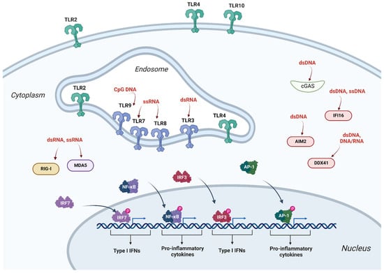
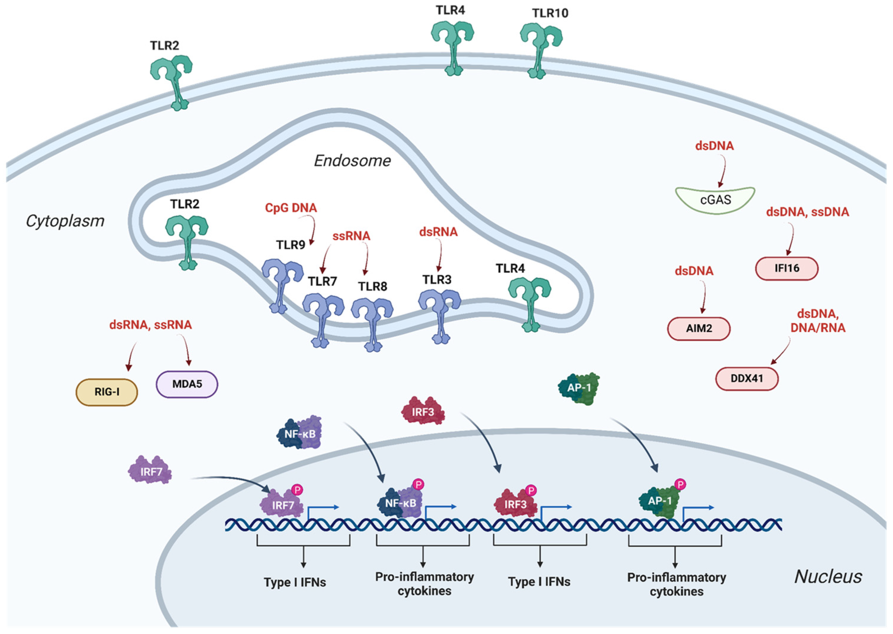
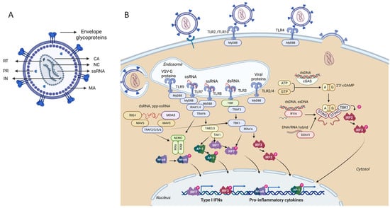
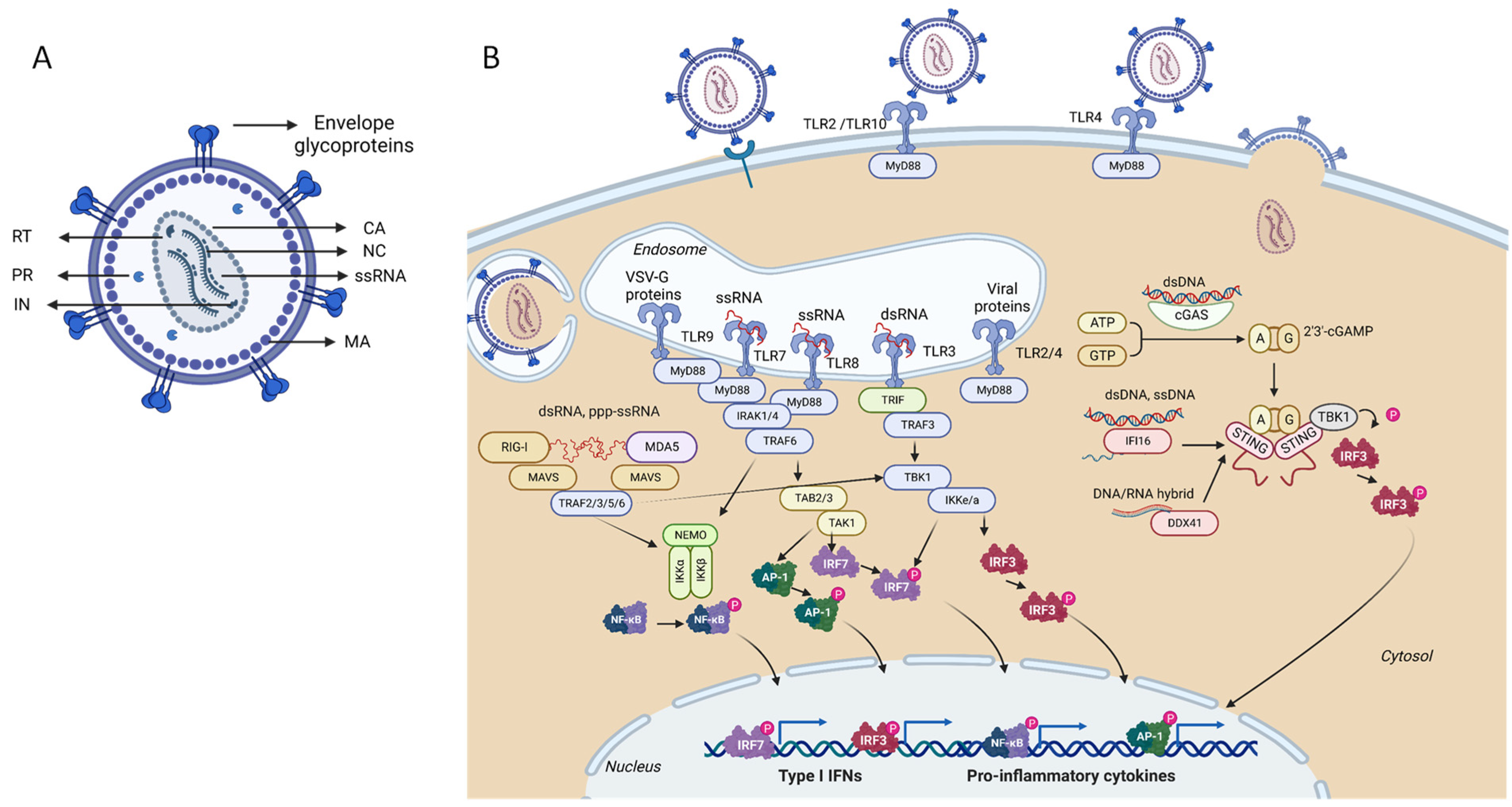
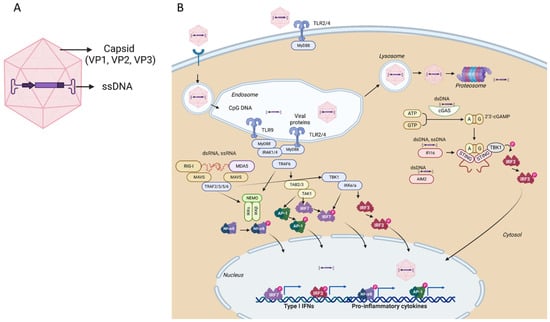
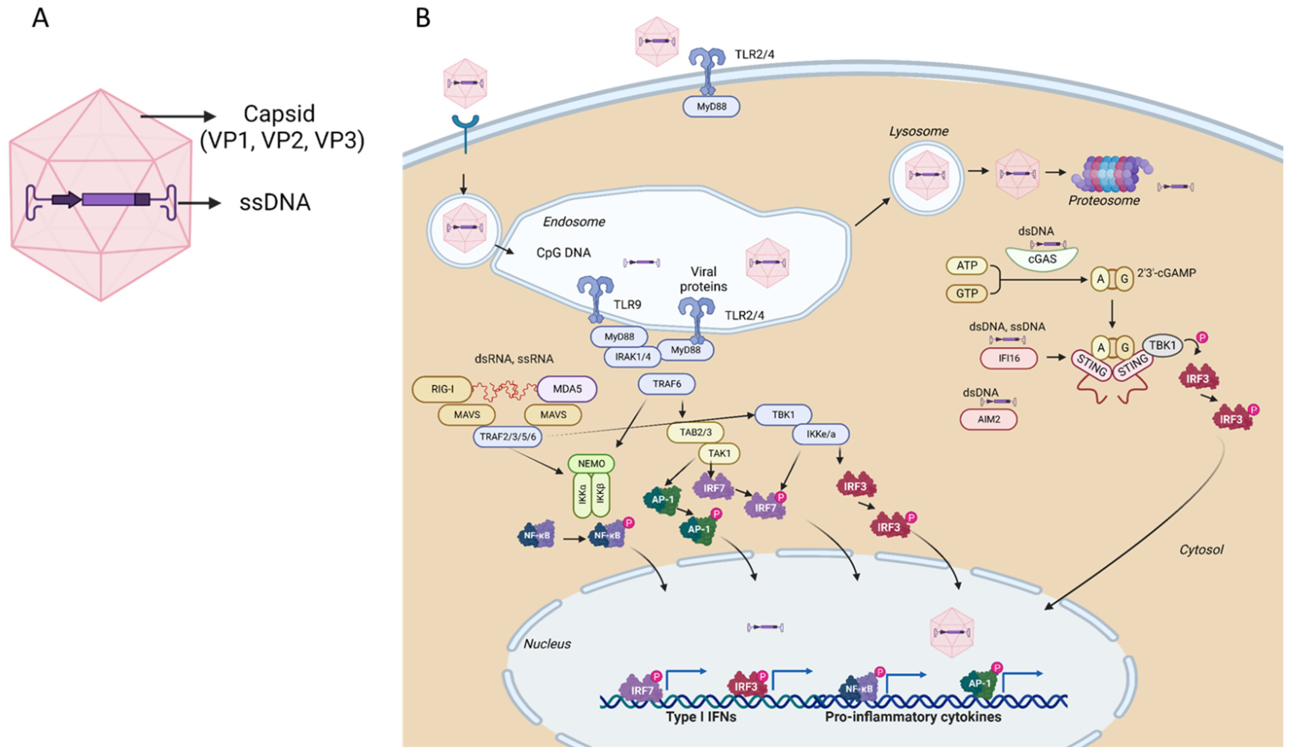
Disclaimer/Publisher’s Note: The statements, opinions and data contained in all publications are solely those of the individual author(s) and contributor(s) and not of MDPI and/or the editor(s). MDPI and/or the editor(s) disclaim responsibility for any injury to people or property resulting from any ideas, methods, instructions or products referred to in the content. |
© 2023 by the author. Licensee MDPI, Basel, Switzerland. This article is an open access article distributed under the terms and conditions of the Creative Commons Attribution (CC BY) license (https://creativecommons.org/licenses/by/4.0/).