


Abstract
Objective: To evaluate the effects of specimen thickness and simulated bruxism loading on the flexural strength of multilayer zirconia at three clinically relevant thicknesses. Materials and Methods: Sixty bar specimens (N = 60) were milled from multilayer zirconia disks (graded 4Y-PSZ dentin/5Y-PSZ enamel composition) (IPS e.max® ZirCAD MT Multi, Ivoclar Vivadent, Schaan, Liechtenstein). Specimens were tested using a modified ISO 6872-based three-point flexural configuration (16 × 4 mm bar geometry) at three thickness levels, 1.5 mm (S), 2.0 mm (M), and 2.5 mm (L), yielding six groups of ten (S1, S2, M1, M2, L1, L2), where the numerical suffix designates uncycled and cycled subgroups respectively. Bruxism simulation was performed for 250,000 cycles at 490.5 N with a 6 mm metallic ball antagonist. Three-point flexural strength testing was performed on a universal testing machine. Surface morphology of cycled specimens was assessed by SEM. Statistical analysis employed a Scheirer–Ray–Hare test and Dunn’s post hoc test with Holm–Bonferroni correction, with significance set at p < 0.05. Results: Specimen thickness exerted a statistically significant effect on flexural strength (p < 0.001). Mean values rose from 267.50 ± 44.207 MPa (S1) and 280.11 ± 69.714 MPa (S2) to 517.00 ± 105.343 MPa (M1) and 604.20 ± 122.469 MPa (M2) to 659.20 ± 145.126 MPa (L1) and 746.90 ± 176.429 MPa (L2). Post hoc analysis confirmed significant differences in all pairwise comparisons involving S against M or L groups, while no significant difference was detected between M and L subgroups at any pairing. Bruxism (loading) exerted no statistically significant main effect on flexural strength in a two-way rank-based analysis accounting for thickness (Scheirer–Ray–Hare, p = 0.277), with no significant thickness × loading interaction (p = 0.876) and no significant loading effect within any individual thickness group after correction for multiple comparisons. SEM imaging revealed a smoother surface topography within the antagonist contact zone relative to surrounding areas, accompanied by larger debris particles at the impact site. Conclusions: Although thickness significantly influenced flexural strength, no statistically significant difference was detected between the 2.0 mm and 2.5 mm groups under the present experimental conditions and sample size. Evaluated only as descriptive ISO 6872 material-classification reference values, the 1.5 mm group’s mean flexural strength falls below, and the 2.0 mm group’s mean meets, the strength class associated with three-unit fixed dental prostheses including molars; because the present modified bar-specimen model does not satisfy ISO 6872’s slenderness recommendation and was not designed to validate a clinical minimum restoration thickness, these comparisons should not be read as an indication-specific clinical thickness recommendation.
1. Introduction
Zirconia has undergone a remarkable transformation over recent decades, transitioning from a niche industrial ceramic into one of the most extensively investigated and clinically adopted materials in contemporary prosthodontics. Its appeal derives from an exceptional combination of mechanical strength, biological compatibility, and chemical stability, properties that have made it a compelling alternative to metal-based frameworks and conventional glass-ceramics alike [1]. The progressive shift toward metal-free restorative systems, driven both by patient esthetic expectations and by advances in computer-aided design and manufacturing (CAD/CAM) technologies, has further accelerated zirconia’s penetration across virtually all fixed prosthodontic indications [2]. From single crowns and fixed dental prostheses to implant superstructures and minimally invasive occlusal veneers, zirconia restorations are now encountered across the full spectrum of clinical complexity [3]. Despite this broad adoption, the material class is far from monolithic; substantial compositional and microstructural heterogeneity exists among commercially available formulations, and understanding the mechanical consequences of this diversity remains an active area of inquiry.
The mechanical basis of zirconia’s performance is rooted in the polymorphic nature of zirconium dioxide, which exists in monoclinic, tetragonal, and cubic crystalline phases, with phase composition strongly governed by temperature and the concentration of stabilizing dopants such as yttria [4]. Conventional 3Y-TZP (yttria-stabilized tetragonal zirconia polycrystal), containing approximately 3 mol% yttria and stabilized predominantly in the tetragonal phase, exhibits flexural strengths in the range of 900–1200 MPa, a property underpinned by stress-induced transformation toughening, wherein propagating cracks trigger a tetragonal-to-monoclinic phase conversion accompanied by a volumetric expansion that arrests further crack advancement [5]. However, the same opacity that characterizes 3Y-TZP, arising primarily from the birefringence of tightly packed tetragonal grains, restricts its suitability in esthetically demanding anterior applications. The clinical imperative to balance translucency against mechanical competence led to the successive development of 4Y-PSZ and 5Y-PSZ formulations (4 and 5 mol% yttria partially stabilized zirconia, respectively), in which elevated yttria content stabilizes a greater proportion of the optically isotropic cubic phase, dramatically improving light transmission at the cost of reduced transformation toughening capacity and, consequently, lower flexural strength [6]. A systematic review of published flexural strength data found overall mean values of 803 ± 233 MPa for 4Y-PSZ and 570 ± 116 MPa for 5Y-PSZ, a statistically significant difference that carries direct clinical implications for material selection by indication [4].
Strength-gradient multilayer zirconia systems were introduced to reconcile the competing demands of translucency and mechanical robustness within a single restoration block. These materials are architecturally engineered so that a highly translucent enamel-simulating layer of 5Y-TZP transitions through intermediate compositions toward a body layer of 3Y-TZP or 4Y-TZP that provides structural load-bearing capacity [7]. The compositional gradient across layers enables restorations with natural-appearing shade and optical depth at the incisal or occlusal surface while preserving adequate strength in the cervical and dentin zones, a design philosophy that has proved sufficiently attractive to drive widespread clinical uptake [6]. Yet the mechanical heterogeneity inherent to such graded architectures introduces interpretive complexity: layer-specific properties such as flexural strength, hardness, and elastic modulus differ substantially across the block, and the manner in which these gradients interact under clinical loading conditions is not fully elucidated [5]. Restoration thickness further modulates this relationship, as demonstrated by fatigue studies in which 0.5 mm multilayer zirconia occlusal veneers exhibited subsurface radial cracking not observed in specimens at or above 1.0 mm, underscoring that geometric factors must be considered alongside compositional ones when predicting mechanical behavior [7].
A dimension of clinical loading that has received comparatively limited attention in the context of multilayer zirconia is the mechanical challenge posed by parafunctional habits, most notably bruxism. The prevalence of bruxism in the adult population ranges widely, with estimates spanning 8–31.4% depending on diagnostic criteria and population studied, and the disorder is recognized as a significant contributor to temporomandibular joint (TMJ) dysfunction as well as a primary driver of pathological tooth wear and restoration fracture [8]. Bruxism generates bite forces that substantially exceed those associated with normal mastication, bite forces recorded during sleep-bruxism episodes have been reported to approach, and in some individuals exceed, the maximum voluntary bite force recorded from the same individual while awake [9], exposing restorations to repetitive high-magnitude loading cycles that bear little resemblance to the conditions evaluated in standard quasi-static mechanical tests [10]. Chewing simulators afford a valuable means of approximating the fatigue environment of the oral cavity under controlled laboratory conditions, allowing investigators to subject restorative materials to cyclical mechanical stress before conventional fracture testing; however, no simulator yet replicates the full complexity of human mastication, and methodological standardization across published studies remains incomplete [11]. Against this background, the authors recognized that clinical evidence guiding the use of zirconia restorations in patients with parafunctional habits is currently scarce, rendering the performance of these materials in such conditions uncertain. To begin addressing this gap at the laboratory level, the authors aimed to investigate the flexural behavior of multilayer zirconia under a simulated bruxism condition and to examine the influence of restoration thickness on this response. The null hypotheses tested were as follows: first, that no statistically significant difference in flexural strength exists among different thicknesses of multilayer zirconia, and second, that no statistically significant difference in flexural strength exists in multilayer zirconia before versus after bruxism simulation. Because multilayer disks are compositionally graded and layer-specific properties differ substantially across the block, a thickness-dependent effect was, in fact, the a priori expectation underlying this design; the first null hypothesis is a formal statistical null to be tested against this expectation, not a claim that the authors anticipated no effect of thickness.
2. Materials and Methods
2.1. Study Design
A total of sixty zirconia bar specimens (N = 60) were fabricated and allocated across six experimental groups according to specimen thickness and testing condition, specifically, whether flexural strength was assessed before or after bruxism simulation. Each group comprised ten specimens (n = 10). The study evaluated three thickness levels: 1.5 mm (group code S), 2.0 mm (group code M), and 2.5 mm (group code L), each subdivided into a uncycled (1) and a cycled (2) subgroup, yielding the groups S1, S2, M1, M2, L1, and L2.
2.2. Specimen Fabrication
All specimens were milled from multilayer zirconia disks (graded 4 mol% (dentine) and 5 mole% (enamel) yttria partially stabilized zirconia (4 and 5Y-PSZ) disks (IPS e.max ZirCAD MT Multi, Ivoclar Vivadent, Schaan, Liechtenstein; “4 mol%” and “5 mol%” denotes molar percent Y2O3 (yttria) content, the standard nomenclature for this material class (4 and 5Y-PSZ), distinct from weight-percent conventions sometimes used elsewhere) using a CAD/CAM milling unit (PrograMill, Ivoclar Vivadent, Schaan, Liechtenstein). Bar geometries were designed in Meshmixer software (version 3.5.474, Autodesk, San Francisco, CA, USA) in accordance with International Organization for Standardization (ISO) 6872, with a fixed length of 16 mm and width of 4 mm. The three thickness levels were produced (S, M, and L). Following milling, all specimens underwent complete sintering in a dedicated furnace (iSINT eco, imes-icore GmbH, Eiterfeld, Germany) according to the manufacturer’s recommended protocol. Sintering was completed prior to the substrate-cementation and bruxism-simulation steps (performed only for the cycled subgroups, S2/M2/L2) and prior to flexural testing: the process sequence for all specimens was mill → sinter → [cement and bruxism-simulate: S2/M2/L2 only] → flexural test.
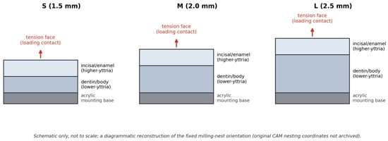
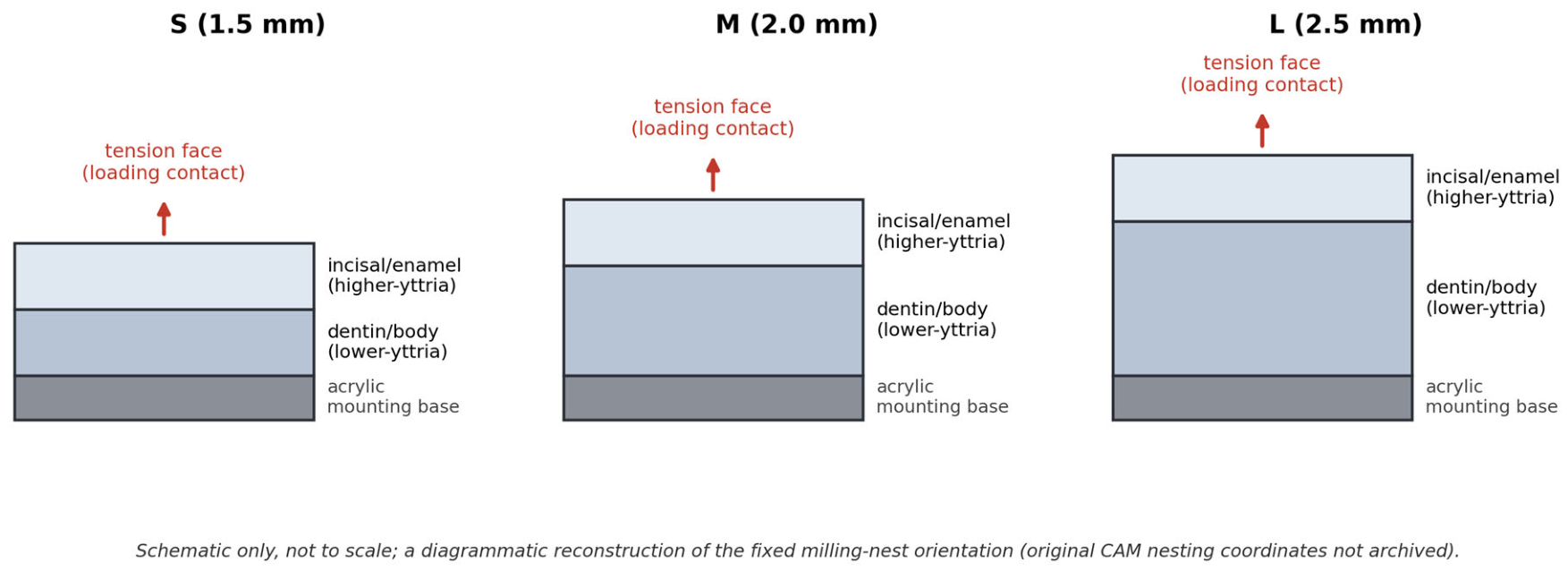
All bar specimens, across all three thickness groups, were milled with a fixed, consistent nesting orientation within the multilayer disk: the incisal/enamel (higher-yttria, more translucent) layer was positioned toward what became the tension face of the specimen during three-point bending, and the dentin/body (lower-yttria) layer was positioned toward the face seated against the acrylic mounting base (Figure 1). Because thicker specimens therefore incorporate a larger absolute and proportional volume of the stronger dentin-zone material beneath the tension face, the thickness effect is confounded with layer composition and should not be interpreted as a pure geometric/volumetric effect. The original CAM nesting files recording exact milling coordinates were not archived; Figure 1 is a diagrammatic reconstruction of this fixed orientation protocol rather than a reproduction of the original milling-software output. The specific disk shade and batch/lot number used are not reported, as this information was not recorded at the time of specimen fabrication and could not be retrieved retrospectively.
Figure 1.
Milling-nest layer orientation held fixed across the three thickness groups (schematic). Layer proportions shown are illustrative and not to scale.
2.3. Substrate Preparation and Cementation for Bruxism Simulation
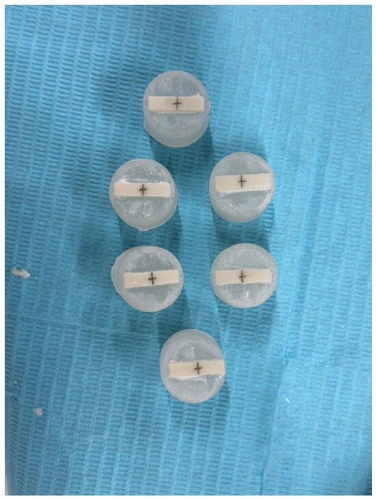
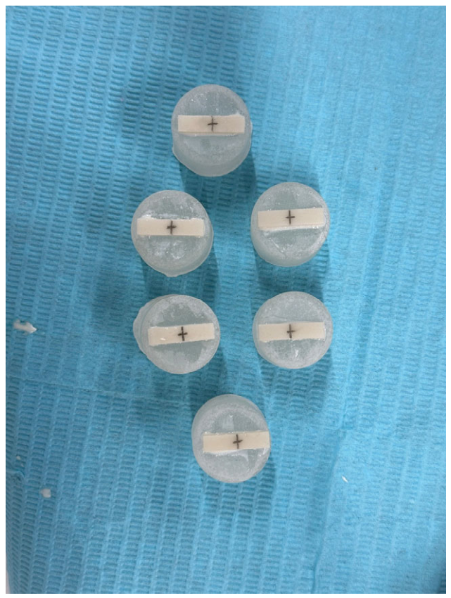
Cylindrical acrylic bases intended to support specimens during bruxism simulation were fabricated from self-curing acrylic resin (Techno Sin Ortho Acrylic, Protechno, Girona, Spain), mixed in accordance with the manufacturer’s instructions and cast into prefabricated rubber cylindrical molds dimensioned to fit the chewing simulator; the cylindrical molds were the standard accessory supplied with the chewing-simulation unit and sized to its specimen-holder sockets; their internal dimensions were not independently measured by the study team, as they were used as supplied and were not a manipulated study variable. The resin was allowed to cure fully before demolding. Prior to cementation, the bonding surfaces of both the acrylic bases and the zirconia specimens assigned to the cycled subgroups (S2, M2, and L2) were air-abraded with 110 µm aluminum oxide particles (Sirio Dental, Meldola, Italy) for 10 s at a standoff distance of 10 mm using a circular motion, then cleaned with an air jet for 20 s in an analogous circular motion. Dual-cure adhesive resin cement (G-CEM ONE, GC Corporation, Tokyo, Japan) was applied to the fitting surface of each specimen. Each specimen was manually positioned at the center of its respective acrylic base and held in place with an adjustable laboratory pressing tool to ensure even cement distribution. Excess cement was removed with a microbrush, and light curing was performed at a distance of 2 mm from each lateral aspect for 20 s per side (Elipar DeepCure-L LED, 3M ESPE, Seefeld, Germany). Cemented assemblies (Figure 2) were then seated within customized metallic sockets, each secured by four anchorage screws applied from different directions to maintain positional stability throughout simulation.
Figure 2.
Cylindrical acrylic base and specimen assembly for simulating bruxism. Center of the specimen is marked.
2.4. Bruxism Simulation
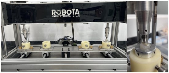
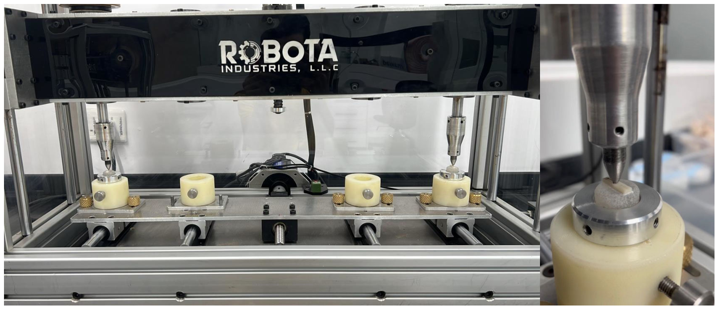
Bruxism simulation was carried out using a universal chewing simulation machine (ROBOTA, Alexandria, Egypt). The geometric center of each specimen in groups S2, M2, and L2 was marked with a pencil prior to loading to confirm consistent antagonist contact at the intended point. The machine accommodated two specimens simultaneously. A metallic ball antagonist with a diameter of 6 mm delivered the cyclic loading. The chewing simulator assembly is illustrated in Figure 3. Operating parameters were configured as follows: vertical movement 2 mm, lateral movement 0.7 mm, vertical speed 55 mm/s, lateral speed 30 mm/s, frequency 1.8 Hz, and a loading weight of 50 kg (490.5 N), for a total of 250,000 cycles. This loading weight is at the lower end of the range of forces reported for bruxism, which in some individuals has been documented to approach or exceed 1000 N. Each complete set required approximately 48 h of machine time; the chewing simulator runs continuously across all specimens in a batch and does not pause or log the cycle count at the moment an individual specimen fractures, a limitation of the equipment relevant to interpreting any cycling failures.
Figure 3.
Chewing simulator assembly.
Upon completion of simulation, specimens were detached from their acrylic bases by brief localized heating from a torch applied laterally for 5 s to soften the cement interface; separation was then achieved with a wax knife without inducing surface damage. Residual cement was removed using a green-wheel silicone polisher, after which specimens were considered ready for flexural strength testing.
2.5. Scanning Electron Microscopy
Following bruxism simulation, one randomly selected specimen underwent surface examination by scanning electron microscopy (SEM; AURA100, Seron Technologies Inc., Uiwang, Republic of Korea). A gold coating was applied prior to imaging (SC7620, Quorum Technologies Ltd., Laughton, East Sussex, UK). Two anatomically distinct regions were imaged: the area corresponding to the antagonist impact site and an area remote from the impact zone. Each region was examined at magnifications of 60× and 1000×, yielding four images in total.
2.6. Flexural Strength Testing
Three-point flexural strength testing was conducted for all six groups (S1, S2, M1, M2, L1, and L2) using a universal testing machine (MultiTest 2.5-i, Mecmesin, Slinfold, West Sussex, UK). The midpoint of each specimen was marked with a pencil to facilitate accurate positioning beneath the loading arm. The machine was configured with the following parameters: 1.5 kN load cell; upper softened switch position at 1000 mm; lower softened switch position at 200 mm; upper force limit 1700 N; lower force limit 100 N; pre-load 10 N; loading arm speed 1 mm/min; and a span distance between support arms of 12 mm.
Flexural strength (σ, MPa) was calculated from σ = 3Pl/2wb2 (ISO 6872 Formula 1 [12]), where P is the fracture load (N), l is the support span (mm), and w and b are the specimen width and thickness (mm), respectively, each measured with a digital caliper immediately prior to testing; specimen edges were chamfered per ISO 6872 edge-finishing guidance after sintering. ISO 6872:2015+A1:2018 permits three-point bending support-roller spacing from 12.0 mm to 40.0 mm; the 12 mm span used here, selected as the minimum of this range so that a single fixture could accommodate all three thickness groups, is therefore within the standard’s permitted range. However, the standard additionally specifies that the ratio of specimen thickness to span (b/L) should not exceed 0.10; at the 12 mm span used, this ratio is 0.125 for the S (1.5 mm) group, 0.167 for the M (2.0 mm) group, and 0.208 for the L (2.5 mm) group, exceeding ISO’s slenderness guidance for all three groups and most markedly for the M and L groups. The present test is therefore more precisely described as a modified three-point flexural test, conducted within ISO 6872’s permitted span range but not satisfying its b/L slenderness recommendation for all three thickness groups.
2.7. Statistical Analysis
No a priori sample-size or power calculation was performed; n = 10 per group (n = 9 for the cycled 1.5 mm subgroup following one cycling failure) was a pragmatic choice consistent with common precedent in comparable in vitro dental-materials flexural-strength studies, rather than a formally justified sample size. The normality of the data distribution and the homogeneity of group variances were assessed using the Shapiro–Wilk test and Levene’s test, respectively. Given the outcomes of these distributional assessments, the data were analyzed with a Scheirer–Ray–Hare test, a rank-based two-way Analysis of Variance (ANOVA) analog, to formally test the thickness main effect, the loading (uncycled/cycled) main effect, and the thickness × loading interaction within a single 3 × 2 factorial model. Dunn’s post hoc test (Holm-Bonferroni corrected) was used to resolve significant effect/s. Pooling the loading effect across all thicknesses would be inappropriate given that thickness dominates the variance; loading was therefore tested within each thickness group using Mann–Whitney U tests with Holm-Bonferroni correction for the three comparisons: S1 vs. S2, M1 vs. M2, and L1 vs. L2. Two-parameter Weibull moduli and characteristic strengths were computed per group by maximum likelihood as an exploratory analysis only: ISO 6872 Annex B recommends a minimum of 15 specimens before Weibull statistics are considered reportable, a threshold not met by the present n = 9–10 groups, and small-sample Weibull estimates for dental zirconia have been shown to be unstable [13]; Weibull parameters are therefore reported as point estimates without confidence intervals, which would themselves be unreliable at this sample size. Statistical analyses were computed using IBM SPSS Statistics, version 22.0 (SPSS Inc., Chicago, IL, USA), evaluated at p < 0.05.
3. Results
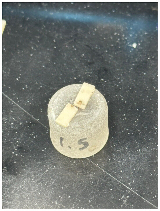
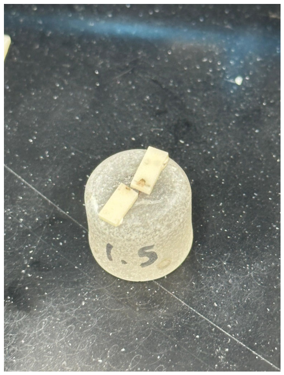
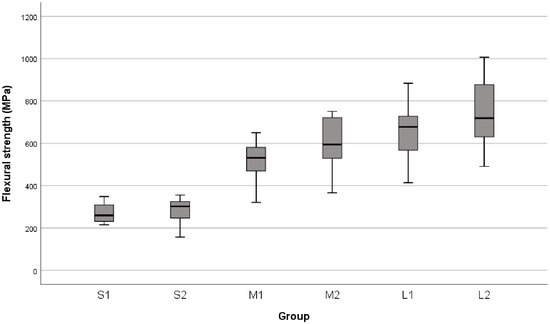
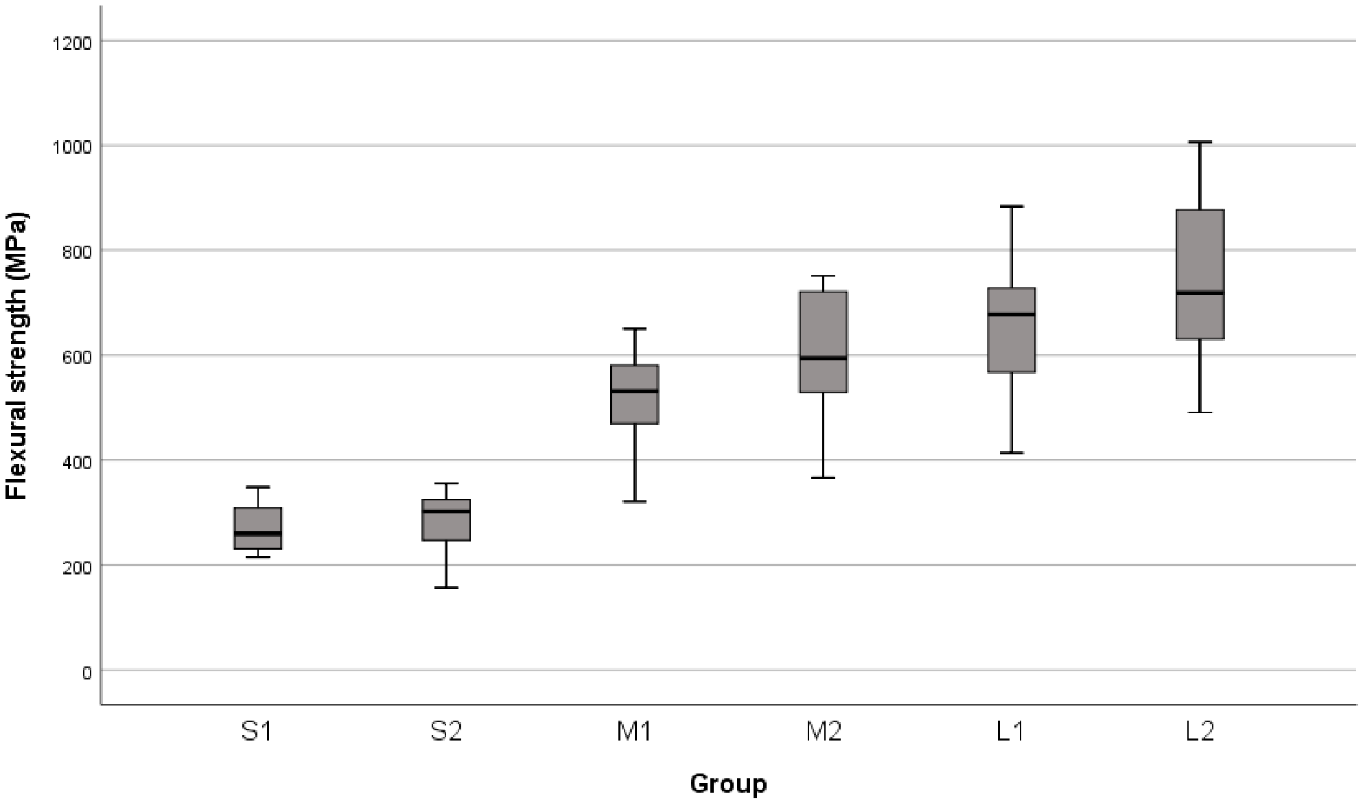
Flexural strength values recorded across all specimen groups are presented in Table 1; because non-parametric inferential tests were used throughout, medians and interquartile ranges (Q1–Q3) are reported alongside the means and standard deviations. The mean flexural strength of the 1.5 mm specimens prior to bruxism simulation (S1) was 267.50 ± 44.207 MPa, rising to 280.11 ± 69.714 MPa following simulation (S2), with the S2 subgroup comprising nine rather than ten specimens owing to the fracture of one specimen (Figure 4) during the bruxism simulation protocol. For the 2.0 mm group, mean flexural strength increased from 517.00 ± 105.343 MPa before simulation (M1) to 604.20 ± 122.469 MPa after simulation (M2). The 2.5 mm group demonstrated the highest recorded values, with L1 yielding a mean of 659.20 ± 145.126 MPa and L2 a mean of 746.90 ± 176.429 MPa. Across all thickness groups, the lowest overall mean was observed in S1 and the highest in L2. A consistent pattern was also noted whereby standard deviations were markedly lower in uncycled subgroups than in their corresponding cycled counterparts, and variability was substantially greater in the M and L groups relative to S. These distributional features are illustrated visually in the box-and-whisker plot (Figure 5), which further conveys the general elevation in flexural strength following bruxism simulation alongside the concurrent reduction in recording consistency.
Table 1.
Descriptive statistics of flexural strength (MPa) by group.
Figure 4.
A bruxism simulation failed specimen of the S2 group.
Figure 5.
Box-and-whisker plot of the flexural strength of the zirconia specimens of different thicknesses (S, M, and L) before (1) and after (2) bruxism simulation. S = 1.5 mm thickness; M = 2.0 mm thickness; L = 2.5 mm thickness.
Prior to inferential testing, the distributional properties of the data were evaluated (Table 2). Shapiro–Wilk testing returned p-values exceeding 0.05 for all groups; thus, the overall dataset was treated as satisfying normality assumptions. Levene’s test for homogeneity of variance yielded a significance value below 0.05, indicating that the assumption of equal variances across groups could not be retained.
Table 2.
Tests of normality and homogeneity of variance.
Significance testing results are summarized in Table 3. The Scheirer–Ray–Hare thickness main effect on flexural strength was statistically significant (p < 0.001), indicating that thickness was a meaningful source of variation. Neither the loading main effect (p = 0.277) nor the thickness × loading interaction (p = 0.876) reached significance.
Table 3.
Significance tests for the effects of thickness and bruxism simulation on flexural strength.
Post hoc pairwise comparisons are presented in Table 4. Dunn’s post hoc test, applied to the pooled thickness groups following the significant Scheirer–Ray–Hare thickness main effect, showed that pooled S differed significantly from both pooled M and pooled L (p < 0.001 for each), while pooled M and L did not differ significantly from one another. The separate, Holm-Bonferroni-corrected Mann–Whitney comparisons of the uncycled-versus-cycled (within-thickness) subgroups were not significant for any thickness group.
Table 4.
Post hoc pairwise comparisons of flexural strength: Dunn’s test (Holm-Bonferroni corrected) for the thickness factor.
The fracture of a specimen in S2 group during cycling is reported here as a failure outcome in its own right, not merely a missing data point, since it directly bears on the interpretation of the S2 group’s mean. To assess survivor bias, a conservative sensitivity analysis was performed treating the fractured specimen as a zero-strength failure (S2, n = 10, one value imputed as 0 MPa): under this conservative scenario the S2 mean falls to 252.1 MPa (SD 110.3), below the S1 mean of 267.5 MPa, i.e., the directional increase for the S group does not survive this conservative treatment, although the S1-versus-S2 comparison remains statistically non-significant either way (Mann–Whitney p = 0.734 under zero-imputation vs. p = 0.811 as analyzed on survivors only). Because the chewing simulator runs continuously without pausing or logging at the moment of an individual specimen’s fracture, the exact cycle count at which this specimen failed could not be captured. Visual inspection of all specimens at removal from the simulator showed no other cycling failures or visible damage prior to flexural testing.
As an exploratory supplement to the inferential tests above, two-parameter Weibull moduli (m) and characteristic strengths (σ0) were estimated per group by maximum likelihood (Table 5). Because the achieved sample sizes (n = 9–10) fall below the minimum of 15 specimens recommended by ISO 6872 Annex B before Weibull statistics are considered reportable, these values are presented as point estimates only, without confidence intervals, and should be interpreted with corresponding caution rather than as a validated reliability characterization.
Table 5.
Exploratory two-parameter Weibull moduli and characteristic strengths by group (point estimates; not reportable per ISO 6872 Annex B at the present sample sizes).
Weibull moduli followed a pattern broadly consistent with the mean/median flexural-strength results (Table 1): moduli were similar across the M and L groups (m ≈ 5.0–6.4) and slightly higher in the S1 group (m = 6.65), with the lowest modulus observed in L2 (m = 4.95), consistent with the greater variability noted in the L group in Table 1.
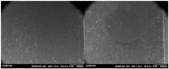
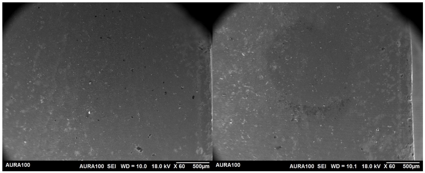
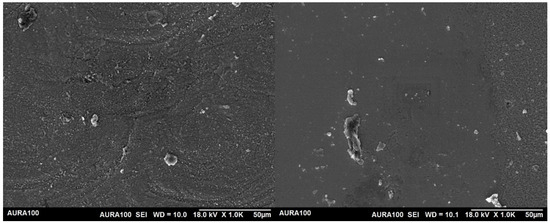
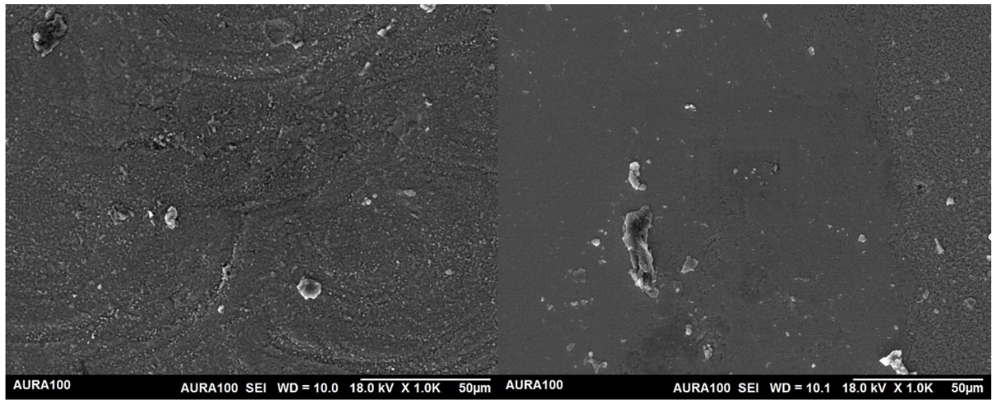
SEM examination of cycled specimens revealed distinct surface features at the antagonist contact site. At 60× magnification, a clearly delineated rim was visible surrounding the contact site on cycled specimens, demarcating the impacted zone from the surrounding unaffected surface (Figure 6). Higher-magnification imaging at 1000× revealed that the surface topography within the impact zone was comparatively smoother than the surrounding area; however, this region was also characterized by a greater abundance of debris particles of larger dimensions than those observed at sites remote from the point of contact (Figure 7).
Figure 6.
SEM imaging at 60× magnification of a zirconia specimen (M2 group) surface away from the impact (1) and at the site of impact (2). Note the rim around the impact site in image 2.
Figure 7.
SEM imaging at 1000× magnification of a zirconia specimen (M2 group) surface away from the impact (1) and at the site of impact (2). Note the smoother surface characteristics within the area of impact, associated with larger and more numerous impact debris compared with the area away from the impact.
4. Discussion
Only the first null hypothesis tested in the present study is rejected. Statistically significant differences in flexural strength were detected among the three thickness groups (p < 0.001). Although the factorial analysis found no statistically significant main effect of bruxism loading on flexural strength and no significant loading effect within any individual thickness group, the consistent directional increase in mean flexural strength following simulation across all three groups constitutes a meaningful and interpretively important finding, even as the inferential result must be faithfully reported.
This directional pattern should, however, be interpreted with two caveats. First, the factorial (Scheirer–Ray–Hare) analysis found no significant thickness × loading interaction, meaning there is no statistical evidence that the direction or magnitude of the loading effect differed across thickness groups; the apparent “consistency” of the directional increase across groups should not be over-read as a robust, replicated pattern. Second, a conservative sensitivity analysis treating the S2 group’s cycling failure as a zero-strength outcome reverses the direction of the S-group comparison, so the directional increase is not in fact observed across all three thickness groups once this failure is accounted for as a failure rather than a missing value.
The thickness-dependent effect on flexural strength was among the most pronounced findings. This progressive, non-linear rise in strength with thickness, pronounced between the S and M groups but plateauing between M and L, is consistent with well-established mechanical principles: increasing material volume raises the structural capacity of a ceramic element to resist crack propagation under load. Abdulmajeed et al. demonstrated in a comparable disk-specimen design that thickness exerted a statistically significant effect on biaxial fracture load across three yttria concentrations, with 1.2 mm specimens outperforming 0.7 mm specimens in every composition group [14]. The present study extends this principle to the clinically relevant range of 1.5–2.5 mm for a multilayer formulation, and the absence of a statistically significant difference between the 2.0 mm and 2.5 mm groups is a clinically consequential observation: no statistically significant difference was detected between the 2.0 mm and 2.5 mm groups under the present experimental conditions and sample size; given the achieved sample size and the observed variance, this should not be interpreted as evidence of equivalence or non-inferiority, since no equivalence margin or non-inferiority analysis with adequate power was defined or performed. This absence of a significant difference should be interpreted cautiously, in line with the equivalence/non-inferiority caveat above, rather than as evidence of a diminishing-returns relationship.
This thickness effect should not, however, be attributed to specimen volume alone. All specimens were milled with a fixed nesting orientation: the incisal/enamel (higher-yttria) layer toward the tension face, the dentin/body (lower-yttria) layer toward the mounting side. Because thicker specimens therefore incorporate a proportionally larger volume of the stronger dentin-zone material beneath the tension face, the present thickness effect is confounded with layer composition. Lohbauer, Schwarz and Belli [15] report dentin-zone flexural strength roughly double that of the incisal zone in several comparable 4/5Y-PSZ multilayer products, and Machry et al. [16] report a similar cervical > transition > incisal strength gradient with quantified static and fatigue strength per layer; both provide plausible quantitative support for a layer-composition contribution to the effect reported here, which cannot be separated from a pure geometric/volumetric effect using the present full-thickness, multilayer specimen design.
The elevation in mean flexural strength observed after bruxism simulation, although statistically non-significant, is an unexpected directional result that warrants mechanistic consideration. The authors propose, as one plausible hypothesis rather than a demonstrated mechanism, that this pattern may reflect the transformation toughening mechanism intrinsic to yttria-stabilized tetragonal zirconia, whereby the repetitive compressive loading cycles imposed during simulation may have induced a controlled tetragonal-to-monoclinic phase conversion at and near specimen surfaces, generating a zone of residual compressive stress that partially counteracted subsequent tensile loading during the flexural test. IPS e.max ZirCAD MT Multi, as a 4Y-PSZ/5Y-PSZ multilayer system, retains a meaningful proportion of transformable tetragonal grains in its body layer, making this mechanism plausible; however, no X-ray diffraction (XRD) or Raman phase analysis was performed on cycled or uncycled specimens in this study, so the proposed mechanism was not directly demonstrated and should be read as a hypothesis for future testing, not an established explanation. The adhesion to zirconia literature corroborates that the stress-induced tetragonal-to-monoclinic transformation is accompanied by a volumetric expansion of approximately 5%, which imposes compressive closure forces on propagating microcracks [17]. Directly comparable data from studies examining flexural strength before and after a bruxism-magnitude simulation in the same multilayer material do not appear to exist in the reviewed literature; however, several studies using different loading regimes and material compositions provide contextually relevant evidence. Abdulmajeed et al. found that mastication simulation at 110 N over 1.2 million cycles had no statistically significant effect on the biaxial fracture load of surviving specimens across all three yttria groups [14], a result consistent with the present study’s non-significant within-thickness comparisons. Similarly, Badr et al. reported that thermo-mechanical loading at equivalent cycle counts did not significantly alter fracture resistance within any zirconia group, irrespective of yttria concentration [18]. These findings collectively suggest that cyclic mechanical loading within physiologically or para-physiologically relevant ranges does not degrade the fracture resistance of adequately thick zirconia specimens, and may, in tetragonal-rich compositions, exert a modest surface-strengthening effect through transformation toughening activation.
A further, more fundamental caveat applies to any mechanistic interpretation of the cycled-vs-uncycled comparison. Only the cycled (S2/M2/L2) specimens underwent air-abrasion, resin cementation, cyclic loading, heat-debonding, and post-debond polishing; the uncycled (S1/M1/L1) specimens underwent none of these steps. The comparison therefore reflects the combined effect of the full simulation workflow, abrasion, cementation, cyclic loading, heat-debonding, and polishing, rather than the isolated effect of cyclic loading, and terms implying that cyclic loading alone strengthened the material should be read with this caveat. A process-matched control group (identical abrasion, cementation, heating, and polishing, but no cyclic loading) was not included in the present design and is recommended for future work to isolate the contribution of cyclic loading itself.
The 500 N load applied during simulation in the present study was selected to approximate the parafunctional forces documented in bruxism patients, which can substantially exceed forces generated during normal mastication [19]. This choice is clinically defensible, and the finite element literature on bruxism further reinforces that such force magnitudes impose biomechanically significant demands on restorative materials and their supporting structures [20]. Nonetheless, the 490.5 N (50 kg) loading weight used in the present study is itself at the lower end of this reported range; higher, more severe bruxism loads, approaching or exceeding 1000 N, could plausibly impose greater cyclic stress on the zirconia substrate and its cement interface than was captured here, potentially accelerating subcritical crack growth or producing a more pronounced strength change than observed at 490.5 N; evaluating multilayer zirconia under a range of loading magnitudes extending toward this upper bound is recommended for future work. Against this loading context, the clinical interpretation of results relative to ISO 6872 thresholds deserves careful consideration. The S group’s mean flexural strength values fall well below the 300 MPa threshold required for luted crowns and three-unit bridges not involving a molar, and substantially below the 500 MPa threshold for bridges spanning molars. Under the present bar-specimen test conditions, which do not satisfy ISO 6872’s b/L slenderness recommendation and reflect the combined simulation workflow rather than cyclic loading alone, this indicates that the S group’s measured flexural strength falls below the relevant ISO 6872 material-classification strength class, which should not, on its own, be read as a validated clinical minimum-thickness recommendation for a specific prosthetic design. The M group’s cycled mean meets the ISO 6872 criterion for three-unit fixed dental prostheses including molars, indicating that the M group’s measured flexural strength meets that same ISO 6872 material-classification strength class under the present test conditions, without extending this to a validated clinical minimum-thickness recommendation, which the present bar-specimen data do not establish. Neither the M nor L groups approach the 800 MPa threshold required for prostheses of four or more units. The absence of a statistically significant strength advantage for the L group relative to M should not, on its own, be read as evidence that a 2.5 mm thickness offers no clinical benefit: the present study did not include an equivalence or non-inferiority design, a predefined clinical margin, or an analysis of the biological cost of the additional tooth reduction such a thickness would require, and thickness selection in practice should continue to be guided by the specific prosthetic indication and established ISO 6872 material-classification criteria rather than by these exploratory bar-specimen comparisons alone. It must be noted that zirconia relies predominantly on luting rather than bonding, and adhesive bonding of zirconia remains an area of ongoing investigation with a guarded prognosis, making the ISO 6872 luted crown criteria the operationally relevant benchmark [17].
The smoother contact-zone surface noted on SEM, despite the co-occurring accumulation of larger debris particles, is consistent with the understanding that highly polished or wear-smoothed zirconia surfaces resist further degradation more favorably than rougher ones, a phenomenon noted in wear studies involving zirconia under cyclic loading conditions [19]. If the smoother contact-zone topography observed here does reflect progressive smoothing of surface asperities under repeated contact loading, a hypothesis that cannot be confirmed from a single cycled specimen without an uncycled SEM control, such smoothing could, in principle, contribute to the slight cycled strength elevation observed, as surface flaws act as crack initiation sites in brittle ceramics; their reduction would theoretically increase the load required to initiate fracture; this mechanistic link remains speculative rather than demonstrated by the present data. Pöppel et al. observed that surface roughness across different zirconia multilayer generations was comparable following polishing and artificial aging, suggesting that surface treatment and cyclic loading converge toward similar surface states regardless of initial composition [21]. Ziębowicz et al. similarly found no crack formation on 5Y-TZP crown surfaces following chewing simulation, reinforcing the view that zirconia surfaces tolerate repeated contact without progressive structural deterioration under moderate loading [22].
The clinical evidence base for monolithic zirconia in bruxism patients remains limited largely to case reports, such as the two-year follow-up by Ouni et al. documenting successful full-arch rehabilitation with CAD/CAM monolithic zirconia in a patient with severe bruxism [23]. While such reports confirm the qualitative plausibility of this material class for parafunctional patients, they do not provide the stratified, thickness-specific mechanical data that the present study contributes.
Several limitations must be acknowledged. The study was conducted entirely in vitro, and extrapolation of bar-specimen three-point flexural strength data to the complex geometry, cementation interface, and biological environment of actual restorations requires caution. The specimen geometry, while conforming to ISO 6872, differs fundamentally from a crown or fixed dental prosthesis, and the chewing simulator, although operating at a clinically relevant force magnitude, cannot reproduce the full vector complexity of bruxist parafunction, salivary chemistry, or the thermal cycling characteristic of the oral environment [11]. No a priori sample-size or power calculation was performed. The relatively small sample size per subgroup (n = 10, reduced to n = 9 in S2 following one specimen fracture during simulation) limits statistical power and contributes to the broad standard deviations observed, particularly in the M and L groups. Restricting the investigation to a single multilayer zirconia product also constrains generalizability across the wider commercial landscape. The cycled-versus-uncycled comparison reflects the combined effect of abrasion, cementation, cyclic loading, heat-debonding, and polishing, not cyclic loading in isolation, since no process-matched, unloaded control group was included. The specimen thickness-to-span (b/L) ratio at the 12 mm test span used exceeds ISO 6872’s slenderness recommendation for all three thickness groups. All specimens were milled with a fixed layer orientation, confounding the thickness effect with layer composition. Surface morphology was examined by SEM on a single representative specimen per condition, without quantitative roughness measurement or EDS elemental analysis of surface debris, so these findings remain descriptive and illustrative rather than generalizable or quantitative. No XRD or Raman phase analysis was performed, so the proposed transformation-toughening mechanism remains an untested hypothesis. The simulation protocol did not include thermocycling, artificial saliva, or a human-enamel/steatite antagonist, and disk shade and batch/lot number were not recorded at the time of specimen fabrication.
Future investigations should incorporate multiple multilayer zirconia formulations with differing yttria gradients to determine whether the transformation-toughening response observed here is consistent across product generations. Cross-technology comparisons of multilayer zirconia products have shown that flexural strength varies meaningfully by manufacturing technology and layer architecture [24], reinforcing the need for such multi-product validation. Studies employing crown-geometry specimens cemented onto tooth-analog substrates, combined with thermocycling protocols and longer simulation durations, would more faithfully replicate clinical conditions. Prospective clinical trials in patients with confirmed bruxism diagnoses, using standardized prosthesis monitoring over multi-year observation periods, are ultimately necessary to translate these laboratory findings into evidence-based clinical guidelines. Additionally, finite element modeling of the present thickness groups under oblique and lateral loading would help characterize stress distribution patterns that static flexural testing alone cannot reveal.
5. Conclusions
This study’s central contribution is less about whether multilayer 4Y-PSZ/5Y-PSZ zirconia can withstand bruxism-level loading than about how much of it a clinician actually needs to prescribe. Cyclic loading itself proved to be the less demanding variable: one specimen of ten failed outright during cycling in the 1.5 mm group, so cyclic loading cannot be characterized as uniformly well tolerated; among specimens that survived cycling to be flexural-tested, strength did not decrease, and transformation toughening and surface smoothing remain plausible. The literature supported hypotheses for this pattern rather than demonstrating mechanisms, weighed against the survivor-bias and combined-workflow caveats. Thickness, not loading history, was the variable that governed clinical performance, and it did so as a threshold rather than a gradient: strength climbed sharply up to 2.0 mm and then leveled off. For clinicians, this reframes restoration design for bruxing patients from a search for maximal bulk to a search for the minimum thickness that clears the relevant ISO 6872 threshold for the intended prosthesis, with the 2.0 mm group meeting the relevant ISO 6872 material-classification strength class for molar-inclusive three-unit restorations under the present bar-specimen test conditions, though this should not, on its own, be read as a validated clinical minimum-thickness recommendation without further crown-geometry and clinical validation. For researchers, the findings argue for directing future mechanical testing away from further characterizing loading tolerance in isolation and toward crown-geometry, thermocycled, multi-product studies capable of establishing whether this same thickness threshold, and the mechanism underlying it, holds outside the simplified bar-specimen model used here.
Author Contributions
Conceptualization, O.A.; methodology, O.A., K.A.M., R.Z.A., W.A.B., A.J.A., and H.A.A.; software, O.A., K.A.M., R.Z.A., W.A.B., A.J.A., and H.A.A.; validation, O.A., K.A.M., R.Z.A., W.A.B., A.J.A., and H.A.A.; formal analysis, O.A., K.A.M., R.Z.A., W.A.B., A.J.A., and H.A.A.; investigation, O.A., K.A.M., R.Z.A., W.A.B., A.J.A., and H.A.A.; resources, O.A., K.A.M., R.Z.A., W.A.B., A.J.A., and H.A.A.; data curation, O.A., K.A.M., R.Z.A., W.A.B., A.J.A., and H.A.A.; writing—original draft preparation, O.A., K.A.M., R.Z.A., W.A.B., A.J.A., and H.A.A.; writing—review and editing, O.A., K.A.M., R.Z.A., W.A.B., A.J.A., and H.A.A.; visualization, O.A., K.A.M., R.Z.A., W.A.B., A.J.A., and H.A.A.; supervision, O.A.; project administration, O.A., and K.A.M.; funding acquisition, O.A. All authors have read and agreed to the published version of the manuscript.
Funding
The project was funded by the Deanship of Scientific Research (DSR) at King Abdulaziz University, Jeddah, Saudi Arabia under grant no. (IPP: 1607-165-2025). The authors, therefore, acknowledge with thanks DSR for technical and financial support.
Institutional Review Board Statement
Not applicable, as this in vitro study did not involve human participants, human data, or animal subjects.
Informed Consent Statement
Not applicable, as this study did not involve humans.
Data Availability Statement
The data presented in this study are available on request from the corresponding author.
Acknowledgments
The authors acknowledge with thanks the Deanship of Scientific Research (DSR) at King Abdulaziz University for technical support and financial support.
Conflicts of Interest
The authors declare no conflicts of interest.
Abbreviations
The following abbreviations are used in this manuscript:
| 4Y-PSZ | 4 mol% yttria partially stabilized zirconia |
| 5Y-PSZ | 5 mol% yttria partially stabilized zirconia |
| 3Y-TZP | 3 mol% yttria-stabilized tetragonal zirconia polycrystal |
| CAD/CAM | Computer-Aided Design/Computer-Aided Manufacturing |
| ISO | International Organization for Standardization |
| SEM | Scanning Electron Microscopy |
| TMJ | Temporomandibular Joint |
| DSR | Deanship of Scientific Research |
References
- Zhang, Y.; Kelly, J.R. Dental Ceramics for Restoration and Metal Veneering. Dent. Clin. N. Am. 2017, 61, 797–819. [Google Scholar] [CrossRef] [Scilit] [PubMed]
- Boanca, C.; Cocos, D.I.; Focsaneanu, S.C.; Earar, K. Mechanical Performance and Fracture Behavior of Fixed Dental Prostheses Under Bending Loads: An In Vitro Comparative Study. Dentistry 2026, 14, 255. [Google Scholar] [CrossRef] [Scilit] [PubMed]
- Sharma, A.R.; Pruthi, T.; Agrawal, A.; Madathil, E.; Tayal, R.; Shukla, K. The evolution, clinical applications, and performance of zirconia in dentistry: A systematic review. IP Indian J. Conserv. Endod. 2026, 11, 34–42. [Google Scholar] [CrossRef] [Scilit]
- Bernauer, S.A.; Lirgg, N.M.; Ioannidis, A.; Zitzmann, N.U.; Rohr, N. Flexural strength of translucent zirconia for single crowns and fixed dental prostheses—A systematic review. J. Prosthodont. Res. 2026, 70, 173–182. [Google Scholar] [CrossRef] [Scilit] [PubMed]
- Ham, J.M.; Lee, J.H.; Ha, S.R.; Seo, D.G.; Ahn, J.S.; Choi, Y.S. Changes in the flexural strength and surface characteristics of different zones of multilayered translucent zirconia during aging process. J. Mater. Sci. Mater. Med. 2025, 36, 78. [Google Scholar] [CrossRef] [Scilit] [PubMed]
- Yılmaz Biçer, E.; Kaynak Öztürk, E.; Bankoglu Güngör, M. Strength-gradient multilayer zirconia ceramics: A narrative review of the mechanical and optical properties. J. Am. Ceram. Soc. 2025, 108, e20711. [Google Scholar] [CrossRef] [Scilit]
- Prott, L.S.; Gierthmuehlen, P.C.; Blatz, M.B.; Zhang, Y. Failure Prediction of Lithium Disilicate and Composition-Gradient Multilayered Zirconia Occlusal Veneers: A Fractographic and Theoretical Analysis. Materials 2025, 18, 4287. [Google Scholar] [CrossRef] [Scilit] [PubMed]
- Gholampour, S.; Gholampour, H.; Khanmohammadi, H. Finite element analysis of occlusal splint therapy in patients with bruxism. BMC Oral Health 2019, 19, 205. [Google Scholar] [CrossRef] [Scilit] [PubMed]
- Nishigawa, K.; Bando, E.; Nakano, M. Quantitative study of bite force during sleep associated bruxism. J. Oral Rehabil. 2001, 28, 485–491. [Google Scholar] [CrossRef] [Scilit] [PubMed]
- Santiago Júnior, O.; Lucas Ferreira, M.V.; Huebner, R. Do chewing simulators influence the test results of dental materials? Systematic review. J. Fundam. Clin. Dent. 2023, 3, 1–18. [Google Scholar] [CrossRef] [Scilit]
- Soriano-Valero, S.; Román-Rodriguez, J.L.; Agustín-Panadero, R.; Bellot-Arcís, C.; Fons-Font, A.; Fernández-Estevan, L. Systematic review of chewing simulators: Reality and reproducibility of in vitro studies. J. Clin. Exp. Dent. 2020, 12, e1189–e1195. [Google Scholar] [CrossRef] [Scilit] [PubMed]
- ISO 6872:2015+A1:2018; Dentistry—Ceramic Materials. International Organization for Standardization: Geneva, Switzerland, 2018.
- Belli, R.; Lohbauer, U. The breakdown of the Weibull behavior in dental zirconias. J. Am. Ceram. Soc. 2021, 104, 4819–4828. [Google Scholar] [CrossRef] [Scilit]
- Abdulmajeed, A.; Sulaiman, T.; Abdulmajeed, A.; Bencharit, S.; Närhi, T. Fracture Load of Different Zirconia Types: A Mastication Simulation Study. J. Prosthodont. 2021, 30, e1–e7. [Google Scholar] [CrossRef] [Scilit] [PubMed]
- Lohbauer, U.; Schwarz, M.; Belli, R. Flexural Strength and Clinical Classification of Different Layers in 4/5Y-PSZ Zirconia Materials. J. Funct. Biomater. 2026, 17, 300. [Google Scholar] [CrossRef] [Scilit] [PubMed]
- Machry, R.V.; Dapieve, K.S.; Cadore-Rodrigues, A.C.; Werner, A.; de Jager, N.; Pereira, G.K.R.; Valandro, L.F.; Kleverlaan, C.J. Mechanical characterization of a multi-layered zirconia: Flexural strength, hardness, and fracture toughness of the different layers. J. Mech. Behav. Biomed. Mater. 2022, 135, 105455. [Google Scholar] [CrossRef] [Scilit] [PubMed]
- Abo Ria, H.I. Adhesion to Zirconia: Factors affect bonding to zirconia. ERURJ 2025, 4, 2190–2209. [Google Scholar] [CrossRef] [Scilit]
- Badr, Z.; Culp, L.; Duqum, I.; Lim, C.H.; Zhang, Y.; Sulaiman, T. Survivability and Fracture Resistance of Monolithic and Multi-Yttria-Layered Zirconia Crowns as a Function of Yttria Content: A Mastication Simulation Study. J. Esthet. Restor. Dent. 2022, 34, 633–640. [Google Scholar] [CrossRef] [Scilit] [PubMed]
- Baldi, A.; Carossa, M.; Comba, A.; Alovisi, M.; Femiano, F.; Pasqualini, D.; Berutti, E.; Scotti, N. Wear Behaviour of Polymer-Infiltrated Network Ceramics, Lithium Disilicate and Cubic Zirconia against Enamel in a Bruxism-Simulated Scenario. Biomedicines 2022, 10, 1682. [Google Scholar] [CrossRef] [Scilit] [PubMed]
- Aslan, D.; Korkmaz, İ.H.; Yanıkoglu, N.; Şensoy, A.T. Influence of Prosthetic Material Properties and Implant Number on Stress Distribution in Implant–Bone Systems Under Bruxism Loading: A Finite Element Study. Biomimetics 2026, 11, 89. [Google Scholar] [CrossRef] [Scilit] [PubMed]
- Pöppel, M.L.; Rosentritt, M.; Sturm, R.; Beuer, F.; Hey, J.; Schmid, A.; Schmidt, F. Fracture Load and Fracture Patterns of Monolithic Three-Unit Anterior Fixed Dental Prostheses after In Vitro Artificial Aging—A Comparison between Color-Gradient and Strength-Gradient Multilayer Zirconia Materials with Varying Yttria Content. J. Clin. Med. 2022, 11, 4982. [Google Scholar] [CrossRef] [Scilit] [PubMed]
- Ziębowicz, A.; Oßwald, B.; Kern, F.; Schwan, W. Effect of Simulated Mastication on Structural Stability of Prosthetic Zirconia Material after Thermocycling Aging. Materials 2023, 16, 1171. [Google Scholar] [CrossRef] [Scilit] [PubMed]
- Ouni, I.; Saidi, W.; Mansour, L. Monolithic Zirconia as Permanent Solution for Bruxism (Two years follow up). AI-Rafidain Dent. J. 2023, 23, 220–227. [Google Scholar] [CrossRef] [Scilit]
- Bakitian, F. Flexural Strength of Translucent Zirconia Materials Produced with Different Multilayer Technologies: An In Vitro Study. Int. J. Dent. 2024, 2024, 8410101. [Google Scholar] [CrossRef] [Scilit] [PubMed]

Disclaimer/Publisher’s Note: The statements, opinions and data contained in all publications are solely those of the individual author(s) and contributor(s) and not of MDPI and/or the editor(s). MDPI and/or the editor(s) disclaim responsibility for any injury to people or property resulting from any ideas, methods, instructions or products referred to in the content. |
© 2026 by the authors. Licensee MDPI, Basel, Switzerland. This article is an open access article distributed under the terms and conditions of the Creative Commons Attribution (CC BY) license.