


Abstract
Traditional enzyme immobilization strategies often rely on chemical crosslinkers or solid carriers, thereby increasing processing complexity and potentially compromising catalytic efficiency. Here, we present a carrier-free approach for generating stable biocatalytic particles by exploiting the intrinsic aggregation behavior of four phylogenetically distinct AA10 LPMO homologues (Kpapp40, Karip40, Alipp40, and Psufp40) as scaffolds for catalytically active inclusion bodies (CatIBs) in Escherichia coli. Each AA10 variant was genetically fused to either mCherry or a thermostable Bacillus α-amylase (BacAmy) and expressed in E. coli BL21(DE3), resulting in the predominant formation of insoluble protein inclusion bodies (IBs). Protein partitioning was quantified by SDS–PAGE densitometry, intracellular localization by confocal microscopy, particle size and morphology by dynamic light scattering and FESEM, and secondary structure by FTIR spectroscopy. All variants assembled into submicron, structured aggregates with hydrodynamic diameters ranging from 620 to 824 nm and were enriched in α-helical and β-sheet secondary structure, consistent with the formation of structured aggregates rather than extensive amorphous misfolding. mCherry IBs retained fluorescence and displayed polar localization in vivo, while BacAmy CatIBs exhibited maximal catalytic activity at 80 °C, maintained substantial activity up to 95 °C, and demonstrated broad pH tolerance with pronounced pH stability from a slightly acidic to a mild alkaline range. FTIR analysis showed that BacAmy CatIBs contained 47–54% α-helical structure, while mCherry IBs contained 42–45% α-helical structure, indicating the preservation of partially native protein conformations within the aggregated state. Differences among variants influenced particle size, dispersity, and aggregate morphology. These findings demonstrate the potential of AA10 LPMO domains as versatile structural modules for engineering thermostable, carrier-free biocatalysts and provide a foundation for expanding their application beyond oxidative polysaccharide cleavage toward sustainable enzyme material design.
1. Introduction
The catalytically active inclusion bodies (CatIBs) have emerged as a versatile class of self-assembled protein materials with considerable potential in biocatalysis and nanobiotechnology. Inclusion bodies (IBs), historically regarded as deposits of misfolded, insoluble, and biologically inactive recombinant proteins formed in the cytoplasm of Escherichia coli, are now recognized as densely packed nanostructured protein aggregates that can retain substantial native-like secondary structure and measurable enzymatic activity under non-denaturing conditions [1,2,3,4,5,6]. These functional IBs provide several advantages over soluble enzymes and conventional matrix-based immobilized biocatalysts, including high local enzyme concentration, inherent mechanical stability, facile purification via centrifugation, and reusability in both batch and continuous-flow bioprocesses. Consequently, CatIBs are increasingly explored as carrier-free immobilized enzymes for sustainable biotransformation, continuous-flow reactors, and the development of protein-based biomaterials. Because they do not require external carrier materials or additional immobilization supports, CatIBs can simplify downstream processing, reduce material inputs, and facilitate enzyme recovery and reuse, making them attractive candidates for sustainable biocatalytic applications [7,8,9,10,11,12].
IB formation can be influenced by intrinsic sequence-encoded aggregation propensity rather than solely by cellular stress during recombinant overexpression [4,5,13,14]. Aggregation-prone sequence motifs, particularly those enriched in hydrophobic residues and β-sheet-forming segments, can promote the formation of structured IBs with amyloid-like structural features. The identification of these aggregation-driving regions has been facilitated by computational prediction tools such as AGGRESCAN, TANGO, and related bioinformatic platforms [13,14,15,16]. These advances have enabled rational protein engineering strategies in which aggregation-prone domains or peptide tags are intentionally fused to target proteins to promote the formation of self-assembling functional IBs or CatIBs, depending on whether the fused partner is catalytic, with tailored morphology and functional properties. Such approaches have successfully generated functional immobilized enzymes across diverse enzyme classes, highlighting the importance of identifying natural protein scaffolds that combine reproducible aggregation behavior with the retention of the activity of the fused catalytic partner within the aggregated state [1,8,13,15,17]. While intrinsic aggregation provides a promising scaffold, functional utility ultimately depends on the retention of accessibility and activity of the fused partner proteins.
A key requirement in CatIB design is selecting an appropriate aggregation scaffold that promotes predictable self-assembly while maintaining the structural integrity and functionality of the fused partner proteins. In our earlier study, we showed that the N-terminal Auxiliary Activity Family 10 (AA10) catalytic module (Ccelp40), hereafter referred to as p40, derived from Caldibacillus cellulovorans, initially characterized as a component of a multidomain β-1,4-mannanase [18], reproducibly accumulated as IBs when heterologously expressed in E. coli. This aggregation behavior was observed even when the domain was fused to multiple functional proteins, enzymes, or bioactive peptides [19]. Notably, the resulting fusion constructs preserved detectable catalytic or biological activity under non-denaturing conditions. These findings indicate that the Ccelp40 domain exhibits an inherent tendency to aggregate while maintaining a structurally robust β-sandwich architecture that may support or be compatible with functional protein presentation within IBs.
Expanding on this observation, we further established that catalytic domains of lytic polysaccharide monooxygenases (LPMOs), specifically selective members of the AA10 family as per the CAZy database, which predominantly comprise bacterial enzymes, exhibit intrinsic, sequence-encoded aggregation behavior in the homologues examined [14]. LPMOs are copper-dependent oxidative enzymes that catalyze the cleavage of recalcitrant polysaccharides, including cellulose and chitin, through oxidative chain scission mechanisms that enhance biomass deconstruction [20,21,22,23,24,25,26]. Structurally, AA10 LPMOs adopt an immunoglobulin-like β-sandwich fold that supports the conserved histidine brace catalytic center, which coordinates a mononuclear copper ion essential for enzymatic activity [22,25]. In our earlier comparative analysis, four evolutionarily distinct AA10 homologues originating from Kallotenue papyrolyticum (Kpapp40), Kibdelosporangium aridum (Karip40), Archangium lipolyticum (Alipp40), and Phytohabitans suffuscus (Psufp40) were found to reproducibly accumulate as submicron-sized IBs upon heterologous expression in E. coli. These aggregates exhibited a pronounced β-sheet content and characteristic nanoscale structural features. Together, these observations suggest that a strong intrinsic tendency toward aggregation is a conserved structural property of these AA10 catalytic domains [14].
Although these findings established AA10 domains as promising scaffolds for IB formation, it remains unclear whether AA10-driven aggregates can function as active carriers for heterologous functional proteins. Transitioning from structural characterization to functional validation requires a systematic evaluation of whether the AA10 scaffold can support the folding, stability, and activity of diverse fusion partners.
In the present study, we investigated the catalytic and functional properties of IBs formed by the four AA10 LPMO homologues (p40-type domains) by generating fusion constructs with the fluorescent protein mCherry and with bacterial α-amylase from Bacillus licheniformis (BacAmy) separately. The mCherry reporter protein was selected due to its well-characterized chromophore maturation, folding robustness, and suitability for monitoring protein folding and intracellular localization within aggregated assemblies [27,28]. In parallel, BacAmy was selected as a representative industrial hydrolase because of its widespread application in starch processing, biofuel production, and food biotechnology, as well as its well-established enzymatic activity assays and characterized stability profile [29,30].
Comparative biochemical, microscopic, and biophysical analyses were used to evaluate IB formation, structural properties, intracellular localization of all fusion proteins, and the catalytic activity of BacAmy fusions. This study aims to assess the potential of selected AA10 domains as scaffolds for self-immobilized biocatalysts, in which the catalytic protein is incorporated directly into the insoluble particle without the need for an external carrier or immobilization matrix. Unlike previous studies that rely on engineered aggregation-inducing tags or external immobilization strategies, this work evaluates naturally occurring AA10 catalytic domains as intrinsic scaffolds for the formation of functional IBs. By comparing four phylogenetically distinct AA10 homologues across two independent fusion partners, this study further examines whether aggregation is an inherent sequence-encoded property of these domains and whether this property can be exploited as a general strategy for engineering functional inclusion bodies and carrier-free biocatalysts.
2. Results
2.1. Expression and IB Formation
Recombinant expression of the four AA10 LPMO homologues as mCherry and BacAmy fusions in E. coli BL21(DE3) resulted in predominant accumulation of the fusion proteins in the insoluble fraction (I), with bands corresponding to the expected molecular masses of 48–50 kDa and 74–76 kDa, respectively. Representative SDS–PAGE analyses of expression and soluble/insoluble partitioning of the mCherry- and BacAmy-fusions are shown in Figure 1 and Figure 2.
Figure 1.
SDS–PAGE analysis of IBs formed upon recombinant expression of mCherry fusions of the four AA10 homologs in E. coli BL21(DE3). Lane M, molecular weight marker. Cultures were sampled at 0, 1, 3, and 6 h after IPTG induction; lanes 1–4 correspond to these time points, with lanes representing total (T), insoluble (I), and soluble (S) protein fractions following cell lysis. Each p40 homologue was expressed at three IPTG concentrations (0.1, 0.5, and 1 mM). Prominent bands at approximately 48–50 kDa correspond to the expected molecular mass of the mCherry fusion proteins. Densitometric estimates of relative soluble and insoluble fractions are summarized in Table 1.
Figure 2.
SDS–PAGE analysis of IBs formed upon recombinant expression of BacAmy fusions of the four AA10 homologs in E. coli BL21(DE3). Lane M, molecular weight marker. Cultures were sampled at 0, 1, 2, 3, and 4 h after IPTG induction; lanes 1–5 correspond to these time points, with lanes representing total (T), insoluble (I), and soluble (S) protein fractions following cell lysis. Each p40 homologue was expressed at three IPTG concentrations (0.1, 0.5, and 1 mM). Prominent bands at approximately 74–76 kDa correspond to the expected molecular mass of the BacAmy fusion proteins. Densitometric estimates of relative soluble and insoluble fractions are summarized in Table 1.
Densitometric analysis for the mCherry fusions (Table 1) revealed that under low induction conditions of 0.1 mM IPTG, the I fraction predominated across all variants, representing 92–95% of the total expressed protein. Under these conditions, mCherry-Alipp40 and mCherry-Kpapp40 exhibited the lowest and highest insolubility, respectively. Increasing the IPTG concentration to 1 mM produced a modest shift in the protein distribution; the I fraction decreased by approximately 5–9%, with a concomitant increase in the soluble fraction (S). The highest solubility levels at 1 mM IPTG were observed for mCherry-Alipp40 and mCherry-Psufp40 (12% and 10%, respectively).
A similar trend was observed for the BacAmy fusions (Table 1), where under low induction conditions (0.1 mM IPTG), the I fraction predominated across all variants, representing 91–94% of the total expressed protein. Under these conditions, BacAmy- Alipp40 and BacAmy-Kpapp40 exhibited the lowest and highest relative insolubility, respectively. Elevating the IPTG concentration to 1 mM resulted in a modest, consistent reduction in the I fraction, averaging 4–7%, accompanied by a marginal improvement in protein solubility. Notably, BacAmy-Alipp40 and BacAmy-Kpapp40 achieved the highest S fraction values at 1 mM IPTG, reaching 12% and 11%, respectively.
Overall, all four homologues, regardless of the fusion partner (mCherry or BacAmy) demonstrated strong intrinsic aggregation tendencies across all inducing conditions.
2.2. Expression Profiling
Expression profiles of mCherry fusions showed increasing relative fluorescence intensity (FI) following induction, with increases becoming apparent after approximately 2–3 h post-induction, whereas uninduced controls remained at baseline FI levels throughout the experiment. Across the 6 h induction period, the variants exhibited broadly similar FI profiles across the IPTG concentrations tested. Across all variants, the highest FI values within the measured time course were observed at 6 h, with endpoint FI responses across induction conditions ranging approximately from 96 to 100% of the highest FI observed for each variant, indicating only minor effects of IPTG concentration on fluorescence output under these conditions (Figure 3).
Figure 3.
Fluorescence-based expression profiling of mCherry IBs formed by fusions of the four AA10 homologues in E. coli cells at 37 °C and three IPTG concentrations (0.1, 0.5 and 1.0 mM). Relative fluorescence intensity (%) of mCherry IBs measured. Fluorescence measurements from induced cultures were corrected by subtracting baseline fluorescence recorded from uninduced cells under the corresponding conditions. All values are mean ± SD, n = 3.
2.3. Intracellular Localization of mCherry IBs
The intracellular distribution of mCherry IBs formed by fusions of the four AA10 homologues was investigated under optimized cultivation conditions (37 °C, 250 rpm, 0.1 mM IPTG). Samples were collected at 1, 3, and 6 h post-induction and analyzed using confocal laser scanning microscopy (CLSM). A time-dependent increase in the proportion of cells containing visible intracellular IBs was observed, indicating progressive intracellular accumulation over the induction period. The mCherry fluorescent signal was localized as spherical IBs at the poles of E. coli (Figure 4).
Figure 4.
Confocal laser scanning microscopy (CLSM) images showing intracellular localization of mCherry IBs formed by fusions of the four AA10 homologues at the poles of recombinantly expressed E. coli BL21(DE3) cells under optimal cultivation conditions (37 °C, 250 rpm, 0.1 mM IPTG). (A) mCherry-Kpapp40, (B) mCherry-Karip40, (C) mCherry-Psufp40, and (D) mCherry-Alipp40. Scale bar = 1 µm.
2.4. Partial Purification and Retention Characteristics of IBs
Protein retention and pellet stability of the IBs were assessed following a sequential washing protocol utilizing PBS, 0.1% Triton X-100, and the commercial bacterial protein extraction reagent, B-PER (Figure S1). This procedure removed loosely associated host-cell impurities while preserving most of the recombinant IB material. As summarized in Table 2, retention yields and pellet characteristics varied depending on the specific variant and fusion partner.
Table 2.
Retention (%) and pellet quality assessment of IBs from mCherry and BacAmy fusions of the four AA10 homologues produced under optimal expression conditions (37 °C, 0.1 mM IPTG).
mCherry-Kpapp40, mCherry-Karip40, BacAmy-Kpapp40, and BacAmy-Karip40 IBs exhibited high mechanical stability, maintaining hard, cohesive pellet characteristics throughout the purification sequence, with retention yields exceeding 90%. Similarly, mCherry-Psufp40 showed high retention (86%) and a hard-pellet consistency. In contrast, BacAmy-Psufp40 and mCherry-Alipp40 IBs exhibited moderate retention levels of 65% and 75%, respectively, accompanied by moderate and hard pellet consistencies. The largest reduction in retention was observed for BacAmy-Alipp40, which produced a soft pellet and retained only 49% of its initial mass, indicating lower retention during purification relative to the other variants. SDS-PAGE analysis corroborated these findings, confirming that the purified IBs retained their primary protein bands of approximately 48–50 kDa for mCherry fusions and 74–76 kDa for BacAmy fusions without obvious evidence of substantial proteolytic degradation.
2.5. Effect of Temperature on α-Amylase Activity of BacAmy CatIBs
BacAmy fusions formed CatIBs, retaining measurable α-amylase activity across the tested temperature range (40–95 °C). As shown in Figure 5, the catalytic activity of all four CatIBs was temperature dependent. Across all variants, relative amylase activity increased steadily from 40 °C, increased further between 50 °C and 70 °C, and reached the highest observed activity at 80 °C. Above 80 °C, the activity declined gradually. Substantial residual activity was retained at 90 °C and 95 °C.
Figure 5.
Temperature dependence of relative α-amylase activity of BacAmy CatIBs over 40–95 °C. All variants showed increasing activity up to 80 °C, followed by declining activity at higher temperatures while retaining residual activity at 90–95 °C. Relative activity was defined as the percentage of the maximum activity measured in the assay. Values represent mean ± SD, n = 3.
Among the four variants, BacAmy-Kpapp40 generally exhibited the highest activity across the temperature range, followed by BacAmy-Karip40, BacAmy-Psufp40, and BacAmy-Alipp40. At 70 °C, all variants displayed relative activities of 95, 90, 89, and 85%, respectively. At 90 °C, the variants retained 84–90% of their peak activity, consistent with the activity trend: Kpapp40 > Karip40 > Psufp40 > Alipp40. Even at 95 °C, all variants retained substantial residual activity, ranging from 50 to 62%, with BacAmy-Kpapp40 showing the highest catalytic activity and BacAmy-Alipp40 the lowest.
The absence of detectable α-amylase activity in the bare p40 controls supports that catalysis was conferred by the BacAmy fusion partner. These results show that BacAmy CatIBs retain catalytic activity over a broad temperature range.
2.6. Thermal Stability of BacAmy CatIBs
The thermal stability of the BacAmy CatIBs was evaluated by monitoring relative α-amylase activity over a 120 min incubation period at temperatures ranging from 40 to 90 °C (Figure 6). All four retained high catalytic activity at moderate temperatures (40–60 °C), maintaining 84–97% of their initial activity even after 120 min.
Figure 6.
Thermal stability profiles of BacAmy CatIBs showing residual relative α-amylase activity during 120 min incubation at 40–90 °C for (A) BacAmy-Kpapp40, (B) BacAmy-Karip40, (C) BacAmy-Psufp40, and (D) BacAmy-Alipp40. Values represent mean ± SD, n = 3.
While activity gradually declined with increasing temperatures, the variants retained substantial activity at 70 °C. After 90 min, BacAmy-Kpapp40 and BacAmy-Karip40 retained 85 and 84%, while BacAmy-Psufp40 and BacAmy-Alipp40 retained 80 and 78% activity, respectively. Activity remained moderately high after 120 min at 70 °C, ranging from 65 to 75%. At 80 °C, the variants reached the 50% activity threshold at varying rates. BacAmy-Psufp40 and BacAmy-Alipp40 reached this threshold after 60 min, with relative catalytic activity declining to 50%, whereas BacAmy-Kpapp40 and BacAmy-Karip40 retained 60 and 55% activity, respectively, after 60 min. After 120 min at 80 °C, residual activity was 35, 30, 25, and 15% for BacAmy-Kpapp40, BacAmy-Karip40, BacAmy-Psufp40, and BacAmy-Alipp40, respectively.
Thermal inactivation became more pronounced at 90 °C, where activity for all variants declined to approximately 40–50% within the first 30 min, with BacAmy-Kpapp40 and BacAmy-Alipp40 showing the highest and lowest residual activities, respectively. After 120 min, all variants exhibited minimal residual activity (1 to 10%), with BacAmy-Alipp40 showing near-complete thermal inactivation, whereas BacAmy-Kpapp40 retained approximately 10% activity.
2.7. Effect of pH on the Catalytic Activity of BacAmy CatIBs
The influence of pH on the amylase activity of the BacAmy CatIBs was evaluated across a broad range (pH 4.0 to 11.0) (Figure 7). All four variants exhibited high catalytic activity between pH 6 and 8, with the highest activity observed at pH 7.0.
Figure 7.
Effect of pH on the relative α-amylase activity of BacAmy CatIBs measured over the pH range 4.0–11.0 for BacAmy-Kpapp40, BacAmy-Karip40, BacAmy-Psufp40, and BacAmy-Alipp40. Values represent mean ± SD, n = 3.
At pH 6.0, 8.0, and 9.0, the variants maintained high relative activity levels ranging from 89 to 99%. Even at the extreme alkaline pH of 11.0, BacAmy-Kpapp40 retained 89% of its relative catalytic activity, and BacAmy-Alipp40 displayed 70%, while the other variants remained above 70%. In contrast, activity declined more sharply under acidic conditions, dropping to 52–65% at pH 4.0.
2.8. pH Stability of BacAmy CatIBs
The pH stability of the BacAmy CatIBs was evaluated by monitoring relative α-amylase activity over 24 h across a pH range of 4 to 11 (Figure 8). Overall, all variants retained higher relative activity between pH 6 and 9, where the relative amylase activity remained close to maximum levels for most of the incubation period. In contrast, activity declined under acidic conditions, particularly at pH 4 and 5, where catalytic activity progressively decreased.
Figure 8.
pH stability profiles of BacAmy CatIBs showing residual relative α-amylase activity over 24 h at pH 4.0–11.0 for (A) BacAmy-Kpapp40, (B) BacAmy-Karip40, (C) BacAmy-Psufp40, and (D) BacAmy-Alipp40. Values represent mean ± SD, n = 3.
Among the four variants, BacAmy-Kpapp40 showed the highest retained activity, preserving 88–100% activity over pH 6–10 for up to 24 h, with only modest declines at pH 11 and pH 5, where 81 and 75% of activity were retained after 24 h. However, its relative amylase decreased substantially from an initial 90% after 10 min to 40% after 24 h at pH 4. BacAmy-Karip40 showed a similar trajectory but a slightly lower stability profile, retaining 70% activity at pH 5 and 32% at pH 4 after 24 h, while maintaining 80–100% activity at pH 6–11.
BacAmy-Psufp40 retained approximately 65% activity at pH 5 after 24 h, and 30% activity at pH 4. The BacAmy-Psufp40 CatIBs remained largely stable across pH 6–11, preserving 79–99% activity after 24 h. BacAmy-Alipp40 was the least stable variant under acidic conditions, dropping to 25% activity at pH 4 and 60% at pH 5 after 24 h. Nevertheless, it remained relatively stable at pH 6–11, with 75–99% of activity retained after 24 h.
2.9. Hydrodynamic Diameter and Dispersity of IBs
The hydrodynamic diameter and dispersity of IBs were characterized via dynamic light scattering (DLS). The analysis revealed submicron-scale aggregates with hydrodynamic diameters ranging from 631 to 824 nm for BacAmy CatIBs and 620 to 760 nm for mCherry IBs across the four AA10 homologues. These values, summarized alongside their corresponding polydispersity indices (PDI) in Table 3, indicate the formation of submicron-scale protein aggregates, as shown by the corresponding intensity-weighted size distributions (Figure S2).
Table 3.
Hydrodynamic diameter and polydispersity index (PDI) of BacAmy CatIBs and mCherry IBs determined by dynamic light scattering (DLS). Values represent mean ± SD, n = 3.
All variants exhibited low to moderate polydispersity, with PDI values ranging from 0.10 to 0.46. Specifically, the Kpapp40 and Karip40 variants showed the lowest PDI values (e.g., BacAmy-Kpapp40 showed PDI = 0.10), while the Alipp40 and Psufp40 variants showed higher PDI values, indicating greater size heterogeneity. The observed PDI trend across both systems (Alipp40 > Psufp40 > Karip40 > Kpapp40) indicates differences in dispersity among the variants.
2.10. Morphology of IBs by FESEM
Field emission scanning electron microscopy (FESEM) analysis revealed that all IBs formed spherical to ellipsoidal particles. As shown in Figure 9, mCherry-Kpapp40, mCherry-Karip40, BacAmy-Kpapp40, and BacAmy-Karip40 produced relatively uniform and densely packed particles. These variants exhibited submicron particle dimensions and comparatively homogeneous morphologies, consistent with the lower dispersity values obtained by DLS. In contrast, mCherry-Psufp40, mCherry-Alipp40, BacAmy-Psufp40, and BacAmy-Alipp40 displayed greater particle-size heterogeneity and increased particle aggregation, often forming densely packed clusters with less clearly defined particle boundaries. Quantitative FESEM image analysis (Table 4) showed mean particle diameters ranging from 673 to 753 nm across the eight variants. BacAmy-Kpapp40 and mCherry-Kpapp40 produced the largest particles, with mean diameters of 753 ± 53 and 737 ± 35 nm, respectively. In contrast, the Psufp40 and Alipp40 variants generally produced smaller particles and exhibited greater size heterogeneity, consistent with their higher DLS dispersity values. The corresponding FESEM-derived particle-size distributions are presented in Supplementary Figure S3.
Figure 9.
Field emission scanning electron microscopy (FESEM) images of IBs formed by fusions to the four AA10 homologues. (A) BacAmy-Kpapp40, (B) BacAmy-Karip40, (C) BacAmy-Psufp40, (D) BacAmy-Alipp40, (E) mCherry-Kpapp40, (F) mCherry-Karip40, (G) mCherry-Psufp40, and (H) mCherry-Alipp40. The scale bar represents 1 μm in all panels. Corresponding quantitative FESEM particle size data, including mean particle diameters, are provided in Table 4.
Table 4.
Mean particle diameters of BacAmy CatIBs and mCherry IBs determined from FESEM image analysis. Values represent mean diameter ± SD (n = 90 particles per variant).
2.11. Secondary-Structure Characteristics by FTIR
FTIR spectroscopy was employed to evaluate the secondary structure of purified fractions of IBs formed by mCherry and BacAmy fusions. All preparations exhibited the characteristic amide bands at ~1650 cm−1 (amide I), ~1540 cm−1 (amide II), and ~1240 cm−1 (amide III) (Figure S4). Deconvolution of the amide I region revealed that both fusion systems exhibited a predominantly α-helical secondary structure, with contributions from β-sheet and β-turn structures (Table 5). For the BacAmy CatIBs, α-helix content ranged from 47 to 54%, with 26 to 29% β-sheet and 19 to 27% β-turn structures. The dominant components at 1642–1660 cm−1 (α-helix), accompanied by bands at 1620–1640 cm−1 (β-sheet) and 1661–1691 cm−1 (β-turn), are consistent with the secondary-structure composition determined from the deconvoluted spectra. Similarly, mCherry IBs displayed ~42–45% α-helix, 29–30% β-sheet, and 26–28% β-turn structures across all four AA10 homologues. The secondary-structure composition was consistent across all variants for both systems (Table 5).
Table 5.
FTIR-derived secondary-structure composition of BacAmy CatIBs and mCherry IBs determined from amide I band deconvolution. Values are mean ± SD (n = 3).
3. Discussion
Recombinant expression of the four AA10 LPMO homologues (Kpapp40, Karip40, Alipp40, and Psufp40) as mCherry and BacAmy fusions consistently resulted in predominant partitioning of the proteins into the insoluble fraction across all induction conditions. This behavior was largely independent of IPTG concentration and fusion partner, indicating a strong aggregation tendency. When considered alongside our previous work, the results support the interpretation that aggregation is likely a sequence-encoded property of the examined AA10 domains under cytoplasmic expression conditions [14]. Importantly, the present study extends this observation by demonstrating that these intrinsically aggregation-prone domains can also support functional protein incorporation and CatIB formation without the need for engineered aggregation-inducing tags or external carrier materials. Previous CatIB systems have frequently relied on engineered aggregation-inducing peptides, self-assembling protein tags, or other artificial aggregation modules to promote particle formation [7,8,9,10,11,12]. In contrast, the AA10 homologues investigated here inherently possess strong aggregation tendencies while remaining compatible with functional fusion partners. This expands the range of naturally occurring protein scaffolds that can be exploited for the generation of functional IBs and CatIBs.
Retention of fluorescence in the mCherry fusions indicates that chromophore maturation and folding are maintained within the aggregated state, while BacAmy fusions retained measurable α-amylase activity, confirming the formation of CatIBs. These findings are consistent with previous observations that constructs based on the AA10 catalytic domain from Caldibacillus cellulovorans (referred to as p40) can generate functional IBs across different fusion partners, including enzymes, bioactive proteins, and peptides [19]. The aggregation behavior was conserved across both reporter and catalytic systems, indicating that the AA10 scaffold plays a dominant role in directing IB formation. The limited influence of IPTG concentration on soluble–insoluble partitioning further supports that aggregation is not primarily driven by expression intensity under the tested conditions. While IB formation in E. coli is often associated with folding stress during heterologous expression [31,32,33], the reproducibility observed here across different fusion partners is consistent with a scaffold-driven aggregation mechanism.
Although all four AA10 homologues consistently promoted IB formation and supported retention of fluorescence or catalytic activity, measurable differences were observed in particle morphology, dispersity, retention yield, and catalytic performance among the variants. These observations are consistent with our previous characterization of the corresponding AA10 domains, which identified distinct aggregation hotspot distributions and differences in aggregation propensity among Kpapp40, Karip40, Alipp40, and Psufp40 [14]. In that study, Kpapp40 and Karip40 formed more compact and homogeneous aggregates, whereas Alipp40 and Psufp40 exhibited greater heterogeneity and polydispersity [14]. Similar trends were observed in the present mCherry fusion IBs and BacAmy fusion CatIBs, suggesting that the aggregation behavior of these particles is influenced, at least in part, by the intrinsic properties of the AA10 scaffold. However, the fused partner protein may also affect particle organization and functionality, and therefore the exact factors responsible for the observed differences between homologues remain unclear.
Fluorescence profiling and confocal microscopy showed that mCherry fusion constructs formed IBs that accumulated as discrete intracellular structures over time following induction, with fluorescence localized as spherical aggregates predominantly at the cell poles. In E. coli, IBs frequently exhibit preferential polar localization, and similar spatial organization has been reported for multiple aggregation-prone systems [3,34]. This spatial bias has been attributed to localized assembly processes or to intracellular redistribution following aggregate formation. In agreement with this, the AA10-derived IBs observed here displayed consistent polar localization across all variants.
The retention of fluorescence within these structures indicates that chromophore maturation and folding are not substantially disrupted during aggregation. This observation contrasts with systems in which folding is more sensitive to expression conditions. For example, Bakholdina et al. [35] demonstrated that GFP-tagged phospholipase A1 formed IBs with detectable fluorescence at 18 °C, whereas IBs formed at 37 °C showed fluorescence close to the negative-control level, indicating compromised folding under conditions that favor aggregation. In the present system, the persistence of fluorescence at 37 °C suggests that AA10-driven aggregation remains compatible with partial folding and chromophore formation within the IB matrix. The use of mCherry as a fusion partner also provides a practical advantage, enabling real-time monitoring of protein folding and aggregation. The development of the characteristic fluorescence signal allows rapid assessment of protein quality during expression, while loss of fluorescence can indicate misfolding or non-functional aggregation, enabling early intervention during process optimization [27,28]. Comparable localization and aggregation patterns have been reported for GFP–ELK16 constructs, which form discrete polar IBs distinct from the diffuse fluorescence of soluble GFP [36], as well as for insoluble GFP variants forming punctate intracellular aggregates [37].
Bacterial α-amylases derived from Bacillus species, particularly Bacillus licheniformis, are well-established industrial biocatalysts extensively employed in starch processing, biofuel generation, and applications across the food, detergent, and textile sectors [29,38,39,40]. Their high catalytic efficiency, structural stability, and the availability of standardized activity assays make them suitable model enzymes for assessing functional preservation within engineered IB systems. In the present study, the retention of amylolytic activity in BacAmy–AA10 CatIBs confirms that the IB scaffold supports folding and preserves the catalytic activity of the fused enzyme. Direct comparison with soluble systems provides important context. Abdel-Fattah et al. [29] reported that a soluble α-amylase from B. licheniformis exhibited optimal activity between 60 and 80 °C and retained approximately 75, 50, and 20% of its activity after 90 min at 70, 75, and 80 °C, respectively. In contrast, the BacAmy–AA10 CatIBs in the present study retained 78–85% activity after 90 min at 70 °C and 30–45% at 80 °C, and 65–75% and 15–35% activity after 2 h at these respective temperatures, suggesting enhanced thermal resilience relative to reported soluble α-amylase systems. Similarly, Suleimanova et al. [41] showed that recombinant α-amylases (AmyBL159Ec/AmyBL159Bs) exhibited rapid thermal inactivation at 70 °C, with substantial activity loss within 2 h, and near-complete inactivation at 80 °C within 30 min. In contrast, the CatIB systems described here retained activity over longer incubation periods under comparable conditions.
A similar trend is observed for pH stability. Abdel-Fattah et al. [29] reported that soluble α-amylase retained full activity between pH 6.0–11.0 but showed rapid inactivation under more extreme conditions. Wu et al. [40] similarly described an α-amylase retaining 80% activity at pH 6.0–7.0 and >60% between pH 8.0 and 10.0, while Hmidet et al. [42] reported relative activities of 89, 96.6, and 90% at pH 5.0, 9.0, and 10.0, respectively, with a marked decline to 34.5% at pH 11.0. In contrast, BacAmy–AA10 CatIBs retained measurable activity under both acidic and alkaline conditions, maintaining approximately 60–75% activity at pH 5.0 and 25–40% at pH 4.0 after extended incubation, consistent with broader tolerance than reported for several soluble α-amylase.
From a biocatalysis perspective, the ability of these aggregates to retain enzymatic activity at elevated temperatures and across a broad pH range highlights their potential as carrier-free immobilized biocatalytic systems. Unlike conventional immobilization strategies that require external supports, the catalytic protein is incorporated directly within the aggregate matrix, simplifying particle recovery and eliminating the need for carrier materials. These characteristics are consistent with the growing interest in CatIBs as reusable and structurally robust biocatalytic materials [7,9,11]. In addition, the structural confinement within IBs may reduce conformational flexibility and enhance resistance to denaturation, properties that might be advantageous for industrial biocatalytic processes where operational stability is critical.
The retention of catalytic activity in the aggregated state is consistent with previous studies demonstrating that enzyme function can be preserved or, in some cases, enhanced in IBs. Wu et al. [36] reported that fusion of the self-assembling peptide ELK16 [(LELELKLK)2] to amadoriase II and β-xylosidase resulted in IBs containing 87.5 and 94.4% of the total recovered enzymatic activity, respectively. The specific activity of the aggregated amadoriase II (1875 U/mg) exceeded that of the soluble native enzyme (1563 U/mg), indicating that aggregation did not compromise catalytic performance and may, under certain conditions, provide a structurally favorable environment for activity retention. Similarly, Tu et al. [43] showed that self-assembling peptide-tagged fluorinase enzymes accumulated as IBs while retaining catalytic activity and, in some constructs, exhibited improved thermal stability and reusability. In addition, these engineered systems promoted spontaneous assembly into nanoscale aggregates in aqueous environments, highlighting the potential of IBs as functional biomaterials.
Quantitative comparison with previously reported CatIB systems is complicated by differences in enzymes, aggregation scaffolds, expression systems, purification procedures, and activity assays. Nevertheless, several studies provide useful numerical benchmarks. Ölçücü et al. [10] reported CatIB-formation efficiencies of up to 76%, selected CatIB purities exceeding 56, and 96% activity retention after 5 days for GFIL8-tagged alcohol dehydrogenase (RADH) CatIBs. Dong et al. [44] demonstrated that 85.4% of leucine dehydrogenase activity was associated with the CatIB fraction of SpyTag/Catcher-mediated aggregates. More recently, lipoxygenase (LOX) CatIBs retained approximately 70% activity after seven reuse cycles and achieved space-time yields of up to 12.05 g L−1 h−1 under continuous-flow operation [11], while engineered β-glucosidase (TsBgl1) CatIBs exhibited >90% activity associated with the precipitate fraction, purities exceeding 85%, yields of approximately 500 mg/L, and specific activities of 3.6–4.2 U/mg [45]. These examples place the performance of the AA10-based CatIBs within the broader context of previously reported CatIB systems.
An initial reusability assessment showed that all BacAmy CatIB variants retained approximately 40–55% of their initial relative activity after ten consecutive assay cycles at 80 °C (Figure S5). Consistent with this trend, specific activities decreased from 22.07–16.54 U/mg at the beginning of the experiment to 12.14–6.40 U/mg after cycle 10 (Table S3). These results demonstrate that the AA10-based CatIBs retain a measurable proportion of their catalytic activity during repeated reuse under relatively stringent assay conditions. As these experiments were performed under the optimum activity conditions rather than conditions optimized for long-term operational stability, the results should be considered a preliminary assessment of reusability. Nevertheless, they support the potential of AA10-based CatIBs as reusable carrier-free biocatalysts and provide a basis for future optimization under process-relevant conditions.
The present work further extends these observations by showing that naturally occurring AA10 catalytic domains can serve as aggregation scaffolds without the need for engineered aggregation-inducing peptide tags or self-assembling motifs. These findings indicate that aggregation does not inherently compromise protein function and, under appropriate conditions, can provide a structurally stabilizing environment that supports enzymatic activity.
Physicochemical characterization further supports the formation of structured, functional aggregates. DLS measurements revealed submicron particle sizes (620–824 nm) with low-to-moderate polydispersity (PDI 0.10–0.46), consistent with FESEM observations of predominantly spherical to ellipsoidal morphologies. Although DLS and FESEM both identified the same overall differences in particle size among the AA10 variants, the absolute particle dimensions obtained by the two techniques are not directly comparable because they measure different aspects of particle size. DLS measures the hydrodynamic diameter of hydrated particles in suspension and is an intensity-weighted technique, whereas FESEM measures dehydrated particles after sample preparation and vacuum drying. As a result, hydration layers, limited particle aggregation in suspension, and drying-induced shrinkage can all contribute to differences in the measured particle dimensions. Despite these differences, both techniques showed that Kpapp40 and Karip40 formed larger, more uniform particles, whereas Alipp40 and Psufp40 formed smaller, more heterogeneous aggregates. This scaffold-dependent variation suggests that IB morphology is influenced by intrinsic sequence features rather than arising from random aggregation, consistent with previous observations that IB formation is governed by primary sequence determinants and intermolecular interactions [4,14,46].
FTIR analysis demonstrated predominantly α-helical secondary structure (42–54%) with moderate β-sheet content (26–30%) in both mCherry IBs and BacAmy CatIBs. The absence of dominant β-sheet enrichment suggests that these aggregates do not exhibit the pronounced β-sheet signature typically associated with amyloid fibrils but instead retain partially native structural characteristics. This interpretation is further supported by the retention of fluorescence in mCherry IBs and catalytic activity in BacAmy CatIBs, indicating that aggregation did not completely disrupt protein folding or function. This is consistent with FTIR-based distinctions between native protein structures and amyloid fibrils, where amyloid formation is associated with pronounced β-sheet signatures [33,47], and aligns with the observed retention of fluorescence and enzymatic activity.
Overall, these findings demonstrate that AA10 LPMO catalytic domains function as intrinsically aggregation-prone yet structurally compatible scaffolds capable of forming functional IBs and CatIBs. Their ability to support folding and activity of heterologous proteins, combined with reproducible aggregation behavior and tunable structural properties across homologues, distinguishes AA10-based systems from conventional immobilization approaches and engineered aggregation tags. These properties highlight their potential for the development of carrier-free biocatalytic platforms and protein-based materials and provide a foundation for future studies aimed at evaluating their performance in multi-enzyme CatIB systems and other biocatalytic applications.
4. Materials and Methods
4.1. Chemicals and Reagents
All reagents were of analytical grade and purchased from Sigma-Aldrich (St. Louis, MO, USA), unless noted otherwise.
4.2. Construction of Expression Plasmids
Four AA10 LPMO homologues were selected based on previous experimental work [14]. The sequences were retrieved from the NCBI GenBank database: Kallotenue papyrolyticum (Kpapp40), Kibdelosporangium aridum (Karip40), Archangium lipolyticum (Alipp40), and Phytohabitans suffuscus (Psufp40).
Standard molecular biology protocols were followed for DNA manipulations and restriction digestions [48]. Fusion gene inserts encoding α-amylase (BacAmy) from Bacillus licheniformis (Uniprot: 1BL1), and mCherry from Anaplasma marginale (UniProt: X5DSL3) were synthesized as codon-optimized gBlock gene fragments (Integrated DNA Technologies, Singapore) for recombinant expression in E. coli BL21 (DE3). The inserts contained NcoI and HindIII restriction sites for ligation into plasmids pETDuet-1-Alipp40, pETDuet-1-Kpapp40, pETDuet-1-Psufp40, and pETDuet-1-Karip40, the construction of which has been described previously [14]. The mCherry and α-amylase gBlock inserts were separately ligated into NcoI and HindIII linearized vectors using the Quick Ligation Kit (NEB, Ipswich, MA, USA), and ligation mixtures were transformed into E. coli DH5α competent cells. Positive clones were confirmed by colony PCR and agarose gel electrophoresis. Verified plasmids were purified and transformed into E. coli BL21(DE3) competent cells for heterologous protein expression. The resulting expression plasmids comprised mCherry and BacAmy fusion variants for each AA10 homolog: mCherry-pETDuet-1-Alipp40, mCherry-pETDuet-1-Kpapp40, mCherry-pETDuet-1-Psufp40, and BacAmy-pETDuet-1-Karip40, and BacAmy-pETDuet-1-Alipp40, BacAmy-pETDuet-1-Kpapp40, BacAmy-pETDuet-1-Psufp40, and BacAmy-pETDuet-1-Karip40, which were used for subsequent protein expression experiments.
4.3. Protein Expression
Recombinant production of mCherry and BacAmy fusion variants was carried out in E. coli BL21(DE3) grown in Luria–Bertani (LB) medium containing ampicillin (100 µg mL−1). A 5 mL overnight preculture of cells carrying the respective expression plasmid was used to inoculate 100 mL of fresh LB medium supplemented with 2% (v/v) of the overnight culture. Cultures were grown at 37 °C with shaking at 200 rpm until reaching mid-exponential phase (OD600 ≈ 0.6–0.8). Protein expression was induced with isopropyl β-D-1-thiogalactopyranoside (IPTG) at final concentrations of 0.1, 0.5, and 1 mM. To minimize clonal variability, all cultures were derived from a single recombinant BL21(DE3) colony and divided into parallel flasks for IPTG induction. Following induction, cultures expressing BacAmy fusion CatIBs were incubated for 4 h, while those expressing mCherry fusion IBs were incubated for 6 h at 37 °C and 250 rpm. Cells were harvested by centrifugation (10,000× g, 15 min, 4 °C), washed once with 1× phosphate-buffered saline (PBS, pH 7.5), centrifuged again, and the resulting pellets were stored at −30 °C until further use.
Cells were resuspended in 5 mL of lysis buffer (50 mM Tris-HCl, 100 mM NaCl, 1 mM EDTA, 1 mM Pefabloc® SC and 2% glycerol, pH 7.5) and disrupted by ultrasonication using a Branson 450 Digital Sonifier (Marshall Scientific, Hampton, NH, USA) for a total of 8 min, employing a 5 s on/10 s off pulse cycle at 35% amplitude. Following sonication, an aliquot of the total protein (TP) sample was collected for SDS–PAGE analysis. The lysate was then centrifuged at 10,000× g for 15 min at 4 °C to separate soluble and insoluble fractions. The supernatant (soluble fraction, S) and pellet (insoluble fraction, I) were collected separately and analyzed by SDS–PAGE to assess protein expression and solubility. Band intensities were quantified densitometrically using ImageJ (version 1.54p, NIH, Bethesda, MD, USA) with background correction applied to all measurements. The relative yield (%) of each fraction was calculated as:
where represents the densitometric area of the band corresponding to either the soluble or insoluble protein fraction and represents the densitometric area of the total protein fraction as measured using ImageJ.
Yield (%) = [An/At] × 100
4.4. Sodium Dodecyl Sulfate–Polyacrylamide Gel Electrophoresis (SDS–PAGE)
Protein samples were combined 1:1 with 2× Laemmli sample buffer and heated at 95 °C for 5 min to ensure complete denaturation. After brief centrifugation to remove condensate, the samples were loaded onto 4–20% Mini-PROTEAN® TGX precast gels (Bio-Rad, Hercules, CA, USA). Electrophoresis was carried out at 100 V for 60 min in running buffer containing 25 mM Tris, 192 mM glycine, and 0.1% (w/v) SDS at pH 8.3. Following separation, gels were stained with Coomassie Brilliant Blue G-250 Bio-Safe Stain (Bio-Rad) and destained with Milli-Q water until bands were clearly visible. Protein bands were visualized using a G:BOX F3 gel documentation system (Syngene, Cambridge, UK), with exposure settings adjusted to keep signal intensities within the linear range of the detector and to avoid signal saturation that could affect densitometric analysis. The Precision Plus Protein™ Dual Color Standard (Bio-Rad Laboratories, Hercules, CA, USA) was run alongside all samples as a molecular weight marker.
4.5. Recombinant Protein Expression Kinetics and Relative Fluorescence Intensity Measurement
To optimize the expression conditions of mCherry fusion IBs, a plate-based expression kinetic assay was performed to measure mCherry fluorescence. Protein expression was carried out as described above. Briefly, 5 mL overnight seed cultures were grown in LB medium supplemented with ampicillin (100 µg/mL) at 37 °C. These cultures were then used to inoculate 50 mL LB (2% v/v inoculum), which was incubated at 37 °C until the cells reached mid-exponential phase (OD600 ≈ 0.6–0.8). Expression kinetics were assessed in Greiner µClear® flat-bottom (chimney-well) 96-well microtiter plates (Greiner Bio-One, Kremsmünster, Austria). Aliquots of 200 µL from cultures at mid-exponential phase were transferred aseptically into individual wells. Protein expression was induced by adding IPTG to final concentrations of 0.1, 0.5, or 1 mM; non-induced wells (without IPTG) served as controls. Fluorescence development was then monitored over a 0–6 h period. Cultivation and real-time fluorescence measurements were performed in a FLUOstar Omega multi-mode microplate reader (BMG Labtech, Ortenberg, Germany) at 37 °C with double-orbital shaking at 200 rpm. mCherry fluorescence was recorded every 5 min over the 0–6 h induction period using an excitation wavelength of 595 nm and an emission wavelength of 630 nm. All experiments were carried out in triplicate, and data are reported as mean values ± standard deviation (SD).
4.6. Confocal Laser Scanning Microscopy (CLSM)
Cells harvested from mCherry fusion IB cultures at 1, 3, and 6 h post-induction under optimal conditions (37 °C, 200 rpm, 0.1 mM IPTG) were pelleted by centrifugation (10,000 rpm, 10 min, 4 °C). The cell pellets were resuspended and washed with 1× PBS or sterile Milli-Q water, then 2.5 µL aliquots of the suspension were mounted onto 1% agarose pads prepared on microscope slides. A coverslip was gently applied, and samples were imaged using an Olympus FV3000RS confocal microscope (Olympus, Tokyo, Japan) at 100× magnification. The fluorescence of the mCherry fusion IBs was captured using the inbuilt excitation (595 nm) and emission (630 nm) settings.
4.7. Partial Purification of IBs
Following cell lysis, the insoluble pellet was washed once with 1× PBS (pH 7.5) and centrifuged (10,000× g, 15 min, 4 °C) to remove loosely associated contaminants. The pellet was then washed with 5 mL of 0.1% (v/v) Triton X-100 (Thermo Fisher Scientific, Scoresby, VIC, Australia) to remove membrane-bound material, followed by centrifugation under the same conditions. Residual detergent was eliminated by rinsing the pellet twice with sterile Milli-Q water. For further purification, 1 g of the washed pellet was resuspended in 4 mL of B-PER™ reagent in phosphate buffer (Thermo Fisher Scientific) supplemented with 5 units of Benzonase® nuclease and incubated at room temperature for 15 min with gentle agitation. The suspension was centrifuged again (10,000× g, 15 min, 4 °C) to recover the purified IB–enriched pellet, which was washed twice more with Milli-Q water and stored at −30 °C until further analysis. Samples collected after each purification step, including the final IB fraction, were resuspended in sterile Milli-Q water or 1× PBS and analyzed by SDS–PAGE to evaluate protein purity, apparent molecular weight, and recovery yield.
4.8. Protein Quantification
Theoretical molar extinction coefficients (ε280) and molecular weights for each fusion variant were predicted from the translated amino acid sequence using the ExPASy ProtParam tool (https://web.expasy.org/protparam/, accessed 4 February 2025). Approximately 3 mg of each purified IB preparation was dissolved in 300 µL solubilization buffer (50 mM Tris-HCl, pH 8.5, 5 mM dithiothreitol (DTT)). The suspension was thoroughly mixed by pipetting and vortexing until fully homogenized. Three 100 µL aliquots were then distributed into separate tubes for triplicate analysis. Solubilization of the IBs was achieved by adding a buffer containing 1–3 M guanidinium hydrochloride (GdmCl) to reach a final protein concentration of 1 mg·mL−1. Samples were incubated at room temperature for 2 h with gentle agitation. Complete solubilization was assessed by the absence of detectable turbidity at 350 nm using a FLUOstar Omega microplate reader (BMG Labtech) after blanking with solubilization buffer containing the corresponding GdmCl concentration. Solubilization was further supported by the absence of a visible pellet following centrifugation at 12,000× g, 20 min, 4 °C. Absorbance at 280 nm (A280) was measured using a NanoDrop One/Onec spectrophotometer (Thermo Fisher Scientific), with the solubilization buffer of corresponding GdmCl concentration used as a blank and baseline correction applied at 350 nm. Protein concentrations were calculated using the Beer–Lambert law.
where represents the molar concentration of protein, is the absorbance at 280 nm, εprotein280 is the theoretical molar extinction coefficient (calculated using ProtParam), and is the optical pathlength (cm).
Cprotein (M) = A280/εprotein280 × l
The protein content (%) of each IB preparation was calculated as:
Measurements were conducted in triplicate, with results expressed as mean ± standard deviation (SD).
Protein contentIBs [%] = [Protein concentrationIBs (mg mL−1)/Amount of IB pellet (mg mL−1)] × 100
4.9. α-Amylase Activity Assay
The α-amylase activity of BacAmy fusion CatIBs was determined using the dinitrosalicylic acid (DNS) colorimetric assay [49], with released reducing sugars measured at 540 nm using a FLUOstar Omega multi-mode microplate reader. All four BacAmy fusion variants (5 mg wet weight each) were assayed using 0.5% (w/v) potato starch as substrate in 50 mM 4-(2-hydroxyethyl)-1-piperazineethanesulfonic acid (HEPES) buffer (pH 7.0). Standard assay mixtures containing substrate and enzyme in a final volume of 100 µL were incubated under optimized assay conditions (80 °C, 10 min). Assay conditions were confirmed to be linear with respect to both incubation time and enzyme concentration. Reactions were terminated by placing the tubes on ice and centrifuged briefly (10,000× g, 5 min, 4 °C). Supernatants were transferred to fresh tubes, mixed with 100 µL DNS reagent and incubated in a boiling water bath for 10 min. Following the addition of 100 µL of Milli-Q, absorbance was measured at 540 nm to determine relative amylase activity (%). One unit (U) of α-amylase activity was defined as the amount of enzyme required to release 1 μmol of reducing sugars, expressed as maltose equivalents, per minute under the assay conditions. Corresponding AA10 IBs lacking BacAmy fusions (Kpapp40, Karip40, Alipp40, and Psufp40) were assayed as negative controls.
4.10. Effect of Temperature and pH on BacAmy Fusion CatIBs Activity and Stability
The catalytic activity of BacAmy fusion CatIBs was assessed at temperatures ranging from 40 to 95 °C. Reaction mixtures containing purified BacAmy fusion CatIBs and substrate were incubated at the indicated temperatures for 10 min, after which relative α-amylase activity was determined as described above. For thermal stability experiments, purified BacAmy fusion CatIB suspensions in 1× PBS or Milli-Q water were incubated without substrate at temperatures ranging from 40 to 90 °C for up to 2 h. Residual α-amylase activity was determined after 10, 30, 60, 90, and 120 min under standard assay conditions.
The effect of pH on catalytic activity was assessed using Britton–Robinson universal buffer [50] adjusted to pH 4–11 with 0.2 M NaOH. Purified BacAmy fusion CatIBs were assayed at each pH under the optimal assay temperature (80 °C) for 10 min, while all other assay conditions remained as described above. For pH stability experiments, purified BacAmy fusion CatIBs were incubated without substrate at pH 4–11 for 15 min, 30 min, 1 h, 1.5 h, 2 h, 3 h, and 24 h prior to determination of residual α-amylase activity under standard assay conditions.
For all experiments, corresponding AA10 IBs lacking BacAmy fusions (Kpapp40, Karip40, Alipp40, and Psufp40) were included as controls.
4.11. Dynamic Light Scattering (DLS)
Hydrodynamic diameters of both mCherry fusion IBs and BacAmy fusion CatIBs were measured by dynamic light scattering (DLS) using a Zetasizer Nano ZSP instrument (Malvern Instruments Ltd., Worcestershire, UK) with 50 µL quartz cuvettes. IB and CatIB suspensions were diluted 50–100-fold in sterile Milli-Q water immediately prior to analysis. Measurements were conducted at 25 °C following a 1 min equilibration period, with the dispersant refractive index set to 1.33 (water) and the particle refractive index to 1.45. Data were acquired in normal resolution mode. Particle size distributions were reported as hydrodynamic diameters corresponding to spheres with equivalent diffusion behavior. Triplicate measurements were performed for each sample, and results are presented as mean ± standard deviation (SD).
4.12. Field Emission Scanning Electron Microscopy (FESEM)
Purified mCherry fusion IBs and BacAmy fusion CatIBs were diluted 1:20 in ultrapure Milli-Q water and gently vortexed to obtain homogeneous suspensions. From each sample, 1 mL was passed through a 0.22 µm polycarbonate IsoporeTM filter (Merck, Bayswater, VIC, Australia) assembled in a 13 mm Swinnex filter holder (Merck). The membrane containing the retained particles was subsequently used as the support for FESEM imaging. Filters were rinsed with 500 µL ultrapure water to remove loosely associated material and dried using compressed air.
Filters were dried under laminar flow, mounted onto SEM stubs using graphite-carbon adhesive tabs, and coated with gold using an Emitech K550 sputter coater (Quorum Technologies, Lewes, UK) under argon. Imaging was conducted on a JEOL JSM-7100F field-emission scanning electron microscope (JEOL, Tokyo, Japan) at accelerating voltages of 5–15 kV. Particle dimensions were measured from FESEM images using ImageJ software (NIH, Bethesda, MD, USA). For each IB and CatIB variant, 90 individual particles were analyzed to determine mean particle diameter and size distribution. Histograms were generated using GraphPad Prism version 8.0.2.
4.13. Fourier-Transform Infrared (FTIR) Spectroscopy
Secondary structure analysis of purified mCherry fusion IBs and BacAmy fusion CatIBs was performed using Fourier transform infrared (FTIR) spectroscopy on a JASCO FT/IR-4700 spectrometer (JASCO, Hachioji, Japan). Samples (5–10 µL IBs or CatIBs resuspended in Milli-Q water or 1× PBS) were applied directly onto a ZnSe attenuated total reflectance (ATR) crystal and air-dried for 10–15 min to minimize residual water interference. Spectra were collected over the range 4000–650 cm−1 at 4 cm−1 resolution using 32 averaged scans to improve the signal-to-noise ratio. Background spectra acquired under identical conditions were subtracted from all sample spectra. Between measurements, the ATR crystal was cleaned with 80% ethanol and Milli-Q water, and reference spectra were collected.
The amide I region (1700–1600 cm−1) was analyzed by spectral deconvolution and curve fitting using OriginPro 2025 (OriginLab Corp., Northampton, MA, USA) following baseline correction. Gaussian peak functions were iteratively fitted to deconvoluted spectra, with peak assignments constrained according to established literature values and validated using the deconvolution workflow described in the Supplementary Material of this study. The relative secondary structure contents (%) were determined by normalizing the integrated amide I area to unity, while second-derivative spectra were used to identify major absorption bands corresponding to specific secondary structural elements. Secondary structure assignments were based on established amide I ranges and characteristic peak positions reported in the literature and summarized in Table S2 [33,47,51]. Iterative Gaussian curve fitting was performed to resolve overlapping spectral components, and the integrated peak areas were subsequently used to estimate the secondary structural composition of each IB and CatIB sample. The relative proportion (%) of each secondary structure element was calculated as:
where represents the integrated area of an individual deconvoluted peak within the amide I region and represents the total integrated area of all fitted spectral components. Full details of the peak fitting workflow are provided in the Supplementary Material.
Quantity of each element (%) = [Ai/At] × 100
5. Conclusions
The present work demonstrates that four phylogenetically distinct AA10 LPMO homologues (Kpapp40, Karip40, Alipp40, and Psufp40) can be repurposed as aggregation-prone modules for constructing structurally defined and functionally competent IB–based biocatalysts in E. coli. Despite their pronounced aggregation propensity, all engineered fusions assembled into structured submicron particles exhibiting reproducible morphology, consistent size distributions, and predominantly α-helical secondary structure. These observations are consistent with the formation of structured aggregates that retains partially native structural characteristics rather than arising predominantly from nonspecific misfolding. Retention of intrinsic mCherry fluorescence across the mCherry fusion variants, together with robust α-amylase activity across the BacAmy fusion variants, suggests that the IB matrix preserves functionally relevant conformations of the fused partner proteins within the aggregated state. All four BacAmy fusion variants displayed strong thermal performance, with maximal activity at 80 °C and measurable catalytic activity retained at temperatures approaching 95 °C, although differences in stability were observed among the individual variants. Broad operational activity was also retained across a wide pH range under neutral to alkaline conditions, indicating that the aggregated state is compatible with sustained catalytic function under these conditions. Differences in particle compactness, retention yield, and dispersity among homologues indicate that primary sequence variations within the AA10 scaffold influence particle architecture and the physical properties of the resulting aggregates. These findings expand the functional scope of AA10 LPMOs beyond oxidative polysaccharide cleavage and support their potential as versatile structural scaffolds for developing stable, carrier-free immobilized enzyme systems for potential biocatalytic and biotechnological applications.
Supplementary Materials
The following supporting information can be downloaded at: https://www.mdpi.com/article/10.3390/catal16070641/s1, Table S1: Amino acid sequences of the expressed mCherry fusion IB and BacAmy fusion CatIB variants; Table S2: Assignment of amide I band positions to secondary structural components used for FTIR spectral deconvolution analysis; Table S3: Initial and final specific activities of BacAmy-fused CatIBs measured under the optimum α-amylase assay conditions (80 °C) during the ten-cycle reusability experiment; Figure S1: SDS–PAGE analysis of mCherry fusion IBs and BacAmy fusion CatIBs during sequential washing and purification; Figure S2: Intensity-weighted dynamic light scattering (DLS) size distributions of mCherry fusion IBs and BacAmy fusion CatIBs; Figure S3: FESEM-derived particle-size distributions of BacAmy CatIBs and mCherry IBs; Figure S4a: ATR-FTIR analysis of secondary-structure features in mCherry fusion IBs and BacAmy fusion CatIBs; Figure S4b: Deconvolution of the amide I region (1700–1600 cm−1) from ATR-FTIR spectra of mCherry fusion IBs and BacAmy fusion CatIBs; Figure S4c: Overlay ATR-FTIR transmittance spectra of mCherry fusion IB variants containing the AA10 homologues Kpapp40, Karip40, Alipp40, and Psufp40; Figure S4d: Overlay ATR-FTIR transmittance spectra of BacAmy fusion CatIB variants containing the AA10 homologues Kpapp40, Karip40, Alipp40, and Psufp40; Figure S5: Reusability of BacAmy fusion CatIBs over ten consecutive reaction cycles performed under the optimum α-amylase assay conditions (80 °C).
Author Contributions
Conceptualization, A.M.H.B. and A.S.; methodology, A.M.H.B. and A.S.; validation, A.M.H.B. and A.S.; formal analysis, A.M.H.B.; investigation, A.M.H.B.; resources, A.S.; data curation, A.M.H.B.; writing—original draft preparation, A.M.H.B.; writing—review and editing, A.M.H.B. and A.S.; visualization, A.M.H.B. and A.S.; supervision, A.S.; project administration, A.S. All authors have read and agreed to the published version of the manuscript.
Funding
A.M.H.B. is supported by a Pakistan Higher Education Commission and international Macquarie Research Excellence Co-Funded Scholarship.
Institutional Review Board Statement
Not applicable.
Informed Consent Statement
Not applicable.
Data Availability Statement
The data supporting the findings of this study are contained within the article and the Supplementary Materials. Additional data are available from the corresponding author upon reasonable request.
Acknowledgments
The authors also acknowledge the Macquarie University Faculty of Science and Engineering Microscopy Unit for access to instrumentation and technical support. The authors also thank Anastasiia Tukova for expert technical assistance and guidance with FTIR measurements and spectral analysis.
Conflicts of Interest
The authors declare no conflicts of interest.
References
- Gifre-Renom, L.; Seras-Franzoso, J.; Rafael, D.; Andrade, F.; Cano-Garrido, O.; Martinez-Trucharte, F.; Ugarte-Berzal, E.; Martens, E.; Boon, L.; Villaverde, A.; et al. The biological potential hidden in inclusion bodies. Pharmaceutics 2020, 12, 157. [Google Scholar] [CrossRef] [Scilit] [PubMed]
- Jäger, V.D.; Lamm, R.; Küsters, K.; Ölçücü, G.; Oldiges, M.; Jaeger, K.-E.; Büchs, J.; Krauss, U. Catalytically-active inclusion bodies for biotechnology—General concepts, optimization, and application. Appl. Microbiol. Biotechnol. 2020, 104, 7313–7329. [Google Scholar] [CrossRef] [Scilit] [PubMed]
- Rueda, F.; Cano-Garrido, O.; Mamat, U.; Wilke, K.; Seras-Franzoso, J.; García-Fruitós, E.; Villaverde, A. Production of functional inclusion bodies in endotoxin-free Escherichia coli. Appl. Microbiol. Biotechnol. 2014, 98, 9229–9238. [Google Scholar] [CrossRef] [Scilit] [PubMed]
- Villaverde, A.; Mar Carrió, M. Protein aggregation in recombinant bacteria: Biological role of inclusion bodies. Biotechnol. Lett. 2003, 25, 1385–1395. [Google Scholar] [CrossRef] [Scilit] [PubMed]
- Ventura, S.; Villaverde, A. Protein quality in bacterial inclusion bodies. Trends Biotechnol. 2006, 24, 179–185. [Google Scholar] [CrossRef] [Scilit] [PubMed]
- Gifre-Renom, L.; Baltà-Foix, R.; Arís, A.; Garcia-Fruitós, E. Nondenaturing solubilization of inclusion bodies from lactic acid bacteria. In Insoluble Proteins: Methods and Protocols; Humana: New York, NY, USA, 2022; pp. 389–400. [Google Scholar]
- Villaverde, A.; García-Fruitós, E.; Rinas, U.; Seras-Franzoso, J.; Kosoy, A.; Corchero, J.L.; Vazquez, E. Packaging protein drugs as bacterial inclusion bodies for therapeutic applications. Microb. Cell Fact. 2012, 11, 76. [Google Scholar] [CrossRef] [Scilit] [PubMed]
- Krauss, U.; Jäger, V.D.; Diener, M.; Pohl, M.; Jaeger, K.-E. Catalytically-active inclusion bodies—Carrier-free protein immobilizates for application in biotechnology and biomedicine. J. Biotechnol. 2017, 258, 136–147. [Google Scholar] [CrossRef] [Scilit] [PubMed]
- Ölçücü, G.; Klaus, O.; Jaeger, K.-E.; Drepper, T.; Krauss, U. Emerging solutions for in vivo biocatalyst immobilization: Tailor-made catalysts for industrial biocatalysis. ACS Sustain. Chem. Eng. 2021, 9, 8919–8945. [Google Scholar] [CrossRef] [Scilit]
- Ölçücü, G.; Baumer, B.; Küsters, K.; Möllenhoff, K.; Oldiges, M.; Pietruszka, J.; Jaeger, K.-E.; Krauss, U. Catalytically active inclusion bodies—Benchmarking and application in flow chemistry. ACS Synth. Biol. 2022, 11, 1881–1896. [Google Scholar] [CrossRef] [Scilit] [PubMed]
- Liu, C.-H.; Gao, R.; Xiong, J.; Yuan, X.; Cao, Y.; Zong, M.-H.; Wu, X.; Lou, W.-Y. Continuous-flow microreactor enhanced biotransformation of linoleic acid using Lipoxygenase of catalytically-active inclusion body. Chem. Eng. Sci. 2025, 302, 120809. [Google Scholar] [CrossRef] [Scilit]
- Baltà-Foix, R.; Roca-Pinilla, R.; López-Cano, A.; Gifre-Renom, L.; Arís, A.; Garcia-Fruitós, E. Functional inclusion bodies. In Microbial Production of High-Value Products; Springer: Cham, Switzerland, 2022; pp. 289–308. [Google Scholar]
- Conchillo-Solé, O.; de Groot, N.S.; Avilés, F.X.; Vendrell, J.; Daura, X.; Ventura, S. AGGRESCAN: A server for the prediction and evaluation of” hot spots” of aggregation in polypeptides. BMC Bioinform. 2007, 8, 65. [Google Scholar] [CrossRef] [Scilit] [PubMed]
- Butt, A.M.H.; Sunna, A. Sequence-encoded aggregation of AA10 LPMO domains as a basis for inclusion body design. Int. J. Mol. Sci. 2026, 27, 1188. [Google Scholar] [CrossRef] [Scilit] [PubMed]
- Bello, M.N.; Sabri, S.; Mohd Yahaya, N.; Mohd Shariff, F.; Mohamad Ali, M.S. Catalytically active inclusion bodies as a potential tool for biotechnology. Biotechnol. Appl. Biochem. 2024, 71, 1235–1242. [Google Scholar] [CrossRef] [Scilit] [PubMed]
- Pesce, G.; Solé, O.; Bárcenas, O.; Ventura, S. AGGRESCAN and its evolution: A two-decade perspective on protein aggregation prediction. Biophys. Rev. 2026, 18, 83–94. [Google Scholar] [CrossRef] [Scilit] [PubMed]
- De Marco, A.; Ferrer-Miralles, N.; Garcia-Fruitós, E.; Mitraki, A.; Peternel, S.; Rinas, U.; Trujillo-Roldán, M.A.; Valdez-Cruz, N.A.; Vázquez, E.; Villaverde, A. Bacterial inclusion bodies are industrially exploitable amyloids. FEMS Microbiol. Rev. 2019, 43, 53–72. [Google Scholar] [CrossRef] [Scilit] [PubMed]
- Sunna, A.; Gibbs, M.D.; Chin, C.W.; Nelson, P.J.; Bergquist, P.L. A gene encoding a novel multidomain β-1, 4-mannanase from Caldibacillus cellulovorans and action of the recombinant enzyme on kraft pulp. Appl. Environ. Microbiol. 2000, 66, 664–670. [Google Scholar] [CrossRef] [Scilit] [PubMed]
- Vijayakumar, S.; Khetan, R.; Sunna, A. The p40 fusion domain as a scaffold for producing functional inclusion bodies. Appl. Microbiol. Biotechnol. 2026, 110, 162. [Google Scholar] [CrossRef] [Scilit] [PubMed]
- Vaaje-Kolstad, G.; Westereng, B.; Horn, S.J.; Liu, Z.; Zhai, H.; Sørlie, M.; Eijsink, V.G. An oxidative enzyme boosting the enzymatic conversion of recalcitrant polysaccharides. Science 2010, 330, 219–222. [Google Scholar] [CrossRef] [Scilit] [PubMed]
- Hemsworth, G.R.; Johnston, E.M.; Davies, G.J.; Walton, P.H. Lytic polysaccharide monooxygenases in biomass conversion. Trends Biotechnol. 2015, 33, 747–761. [Google Scholar] [CrossRef] [Scilit] [PubMed]
- Vaaje-Kolstad, G.; Forsberg, Z.; Loose, J.S.; Bissaro, B.; Eijsink, V.G. Structural diversity of lytic polysaccharide monooxygenases. Curr. Opin. Struct. Biol. 2017, 44, 67–76. [Google Scholar] [CrossRef] [Scilit] [PubMed]
- Bissaro, B.; Várnai, A.; Røhr, Å.K.; Eijsink, V.G. Oxidoreductases and reactive oxygen species in conversion of lignocellulosic biomass. Microbiol. Mol. Biol. Rev. 2018, 82, e00029-18. [Google Scholar] [CrossRef] [Scilit] [PubMed]
- Bissaro, B.; Kommedal, E.; Røhr, Å.K.; Eijsink, V.G. Controlled depolymerization of cellulose by light-driven lytic polysaccharide oxygenases. Nat. Commun. 2020, 11, 890. [Google Scholar] [CrossRef] [Scilit] [PubMed]
- Forsberg, Z.; Sørlie, M.; Petrović, D.; Courtade, G.; Aachmann, F.L.; Vaaje-Kolstad, G.; Bissaro, B.; Røhr, Å.K.; Eijsink, V.G. Polysaccharide degradation by lytic polysaccharide monooxygenases. Curr. Opin. Struct. Biol. 2019, 59, 54–64. [Google Scholar] [CrossRef] [Scilit] [PubMed]
- Tandrup, T.; Muderspach, S.J.; Banerjee, S.; Santoni, G.; Ipsen, J.Ø.; Hernández-Rollán, C.; Nørholm, M.H.; Johansen, K.S.; Meilleur, F.; Lo Leggio, L. Changes in active-site geometry on X-ray photoreduction of a lytic polysaccharide monooxygenase active-site copper and saccharide binding. IUCrJ 2022, 9, 666–681. [Google Scholar] [CrossRef] [Scilit] [PubMed]
- Chaurasia, R.; Liang, C.; How, K.; Vieira, D.S.; Vinetz, J.M. mCherry Fusion Proteins Facilitate Production of Recombinant, Cysteine-Rich Leptospira interrogans Proteins in Escherichia coli. Res. Sq. 2023, preprint. [Google Scholar] [CrossRef] [Scilit] [PubMed]
- Shaner, N.C.; Campbell, R.E.; Steinbach, P.A.; Giepmans, B.N.; Palmer, A.E.; Tsien, R.Y. Improved monomeric red, orange and yellow fluorescent proteins derived from Discosoma sp. red fluorescent protein. Nat. Biotechnol. 2004, 22, 1567–1572. [Google Scholar] [CrossRef] [Scilit] [PubMed]
- Abdel-Fattah, Y.R.; Soliman, N.A.; El-Toukhy, N.M.; El-Gendi, H.; Ahmed, R.S. Production, purification, and characterization of thermostable α-amylase produced by Bacillus licheniformis isolate AI20. J. Chem. 2013, 2013, 673173. [Google Scholar] [CrossRef] [Scilit]
- Pervaiz, A.; Malik, B.; Rashid, N. Enhancing soluble gene expression of α-amylase Bacillus licheniformis and purification of recombinant protein. Adv. Life Sci. 2017, 7, 5–10. [Google Scholar] [CrossRef]
- Baneyx, F.; Mujacic, M. Recombinant protein folding and misfolding in Escherichia coli. Nat. Biotechnol. 2004, 22, 1399–1408. [Google Scholar] [CrossRef] [Scilit] [PubMed]
- Rinas, U.; Garcia-Fruitós, E.; Corchero, J.L.; Vázquez, E.; Seras-Franzoso, J.; Villaverde, A. Bacterial inclusion bodies: Discovering their better half. Trends Biochem. Sci. 2017, 42, 726–737. [Google Scholar] [CrossRef] [Scilit] [PubMed]
- Singh, A.; Upadhyay, V.; Singh, A.; Panda, A.K. Structure-function relationship of inclusion bodies of a multimeric protein. Front. Microbiol. 2020, 11, 876. [Google Scholar] [CrossRef] [Scilit] [PubMed]
- Winkler, J.; Seybert, A.; König, L.; Pruggnaller, S.; Haselmann, U.; Sourjik, V.; Weiss, M.; Frangakis, A.S.; Mogk, A.; Bukau, B. Quantitative and spatio-temporal features of protein aggregation in Escherichia coli and consequences on protein quality control and cellular ageing. EMBO J. 2010, 29, 910–923. [Google Scholar] [CrossRef] [Scilit] [PubMed]
- Bakholdina, S.I.; Stenkova, A.M.; Bystritskaya, E.P.; Sidorin, E.V.; Kim, N.Y.; Menchinskaya, E.S.; Gorpenchenko, T.Y.; Aminin, D.L.; Shved, N.A.; Solov’eva, T.F. Studies on the structure and properties of Membrane Phospholipase A1 inclusion bodies formed at low growth temperatures using GFP fusion strategy. Molecules 2021, 26, 3936. [Google Scholar] [CrossRef] [Scilit] [PubMed]
- Wu, W.; Xing, L.; Zhou, B.; Lin, Z. Active protein aggregates induced by terminally attached self-assembling peptide ELK16 in Escherichia coli. Microb. Cell Fact. 2011, 10, 9. [Google Scholar] [CrossRef] [Scilit] [PubMed]
- Raghunathan, G.; Munussami, G.; Moon, H.; Paik, H.-J.; An, S.S.A.; Kim, Y.-S.; Kang, S.; Lee, S.-G. A variant of green fluorescent protein exclusively deposited to active intracellular inclusion bodies. Microb. Cell Fact. 2014, 13, 989. [Google Scholar] [CrossRef] [Scilit] [PubMed]
- Li, C.; Du, M.; Cheng, B.; Wang, L.; Liu, X.; Ma, C.; Yang, C.; Xu, P. Close relationship of a novel Flavobacteriaceae α-amylase with archaeal α-amylases and good potentials for industrial applications. Biotechnol. Biofuels 2014, 7, 18. [Google Scholar] [CrossRef] [Scilit] [PubMed]
- Souza, P.M.d.; Magalhães, P.d.O.e. Application of microbial α-amylase in industry-A review. Braz. J. Microbiol. 2010, 41, 850–861. [Google Scholar] [CrossRef] [Scilit] [PubMed]
- Wu, X.; Wang, Y.; Chen, X.; Chen, J. Purification and biochemical characterization of a thermostable and acid-stable alpha-amylase from Bacillus licheniformis B4-423. Int. J. Biol. Macromol. 2018, 109, 329–337. [Google Scholar] [CrossRef] [Scilit] [PubMed]
- Suleimanova, E.R.; Klochkova, E.A.; Validov, S.Z.; Kolomytseva, M.P.; Chernykh, A.M.; Trachtmann, N.V. Recombinant forms of α-amylase AmyBL159 from a thermophilic bacterium Bacillus licheniformis MGMM159: The effect of the expression system on the enzyme properties. Microorganisms 2025, 13, 2747. [Google Scholar] [CrossRef] [Scilit] [PubMed]
- Hmidet, N.; Bayoudh, A.; Berrin, J.G.; Kanoun, S.; Juge, N.; Nasri, M. Purification and biochemical characterization of a novel α-amylase from Bacillus licheniformis NH1: Cloning, nucleotide sequence and expression of amyN gene in Escherichia coli. Process Biochem. 2008, 43, 499–510. [Google Scholar] [CrossRef] [Scilit]
- Tu, C.; Zhou, J.; Peng, L.; Man, S.; Ma, L. Self-assembled nano-aggregates of fluorinases demonstrate enhanced enzymatic activity, thermostability and reusability. Biomater. Sci. 2020, 8, 648–656. [Google Scholar] [CrossRef] [Scilit] [PubMed]
- Dong, W.; Sun, H.; Chen, Q.; Hou, L.; Chang, Y.; Luo, H. SpyTag/Catcher chemistry induces the formation of active inclusion bodies in E. coli. Int. J. Biol. Macromol. 2022, 199, 358–371. [Google Scholar] [CrossRef] [Scilit] [PubMed]
- Liu, G.; Wu, J.; Cao, S.; Han, S.; Pang, Y.; Lou, H.; Yang, D.; Qiu, X. Synergistic construction of structurally uniform, highly stable, and recyclable β-glucosidase aggregates via surface carboxyl engineering and catalytically active inclusion bodies crosslinking. Int. J. Biol. Macromol. 2025, 330, 147949. [Google Scholar] [CrossRef] [Scilit] [PubMed]
- Ami, D.; Natalello, A.; Gatti-Lafranconi, P.; Lotti, M.; Doglia, S.M. Kinetics of inclusion body formation studied in intact cells by FT-IR spectroscopy. FEBS Lett. 2005, 579, 3433–3436. [Google Scholar] [CrossRef] [Scilit] [PubMed]
- Zandomeneghi, G.; Krebs, M.R.; McCammon, M.G.; Fändrich, M. FTIR reveals structural differences between native β-sheet proteins and amyloid fibrils. Protein Sci. 2004, 13, 3314–3321. [Google Scholar] [CrossRef] [Scilit] [PubMed]
- Sambrook, J.; Russell, D.W. Detection of DNA in agarose gels. In Molecular Cloning, A Laboratory Manual, 3rd ed.; Cold Spring Harbor Laboratory Press: Long Island, NY, USA, 2001; pp. 5–14. [Google Scholar]
- Bailey, M.J.; Biely, P.; Poutanen, K. Interlaboratory testing of methods for assay of xylanase activity. J. Biotechnol. 1992, 23, 257–270. [Google Scholar] [CrossRef] [Scilit]
- Britton, H.T.S.; Robinson, R.A. CXCVIII.—Universal buffer solutions and the dissociation constant of veronal. J. Chem. Soc. 1931, 134, 1456–1462. [Google Scholar] [CrossRef] [Scilit]
- Li, H.; Lantz, R.; Du, D. Vibrational approach to the dynamics and structure of protein amyloids. Molecules 2019, 24, 186. [Google Scholar] [CrossRef] [Scilit] [PubMed]
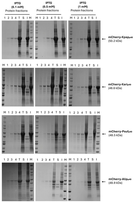
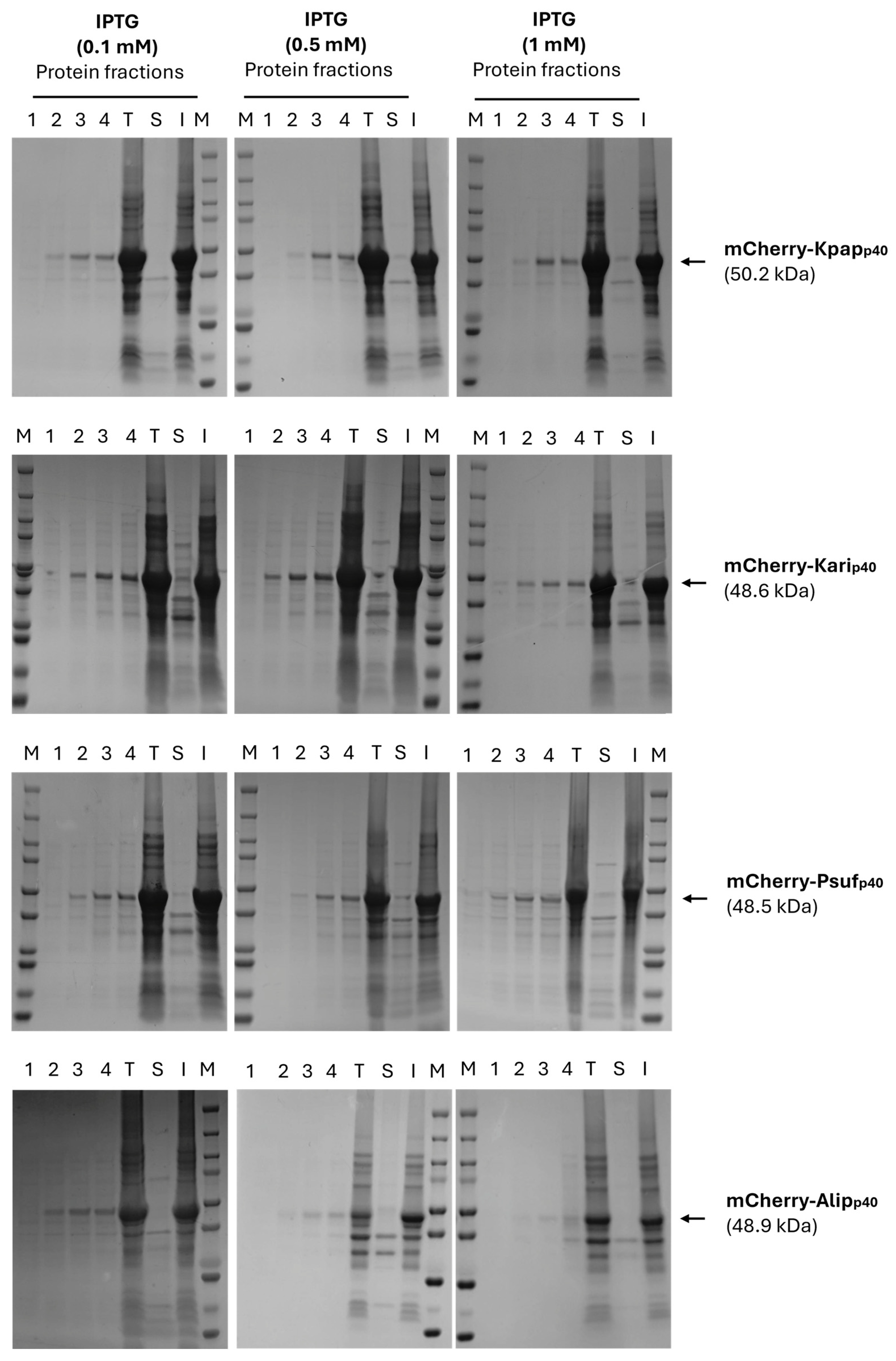
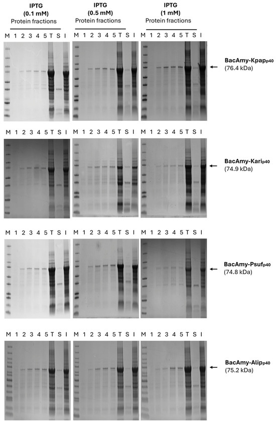
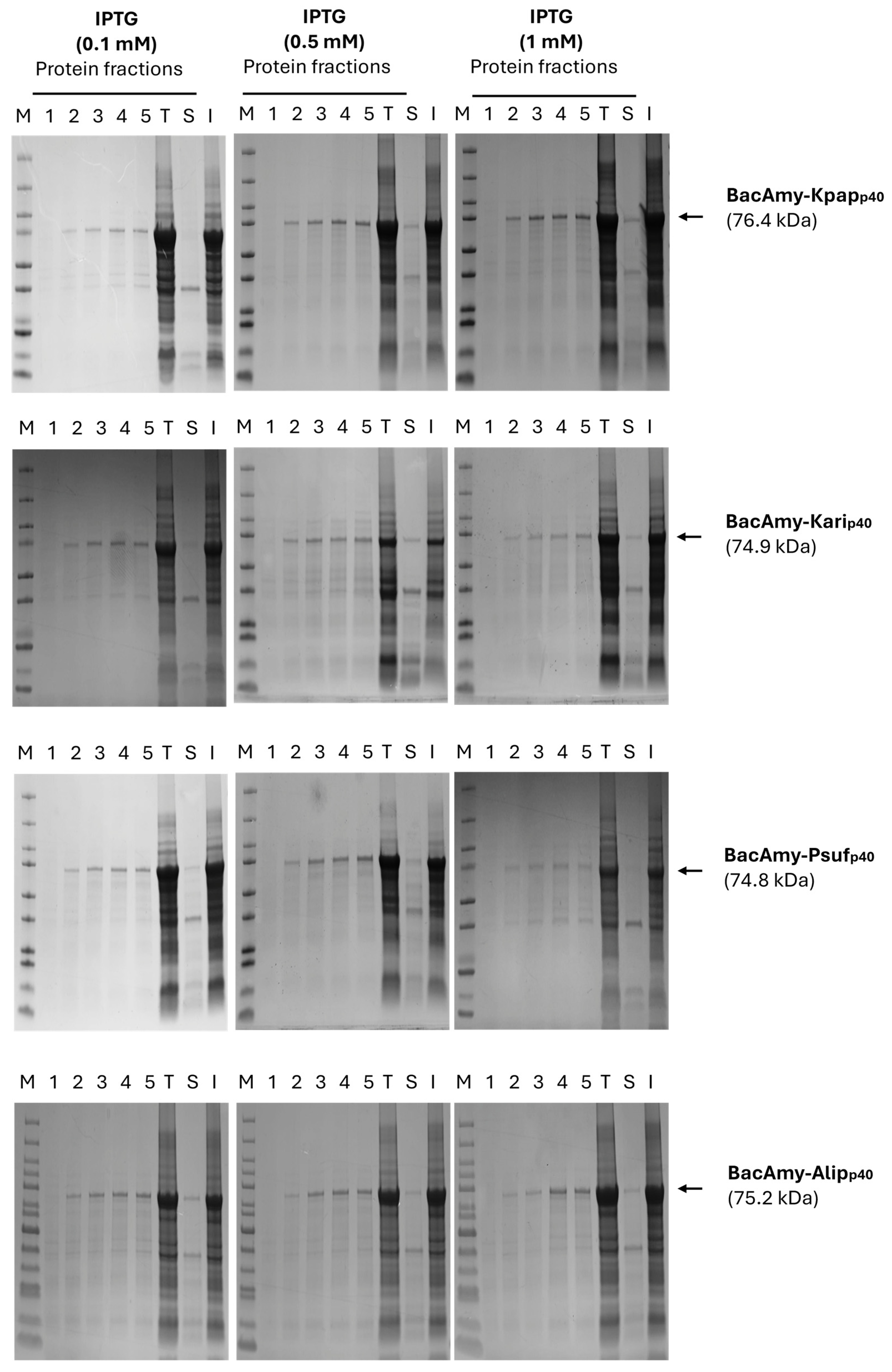
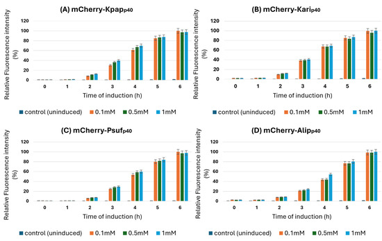
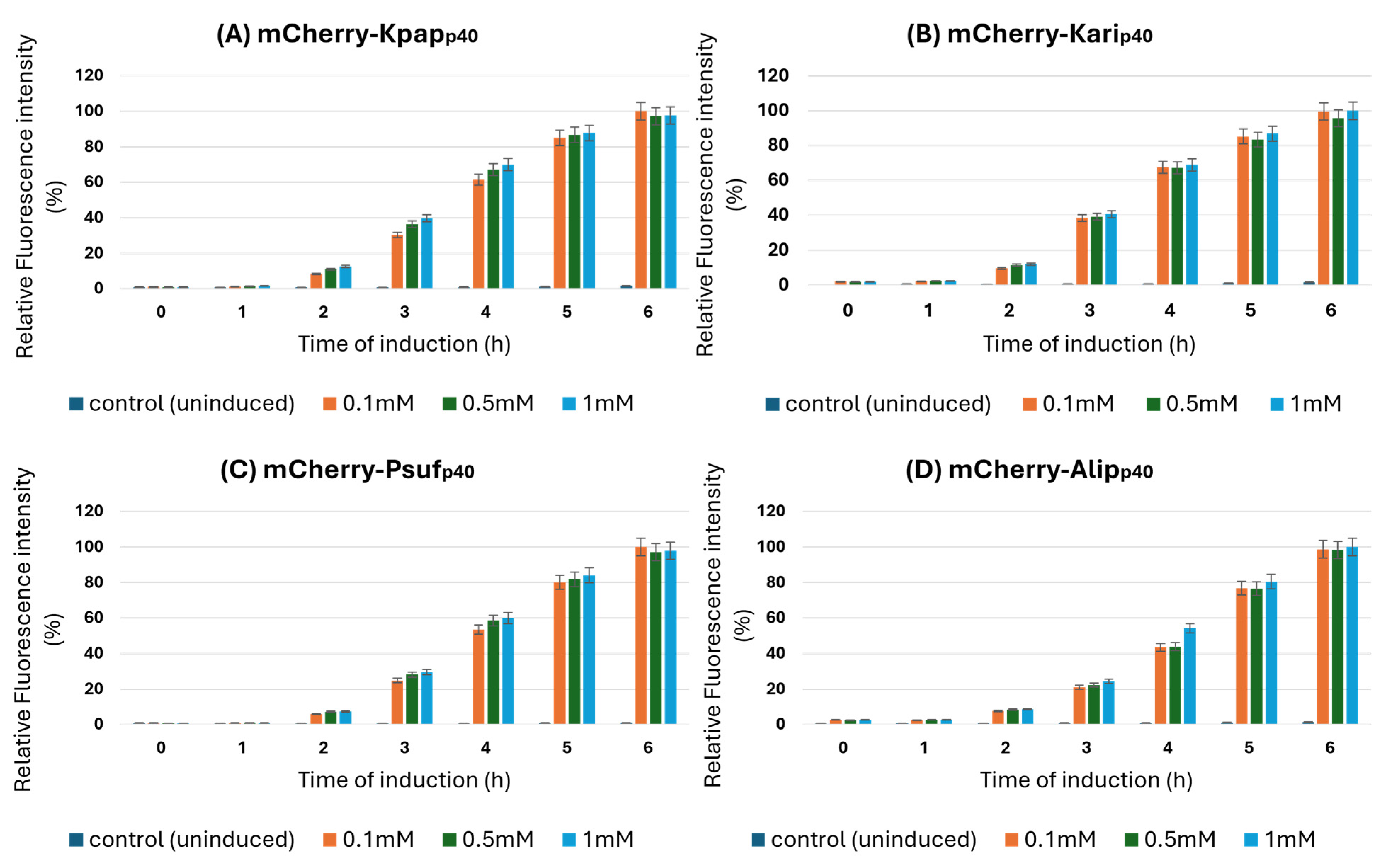
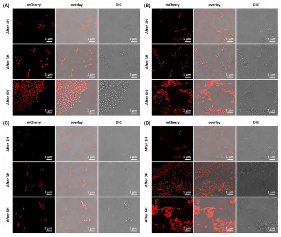
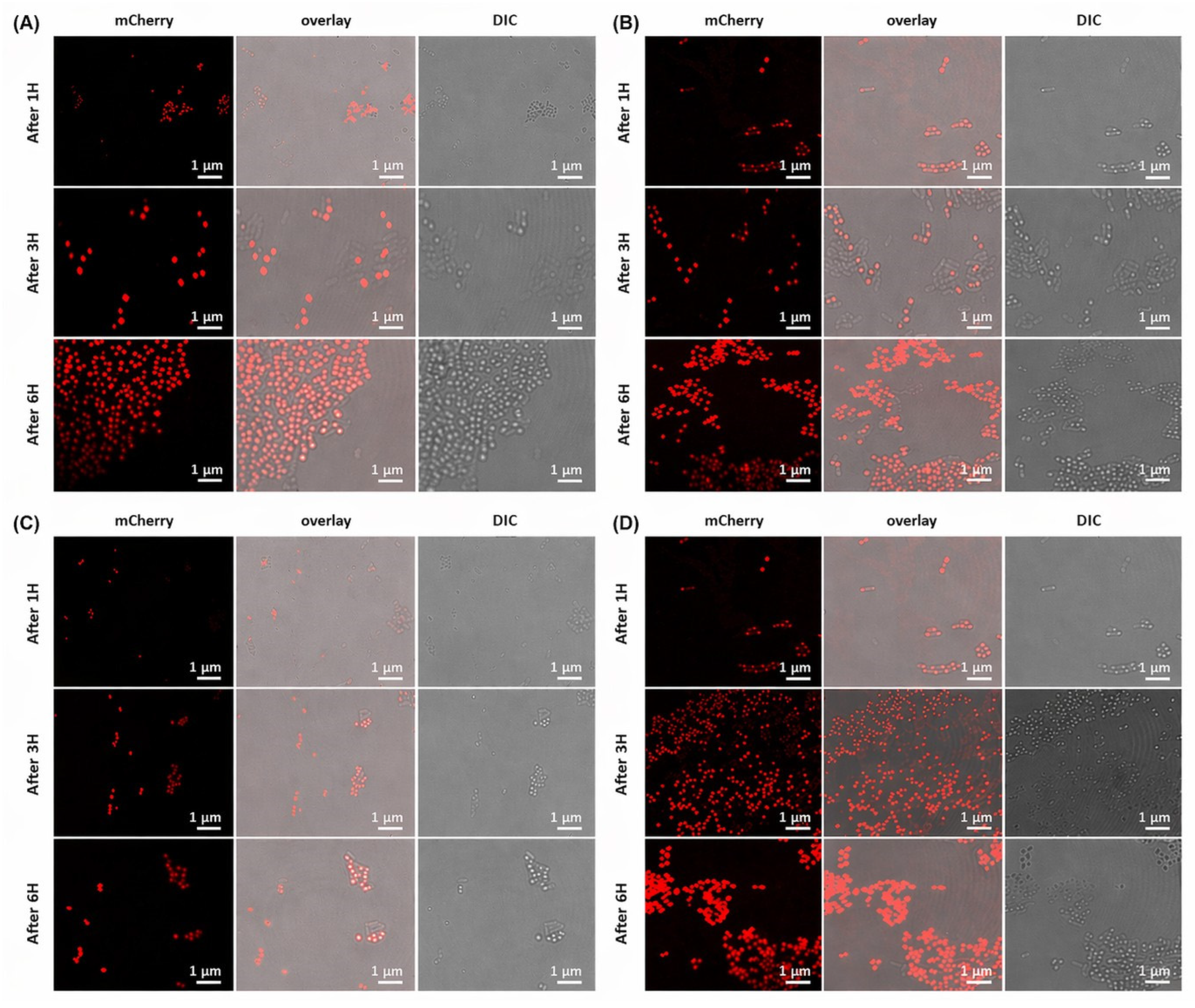
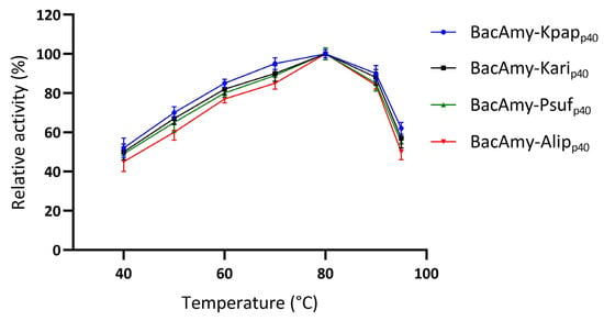
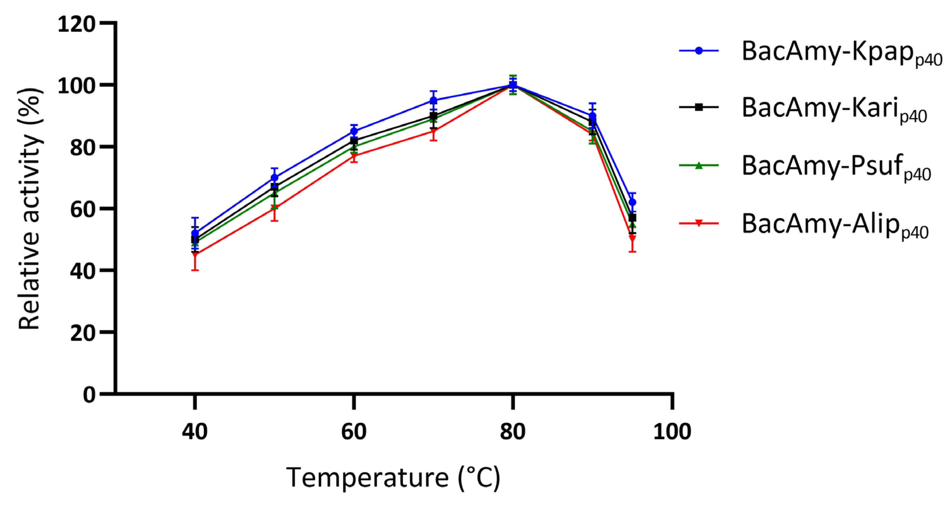
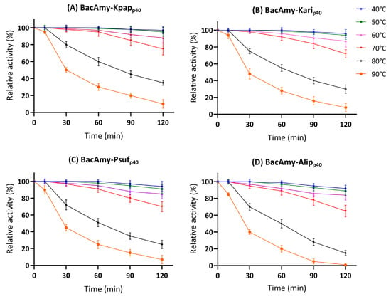
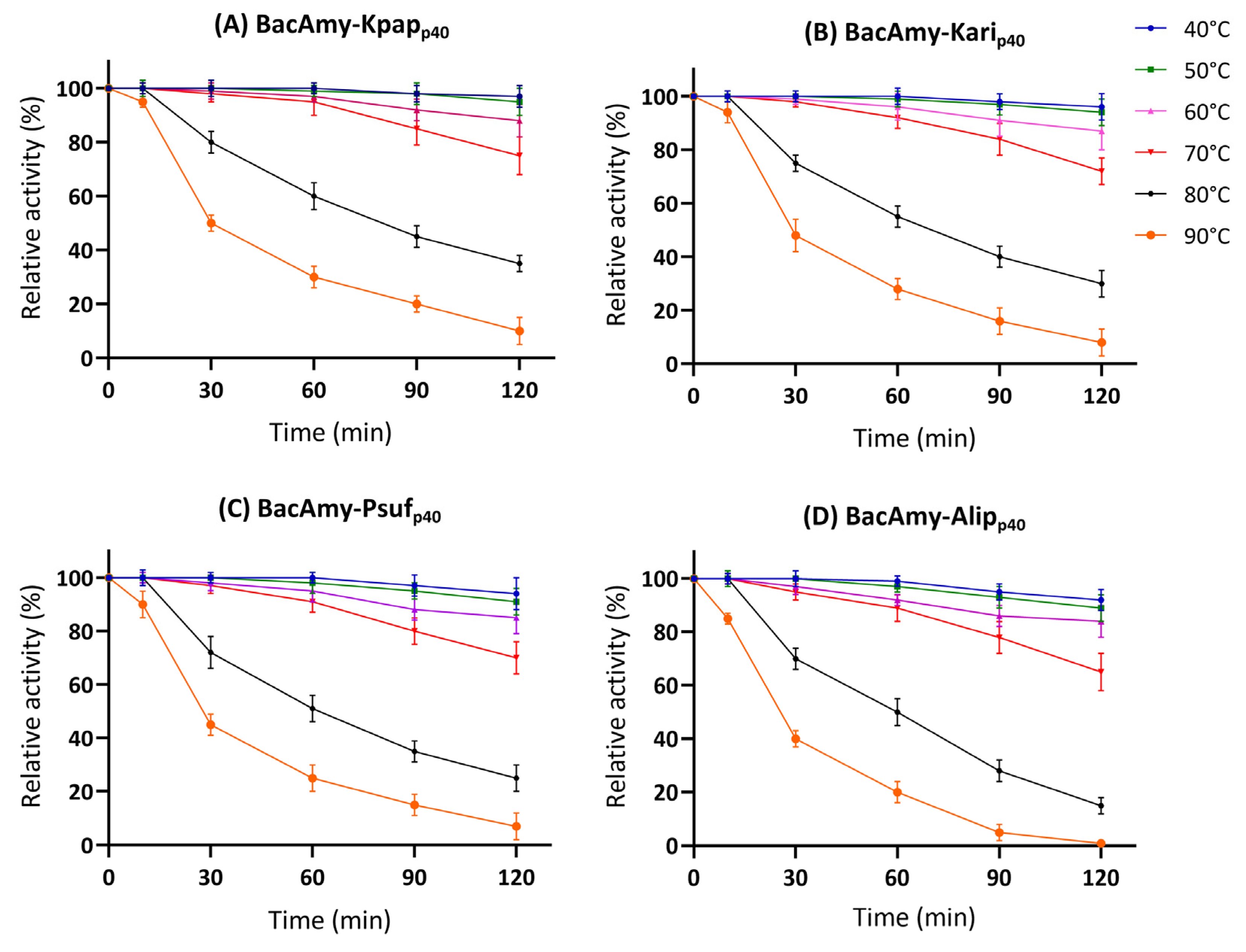
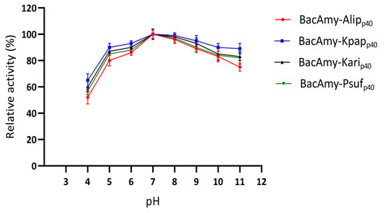
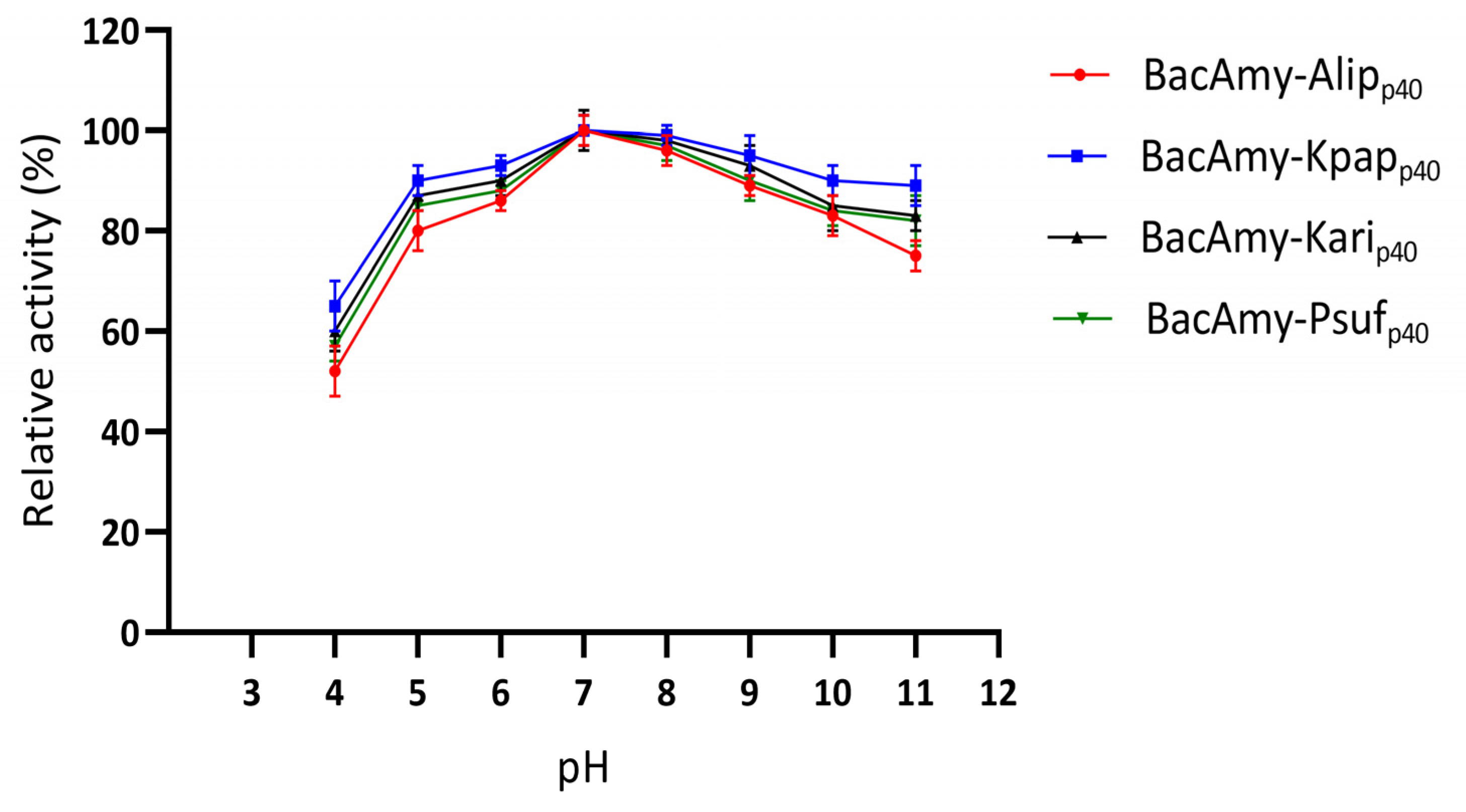
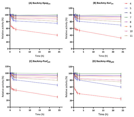
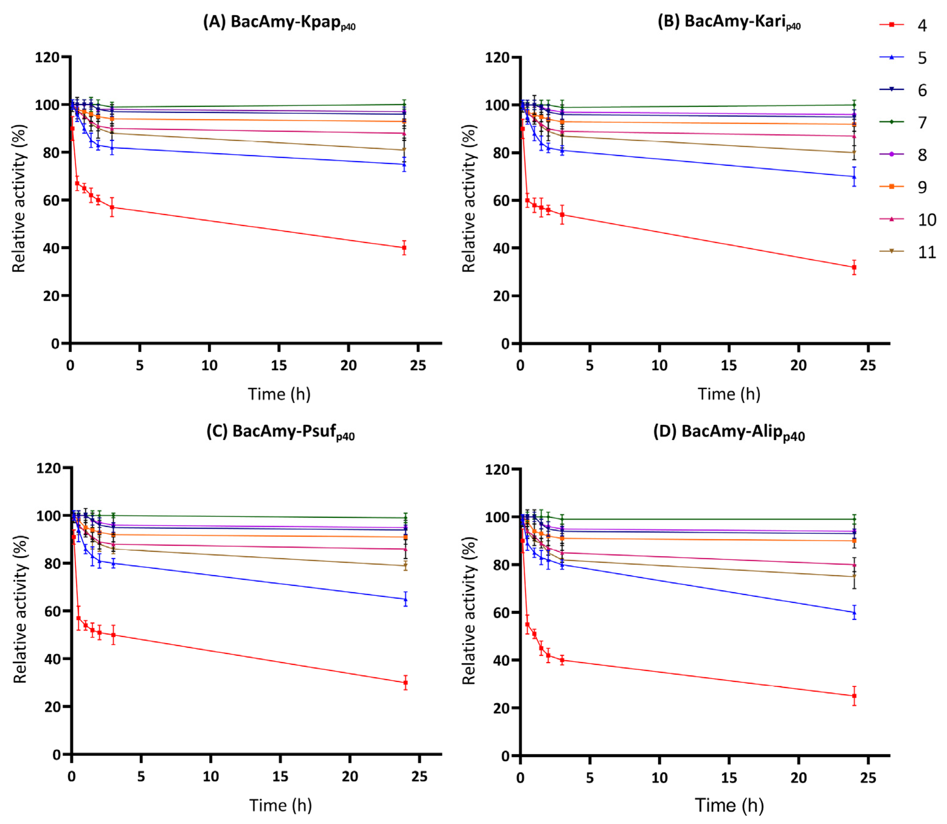
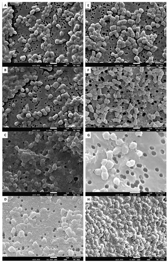
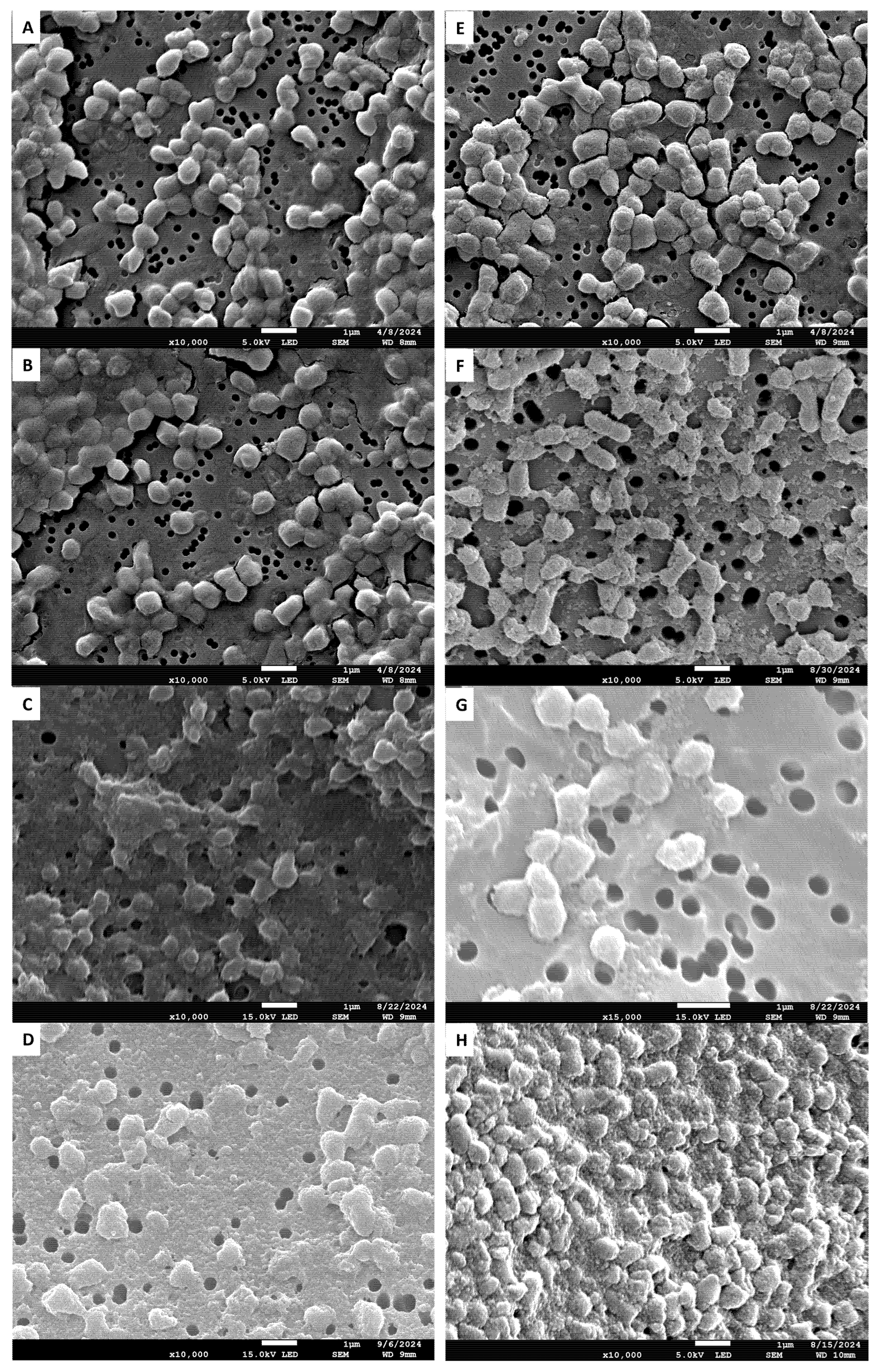

Disclaimer/Publisher’s Note: The statements, opinions and data contained in all publications are solely those of the individual author(s) and contributor(s) and not of MDPI and/or the editor(s). MDPI and/or the editor(s) disclaim responsibility for any injury to people or property resulting from any ideas, methods, instructions or products referred to in the content. |
© 2026 by the authors. Licensee MDPI, Basel, Switzerland. This article is an open access article distributed under the terms and conditions of the Creative Commons Attribution (CC BY) license.