


Abstract
Trans-anethole oxygenase (TAO) exhibits broad arylpropene substrate specificity but has low activity in converting isoeugenol to high-value vanillin. Herein, we employed computer-aided rational design to engineer TAO from Pseudomonas putida (PpTAO) for enhanced catalytic efficiency toward isoeugenol. Structural modeling and AlphaFold 3 docking identified two key catalytic residues, Arg86 and His118. Through substrate channel engineering and computation-guided mutagenesis, a series of targeted variants were constructed. Three variants, H93A, Q207R/G249C, and I59T/F62T, showed significant improvements in whole-cell performance, with activity increases of 1.8-, 2.13-, and 4.83-fold over the wild type (WT), respectively. Purified enzyme kinetics corroborated these findings, as reflected in kcat/Km values that reached 1.6, 2.1, and 4.7 times that of the WT. Mechanistic molecular dynamics simulations revealed that H93A enhances activity by widening the access tunnel, whereas Q207R/G249C exerts beneficial distal effects. Notably, the I59T/F62T variant significantly increases substrate affinity by optimizing hydrophobic interactions within the binding pocket. These results validate the efficacy of computational modeling in enzyme redesign and provide a robust biocatalyst for the sustainable biosynthesis of vanillin.
1. Introduction
Vanillin is an aromatic aldehyde widely used in the flavor and fragrance industry, with great global demand in the food, pharmaceutical, and chemical industries. The chemical synthesis of vanillin from isoeugenol is a mature process [1,2,3,4,5,6,7], typically involving the oxidation of isoeugenol using oxygen or hydrogen peroxide in the presence of catalysts such as methyltrioxorhenium (CH3ReO3) or cobalt/nickel salts [8]. However, this approach suffers from three major drawbacks: poor selectivity control, severe environmental pollution, and harsh reaction conditions. These limitations have driven a shift toward greener biosynthetic alternatives [9,10,11,12,13,14,15,16]. Despite their potential, the industrial application of these biological routes faces key bottlenecks, including the low catalytic efficiency of natural enzymes and the toxicity of vanillin to host cells. Therefore, enhancing enzymatic efficiency through rational engineering remains a core scientific challenge to overcome these industrial barriers [17].
Some oxygenases with activity toward isoeugenol were discovered. For example, lipoxygenase, a type of iron-containing dioxygenase, extracted from soybean was applied for the biocatalysis of isoeugenol to form vanillin [12]. The resveratrol-cleaving dioxygenase NOV1 was initially reported to oxidize isoeugenol with limited efficiency [18]; however, recent rationally guided engineering, such as the S283F variant, has significantly enhanced its catalytic turnover and kinetic stability [19]. Furthermore, optimized NOV1-based whole-cell systems have lately demonstrated high effectiveness in producing vanillin from lignin-derived isoeugenol under industrially relevant conditions [20]. On the other hand, an isoeugenol monooxygenase (PpIEM) derived from P. putida IE27 has been purified, cloned and overexpressed in E. coli BL21 (DE3), and another IEM was identified from P. nitroreducens (PnIEM), both of which were capable of catalyzing the oxidation of isoeugenol to vanillin [21,22]. Trans-anethole oxygenase (PpTAO) was first identified as a novel flavoprotein monooxygenase from Pseudomonas putida JYR-1 by Han and colleagues [23]. This enzyme enables the strain to utilize trans-anethole as a primary carbon source for growth, while also exhibiting catalytic activity toward isoeugenol, O-methylisoeugenol, and isosafrole. Early characterization using recombinant TAO suggested that residues Trp38, Thr43, and Tyr55 were essential for flavin reduction, based on the assumption of a flavin-dependent mechanism [24]. However, structural elucidation of the crystal structure of trans-anethole oxygenase from Stenotrophomonas maltophilia (TAOSm) revealed that it represents a new class of bacterial heme-containing oxygenases [25]. The team truncated disordered terminal regions and incorporated a SUMO solubility tag to achieve successful soluble expression of TAOSm, and the resulting crystal structure revealed that TAO contains a tightly bound heme b and utilizes molecular oxygen (O2) exclusively for catalysis. Critically, the enzyme functions independently of exogenous cofactors such as NAD(P)H or FAD. This discovery overturned previous hypotheses regarding flavin and nicotinamide dependence, redefining TAO as a unique heme-dependent oxygenase.
In addition to exploring the TAO catalytic mechanism, directed evolution of TAO was carried out to enhance the catalytic efficiency toward isosafrole through construction of a TAO mutant library via error-prone PCR [26]. Four variants were identified with significantly improved activity, achieving a 1.45-fold increase in conversion rates compared to the wild-type strain. Specifically, the variant 4C7 exhibited 2.05-fold and 2.38-fold higher conversion rates to p-anisaldehyde and veratraldehyde, respectively. Another variant, 5F4, demonstrated broader improvements, with conversion rates to p-anisaldehyde, veratraldehyde, piperonal, and vanillin increasing by 1.91, 2.08, 1.33, and 1.92-fold, respectively. Qi Ye, et al. implemented a multi-platform computational design strategy (PROSS, FireProt, Rosetta, and FoldX) for engineered mutants to enhance TAO activity in vanillin biosynthesis, which showed that mutant F34Y achieved a product yield of 88.7% at an isoeugenol concentration of 10 mM, while the G117I and T138L variants demonstrated yield improvements exceeding 40% [27]. These advancements highlight the integration of computational-guided strategies in enzyme engineering, which facilitates the identification of key functional residues and narrows down the library of potential candidates for experimental validation.
In this study, we modeled PpTAO using an online prediction tool for structural modeling based on a reference crystal structure (entry ID:8S1J). By combining bioinformatics analysis with structural characterization, we analyzed the effects of specific structural domains on the catalytic mechanism. Using a set of local algorithms as a strategy to cross-screen mutation sites, such as substrate accessibility (channel engineering), affinity calculation using Schrödinger Maestro and Discovery Studio, and Molecular Mechanics–Poisson Boltzmann Surface Area (MM-PBSA), guiding mutant design, we developed PpTAO variants with enhanced catalytic activity toward isoeugenol. The catalytic efficiencies of the designed mutants were validated through wet-lab experiments, followed by molecular dynamics (MD) simulations and structural analysis to elucidate how specific mutations facilitate the observed activity improvements. This work provides a valuable framework for the rational redesign of TAO and serves as a reference for the engineering of structurally analogous enzymes.
2. Results and Discussion
2.1. Structural Modeling and Validation of the Heme Cofactor
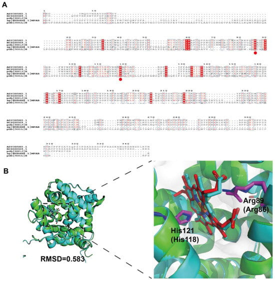
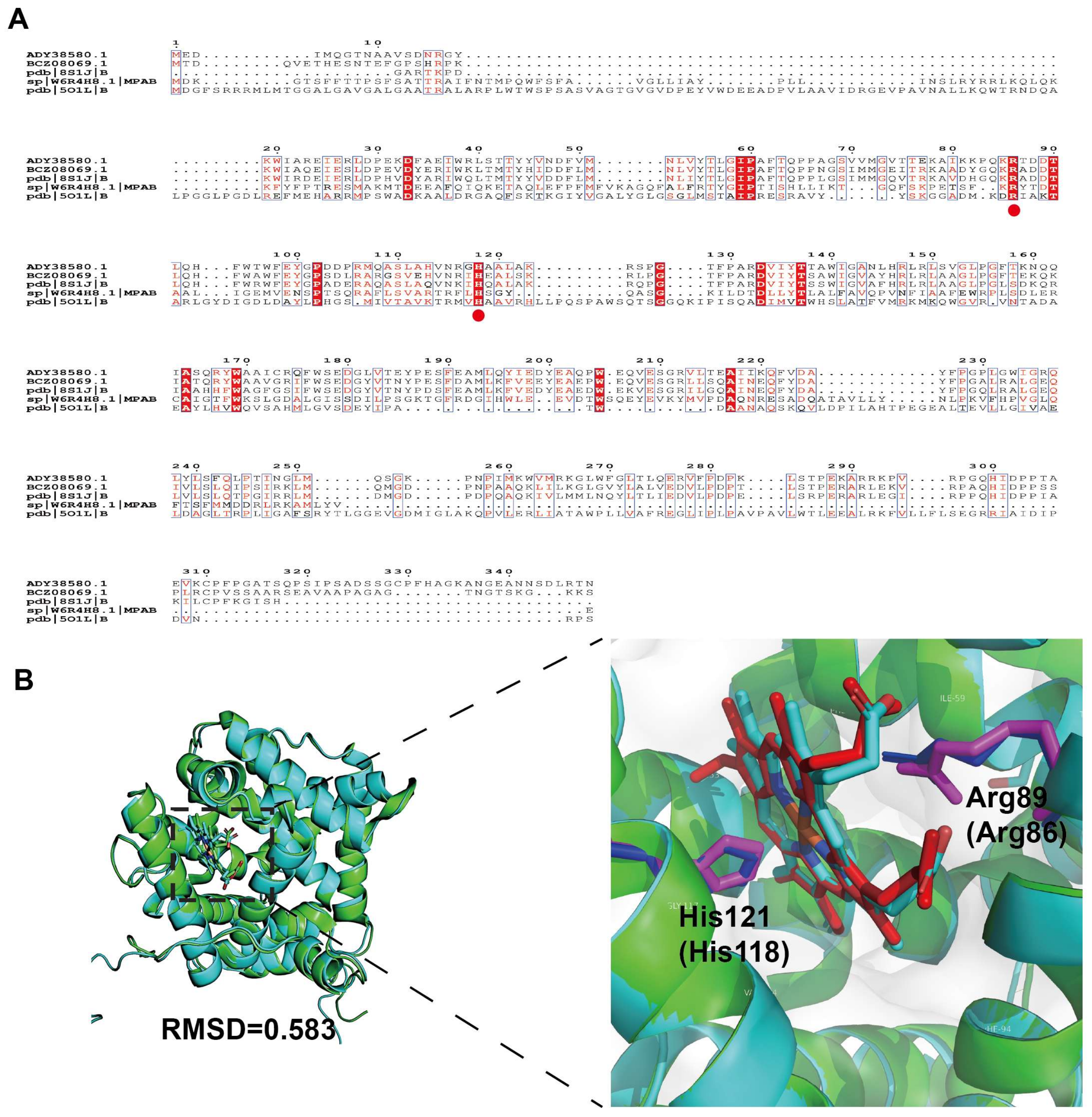
Considering that the recently reported crystal structure of TAOSm (PDB entry: 8S1J) from S. maltophilia indicated that it is a heme-dependent oxygenase, utilizing heme b as its essential cofactor [25], we suspected that the targeted PpTAO from P. putida JYR-1, which shares 62.7% sequence identity with the TAOSm homolog, may also possibly belong to heme-dependent oxygenase. Multiple sequence alignment with four other characterized oxygenases (TAOPs, NCBI GenBank entry: BCZ08069.1; TAOSm, RCSB PDB entry: 8S1J; mpaB, UniProt entry: W6R4H8.1; LCP, RCSB PDB entry: 5O1L) further demonstrated highly conserved catalytic motifs across these species (Figure 1A), suggesting a shared evolutionary mechanism for aryl-alkene oxidation. Therefore, to facilitate rational engineering, the three-dimensional structure of P. putida TAO was predicted using AlphaFold 3. A structural superposition of our de novo model with the S. maltophilia crystal structure (PDB 8S1J) revealed remarkable architectural conservation (Figure 1B). Aside from the disordered regions at the N- and C-termini, the two structures were virtually identical in their core catalytic domains (Figure 1B). The overall architectures are highly conserved, featuring nearly identical geometry and volume of the heme-binding pockets. Both structures harbor a conserved proximal ligand, His (His121 and His118), and a distal axial residue, Arg (Arg89 and Arg86), within the catalytic center. In addition, the root-mean-square deviation (RMSD) between the models indicates that the heme-binding environment and substrate access channels are highly conserved (Figure 1B).
Figure 1.
(A) Multiple sequence alignment of PpTAO with four representative bacterial heme-dependent oxygenases. (B) Structural superposition of the AlphaFold 3-predicted model of PpTAO (blue) with the crystal structure of TAOSm (PDB ID: 8S1J, green). The conserved catalytic dyad residues are highlighted: Arg89 and His121 of 8S1J are shown in light blue, while the corresponding residues Arg86 and His118 of PpTAO are shown in purple.
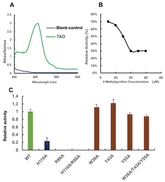
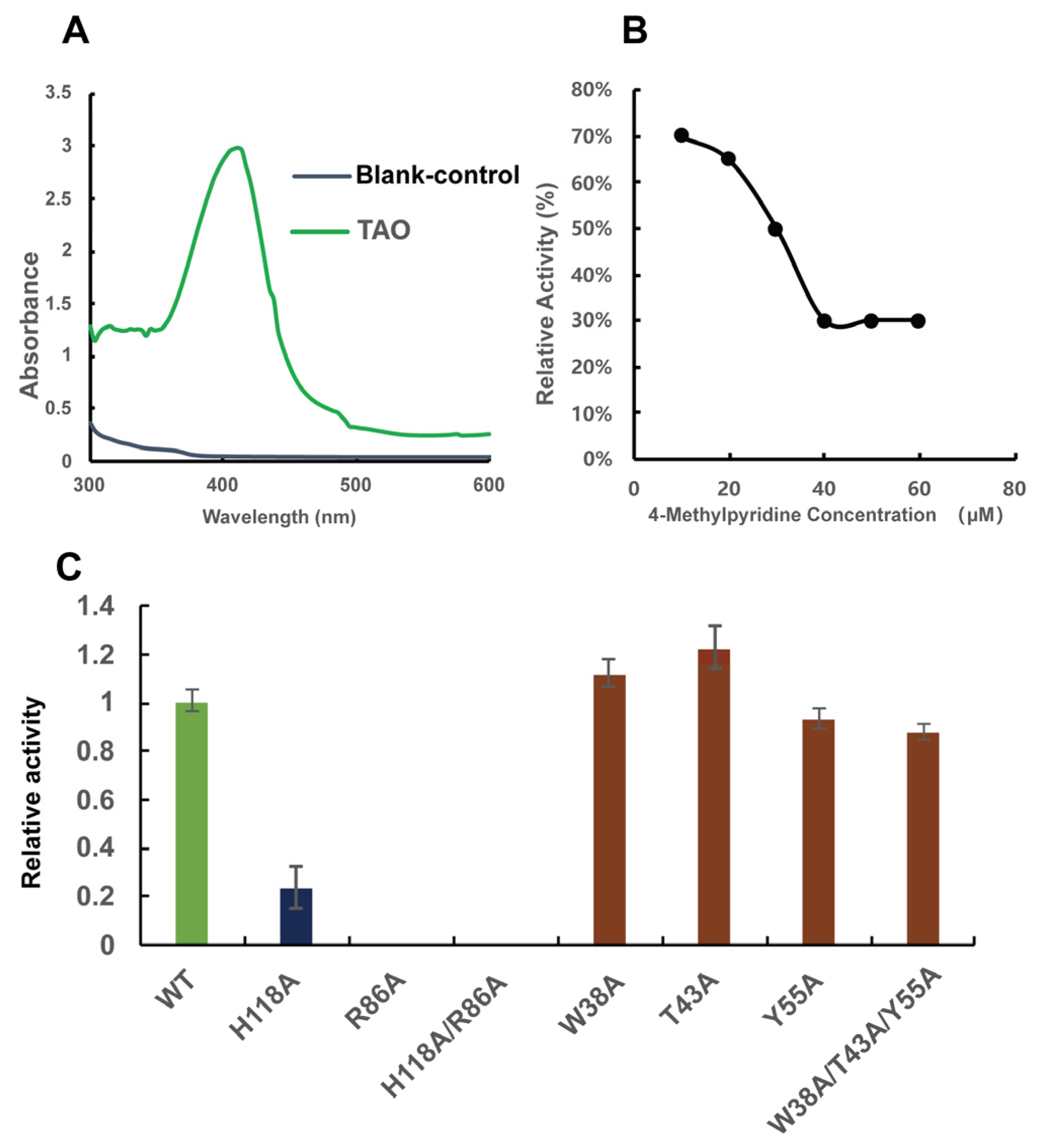
The structural model of TAO from P. putida JYR-1 generated by AlphaFold 3 revealed a distinct heme-binding pocket. Although the prior literature predominantly classified this enzyme as flavin-dependent [28], we sought to empirically confirm the presence of a heme cofactor via spectroscopic analysis. Heme-containing proteins typically exhibit a characteristic Soret band in the visible region between 380 and 450 nm. Thus, we measured the UV–visible absorption spectrum of the purified PpTAO enzyme. As shown in Figure 2, the absorption spectrum of the purified PpTAO exhibited a prominent Soret peak at 414 nm, consistent with the signature of a heme-coordinated protein (Figure 2A), indicative of a heme cofactor. Furthermore, pyridine can act as an exogenous axial ligand, directly coordinating with the iron atom in the heme prosthetic group, thereby competitively inhibiting the enzyme’s normal function. Inhibition assays using 4-methylpyridine, a known heme ligand, showed that the relative enzymatic activity decreased in a dose-dependent manner with increasing concentrations of the inhibitor; when the concentration exceeded 45 μM, the enzyme activity was only 30% of the original (Figure 2B). Combined with structural modeling, these results confirm that PpTAO is a heme-dependent oxygenase. This is consistent with reports on its homologs, rather than being flavin-dependent as previously hypothesized. Furthermore, the predicted catalytic residues (Arg86 and His118) and three putative flavin-binding residues (Trp38, Thr43, and Tyr55) were mutated to alanine to verify their functional roles. As shown in Figure 2C, variants R86A, H118A, and R86A/H118A exhibited a complete loss of catalytic activity, confirming the essential roles of these two residues. In contrast, mutations at the putative flavin-binding sites (W38A, T43A, Y55A, and the triple mutant) showed negligible impact on activity. These results suggest that the previously hypothesized flavin-dependence may be incorrect.
Figure 2.
(A) UV–visible absorption spectrum of PpTAO-purified enzyme. (B) Inhibition of TAO catalytic activity toward isoeugenol by 4-methylpyridine. (C) Mutating both key residues H118 and R86, along with the three putative flavin-binding residues (reddish-brown) W38, T43, and Y55, to alanine, and the resulting catalytic activity.
2.2. Binding Mode of Isoeugenol Within the PpTAO Active Site
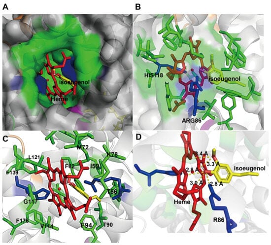
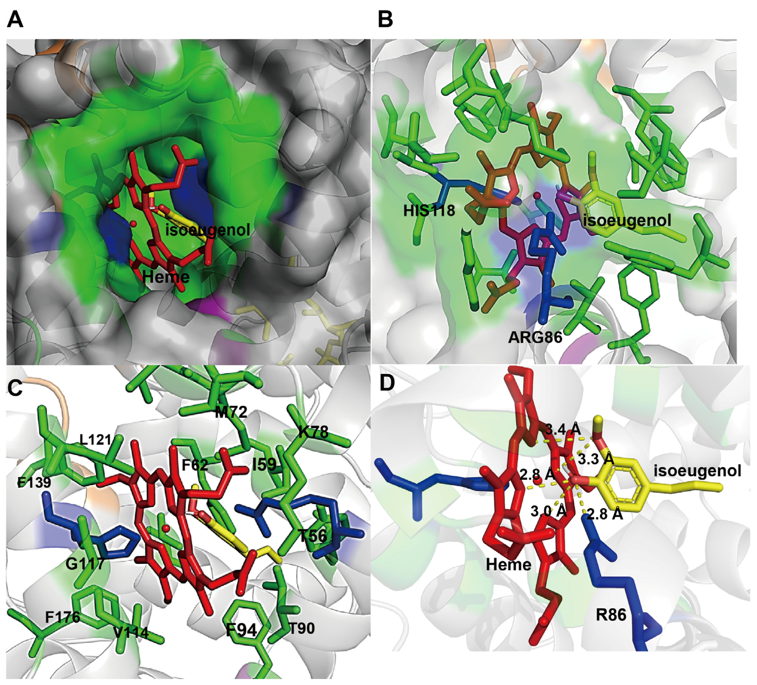
To determine the binding conformation of isoeugenol within the PpTAO active site, further isoeugenol molecular docking was performed using AutoDock Vina. Figure 3A presents a representative view of the heme-binding pocket, illustrating the docked position of isoeugenol relative to the prosthetic group. Isoeugenol is positioned within the distal heme-binding pocket, situated in close proximity to the distal axial residue, Arg86 (Figure 3B). The catalytic cavity surrounding the heme is formed by a cluster of residues, including Phe94, Thr90, Val114, Phe176, Gly117, Phe139, Leu121, Phe62, Met72, Ile59, Lys78, and Thr56 (Figure 3C). These residues constitute the structural framework of the heme-binding pocket, and are highly conserved in homologous oxygenases, underscoring their functional importance. Detailed analysis of the docking pose reveals a sophisticated hydrogen bonding network that stabilizes the substrate (Figure 3D). Isoeugenol forms a direct hydrogen bond with the distal arginine with a distance of 2.8 Å. Furthermore, the substrate is anchored by four hydrogen bonds with the heme prosthetic group, with bond distances of 3.0 Å, 2.8 Å, 3.4 Å, and 3.3 Å, respectively. The substrate is stabilized through a combination of hydrogen bonds and van der Waals forces, primarily interacting with the catalytic residue Arg86 and the heme cofactor. This tight interaction network positions the propenyl side chain of isoeugenol in an optimal orientation for the subsequent oxidative reaction.
Figure 3.
(A) Representative view of the heme-binding pocket, with heme in red and isoeugenol in yellow. (B) Close-up view of the active site residues surrounding isoeugenol; the two critical catalytic residues, Arg86 and His118, are highlighted in blue. (C) Surface representation of the catalytic pocket formed by conserved amino acid residues (shown in green, residues F94, T90, V114, F176, G117, F139, L121, F62, M72, I59, K78, and T56). (D) The hydrogen-bonding between isoeugenol, Arg86, and the heme cofactor. Hydrogen bonds are represented by yellow dashed lines, with bond distances labeled in Angstroms (Å).
2.3. Structure-Guided Engineering of the PpTAO Substrate Access Tunnel
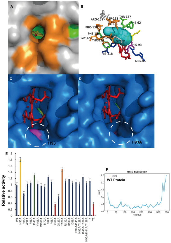
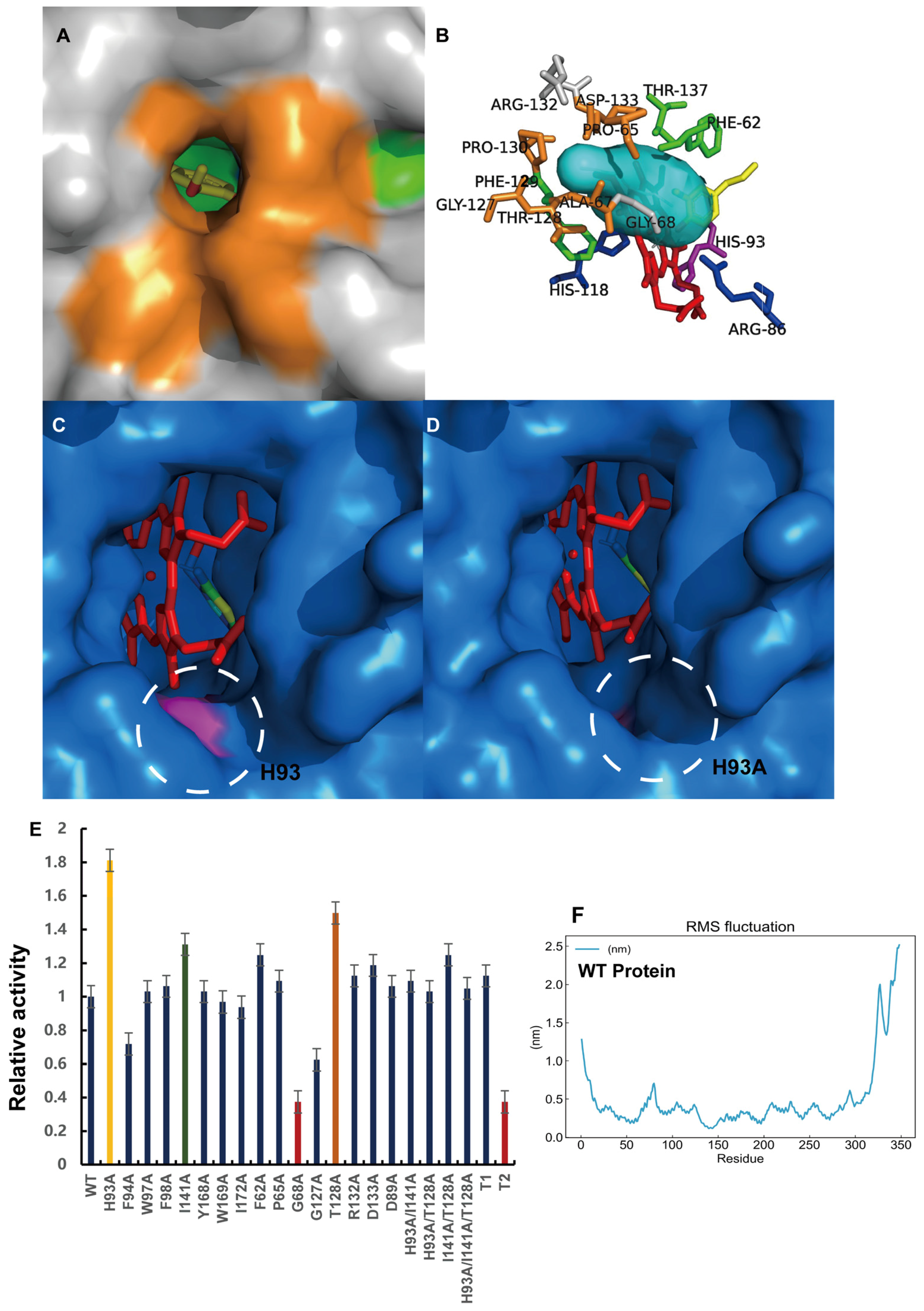
After modeling the PpTAO structure, we investigated structural modifications to broaden the substrate-binding pocket and access tunnels. Structural analysis revealed that the heme prosthetic group occupies a significant portion of the binding cavity, leaving limited space for substrate orientation. On the side adjacent to His118, we identified a highly constricted entry point, hypothesized to be a key access route for the substrate to reach the catalytic center (Figure 4A). According to the induced-fit mechanism, we postulated that the PpTAO protein scaffold, particularly the residues flanking the access tunnel, undergoes conformational rearrangements upon initial contact with the substrate. This structural flexibility facilitates the entry of isoeugenol into the core catalytic region. Using CAVER, we identified a narrow access tunnel (Tunnel 1) composed of residues Pro65, Ala67, Gly127, Thr128, Arg132, and Asp133 (Figure 4B). The bottleneck radius of this tunnel appeared to be a limiting factor for substrate diffusion. Consequently, we targeted these residues, along with a non-conservative residue located at the front of the heme pocket, His93, for “space-expansion” mutagenesis (Figure 4B,C). As an initial conservative strategy, we employed alanine scanning to minimize steric hindrance. Substituting bulky side chains with the methyl group of alanine removes steric bulk while maintaining a minimal hydrophobic scaffold, thereby precisely expanding the channel volume without collapsing the local fold. Experimental validation of the single-point mutants revealed that H93A exhibited the most significant improvement, with a 1.8-fold increase in whole-cell catalytic efficiency over the wild type (WT). Other variants, including I141A and T128A, showed modest improvements of 30% and 50%, respectively (Figure 4E). Next, we attempted to drastically widen the entire tunnel entrance. Two multi-point variants where bottleneck residues were replaced by alanine or glycine (Figure S1A,B), T1 (P65A/T128A/P130A/R132A/D133A) and T2 (P65G/T128G/P130G/R132G/D131G), were constructed, but this “global widening” strategy proved unsuccessful. The T1 variant maintained activity equivalent to the WT, while the T2 variant showed a significant decrease in activity. Furthermore, combinatorial mutations of the beneficial single points (H93A/T128A, H93A/I141A, T128A/I141A, and the triple mutant H93A/I141A/T128A) did not yield synergistic improvements (Figure 4E), suggesting that the local microenvironment around His93 is the primary determinant for the observed activity enhancement.
Figure 4.
(A) Visualization of the narrow substrate access tunnel and the amino acid composition of the catalytic pocket (the yellow section indicates the composition of the channel opening residues). (B) Schematic representation of the substrate channel and amino acid residues. (C) Structural localization of His93 at the periphery of the heme-binding pocket. (D) Surface representation of the H93A variant. (E) Comparison of whole-cell catalytic performance for tunnel-engineered variants, including single-point and multi-point mutations, relative to the wild type (WT). (F) Root-mean-square fluctuation (RMSF) profile of individual amino acid residues in the wild-type (WT) PpTAO.
Molecular dynamics (MD) simulations were performed to elucidate why the multi-point glycine mutations failed to enhance activity. The RMSF (root-mean-square fluctuation) analysis (Figure 4F) revealed that residues Pro65, Thr128, and Pro130 are located within highly flexible loop regions. While Arg132 and Asp133 exhibit lower baseline flexibility, the introduction of glycine, a residue known for its high conformational freedom, significantly increased the local structural fluctuations. This excessive flexibility likely leads to a “closed” state of the tunnel during substrate binding, obstructing entry to the catalytic center. In contrast, His93, located at the rigid periphery of the heme-binding pocket, maintains a stable conformation. Substituting His93 with alanine (H93A) creates a stable “cavity” that facilitates substrate access without compromising structural integrity, illustrating the formation of an enlarged access cavity (pit) upon mutation (Figure 4C,D, Table S4). Based on these findings, we performed saturation mutagenesis at the H93 position to further optimize the site (Figure S1C). However, no variant outperformed H93A, suggesting that H93A offers a near-optimal balance of space and stability for isoeugenol conversion.
2.4. Affinity-Based Rational Design for Enhanced Substrate Binding
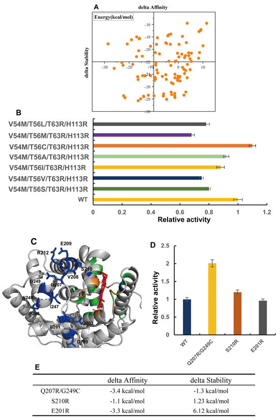
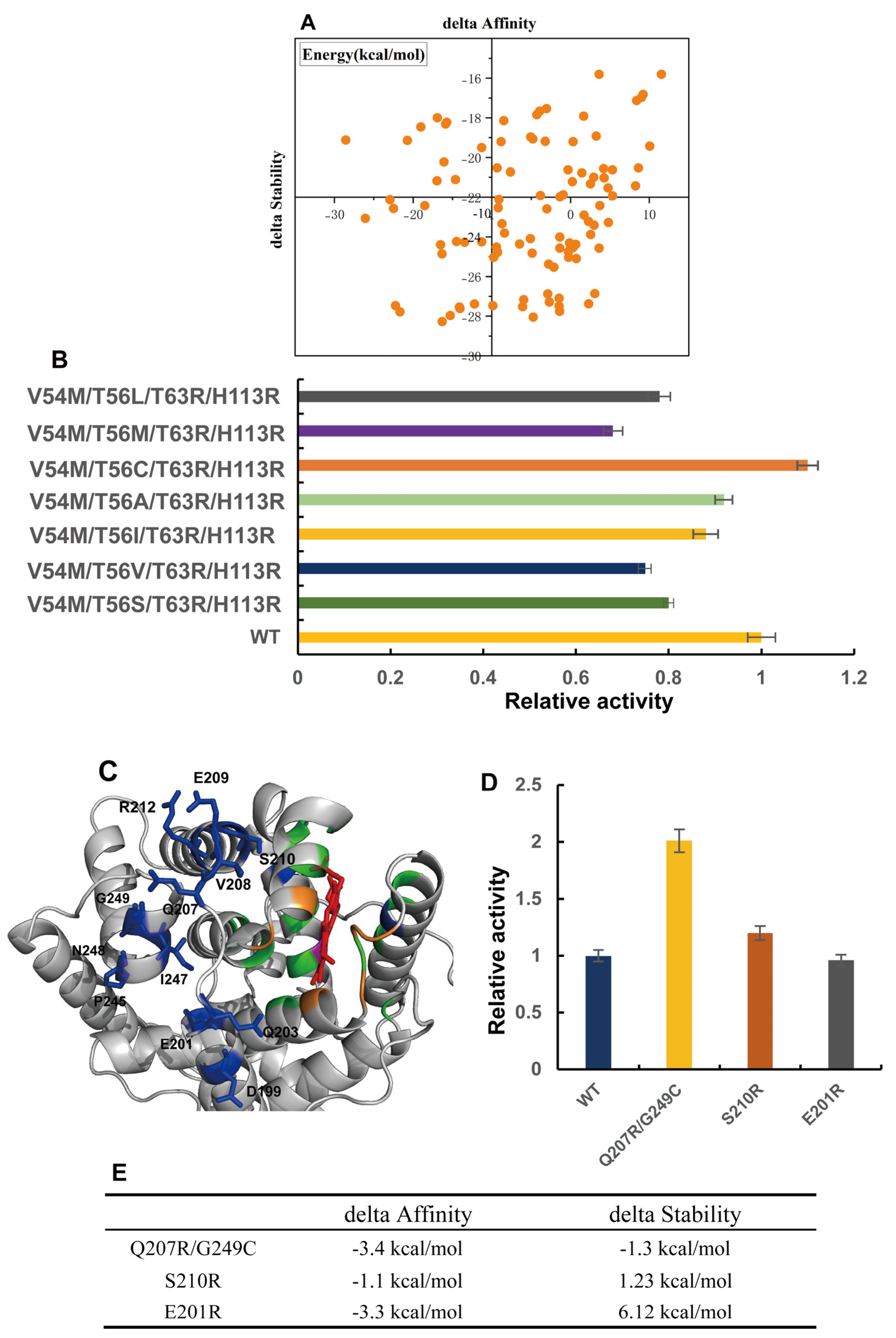
To further enhance the catalytic efficiency, we employed Schrödinger’s FEP+ (Free Energy Perturbation) to predict beneficial mutations. Simultaneously, the Virtual Amino Acid Mutation module in Discovery Studio was used to assess the energetic consequences of substitutions on both binding free energy (ΔΔGbind) and protein thermostability (ΔΔGstability). By integrating cross-platform screening, we performed an exhaustive in silico alanine scanning of all residues constituting the heme-binding pocket and substrate cavity. Using a stringent threshold of ΔAffinity < −3 kcal/mol, we identified four high-frequency target residues: Val54, Thr56, Thr63, and His113. Subsequently, these positions were subjected to in silico saturation mutagenesis using Schrödinger’s Residue Scanning Calculations module (Figure S2). As illustrated in Figure 5A, we prioritized variants based on two criteria: ΔAffinity < −25 kcal/mol to ensure strong substrate binding, and ΔStability < −10 kcal/mol to maintain structural integrity. This dual-parameter filtering yielded seven promising candidates. However, subsequent experimental validation through whole-cell biocatalysis revealed that none of these variants exhibited a significant improvement in activity compared to the wild-type or the H93A mutant (Figure 5B). This discrepancy suggests that while these mutations may enhance binding or stability, they might simultaneously compromise the catalytic geometry or the activation of the heme-iron intermediate.
Figure 5.
(A) Distribution of calculated binding affinity (ΔAffinity) and structural stability (ΔStability) for the virtual mutation library. (B) Relative catalytic activities of the quintuple mutants constructed on the H93A scaffold. (C) Structural representation of TAO highlighting the targeted distal residues (represented as spheres/sticks) located on peripheral α-helices, selected to avoid direct interference with the heme-binding environment. (D) Comparison of whole-cell catalytic efficiency for the wild type (WT) and variants Q207R/G249C, S210R, and E201R toward isoeugenol. (E) Experimental validation of the whole-cell catalytic performance for the three lead variants identified through computational screening. Data represent the mean ± SD from triplicate experiments.
Since modifications within the primary heme-binding pocket and substrate access tunnels yielded limited improvements, we hypothesized that direct alterations to the catalytic core might be too disruptive to the enzyme’s delicate structural integrity. Consequently, we shifted our focus toward distal residues located away from the active site (Figure 5C). To minimize direct structural interference, we specifically targeted residues located on three distinct α-helices that do not contribute to the heme-pocket framework, yet remain in structural proximity to residues flanking the pocket. A total of twelve distal residues—Asn248, Gly249, Ile247, Pro245, Gln207, Val208, Ser210, Arg212, Asp199, Glu209, Glu201, and Gln203—were selected for computational analysis. Following the previously established in silico screening protocol for binding affinity and structural stability, Schrödinger’s Residue Scanning identified only three viable combinatorial variants (Figure 5E). Experimental validation through whole-cell biocatalysis revealed that the double mutant Q207R/G249C significantly outperformed the wild type, exhibiting a 2.13-fold increase in catalytic efficiency (Figure 5D). This result underscores the potential of distal site engineering in modulating the catalytic environment of TAO, likely through long-range conformational effects or improved global stability.
2.5. MM-PBSA Calculation-Guided Rational Design of TAO Variants
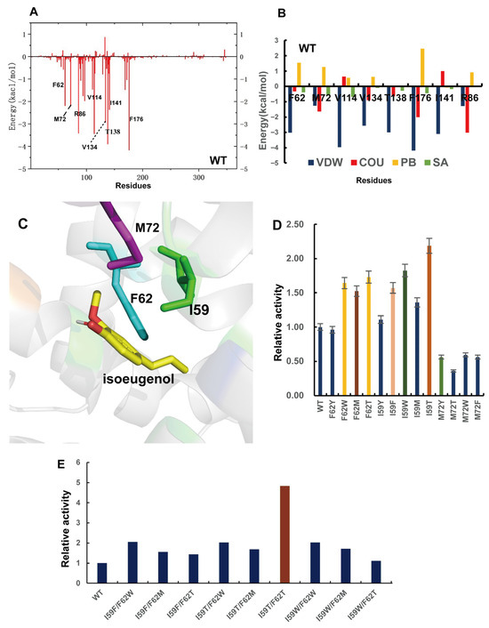
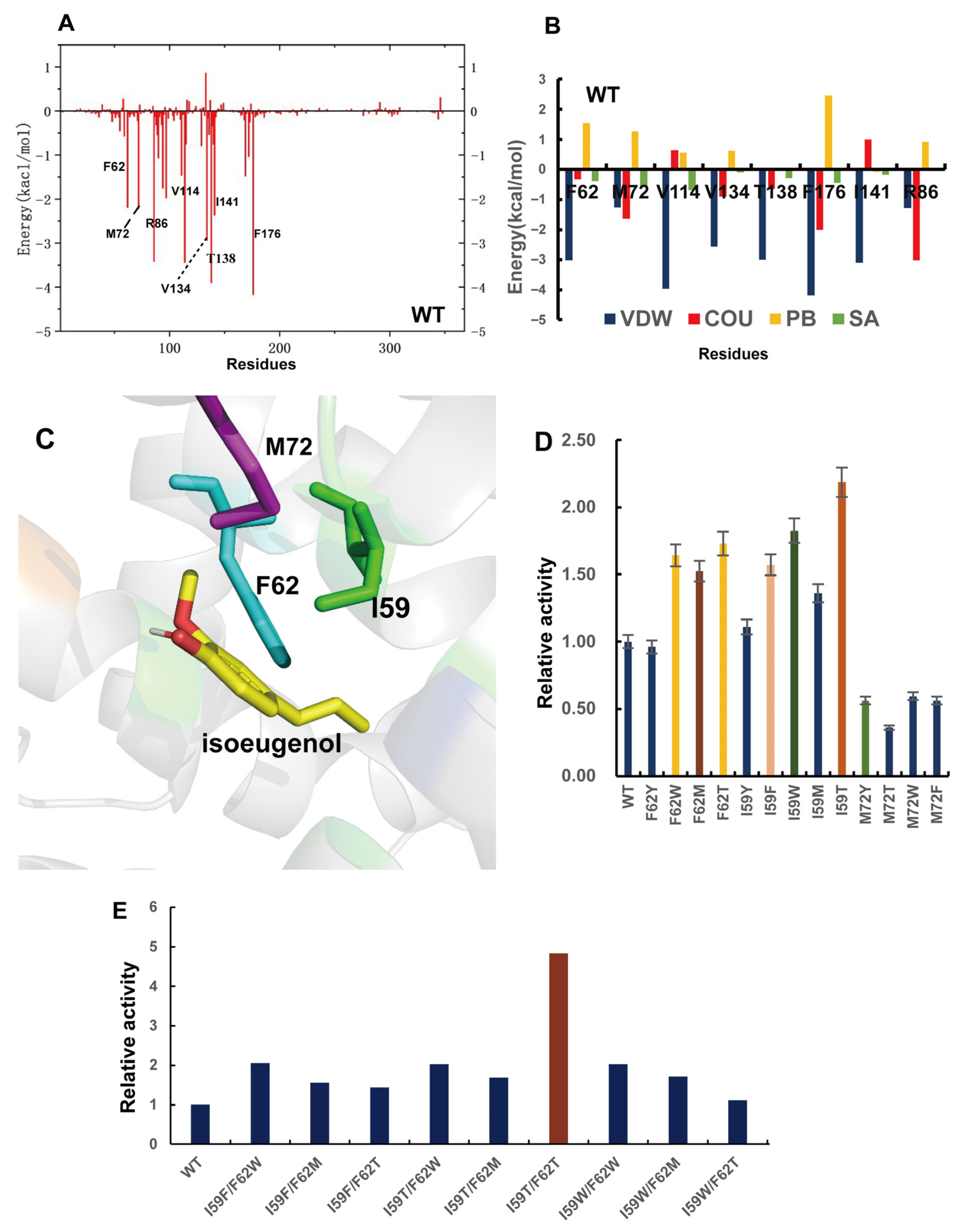
To gain deeper insights into the energetic basis of substrate recognition, we performed MM-PBSA (Molecular Mechanics–Poisson Boltzmann Surface Area) calculations based on the molecular dynamics trajectories of the PpTAO. It combines molecular mechanics (MM) with the Poisson–Boltzmann (PB) equation to account for solvation effects. As illustrated in Figure 6A,B, the total binding free energy was decomposed into individual residue-level contributions to identify key residues stabilizing isoeugenol (Figure 6A). The energy decomposition revealed that the catalytic residue Arg86 primarily stabilizes the substrate through strong electrostatic interactions. Meanwhile, a cluster of hydrophobic and polar residues, including Phe62, Met72, Val114, Val134, Thr138, Phe176, and Ile141, provided significant van der Waals interactions. These residues create a favorable hydrophobic microenvironment that facilitates substrate desolvation and orientation within the catalytic pocket (Figure 6B).
Figure 6.
(A) Total per-residue binding free energy (ΔGbind) contribution profile for all residues in the WT-isoeugenol complex. (B) Detailed energy decomposition into van der Waals (ΔEvdw), electrostatic (ΔEele), and solvation (ΔGsolv) components for key high-contribution residues. (C) Structural visualization of the selected mutation sites (Ile59, Phe62, and Met72) within the substrate-binding pocket. (D) Comparison of relative whole-cell catalytic efficiency for single-point mutants at positions Phe62, Met72, and Ile59. (E) Comparison of whole-cell catalytic efficiency for the combined double variants.
Based on the prevalence of hydrophobic (Ile, Val, Phe) and sulfur-containing or polar residues (Met, Thr) in the high-contribution group, we targeted three conserved residues in close proximity to the substrate for site-directed mutagenesis (Figure 6C). We strategically introduced aromatic residues (Tyr, Phe, Trp) to enhance π−π stacking or hydrophobic effects with the aromatic ring of isoeugenol. Additionally, Met and Thr were selected based on their favorable energetic scores in the MM-PBSA analysis. Experimental validation identified several high-performing single-point mutants. Notably, I59T and I59W exhibited 2.18-fold and 1.83-fold increases in whole-cell catalytic efficiency, respectively, while F62W and F62M showed improvements of 1.64-fold and 1.52-fold (Figure 6D).
Subsequent combinatorial mutagenesis of these beneficial sites yielded nine double mutants. Among them, the I59T/F62T variant demonstrated the most significant improvement, achieving a 4.83-fold increase in catalytic activity compared to the WT (Figure 6E).
2.6. Molecular Dynamics Analysis and Kinetic Characterization of TAO Variants
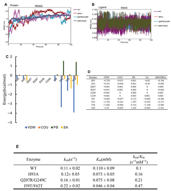
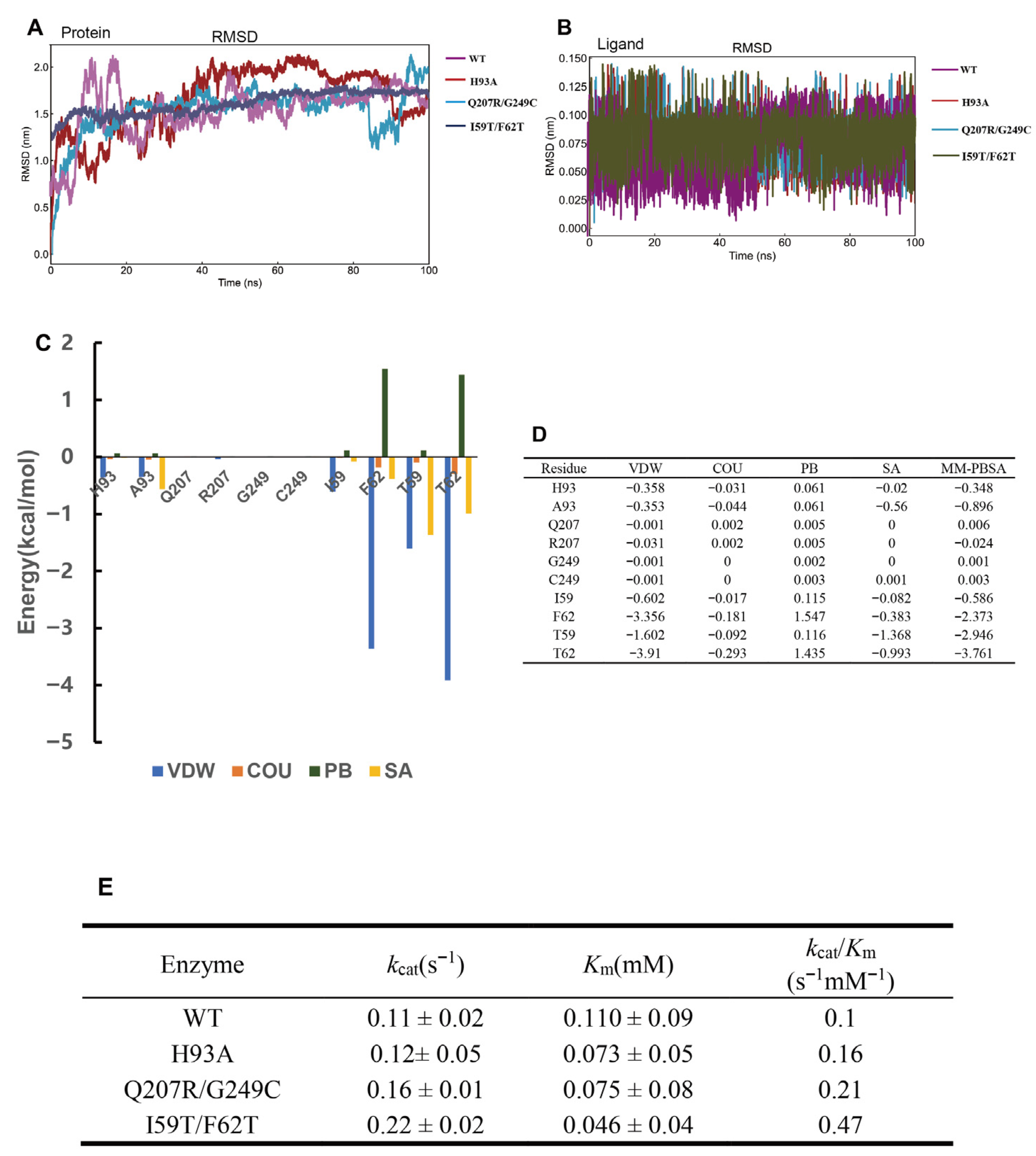
We performed comprehensive MD simulations (Figure 7A,B) and MM-PBSA calculations (Figure S2) for all variants. The MM-PBSA analysis of H93A, Q207R/G249C, and I59T/F62T elucidated the specific energetic contributions of these mutations to substrate binding (Figure 7C,D). For the H93A variant, substituting histidine with alanine primarily increased the non-polar solvation energy, while van der Waals forces, electrostatic energy, and polar solvation energy remained largely unchanged. In contrast, the Q207R/G249C variant showed negligible changes in total binding free energy. The enhanced catalytic efficiency of Q207R/G249C is likely attributed to distal effects, where mutations distant from the active site induce subtle conformational shifts. These changes are propagated through the protein’s secondary structures, modulating the active site’s geometry, electrostatic environment, or dynamic properties to favor catalysis. For the Q207R/G249C variant, SASA analysis indicated a trend toward structural compaction over the simulation (Figure S2). Notably, the Arg86–substrate distance distribution narrowed significantly relative to the wild type, adopting a unimodal profile centered at approximately 2.8–3.0 Å (Figure S6). This stabilization of the key catalytic contact is consistent with the reduced solvent exposure and supports the proposed conformational propagation mechanism.
Figure 7.
(A) RMSD curves and RMSD distribution of WT and multiple mutants in MD simulations. (B) RMSD curves and RMSD distribution of substrate and multiple mutants in MD simulations. (C,D) Detailed analysis of individual binding energy components—including van der Waals (ΔEvdw), electrostatic (ΔEele), polar solvation (ΔGpolar), and non-polar solvation (ΔGnonpolar) energies—for H93A, Q207R/G249C, and I59T/F62T relative to the wild type (WT). (E) Steady-state kinetic parameters of WT TAO and engineered variants.
For the I59T/F62T variant, MM-PBSA calculations revealed a total per-residue binding free energy contribution of −6.71 kcal/mol from the two mutated positions (T59: −2.95 kcal/mol; T62: −3.76 kcal/mol; Figure 7D), representing a 2.8-fold enhancement over the corresponding wild-type residue F62 (−2.37 kcal/mol). This improvement is driven predominantly by a 1.6-fold increase in van der Waals interactions (ΔEVDW increased from −3.36 to −5.51 kcal/mol combined) and a 6.2-fold gain in non-polar solvation free energy (ΔGSA increased from −0.38 to −2.36 kcal/mol combined). These energetic gains reflect more efficient desolvation of the substrate’s non-polar moiety and strengthened van der Waals packing with the hydrophobic patches adjacent to the heme, thereby driving tighter substrate–protein association. Consistent with this interpretation, the variant exhibited reduced SASA values and stable RMSD profiles over the 100 ns simulation (Figure 7A and Figure S3). The activity enhancement of PpTAO I59T mirrors findings for the I62 mutant in TAOSm [25], highlighting a conserved substrate-recognition mechanism within this enzyme family. Despite their different origins, the spatial alignment of these conserved residues identifies this locus as a universal engineering “hotspot”.
The determination of steady-state kinetic parameters for the variants H93A, Q207R/G249C, and I59T/F62T is summarized in Figure 7E, and the enzymatic kinetic profiles of the WT and its variants are depicted in Figure S4. Initially, the purification of recombinant TAO proved to be a significant challenge. We hypothesized that the highly disordered, non-catalytic sequences at both the N- and C-termini hindered the accessibility of the His-tag. These disordered regions likely impose steric hindrance or “shielding” effects through transient intramolecular interactions, effectively burying the His-tag and preventing its binding to the Ni-NTA resin. By truncating these unstructured terminal segments, we successfully achieved high-purity protein, facilitating subsequent biochemical characterization. The WT TAO exhibited a relatively high Km (0.110 ± 0.09 mM) and a low turnover frequency (kcat = 0.11 ± 0.02 s−1), resulting in a baseline catalytic efficiency of 0.1 s−1 mM−1. The H93A variant, designed for tunnel widening, primarily impacted substrate affinity; its Km decreased to 0.073 ± 0.05 mM, leading to a 1.6-fold increase in efficiency (0.16 s−1 mM−1). This suggests that widening the access portal facilitates substrate entry without significantly altering the rate-limiting step of the catalytic cycle. The distal variant Q207R/G249C demonstrated improvements in both binding and turnover, with a kcat of 0.16 s−1 and a Km of 0.075 mM. The resulting 2.1-fold increase in catalytic efficiency (0.21 s−1 mM−1) supports the hypothesis that remote conformational shifts can effectively fine-tune the active-site environment. The most dramatic improvement was observed in the I59T/F62T variant, which was engineered to enhance hydrophobic interactions. This variant achieved a synergistic optimization of both parameters: the kcat doubled to 0.22 s−1, while the Km significantly dropped to 0.046 ± 0.04 mM (a 58% reduction compared to WT). Consequently, the overall catalytic efficiency reached 0.47 s−1 mM−1, representing a 4.7-fold increase over the WT. These kinetic data are highly consistent with our MM-PBSA and SASA calculations, confirming that strengthening the hydrophobic driving force within the binding pocket is the most potent strategy for enhancing TAO activity.
In whole-cell biocatalysis assays, the engineered variants demonstrated a clear progression in performance compared to the wild type. The WT TAO exhibited a conversion rate of only 6.36%. The H93A variant, optimized for tunnel access, increased the conversion to 11%, while the distal-site variant Q207R/G249C reached 13.89%. Most notably, the binding-affinity-optimized variant I59T/F62T showed a dramatic improvement, achieving a conversion rate of 55.3%. These results validate the efficacy of our multi-tiered rational design approach, with the optimization of the binding pocket residues (Ile59 and Phe62) providing the most substantial contribution to overall catalytic turnover.
3. Materials and Methods
3.1. Experimental Section
3.1.1. Materials and Strains
Protein expression and whole-cell biocatalysis were performed using E. coli BL21(DE3). The pETDuet-1 vector was employed for gene cloning and heterologous expression. All chemical reagents used in this study were of analytical or chromatographic grade.
3.1.2. Construction of Recombinant Expression Vectors and Strains
Plasmids and strains constructed in this study are summarized in Table S2. The reference amino acid sequence for TAO was obtained from the NCBI database (Accession ID: HQ889281.1). The TAO gene and the pETDuet-1 vector were amplified using specific primers designed to incorporate 18 bp homologous arms. The recombinant expression vector pETDuet-1-TAO was constructed via homologous recombination. Site-directed mutagenesis was subsequently performed using a PCR-based strategy. Primers were synthesized by Azenta (Suzhou, China).
3.1.3. PCR and Molecular Cloning Protocols
The PCR reaction mixture (50 μL) consisted of 1 μL of template DNA (plasmid or TAO gene), 1 μL each of forward and reverse primers (10 μM), 25 μL of 2× PrimeSTAR Max DNA Polymerase (purchased from Takara, Beijing, China), and nuclease-free water. The thermal cycling conditions were as follows: initial denaturation at 94 °C for 5 min; followed by 30 cycles of 94 °C for 20 s (denaturation), 55 °C for 20 s (annealing), and 72 °C for 1 min (extension); with a final extension at 72 °C for 10 min. To remove the parental template, 1 μL of DpnI (purchased from Takara, Beijing, China) restriction enzyme was added to the PCR products, followed by incubation at 37 °C for 1 h. The amplified fragments were analyzed and purified using 1% (w/v) agarose gel electrophoresis. The homologous recombination reaction (10 μL) comprised 3 μL of linearized vector, 1 μL of the gene insert, 5 μL of 2 × CE Mix V3 (purchased from Vazyme, Nanjing, China), and distilled water. The ligation products were transformed into competent E. coli BL21(DE3) cells to generate the mutant expression strains. All primers and strains utilized in this work are detailed in Tables S2 and S3.
3.1.4. Whole-Cell Biocatalysis
Single colonies of E. coli BL21(DE3) harboring pETDuet-1-TAO were inoculated into LB medium supplemented with ampicillin (Amp) and cultured overnight at 37 °C and 220 rpm. Seed cultures were inoculated at a 1% (v/v) ratio into 100 mL of LB medium containing Amp. The fermentation was maintained at 37 °C until the OD600 reached 0.6–0.8. Protein expression was induced by the addition of IPTG to a final concentration of 0.4 mM, followed by incubation at 25 °C and 220 rpm for 10 h. Cells were harvested by centrifugation at 8000 rpm for 10 min, and the supernatant was discarded. The wet cell pellets were weighed and resuspended in PBS buffer (pH 7.4) to a cell density of 16.6% (w/v). Isoeugenol was prepared as a stock solution in dimethyl sulfoxide (DMSO) at a 1:1 (v/v) ratio. The substrate was slowly added to the cell suspension to achieve a final isoeugenol concentration of 3.05 mM, with DMSO constituting 0.6% of the total reaction volume. The bioconversion was performed at 30 °C and 220 rpm for 5 h. Post reaction, the mixture was centrifuged at 12,000 rpm for 5 min, and the supernatant was collected for further analysis. Ampicillin was purchased from Sangon Biotech (Shanghai, China); Isoeugenol was purchased from Adamas (Titan Scientific, Shanghai, China); and DMSO was purchased from Sinopharm (Shanghai, China).
3.1.5. Protein Purification
Protein purification was performed using a 1 mL HisTrap HP (purchased from GE Healthcare, Chicago, IL, USA) affinity column. The supernatant containing soluble proteins was filtered through a 0.22 μm inorganic membrane and maintained on ice. Prior to loading, the column was equilibrated with 10 column volumes (CVs) of Binding Buffer (20 mmol·L−1 of phosphate, 20 mmol·L−1 of imidazole, and 500 mmol·L−1 of NaCl, pH 7.4). Subsequently, 10 mL of the crude enzyme extract was loaded onto the column at a flow rate of 0.5 mL·min−1. To remove unbound impurity proteins, the column was washed with 10 CV of Binding Buffer at a flow rate of 1 mL·min−1. The target protein was then eluted using a gradient of Elution Buffer (20 mmol·L−1 of phosphate, 500 mmol·L−1 of imidazole, and 500 mmol·L−1 of NaCl, pH 7.4) over 20 CV, with fractions collected according to the UV absorbance peaks. Finally, the column was re-equilibrated with 10 CV of Binding Buffer for further use (Figure S5).
3.1.6. Enzyme Kinetic Analysis
Kinetic parameters were determined using an enzyme concentration of 20 μM and varying concentrations of isoeugenol (dissolved in DMSO) ranging from 0.1 to 20 mM. Reactions were carried out in a 500 μL volume of Tris-HCl buffer (pH 7.4) at 30 °C. The formation of aromatic aldehydes was monitored using a microplate reader (The Epoch2™, BioTek, Winooski, VT, USA) at 340 nm. Kinetic data were fitted to the Michaelis–Menten equation using Origin 2026 (v10.3).
3.1.7. HPLC Analysis
The concentrations of isoeugenol and vanillin were quantified via high-performance liquid chromatography (HPLC) using a Se-Fen Amethyst C18-H column (purchased from Sepax Technologies Suzhou, China). The mobile phase consisted of 65% (v/v) methanol (purchased from Titan Scientific Shanghai, China) containing 0.01% glacial acetic acid (purchased from Sinopharm Shanghai, China). The following chromatographic conditions were employed: column temperature of 30 °C, flow rate of 1 mL/min, UV detection at 270 nm, and an injection volume of 10 μL. Samples were diluted with methanol to precipitate proteins and ensure complete dissolution of substrates and products. After centrifugation for 5 min and filtration through a syringe filter, the samples were analyzed.
3.2. Computational Section
3.2.1. Structural Modeling and Quality Assessment
The amino acid sequence of TAO was retrieved from the NCBI database (Accession ID: HQ889281.1). Structural templates with homologous catalytic domains were identified via sequence alignment. The three-dimensional structure of TAO, including the heme prosthetic group, was predicted using AlphaFold 3 [28]. Model quality was rigorously evaluated using the Predicted Local Distance Difference Test (pLDDT) and Predicted Aligned Error (PAE). Regions with pLDDT scores below 50 were considered unstructured. PAE values were analyzed to assess the relative positional confidence between residues, providing insights into inter-domain stability.
3.2.2. Molecular Docking
Molecular docking was performed using AutoDock Vina 1.5.7. The AlphaFold 3-generated TAO model and ligand structures were preprocessed using AutoDock Tools [29], which included the removal of water molecules and the addition of polar hydrogens. The search space (grid box) was centered on the heme-binding pocket with coordinates x = −6.192, y = 0.383, and z = 8.479. The cubic box dimensions were set to 24.1 Å per side (grid spacing of 1 Å). For each simulation, 50 output conformations were generated, and docking was performed in triplicate. Optimal binding poses were selected based on geometric orientation relative to the heme center, spatial fitting, and lowest binding affinity (kcal/mol).
3.2.3. Molecular Dynamics (MD) Simulations
MD simulations were conducted using the GROMACS 2023 package [30], initiated from the protein–ligand complex obtained via docking. To address the electronic complexity of the heme b prosthetic group, we employed a bonded model for the active site. The heme iron was modeled in its catalytically active Compound I (Cpd I) state, characterized by an oxo-iron(IV) species (Fe4+=O) and a porphyrin π-cation radical [31,32]. This intermediate was assigned to the ground doublet spin state. Atomic partial charges for the Cpd I group and the axial cysteine residue were derived using the Restrained Electrostatic Potential (RESP) procedure based on Density Functional Theory (DFT) calculations at the B3LYP/6-31G* level [31,32]. The coordination sphere, including the Fe=O double bond and the Fe-N and Fe-S bonds, along with their respective force constants, were explicitly defined via the Seminario method within the MCPB.py workflow [31]. These parameters were integrated into the Amber99SB force field to ensure structural and electronic stability during molecular dynamics (MD) simulations and MM-PBSA calculations. All parameters were converted to GROMACS format using the acpype tool [33].
Topology files were generated using the GAFF [34,35] force field for ligands and the AMBER99SB [36] force field for the protein. The system was solvated in a periodic boundary box using the SPC water model and neutralized with Na+ and Cl− ions. Energy minimization was performed using the steepest descent (SD) method followed by the conjugate gradient (CG) method for 10,000 steps each. Subsequently, the system underwent NVT (canonical) and NPT (isothermal–isobaric) equilibration. Production runs were analyzed over a 100 ns trajectory, with binding free energy calculations performed via the MM-PBSA method using snapshots sampled from the final 30 ns (70–100 ns interval). DuIvyTools (Version v0.6.0) was used for visualization [37].
3.2.4. Rational Mutant Design and Screening
Predictive mutagenesis and affinity screenings were performed using two complementary platforms: Discovery Studio [38]: The “Prepare Protein” and “Simulation” tools were used to apply the CHARMM force field to the complex. Targeted amino acid substitutions were simulated using the “Design Protein” module. Schrödinger Maestro 13.5 [39]: Following structure refinement via the “Protein Preparation Wizard,” the Residue Scanning module was utilized to calculate changes in binding affinity (ΔΔGbind) upon mutation, guiding the selection of high-priority variants for experimental validation.
4. Conclusions
In this study, by using AlphaFold 3 to predict the structure of PpTAO and compare it with the crystal structure of TAOSm (PDB ID: 8S1J), we speculated that a heme-binding pocket exists. Experiments, including the detection of characteristic Soret band absorption, pyridine inhibition, and complete loss of activity upon mutation of key catalytic residues, have preliminarily confirmed that this enzyme is a heme-dependent oxidase.
We successfully enhanced the catalytic performance of PpTAO toward isoeugenol through rational engineering. Three variants, H93A, Q207R/G249C, and I59T/F62T, showed significant improvements with activity increases of 1.8-, 2.13-, and 4.83-fold over WT, respectively. Molecular dynamics simulations revealed that the bottleneck mutation H93A further optimized the catalytic environment by widening the access path while providing essential steric support to maintain the tunnel’s geometric integrity. For the Q207R/G249C variant, the 2.1-fold improvement is attributed to distal structural stabilization, which triggers a global compaction of the enzyme architecture and reduces the entropic penalty for binding. The I59T/F62T variant, located in the binding pocket, achieved a 4.7-fold increase in catalytic efficiency (kcat/Km). MM-PBSA calculations revealed that the threonine substitutions at positions 59 and 62 contribute a combined binding free energy of −6.71 kcal/mol, representing a 2.8-fold enhancement over the wild-type F62 residue (−2.37 kcal/mol). This energetic improvement is driven by a 1.6-fold increase in van der Waals interactions (ΔEVDW) and a 6.2-fold gain in non-polar solvation free energy (ΔGSA). Together, these gains reflect more efficient desolvation of the substrate’s non-polar moiety and strengthened van der Waals packing with hydrophobic patches adjacent to the heme.
In summary, the above enzyme redesign strategies based on computational modeling promote the modification of TAO from different perspectives: improving yield and enhancing enzyme activity, providing a powerful biocatalyst for the sustainable biosynthesis of vanillin.
Supplementary Materials
The following supporting information can be downloaded at https://www.mdpi.com/article/10.3390/catal16050374/s1, Table S1: Discovery Studio Mutation Energy, Schrödinger delta Affinity and delta Stability; Table S2: Strains and Plasmids used in this research; Table S3: Primers for preparing TAO mutants; Table S4: Quantitative Comparison of Substrate Access Tunnel Properties between WT and H93A; Figure S1: (A) Schematic illustration of channel mouth widening in the T1 mutant. (B) Schematic illustration of channel mouth widening in the T2 mutant. (C) Whole-cell catalysis results for single-point saturation mutations in H93; Figure S2: The energy decomposition of amino acid residues in TAO and its mutants; Figure S3: Solvent-accessible surface area curves for wild-type and mutants; Figure S4: Kinetic analysis of wild-type and mutants; Figure S5: SDS-PAGE of wild-type and mutant proteins (predicted mass 36 kDa after truncation of 13 N-terminal and 18 C-terminal residues). Lanes 1–4: supernatants of WT, H93A, Q207R/G249C, and I59T/F62T, respectively. Lanes 5–8: purified enzymes of WT, H93A, Q207R/G249C, and I59T/F62T, respectively; Figure S6: (A) MD snapshot of the I59T/F62T–isoeugenol complex. (B) MD snapshot of the Q207R/G249C–isoeugenol complex. (C) Arg86–substrate distance distribution for WT and Q207R/G249C.
Author Contributions
F.H.: Writing—Original Draft Preparation, Visualization, Methodology, Investigation, Formal Analysis, Data Curation. P.C.: Supervision, Investigation. D.W.: Supervision, Investigation. P.Z.: Writing—Review and Editing, Data Curation, Funding Acquisition, Supervision. All authors have read and agreed to the published version of the manuscript.
Funding
We greatly appreciate the financial support from the National Key Research and Development Program of the Ministry of Science and Technology (No. 2025YFA0921202).
Data Availability Statement
All data supporting the findings of its Supplementary Information Files. The original kinetic data and whole-cell bioconversion results are provided in Section 2 and Supplementary Tables.
Acknowledgments
We are grateful to the support of the high-performance computing cluster platform for bioengineering of Jiangnan University, and very grateful to Sobereva for his assistance; we would also like to thank Dulvy for providing the plotting tools.
Conflicts of Interest
The authors declare no conflict of interest.
References
- Kurek, M.; Gren, I. Microbial transformation of vanillic acid. Postep. Mikrobiol. 2014, 53, 25–34. [Google Scholar]
- Zheng, L.R.; Zheng, P.; Sun, Z.H.; Bai, Y.B.; Wang, J.; Guo, X.F. Production of vanillin from waste residue of rice bran oil by Aspergillus niger and Pycnoporus cinnabarinus. Bioresour. Technol. 2007, 98, 1115–1119. [Google Scholar] [CrossRef] [Scilit] [PubMed]
- Zhang, Z.G.; Jiang, S.K.; Shen, X.; Lin, J.C.; Yi, Y.; Ji, X.J. Biocatalytic synthesis of vanillin from biomass-derived compounds: A review. Catal. Today 2025, 445, 13. [Google Scholar] [CrossRef] [Scilit]
- Wangrangsimagul, N.; Klinsakul, K.; Vangnai, A.S.; Wongkongkatep, J.; Inprakhon, P.; Honda, K.; Ohtake, H.; Kato, J.; Pongtharangkul, T. Bioproduction of vanillin using an organic solvent-tolerant Brevibacillus agri 13. Appl. Microbiol. Biotechnol. 2012, 93, 555–563. [Google Scholar] [CrossRef] [Scilit]
- Saeed, S.; Baig, U.U.R.; Tayyab, M.; Altaf, I.; Irfan, M.; Raza, S.Q.; Nadeem, F.; Mehmood, T. Valorization of banana peels waste into biovanillin and optimization of process parameters using submerged fermentation. Biocatal. Agric. Biotechnol. 2021, 36, 7. [Google Scholar] [CrossRef] [Scilit]
- Ruhl, I.A.; Woodworth, S.P.; Haugen, S.J.; Alt, H.M.; Beckham, G.T.; Johnson, C.W. Production of Vanillin from Ferulic Acid by Pseudomonas putida KT2440 Using Metabolic Engineering and In Situ Product Recovery. Microb. Biotechnol. 2025, 18, 11. [Google Scholar] [CrossRef] [Scilit]
- Jiang, W.K.; Chen, X.Y.; Feng, Y.F.; Sun, J.X.; Jiang, Y.J.; Zhang, W.M.; Xin, F.X.; Jiang, M. Current Status, Challenges, and Prospects for the Biological Production of Vanillin. Fermentation 2023, 9, 15. [Google Scholar] [CrossRef] [Scilit]
- He, J.Y.; Zhu, K. Study on Synthesis of Vanillin from Isoeugenol by Catalytic Hydrogen Peroxide Oxidation with Methyltrioxorhenium. Appl. Chem. Ind. 2019, 48, 550–553. [Google Scholar]
- Ni, J.; Wu, Y.T.; Tao, F.; Peng, Y.; Xu, P. A Coenzyme-Free Biocatalyst for the Value-Added Utilization of Lignin-Derived Aromatics. J. Am. Chem. Soc. 2018, 140, 16001–16005. [Google Scholar] [CrossRef] [Scilit]
- Lyu, G.J.; Yoo, C.G.; Pan, X.J. Alkaline oxidative cracking for effective depolymerization of biorefining lignin to mono-aromatic compounds and organic acids with molecular oxygen. Biomass Bioenergy 2018, 108, 7–14. [Google Scholar] [CrossRef] [Scilit]
- Franco, A.; De, S.; Balu, A.M.; Garcia, A.; Luque, R. Mechanochemical synthesis of graphene oxide-supported transition metal catalysts for the oxidation of isoeugenol to vanillin. Beilstein J. Org. Chem. 2017, 13, 1439–1445. [Google Scholar] [CrossRef] [Scilit]
- Liu, H.M.; Zou, Y.; Yao, C.Y.; Yang, Z. Enzymatic synthesis of vanillin and related catalytic mechanism. Flavour Frag. J. 2020, 35, 51–58. [Google Scholar] [CrossRef] [Scilit]
- Singh, S.; Singh, S.; Budakoti, A.; Kumari, N.; Verma, R.S.; Negi, A.S.; Shanker, K.; Tandon, S.; Kalra, A.; Gupta, A. An environmentally benign process to synthesize vanillin and other substituted phenyl aldehydes using natural phenylpropenes. Food Chem. 2025, 463, 8. [Google Scholar] [CrossRef] [Scilit] [PubMed]
- Xu, L.X.; Liaqat, F.; Sun, J.Z.; Khazi, M.I.; Xie, R.R.; Zhu, D.C. Advances in the vanillin synthesis and biotransformation: A review. Renew. Sust. Energ. Rev. 2024, 189, 12. [Google Scholar] [CrossRef] [Scilit]
- Guo, Y.; Zhou, L.Y.; Lai, W.S.; Qian, Z.L.; Yu, H.S.; Cai, M.H. Metabolic and enzyme rewiring enables high-production of vanillin in unconventional yeast. Metab. Eng. 2026, 93, 158–167. [Google Scholar] [CrossRef] [Scilit] [PubMed]
- Yue, D.; Xue-Feng, C.; Huan, L.; Yuan-Yuan, Z.; Guang-Yan, M.; Rong-Jing, Z. Research progress on vanillin biosynthesis. Microbiol. China 2020, 47, 3679–3688. [Google Scholar]
- Huaixiang, T.; Shuyang, H.; Ningwei, H.; Chen, C.; Haiyan, Y.; Chang, G. Research Progress in the Effect of Vanillin Production Methods on Its Flavor Quality. Food Sci. 2025, 46, 7–15. [Google Scholar]
- McAndrew, R.P.; Sathitsuksanoh, N.; Mbughuni, M.M.; Heins, R.A.; Pereira, J.H.; George, A.; Sale, K.L.; Fox, B.G.; Simmons, B.A.; Adams, P.D. Structure and mechanism of NOV1, a resveratrol-cleaving dioxygenase. Proc. Natl. Acad. Sci. USA 2016, 113, 14324–14329. [Google Scholar] [CrossRef] [Scilit]
- De Simone, M.; Alvigini, L.; Alonso-Cotchico, L.; Brissos, V.; Caroli, J.; Lucas, M.F.; Monza, E.; Melo, E.P.; Mattevi, A.; Martins, L.O. Rationally Guided Improvement of NOV1 Dioxygenase for the Conversion of Lignin-Derived Isoeugenol to Vanillin. Biochemistry 2022, 62, 419–428. [Google Scholar] [CrossRef] [Scilit]
- Marić, I.; Guo, Y.; Fürst, M.J.L.J.; Van Aelst, K.; Van den Bosch, S.; De Simone, M.; Martins, L.O.; Sels, B.F.; Fraaije, M.W. A One-Pot, Whole-Cell Biocatalysis Approach for Vanillin Production using Lignin Oil. Adv. Synth. Catal. 2023, 365, 3987–3995. [Google Scholar] [CrossRef] [Scilit]
- Ryu, J.Y.; Seo, J.; Unno, T.; Ahn, J.H.; Yan, T.; Sadowsky, M.J.; Hur, H.G. Isoeugenol monooxygenase and its putative regulatory gene are located in the eugenol metabolic gene cluster in Pseudomonas nitroreducens Jin1. Arch. Microbiol. 2010, 192, 201–209. [Google Scholar] [CrossRef] [Scilit]
- Yamada, M.; Okada, Y.; Yoshida, T.; Nagasawa, T. Purification, characterization and gene cloning of isoeugenol-degrading enzyme from Pseudomonas putida IE27. Arch. Microbiol. 2007, 187, 511–517. [Google Scholar] [CrossRef] [Scilit] [PubMed]
- Han, D.; Ryu, J.Y.; Kanaly, R.A.; Hur, H.G. Isolation of a gene responsible for the oxidation of trans-anethole to para-anisaldehyde by Pseudomonas putida JYR-1 and its expression in Escherichia coli. Appl. Environ. Microbiol. 2012, 78, 5238–5246. [Google Scholar] [CrossRef] [Scilit] [PubMed]
- Han, D.; Sadowsky, M.J.; Chong, Y.; Hur, H.G. Characterization of a self-sufficient trans-anethole oxygenase from Pseudomonas putida JYR-1. PLoS ONE 2013, 8, e73350. [Google Scholar] [CrossRef] [Scilit] [PubMed]
- Purwani, N.N.; Rozeboom, H.J.; Willers, V.P.; Wijma, H.J.; Fraaije, M.W. Discovery of a new class of bacterial heme-containing CC cleaving oxygenases. New Biotechnol. 2024, 83, 82–90. [Google Scholar] [CrossRef] [Scilit] [PubMed]
- Liu, S.Q.; Zheng, P.; Chen, P.C.; Deng, Z.M.; Xing, C.G.; Zhao, X.J. Construction of a recombinant strain for biosynthesis of heliotropin and its product identification. Food Ferment. Ind. 2018, 44, 1–7. [Google Scholar]
- Ye, Q.; Zhao, F.; Wei, H.; Tang, Y.; Song, Y.; Xu, W. Computational and Experimental Engineering of trans-Anethole Oxygenase for Enhancing Activity in Industrial Vanillin Biosynthesis. J. Agric. Food Chem. 2025, 73, 17114–17127. [Google Scholar] [CrossRef] [Scilit]
- Abramson, J.; Adler, J.; Dunger, J.; Evans, R.; Green, T.; Pritzel, A.; Ronneberger, O.; Willmore, L.; Ballard, A.J.; Bambrick, J.; et al. Accurate structure prediction of biomolecular interactions with AlphaFold 3. Nature 2024, 630, 493–500. [Google Scholar] [CrossRef] [Scilit]
- Forli, S.; Huey, R.; Pique, M.E.; Sanner, M.F.; Goodsell, D.S.; Olson, A.J. Lennard-Jones Potential and Dummy Atom Settings to Overcome the AUTODOCK Limitation in Treating Flexible Ring Systems. J. Chem. Inf. Model. 2007, 47, 1481–1492. [Google Scholar] [CrossRef] [Scilit]
- Anselmi, M.; Pisabarro, M.T. Exploring Multiple Binding Modes Using Confined Replica Exchange Molecular Dynamics. J. Chem. Theory Comput. 2015, 11, 3906–3918. [Google Scholar] [CrossRef] [Scilit]
- Song, X.; Wang, Q.; Zhu, X.; Fang, W.; Liu, X.; Shi, C.; Chang, Z.; Jiang, H.; Wang, B. Unraveling the Catalytic Mechanism of Taxadiene-5α-hydroxylase from Crystallography and Computational Analyses. ACS Catal. 2024, 14, 3912–3925. [Google Scholar] [CrossRef] [Scilit]
- Fang, C.Y.; Zhang, L.P.; Wang, Y.C.; Xiong, W.L.; Yan, Z.R.; Zhang, W.J.; Zhang, Q.B.; Wang, B.J.; Zhu, Y.G.; Zhang, C.S. Discovery and Biosynthesis of Cihanmycins Reveal Cytochrome P450-Catalyzed Intramolecular C-O Phenol Coupling Reactions. J. Am. Chem. Soc. 2024, 146, 16478–16489. [Google Scholar] [CrossRef] [Scilit]
- Chen, Q.; Chao, Z.; Wang, K.; Wang, X.; Meng, H.; Liu, X.; Shan, X.; Zhou, J. Simplification of Corticosteroids Biosynthetic Pathway by Engineering P450BM3. ACS Catal. 2024, 14, 4117–4129. [Google Scholar] [CrossRef] [Scilit]
- Wang, J.; Wolf, R.M.; Caldwell, J.W.; Kollman, P.A.; Case, D.A. Development and testing of a general AMBER force field. J. Comput. Chem. 2004, 25, 1157–1174. [Google Scholar] [CrossRef] [Scilit]
- Vassetti, D.; Pagliai, M.; Procacci, P. Assessment of GAFF2 and OPLS-AA General Force Fields in Combination with the Water Models TIP3P, SPCE, and OPC3 for the Solvation Free Energy of Druglike Organic Molecules. J. Chem. Theory Comput. 2019, 15, 1983–1995. [Google Scholar] [CrossRef] [Scilit]
- Hornak, V.; Abel, R.; Okur, A.; Strockbine, B.; Roitberg, A.; Simmerling, C. Comparison of multiple Amber force fields and development of improved protein backbone parameters. Proteins Struct. Funct. Bioinform. 2006, 65, 712–725. [Google Scholar] [CrossRef] [Scilit]
- Hahn, C. DuIvyTools, version 0.6.0. Computer Software. Zenodo: Geneva, Switzerland, 2024. [CrossRef]
- Dassault Systèmes. Discovery Studio; Dassault Systèmes: San Diego, CA, USA, 2024. [Google Scholar]
- Maestro, version 13.5. Computer Software. Schrödinger, L.L.C.: New York, NY, USA, 2023.
Disclaimer/Publisher’s Note: The statements, opinions and data contained in all publications are solely those of the individual author(s) and contributor(s) and not of MDPI and/or the editor(s). MDPI and/or the editor(s) disclaim responsibility for any injury to people or property resulting from any ideas, methods, instructions or products referred to in the content. |
© 2026 by the authors. Licensee MDPI, Basel, Switzerland. This article is an open access article distributed under the terms and conditions of the Creative Commons Attribution (CC BY) license.