Article

Preparation of Ce Doped BiVO4 Magnetic Composite and Its Photocatalytic Degradation Performance for Rhodamine B


1
State Key Laboratory of Advanced Optical Polymer and Manufacturing Technology, College of Environment and Safety Engineering, Qingdao University of Science and Technology, Qingdao 266042, China
2
State Key Laboratory of Advanced Optical Polymer and Manufacturing Technology, College of Chemistry and Molecular Engineering, Qingdao University of Science and Technology, Qingdao 266042, China
*
Authors to whom correspondence should be addressed.
Catalysts 2026, 16(5), 372; https://doi.org/10.3390/catal16050372
Submission received: 27 January 2026 / Revised: 15 April 2026 / Accepted: 20 April 2026 / Published: 22 April 2026

Abstract

A Ce-doped photocatalytic composite with easy solid–liquid separation capability was prepared and a heterojunction was constructed between BiVO4 and Fe3O4 via a co-precipitation method. A variety of characterization techniques were employed, such as X-ray diffraction (XRD), Fourier transform infrared spectroscopy (FTIR), X-ray photoelectron spectroscopy (XPS), ultraviolet–visible spectroscopy (UV-vis), transmission electron microscopy (TEM) and energy-dispersive X-ray spectroscopy (EDS), as well as other related methods. Its photocatalytic performance for the degradation of Rhodamine B (RhB) was also studied. The results indicate that the photocatalytic efficiency of BiVO4/Fe3O4 is 1.4 times that of the pure BiVO4 matrix. In particular, the photocatalytic efficiency of Ce1.5%-BiVO4/Fe3O4 was 2.2 times higher than that of the pure BiVO4 matrix, and a 100% degradation rate of RhB was achieved within 30 min. The introduction of Fe3O4 not only forms a heterojunction with BiVO4, increasing the active sites and surface oxygen vacancies of the material and effectively suppressing the recombination of photogenerated electron (e-)-hole (h+) pairs, but it also enables the rapid separation of the material from the wastewater solution by the magnetic properties of Fe3O4. Additionally, the partial substitution of Ce for Bi in the BiVO4 lattice reduces the bandgap energy, which enhances the utilization efficiency of visible light and improves the photocatalytic performance of the composite material. The mechanism of RhB degradation by Ce1.5%-BiVO4/Fe3O4 composite materials is also analyzed in this study. Quenching experiments and EPR tests revealed that h+ and · O 2 - were the primary reactive species in the degradation process.

1. Introduction

In recent years, with the accelerating pace of urbanization and industrialization, environmental issues such as energy shortages, greenhouse effects and water pollution have become increasingly severe [1,2]. Wastewater from the textile, dyeing, leather and papermaking industries contains substantial amounts of organic dyes, which pose severe threats to the environment if discharged without proper treatment. Therefore, effective treatment of dye wastewater is of paramount importance.
Bismuth vanadate (BiVO4) is a visible-light-responsive semiconductor material that has attracted extensive research interest in photocatalysis due to its relatively narrow bandgap (~2.4 eV), excellent chemical stability, non-toxicity, and low cost [3,4]. However, recent studies have revealed that pure BiVO4 suffers from high recombination rates of photogenerated electron (e-)–hole (h+) pairs and low charge migration efficiency, resulting in suboptimal photocatalytic performance [5].
To enhance the photocatalytic activity of BiVO4, various modification strategies have been employed, including doping with noble metal ions, non-metal ions, rare earth elements and p-type semiconductor materials. For instance, Yang et al. [6] achieved efficient charge carrier separation and transfer by uniformly coating decahedral BiVO4 with Cu2O nanospheres and Ag nanoparticles. Jiang et al. [7] prepared F-doped BiVO4 photocatalysts via a facile one-step alcohol–hydrothermal method using NH4F as the doping precursor, which exhibited enhanced crystallinity, surface oxygen vacancy density, and photogenerated charge carrier separation efficiency compared with pure-phase BiVO4. Lu et al. [8] synthesized CeO2-doped BiVO4 nanophotocatalysts through a hydrothermal method, leading to the formation of a p-n heterojunction that reduced e--h+ recombination and improved photocatalytic performance.
Additionally, facile recovery of photocatalysts from wastewater represents a critical practical challenge. Previous studies have demonstrated that Fe3O4 serves as a versatile support material, functioning not only as a functionalized matrix for metals, organic catalysts, N-heterocyclic carbenes and chiral catalysts, but also as a carrier for homogeneous catalytically active metals such as Pd, Pt, Cu, Ni, Co and Ir, thereby constructing stable heterogeneous catalysts with magnetic recyclability [9,10]. This property is crucial for recovering photocatalysts from wastewater and preventing secondary pollution.
In this work, BiVO4-based magnetic composite photocatalysts were prepared using a co-precipitation method. First, BiVO4 was coupled with Fe3O4 to form a composite system that not only enables magnetic recovery and reuse after organic wastewater treatment, but also promotes efficient separation of photogenerated electron–hole pairs through heterojunction formation. Subsequently, the material properties were optimized via doping with the rare earth element Ce, which significantly broadened the visible-light response range of BiVO4 and reduced the recombination rate of photogenerated e--h+ pairs, thereby enhancing photocatalytic degradation performance. The obtained materials were characterized by XRD, FTIR, XPS, UV-Vis, TEM, EDS, PL, Mott–Schottky analysis, EIS, BET, and EPR and their photocatalytic performance for RhB degradation was evaluated. Furthermore, the degradation mechanism was investigated through radical scavenging experiments and EPR tests, and the reusability of the photocatalysts was examined. The results indicate that the as-prepared Ce1.5%-BiVO4/Fe3O4 nanocomposite features low cost, excellent stability and magnetic separability, and exhibits significantly enhanced photocatalytic performance compared with pure BiVO4. The catalytic system constructed in this study provides a scalable and feasible solution for water treatment technologies. These findings enrich the research system of Ce1.5%-BiVO4/Fe3O4 photocatalysts and offer technical support for promoting environmental sustainability.

2. Results and Discussion

2.1. Catalyst Characterization

2.1.1. XRD Analysis

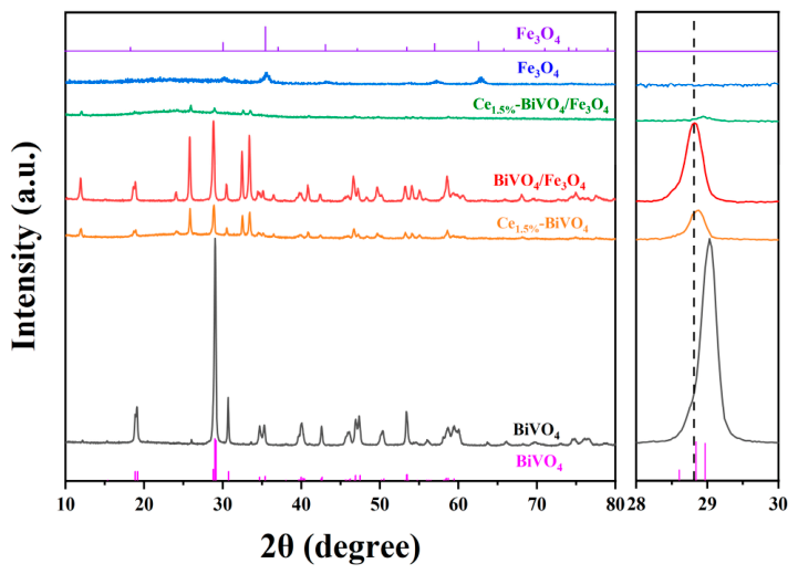
The XRD patterns of the prepared samples are shown in Figure 1. The characteristic diffraction peaks of BiVO4 appear at 19.1, 29.1, 30.6, 34.6, 35.3, 40.0, 42.6, 46.2, 46.8, 47.4, 50.4 and 53.4°, corresponding to the (011), (121), (040), (200), (002), (211), (051), (132), (240), (042), (202) and (161) crystal planes of BiVO4, respectively. All diffraction peaks are in excellent agreement with the standard BiVO4 pattern (JCPDS Card No. 014-0688), and no impurity peaks are detected, confirming the successful synthesis of monoclinic BiVO4 [11]. The main characteristic peaks of Ce1.5%–BiVO4, BiVO4/Fe3O4 and Ce1.5%–BiVO4/Fe3O4 clearly appear, demonstrating that the original crystal structure of the material is maintained after Ce doping and Fe3O4 compositing [12]. The most prominent peaks of BiVO4/Fe3O4 exhibit a slight leftward shift compared with the standard BiVO4 pattern, likely attributable to lattice distortion induced by heterojunction formation [13]. Upon Ce doping, the XRD peaks shift slightly rightward relative to BiVO4/Fe3O4. This phenomenon can be attributed to the fact that the ionic radius of Ce3+(1.14 Å) is slightly smaller than that of Bi3+(1.17 Å) and the partial substitution of Ce3+ for Bi3+ in the BiVO4 lattice leads to a slight contraction of the BiVO4 lattice, which proves the successful doping of Ce3+ [14]. Due to the relatively weak diffraction intensity of cubic Fe3O4, its characteristic peaks are barely observable in the BiVO4/Fe3O4 pattern. The diffraction peak intensity of the samples significantly decreased after Ce doping, as observed in Ce1.5BiVO4 and Ce1.5%-BiVO4/Fe3O4, so this phenomenon is more likely attributed to reduced crystallinity or local disorder induced by Ce doping, leading to an overall decrease in diffraction intensity. Related research needs to be further deepened. In general, moderate reduction in crystallinity is beneficial for increasing surface defects and exposing more active sites to enhance photocatalytic efficiency [15,16,17].

2.1.2. FTIR

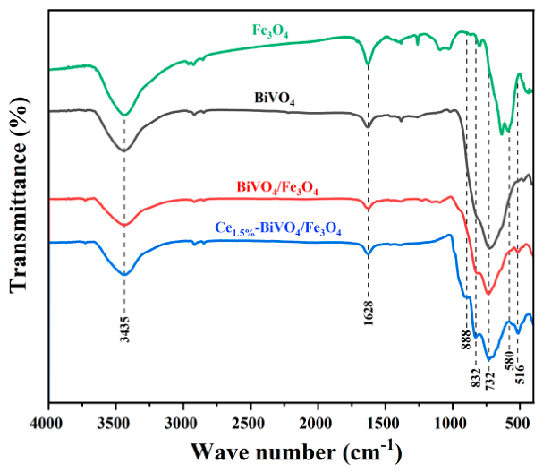
The FTIR spectra of BiVO4, Fe3O4, BiVO4/Fe3O4 and Ce1.5%-BiVO4/Fe3O4 are presented in Figure 2. The characteristic peaks at 3435 cm−1 and 1628 cm−1 correspond to the -OH stretching and bending vibrations of water molecules. The bands at 732 cm−1and 832 cm−1 are attributed to symmetric and asymmetric V-O stretching vibrations in BiVO4 [18], which are observed in the spectra of BiVO4, BiVO4/Fe3O4 and Ce1.5%-BiVO4/Fe3O4. The characteristic Fe-O stretching vibration of Fe3O4 at 580 cm−1 is red-shifted to 516 cm−1 in BiVO4/Fe3O4 and Ce1.5%-BiVO4/Fe3O4, indicating the formation of Fe-O-X chemical bonds in the composite [19,20], which suggests that Fe3O4 is homogeneously coupled with BiVO4. In the spectrum of Ce1.5%-BiVO4/Fe3O4, the peak at 888 cm−1 corresponds to Ce-O bonds [21,22].

2.1.3. XPS Analysis

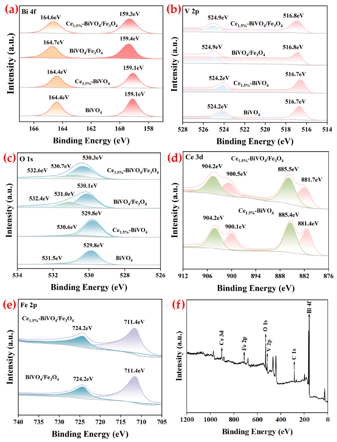
Figure 3a displays the Bi 4f high-resolution XPS spectra. The pure BiVO4 exhibits two distinct peaks at binding energies of 159.1 and 164.4 eV, corresponding to the Bi3+ 4f7/2 and 4f5/2 orbitals, respectively, which aligns with the reported electronic structure of monoclinic BiVO4 [4]. Notably, the Bi 4f peak positions in the Ce1.5%-BiVO4 sample remain largely unchanged, indicating that Ce doping does not significantly alter the local chemical environment or valence state of Bi. In contrast, the Bi 4f peaks for the BiVO4/Fe3O4 composite shift entirely towards higher binding energies. This positive shift is typically attributed to a decrease in electron cloud density around the Bi atoms, suggesting the presence of strong interfacial interactions between Fe3O4 and BiVO4 that may facilitate electron transfer from BiVO4 to Fe3O4 [23]. The V 2p spectrum (Figure 3b) of pure BiVO4 shows characteristic peaks at 516.7 and 524.2 eV, assigned to the V5+ 2p3/2 and 2p1/2 states, respectively [24]. Consistent with the trend observed in the Bi 4f spectra, the V 2p peaks in BiVO4/Fe3O4 also exhibit a distinct positive shift, further confirming a reduction in the surface electron density of V atoms [25,26].
Deconvolution of the O 1s spectra (Figure 3c) reveals that the spectrum for pure BiVO4 can be fitted with two peaks at 529.8 and 531.5 eV. The O 1s spectrum of Ce1.5%-BiVO4 is fitted with peaks at 529.8 and 530.6 eV, where the peak at 530.6 eV is commonly associated with oxygen vacancies (OVs) or low-coordinated oxygen species. The O 1s spectrum of BiVO4/Fe3O4 exhibits a more complex structure, which can be deconvoluted into three sub-peaks at 530.1 eV (lattice oxygen, Bi-O), 531.0 eV (oxygen vacancies, OVs), and 532.4 eV (chemisorbed oxygen species, such as -OH) [27]. Comparing the relative area of the peaks corresponding to oxygen vacancies reveals that the OVs signals for both Ce1.5%-BiVO4 and BiVO4/Fe3O4 are significantly stronger than that of pure BiVO4. This indicates that both Ce doping and the formation of the BiVO4/Fe3O4 composite induce a higher density of oxygen vacancies [28].
The Ce 3d spectrum (Figure 3d) reveals peaks at 881.7 and 885.5 eV for Ce3+, and at 900.1 and 904.2 eV for Ce4+ [29,30,31], confirming the coexistence of Ce3+ and Ce4+ species. The Fe 2p spectrum (Figure 3e) displays broadened peaks at 711.4 and 724.2 eV, consistent with the coexistence of Fe2+ and Fe3+ in Fe3O4 [32]. The survey spectrum (Figure 3f) clearly identifies signals from Bi, V, O, Ce, and Fe, corroborating successful composite formation.

2.1.4. UV-Vis Diffuse Reflectance Spectroscopy and Mott–Schottky Plots

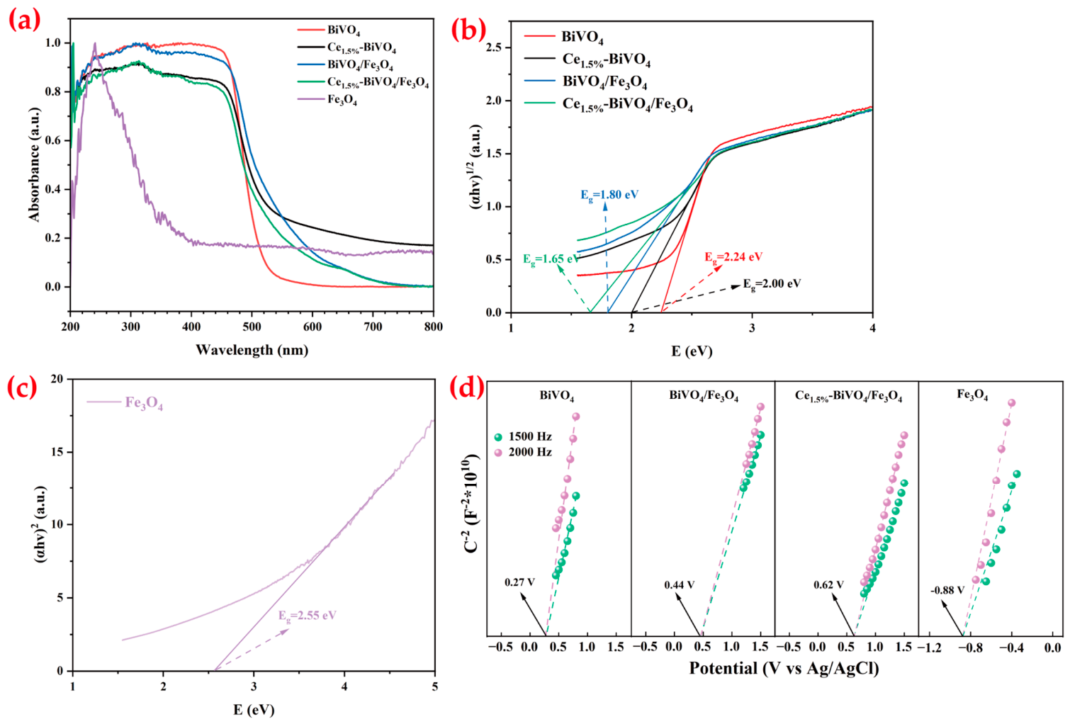
The UV-Vis diffuse reflectance spectra are shown in Figure 4a. All samples exhibit absorption in the ultraviolet region (200–500 nm). Both BiVO4/Fe3O4 and Ce1.5%-BiVO4/Fe3O4 show further red-shifted absorption edges extending to 750 nm, substantially enhancing visible light absorption and providing a reliable basis for improved photocatalytic performance. The band positions of BiVO4, Ce1.5%-BiVO4, Fe3O4, BiVO4/Fe3O4, and Ce1.5%-BiVO4/Fe3O4 were determined via UV-Vis diffuse reflectance spectra and Mott–Schottky curves (Figure 4d). The bandgap (Eg) (an effective optical response of the composite) of the synthesized photocatalysts is typically estimated using the Kubelka–Munk method, which is expressed by the equation (αhν)n = A (hν-Eg). Herein, α, h, ν, and A denote the absorption coefficient, Planck constant, light frequency, and a constant, respectively. The value of n depends on the transition characteristics of the semiconductor, and Eg can be derived by plotting (αhν)n against it. Since BiVO4 is a direct allowed transition semiconductor [33], n = 1/2 is adopted. Plotting (αhν)1/2 versus hν (Figure 4b) yields a bandgap of approximately 2.24 eV for BiVO4. Fe3O4 is a metal oxide, so n = 2 is used [34]. By plotting (αhν)1/2 versus hν, the bandgap of Fe3O4 is estimated to be about 2.55 eV (Figure 4c). The calculated bandgap values (Figure 4b) are 2.00 eV for Ce1.5%-BiVO4, 1.80 eV for BiVO4/Fe3O4, and 1.65 eV for Ce1.5%-BiVO4/Fe3O4. The Ce1.5%-BiVO4/Fe3O4 composite possesses the smallest bandgap (1.65 eV), which requires the lowest excitation energy for electron transition from the valence band (VB) to the conduction band (CB), thereby maximizing the visible light response and utilization efficiency. The bandgap value of 1.65 eV for Ce1.5%-BiVO4/Fe3O4 is unusually low and it should be considered as an apparent/effective optical response of the composite, rather than a conventional intrinsic bandgap. The strong narrowing of the bandgap can be attributed to the chemical bonds formed between BiVO4 and Ce species [35]. Enhancing visible light utilization is highly beneficial for improving the photocatalytic efficiency of the catalysts [36].
The Mott–Schottky plots in Figure 4d show that the flat-band potentials (VFB) of the BiVO4, BiVO4/Fe3O4, and Ce1.5%-BiVO4/Fe3O4 samples are 0.27, 0.44, and 0.62 V (vs. Ag/AgCl), respectively. Compared with BiVO4, the flat-band potentials of BiVO4/Fe3O4 and Ce1.5%-BiVO4/Fe3O4 exhibit a significant positive shift, indicating reduced band bending and a lower Schottky barrier. This can be attributed to the decreased Fermi level, which enhances the photogenerated electron transfer efficiency of the composite photocatalysts [37]. The even more positive flat-band potential observed for the Ce-doped composite indicates that Ce incorporation may significantly modify the band structure of BiVO4. Ce ions, with their unique 4f electronic configuration, act as electron acceptors in the BiVO4 lattice, introducing localized defect states near the conduction band. These defect states may trap photogenerated electrons, further reducing the electron density in the surface region of BiVO4 and leading to a more pronounced positive shift in the flat-band potential. In addition, the Mott–Schottky curves measured at frequencies of 1500 Hz and 2000 Hz display positive slopes for both BiVO4 and Fe3O4, confirming that both are n-type semiconductors [38]. The flat-band potentials (vs. Ag/AgCl) of BiVO4 and Fe3O4 are determined to be 0.27 and −0.88 V, respectively, from the Mott–Schottky plots. Typically, the CB potential (vs. NHE, pH = 7) is approximately 0.2 eV more negative than the flat-band potential. Based on Equation (1), the CB potentials of BiVO4 and Fe3O4 are calculated to be 0.47 and −0.68 V (vs. NHE, pH = 7), respectively. Furthermore, according to Equation (2), the corresponding VB potentials are determined to be +2.71 and +1.87 V (vs. NHE, pH = 7).
E NHE ,   pH = 7 = E Ag/AgCl + 0.197
E g = E VB E CB

2.1.5. PL and EIS Nyquist Analysis

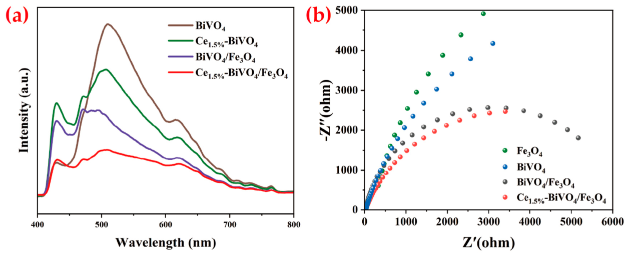
The separation efficiency of photogenerated electron–hole pairs was characterized by photoluminescence (PL) spectroscopy. The PL emission peaks originate from the recombination of photogenerated carriers, and their intensity directly reflects the separation efficiency of electron–hole pairs. Generally, a lower fluorescence intensity indicates greater suppression of carrier recombination and a higher separation efficiency [39]. As shown in Figure 5a, under excitation at 375 nm, BiVO4, Ce1.5%-BiVO4, BiVO4/Fe3O4 and Ce1.5%-BiVO4/Fe3O4 exhibit a distinct emission peak at approximately 508 nm, which is attributed to the recombination of photoexcited electrons and holes. Notably, the PL intensity of Ce1.5%-BiVO4/Fe3O4 is significantly lower than that of pure BiVO4, indicating that the introduction of Ce effectively suppresses the recombination of photogenerated carriers and enhances the separation of electron–hole pairs [40]. Meanwhile, Fe3O4 possesses favorable electronic conductivity, which facilitates the migration of photogenerated carriers [41]. Therefore, the improved charge separation and migration efficiency synergistically enhance the photocatalytic performance of the composite.
Electrochemical impedance spectroscopy (EIS) is commonly employed to investigate the migration resistance and separation efficiency of photogenerated charge carriers in semiconductors. The interfacial resistance at the electrode surface is reflected by the arc radius in the Nyquist plots. Generally, a smaller interfacial resistance leads to higher charge transfer efficiency, which corresponds to superior material performance [42]. As shown in Figure 5b, the Nyquist plots of all composite samples exhibit a semicircular arc in the high-frequency region. Compared with pure BiVO4 and pure Fe3O4, the BiVO4/Fe3O4 and Ce1.5%-BiVO4/Fe3O4 samples display smaller arc radii. Among them, the Ce1.5%-BiVO4/Fe3O4 composite exhibits the smallest arc radius, indicating that this system possesses the lowest migration resistance for photogenerated charge carriers, thereby effectively suppressing their recombination. The above photoelectrochemical analysis reveals that the introduction of an appropriate amount of Ce and Fe3O4 significantly increases the number of photogenerated charge carriers.

2.1.6. TEM Analysis

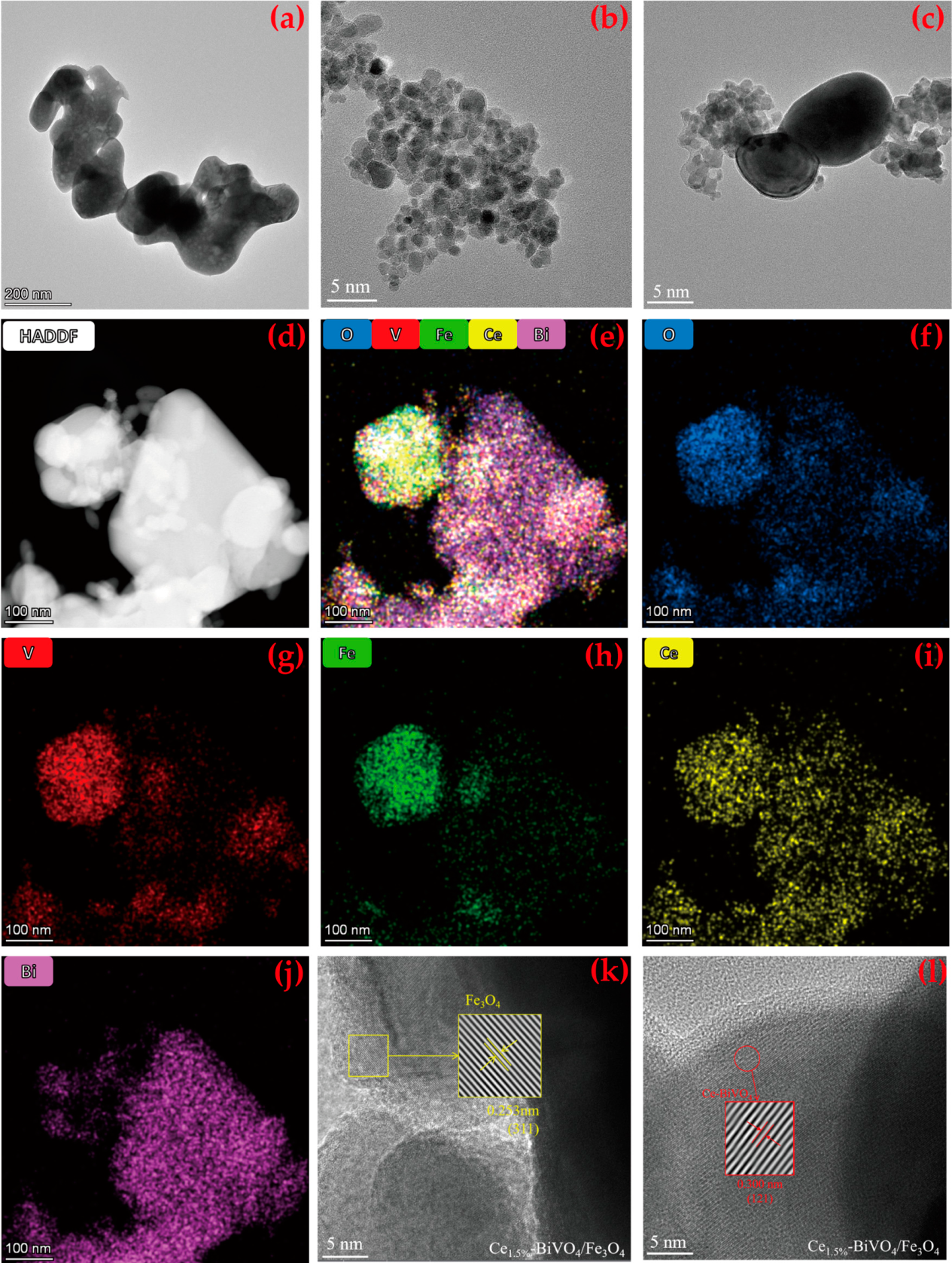
Figure 6 presents the TEM images of BiVO4, Fe3O4, BiVO4/Fe3O4 (Figure 6a–c) and high-angle annular dark-field (HAADF) imaging with elemental mapping of Ce1.5%-BiVO4/Fe3O4 (Figure 6d–j), which reveal uniform distribution of O, V, Fe, Ce, and Bi elements, confirming successful doping of Ce into the BiVO4 matrix. High-resolution TEM images (Figure 6k–l) show lattice fringes with interplanar spacings of 0.253 nm, corresponding to the (311) plane of cubic Fe3O4 [43], and 0.300 nm, corresponding to the (121) plane of monoclinic BiVO4. This slight decrease in interplanar spacing may result from lattice contraction induced by Ce doping [44,45]. The clear interfacial region between BiVO4 and Fe3O4 demonstrates heterojunction formation, which is beneficial for photocatalytic enhancement [28].
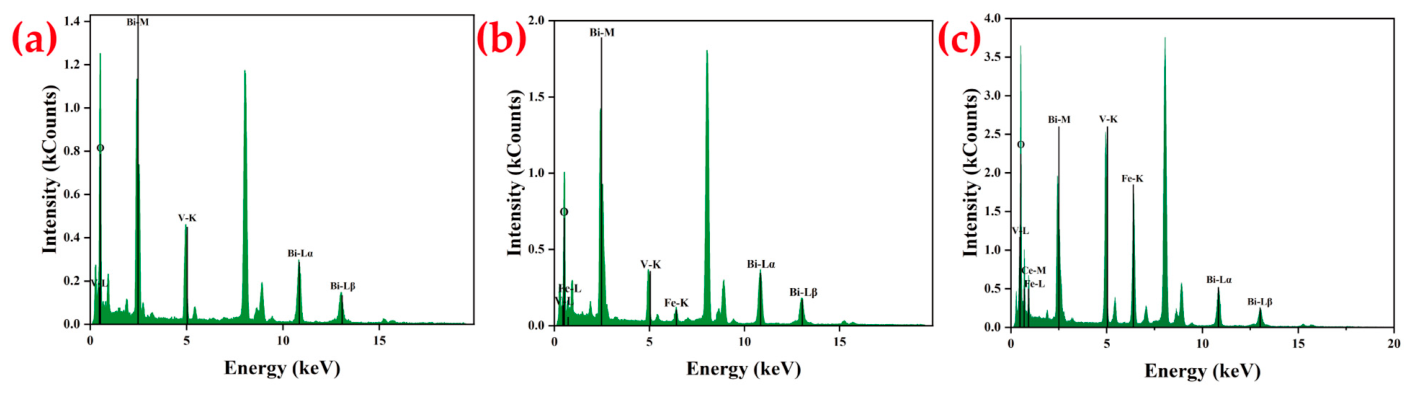
The EDS spectra of BiVO4, BiVO4/Fe3O4, and Ce1.5%-BiVO4/Fe3O4 (Figure 7) confirm the presence of Bi, V, and O in BiVO4; Bi, V, O, and Fe in BiVO4/Fe3O4; and Bi, V, O, Fe, and Ce in Ce1.5%-BiVO4/Fe3O4, further validating successful material synthesis.
Table 1 shows the semi-qualitative analysis data of each element in the elemental mapping images of BiVO4, BiVO4/Fe3O4, and Ce1.5%-BiVO4/Fe3O4 samples. It summarizes the mass fractions of key elements (O, V, Fe, Ce, Bi) in BiVO4, BiVO4/Fe3O4, and Ce1.5%-BiVO4/Fe3O4 samples prepared by the co-precipitation method, with the data obtained from EDS mapping analysis. The data presented in Table 1 were acquired from random regions of different samples during characterization, and are affected by sampling depth and local inhomogeneity. Although they can reflect the inhomogeneous elemental distribution in the corresponding regions, the elemental contents among different samples are not directly comparable. Therefore, these semi-quantitative results can only confirm the successful incorporation of the elements, but cannot fully represent the true proportions of each element in the samples.

2.1.7. BET Analysis

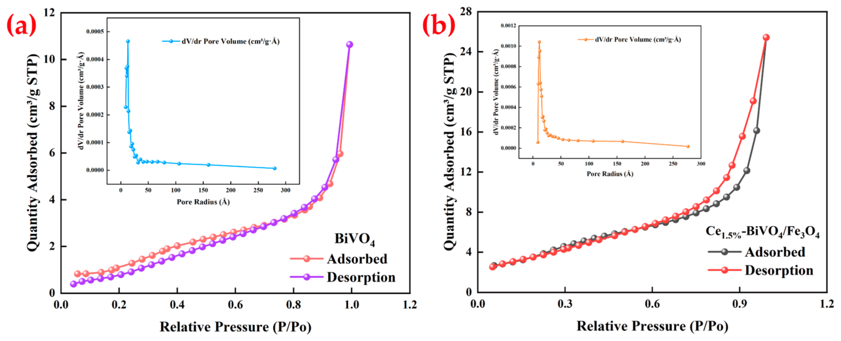
Figure 8 shows the N2 adsorption–desorption isotherms and pore size distribution curves of pure BiVO4 and Ce1.5%-BiVO4/Fe3O4. N2 adsorption–desorption measurements were performed to investigate the effects of Ce doping and Fe3O4 composite modification on the specific surface area and pore structure of BiVO4. The Barrett Joyner Halenda (BJH) model was applied to analyze the pore size distribution of the samples. As displayed in Figure 8a,b, the Ce1.5%-BiVO4/Fe3O4 composite exhibits a typical Type IV isotherm, confirming the existence of a mesoporous structure. The specific surface areas of BiVO4 and Ce1.5%-BiVO4/Fe3O4 are 4.78 and 14.63 m2/g, while their average pore diameters are 6.5290 and 6.0273 nm, respectively. Compared with the pure material, the larger specific surface area of the composite effectively enlarges the contact interface between the catalyst and reaction substrates and provides more active sites for the photocatalytic reaction [46].

2.2. Photocatalytic Performance

2.2.1. Photocatalytic Activity

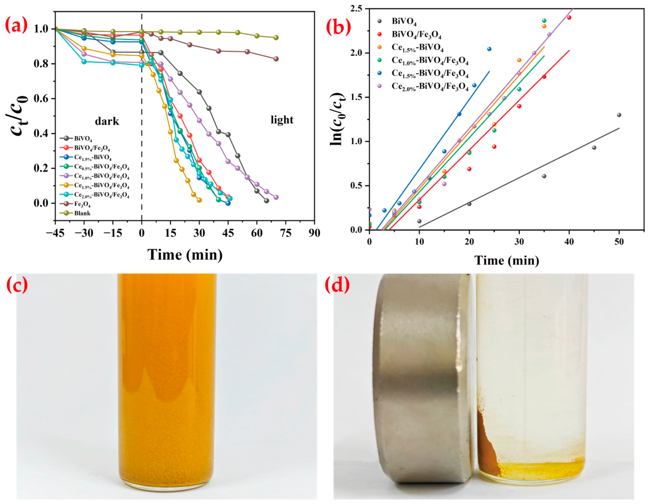
Figure 9a displays the adsorption and photodegradation curves of RhB with and without catalysts. It can be observed that all samples basically reach the adsorption equilibrium within 45 min. The degradation efficiency first increases and then decreases with increasing Ce content. Ce1.5%-BiVO4/Fe3O4 exhibits the optimal performance, achieving 100% RhB removal within 30 min. Within the same 30 min period, the degradation rates of RhB using BiVO4, BiVO4/Fe3O4, Ce1.5%-BiVO4, Ce1.0–BiVO4/Fe3O4, and Ce2.0–BiVO4/Fe3O4 as photocatalysts were only 37.6%, 75.2%, 85.3%, 81.3%, and 83.9%, respectively. Meanwhile, Figure 9a compares the degradation efficiencies of BiVO4, BiVO4/Fe3O4, Ce1.5%-BiVO4, and Ce1.5%-BiVO4/Fe3O4, which require 65, 45, 40, and 30 min for complete RhB degradation respectively. Fe3O4 exhibited negligible photocatalytic activity toward RhB degradation under visible light irradiation. The modified composites demonstrate significant performance enhancement, with BiVO4/Fe3O4, Ce1.5%-BiVO4 and Ce1.5%-BiVO4/Fe3O4 showing 1.4-fold, 1.6-fold, and 2.2-fold improvements in degradation efficiency compared with pure BiVO4. Heterojunction formation between BiVO4 and Fe3O4 promotes photogenerated charge separation and suppresses recombination. Ce doping further enhances performance. This is mainly due to the fact that when an appropriate amount of Ce is doped into BiVO4/Fe3O4, the Ce4+ ions capture some electrons and reduce to Ce3+ ions, thereby preventing the recombination of e and h+ in BiVO4/Fe3O4 photocatalysts and improving the photocatalytic degradation efficiency. In addition, Ce ions possess a rich energy level structure, which narrows the bandgap of BiVO4/Fe3O4 photocatalysts and enhances visible light absorption (as shown in Figure 4b). However, excessive Ce doping introduces defects that act as recombination centers, deteriorating photocatalytic activity. The reaction rate constants (Figure 9b) are 0.028, 0.056, 0.064, and 0.079 min−1 for BiVO4, BiVO4/Fe3O4, Ce1.5%-BiVO4 and Ce1.5%-BiVO4/Fe3O4, respectively. As shown in Table 2, the goodness-of-fit indicators (R2 values) of the degradation models for BiVO4, BiVO4/Fe3O4, Ce1.5%-BiVO4, and Ce1.5%-BiVO4/Fe3O4 are 0.95, 0.97, 0.95, and 0.99, respectively, all of which are greater than 0.9, indicating an excellent model fit [47]. The adjusted R2 values are 0.94, 0.96, 0.94, and 0.99, respectively, with corresponding differences of 0.01, 0.01, 0.01, and 0.00, all less than 0.1, demonstrating that the models are reasonable and reliable [48]. Meanwhile, total organic carbon (TOC) analysis was performed on Ce1.5%-BiVO4/Fe3O4. After 30 min of visible light irradiation, the TOC removal efficiency of the target pollutant solution reached 90.20%, verifying its promising application potential in wastewater treatment. As summarized in Table 3, the photocatalytic degradation performance of various reference catalysts toward RhB was investigated in this study, all of which exhibited relatively limited activity. This table is only for general comparison with existing literature, rather than a one-to-one quantitative benchmark comparison under identical experimental conditions. From Table 3, the Ce1.5%-BiVO4/Fe3O4 composite prepared in this work demonstrated the superior photocatalytic activity among all tested catalysts, with the detailed performance parameters presented in the table.

2.2.2. Magnetic Separation Performance

The magnetic separability of Ce1.5%-BiVO4/Fe3O4 was evaluated. Figure 9c shows the uniform dispersion of the catalyst in solution, while Figure 9d presents a separation effect diagram of sample and solution. The results indicate that under the influence of a magnet, the solution became clear within 1 min, achieving rapid separation of the Ce1.5%-BiVO4/Fe3O4 sample after degrading RhB from the aqueous phase. Therefore, Ce1.5%-BiVO4/Fe3O4 possesses excellent paramagnetic properties, which can be utilized to achieve rapid separation and recycling of Ce1.5%-BiVO4/Fe3O4 from the aqueous phase.

2.2.3. Reusability Performance

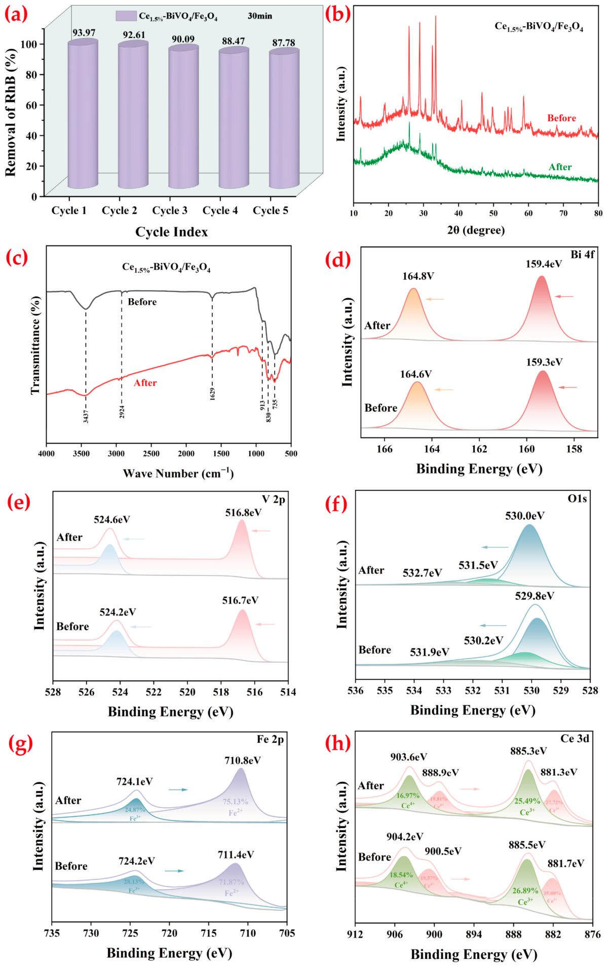
The stability of Ce1.5%-BiVO4/Fe3O4 was assessed through five consecutive cycles of RhB degradation (30 min per cycle). As shown in Figure 10a, the RhB removal rate gradually decreases with cycling but remains above 90% after five cycles, demonstrating excellent reusability and commercial potential [51,52].
As shown in Figure 10b,c, for the recycled Ce1.5%-BiVO4/Fe3O4 composite after cyclic degradation experiments, no obvious shift in the XRD characteristic diffraction peaks is observed, and no significant difference appears in the FTIR spectra. This confirms that the crystal structure of the material remains stable after cycling tests. However, the intensity of the diffraction peaks decreased, indicating that a small number of defects may form inside the material after recycling, which may lead to a slight reduction in photocatalytic efficiency. XPS analysis was conducted to further investigate the interfacial behavior and variations in the chemical states of elements within the Ce1.5%-BiVO4/Fe3O4 composite. In the Bi 4f and V 2p spectra, the characteristic peaks of the Ce1.5%-BiVO4/Fe3O4 composite shifted toward higher binding energies by approximately 0.2 and 0.4 eV, respectively. Similarly, the OL peak in the O 1s spectrum exhibited a notable shift to a higher binding energy (Figure 10d–h). In contrast, the Fe 2p3/2 spectrum was deconvoluted into two components corresponding to Fe2+ and Fe3+ species, with their binding energies shifting to lower values by about 0.6 and 0.1 eV, respectively, indicating electron enrichment around the iron atoms (Figure 10g). Nevertheless, such a shift may also arise from other factors as the test is based on ex situ measurements, so further investigation is required to verify the conclusions. Moreover, upon light irradiation, the proportion of Fe3+ species decreased from 28.13% to 24.87%, while that of Fe2+ increased from 71.87% to 75.13% [28]. A similar trend was observed for the Ce species, with the Ce4+ ratio decreasing from 38.11% to 36.78% and the Ce3+ ratio increasing from 61.89% to 63.21% (Figure 10h). This may be attributed to the fact that under visible light irradiation, Fe3+ and Ce4+ in the system can effectively capture photogenerated electrons and be reduced to Fe2+ and Ce3+, thereby significantly enhancing the overall charge separation efficiency at the BiVO4/Fe3O4 interface.

2.3. Degradation Mechanism

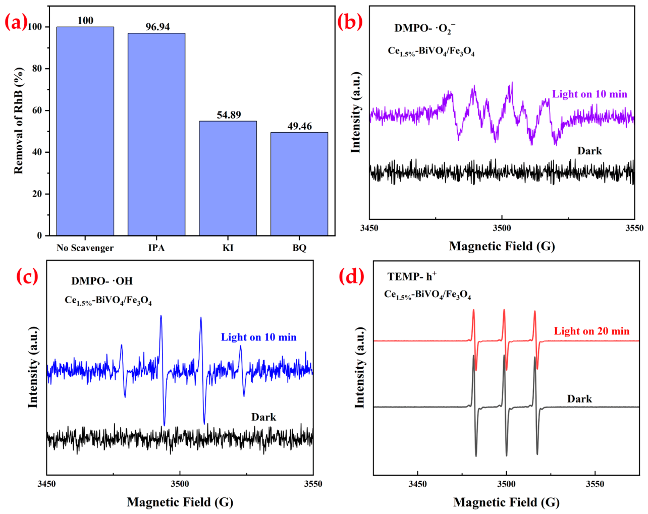
Radical scavenging experiments were conducted to identify the primary active species in the Ce1.5%-BiVO4/Fe3O4 system. Potassium iodide (KI), isopropanol (IPA), and benzoquinone (BQ) were employed as scavengers for holes (h+), hydroxyl radicals (·OH) and superoxide radicals ( · O 2 - ), respectively [53]. As shown in Figure 11a, the addition of IPA causes only a 3.06% decrease in degradation efficiency (96.94% retention), whereas KI and BQ addition reduces efficiency to 54.89% and 49.46%, respectively. These results confirm that h+ and · O 2 - are the predominant reactive species, while ·OH plays a weak role. EPR tests were performed using DMPO and TEMP as spin-trapping agents (Figure 11b–d) to further identify the reactive oxygen species generated in the Ce1.5%-BiVO4/Fe3O4 photocatalytic system. Compared with the smooth baseline measured in the dark, the characteristic sextet signal of · O 2 - was significantly enhanced under photoexcitation (Figure 11b), confirming the abundant production of · O 2 - radicals. No obvious characteristic peaks were observed under dark conditions, while a weak signal corresponding to ·OH was detected upon light irradiation (Figure 11c), indicating that ·OH participated in the degradation process. Meanwhile, the 1:1:1 triplet signal intensity of h+ under illumination was remarkably lower than that in the dark (Figure 11d), suggesting that h+ was continuously consumed during the photocatalytic reaction. These EPR results were consistent with those of radical quenching experiments, further verifying that the active species produced by Ce1.5%-BiVO4/Fe3O4 played a crucial role in the photocatalytic degradation of RhB [45,54].
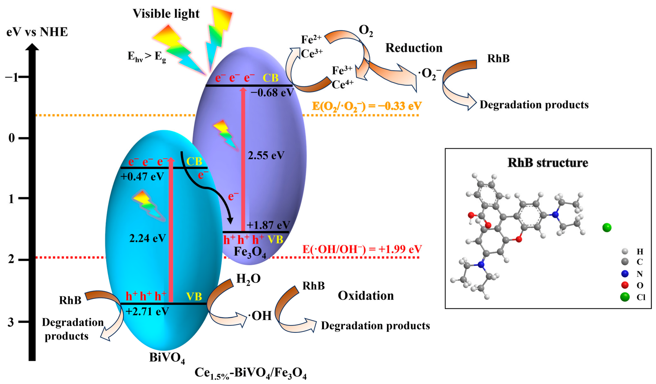
There are two possible reaction mechanisms for the photocatalytic degradation of RhB by Ce1.5%-BiVO4/Fe3O4 under visible light irradiation. According to the traditional type-II photocatalytic mechanism, BiVO4 and Fe3O4 are simultaneously excited under visible light irradiation. Subsequently, a large number of electrons jump from VB to CB within the same semiconductor, leaving h+ in the VB. CB potential of Fe3O4 (Eg = −0.68 eV vs. NHE) is more negative than that of BiVO4 (Eg = +0.47 eV vs. NHE), so the electrons in the CB of Fe3O4 can jump to the CB of BiVO4. In contrast, VB potential of BiVO4 (Eg = +2.71 eV vs. NHE) is more positive than that of Fe3O4 (Eg = +1.87 eV vs. NHE), which allows the migration of h+ from BiVO4 to Fe3O4. However, the type-II photocatalytic mechanism cannot explain the generation of ·OH and · O 2 - radicals, because the VB potential of Fe3O4 cannot reach the potential of H2O/·OH (+1.99 eV), and the CB potential of BiVO4 also cannot reach the potential of O2/ · O 2 - (−0.33 eV). Based on these findings from Mott–Schottky analysis, UV-Vis, radical trapping, XPS and EPR, this study speculates that the most plausible charge-transfer pathway of Ce-BiVO4/Fe3O4 photocatalyst follows a Z-scheme charge transfer mechanism [55]. The detailed mechanism of electron transfer remains to be further investigated due to current limitations in research techniques. Based on the available data, the photocatalytic mechanism is speculated as illustrated in Figure 12. Under visible light irradiation, electrons are excited from the VB to CB in both BiVO4 and Fe3O4, generating holes in the VB (Equation 3). Owing to the conductivity of Fe3O4, electrons in the CB of BiVO4 (Eg = +0.47 eV νs. NHE) migrate to the VB of Fe3O4 (Eg = +1.87 eV νs. NHE) at the heterojunction interface through a Z-scheme charge transfer pathway [55] (Equation (4)), where electron–hole recombination occurs, prolonging electron lifetime and promoting charge separation. Subsequently, electrons in the CB of Fe3O4 (Eg = −0.68 eV νs. NHE) reduce Fe3+ to Fe2+ (Equation 5), and Fe2+ reacts with adsorbed O2 to generate · O 2 - (−0.33 eV) (Equation 6) [53]. Meanwhile, some electrons are captured by Ce4+ to form Ce3+ (Equation 7), preventing electron–hole recombination. The Ce3+ species can also react with adsorbed oxygen to produce · O 2 - (Equation 8). Simultaneously, holes in the VB of BiVO4 (Eg = +2.71 eV νs. NHE) directly oxidize RhB molecules. Ultimately, RhB is decomposed by h+ (Equation 9) and · O 2 - (Equation 10). It should be noted that Figure 12 is merely a schematic based on the band structures of pure-phase BiVO4 and pure-phase Fe3O4, and does not fully represent the intrinsic true energy level structure of the composite photocatalyst. Ce doping introduces localized energy levels and lattice distortions that alter the band structure of BiVO4, while the heterojunction interface between BiVO4 and Fe3O4 induces band bending. These effects, which are not captured by pure-phase data, mean the diagram cannot fully reflect the actual electronic structure of the Ce1.5%-BiVO4/Fe3O4 ternary composite. Compared with the degradation mechanism of RhB reported by Luo et al. [49] for the Ce-doped BiVO4 system, the mechanism proposed herein exhibits distinct characteristics. In the Ce-BiVO4 system, the photocatalytic activity primarily relies on Ce4+ acting as an electron trap to form Ce3+, which subsequently promotes the generation of ·OH as the dominant reactive species. Furthermore, the separation efficiency in that system is modulated by the doping concentration through its influence on the space-charge region potential [49]. In contrast, the BiVO4/Fe3O4 composite developed in this work employs a Z-scheme heterojunction to facilitate interfacial charge separation. Coupled with the synergistic effect between the Fe3+/Fe2+ redox cycle and the Ce4+/Ce3+ transition, the present system utilizes ·O2- and h+ as the main active species for direct pollutant degradation. Moreover, the role of Ce in this composite extends beyond simple electron trapping to include band-structure modulation, forming multiple charge-transfer channels with Fe3O4 that collectively enhance carrier separation and the generation of reactive species. This integrated “heterojunction-doping” synergy thus establishes a more efficient and robust photocatalytic mechanism.
Photocatalyst + hν → e- + h+
eCB-(BiVO4) + hVB+(Fe3O4) → recombination
e- + Fe3+ → Fe2+
Fe 2 + + O 2 · O 2 - Fe 3 +
Ce4+ + e- → Ce3+
Ce 3 + + O 2 · O 2 - Ce 4 +
RhB + h+ → degradation products
RhB + · O 2 - degradation products

3. Materials and Methods

3.1. Reagents and Instruments

Sodium metavanadate (NaVO3, 99.0%), bismuth chloride (BiCl3), cerium nitrate hexahydrate (Ce(NO3)3·6H2O, 99.95%), nano-sized iron(II,III) oxide (Fe3O4, 99.5%), ammonium hydroxide (NH3·H2O, 25.0–28.0%) and concentrated hydrochloric acid (HCl, 36.0–38.0%) were purchased from Sinopharm Chemical Reagent Co., Ltd. (Shanghai, China). All reagents were of analytical grade and used as received without further purification.
Instruments used: D8 Advance X-ray diffractometer (XRD, Bruker, Billerfeld, Germany); Nicolet 6700 Fourier transform infrared spectrometer (FTIR, Thermo Fisher Scientific, Waltham, MA, USA); ESCALAB XI+ X-ray photoelectron spectrometer (XPS, Thermo Fisher Scientific, Waltham, MA, USA); Cary 5000 UV-Vis spectrophotometer (Agilent Technologies, Santa Clara, CA, USA); JEM-F200 transmission electron microscope (TEM, JEOL, Tokyo Metropolis, Tokyo, Japan). Photoluminescence emission (PL) spectra of the samples were detected using a fluorescence spectrophotometer (F97Pro, Shanghai Lengguang Technology Co., Ltd., Shanghai, China) equipped with a 450 W Xe lamp as the excitation source. Electrochemical impedance spectroscopy (EIS) measurements were performed using Na2SO4 (0.3 mol/L) with frequency ranging between 0.1 and 106 Hz and an amplitude of 0.01 V. The Mott–Schottky curves were obtained by an electrochemical workstation (CHI660E, Shanghai Chenhua Instruments Co., Ltd., Shanghai, China) with a potential range of −0.5 to 1 V and a test frequency of 3000 Hz. The surface area and pore size were measured by a surface area and porosity analyzer (ASAP2460-2, Mike Company, Norcross, GA, USA).

Electron Paramagnetic Resonance (EPR)

All EPR spectra were recorded at room temperature using a Bruker EMXnano spectrometer (USA), with specific conditions for each active species as follows:
O 2 - : A total of 10 mg of catalyst was dispersed in 1 mL of methanol containing 50 mM DMPO. The mixture was irradiated with visible light (300 W xenon lamp, λ > 420 nm) for 10 min, and the spectrum was recorded immediately. A characteristic six-line spectrum of the DMPO- · O 2 - adduct was observed (a(N) ≈ 1.5 mT, a(H) ≈ 1.1 mT).
OH: A total of 10 mg of catalyst was dispersed in 1 mL of deionized water containing 50 mM DMPO. The mixture was irradiated following the same method described above. A characteristic four-line spectrum of the DMPO- ·OH adduct was observed (a(N) ≈ a(H) ≈ 1.5 mT).
h+: A total of 10 mg of catalyst was dispersed in 1 mL of deionized water containing 0.1 mM TEMPO. EPR spectra were recorded before visible light irradiation (dark state—the mixture was kept in the dark for 5 min, and then the EPR spectrum was recorded to establish the baseline signal intensity of TEMPO) and after 20 min of irradiation (irradiated with visible light from a 300 W xenon lamp with λ > 420 nm for 20 min, and the spectrum was recorded immediately afterward), respectively. Photogenerated holes oxidize TEMPO (1:1:1 triplet, g ≈ 2.006) into an EPR-silent oxoammonium cation, resulting in a decrease in signal intensity under light irradiation compared with the dark state.

3.2. Photocatalyst Preparation

3.2.1. Synthesis of BiVO4

A total of 2 mmol of BiCl3 and 2 mmol of NaVO3 were dissolved separately in 1.0 mL of 6 mol·L−1 dilute HCl and 19.0 mL of deionized water, respectively. Under ultrasonication, the bismuth chloride solution was added dropwise to the sodium metavanadate solution. The pH was adjusted to 8–9 with ammonia water. Subsequently, the mixture was mechanically stirred continuously at 500–600 r·min−1 at 80 °C for 0.5 h. After completion of the reaction, the resulting product was centrifuged at 4000 r·min−1 for 10 min at room temperature. The supernatant was discarded and the solid product was collected. The solid was washed with deionized water and anhydrous ethanol three times each, followed by centrifugation at 4000 r·min−1 for 10 min after each washing step. The final product was vacuum-dried at 80 °C for 12 h. The dried sample was ground and calcined in a muffle furnace at 400 °C for 2 h (heating rate: 5 °C·min−1).

3.2.2. Synthesis of Ce-BiVO4, BiVO4/Fe3O4 and Ce-BiVO4/Fe3O4 Composites

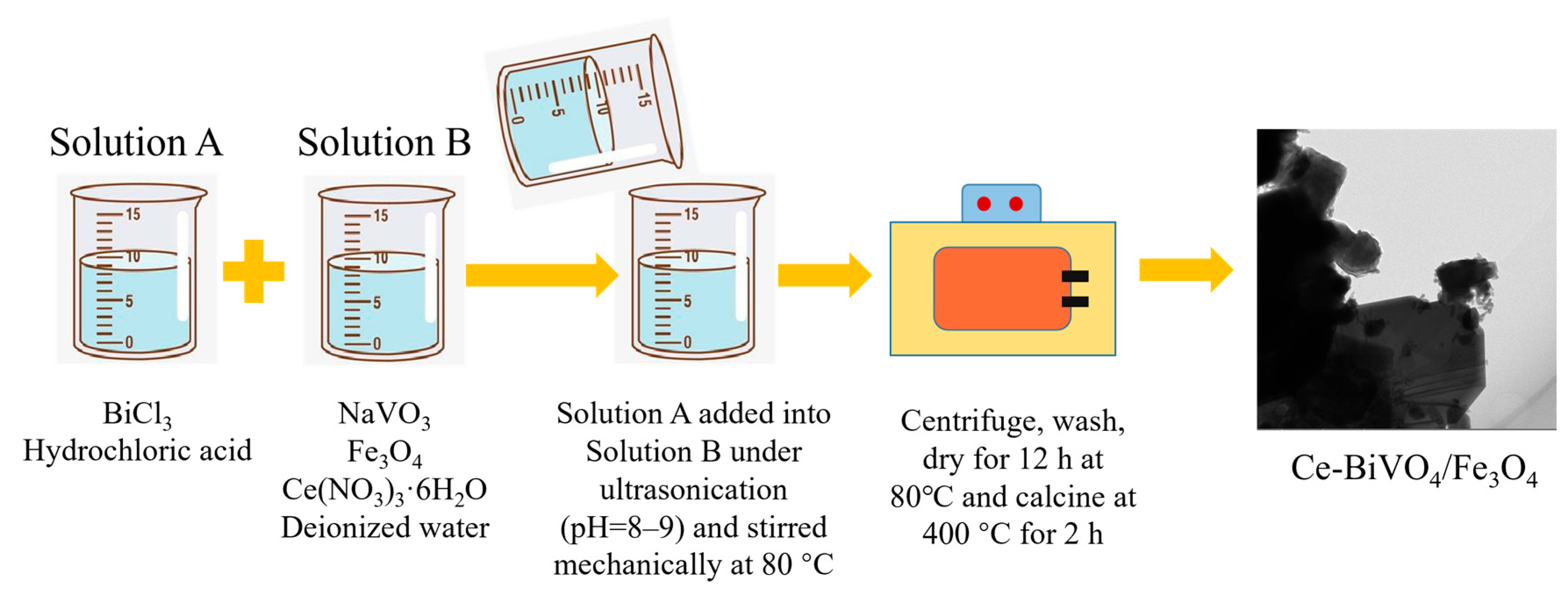
The preparation procedure is illustrated in Scheme 1. A total of 2 mmol of BiCl3 and 2 mmol of NaVO3 were dissolved separately in 1.0 mL of HCl (6mol·L−1) and 19.0 mL of deionized water. To the sodium metavanadate solution, 0.0648 g of Fe3O4 was added (Fe3O4:BiVO4 mass ratio = 10%). For cerium-doped samples, Ce(NO3)3·6H2O was added at Ce:V molar ratios of 0.5%, 1.0%, 1.5% and 2.0%, respectively. Under ultrasonic irradiation, the bismuth chloride solution was added dropwise into the sodium metavanadate solution, and the pH was adjusted to 8–9 with ammonia water. The mixture was reacted at 80 °C for 0.5 h under mechanical stirring at 500–600 r·min−1. After the reaction, the product was centrifuged at 4000 r·min−1 for 10 min at room temperature. The solid product was washed three times with deionized water and anhydrous ethanol, respectively, followed by centrifugation at 4000 r·min−1 for 10 min after each washing step. The final product was vacuum-dried at 80 °C for 12 h, ground, and then calcined at 400 °C for 2 h with a heating rate of 5 °C·min−1.

3.3. Photocatalytic Degradation Experiments

A 10.00 mg·L−1 rhodamine B (RhB) solution was prepared, and its initial absorbance was measured using a UV–visible spectrophotometer at λ= 554 nm. Then, 0.1000 g of photocatalyst was added to 100 mL of the RhB solution with a concentration of 10.00 mg·L−1. The suspension was magnetically stirred at 400–500 r·min−1 for 45 min at room temperature in the dark and without aeration or oxygen supply to achieve adsorption–desorption equilibrium. Subsequently, the photocatalytic reaction was carried out under visible light irradiation using a 300 W xenon lamp (the light intensity was maintained at 600 mW/cm2) with optical filters, at a distance of approximately 10 cm from the liquid surface. Cooling water was continuously circulated to maintain room temperature. Samples were periodically collected, separated and measured until complete RhB degradation was achieved. After each photocatalytic test, BiVO4/Fe3O4 and Ce1.5%-BiVO4/Fe3O4 were magnetically recovered for cyclic reusability evaluation.
The degradation efficiency was calculated using Equation (11):
R h B   r e m o v a l   r a t e = C 0 C t C 0 × 100 %  
c0 and ct represent the initial and instantaneous concentrations of RhB.
The kinetic constants of the samples were calculated via the Langmuir–Hinshelwood model.
r = d c d t = k K c t + K c 0  
In the formula, r is the reaction rate, c0 is the initial concentration of the reactant, ct is the reactant concentration at time t, t is the illumination time, K is the reaction rate constant under the given experimental conditions, and k is the adsorption constant of the reactant.
In the experiments in this paper, the initial concentration is relatively low, and Kc0 can be neglected. Therefore, the model is simplified to a first-order equation:
r = d c d t   =   k K c t =   K α c t
i.e.,
ln C t C 0 = K α t
where Kα is the first-order reaction rate constant.

4. Conclusions

In this study, BiVO4/Fe3O4 and Ce-BiVO4/Fe3O4 composite photocatalysts were successfully synthesized via a facile co-precipitation method. The materials were characterized by XRD, FTIR, XPS, UV-Vis, TEM, EDS, PL, Mott–Schottky analysis, EIS, BET, and EPR. Photocatalytic investigations revealed that BiVO4/Fe3O4, Ce1.5%-BiVO4, and Ce1.5%-BiVO4/Fe3O4 exhibited 1.4-fold, 1.6-fold, and 2.2-fold enhancements in RhB degradation efficiency compared with pure BiVO4, respectively. Heterojunction formation between BiVO4 and Fe3O4 promoted photogenerated charge separation, while Ce doping introduced electron-trapping centers via Ce4+/Ce3+ redox cycling and reduced the bandgap energy, thereby expanding visible light absorption and improving photocatalytic efficiency. Radical scavenging experiments and EPR tests confirmed that h+ and · O 2 - are the primary active species responsible for RhB degradation. The magnetic properties of Fe3O4 enable rapid catalyst recovery and reuse, offering promising prospects for practical applications in RhB wastewater treatment.

Author Contributions

Conceptualization, J.L. and Z.W. (Zhanchao Wu); methodology, Z.W. (Zuo Wen); software, D.Z.; validation, D.Z., Y.X. and H.S.; formal analysis, Y.X.; investigation, J.Y.; data curation, H.X.; writing—original draft preparation, J.Y.; writing—review and editing, J.L. and Z.W. (Zhanchao Wu); visualization, Z.H. and T.Z.; supervision, Z.W. (Zhanchao Wu); project administration, S.K.; funding acquisition, S.K. All authors have read and agreed to the published version of the manuscript.

Funding

This research was funded by the Major Scientific and Technological Innovation Project of Shandong Province (2021CXGC011206), China.

Data Availability Statement

Data will be made available on request.

Conflicts of Interest

The authors declare no conflicts of interest.

References

  1. Li, F.; Hou, J.; Geng, P.; Chang, Y.; Zhang, M.; Meng, X. Ag3PO4/PtNPs-NaYF4: Yb, Tm, an up-conversion photocatalyst for enhanced degradation efficiency of pollutants in water. J. Rare Earths 2025, 43, 928–939. [Google Scholar] [CrossRef] [Scilit]
  2. Xu, S.; Li, M.; Wang, Y.; Gao, C.; Xu, R.; Jin, Z. Enhanced photocatalytic hydrogen production from Co-MOF/CN by nitrogen and sulfur co-doped coal-based carbon quantum dots. J. Rare Earths 2024, 42, 838–850. [Google Scholar] [CrossRef] [Scilit]
  3. Huang, Z.F.; Pan, L.; Zou, J.J.; Zhang, X.W.; Wang, L. Nanostructured bismuth vanadatebased materials for solar-energy-driven water oxidation: A review on recent progress. Nanoscale 2014, 6, 14044–14063. [Google Scholar] [CrossRef] [Scilit]
  4. Cao, T.P.; Li, Y.J.; Sun, D.W. Construction of Double S-scheme YVO4/TiO2/BiVO4 Heterojunction and Its Photocatalytic CO2 Reduction Performance. Chem. J. Chin. Univ. 2024, 45, 151–159. [Google Scholar] [CrossRef]
  5. Yang, Y.; Wang, S.; Wang, W.D.; Chen, L. Photocatalytic CO2 reduction performance of Z-scheme Ag-Cu2O/BiVO4 photocatalyst. Chin. J. Inorg. Chem. 2024, 40, 895–906. [Google Scholar] [CrossRef]
  6. Jiang, H.Y.; Zhang, F.; Yu, S.G.; Li, Y.Z. Efficient F-Doped BiVO4 Photocatalyst Synthesized by One-Step Alcohol-Hydrothermal Method. Chin. J. Inorg. Chem. 2019, 35, 695–702. [Google Scholar] [CrossRef]
  7. Lu, C.C.; Alshahrani, T.; Wu, R.J.; Fegade, U.; Jethave, G.; Al-Ahmed, A.; Khan, F.; Kolate, S. Synthesis of CeO2–BiVO4 nano photocatalyst material and used for the degradation of gaseous formaldehyde. Opt. Mater. 2023, 144, 114310. [Google Scholar] [CrossRef] [Scilit]
  8. Gawande, M.B.; Branco, P.S.; Varma, R.S. Nano-magnetite (Fe3O4) as a support for recyclable catalysts in the development of sustainable methodologies. Chem. Soc. Rev. 2013, 42, 3371–3393. [Google Scholar] [CrossRef] [Scilit]
  9. Huang, Y.Q.; Liu, Y.; Zhang, H.L. Construction of GOx@Fe3O4-HNTs Microcapsule Reactor and Its Multi-enzyme Cascade Catalytic Performance. Chem. J. Chin. Univ. 2024, 45, 80–91. [Google Scholar]
  10. Tian, Z.H.; Gao, P.P.; Yu, R.H. Preparation of Adsorption-degradation Collagen/ZnO Composites Based on Chrome-tanned Leather Shavings. Chem. J. Chin. Univ. 2024, 45, 20230416. [Google Scholar] [CrossRef]
  11. Fu, W.J.; Wei, C.; Xu, Y.; Gongbao Guo, G.B.; Li, J.H.; Jiang, H.S.; Luo, H.J.; Xu, S.N.; Zuo, J. Highly dispersed BiVO4 nanoparticles supported by pseudoboehmite for effective removal of antibiotics. Colloids Surf. A 2024, 699, 134662. [Google Scholar] [CrossRef] [Scilit]
  12. Pradhan, D.; Biswal, S.-K.; Singhal, R.; Panda, P.-K.; Dash, S.-K. Green nanoarchitectonics Ce-Co3O4/BiVO4 (p-n) heterojunction nanocomposite: Dual functionality for photodegradation of Congo red and supercapacitor applications. Surf. Interfaces 2024, 52, 104954. [Google Scholar] [CrossRef] [Scilit]
  13. Yang, J. Structural Regulation of Indium-Based Metal Sulfides and Their Photocatalytic CO2 Reduction Performance. Ph.D. Thesis, Jiangsu University, Zhenjiang, China, 2022. [Google Scholar] [CrossRef]
  14. Zhou, Q.; Ren, W.Y.; Wang, Y.Y.; Zhang, W.T.; Zhang, P.C.; Huang, Y. Synthesis and visible-light photocatalytic performance of Ce-BiVO4/SrTiO3 Z-scheme heterojunction for H2 production and organic pollutant degradation. J. Alloys Compd. 2025, 1010, 0925–8388. [Google Scholar] [CrossRef] [Scilit]
  15. Tripathy, S.R.; Baral, S.S. Defect Engineering in Semiconductor Photocatalysts: Enhancing Photocatalytic Activity for Green Energy Production. Adv. Energy Sustain. Res. 2025, 6, 2500110. [Google Scholar] [CrossRef] [Scilit]
  16. Wang, T.; Xiao, H.; Gao, Y.; Xu, J.; Zhang, Z.; Bian, H.; Sun, T. Ag2O/TiO2 hollow microsphere heterostructures with exposed high-energy {001} crystal facets and high photocatalytic activities. J. Mater. Sci. Mater. Electron. 2020, 31, 11496–11507. [Google Scholar] [CrossRef] [Scilit]
  17. Zhang, L.P.; Gonçalves, A.A.S.; Jaroniec, M. Identification of preferentially exposed crystal facets by X-ray diffraction. RSC Adv. 2020, 10, 5585–5589. [Google Scholar] [CrossRef] [Scilit]
  18. Liu, W.; Zhao, G.S.; An, M.Z.; Chang, L.M. Solvothermal synthesis of nanostructured BiVO4 with highly exposed (010) facets and enhanced sunlight-driven photocatalytic properties. Appl. Surf. Sci. 2015, 357, 1053–1063. [Google Scholar] [CrossRef] [Scilit]
  19. Shi, T.; Zheng, Z.H.; Liu, H.; Wu, D.Z.; Wang, X.D. Configuration of Multifunctional Polyimide/Graphene/Fe3O4 Hybrid Aerogel-Based Phase-Change Composite Films for Electromagnetic and Infrared Bi-Stealth. Nanomaterials 2021, 11, 3038. [Google Scholar] [CrossRef] [Scilit] [PubMed]
  20. Wu, K.H.; Huang, W.C.; Wang, J.C.; Hung, W.C. Infrared stealth and microwave absorption properties of reduced graphene oxide functionalized with Fe3O4. Mater. Sci. Eng. B 2022, 276, 115575. [Google Scholar] [CrossRef] [Scilit]
  21. Zhang, S.; Zhang, Z.; Wang, C.; Khan, S.; Wang, L.; Zhang, R.; Zhang, X.; Zhou, J.; Duan, J. Coffee Extract Mediated Foam-Structured Cerium Dioxide Nanoparticles: Green Synthesis and Enhanced Photocatalytic Bactericidal Efficiency. Sep. Purif. Technol. 2025, 361, 131310. [Google Scholar] [CrossRef] [Scilit]
  22. Atran, A.A.; Ibrahim, A.F.; Awwad, S.N.; Hamdy, S.M. Iron incorporated porous cerium oxide nanoparticles as an efficient photocatalyst for different hazardous elimination. J. Rare Earths 2025, 43, 726–735. [Google Scholar] [CrossRef] [Scilit]
  23. Qiu, Y.; Pan, X.; Wu, T.; Zhong, Y.; Xu, H.; Mei, Z.; Zhang, L. Construction of BiOBr/BNQDs heterostructure photocatalyst and performance studies of photocatalytic degradation of RhB. Catalysts 2025, 15, 771. [Google Scholar] [CrossRef] [Scilit]
  24. Liu, C.; Mao, S.; Shi, M.X.; Hong, X.Y.; Wang, D.T.; Wang, F.Y.; Xia, M.Z.; Chen, Q. Enhanced photocatalytic degradation performance of BiVO4/BiOBr through combining Fermi level alteration and oxygen defect engineering. Chem. Eng. J. 2022, 449, 137757. [Google Scholar] [CrossRef] [Scilit]
  25. Fang, H.; Han, Y.; Feng, X.; Ji, W.; Au, C.-T. S-scheme heterojunction g-C3N4/Ag/AgNCO for efficient tetracycline removal in a photo-assisted peroxymonosulfate system. Sep. Purif. Technol. 2022, 296, 121210. [Google Scholar] [CrossRef] [Scilit]
  26. Li, X.; Shen, X.; Qiu, Y.; Zhu, Z.; Zhang, H.; Yin, D. Fe3O4 quantum dots mediated P-g-C3N4/BiOI as an efficient and recyclable Z-scheme photo-Fenton catalyst for tetracycline degradation and bacterial inactivation. J. Hazard. Mater. 2023, 456, 131677. [Google Scholar] [CrossRef] [Scilit] [PubMed]
  27. Sun, Z.; Zhang, Y. S-scheme heterojunction and double oxygen vacancies for BiOBr/MnO2 synergistically promote photocatalytic CO2 reduction. J. Alloy Comp. 2025, 1037, 62–66. [Google Scholar] [CrossRef] [Scilit]
  28. Zhang, J.; Fang, C.; Cheng, B.; Wang, C.; Zhou, X.; Zhou, Z.; Bedia, J.; Belver, C.; Chen, S.; Xiang, M.; et al. Regulating electron transfer in Fe3O4/BiVO4 Z-scheme heterojunction for synergistic photocatalytic degradation of tri(2,3-dichloropropyl) phosphate. Chem. Eng. J. 2025, 506, 160094. [Google Scholar] [CrossRef] [Scilit]
  29. Zhang, Y.H.; Peng, M.X.; Yue, L.J.; Chen, J.L.; Gong, F.L.; Xie, K.F.; Fang, S.M. A room-temperature aniline sensor based on Ce doped ZnO porous nanosheets with abundant oxygen vacancies. J. Alloy Compd. 2021, 885, 160988. [Google Scholar] [CrossRef] [Scilit]
  30. Cai, Y.T.; Zhang, F.F.; Liu, J.H.; Tao, J.B.; Hao, S.Y. Effects of experimental parameters on photocatalytic efficiency of CeO2@C prepared using acid dyes as carbon source. J. Chin. Soc. Rare Earths 2024, 42, 489–496. [Google Scholar]
  31. Cao, T.P.; Li, Y.J.; Sun, D.W. Reconstruction of CeO2 nanofibers with Bi4O5Br2/Bi12O17Br2 heterojunction for enhanced visible-light photocatalytic formaldehyde degradation. J. Chin. Soc. Rare Earths 2024, 42, 689–697. [Google Scholar]
  32. Xu, G.L.; Du, M.L.; Li, T.; Guan, Y.P.; Guo, C. Facile synthesis of magnetically retrievable Fe3O4/BiVO4/CdS heterojunction composite for enhanced photocatalytic degradation of tetracycline under visible light. Sep. Purif. Technol. 2021, 275, 119157. [Google Scholar] [CrossRef] [Scilit]
  33. Chen, H.-Y.; Wu, T.; Lin, Y.-W. Insights into the PDS activation towards Rhodamine B removal mechanism by a MXene-derived Z-Scheme BiVO4/Cu2O/Ti3C2Tx photocatalysts. Surf. Interfaces 2026, 88, 109059. [Google Scholar] [CrossRef] [Scilit]
  34. Zhu, B.; Song, D.; Jia, T.; Sun, W.; Wang, D.; Wang, L.; Guo, J.; Jin, L.; Zhang, L.; Tao, H. Effective Visible Light-Driven Photocatalytic Degradation of Ciprofloxacin over Flower-like Fe3O4/Bi2WO6 Composites. ACS Omega 2021, 6, 1647−1656. [Google Scholar] [CrossRef] [Scilit] [PubMed]
  35. Biswas, M.; Oh, W. Synthesis of BiVO4-GO-PVDF nanocomposite: An excellent, newly designed material for high photocatalytic activity towards organic dye degradation by tuning band gap energies. Solid State Sci. 2018, 80, 22–30. [Google Scholar] [CrossRef] [Scilit]
  36. Aadnan, I.; Zegaoui, O.; Daou, I.; da Silva, J.C.E. Synthesis and physicochemical characterization of a ZnO-Chitosan hybrid-biocomposite used as an environmentally friendly photocatalyst under UV-A and visible light irradiations. J. Environ. Chem. Eng. 2020, 8, 104260. [Google Scholar] [CrossRef] [Scilit]
  37. Li, Y.; Liu, Z.; Qi, W.; Shi, H.; Wang, K.; Wang, X.; Wang, P.; Chen, F.; Yu, H. Bifunctional reality of Gd doping to boost the photocatalytic H2O2 production of Au/BiVO4. Appl. Surf. Sci. 2024, 656, 159664. [Google Scholar] [CrossRef] [Scilit]
  38. Wu, P.; Wu, Y.; Shi, Z.; Chen, B.; Wen, J.; Na, S.; Xia, W.; Yoriya, S.; He, P.; Huang, K.; et al. Z-Scheme heterojunction CdIn2S4/BiVO4 with a spherical structure for photocatalytic CO2 reduction. ACS Appl. Nano Mater. 2025, 8, 5979–5991. [Google Scholar] [CrossRef] [Scilit]
  39. Tang, Q.-Y.; Yang, M.-J.; Yang, S.-Y.; Xu, Y.-H. Enhanced photocatalytic degradation of glyphosate over 2D CoS/BiOBr heterojunctions under visible light irradiation. J. Hazard. Mater. 2021, 407, 436–446. [Google Scholar] [CrossRef] [Scilit]
  40. Shen, M.; Cai, X.; Cao, B.; Cao, J.; Zhao, P.; Xu, Y. Construction of an S-scheme AgBr/BiOBr heterojunction by in situ hydrolysis for highly efficient photocatalytic reduction of CO2 into CO. J. Alloy Comp. 2024, 1009, 112–121. [Google Scholar] [CrossRef] [Scilit]
  41. Li, K.; Chen, M.; Chen, L.; Xue, W.; Pan, W.; Han, Y. Investigating the Performance and Stability of Fe3O4/Bi2MoO6/g-C3N4 Magnetic Photocatalysts for the Photodegradation of Sulfonamide Antibiotics under Visible Light Irradiation. Processes 2023, 11, 1749. [Google Scholar] [CrossRef] [Scilit]
  42. Liu, Z.; Lu, G.; Yang, R.; Li, Z.; Wang, F. Fabrication of Z-scheme CeVO4/BiVO4 heterojunction and its enhanced photocatalytic degradation of NFX. Processes 2024, 12, 1614. [Google Scholar] [CrossRef] [Scilit]
  43. Lu, W.; Shen, Y.; Xie, A.; Zhang, W. Green synthesis and characterization of superparamagnetic Fe3O4 nanoparticles. J. Magn. Magn. Mater. 2010, 322, 1828–1833. [Google Scholar] [CrossRef] [Scilit]
  44. Guo, J.; Chen, X.; Yu, Y. Synthesis and properties of a Cu2O decorated BiVO4 nanocomposite photocatalyst. iScience 2025, 28, 113896. [Google Scholar] [CrossRef] [Scilit]
  45. Sriwichai, S.; Sakulsermsuk, S.; Duangjam, S.; Wetchakun, K.; Wetchakun, N. Synergistic effects of Ce and Mo co-doping for enhancing photocatalytic activity of BiVO4 on methyl orange degradation. Mater. Sci. Semicond. Process. 2024, 184, 1369–8001. [Google Scholar] [CrossRef] [Scilit]
  46. Yu, J.-B.; Yu, J.; Zhang, D.-H.; Xu, K.-G.; Qiu, K.; Liu, J.; Wu, Z.; Lu, H.; Xuan, R.; Xiong, Y.; et al. Cerium and chitosan co-modified ZnO composites for enhancing visible light photocatalytic degradation of Rhodamine B. J. Rare Earths 2026, 44, 196–204. [Google Scholar] [CrossRef] [Scilit]
  47. Eskandari, P.; Amarloo, E.; Zangeneh, H.; Rezakazemi, M.; Zamani, M.-R.; Aminabhavi, T.-M. Photocatalytic activity of visible-light-driven L-proline-TiO2/BiOBr nanostructured materials for dyes degradation: The role of generated reactive species. J. Environ. Manag. 2023, 326, 116691. [Google Scholar] [CrossRef] [Scilit] [PubMed]
  48. Foroughipour, M.; Nezamzadeh-Ejhieh, A. CaTiO3/g-C3N4 heterojunction-based composite photocatalyst: Part I: Experimental design, kinetics, and scavenging agents’ effects in photocatalytic degradation of gemifloxacin. Chemosphere 2023, 334, 139019. [Google Scholar] [CrossRef] [Scilit] [PubMed]
  49. Luo, X.-L.; Liu, C.-J.; Chen, M.-J.; Zhang, S.-S.; Xu, Y.-H. Electrochemical Performance and Enhanced Photocatalytic Activity of Ce-doped BiVO4 under Visible Light Irradiation. Mater. Res. Bull. 2017, 94, 428–434. [Google Scholar] [CrossRef] [Scilit]
  50. Zhu, G.Q.; Hojamberdiev, M.; Que, W.X.; Liu, P. Hydrothermal synthesis and visible-light photocatalytic activity of porous peanut-like BiVO4 and BiVO4/Fe3O4 submicron structures. Ceram. Int. 2013, 39, 9163–9172. [Google Scholar] [CrossRef] [Scilit]
  51. Kumar, B.V.; Prasad, M.D.; Vithal, M. Enhanced visible light photocatalytic activity of Sn doped Bi2WO6 nanocrystals. Mater. Lett. 2015, 152, 200–202. [Google Scholar] [CrossRef] [Scilit]
  52. Lovisa, L.; Nunes, T.; Wils, R.; Longo, E.; Daldind, M.; Bomio, M.; Motta, F. Synthesis and evaluation of photocatalytic and photoluminescent properties of Zn2+-doped Bi2WO6. Dalton Trans. 2022, 51, 17700–17710. [Google Scholar] [CrossRef] [Scilit] [PubMed]
  53. Zhai, Y.Q.; Yin, Y.J.; Liu, X.; Li, Y.M.; Wang, J.Y.; Liu, C.C.; Bian, G. Novel Magnetically Separable BiVO4/Fe3O4 Photocatalyst: Synthesis and Photocatalytic Performance under Visible-light Irradiation. Mater. Res. Bull. 2017, 89, 297–306. [Google Scholar] [CrossRef] [Scilit]
  54. Cuadra, J.G.; Molina-Prados, S.; Mínguez-Vega, G.; Estrada, A.C.; Trindade, T.; Oliveira, C.; Seabra, M.P.; Labrincha, J.; Porcar, S.; Cadena, R.; et al. Multifunctional Silver-Coated Transparent TiO2 Thin Films for Photocatalytic and Antimicrobial Applications. Appl. Surf. Sci. 2023, 617, 156519. [Google Scholar] [CrossRef] [Scilit]
  55. EI-Sabban, H.-A.; Hegazey, R.-M.; Hamdy, A.; Moustafa, Y. Study on highly efficient Z-scheme p-n heterojunction Fe3O4/N-Bi2MoO6: Synthesis, characterization and visible-light-excited photocatalytic activity. J. Mol. Struct. 2022, 1269, 133755. [Google Scholar] [CrossRef] [Scilit]
Figure 1. XRD of BiVO4, Fe3O4, BiVO4/Fe3O4, Ce-BiVO4 and Ce-BiVO4/Fe3O4.
Catalysts 16 00372 g001
Figure 2. FTIR spectra of BiVO4, Fe3O4, BiVO4/Fe3O4, Ce1.5%-BiVO4/Fe3O4.
Catalysts 16 00372 g002
Figure 3. XPS spectra of BiVO4, Ce1.5%-BiVO4 and BiVO4/Fe3O4 samples: Bi 4f narrow scan (a), V 2p narrow scan (b), O 1s narrow scan (c), Ce 3d narrow scan (d), Fe 2p narrow scan (e) and full spectrum (f).
Catalysts 16 00372 g003
Figure 4. UV-vis spectra of BiVO4, Fe3O4, Ce1.5%-BiVO4, BiVO4/Fe3O4 and Ce1.5%-BiVO4/Fe3O4 (a), Kubelka–Munk plots of BiVO4, Ce1.5%-BiVO4, BiVO4/Fe3O4 and Ce1.5%-BiVO4/Fe3O4 (b), Kubelka–Munk plot of Fe3O4 (c), Mott–Schottky plots of BiVO4, BiVO4/Fe3O4, Ce1.5%-BiVO4/Fe3O4 and Fe3O4 (d).
Catalysts 16 00372 g004
Figure 5. Photoluminescence spectra of BiVO4, Ce1.5%-BiVO4, BiVO4/Fe3O4 and Ce1.5%-BiVO4/Fe3O4 (a); EIS Nyquist plots of BiVO4, Fe3O4, BiVO4/Fe3O4 and Ce1.5%-BiVO4/Fe3O4 (b).
Catalysts 16 00372 g005
Figure 6. TEM images of BiVO4, Fe3O4 and BiVO4/Fe3O4 (ac); HAADF image of the Ce1.5%-BiVO4/Fe3O4 specimen (d); EDX mapping images of the Ce1.5%-BiVO4/Fe3O4 specimen (ej) and HRTEM of Ce1.5%-BiVO4/Fe3O4 (k,l).
Catalysts 16 00372 g006
Figure 7. EDS spectra of BiVO4 (a), BiVO4/Fe3O4 (b) and Ce1.5%-BiVO4/Fe3O4(c).
Catalysts 16 00372 g007
Figure 8. N2 adsorption–desorption isotherms and pore size distribution curves of BiVO4 (a) and Ce1.5%-BiVO4/Fe3O4 (b).
Catalysts 16 00372 g008
Figure 9. Adsorption and degradation curves of RhB with and without catalysts (a). The kinetic rate constant for RhB degradation (b); Ce1.5%-BiVO4/Fe3O4 photocatalyst evenly dispersed in solution (c); Ce1.5%-BiVO4/Fe3O4 under external magnetic field for 1 min (d).
Catalysts 16 00372 g009
Figure 10. Five cycles of photocatalytic degradation of RhB dye using Ce1.5%-BiVO4/Fe3O4 as photocatalyst (a). Comparison of XRD (b), FTIR (c) and XPS (dh) analysis of Ce1.5%-BiVO4/Fe3O4 before and after five cycling tests.
Catalysts 16 00372 g010
Figure 11. Degradation rate of RhB dyes in the presence of quenching agents for Ce1.5%-BiVO4/Fe3O4 (a); EPR signals of DMPO- · O 2 - (b), DMPO-•OH (c) and TEMPO-h+ (d) for Ce1.5%-BiVO4/Fe3O4.
Catalysts 16 00372 g011
Figure 12. Photocatalytic degradation mechanism of RhB by Ce1.5%-BiVO4/Fe3O4 under visible light irradiation.
Catalysts 16 00372 g012
Scheme 1. The preparation flowchart of Ce-BiVO4/Fe3O4.
Catalysts 16 00372 sch001
Table 1. The mass fraction of the composite elements in BiVO4, BiVO4/Fe3O4 and Ce1.5%-BiVO4/Fe3O4 samples obtained from EDS mapping.
Sample ElementsO (%)V (%)Fe (%)Ce (%)Bi (%)
BiVO418.4117.76--63.83
BiVO4/Fe3O412.8012.213.38-71.62
Ce1.5%-BiVO4/Fe3O413.5828.1724.084.1330.04
Table 2. Goodness-of-fit indicators (R2 values).
SampleBiVO4BiVO4/Fe3O4Ce1.5%-BiVO4Ce1.5%-BiVO4/Fe3O4
R20.94720.96820.95030.9900
Adjusted R20.93670.96020.94210.9874
Kα/(min−1)0.02780.0560.0640.079
Table 3. Degradation performance of RhB over different photocatalysts.
CatalystRhB Concentration (mg/L)ct/c0Degradation Time (min)Reference
BiVO4-Ce50.8240[49]
BiVO4/Fe3O4100120[50]
Ce1.5%-BiVO4/Fe3O410030This work
Disclaimer/Publisher’s Note: The statements, opinions and data contained in all publications are solely those of the individual author(s) and contributor(s) and not of MDPI and/or the editor(s). MDPI and/or the editor(s) disclaim responsibility for any injury to people or property resulting from any ideas, methods, instructions or products referred to in the content.