Next Issue
Volume 12, November
Previous Issue
Volume 12, September


Cancers, Volume 12, Issue 10 (October 2020) – 354 articles

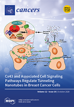
Cover Story (view full-size image): Tunneling nanotubes (TNTs) and similar cellular protrusions are emerging as an important intercellular communication network that cancer cells exploit to promote growth and overcome antitumoral insults. Cx43, a major gap junction protein often present in TNTs, appears to stimulate their formation. There are numerous bidirectional interactions between Cx43 and key cancer signaling pathways, and the presence or absence of Cx43 appears to modulate these pathways and how they regulate TNT formation. Understanding and targeting the intricate interplay between Cx43 and cell signaling pathways and their effect on TNTs may provide novel therapeutic opportunities for malignant tumors. View this paper.
  • Issues are regarded as officially published after their release is announced to the table of contents alert mailing list.
  • You may sign up for e-mail alerts to receive table of contents of newly released issues.
  • PDF is the official format for papers published in both, html and pdf forms. To view the papers in pdf format, click on the "PDF Full-text" link, and use the free Adobe Reader to open them.
Order results
Result details
Section
Select all
Export citation of selected articles as:
4 pages, 534 KiB  
Editorial
The Tumor Microenvironment of Pancreatic Cancer
by Eva Karamitopoulou
Cancers 2020, 12(10), 3076; https://doi.org/10.3390/cancers12103076 - 21 Oct 2020
Cited by 14 | Viewed by 3342
Abstract
Pancreatic ductal adenocarcinoma (PDAC) has a dismal prognosis along with rising incidence rates and will be responsible for many cancer deaths in the future [...] Full article
(This article belongs to the Special Issue Tumor Microenvironment of Pancreatic Cancer)
Show Figures

Figure 1

16 pages, 4594 KiB  
Article
Occurrence of Fibrotic Tumor Vessels in Grade I Meningiomas Is Strongly Associated with Vessel Density, Expression of VEGF, PlGF, IGFBP-3 and Tumor Recurrence
by Katharina Hess, Dorothee Cäcilia Spille, Alborz Adeli, Peter B. Sporns, Karina Zitta, Lars Hummitzsch, Julian Pfarr, Walter Stummer, Benjamin Brokinkel, Rouven Berndt and Martin Albrecht
Cancers 2020, 12(10), 3075; https://doi.org/10.3390/cancers12103075 - 21 Oct 2020
Cited by 7 | Viewed by 2892
Abstract
Angiogenesis is a key feature during oncogenesis and remains a potential target of antiangiogenic therapy. While commonly described in high-grade lesions, vascularization and its correlation with prognosis in grade I meningiomas is largely unexplored. In the histological classification, not only the number but [...] Read more.
Angiogenesis is a key feature during oncogenesis and remains a potential target of antiangiogenic therapy. While commonly described in high-grade lesions, vascularization and its correlation with prognosis in grade I meningiomas is largely unexplored. In the histological classification, not only the number but also the composition of blood vessels seems to be important. Therefore, tumor vessel density and fibrosis were correlated with clinical and imaging variables and prognosis in 295 patients with intracranial grade I meningioma. Expression of pro-angiogenic proteins within the meningiomas was investigated by proteome analyses and further validated by immunohistochemical staining. Fibrotic tumor vessels (FTV) were detected in 48% of all tumors and strongly correlated with vessel density, but not with the histopathological tumor subtype. Occurrence of FTV was correlated with a 2-fold increased risk of recurrence in both univariate and multivariate analyses. Explorative proteome analyses revealed upregulation of VEGF (vascular endothelial growth factor), PlGF (placental growth factor), and IGFBP-3 (insulin-like growth factor-binding protein-3) in tumors displaying FTV. Immunohistochemical analyses confirmed strong correlations between tumor vessel fibrosis and expression of VEGF, PlGF, and IGFBP-3. Presence of FTV was strongly associated with disruption of the arachnoid layer on preoperative MRI in univariate and multivariate analyses. In summary, the occurrence of fibrotic tumor vessels in grade I meningiomas is strongly associated with vessel density, disruption of the arachnoid layer, expression of VEGF, PlGF, IGFBP-3 and tumor recurrence. Full article
(This article belongs to the Section Cancer Causes, Screening and Diagnosis)
Show Figures

Figure 1

13 pages, 2665 KiB  
Article
De Novo T790M Mutation in an L858R Epidermal Growth Factor Receptor Mutant-Associated Lung Adenocarcinoma
by Takumi Fujiwara, Tetsu Kobayashi, Taro Yasuma, Corina N. D’Alessandro-Gabazza, Masaaki Toda, Hajime Fujimoto, Kentaro Fujiwara, Atsuro Takeshita, Kota Nishihama, Tomohito Okano, Valeria Fridman D’Alessandro, Yoshiyuki Takei, Osamu Hataji and Esteban C Gabazza
Cancers 2020, 12(10), 3074; https://doi.org/10.3390/cancers12103074 - 21 Oct 2020
Cited by 3 | Viewed by 2555
Abstract
Background: Lung cancer is the leading cause of mortality for cancer worldwide. A point mutation in exon 21 of the epidermal growth factor receptor resulting in the substitution of arginine for leucine at position 858 (L858R) is a frequent cause of lung adenocarcinoma. [...] Read more.
Background: Lung cancer is the leading cause of mortality for cancer worldwide. A point mutation in exon 21 of the epidermal growth factor receptor resulting in the substitution of arginine for leucine at position 858 (L858R) is a frequent cause of lung adenocarcinoma. Tyrosine kinase inhibitors are effective for treating patients with lung cancer associated with mutant epidermal growth factor receptors but most tumors become resistant shortly after treatment. The substitution of methionine for threonine at position 790 (T790M) on exon 20 is the most frequently acquired mutation leading to resistance to tyrosine kinase inhibitors. Whether the T790M mutation occurred after tyrosine kinase inhibitor therapy or it already existed before therapy is unclear. Methods: Here, we developed mice with tetracycline-inducible lung-specific expression of the full-length genomic DNA of the human epidermal growth factor receptor containing an L858R mutation or both L858R and T790M mutations and evaluated de novo T790M mutation in untreated transgenic mice carrying a single L858R EGFR mutation. Results: The L858R mutation-associated lung adenocarcinoma acquired de novo T790 mutation without previous therapy. Conclusions: The results of this study suggest that lung tumors may spontaneously acquire T790M mutations without any drug-related selective pressure. Full article
(This article belongs to the Special Issue EGFR Signaling in Cancer)
Show Figures

Figure 1

16 pages, 2900 KiB  
Article
Evolution of Mutational Landscape and Tumor Immune-Microenvironment in Liver Oligo-Metastatic Colorectal Cancer
by Alessandro Ottaiano, Michele Caraglia, Annabella Di Mauro, Gerardo Botti, Angela Lombardi, Jerome Galon, Amalia Luce, Luigi D’Amore, Francesco Perri, Mariachiara Santorsola, Fabienne Hermitte, Giovanni Savarese, Fabiana Tatangelo, Vincenza Granata, Francesco Izzo, Andrea Belli, Stefania Scala, Paolo Delrio, Luisa Circelli and Guglielmo Nasti
Cancers 2020, 12(10), 3073; https://doi.org/10.3390/cancers12103073 - 21 Oct 2020
Cited by 26 | Viewed by 2963
Abstract
Genetic dynamics underlying cancer progression are largely unknown and several genes involved in highly prevalent illnesses (e.g., hypertension, obesity, and diabetes) strongly concur to cancer phenotype heterogeneity. To study genotype-phenotype relationships contributing to the mutational evolution of colorectal cancer (CRC) with a focus [...] Read more.
Genetic dynamics underlying cancer progression are largely unknown and several genes involved in highly prevalent illnesses (e.g., hypertension, obesity, and diabetes) strongly concur to cancer phenotype heterogeneity. To study genotype-phenotype relationships contributing to the mutational evolution of colorectal cancer (CRC) with a focus on liver metastases, we performed genome profiling on tumor tissues of CRC patients with liver metastatic disease and no co-morbidities. We studied 523 cancer-related genes and tumor-immune microenvironment characteristics in primary and matched metastatic tissues. We observed a loss of KRAS and SMAD4 alterations and a high granzyme-B+ T-cell infiltration when the disease did not progress. Conversely, gain in KRAS, PIK3CA and SMAD4 alterations and scarce granzyme-B+ T-cells infiltration were observed when the tumor evolved towards a poly-metastatic spread. These findings provide novel insights into the identification of tumor oligo-metastatic status, indicating that some genes are on a boundary line between these two clinical settings (oligo- vs. poly-metastatic CRC). We speculate that the identification of these genes and modification of their evolution could be a new approach for anti-cancer therapeutic strategies. Full article
(This article belongs to the Section Cancer Biomarkers)
Show Figures

Graphical abstract

15 pages, 3909 KiB  
Article
Electrotransfer of IL-15/IL-15Rα Complex for the Treatment of Established Melanoma
by Shawna A. Shirley, Cathryn G. Lundberg and Richard Heller
Cancers 2020, 12(10), 3072; https://doi.org/10.3390/cancers12103072 - 21 Oct 2020
Cited by 7 | Viewed by 1918
Abstract
Gene electrotransfer (GET) is a safe, reliable, and effective method of delivering plasmid DNA (pDNA) to solid tumors. GET has been previously used to deliver interleukin-15 (IL-15) to mouse melanoma, resulting in long-term tumor regression and the survival of a percentage of treated [...] Read more.
Gene electrotransfer (GET) is a safe, reliable, and effective method of delivering plasmid DNA (pDNA) to solid tumors. GET has been previously used to deliver interleukin-15 (IL-15) to mouse melanoma, resulting in long-term tumor regression and the survival of a percentage of treated animals after challenge. To enhance this effect, we evaluated modulating the expression levels of IL-15 and co-expressing its receptor, IL-15Rα. GET was used to deliver plasmids encoding IL-15 and IL-15Rα to established B16.F10 tumors on days 0, 4, and 7. Two delivery protocols that yielded different expression profiles were utilized. Mice that were tumor-free for 50 days were then challenged with B16.F10 cells on the opposite flank and monitored for an additional 50 days. The amount of IL-15 expressed and the presence or absence of IL-15Rα in the treated tumors did not significantly affect the tumor regression and long-term survival. Upon challenge, however, low levels of IL-15 were more protective and resulted in a greater production of anti-tumor cytokines such as IFN-γ and MIP-1β and a greater amount of CD11b+ and CD3e+ cells infiltrating tumors. While mice with high levels of IL-15 showed CD11b+ and CD3e+ cell infiltrate, there was a substantial presence of NK cells that was absent in other treated groups. We can conclude that the level of IL-15 expressed in tumors after GET is an important determinant of the therapeutic outcome, a finding that will help us finetune this type of therapy. Full article
(This article belongs to the Section Cancer Immunology and Immunotherapy)
Show Figures

Figure 1

21 pages, 396 KiB  
Review
The Signaling Duo CXCL12 and CXCR4: Chemokine Fuel for Breast Cancer Tumorigenesis
by Karolina A. Zielińska and Vladimir L. Katanaev
Cancers 2020, 12(10), 3071; https://doi.org/10.3390/cancers12103071 - 21 Oct 2020
Cited by 43 | Viewed by 4292
Abstract
The CXCL12/CXCR4 signaling pathway has emerged in the recent years as a key player in breast cancer tumorigenesis. This pathway controls many aspects of breast cancer development including cancer cell proliferation, motility and metastasis to all target organs. Moreover, the CXCL12/CXCR4 cascade affects [...] Read more.
The CXCL12/CXCR4 signaling pathway has emerged in the recent years as a key player in breast cancer tumorigenesis. This pathway controls many aspects of breast cancer development including cancer cell proliferation, motility and metastasis to all target organs. Moreover, the CXCL12/CXCR4 cascade affects both immune and stromal cells, creating tumor-supporting microenvironment. In this review, we examine state-of-the-art knowledge about detrimental roles of the CXCL12/CXCR4 signaling, discuss its therapeutic potential and suggest further research directions beneficial both for basic research and personalized medicine in breast cancer. Full article
Show Figures

Figure 1

30 pages, 2606 KiB  
Review
The Diagnostic and Prognostic Value of a Liquid Biopsy for Esophageal Cancer: A Systematic Review and Meta-Analysis
by Daisuke Matsushita, Takaaki Arigami, Keishi Okubo, Ken Sasaki, Masahiro Noda, Yoshiaki Kita, Shinichiro Mori, Yoshikazu Uenosono, Takao Ohtsuka and Shoji Natsugoe
Cancers 2020, 12(10), 3070; https://doi.org/10.3390/cancers12103070 - 21 Oct 2020
Cited by 8 | Viewed by 2936
Abstract
Esophageal cancer is among the most aggressive diseases, and circulating tumor cells (CTCs) have been recognized as novel biomarkers for various cancers over the past two decades, including esophageal cancer. CTCs might provide crucial clinical information for predicting cancer prognosis, monitoring therapeutic responses [...] Read more.
Esophageal cancer is among the most aggressive diseases, and circulating tumor cells (CTCs) have been recognized as novel biomarkers for various cancers over the past two decades, including esophageal cancer. CTCs might provide crucial clinical information for predicting cancer prognosis, monitoring therapeutic responses or recurrences, or elucidating the mechanism of metastasis. The isolation of CTCs is among the applications of a “liquid biopsy”. There are various technologies for liquid biopsies, and they are classified into two main methods: cytometric or non-cytometric techniques. Here, we review a total of 57 eligible articles to summarize various technologies for the use of a liquid biopsy in esophageal cancer and perform a meta-analysis to assess the clinical utility of liquid biopsies as a prognostic and diagnostic biomarker technique. For prognostic evaluation, the pooled hazard ratio in the cytometric assay is relatively higher than that of the non-cytometric assay. On the other hand, a combination of multiple molecules, using a non-cytometric assay, might be a favorable biomarker technique for the early diagnosis of esophageal cancer. Although determining strong evidence for a biomarker by using a liquid biopsy is still challenging, our meta-analysis might be a milestone for the future development of liquid biopsies in use with esophageal cancer. Full article
(This article belongs to the Special Issue Non-Invasive Early Detection of Cancers)
Show Figures

Figure 1

18 pages, 2272 KiB  
Review
ELAVL1 Role in Cell Fusion and Tunneling Membrane Nanotube Formations with Implication to Treat Glioma Heterogeneity
by Natalia Filippova and Louis B. Nabors
Cancers 2020, 12(10), 3069; https://doi.org/10.3390/cancers12103069 - 21 Oct 2020
Cited by 18 | Viewed by 2900
Abstract
Homotypic and heterotypic cell fusions via permanent membrane fusions and temporal tunneling nanotube formations in the glioma microenvironment were recently documented in vitro and in vivo and mediate glioma survival, plasticity, and recurrence. Chronic inflammation, a hypoxic environment, aberrant mitochondrial function, and ER [...] Read more.
Homotypic and heterotypic cell fusions via permanent membrane fusions and temporal tunneling nanotube formations in the glioma microenvironment were recently documented in vitro and in vivo and mediate glioma survival, plasticity, and recurrence. Chronic inflammation, a hypoxic environment, aberrant mitochondrial function, and ER stress due to unfolded protein accumulation upregulate cell fusion events, which leads to tumor heterogeneity and represents an adaptive mechanism to promote tumor cell survival and plasticity in cytotoxic, nutrient-deprived, mechanically stressed, and inflammatory microenvironments. Cell fusion is a multistep process, which consists of the activation of the cellular stress response, autophagy formation, rearrangement of cytoskeletal architecture in the areas of cell-to-cell contacts, and the expression of proinflammatory cytokines and fusogenic proteins. The mRNA-binding protein of ELAV-family HuR is a critical node, which orchestrates the stress response, autophagy formation, cytoskeletal architecture, and the expression of proinflammatory cytokines and fusogenic proteins. HuR is overexpressed in gliomas and is associated with poor prognosis and treatment resistance. Our review provides a link between the HuR role in the regulation of cell fusion and tunneling nanotube formations in the glioma microenvironment and the potential suppression of these processes by different classes of HuR inhibitors. Full article
Show Figures

Figure 1

24 pages, 645 KiB  
Review
Ion Channels as Therapeutic Targets in High Grade Gliomas
by Michaela Griffin, Raheela Khan, Surajit Basu and Stuart Smith
Cancers 2020, 12(10), 3068; https://doi.org/10.3390/cancers12103068 - 21 Oct 2020
Cited by 20 | Viewed by 4002
Abstract
Glioblastoma multiforme (GBM) is a lethal brain cancer with an average survival of 14–15 months even with exhaustive treatment. High grade gliomas (HGG) represent the leading cause of CNS cancer-related death in children and adults due to the aggressive nature of the tumour [...] Read more.
Glioblastoma multiforme (GBM) is a lethal brain cancer with an average survival of 14–15 months even with exhaustive treatment. High grade gliomas (HGG) represent the leading cause of CNS cancer-related death in children and adults due to the aggressive nature of the tumour and limited treatment options. The scarcity of treatment available for GBM has opened the field to new modalities such as electrotherapy. Previous studies have identified the clinical benefit of electrotherapy in combination with chemotherapeutics, however the mechanistic action is unclear. Increasing evidence indicates that not only are ion channels key in regulating electrical signaling and membrane potential of excitable cells, they perform a crucial role in the development and neoplastic progression of brain tumours. Unlike other tissue types, neural tissue is intrinsically electrically active and reliant on ion channels and their function. Ion channels are essential in cell cycle control, invasion and migration of cancer cells and therefore present as valuable therapeutic targets. This review aims to discuss the role that ion channels hold in gliomagenesis and whether we can target and exploit these channels to provide new therapeutic targets and whether ion channels hold the mechanistic key to the newfound success of electrotherapies. Full article
Show Figures

Figure 1

17 pages, 1295 KiB  
Review
Therapy Resistance, Cancer Stem Cells and ECM in Cancer: The Matrix Reloaded
by Kousik Kesh, Vineet K. Gupta, Brittany Durden, Vanessa Garrido, Beatriz Mateo-Victoriano, Shweta P. Lavania and Sulagna Banerjee
Cancers 2020, 12(10), 3067; https://doi.org/10.3390/cancers12103067 - 21 Oct 2020
Cited by 37 | Viewed by 3919
Abstract
The extracellular matrix (ECM) has remained an enigmatic component of the tumor microenvironment. It drives metastasis via its interaction with the integrin signaling pathway, contributes to tumor progression and confers therapy resistance by providing a physical barrier around the tumor. The complexity of [...] Read more.
The extracellular matrix (ECM) has remained an enigmatic component of the tumor microenvironment. It drives metastasis via its interaction with the integrin signaling pathway, contributes to tumor progression and confers therapy resistance by providing a physical barrier around the tumor. The complexity of the ECM lies in its heterogeneous composition and complex glycosylation that can provide a support matrix as well as trigger oncogenic signaling pathways by interacting with the tumor cells. In this review, we attempt to dissect the role of the ECM in enriching for the treatment refractory cancer stem cell population and how it may be involved in regulating their metabolic needs. Additionally, we discuss how the ECM is instrumental in remodeling the tumor immune microenvironment and the potential ways to target this component in order to develop a viable therapy. Full article
Show Figures

Graphical abstract

22 pages, 1531 KiB  
Review
Cutting the Brakes on Ras—Cytoplasmic GAPs as Targets of Inactivation in Cancer
by Arianna Bellazzo and Licio Collavin
Cancers 2020, 12(10), 3066; https://doi.org/10.3390/cancers12103066 - 21 Oct 2020
Cited by 7 | Viewed by 3372
Abstract
The Ras pathway is frequently deregulated in cancer, actively contributing to tumor development and progression. Oncogenic activation of the Ras pathway is commonly due to point mutation of one of the three Ras genes, which occurs in almost one third of human cancers. [...] Read more.
The Ras pathway is frequently deregulated in cancer, actively contributing to tumor development and progression. Oncogenic activation of the Ras pathway is commonly due to point mutation of one of the three Ras genes, which occurs in almost one third of human cancers. In the absence of Ras mutation, the pathway is frequently activated by alternative means, including the loss of function of Ras inhibitors. Among Ras inhibitors, the GTPase-Activating Proteins (RasGAPs) are major players, given their ability to modulate multiple cancer-related pathways. In fact, most RasGAPs also have a multi-domain structure that allows them to act as scaffold or adaptor proteins, affecting additional oncogenic cascades. In cancer cells, various mechanisms can cause the loss of function of Ras inhibitors; here, we review the available evidence of RasGAP inactivation in cancer, with a specific focus on the mechanisms. We also consider extracellular inputs that can affect RasGAP levels and functions, implicating that specific conditions in the tumor microenvironment can foster or counteract Ras signaling through negative or positive modulation of RasGAPs. A better understanding of these conditions might have relevant clinical repercussions, since treatments to restore or enhance the function of RasGAPs in cancer would help circumvent the intrinsic difficulty of directly targeting the Ras protein. Full article
Show Figures

Figure 1

3 pages, 173 KiB  
Editorial
Circulating Tumor Cells: From the Laboratory to the Cancer Clinic
by Noriyoshi Sawabata
Cancers 2020, 12(10), 3065; https://doi.org/10.3390/cancers12103065 - 20 Oct 2020
Cited by 4 | Viewed by 1750
Abstract
Circulating tumor cells (CTCs) are cells that are separated from the primary tumor, move through the bloodstream, and spread from the original tumor to other sites, causing cancer metastasis [...] Full article
(This article belongs to the Special Issue Circulating Tumor Cells: From the Laboratory to the Cancer Clinic)
12 pages, 1652 KiB  
Article
Polyphenol Intake and Gastric Cancer Risk: Findings from the Stomach Cancer Pooling Project (StoP)
by Facundo Vitelli-Storelli, Marta Rossi, Claudio Pelucchi, Matteo Rota, Domenico Palli, Monica Ferraroni, Nuno Lunet, Samantha Morais, Lizbeth López-Carrillo, David Georgievich Zaridze, Dmitry Maximovich, María Rubín García, Gemma Castaño-Vinyals, Nuria Aragonés, Manuela Garcia de la Hera, Raúl Ulises Hernández-Ramírez, Eva Negri, Rossella Bonzi, Mary H. Ward, Areti Lagiou, Pagona Lagiou, Malaquías López-Cervantes, Paolo Boffetta, M. Constanza Camargo, Maria Paula Curado, Zuo-Feng Zhang, Jesus Vioque, Carlo La Vecchia and Vicente Martín Sánchezadd Show full author list remove Hide full author list
Cancers 2020, 12(10), 3064; https://doi.org/10.3390/cancers12103064 - 20 Oct 2020
Cited by 9 | Viewed by 3895
Abstract
Phenolic compounds may exert a favorable effect on the risk of several cancer types, including gastric cancer (GC). However, selected polyphenol classes have not been adequately investigated in relation to GC. The aim of this study is to evaluate the association between the [...] Read more.
Phenolic compounds may exert a favorable effect on the risk of several cancer types, including gastric cancer (GC). However, selected polyphenol classes have not been adequately investigated in relation to GC. The aim of this study is to evaluate the association between the intake of polyphenols in relation to GC risk. We used data from the Stomach cancer Pooling (StoP) Project, including 10 studies from six countries (3471 GC cases and 8344 controls). We carried out an individual participant data pooled analysis using a two-stage approach. The summary odds ratios (ORs) of GC for each compound, and the corresponding 95% confidence intervals (95% CI), were computed by pooling study specific ORs obtained through multivariate logistic regression, using random effect models. Inverse associations with GC emerged for total polyphenols (OR = 0.67, 95% CI = 0.54–0.81, for the highest versus lowest quartile of intake), total flavonoids (OR = 0.73, 95% CI = 0.55–0.90), anthocyanidins (OR = 0.74, 95% CI = 0.56–0.92), flavanols (OR = 0.77, 95% CI = 0.66–0.88), flavanones (OR = 0.57, 95%CI = 0.44–0.69), total phenolic acids (OR = 0.75, 95%CI = 0.55–0.94), and hydroxybenzoic acids (OR = 0.73, 95%CI = 0.57–0.89). Results were consistent across strata of age, sex, social class, and smoking habit. Suggestive inverse associations were also found for flavonols (OR = 0.76, 95%CI = 0.51–1.01) and hydroxycinnamic acids (OR = 0.82, 95%CI = 0.58–1.06). Further investigations from longitudinal data are needed to confirm this association. Full article
(This article belongs to the Section Cancer Epidemiology and Prevention)
Show Figures

Figure 1

20 pages, 3465 KiB  
Review
Consensus Definition and Prediction of Complexity in Transurethral Resection or Bladder Endoscopic Dissection of Bladder Tumours
by Mathieu Roumiguié, Evanguelos Xylinas, Antonin Brisuda, Maximillian Burger, Hugh Mostafid, Marc Colombel, Marek Babjuk, Joan Palou Redorta, Fred Witjes and Bernard Malavaud
Cancers 2020, 12(10), 3063; https://doi.org/10.3390/cancers12103063 - 20 Oct 2020
Cited by 7 | Viewed by 2254
Abstract
Ten senior urologists were interrogated to develop a predictive model based on factors from which they could anticipate complex transurethral resection of bladder tumours (TURBT). Complexity was defined by consensus. Panel members then used a five-point Likert scale to grade those factors that, [...] Read more.
Ten senior urologists were interrogated to develop a predictive model based on factors from which they could anticipate complex transurethral resection of bladder tumours (TURBT). Complexity was defined by consensus. Panel members then used a five-point Likert scale to grade those factors that, in their opinion, drove complexity. Consensual factors were highlighted through two Delphi rounds. Respective contributions to complexity were quantitated by the median values of their scores. Multivariate analysis with complexity as a dependent variable tested their independence in clinical scenarios obtained by random allocation of the factors. The consensus definition of complexity was “any TURBT/En-bloc dissection that results in incomplete resection and/or prolonged surgery (>1 h) and/or significant (Clavien-Dindo ≥ 3) perioperative complications”. Logistic regression highlighted five domains as independent predictors: patient’s history, tumour number, location, and size and access to the bladder. Receiver operating characteristic (ROC) analysis confirmed good discrimination (AUC = 0.92). The sum of the scores of the five domains adjusted to their regression coefficients or Bladder Complexity Score yielded comparable performance (AUC = 0.91, C-statistics, p = 0.94) and good calibration. As a whole, preoperative factors identified by expert judgement were organized to quantitate the risk of a complex TURBT, a crucial requisite to personalise patient information, adapt human and technical resources to individual situations and address TURBT variability in clinical trials. Full article
(This article belongs to the Special Issue Urological Cancer 2020)
Show Figures

Graphical abstract

1 pages, 164 KiB  
Erratum
Erratum: Ju, Z., et al. Integrative Analyses of Multilevel Omics Reveal Preneoplastic Breast to Possess a Molecular Landscape That Is Globally Shared with Invasive Basal-Like Breast Cancer (Running Title: Molecular Landscape of Basal-Like Breast Cancer Progression). Cancers 2020, 12, 722
by Zhenlin Ju, Anjana Bhardwaj, Matthew D. Embury, Harpreet Singh, Preethi H. Gunaratne, Isabelle Bedrosian and Jing Wang
Cancers 2020, 12(10), 3062; https://doi.org/10.3390/cancers12103062 - 20 Oct 2020
Cited by 1 | Viewed by 1178
Abstract
There is an error in the title of the paper [...] Full article
18 pages, 717 KiB  
Review
Risks of Solid and Lymphoid Malignancies in Patients with Myeloproliferative Neoplasms: Clinical Implications
by Mette Brabrand and Henrik Frederiksen
Cancers 2020, 12(10), 3061; https://doi.org/10.3390/cancers12103061 - 20 Oct 2020
Cited by 15 | Viewed by 2667
Abstract
In the past decade, several studies have reported that patients with chronic myeloproliferative neoplasms (MPNs) have an increased risk of second solid cancer or lymphoid hematological cancer. In this qualitative review study, we present results from studies that report on these cancer risks [...] Read more.
In the past decade, several studies have reported that patients with chronic myeloproliferative neoplasms (MPNs) have an increased risk of second solid cancer or lymphoid hematological cancer. In this qualitative review study, we present results from studies that report on these cancer risks in comparison to cancer incidences in the general population or a control group. Our literature search identified 12 such studies published in the period 2009–2018 including analysis of more than 65,000 patients. The results showed that risk of solid cancer is 1.5- to 3.0-fold elevated and the risk of lymphoid hematological cancer is 2.5- to 3.5-fold elevated in patients with MPNs compared to the general population. These elevated risks apply to all MPN subtypes. For solid cancers, particularly risks of skin cancer, lung cancer, thyroid cancer, and kidney cancer are elevated. The largest difference in cancer risk between patients with MPN and the general population is seen in patients below 80 years. Cancer prognosis is negatively affected due to cardiovascular events, thrombosis, and infections by a concurrent MPN diagnosis mainly among patients with localized cancer. Our review emphasizes that clinicians caring for patients with MPNs should be aware of the very well-documented increased risk of second non-myeloid cancers. Full article
(This article belongs to the Special Issue New Insights into Myeloproliferative Neoplasms)
Show Figures

Figure 1

21 pages, 14354 KiB  
Article
Specific Effects of Trabectedin and Lurbinectedin on Human Macrophage Function and Fate—Novel Insights
by Adrián Povo-Retana, Marina Mojena, Adrian B. Stremtan, Victoria B. Fernández-García, Ana Gómez-Sáez, Cristina Nuevo-Tapioles, José M. Molina-Guijarro, José Avendaño-Ortiz, José M. Cuezva, Eduardo López-Collazo, Juan F. Martínez-Leal and Lisardo Boscá
Cancers 2020, 12(10), 3060; https://doi.org/10.3390/cancers12103060 - 20 Oct 2020
Cited by 11 | Viewed by 4467
Abstract
Background: Tumor-associated macrophages (TAMs) play a crucial role in suppressing the immunosurveillance function of the immune system that prevents tumor growth. Indeed, macrophages can also be targeted by different chemotherapeutic agents improving the action over immune checkpoints to fight cancer. Here we describe [...] Read more.
Background: Tumor-associated macrophages (TAMs) play a crucial role in suppressing the immunosurveillance function of the immune system that prevents tumor growth. Indeed, macrophages can also be targeted by different chemotherapeutic agents improving the action over immune checkpoints to fight cancer. Here we describe the effect of trabectedin and lurbinectedin on human macrophage cell viability and function. Methods: Blood monocytes from healthy donors were differentiated into macrophages and exposed to different stimuli promoting functional polarization and differentiation into tumor-associated macrophages. Cells were challenged with the chemotherapeutic drugs and the effects on cell viability and function were analyzed. Results: Human macrophages exhibit at least two different profiles in response to these drugs. One-fourth of the blood donors assayed (164 individuals) were extremely sensitive to trabectedin and lurbinectedin, which promoted apoptotic cell death. Macrophages from other individuals retained viability but responded to the drugs increasing reactive oxygen production and showing a rapid intracellular calcium rise and a loss of mitochondrial oxygen consumption. Cell-membrane exposure of programmed-death ligand 1 (PD-L1) significantly decreased after treatment with therapeutic doses of these drugs, including changes in the gene expression profile of hypoxia-inducible factor 1 alpha (HIF-1α)-dependent genes, among other. Conclusions: The results provide evidence of additional onco-therapeutic actions for these drugs. Full article
(This article belongs to the Collection Mechanism of Immunotherapy in Cancers)
Show Figures

Graphical abstract

12 pages, 1796 KiB  
Article
EQD2 Analyses of Vaginal Complications in Exclusive Brachytherapy for Postoperative Endometrial Carcinoma
by Yaowen Zhang, Balbino Fornes, Gabriela Gómez, Irene Bentoldrà, Clara Carmona, Antonio Herreros, Sebastià Sabater, Inmaculada Nicolás, Yan Li, Joan Sánchez, Albert Biete, Aureli Torné, Carlos Ascaso and Ángeles Rovirosa
Cancers 2020, 12(10), 3059; https://doi.org/10.3390/cancers12103059 - 20 Oct 2020
Cited by 6 | Viewed by 2419
Abstract
Background: To evaluate whether EQD2(α/β = 3Gy) at 2 cm3 of the most exposed area of the vagina is related to late vaginal toxicity in postoperative endometrial cancer (PEC) patients (p) treated with exclusive brachytherapy (BT). Methods: From 2014 to 2017, [...] Read more.
Background: To evaluate whether EQD2(α/β = 3Gy) at 2 cm3 of the most exposed area of the vagina is related to late vaginal toxicity in postoperative endometrial cancer (PEC) patients (p) treated with exclusive brachytherapy (BT). Methods: From 2014 to 2017, 43p were included in this study. BT was administered: 3-fractions of 6Gy in 37p and 2-fractions of 7.5Gy in 6p. The dose was prescribed at a depth of 5 mm from the applicator surface with dose-point optimization based on distance. The active treatment length was 2.5 cm. CTV-D90 and the dose to the most exposed 2 cm3 of the vagina was calculated for each patient. Late toxicity of the bladder and rectum was assessed using Radiation Therapy Oncology Group (RTOG) criteria, and vaginal toxicity by objective Late Effects Normal Tissue Task Force (LENT)-Subjective, Objective, Management, Analytic (SOMA) (LENT-SOMA) criteria. Statistics: frequency tables, mean, median, range, standard deviation, and box plot. Results: The median follow-up was 51 months (12–68). 20 p (46.5%) and 2 p (4.7%) developed G1 and G2 vaginal complications, respectively. Only 1/2 p-G2 receiving EQD2(α/β = 3Gy) at 2 cm3 >68Gy presented vaginal shortening and 18/20 p-G1 received doses < 68Gy. Conclusions: PECp receiving exclusive brachytherapy with doses < 68Gy EQD2(α/β = 3Gy) at 2 cm2 of the vagina presented only G0–G1 vaginal toxicity, except for one with bleeding telangiectasias. Larger prospective studies are necessary to confirm the present results. Full article
(This article belongs to the Special Issue Interventional Radiotherapy in Gynecological Cancer)
Show Figures

Figure 1

21 pages, 3123 KiB  
Article
Novel Therapeutic Insights in Dedifferentiated Liposarcoma: A Role for FGFR and MDM2 Dual Targeting
by Bérengère Dadone-Montaudié, Audrey Laroche-Clary, Aline Mongis, Emmanuel Chamorey, Ilaria Di Mauro, Vanessa Chaire, Pascal Finetti, Renaud Schiappa, François Le Loarer, Isabelle Birtwisle-Peyrottes, Jean-François Michiels, François Bertucci, Florence Pedeutour, Antoine Italiano and Laurence Bianchini
Cancers 2020, 12(10), 3058; https://doi.org/10.3390/cancers12103058 - 20 Oct 2020
Cited by 10 | Viewed by 2638
Abstract
We aimed to evaluate the therapeutic potential of the pan-FGFR inhibitor erdafitinib to treat dedifferentiated liposarcoma (DDLPS). FGFR expression and their prognostic value were assessed in a series of 694 samples of well-differentiated/dedifferentiated liposarcoma (WDLPS/DDLPS). The effect of erdafitinib—alone or in combination with [...] Read more.
We aimed to evaluate the therapeutic potential of the pan-FGFR inhibitor erdafitinib to treat dedifferentiated liposarcoma (DDLPS). FGFR expression and their prognostic value were assessed in a series of 694 samples of well-differentiated/dedifferentiated liposarcoma (WDLPS/DDLPS). The effect of erdafitinib—alone or in combination with other antagonists—on tumorigenicity was evaluated in vitro and in vivo. We detected overexpression of FGFR1 and/or FGFR4 in a subset of WDLPS and DDLPS and demonstrated correlation of this expression with poor prognosis. Erdafitinib treatment reduced cell viability, inducing apoptosis and strong inhibition of the ERK1/2 pathway. Combining erdafitinib with the MDM2 antagonist RG7388 exerted a synergistic effect on viability, apoptosis, and clonogenicity in one WDLPS and two DDLPS cell lines. Efficacy of this combination was confirmed in vivo on a DDLPS xenograft. Importantly, we report the efficacy of erdafitinib in one patient with refractory DDLPS showing disease stabilization for 12 weeks. We provide evidence that the FGFR pathway has therapeutic potential for a subset of DDLPS and that an FGFR1/FGFR4 expression might constitute a powerful biomarker to select patients for FGFR inhibitor clinical trials. In addition, we show that combining erdafitinib with RG7388 is a promising strategy for patients with DDLPS that deserves further investigation in the clinical setting. Full article
(This article belongs to the Special Issue Sarcomas: New Biomarkers and Therapeutic Strategies)
Show Figures

Figure 1

20 pages, 1788 KiB  
Review
From Conventional Therapies to Immunotherapy: Melanoma Treatment in Review
by Lukasz Kuryk, Laura Bertinato, Monika Staniszewska, Katarzyna Pancer, Magdalena Wieczorek, Stefano Salmaso, Paolo Caliceti and Mariangela Garofalo
Cancers 2020, 12(10), 3057; https://doi.org/10.3390/cancers12103057 - 20 Oct 2020
Cited by 48 | Viewed by 6047
Abstract
In this review, we discuss the use of oncolytic viruses and checkpoint inhibitors in cancer immunotherapy in melanoma, with a particular focus on combinatory therapies. Oncolytic viruses are promising and novel anti-cancer agents, currently under investigation in many clinical trials both as monotherapy [...] Read more.
In this review, we discuss the use of oncolytic viruses and checkpoint inhibitors in cancer immunotherapy in melanoma, with a particular focus on combinatory therapies. Oncolytic viruses are promising and novel anti-cancer agents, currently under investigation in many clinical trials both as monotherapy and in combination with other therapeutics. They have shown the ability to exhibit synergistic anticancer activity with checkpoint inhibitors, chemotherapy, radiotherapy. A coupling between oncolytic viruses and checkpoint inhibitors is a well-accepted strategy for future cancer therapies. However, eradicating advanced cancers and tailoring the immune response for complete tumor clearance is an ongoing problem. Despite current advances in cancer research, monotherapy has shown limited efficacy against solid tumors. Therefore, current improvements in virus targeting, genetic modification, enhanced immunogenicity, improved oncolytic properties and combination strategies have a potential to widen the applications of immuno-oncology (IO) in cancer treatment. Here, we summarize the strategy of combinatory therapy with an oncolytic vector to combat melanoma and highlight the need to optimize current practices and improve clinical outcomes. Full article
(This article belongs to the Special Issue Oncolytic Virus Immunotherapy)
Show Figures

Figure 1

23 pages, 3444 KiB  
Article
Contribution of miRNAs, tRNAs and tRFs to Aberrant Signaling and Translation Deregulation in Lung Cancer
by Ilias Skeparnias, Dimitrios Anastasakis, Katerina Grafanaki, George Kyriakopoulos, Panagiotis Alexopoulos, Dimitrios Dougenis, Andreas Scorilas, Christos K. Kontos and Constantinos Stathopoulos
Cancers 2020, 12(10), 3056; https://doi.org/10.3390/cancers12103056 - 20 Oct 2020
Cited by 7 | Viewed by 2737
Abstract
Transcriptomics profiles of miRNAs, tRNAs or tRFs are used as biomarkers, after separate examination of several cancer cell lines, blood samples or biopsies. However, the possible contribution of all three profiles on oncogenic signaling and translation as a net regulatory effect, is under [...] Read more.
Transcriptomics profiles of miRNAs, tRNAs or tRFs are used as biomarkers, after separate examination of several cancer cell lines, blood samples or biopsies. However, the possible contribution of all three profiles on oncogenic signaling and translation as a net regulatory effect, is under investigation. The present analysis of miRNAs and tRFs from lung cancer biopsies indicated putative targets, which belong to gene networks involved in cell proliferation, transcription and translation regulation. In addition, we observed differential expression of specific tRNAs along with several tRNA-related genes with possible involvement in carcinogenesis. Transfection of lung adenocarcinoma cells with two identified tRFs and subsequent NGS analysis indicated gene targets that mediate signaling and translation regulation. Broader analysis of all major signaling and translation factors in several biopsy specimens revealed a crosstalk between the PI3K/AKT and MAPK pathways and downstream activation of eIF4E and eEF2. Subsequent polysome profile analysis and 48S pre-initiation reconstitution experiments showed increased global translation rates and indicated that aberrant expression patterns of translation initiation factors could contribute to elevated protein synthesis. Overall, our results outline the modulatory effects that possibly correlate the expression of important regulatory non-coding RNAs with aberrant signaling and translation deregulation in lung cancer. Full article
(This article belongs to the Section Molecular Cancer Biology)
Show Figures

Graphical abstract

26 pages, 2762 KiB  
Review
New Developments in Imaging for Sentinel Lymph Node Biopsy in Early-Stage Oral Cavity Squamous Cell Carcinoma
by Rutger Mahieu, Josanne S. de Maar, Eliane R. Nieuwenhuis, Roel Deckers, Chrit Moonen, Lejla Alic, Bennie ten Haken, Bart de Keizer and Remco de Bree
Cancers 2020, 12(10), 3055; https://doi.org/10.3390/cancers12103055 - 20 Oct 2020
Cited by 29 | Viewed by 6557
Abstract
Sentinel lymph node biopsy (SLNB) is a diagnostic staging procedure that aims to identify the first draining lymph node(s) from the primary tumor, the sentinel lymph nodes (SLN), as their histopathological status reflects the histopathological status of the rest of the nodal basin. [...] Read more.
Sentinel lymph node biopsy (SLNB) is a diagnostic staging procedure that aims to identify the first draining lymph node(s) from the primary tumor, the sentinel lymph nodes (SLN), as their histopathological status reflects the histopathological status of the rest of the nodal basin. The routine SLNB procedure consists of peritumoral injections with a technetium-99m [99mTc]-labelled radiotracer followed by lymphoscintigraphy and SPECT-CT imaging. Based on these imaging results, the identified SLNs are marked for surgical extirpation and are subjected to histopathological assessment. The routine SLNB procedure has proven to reliably stage the clinically negative neck in early-stage oral squamous cell carcinoma (OSCC). However, an infamous limitation arises in situations where SLNs are located in close vicinity of the tracer injection site. In these cases, the hotspot of the injection site can hide adjacent SLNs and hamper the discrimination between tracer injection site and SLNs (shine-through phenomenon). Therefore, technical developments are needed to bring the diagnostic accuracy of SLNB for early-stage OSCC to a higher level. This review evaluates novel SLNB imaging techniques for early-stage OSCC: MR lymphography, CT lymphography, PET lymphoscintigraphy and contrast-enhanced lymphosonography. Furthermore, their reported diagnostic accuracy is described and their relative merits, disadvantages and potential applications are outlined. Full article
(This article belongs to the Special Issue Emerging Concepts in Treatment of Laryngeal Cancer)
Show Figures

Figure 1

21 pages, 10470 KiB  
Article
A Methylation-Based Reclassification of Bladder Cancer Based on Immune Cell Genes
by Qizhan Luo and Thomas-Alexander Vögeli
Cancers 2020, 12(10), 3054; https://doi.org/10.3390/cancers12103054 - 20 Oct 2020
Cited by 26 | Viewed by 3862
Abstract
Background: Bladder cancer is highly related to immune cell infiltration. This study aimed to develop a new classification of BC molecular subtypes based on immune-cell-associated CpG sites. Methods: The genes of 28 types of immune cells were obtained from previous studies. Then, methylation [...] Read more.
Background: Bladder cancer is highly related to immune cell infiltration. This study aimed to develop a new classification of BC molecular subtypes based on immune-cell-associated CpG sites. Methods: The genes of 28 types of immune cells were obtained from previous studies. Then, methylation sites corresponding to immune-cell-associated genes were acquired. Differentially methylated sites (DMSs) were identified between normal samples and bladder cancer samples. Unsupervised clustering analysis of differentially methylated sites was performed to divide the sites into several subtypes. Then, the potential mechanism of different subtypes was explored. Results: Bladder cancer patients were divided into three groups. The cluster 3 subtype had the best prognosis. Cluster 1 had the poorest prognosis. The distribution of immune cells, level of expression of checkpoints, stromal score, immune score, ESTIMATEScore, tumor purity, APC co_inhibition, APC co_stimulation, HLA, MHC class_I, Type I IFN Response, Type II IFN Response, and DNAss presented significant differences among the three subgroups. The distribution of genomic alterations was also different. Conclusions: The proposed classification was accurate and stable. BC patients could be divided into three subtypes based on the immune-cell-associated CpG sites. Specific biological signaling pathways, immune mechanisms, and genomic alterations were varied among the three subgroups. High-level immune infiltration was correlated with high-level methylation. The lower RNAss was associated with higher immune infiltration. The study of the intratumoral immune microenvironment may provide a new perspective for BC therapy. Full article
(This article belongs to the Collection Application of Bioinformatics in Cancers)
Show Figures

Figure 1

20 pages, 8517 KiB  
Article
Cancer-Associated Stemness and Epithelial-to-Mesenchymal Transition Signatures Related to Breast Invasive Carcinoma Prognostic
by Iulia-Monica Groza, Cornelia Braicu, Ancuta Jurj, Oana Zanoaga, Raduly Lajos, Paul Chiroi, Roxana Cojocneanu, Diana Paun, Alexandru Irimie, Schuyler S. Korban, Patriciu Achimas-Cadariu and Ioana Berindan-Neagoe
Cancers 2020, 12(10), 3053; https://doi.org/10.3390/cancers12103053 - 20 Oct 2020
Cited by 16 | Viewed by 3342
Abstract
Breast cancer is one of the most common oncological diseases in women, as its incidence is rapidly growing, rendering it unpredictable and causing more harm than ever before on an annual basis. Alterations of coding and noncoding genes are related to tumorigenesis and [...] Read more.
Breast cancer is one of the most common oncological diseases in women, as its incidence is rapidly growing, rendering it unpredictable and causing more harm than ever before on an annual basis. Alterations of coding and noncoding genes are related to tumorigenesis and breast cancer progression. In this study, several key genes associated with epithelial-to-mesenchymal transition (EMT) and cancer stem cell (CSC) features were identified. EMT and CSCs are two key mechanisms responsible for self-renewal, differentiation, and self-protection, thus contributing to drug resistance. Therefore, understanding of the relationship between these processes may identify a therapeutic vulnerability that can be further exploited in clinical practice, and evaluate its correlation with overall survival rate. To determine expression levels of altered coding and noncoding genes, The Cancer Omics Atlas (TCOA) are used, and these data are overlapped with a list of CSCs and EMT-specific genes downloaded from NCBI. As a result, it is observed that CSCs are reciprocally related to EMT, thus identifying common signatures that allow for predicting the overall survival for breast cancer genes (BRCA). In fact, common CSCs and EMT signatures, represented by ALDH1A1, SFRP1, miR-139, miR-21, and miR-200c, are deemed useful as prognostic biomarkers for BRCA. Therefore, by mapping changes in gene expression across CSCs and EMT, suggesting a cross-talk between these two processes, we have been able to identify either the most common or specific genes or miRNA markers associated with overall survival rate. Thus, a better understanding of these mechanisms will lead to more effective treatment options. Full article
Show Figures

Figure 1

21 pages, 1387 KiB  
Review
Impact of MYC on Anti-Tumor Immune Responses in Aggressive B Cell Non-Hodgkin Lymphomas: Consequences for Cancer Immunotherapy
by A. Vera de Jonge, Tuna Mutis, Margaretha G. M. Roemer, Blanca Scheijen and Martine E. D. Chamuleau
Cancers 2020, 12(10), 3052; https://doi.org/10.3390/cancers12103052 - 20 Oct 2020
Cited by 11 | Viewed by 3821
Abstract
Patients with MYC overexpressing high grade B cell lymphoma (HGBL) face significant dismal prognosis after treatment with standard immunochemotherapy regimens. Recent preclinical studies indicate that MYC not only contributes to tumorigenesis by its effects on cell proliferation and differentiation, but also plays an [...] Read more.
Patients with MYC overexpressing high grade B cell lymphoma (HGBL) face significant dismal prognosis after treatment with standard immunochemotherapy regimens. Recent preclinical studies indicate that MYC not only contributes to tumorigenesis by its effects on cell proliferation and differentiation, but also plays an important role in promoting escape from anti-tumor immune responses. This is of specific interest, since reversing tumor immune inhibition with immunotherapy has shown promising results in the treatment of both solid tumors and hematological malignancies. In this review, we outline the current understanding of impaired immune responses in B cell lymphoid malignancies with MYC overexpression, with a particular emphasis on diffuse large B cell lymphoma. We also discuss clinical consequences of MYC overexpression in the treatment of HGBL with novel immunotherapeutic agents and potential future treatment strategies. Full article
Show Figures

Figure 1

20 pages, 5628 KiB  
Article
SFCE-RAPIRI Phase I Study of Rapamycin Plus Irinotecan: A New Way to Target Intra-Tumor Hypoxia in Pediatric Refractory Cancers
by Sarah Jannier, Véronique Kemmel, Consuelo Sebastia Sancho, Agathe Chammas, Amelia-Naomie Sabo, Erwan Pencreach, Françoise Farace, Marie Pierre Chenard, Benoit Lhermitte, Birgit Geoerger, Isabelle Aerts, Didier Frappaz, Pierre Leblond, Nicolas André, Stephane Ducassou, Nadège Corradini, Anne Isabelle Bertozzi, Eric Guérin, Florence Vincent, Michel Velten and Natacha Entz-Werleadd Show full author list remove Hide full author list
Cancers 2020, 12(10), 3051; https://doi.org/10.3390/cancers12103051 - 20 Oct 2020
Cited by 4 | Viewed by 2322
Abstract
Hypoxic environment is a prognostic factor linked in pediatric cancers to a worse outcome, favoring tumor progression and resistance to treatments. The activation of mechanistic Target Of Rapamycin (mTor)/hypoxia inducible factor (HIF)-1 pathway can be targeted by rapamycin and irinotecan, respectively. Therefore, we [...] Read more.
Hypoxic environment is a prognostic factor linked in pediatric cancers to a worse outcome, favoring tumor progression and resistance to treatments. The activation of mechanistic Target Of Rapamycin (mTor)/hypoxia inducible factor (HIF)-1 pathway can be targeted by rapamycin and irinotecan, respectively. Therefore, we designed a phase I trial associating both drugs in pediatric refractory/relapsing solid tumors. Patients were enrolled according to a 3 + 3 escalation design with ten levels, aiming to determine the MTD (maximum tolerated dose) of rapamycin plus irinotecan. Rapamycin was administered orally once daily in a 28-day cycle (1 to 2.5 mg/m2/day), associating biweekly intravenous irinotecan (125 to 240 mg/m2/dose). Toxicities, pharmacokinetics, efficacy analyses, and pharmacodynamics were evaluated. Forty-two patients, aged from 2 to 18 years, were included. No MTD was reached. Adverse events were mild to moderate. Only rapamycin doses of 1.5 mg/m2/day reached over time clinically active plasma concentrations. Tumor responses and prolonged stable disease were associated with a mean irinotecan area under the curve of more than 400 min.mg/L. Fourteen out of 31 (45.1%) patients had a non-progressive disease at 8 weeks. Most of them were sarcomas and brain tumors. For the phase II trial, we can then propose biweekly 125 mg/m2 irinotecan dose with a pharmacokinetic (PK) follow-up and a rapamycin dose of 1.5 mg/m2/day, reaching a blood concentration above 10 g/L. Full article
Show Figures

Figure 1

15 pages, 3287 KiB  
Article
Cetuximab-Mediated Protection from Hypoxia- Induced Cell Death: Implications for Therapy Sequence in Colorectal Cancer
by Hans Urban, Gabriele D. Maurer, Anna-Luisa Luger, Nadja I. Lorenz, Benedikt Sauer, Christopher Stroh, Jörg Trojan, Michel Mittelbronn, Joachim P. Steinbach, Patrick N. Harter and Michael W. Ronellenfitsch
Cancers 2020, 12(10), 3050; https://doi.org/10.3390/cancers12103050 - 20 Oct 2020
Cited by 1 | Viewed by 2833
Abstract
Monoclonal antibodies like cetuximab, targeting the epidermal growth factor receptor (EGFR), and bevacizumab, targeting the vascular endothelial growth factor (VEGF), are an integral part of treatment regimens for metastasized colorectal cancer. However, inhibition of the EGFR has been shown to protect human glioma [...] Read more.
Monoclonal antibodies like cetuximab, targeting the epidermal growth factor receptor (EGFR), and bevacizumab, targeting the vascular endothelial growth factor (VEGF), are an integral part of treatment regimens for metastasized colorectal cancer. However, inhibition of the EGFR has been shown to protect human glioma cells from cell death under hypoxic conditions. In colon carcinoma cells, the consequences of EGFR blockade in hypoxia (e.g., induced by bevacizumab) have not been evaluated yet. LIM1215 and SW948 colon carcinoma and LNT-229 glioblastoma cells were treated with cetuximab, PD153035, and erlotinib and analyzed for cell density and viability. The sequential administration of either cetuximab followed by bevacizumab (CET->BEV) or bevacizumab followed by cetuximab (BEV->CET) was investigated in a LIM1215 (KRAS wildtype) and SW948 (KRAS mutant) xenograft mouse model. In vitro, cetuximab protected from hypoxia. In the LIM1215 model, a survival benefit with cetuximab and bevacizumab monotherapy was observed, but only the sequence CET->BEV showed an additional benefit. This effect was confirmed in the SW948 model. Our observations support the hypothesis that bevacizumab modulates the tumor microenvironment (e.g., by inducing hypoxia) where cetuximab could trigger protective effects when administered later on. The sequence CET->BEV therefore seems to be superior as possible mutual adverse effects are bypassed. Full article
Show Figures

Graphical abstract

3 pages, 160 KiB  
Editorial
Novel Therapies for Hepatocellular Carcinoma
by Lorenza Rimassa
Cancers 2020, 12(10), 3049; https://doi.org/10.3390/cancers12103049 - 20 Oct 2020
Cited by 2 | Viewed by 1858
Abstract
Since 2007, for patients with advanced- or intermediate-stage hepatocellular carcinoma (HCC) unsuitable for locoregional treatments and with preserved liver function, the multikinase inhibitor (MKI) sorafenib has been the worldwide standard of care [...] Full article
(This article belongs to the Special Issue Novel Therapies for Hepatocellular Carcinoma)
13 pages, 1813 KiB  
Article
Exploratory Analysis of Lenvatinib Therapy in Patients with Unresectable Hepatocellular Carcinoma Who Have Failed Prior PD−1/PD-L1 Checkpoint Blockade
by Tomoko Aoki, Masatoshi Kudo, Kazuomi Ueshima, Masahiro Morita, Hirokazu Chishina, Masahiro Takita, Satoru Hagiwara, Hiroshi Ida, Yasunori Minami, Masakatsu Tsurusaki and Naoshi Nishida
Cancers 2020, 12(10), 3048; https://doi.org/10.3390/cancers12103048 - 20 Oct 2020
Cited by 38 | Viewed by 5340
Abstract
Although programmed cell death protein 1 (PD−1)/PD-ligand 1 (PD-L1) blockade is effective in a subset of patients with hepatocellular carcinoma (HCC), its therapeutic response is still unsatisfactory. Alternatively, the potential impact of the lenvatinib in patients who showed tumor progression on PD−1/PD-L1 blockade [...] Read more.
Although programmed cell death protein 1 (PD−1)/PD-ligand 1 (PD-L1) blockade is effective in a subset of patients with hepatocellular carcinoma (HCC), its therapeutic response is still unsatisfactory. Alternatively, the potential impact of the lenvatinib in patients who showed tumor progression on PD−1/PD-L1 blockade is unknown. In this work, we evaluated the safety and efficacy of lenvatinib administration after PD-1/PD-L1 checkpoint blockade. The outcome and safety of lenvatinib administered after PD-1/PD-L1 blockade failure was analyzed retrospectively in 36 patients. Tumor growth was assessed every 4–8 weeks using modified Response Evaluation Criteria in Solid Tumors. The mean relative dose intensity of lenvatinib was 87.6% and 77.8% in patients receiving a starting dose of 8 (interquartile range (IQR), 77.5–100.0) mg and 12 (IQR, 64.4–100.0) mg, respectively. Since lenvatinib therapy initiation, the median progression-free survival was 10 months (95% confidence interval (CI): 8.3–11.8) and the median overall survival was 15.8 months (95% CI: 8.5–23.2). The objective response rate was 55.6%, and the disease control rate was 86.1%. No particular safety concerns were observed. Lenvatinib demonstrated considerable antitumor effects with acceptable safety in patients with progressive and unresectable HCC when administered right after PD-1/PD-L1 blockade failure. Full article
(This article belongs to the Section Cancer Immunology and Immunotherapy)
Show Figures

Figure 1

26 pages, 945 KiB  
Article
Comprehensive Analysis of Tumour Sub-Volumes for Radiomic Risk Modelling in Locally Advanced HNSCC
by Stefan Leger, Alex Zwanenburg, Karoline Leger, Fabian Lohaus, Annett Linge, Andreas Schreiber, Goda Kalinauskaite, Inge Tinhofer, Nika Guberina, Maja Guberina, Panagiotis Balermpas, Jens von der Grün, Ute Ganswindt, Claus Belka, Jan C. Peeken, Stephanie E. Combs, Simon Boeke, Daniel Zips, Christian Richter, Mechthild Krause, Michael Baumann, Esther G.C. Troost and Steffen Löckadd Show full author list remove Hide full author list
Cancers 2020, 12(10), 3047; https://doi.org/10.3390/cancers12103047 - 19 Oct 2020
Cited by 16 | Viewed by 2601
Abstract
Imaging features for radiomic analyses are commonly calculated from the entire gross tumour volume (GTVentire). However, tumours are biologically complex and the consideration of different tumour regions in radiomic models may lead to an improved outcome prediction. Therefore, we investigated the [...] Read more.
Imaging features for radiomic analyses are commonly calculated from the entire gross tumour volume (GTVentire). However, tumours are biologically complex and the consideration of different tumour regions in radiomic models may lead to an improved outcome prediction. Therefore, we investigated the prognostic value of radiomic analyses based on different tumour sub-volumes using computed tomography imaging of patients with locally advanced head and neck squamous cell carcinoma. The GTVentire was cropped by different margins to define the rim and the corresponding core sub-volumes of the tumour. Subsequently, the best performing tumour rim sub-volume was extended into surrounding tissue with different margins. Radiomic risk models were developed and validated using a retrospective cohort consisting of 291 patients in one of the six Partner Sites of the German Cancer Consortium Radiation Oncology Group treated between 2005 and 2013. The validation concordance index (C-index) averaged over all applied learning algorithms and feature selection methods using the GTVentire achieved a moderate prognostic performance for loco-regional tumour control (C-index: 0.61 ± 0.04 (mean ± std)). The models based on the 5 mm tumour rim and on the 3 mm extended rim sub-volume showed higher median performances (C-index: 0.65 ± 0.02 and 0.64 ± 0.05, respectively), while models based on the corresponding tumour core volumes performed less (C-index: 0.59 ± 0.01). The difference in C-index between the 5 mm tumour rim and the corresponding core volume showed a statistical trend (p = 0.10). After additional prospective validation, the consideration of tumour sub-volumes may be a promising way to improve prognostic radiomic risk models. Full article
(This article belongs to the Special Issue Advances in Head and Neck Squamous Cell Carcinoma (HNSCC))
Show Figures

Figure 1

Previous Issue
Next Issue
Back to TopTop