


Abstract
Congenital malformations, iatrogenic injuries, and acquired diseases of the ureter often lead to structural or functional defects, yet tissue engineering approaches for ureteral repair and reconstruction remain limited. In this study, a photo-crosslinkable bladder acellular matrix (BAMMA) was synthesized from porcine bladder acellular matrix (BAM) through enzymatic digestion and methacrylic anhydride modification. The mechanical and biological properties of GelMA, GelMA/BAMMA, and BAMMA hydrogels were comprehensively characterized, and 3D-bioprinted tubular scaffolds were fabricated using a GelMA/BAMMA hydrogel and adipose-derived mesenchymal stem cells (ADSCs) mixture as the bioink and subsequently embedded in the rat omentum. The GelMA/BAMMA hybrid hydrogel combined the favorable mechanical properties of GelMA with the biological activity of BAMMA, supporting the growth and proliferation of ADSCs. One week after omental embedding, histological and immunohistochemical analyses revealed enhanced tissue remodelling, neovascularization and mitigated local inflammatory response in the ADSC-seeded scaffolds compared with the unseeded controls. These findings demonstrate that ADSC-loaded GelMA/BAMMA bioprinted tubular scaffolds can undergo tissue remodeling and vascularization in a rat omental model, providing a feasible hydrogel bioink for ureteral tissue engineering and a foundation for future research on ureteral repair.
1. Introduction
Congenital malformations of the ureter, as well as acquired conditions such as iatrogenic injuries, tumors, inflammation, trauma, and infections, can all lead to structural or functional defects of the ureter [1,2]. Among these, the management of long-segment (>5 cm) ureteral strictures and defects in the upper and middle ureter remains a highly complex and challenging clinical dilemma for urologists [3,4]. Currently, surgical reconstruction of long-segment ureteral defects often involves the use of autologous tissues or organs, including ileum, appendix, buccal mucosa, lingual mucosa, bladder muscle flaps, and blood vessels [5]. However, these surgical reconstructions are frequently associated with a range of postoperative complications, such as mucus secretion, stone formation, recurrent strictures, urinary tract infections, and renal impairment [6,7]. Therefore, there is an urgent need to develop a novel alternative for repairing long-segment ureteral defects that can restore the ureter’s anatomical structure and physiological function, reduce the incidence of complications, and improve patients’ quality of life.
In recent years, with the rapid development and innovation of tissue-engineering technologies, tissue-engineered ureters based on seed cells and scaffold materials have emerged as a promising therapeutic approach for the repair and reconstruction of long-segment ureteral defects [8,9]. Seed cells are the foundation of tissue engineering research. Currently, in the field of ureteral tissue engineering, adipose-derived mesenchymal stem cells (ADSCs) are the most commonly used seed cells [10,11]. This is because ADSCs are derived from adipose tissue, which is widely distributed throughout the human body, making them readily accessible and easy to harvest. Moreover, ADSCs possess excellent differentiation and paracrine capabilities, positioning them as a crucial source of seed cells for the engineered reconstruction of the ureter [12].
The selection of scaffold materials is particularly important. Currently, the most commonly used scaffold materials in ureteral tissue engineering fall into two main categories: synthetic and natural biomaterials. Natural biomaterials possess good biodegradability, biocompatibility, and other properties that mimic the extracellular matrix (ECM), making them highly attractive scaffold materials in tissue engineering [13,14]. Among natural biomaterials, decellularized matrix materials, which remove cellular components that could induce immune responses while preserving the ECM structure and composition of the original organ or tissue, facilitate cell adhesion, growth, proliferation, and differentiation. Consequently, they are widely used in tissue engineering research [15]. Decellularized matrices derived from various tissues or organs have been used in ureteral tissue-engineering studies, including small intestinal submucosa, amniotic membrane, bladder, and ureter [16]. The urinary tract, including the bladder and ureter, shares a common embryological origin from the urogenital sinus and similar tissue composition, characterized by a urothelial lining and smooth muscle layer [17]. Bladder ECM contains tissue-specific bioactive factors, including growth factors and cytokines that are relevant to urinary tract regeneration. Previous studies have demonstrated that bladder acellular matrix (BAM) retains these bioactive components and can promote cell adhesion, proliferation, and differentiation relevant to urinary tract tissues [18]. Therefore, bladder ECM was selected as a tissue-specific ECM source expected to provide a more favorable microenvironment for ureteral tissue engineering than non-urinary ECM sources. Furthermore, hydrogels derived from natural biomaterials can effectively mimic the environment of tissues, providing a suitable ECM microenvironment for seeded cells. Gelatin is a natural polymer derived from the hydrolysis of collagen. However, gelatin lacks a stable triple-helix structure, resulting in poor mechanical properties and uncontrolled degradation [19]. Gelatin modified with methacryloyl side groups, known as GelMA, has gained increasing attention due to its tunable biological functions and moderately enhanced mechanical performance. Through crosslinking, GelMA forms a hydrogel that is biocompatible, sustainable, and biomimetic, providing the structural and biological properties required for tissue regeneration and cell culture. These characteristics make it suitable for tissue integration and regeneration [20,21]. However, despite its widespread use in various tissue engineering applications, GelMA lacks tissue-specific biological cues and has a limited capacity to direct cell differentiation toward urinary tract lineages. Its insufficient bioactivity for promoting urothelial and smooth muscle regeneration remains a significant limitation for urinary tract applications. To address these limitations, the incorporation of tissue-specific ECM components has been proposed as a strategy to enhance the biological performance of GelMA-based hydrogels. There are few reports on the use of GelMA for ureteral tissue engineering.
In the field of in vitro construction of tissue-engineered ureters, approaches have evolved from the initial use of materials such as small intestinal submucosa decellularized matrix and polytetrafluoroethylene alone to fabricate engineered ureters, toward the combined use of scaffold materials and seed cells for co-construction [16]. With technological advancements, three-dimensional (3D) bioprinting has emerged as a novel approach for the biomimetic construction of engineered ureters with a multilayer structure resembling the native ureteral wall. By employing layer-by-layer printing, 3D bioprinters enable precise spatial control over the placement of functional components—including biomaterials, biochemical agents, and living cells—within different layers, thereby replicating the normal physiological structure of target organs. This technology is currently a research hotspot in organ tissue engineering and has been preliminarily applied to the tissue engineering and regeneration of various tissues and organs, including the skin, vascular systems, and cartilage [22,23].
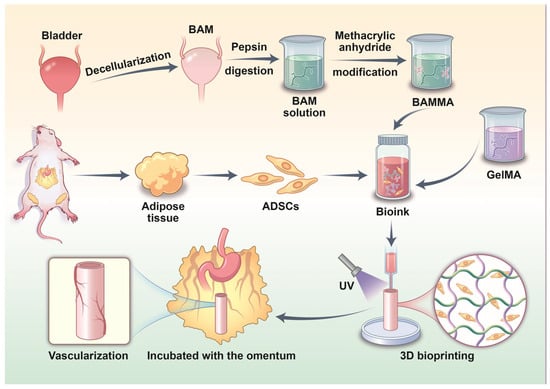
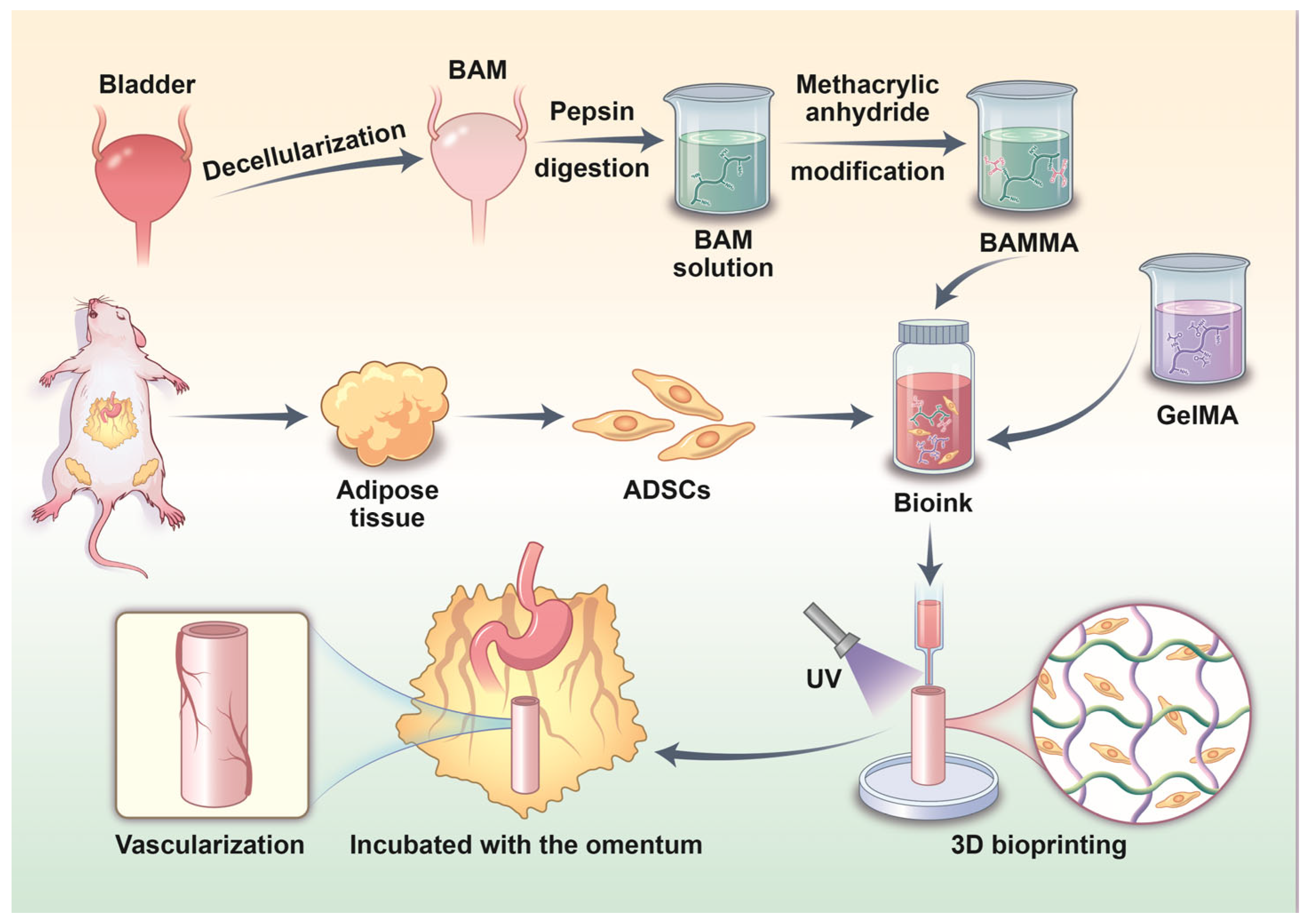
In this study, the prepared porcine bladder acellular matrix (BAM) was modified with methacrylic anhydride (MA) to obtain a photo-crosslinkable bladder acellular matrix (BAMMA). The mechanical and biological properties of GelMA, BAMMA, and GelMA/BAMMA hydrogels were comprehensively characterized in vitro, and the GelMA/BAMMA hydrogel was selected as the bioink matrix. Using a mixture of GelMA/BAMMA hydrogel and ADSCs as the bioink, ADSC-loaded tubular scaffolds for ureteral tissue engineering were fabricated via 3D bioprinting. The bioprinted tubular scaffolds were subsequently embedded in the rat omentum, and tissue remodeling within the scaffolds and neovascularization in the surrounding tissue were observed (Scheme 1).
Scheme 1.
Schematic illustration of 3D bioprinting of tubular scaffolds using GelMA/BAMMA hydrogel and ADSCs as bioink for subsequent omental incubation.
2. Materials and Methods
2.1. Synthesis and Detection of the BAMMA
The porcine bladder tissues were acquired from a local market. The BAM was prepared as described in our previous study [24]. The acquired porcine bladder tissues were cut into small pieces and cryopreserved at −80 °C for 24 h before thawing at room temperature. The porcine bladder tissues were submerged in 1% Triton X-100 (Sigma-Aldrich, St. Louis, MO, USA) for 12 h and then rinsed with deionized water for 2 h. Subsequently, 1% sodium dodecyl sulfate (SDS, Sigma-Aldrich, USA) was used as the decellularization solution for 12 h. Finally, the porcine bladder tissues were rinsed with deionized water for 24 h to obtain the BAM. During decellularization, the solution was continuously stirred at 60 rpm. The synthesis of BAMMA is referred to in our previous research [25]. Briefly, the prepared BAM was lyophilized and ground into powder, then digested with pepsin (Sigma-Aldrich, USA) at 37 °C for 48 h to obtain the BAM solution. Subsequently, the pH of the BAM solution was adjusted to 8–9. The methacrylic anhydride (MA, Sigma-Aldrich, USA) was added to the solution, reacted in the dark at 4 °C for 24 h, and then dialyzed and freeze-dried to synthesize the BAMMA. The synthesized BAMMA was sterilized with 60Co radiation.
The prepared BAM and the native bladder were fixed in 10% formalin solution, embedded in paraffin, and sectioned at 5 μm. The decellularization efficacy was evaluated using Hematoxylin and eosin (H&E), Masson’s trichrome (Masson), 4, 6-diamidino-2-phenylindole (DAPI) staining, and DNA quantification. The prepared BAM samples (n = 4) and native bladder samples (n = 4) were dabbed dry with tissue paper and weighed. The Genomic DNA Kit (Tiangen, Beijing, China) was used to extract DNA, which was expressed as ng/mg wet weight of the samples. The BAM and BAMMA were detected using 1H NMR. The samples of BAM and BAMMA, placed into separate EP tubes, were added to approximately 1 mL of deuterium oxide. The 1H NMR test was performed using a 400/500 MHz Nuclear Magnetic Resonance spectrometer (Bruker, Avance NEO, Rheinstetten, Germany). The degree of methacrylate was confirmed using a 2,4,6-trinitrobenzene sulfonic acid (TNBS) assay kit (Sigma-Aldrich, USA). Briefly, the prepared BAMMA was dissolved in 0.1 M sodium bicarbonate buffer (pH 8.5) to a final concentration of 200 µg/mL. Subsequently, 250 µL of TNBS reagent was added to 0.5 mL of the sample solution, and the mixture was incubated at 37 °C for 2 h. The reaction was terminated by the addition of 250 µL of 10% SDS and 125 µL of 1 M hydrochloric acid (Sigma-Aldrich, USA). The absorbance was measured at 335 nm using a spectrophotometer. The BAM before methacrylation served as a control. Each group included three samples. The extent of functionalization was calculated based on the difference in residual free amino groups between BAMMA and BAM, using the following equation:
Considering the potential batch-to-batch variation in ECM-derived biomaterials, the methacrylation degree was tested for each BAMMA batch thereafter. Raw material origin, pepsin digestion time, pH, feeding ratio of methacrylic anhydride, and reaction temperature were strictly controlled to reduce batch variation.
2.2. Preparation of the GelMA, GelMA/BAMMA and BAMMA Hydrogels
The purchased GelMA (EFL-GM-60, Engineering For Life, Suzhou, China) was dissolved in phosphate-buffered saline (PBS) to prepare a 10% (w/v) GelMA solution. Similarly, the synthesized BAMMA was dissolved in PBS to prepare a 10% (w/v) BAMMA solution. Equal volumes of the two stock solutions were mixed at a volume ratio of 1:1 to obtain the 5% (w/v) GelMA/5% (w/v) BAMMA (GelMA/BAMMA) mixed solution. The GelMA, GelMA/BAMMA, and BAMMA solutions with lithium phenyl-2,4,6-trimethylbenzoylphosphinate (LAP, Sunp Biotech, Beijing, China) at a final concentration of 0.25% (w/v) were exposed to 405 nm ultraviolet (UV) light for 1 min to obtain the GelMA, GelMA/BAMMA, and BAMMA hydrogels. Based on published literature and commercial product specifications, a LAP photoinitiator concentration of 0.25% (w/v) was selected, as this concentration provides sufficient crosslinking efficiency for methacrylated hydrogels under 405 nm UV light while maintaining high cell viability [26].
2.3. Scanning Electron Microscopy (SEM)
About 500 μL of the GelMA, GelMA/BAMMA, and BAMMA solutions with LAP at a final concentration of 0.25% (w/v) were added to 2 mL EP tubes, respectively, and exposed to 405 nm UV light for 1 min. The GelMA, GelMA/BAMMA and BAMMA hydrogels were lyophilized and coated with 2 μm AuPd using a sputter coater. Micrographs were captured with a scanning electron microscope (Tescan Mira, Brno, Czechia). The average pore size and porosity of the lyophilized hydrogels were quantified from SEM micrographs using ImageJ software (version 1.54u7). For each hydrogel sample, one SEM image was randomly selected, and three visual fields possessing uniform pore characteristics were randomly selected; the averaged value was taken as the mean pore size of the sample. Porosity was calculated as the ratio of the pore area to the total area in the SEM image. Three samples were tested per group.
2.4. Rheological Analysis
The rheological behavior of the GelMA, GelMA/BAMMA and BAMMA hydrogels was assessed using a Rheometer (Anton Paar MCR 302, Graz, Austria) with a 25 mm plate. The relationship between shear rate and viscosity of the GelMA, GelMA/BAMMA, and BAMMA hydrogels was evaluated in the shear rate range of 0.1 to 1000 s−1 at a temperature of 37 °C. Oscillatory temperature sweep measurements were performed at 1% strain and a frequency of 1 Hz. The temperature was decreased from 37 °C to 4 °C at a constant cooling rate, and the storage modulus (G′) and loss modulus (G″) were recorded as functions of temperature.
2.5. Swelling Ratio
The GelMA, GelMA/BAMMA, and BAMMA hydrogels were molded into cylindrical shapes with a diameter of 10 mm and a height of 5 mm using a mold. The cylindrical hydrogels were weighed (W0). Subsequently, the hydrogels were soaked in PBS and removed after 1, 3, 6, 12, 24, and 48 h. The surface moisture of the hydrogels was gently wiped off with absorbent paper and then weighed (Wt). Each group included three samples. The swelling ratio was calculated using the formula:
2.6. Degradation Ratio
Similarly, the GelMA, GelMA/BAMMA, and BAMMA hydrogels were molded into cylindrical shapes with a diameter of 10 mm and a height of 5 mm. The cylindrical hydrogels were lyophilized and then weighed (W0). The hydrogels were immersed in PBS containing 2 U/mL Collagenase II (LABLEAD, Beijing, China) and removed after 2, 4, 8, 16 and 24 h. The residual hydrogels were lyophilized and then weighed (W1). Each group included three samples. The degradation ratio was calculated using the formula:
2.7. Compression Testing
The GelMA, GelMA/BAMMA and BAMMA hydrogels were also molded into cylindrical shapes with a diameter of 10 mm and a height of 5 mm. The hydrogels underwent compression testing on a universal mechanical testing machine (Instron, 68SC-1, Norwood, MA, USA) at a compression speed of 1 mm/min and a maximum strain of 60%. The stress–strain data were utilized to measure the compression curve and compression modulus. Three samples were tested per group.
2.8. Tensile Testing
The GelMA, GelMA/BAMMA, and BAMMA hydrogels were molded into dumbbell shapes with a length of 10 mm, a width of 2 mm, and a thickness of 2.5 mm using a mold. The dumbbell hydrogels were subjected to mechanical testing on a universal testing machine (Instron, 68SC-1, USA) at a stretching speed of 5 mm/min. Young’s modulus was calculated from the stress–strain data. Each group included three samples.
2.9. Cytotoxicity Analysis of the GelMA, GelMA/BAMMA and BAMMA Hydrogels
The hydrogel extracts were prepared according to the ISO 10993-12 standard for biological evaluation of medical devices. Briefly, 1 mL of each hydrogel precursor solution was crosslinked in a culture dish under 405 nm UV light for 1 min. Subsequently, 10 mL of Dulbecco’s Modified Eagle Medium (DMEM, Gibco, Grand Island, NY, USA) containing 10% fetal bovine serum (FBS, Gibco, USA) was added to each culture dish. The hydrogels were incubated in the medium at 37 °C for 24 h. The resulting extracts were filtered through a 0.22 μm membrane and used directly (undiluted) for cytotoxicity assessment. The control group consisted of regular DMEM (10% FBS) that was incubated under identical conditions (37 °C for 24 h in a culture dish) without hydrogel contact, serving as a baseline to account for any effects of the incubation conditions on the culture medium. The purchased third-passage rat ADSCs (HTX2229, Otwo Biotech, Shenzhen, China) were seeded in 96-well plates at a concentration of 2000 cells/well and cultured in DMEM (10% FBS) for 24 h at 37 °C with 5% humidified CO2. After that, the culture medium was replaced with the hydrogel extract and the control DMEM. On days 1, 3, and 5, the culture medium was aspirated, the ADSCs were washed twice with PBS, and then incubated with 100 μL live/dead staining solution (Invitrogen, Carlsbad, CA, USA) at 37 °C for 15 min. The ADSCs were observed under a confocal laser scanning microscope (Zeiss, LSM900, Jena, Germany). Additionally, the ADSCs were incubated with 10 μL of Cell Counting Kit-8 reagent (CCK-8, Invitrogen, USA) in 5% humidified CO2 for 2 h at 37 °C on days 1, 3, and 5. The absorbance at 450 nm was measured using a microplate reader (Molecular Devices, Sunnyvale, CA, USA).
2.10. Three-Dimensional Bioprinting of the Grid Scaffolds and Tubular Scaffolds
The ADSCs were incubated with PKH67 (Solarbio, Beijing, China) for 5 min at 37 °C, followed by an additional 15 min at 4 °C. After labeling, the cells were washed twice with PBS and resuspended in DMEM containing 10% FBS. The GelMA/BAMMA solution and labeled ADSCs (1 × 10⁷ cells/mL) were used as the bioink for 3D bioprinting (SunP BioMaker 4, Sunp Biotech, Beijing, China) of the grid scaffolds and tubular scaffolds. Additionally, the tubular scaffolds were printed using only the GelMA/BAMMA solution without cells as the bioink, serving as the control group. The 3D bioprinting parameters were as follows: print nozzle temperature at 20 °C, print platform temperature at 4 °C, extrusion speed at 0.35 mm3/s, print speed at 4 mm/s, and print layer height at 0.3 mm. Based on our preliminary studies and the thermosensitive behavior of the GelMA/BAMMA hydrogel, the nozzle temperature was set at 20 °C to keep the bioink flowable during extrusion, thereby ensuring smooth bioprinting and reducing shear stress on encapsulated cells. The platform temperature was set at 4 °C to induce rapid sol–gel transition upon deposition. Cooling from 20 °C to 4 °C triggered a rapid viscosity increase and solidification of the GelMA/BAMMA hydrogel, which maintained the structural integrity and shape fidelity of the printed constructs [24,27,28]. For the grid scaffolds, they were exposed to 405 nm UV light for 1 min after bioprinting to complete crosslinking. During the bioprinting of tubular scaffolds, after every 10 layers were printed, the construct was exposed to 405 nm UV light for 15 s, followed by an additional 1-min UV exposure upon completion of the bioprinting. The printed grid scaffolds and tubular scaffolds were cultured in a culture dish. For the printed tissue-engineered ureter, a 2 mm-diameter soft tube was inserted into its lumen to maintain its shape. After 3 days of cultivation, a confocal laser scanning microscope (Zeiss, Germany) was used to observe the distribution of the labeled ADSCs in the printed grid scaffolds and tubular scaffolds.
The printability of the hydrogel was semi-quantitatively evaluated by calculating the printability factor (n = 3). The printability factor (Pr) was determined by analyzing printed macropores of bioprinted double-layer grid scaffolds and calculated from the following equation based on the circularity (C), perimeter (L), and area (A) of measured pores. For this metric, a Pr value close to 1 indicates that the printed pores closely match the designed square geometry, representing high shape fidelity and favourable printability. Deviation away from 1 signifies distorted, fused, or collapsed pores, reflecting poor printing performance.
2.11. Omentum Embedding of Bioprinted Tubular Scaffolds
Eight-week-old SD rats (SPF Biotechnology, Beijing, China) were randomly divided into two groups: tubular scaffolds loaded with labeled ADSCs served as the experimental group (n = 3), while those without labeled ADSCs served as the control group (n = 3). All animal experiments were conducted in accordance with the Chinese national standard GB/T 35892-2018 “Guidelines for ethical review of laboratory animal welfare” and were approved by the Ethics Committee of the Chinese PLA Air Force Medical Center (No. 2024-44-PJ01). The rats were anesthetized by an intraperitoneal injection of pentobarbital (30 mg/kg), and the surgical area was disinfected with iodophor, and a sterile sheet was spread. The tubular scaffolds were sutured onto the omentum, and the incision was sutured layer by layer. After one week, the rats were sacrificed, and the tubular scaffolds were retrieved and fixed in 10% formalin solution for histological analysis.
2.12. Histological Analysis
The soft tube inside the retrieved scaffold sample was removed after the sample had been fixed with 10% formalin solution. Scaffold samples were stained with H&E and Masson’s trichrome staining to assess tissue remodelling. The percentages of tissue-remodeling area and cell migration distance were calculated using ImageJ software. One randomly selected section per animal and three randomly selected fields of view per section were used for statistical analysis. Quantitative metrics obtained from the three fields of view for each animal were averaged to generate a single representative value for each biological replicate (n = 3 per group). Tissue-remodeling area: The percentage of tissue remodeling area was defined as the ratio of the area showing cell infiltration and new tissue formation within the hydrogel matrix layer to the total area. Percentage of cell migration distance: This parameter was defined as the ratio of the distance from the outer surface of the scaffold to the farthest point of cell infiltration within the hydrogel matrix, relative to the total thickness of the hydrogel matrix wall. The percentage was calculated as:
Tubular scaffolds sections were stained with DAPI (Solarbio, China), and the distribution of cells was observed using a confocal laser scanning microscope (Zeiss, Germany). Vascularization and local inflammatory response of the scaffolds wrapped in the omentum were evaluated by immunohistochemical (IHC) analysis. For IHC staining, tissue sections were immersed in antigen retrieval solution and heated to 95–100 °C for 30 min. Endogenous peroxidase activity was blocked by incubation with 3% hydrogen peroxide solution. The sections were then blocked with 4% goat serum and incubated overnight at 4 °C with primary antibodies: rabbit polyclonal anti-CD31 (1:2000, ab182981, Abcam, Waltham, MA, USA), rabbit anti-TNF(1:1000, ab205587, Abcam, USA), and rabbit anti-IL-10 (1:500, PA5-94918, Invitrogen, USA). After washing, the sections were incubated with the appropriate species-matched secondary antibody for 30 min at 37 °C. Subsequently, the sections were treated with a streptavidin-horseradish peroxidase complex, diaminobenzidine (DAB) solution (Dako, Agilent, Glostrup, Denmark), and counterstained with hematoxylin (Dako, Agilent, Denmark). Photomicrographs were captured using a Nikon Eclipse 80i microscope (Nikon, Tokyo, Japan). For quantitative evaluation of the number of vessels and CD31-positive area, a single section was examined per animal, with three randomly chosen microscopic fields per section. CD31-positive immunostaining (brown) was recognized by setting a consistent color threshold, and both the number of vessels and CD31-positive area were quantified using ImageJ software.
2.13. Statistical Analysis
Statistical analysis was performed using GraphPad Prism 11.0. The quantitative data were expressed as mean ± standard deviation. The differences between groups were estimated using a two-tailed Student’s t test or a one-way analysis of variance with Bonferroni post hoc test. The assumptions of normality (assessed using the Shapiro–Wilk test) and homogeneity of variance (assessed using Levene’s test) were verified before applying parametric tests. Where assumptions were not met, appropriate non-parametric alternatives were considered. Statistical significance was defined as p < 0.05.
3. Results
3.1. Synthesis of BAMMA
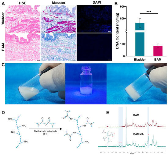
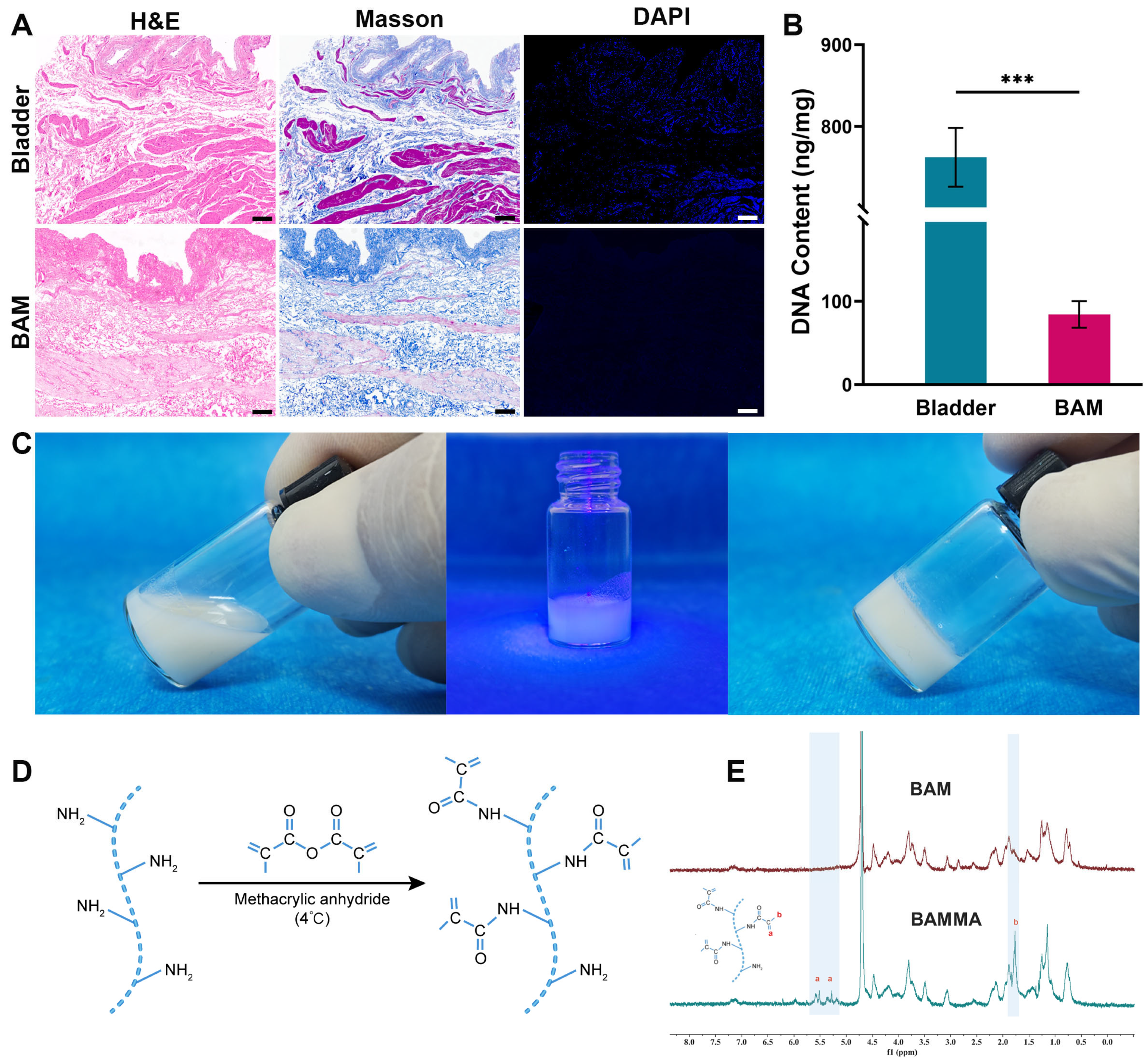
H&E, Masson, and DAPI staining demonstrated that the prepared BAM was successfully decellularized, as evidenced by the absence of nuclei and the preservation of collagen structure (Figure 1A). Moreover, DNA quantification revealed that the residual DNA content in the BAM (84.20 ± 15.88 ng/mg) was significantly lower than that in the native bladder tissue (762.30 ± 35.99 ng/mg, p < 0.001) (Figure 1B). In the preparation of BAMMA, the amino groups of the BAM solution react with the anhydride groups of methacrylic anhydride via amidation, forming amide bonds and thus endowing the BAMMA with photocrosslinking properties (Figure 1D). Moreover, 1H NMR spectroscopy revealed the appearance of two new peaks at 5.25 ppm and 5.53 ppm in BAMMA (Figure 1E). The prepared BAMMA solution containing 0.25% (v/v) LAP was exposed to 405 nm UV light for 1 min to obtain BAMMA hydrogels (Figure 1C). After methacrylate, the degree of amino group substitution in BAM following methacrylate was assessed via TNBS assay, which revealed a methacrylate percentage of 57.63 ± 9.84% for BAMMA.
Figure 1.
Synthesis and evaluation of BAMMA. (A) H&E, Masson, and DAPI staining of the native bladder and BAM (Scale bar = 200 μm). (B) The DNA content of the native bladder and BAM. (C) The preparation of BAMMA hydrogel. (D) The amino groups react with the anhydride groups of methacrylic anhydride to form amide bonds. (E) 1H NMR spectroscopy. a indicates vinylic hydrogen; b indicates methyl hydrogen. All statistical data are represented as mean ± SD (n = 4, *** p < 0.001).
3.2. Characterization of the GelMA, GelMA/BAMMA and BAMMA Hydrogels
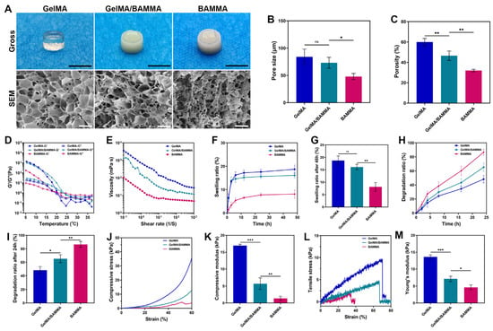
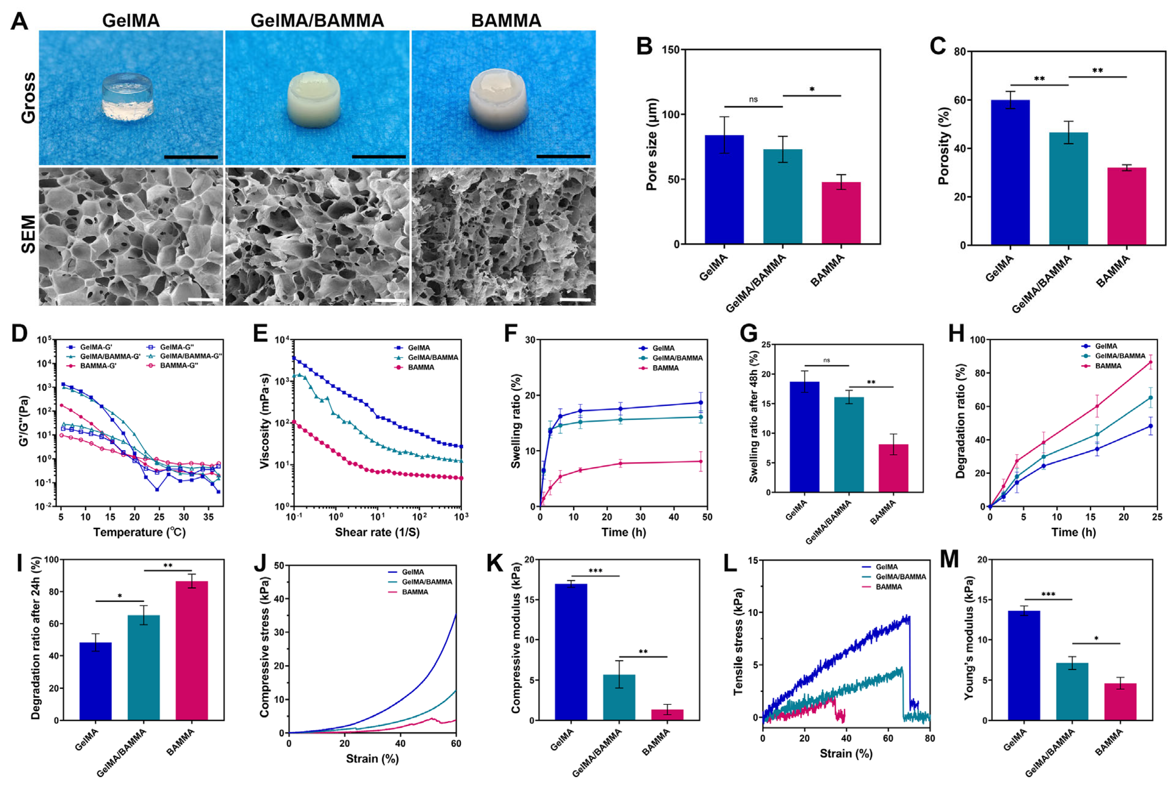
Gross observation revealed that the GelMA hydrogel was transparent, the BAMMA hydrogel appeared white, and the GelMA/BAMMA hydrogel was opaque with a whitish tint. SEM analysis demonstrated that the GelMA hydrogel possessed a more porous structure than the BAMMA hydrogel, with the GelMA/BAMMA hydrogel showing an intermediate morphology (Figure 2A). Regarding pore size, GelMA (84.12 ± 14.05 μm) and GelMA/BAMMA (73.10 ± 10.07 μm) showed no statistically significant difference, whereas both were significantly larger than BAMMA (47.86 ± 5.73 μm, p < 0.05) (Figure 2B). In terms of porosity, GelMA (59.97 ± 3.56%) exhibited the highest value among all groups, with significant differences compared with both GelMA/BAMMA (46.55 ± 4.65%) and BAMMA (32.00 ± 1.18%). Additionally, the porosity of GelMA/BAMMA was also significantly higher than that of BAMMA (p < 0.01) (Figure 2C). Temperature sweep tests revealed that all three hydrogel groups underwent reversible sol–gel transitions with temperature changes. At elevated temperatures, the storage modulus (G′) was lower than the loss modulus (G″), indicating that the hydrogel was in a flowable liquid state. As the temperature decreased, G′ became greater than G″, signifying that the hydrogel transitioned from a liquid to a solid state (Figure 2D). All three hydrogel groups exhibited favorable shear-thinning behavior, characterized by a gradual decrease in viscosity with increasing shear rate. Compared with the GelMA and BAMMA hydrogels, the GelMA/BAMMA hydrogel displayed an intermediate viscosity (Figure 2E). The swelling ratio curves indicated that all three hydrogel groups reached swelling equilibrium after 48 h (Figure 2F). At 48 h, the swelling ratio of the GelMA group (18.71 ± 1.81%) showed no significant difference from that of the GelMA/BAMMA group (16.10 ± 1.11%), but was significantly higher than that of the BAMMA group (8.11 ± 1.76%, p < 0.01) (Figure 2G). The degradation ratio curves showed that the degradation rates of all three hydrogel groups increased almost linearly over time (Figure 2H). At 24 h, the BAMMA group (86.54 ± 4.31%) was almost completely degraded, which was significantly higher than that of the GelMA/BAMMA group (65.27 ± 5.99%, p < 0.01) and the GelMA group (48.28 ± 5.40%, p < 0.001), with the GelMA group exhibiting the slowest degradation rate (Figure 2I). The compressive stress–strain curves showed that the compressive performance of the GelMA group was significantly superior to that of the GelMA/BAMMA group and the BAMMA group (Figure 2J). The compressive modulus of the GelMA group (16.95 ± 0.43 kPa) was significantly higher than that of the GelMA/BAMMA group (5.70 ± 1.67 kPa, p < 0.001) and the BAMMA group (1.35 ± 0.64 kPa, p < 0.001). Additionally, the compressive modulus of the GelMA/BAMMA group was higher than that of the BAMMA group (p < 0.01) (Figure 2K). The tensile curves indicated that the GelMA group exhibited good tensile properties, which were significantly superior to those of the GelMA/BAMMA group and the BAMMA group (Figure 2L). Young’s modulus of the GelMA/BAMMA group (7.11 ± 0.78 kPa) and the BAMMA group (4.62 ± 0.73 kPa) was significantly lower than that of the GelMA group (13.62 ± 0.59 kPa, p < 0.001), while the Young’s modulus of the GelMA/BAMMA group was higher than that of the BAMMA group (p < 0.05) (Figure 2M).
Figure 2.
Characterization of the GelMA, GelMA/BAMMA and BAMMA hydrogels. (A) Gross observation and SEM results (Gross, scale bar = 1 cm; SEM, scale bar = 200 μm). (B) Pore size. (C) Porosity. (D) Temperature/storage and loss modulus of the rheological analysis. (E) Shear rate/viscosity of the rheological analysis. (F) Swelling ratio curve. (G) Swelling ratio after 48 h. (H) Degradation ratio curve. (I) Degradation ratio after 24 h. (J) Compressive stress-strain curves. (K) Compressive modulus. (L) Tensile stress curves. (M) Young’s modulus. All statistical data are represented as mean ± SD (n = 3, * p < 0.05, ** p < 0.01 and *** p < 0.001).
3.3. Biocompatibility of the GelMA, GelMA/BAMMA and BAMMA Hydrogels
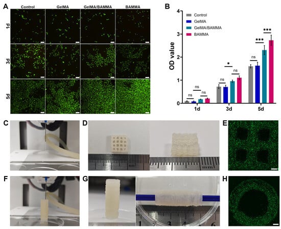
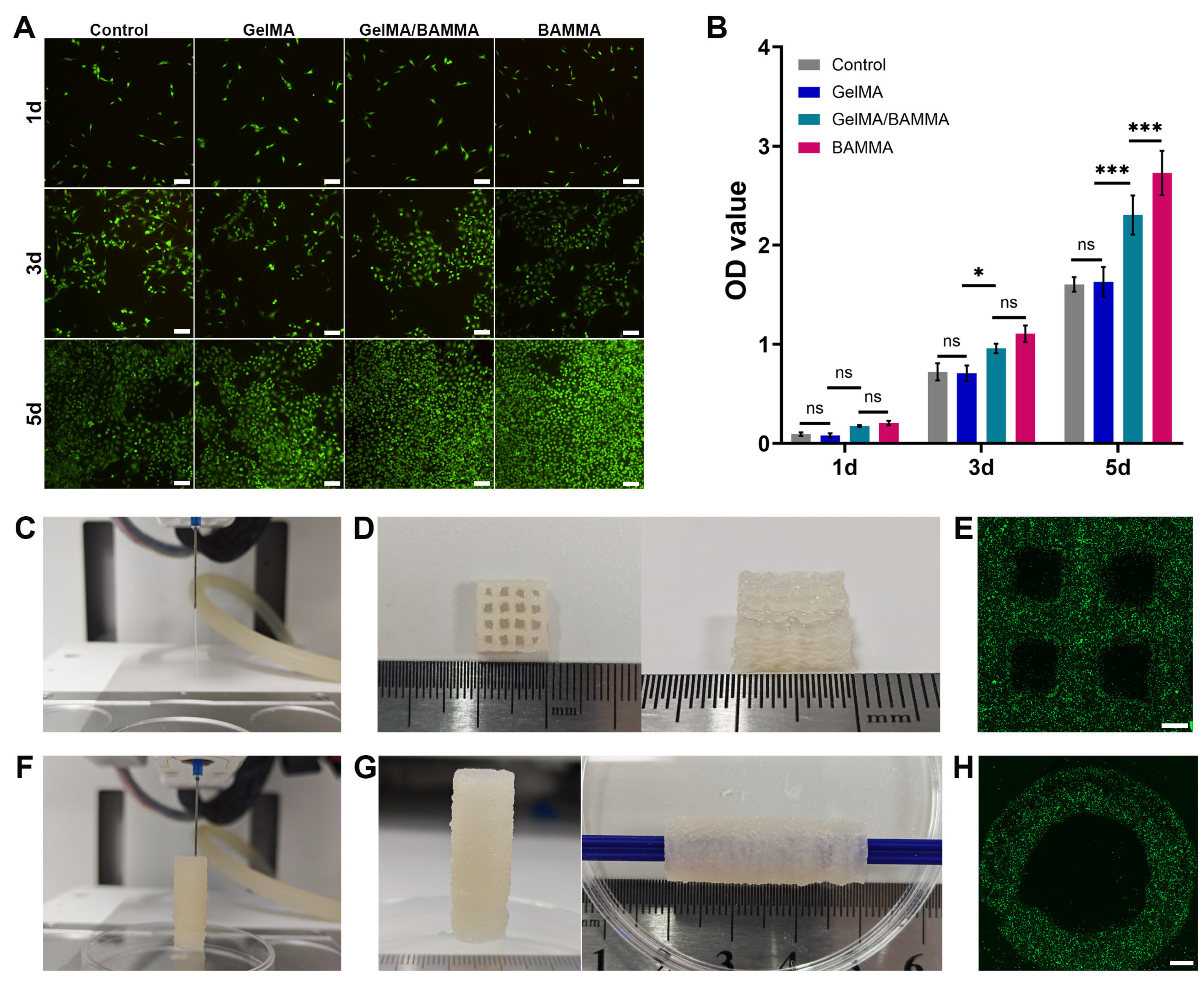
Live/dead staining showed that the ADSCs could survive in the extracts of all three groups of hydrogels, with no obvious cell death observed (Figure 3A). CCK-8 assay showed no significant differences in OD values among groups at day 1. At day 3, there was no significant difference between the control group and the GelMA group; the OD value of the GelMA/BAMMA group was higher than that of the GelMA group (p < 0.05), while no significant difference was observed between the GelMA/BAMMA group and the BAMMA group. At day 5, there remained no significant difference between the control group and the GelMA group; the OD value of the GelMA/BAMMA group was significantly higher than that of the GelMA group (p < 0.001), but significantly lower than that of the BAMMA group (p < 0.001) (Figure 3B).
Figure 3.
Cytotoxicity test of the GelMA, GelMA/BAMMA, and BAMMA hydrogels and 3D bioprinting. (A) Live/dead staining (Green: live cells; Red: dead cells; Scale bar = 100 μm). (B) CCK-8 test. (C–H) Printing performance of the GelMA/BAMMA hydrogel. (C) The 3D bioprinter extruded the hydrogel. (D) 3D bioprinted grid scaffolds. (E) Confocal laser scanning microscope observation of the grid scaffolds (Scale bar = 500 μm). (F) The tubular scaffolds were printed using the 3D bioprinter. (G) 3D bioprinted tubular scaffolds. (H) Confocal laser scanning microscope observation of the tubular scaffolds (Scale bar = 500 μm). All statistical data are represented as mean ± SD (n = 3, * p < 0.05, *** p < 0.001, ns—not significant).
3.4. 3D Bioprinting the Grid Scaffolds and Tubular Scaffolds
The GelMA/BAMMA solution formed continuous thin filaments when extruded through the 3D bioprinter (Figure 3C). The rectangular grid scaffolds measuring 1 cm in length, 1 cm in width, and 0.5 cm in height were successfully printed using the 3D bioprinter (Figure 3D). The printability factor (Pr) of the hydrogel was 1.05 ± 0.04 (n = 3). This value is close to 1, demonstrating that the printed macropores of double-layer grid scaffolds approximated the pre-designed square geometry and confirming favourable printing shape fidelity. During the printing of the tubular scaffolds using the 3D bioprinter, crosslinking was performed using 405 nm UV light for 15 s every 10 layers (Figure 3F), and the tubular scaffolds with a diameter of 1 cm and a length of 3 cm were successfully fabricated (Figure 3G). Confocal laser scanning microscope observation revealed that the labeled ADSCs were uniformly distributed within the bioprinted grid scaffolds and the tubular scaffolds (Figure 3E,H).
3.5. Histological Examination
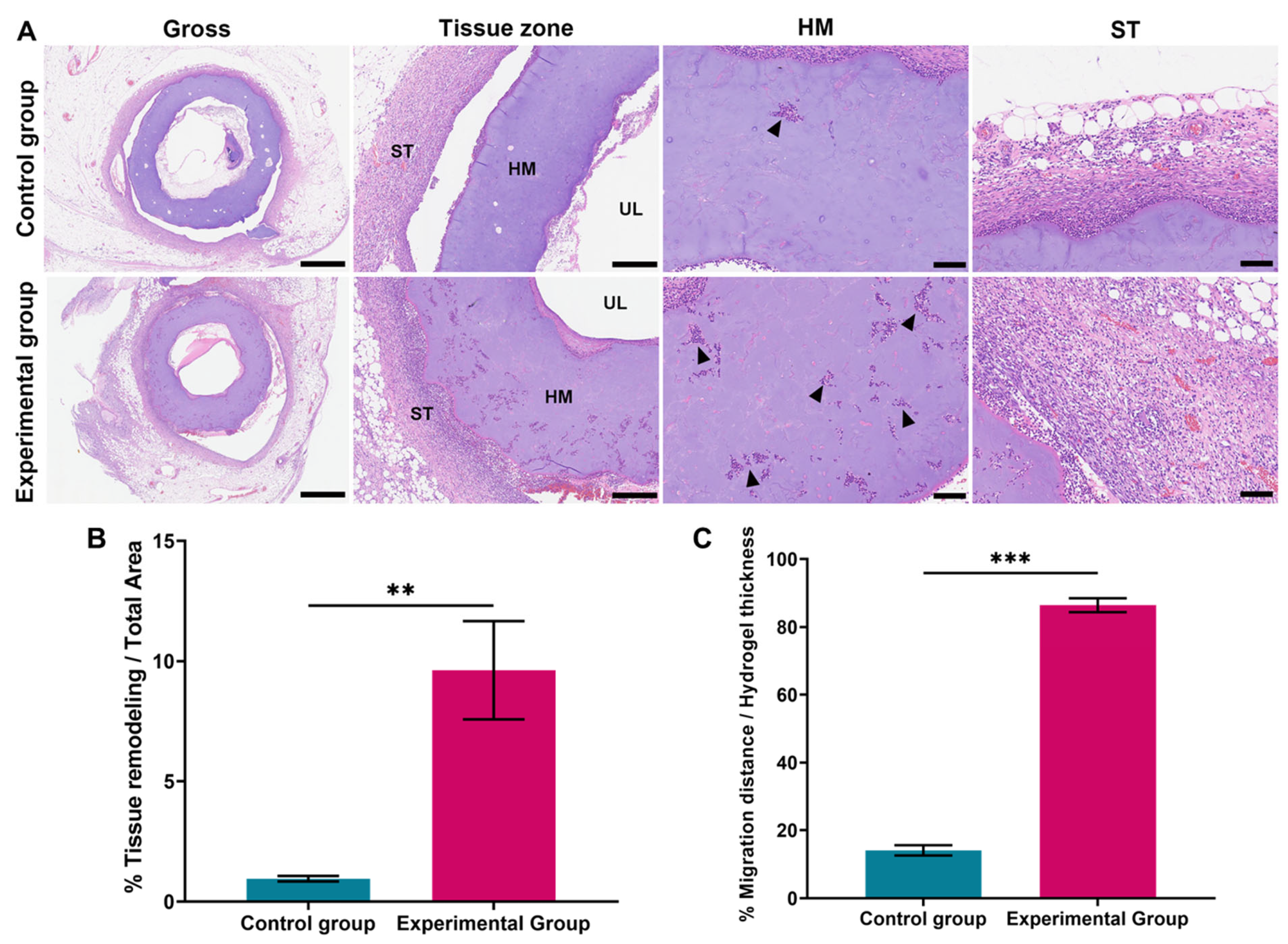
One week after omental embedding, H&E staining revealed that the bioprinted tubular scaffolds were completely encapsulated by the surrounding tissue, with clear delineation of their layered architecture. The ureteral lumen (UL) was located in the center, surrounded by the hydrogel matrix (HM) as the middle layer, and the outermost layer consisted of the surrounding tissue (ST) that encapsulated the construct. Masson’s trichrome staining clearly demonstrated collagen deposition at the implantation site (Figure S1). Collagen fibres in the ADSC-laden experimental group appeared thicker with a relatively loose arrangement, whereas thinner and densely. Cell infiltration and tissue remodeling were observed within the hydrogel matrix layer, and vessel-like structures were visible in the outermost surrounding tissue (Figure 4A). The area of tissue remodeling within the scaffolds was significantly larger in the experimental group (9.62 ± 2.04%) than in the control group (0.96 ± 0.12%, p < 0.01) (Figure 4B). Similarly, the percentage of cell migration distance within the scaffolds was markedly better in the experimental group (86.42 ± 2.06%) than in the control group (14.16 ± 1.54%, p < 0.001) (Figure 4C).
Figure 4.
Histological examination of the bioprinted tubular scaffolds. (A) H&E staining of scaffolds in the experimental and control groups (Gross, scale bar = 1.25 cm; Tissue zone, scale bar = 312.5 μm; HM and ST, scale bar = 100 μm). (B) The area of tissue remodeling. (C) The percentage of cell migration distance. UL: ureteral lumen; HM: hydrogel matrix; ST: surrounding tissue. Black triangles indicate tissue remodeling. All statistical data are represented as mean ± SD (n = 3, ** p < 0.01, *** p < 0.001).
3.6. Immunohistochemical Analyses
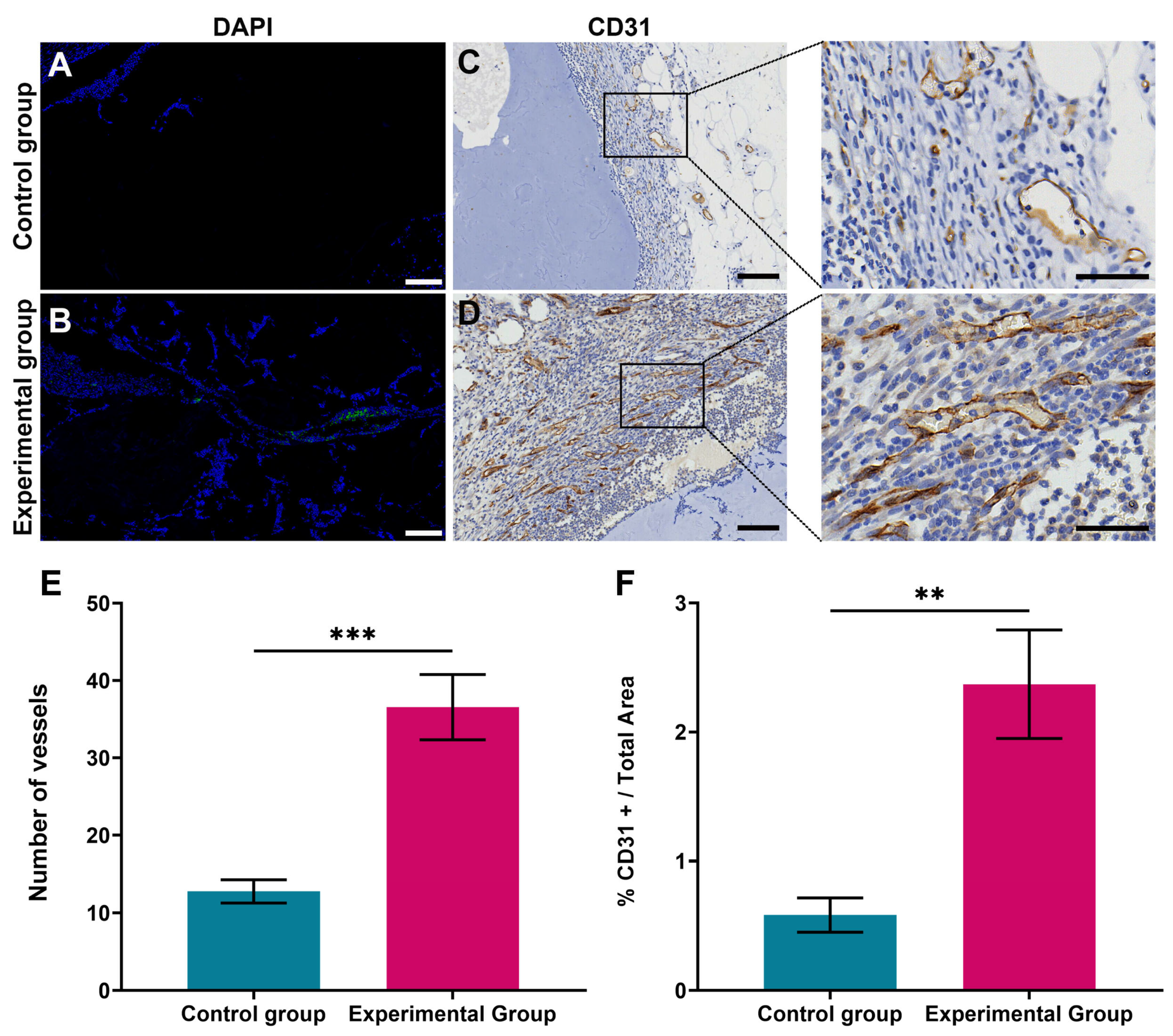
DAPI staining revealed that only a few cells were distributed on the hydrogel matrix in the control group (Figure 5A), whereas the experimental group showed a large number of cells on the hydrogel matrix, along with green fluorescently labeled ADSCs (Figure 5B). Vessel-like structures were observed in the surrounding tissue of the scaffolds in both the control and experimental groups by IHC staining. Higher magnification clearly delineated the vascular structures (Figure 5C,D). The number of vessels in the surrounding tissue of the scaffolds was significantly higher in the experimental group (36.56 ± 4.22) than in the control group (12.78 ± 1.50 p < 0.001) (Figure 5E). Furthermore, the CD31-positive area in the surrounding tissue of the experimental group (2.37 ± 0.42%) was also significantly greater than that in the control group (0.58 ± 0.13%, p < 0.01) (Figure 5F).
Figure 5.
Immunohistochemical analyses of the bioprinted tubular scaffolds. (A,B) DAPI staining of scaffolds in the experimental and control groups (DAPI: blue; Labeled ADSCs: green; Scale bar = 100 μm). (C,D) CD31 immunohistochemical staining; brown color indicates positive CD31 expression (scale bar = 100 μm; Magnified figures, scale bar = 50 μm). (E) The number of CD31-positive vessels. (F) The positive area of CD31. All statistical data are represented as mean ± SD (n = 3, ** p< 0.01, *** p < 0.001).
Immunohistochemical staining of TNF-α and IL-10 was carried out to characterise the local inflammatory response toward the implanted hydrogel constructs. The GelMA/BAMMA scaffold group loaded with ADSCs exhibited relatively low expression of the pro-inflammatory marker TNF-α, while stronger TNF-α immunoreactivity was observed in the cell-free GelMA/BAMMA control group. In contrast, the anti-inflammatory cytokine IL-10 was more abundantly distributed around the implants in the ADSC-laden group (Figure S2).
4. Discussion
Hydrogels have attracted significant interest in tissue engineering due to their mechanical strength, biocompatibility, biodegradability, and resemblance to the in vivo extracellular matrix [29]. The three-dimensional hydrophilic polymer network of hydrogels enables them to absorb and retain large amounts of biological fluids and water, endowing them with a soft gel-like consistency and exhibiting biocompatibility highly similar to that of the ECM. As a result, hydrogels are widely used in tissue engineering for the repair and reconstruction of tissues or organs [30,31]. However, there are few reports on the use of hydrogels for ureteral tissue engineering.
In this study, a mixture of two hydrogels, BAMMA and GelMA, was selected as the scaffold material for tubular scaffolds. During the synthesis of BAMMA, porcine BAM prepared by decellularization was used as the raw material. BAM was prioritized over other decellularized matrices because the bladder and ureter share homologous urinary tract histology, with comparable layered structures of urothelium, lamina propria and smooth muscle. It retains urinary-tract-specific bioactive cues that are better suited for ureteral regeneration than ECM derived from heterologous tissues such as small intestinal submucosa or dermis. BAM was digested with pepsin to generate peptide fragments with exposed amino groups, which were subsequently modified by methacrylic anhydride to form amide bonds, thereby synthesizing BAMMA with rapid photocrosslinking properties.
The commercial GelMA adopted in this work possesses a methacrylation degree of 60 ± 5%. The degree of methacrylation directly regulates the crosslink density and elastic modulus of photocrosslinkable hydrogels; a higher methacrylation degree generally produces a stiffer hydrogel. GelMA with a methacrylation degree near 60% is commonly applied in soft tissue reconstruction because its mechanical behavior better mimics the modulus of native soft tissues. In contrast to GelMA, BAMMA consists of heterogeneous peptide fragments digested from bladder decellularized ECM without uniform repeating sequences, which prevents accurate calculation of methacrylation degree from 1H NMR spectra. Accordingly, we employed the TNBS assay, which quantifies the reduction in free primary amino groups, to assess the modification level of BAMMA. Three independent preliminary batches of BAMMA were characterized by TNBS, and the average methacrylation degree was approximately 57.63%. Given the potential batch variation in ECM-derived materials, the methacrylation degree of each prepared BAMMA batch was routinely tested. Only batches with a methacrylation degree between 50% and 70% were used for downstream experiments to stabilize physicochemical and biological performance.
The successful decellularization and methacrylation of BAM were confirmed through histological staining, DNA quantification, 1H NMR spectroscopy, and TNBS assay. Biologically, BAMMA exhibited favorable cytocompatibility and the ability to promote ADSC proliferation, which can be attributed to the retention of bioactive ECM components—including vascular endothelial growth factor, platelet-derived growth factor-BB, and keratinocyte growth factor—that remain preserved through the pepsin digestion and methacrylation process [32,33,34]. This biological bioactivity is a key advantage of ECM-derived hydrogels over synthetic alternatives, as the retained growth factors provide intrinsic tissue-specific cues that support cellular activities without the need for exogenous supplementation. Furthermore, the rapid biodegradability of BAMMA, stemming from its natural ECM origin, is advantageous for applications requiring rapid tissue integration but presents challenges for long-term structural support. Mechanically, however, BAMMA alone demonstrated insufficient structural integrity for 3D bioprinting applications. The low storage modulus, weak compressive resistance, and poor extensibility of pure BAMMA hydrogels rendered them inadequate for maintaining shape fidelity during extrusion and prone to fragmentation under physiological loading conditions. These mechanical limitations are consistent with the inherent characteristics of ECM-derived hydrogels, which typically prioritize biological functionality over mechanical strength due to their natural origin and rapid biodegradability. GelMA, as a methacrylated gelatin-based hydrogel, provides complementary mechanical reinforcement—including higher storage modulus, improved printability, and enhanced structural stability—while maintaining cytocompatibility.
The combination of BAMMA (contributing tissue-specific bioactivity and growth factor retention) with GelMA (providing mechanical support and printability) was therefore designed to achieve a synergistic bioink system that satisfies both the biological and mechanical requirements for 3D bioprinting of tubular scaffolds. This composite strategy leverages the respective strengths of each component while compensating for their individual limitations, representing a rational material design approach for bioprinting applications.
The introduction of GelMA into the BAMMA hydrogel system was based on our previous experience with 3D bioprinting and the mechanical properties of GelMA [24,35,36]. The blending strategy was designed to address the specific mechanical shortcomings of BAMMA while preserving its favorable biological characteristics. The interconnected pore network of the GelMA/BAMMA composite is functionally significant, providing essential channels for oxygen, carbon dioxide, and nutrient exchange—a prerequisite for cell survival and tissue integration within thick constructs [37]. It should be noted that the mean pore size was smaller in the GelMA/BAMMA group than in the pure GelMA group. Unlike relatively uniform GelMA chains, BAMMA is composed of heterogeneous, rigid ECM fragments from decellularized bladder matrix, which possess abundant physical interchain interactions including hydrogen bonds and electrostatic attraction. These interactions induce early pre-aggregation of BAMMA domains prior to covalent photocrosslinking, modulating liquid–liquid phase separation kinetics and limiting the size of water-rich pore-forming domains during gelation and freeze-drying. Despite the reduced pore dimension, the composite hydrogel maintained interconnected porosity favourable for cell migration and nutrient diffusion.
However, the early micro-aggregation only forms isolated discrete domains rather than a long-range continuous network. After full photocuring, pure BAMMA fails to build a percolating covalent skeleton between these separated aggregates, leading to a loose, discontinuous crosslinked architecture rich in structural defects, which in turn accounts for its rapid degradation and inferior mechanical performance. The substantially increased storage modulus and mechanical strength upon GelMA incorporation meet the printability and structural stability requirements for extrusion-based bioprinting. From a biological perspective, the slower degradation rate of the GelMA/BAMMA composite compared with pure BAMMA represents a deliberate design consideration. Under ideal circumstances, the biodegradation rate of the scaffold material needs to match the rate of new tissue formation [38]. The extended degradation profile of the composite hydrogel provides a longer temporal window for tissue regeneration while maintaining structural support, which is particularly important for ureteral tissue engineering where the scaffold must maintain luminal patency during the initial healing phase. However, it should be noted that the actual in vivo degradation profile of the GelMA/BAMMA hydrogel may differ substantially from the in vitro enzymatic degradation results, as in vivo degradation is influenced by multiple factors including enzymatic activity, mechanical loading, cellular remodeling, and immune-mediated degradation. This in vivo degradation behavior remains to be validated through future implantation studies. The retained biocompatibility of the GelMA/BAMMA composite, with maintained ability to support ADSC proliferation, confirms that the blending strategy successfully preserved the biological advantages of BAMMA while achieving the necessary mechanical properties. Collectively, the GelMA/BAMMA hybrid hydrogel represents a rationally designed bioink that achieves a balance between biological functionality and mechanical printability, making it suitable for 3D bioprinting of tubular scaffolds for ureteral tissue engineering applications.
Currently, 3D printing is among the most widely used techniques in tissue engineering [39]. By enabling layer-by-layer printing, 3D bioprinters can precisely control the spatial positioning of functional components—such as biomaterials, biochemical agents, and living cells—within different layers, thereby mimicking the normal physiological structure of target organs. This technology is a current research hotspot in organ reconstruction via tissue engineering and has been preliminarily applied to various tissues and organs, including skin, vascular systems, and cartilage [40]. 3D bioprinting technology provides a new approach to biomimetically construct engineered ureters with the multilayered structure of the ureteral wall. In this study, an extrusion-based 3D bioprinter was used to print tubular scaffolds. The printing fidelity of extrusion-based 3D bioprinting was highly dependent on the rheological properties of the hydrogel bioink [41]. The shear-thinning and thermosensitive properties of the GelMA/BAMMA hydrogel are critical determinants of its printability. Shear-thinning and thermosensitive properties endowed the GelMA/BAMMA hydrogel with excellent printability. Shear-thinning reduced the material’s viscosity under shear stress in the nozzle, facilitating smooth extrusion and minimizing damage to the encapsulated cells. Once the shear stress was removed, the viscosity rapidly recovered, effectively maintaining the printed construct’s morphology and preventing structural collapse and spreading. Thermosensitive properties enabled a temperature-induced sol–gel phase transition, allowing the hydrogel to maintain good fluidity during printing and rapidly solidify upon cooling after deposition, thereby maximizing the structural stability of the 3D-printed construct. The uniform distribution of ADSCs within the tubular scaffolds was confirmed by confocal microscopy. This finding further demonstrates that the bioprinting process does not compromise cell viability or spatial distribution, which is essential for achieving homogeneous tissue regeneration throughout the tubular scaffold. During the printing of tubular scaffolds, we attempted to fabricate long-segment (>5 cm) constructs. When the printing height exceeded 3 cm, the entire tubular scaffolds began to wobble or even tilt during further printing. We considered the possible reason for this issue to be a lack of internal support, which rendered the hollow tubular scaffolds unstable as the printing height increased. Despite this length limitation, 3D bioprinting remains a promising approach for ureteral tissue engineering due to its precise spatial control over multilayered structures and uniform cell distribution. The length limitation may be overcome in future work through optimization of printing parameters, bioink formulation, and printing strategies. Additionally, we are trying to solve this problem by employing suspension printing.
After the tissue-engineered scaffold is implanted into the body, its living environment transitions from an in vitro culture medium environment to an in vivo interstitial fluid environment [42,43]. Rapid establishment of blood circulation is required to provide nutrients and oxygen to the seeded cells, enabling their survival and subsequent functional performance [44,45]. Therefore, rapid vascularization is a critical step in tissue-engineering-based repair and reconstruction, directly determining the success of regenerative outcomes. In this study, the significantly enhanced tissue remodeling and vascularization observed in tubular scaffolds seeded with ADSCs compared with those without ADSCs can be attributed to multiple synergistic mechanisms.
Residual bioactive factors within the GelMA/BAMMA hydrogel contributed to the pro-angiogenic effect. Furthermore, the ADSCs can directly participate in new blood vessel formation by differentiating into endothelial cells and pericyte-like cells, and can also indirectly contribute to neovascularization by secreting pro-angiogenic growth factors and extracellular vesicles [46,47]. Hence, ADSCs may promote angiogenesis in the tissue surrounding the bioprinted tubular scaffolds through multiple pathways. Complementing the intrinsic bioactivity of the hydrogel and the paracrine and differentiation effects of ADSCs, the omentum, as a highly vascularized tissue, provides a favorable microenvironment for neovascularization through its rich supply of growth factors, cytokines, and progenitor cells. Our previous studies confirmed that embedding a tissue-engineered scaffold in the omentum could enhance vascular responses [33,48,49]. However, in the present study, we only detected robust angiogenesis in the host tissue surrounding the bioprinted scaffolds, while evident vascular ingrowth inside the hydrogel matrix was not observed. This phenomenon may be ascribed to the limited omental incubation duration, or the absence of predefined channels that guide ordered vascular regeneration within the scaffold matrix. Collectively, the tissue-specific GelMA/BAMMA hydrogel, ADSC delivery, and omental incubation exerted combined pro-angiogenic effects, supporting the formation of vascular structures in the peri-scaffold tissue.
Although BAMMA was derived from xenogeneic porcine bladder decellularized matrix, obvious immune rejection and severe foreign-body reaction were not detected after implantation. This favourable immune profile can be mainly attributed to the decellularization process, which substantially removes cellular antigens and greatly mitigates the intrinsic immunogenicity of the raw ECM tissue. Moreover, the ADSCs encapsulated within the hydrogel further modulated the local inflammatory microenvironment [50]. Consistently, IHC staining for TNF-α and IL-10 revealed a milder inflammatory response in the ADSC-laden group relative to the cell-free control, supporting the immunomodulatory capacity of ADSCs. We observed cell and tissue migration into the implanted hydrogel scaffolds in the experimental group, yet mature vascular channels were not formed within the hydrogel matrix at the examined time point. Masson’s trichrome staining further showed that the ADSC-laden constructs were surrounded by thicker and loosely organized collagen fibres. Collectively, these findings indicate that the ADSC-laden GelMA/BAMMA hydrogel scaffold retains the capacity to facilitate tissue remodelling after omental incubation. Notably, omental incubation carries additional strategic significance for our ureteral reconstruction design. Importantly, this omental incubation serves as a pre-vascularization preparation step for subsequent ureteral reconstruction. Benefiting from its favourable mobility, the omentum-wrapped construct will be directly used for ureteral replacement without separation of the scaffold from the omentum. This staged embedding and transplantation approach is clinically applicable, and the retained omentum acts as a vascular pedicle to maintain graft perfusion after transfer to the ureteral defect.
Although we successfully printed tubular scaffolds using a 3D bioprinter and GelMA/BAMMA hydrogel, the printed scaffolds were only 3 cm in length, and long-segment (>5 cm) tubular scaffolds could not be achieved. Furthermore, while we performed in vitro biodegradation experiments on the GelMA/BAMMA hydrogel, the dynamic balance between scaffold degradation and tissue regeneration is a crucial indicator for evaluating hydrogel materials; however, this study did not conduct in vivo biodegradation studies of the tubular scaffolds. In addition, this study only accomplished 3D bioprinting and omental embedding of the tubular scaffolds, without performing in situ repair and reconstruction experiments following ureteral injury. Future research will focus on addressing the aforementioned issues, aiming to achieve long-segment 3D bioprinting of tubular scaffolds, conduct in vivo biodegradation studies of tubular scaffolds, further improve the bioink materials required for 3D bioprinting, and complete in vivo experiments on the repair and reconstruction of ureteral defects using bioprinted tubular scaffolds, thereby comprehensively evaluating the efficacy of bioprinted tubular scaffolds in repairing and reconstructing ureteral defects.
5. Conclusions
In this study, the synthesized BAMMA hydrogel not only exhibited rapid gelation properties but also possessed favorable biodegradability and the ability to promote the proliferation of ADSCs, demonstrating the stability of the BAMMA synthesis system. Furthermore, the GelMA/BAMMA hybrid hydrogel, prepared by thorough mixing of GelMA and BAMMA, combined the favorable mechanical properties of GelMA with the excellent biological properties of BAMMA, providing a high-performance hydrogel for 3D bioprinting in ureteral tissue engineering. Using a mixture of GelMA/BAMMA hydrogel and ADSCs as the bioink, the tubular scaffolds were successfully printed via a 3D bioprinter, and uniform distribution of ADSCs within the constructs was observed. Subsequently, the 3D-bioprinted tubular scaffolds were embedded in the omentum, and tissue remodeling within the scaffolds and the formation of a surrounding neovascular network could be observed.
Supplementary Materials
The following supporting information can be downloaded at https://www.mdpi.com/article/10.3390/mi17111261/s1: Figure S1: Masson’s trichrome staining of scaffolds in the experimental and control groups (Scale bar: 100 μm); Figure S2: Immunohistochemical staining for TNF-α and IL-10 of scaffolds in the experimental and control groups (Scale bar: 50 μm).
Author Contributions
Conceptualization, P.W. and S.X.; methodology, K.B. and D.L.; software, Y.H.; validation, Y.H.; formal analysis, D.L.; investigation, W.F. and F.L.; resources, W.F. and S.X.; data curation, D.L.; writing—original draft preparation, and Y.H.; writing—review and editing, D.M. and S.X.; visualization, Y.H.; supervision, W.F. and P.W.; project administration, D.M. and F.L.; funding acquisition, P.W. and S.X. All authors have read and agreed to the published version of the manuscript.
Funding
This research was funded by the Beijing Natural Science Foundation (7244413); The Youth Project of the Talent Cultivation Plan of the Chinese PLA Air Force Medical Center (2025RCQN10); Hainan Provincial Natural Science Foundation (823QN367) and “Nanhai New Star” Projects.
Institutional Review Board Statement
All animal experiments were approved by the Ethics Committee of the Chinese PLA Air Force Medical Center (No. 2024-44-PJ01).
Informed Consent Statement
Not applicable.
Data Availability Statement
The data that support the findings of this study are available from the corresponding authors upon reasonable request.
Conflicts of Interest
The authors declare no conflict of interest.
Abbreviations
The following abbreviations are used in this manuscript:
| GelMA | Gelatin methacryloyl |
| BAMMA | Photo-crosslinkable bladder acellular matrix |
| BAM | Bladder acellular matrix |
| ADSCs | Adipose-derived mesenchymal stem cells |
| ECM | Extracellular matrix |
| MA | Methacrylic anhydride |
| SDS | Sodium dodecyl sulfate |
| H&E | Hematoxylin and eosin |
| Masson | Masson’s trichrome |
| DAPI | 4, 6-diamidino-2-phenylindole |
| TNBS | 2,4,6-trinitrobenzene sulfonic acid |
| DMEM | Dulbecco’s Modified Eagle Medium |
| FBS | Fetal bovine serum |
| LAP | Lithium phenyl-2,4,6-trimethylbenzoylphosphinate |
| UV | Ultraviolet |
| IHC | Immunohistochemical |
| DAB | Diaminobenzidine |
| SEM | Scanning electron microscopy |
| UL | Ureteral lumen |
| HM | Hydrogel matrix |
| ST | Surrounding tissue |
References
- O’Meara, S.; Cunnane, E.M.; Croghan, S.M.; Cunnane, C.V.; Walsh, M.T.; O’Brien, F.J.; Davis, N.F. Mechanical characteristics of the ureter and clinical implications. Nat. Rev. Urol. 2024, 21, 197–213. [Google Scholar] [CrossRef] [Scilit] [PubMed]
- Casarin, M.; Toniolo, I.; Todesco, M.; Carniel, E.L.; Astolfi, L.; Morlacco, A.; Moro, F.D. Mechanical characterization of porcine ureter for the evaluation of tissue-engineering applications. Front. Bioeng. Biotechnol. 2024, 12, 1412136. [Google Scholar] [CrossRef] [Scilit] [PubMed]
- Ordorica, R.; Wiegand, L.R.; Webster, J.C.; Lockhart, J.L. Ureteral replacement and onlay repair with reconfigured intestinal segments. J. Urol. 2014, 191, 1301–1306. [Google Scholar] [CrossRef] [Scilit] [PubMed]
- Xiong, S.; Zhu, W.; Li, X.; Zhang, P.; Wang, H.; Li, X. Intestinal interposition for complex ureteral reconstruction: A comprehensive review. Int. J. Urol. 2020, 27, 377–386. [Google Scholar] [CrossRef] [Scilit] [PubMed]
- Kapetanos, K.; Light, A.; Thakare, N.; Mahbubani, K.; Saeb-Parsy, K.; Saeb-Parsy, K. Bioengineering solutions for ureteric disorders: Clinical need, challenges and opportunities. BJU Int. 2022, 130, 408–419. [Google Scholar] [CrossRef] [Scilit] [PubMed]
- Yamamoto, S.; Matsui, K.; Kinoshita, Y.; Sasaki, H.; Sekine, H.; Saito, Y.; Nakayama, Y.; Kume, H.; Kimura, T.; Yokoo, T.; et al. Successful reconstruction of the rat ureter by a syngeneic collagen tube with a cardiomyocyte sheet. Regen. Ther. 2023, 24, 561–567. [Google Scholar] [CrossRef] [Scilit] [PubMed]
- Fink, E.E.; Sona, S.; Tran, U.; Desprez, P.E.; Bradley, M.; Qiu, H.; Eltemamy, M.; Wee, A.; Wolkov, M.; Nicolas, M.; et al. Single-cell and spatial mapping Identify cell types and signaling Networks in the human ureter. Dev. Cell 2022, 15, 1899–1916.e6. [Google Scholar] [CrossRef] [Scilit] [PubMed]
- Ławkowska, K.; Rosenbaum, C.; Petrasz, P.; Kluth, L.; Koper, K.; Drewa, T.; Pokrywczynska, M.; Adamowicz, J.; Trauma and Reconstructive Urology Working Party of the European Association of Urology Young Academic Urologists. Tissue engineering in reconstructive urology-The current status and critical insights to set future directions-critical review. Front. Bioeng. Biotechnol. 2023, 10, 1040987. [Google Scholar] [CrossRef] [Scilit] [PubMed]
- Hou, C.; Gu, Y.; Yuan, W.; Zhang, W.; Xiu, X.; Lin, J.; Gao, Y.; Liu, P.; Chen, X.; Song, L. Application of microfluidic chips in the simulation of the urinary system microenvironment. Mater. Today Bio 2023, 19, 100553. [Google Scholar] [CrossRef] [Scilit] [PubMed]
- Baghaei, H.; Faegh, A.; Jahani, S.; Hekmati, P.; Majidi Zolbin, M. Stem cell therapy for urethra and ureter tissue engineering: A systematic literature review on animal studies. Regen. Ther. 2025, 30, 92–99. [Google Scholar] [CrossRef] [Scilit] [PubMed]
- Biniazan, F.; Stoian, A.; Haykal, S. Adipose-Derived Stem Cells: Angiogenetic Potential and Utility in Tissue Engineering. Int. J. Mol. Sci. 2024, 25, 2356. [Google Scholar] [CrossRef] [Scilit] [PubMed]
- Liu, M.; Chen, J.; Cao, N.; Zhao, W.; Gao, G.; Wang, Y.; Fu, Q. Therapies Based on Adipose-Derived Stem Cells for Lower Urinary Tract Dysfunction: A Narrative Review. Pharmaceutics 2022, 14, 2229. [Google Scholar] [CrossRef] [Scilit] [PubMed]
- Abdulghani, S.; Mitchell, G.R. Biomaterials for In Situ Tissue Regeneration: A Review. Biomolecules 2019, 9, 750. [Google Scholar] [CrossRef] [Scilit] [PubMed]
- Duan, L.; Wang, Z.; Fan, S.; Wang, C.; Zhang, Y. Research progress of biomaterials and innovative technologies in urinary tissue engineering. Front. Bioeng. Biotechnol. 2023, 11, 1258666. [Google Scholar] [CrossRef] [Scilit] [PubMed]
- Lee, J.S.; Choi, Y.S.; Cho, S.W. Decellularized Tissue Matrix for Stem Cell and Tissue Engineering. In Biomimetic Medical Materials; Advances in Experimental Medicine and Biology; Springer: Singapore, 2018; Volume 1064, pp. 161–180. [Google Scholar] [CrossRef] [Scilit] [PubMed]
- Janke, H.P.; de Jonge, P.K.J.D.; Feitz, W.F.J.; Oosterwijk, E. Reconstruction Strategies of the Ureter and Urinary Diversion Using Tissue Engineering Approaches. Tissue Eng. Part B Rev. 2019, 3, 237–248. [Google Scholar] [CrossRef] [Scilit] [PubMed]
- Liaw, A.; Cunha, G.R.; Shen, J.; Cao, M.; Liu, G.; Sinclair, A.; Baskin, L. Development of the human bladder and ureterovesical junction. Differentiation 2018, 103, 66–73. [Google Scholar] [CrossRef] [Scilit] [PubMed]
- Yang, B.; Zhang, Y.; Zhou, L.; Sun, Z.; Zheng, J.; Chen, Y.; Dai, Y. Development of a porcine bladder acellular matrix with well-preserved extracellular bioactive factors for tissue engineering. Tissue Eng. Part C Methods 2010, 16, 1201–1211. [Google Scholar] [CrossRef] [Scilit] [PubMed]
- Asl, S.K.; Rahimzadegan, M.; Asl, A.K. Progress in cardiac tissue engineering and regeneration: Implications of gelatin-based hybrid scaffolds. Int. J. Biol. Macromol. 2024, 261, 129924. [Google Scholar] [CrossRef] [Scilit] [PubMed]
- Lv, B.; Lu, L.; Hu, L.; Cheng, P.; Hu, Y.; Xie, X.; Dai, G.; Mi, B.; Liu, X.; Liu, G. Recent advances in GelMA hydrogel transplantation for musculoskeletal disorders and related disease treatment. Theranostics 2023, 13, 2015–2039. [Google Scholar] [CrossRef] [Scilit] [PubMed]
- Kurian, A.G.; Singh, R.K.; Patel, K.D.; Lee, J.H.; Kim, H.W. Multifunctional GelMA platforms with nanomaterials for advanced tissue therapeutics. Bioact. Mater. 2021, 8, 267–295. [Google Scholar] [CrossRef] [Scilit] [PubMed]
- Murphy, S.V.; Atala, A. 3D bioprinting of tissues and organs. Nat. Biotechnol. 2014, 32, 773–785. [Google Scholar] [CrossRef] [Scilit] [PubMed]
- Agarwal, T.; Onesto, V.; Banerjee, D.; Guo, S.; Polini, A.; Vogt, C.; Viswanath, A.; Esworthy, T.; Cui, H.; O’Donnell, A.; et al. 3D bioprinting in tissue engineering: Current state-of-the-art and challenges towards system standardization and clinical translation. Biofabrication 2025, 17, 042003. [Google Scholar] [CrossRef] [Scilit] [PubMed]
- Ling, Z.; Zhang, H.; Zhao, J.; Wang, P.; An, Z.; Xiao, S.; Sun, Y.; Fu, W. Electrostimulation-Based Decellularized Matrix Bladder Patch Promotes Bladder Repair in Rats. ACS Biomater. Sci. Eng. 2024, 10, 6498–6508. [Google Scholar] [CrossRef] [Scilit] [PubMed]
- Li, D.; Wang, P.; Bi, K.; He, Y.; Zhang, J.; An, Z.; Wang, Z.; Ji, L.; Li, J.; Mu, D.; et al. The engineered bladder patch with a three-layer structure promotes the regeneration and functional recovery of the bladder in a rabbit model. Bioeng. Transl. Med. 2026, 11, e70140. [Google Scholar] [CrossRef] [Scilit] [PubMed]
- Nguyen, A.K.; Goering, P.L.; Reipa, V.; Narayan, R.J. Toxicity and photosensitizing assessment of gelatin methacryloyl-based hydrogels photoinitiated with lithium phenyl-2,4,6-trimethylbenzoylphosphinate in human primary renal proximal tubule epithelial cells. Biointerphases 2019, 14, 021007. [Google Scholar] [CrossRef] [Scilit] [PubMed]
- Lipari, S.; Marfoglia, A.; Sorrentino, G.; Cazalbou, S.; Pilloux, L.; Sacco, P.; Donati, I. Thermally Cured Gelatin-Methacryloyl Hydrogels Form Mechanically Modulating Platforms for Cell Studies. Biomacromolecules 2025, 26, 5086–5095. [Google Scholar] [CrossRef] [Scilit] [PubMed]
- Rebers, L.; Granse, T.; Tovar, G.E.M.; Southan, A.; Borchers, K. Physical Interactions Strengthen Chemical Gelatin Methacryloyl Gels. Gels 2019, 5, 4. [Google Scholar] [CrossRef] [Scilit] [PubMed]
- Ho, T.C.; Chang, C.C.; Chan, H.P.; Chung, T.W.; Shu, C.W.; Chuang, K.P.; Duh, T.H.; Yang, M.H.; Tyan, Y.C. Hydrogels: Properties and Applications in Biomedicine. Molecules 2022, 27, 2902. [Google Scholar] [CrossRef] [Scilit] [PubMed]
- Goswami, A.K.; Giduturi, V.K.; Yerramilli, S.N.; Chauhan, V.S.; Yadav, N. Tissue engineering: Hydrogel scaffolds and mechanical properties as key design parameters. Adv. Colloid Interface Sci. 2026, 347, 103691. [Google Scholar] [CrossRef] [Scilit] [PubMed]
- Kesharwani, P.; Alexander, A.; Shukla, R.; Jain, S.; Bisht, A.; Kumari, K.; Verma, K.; Sharma, S. Tissue regeneration properties of hydrogels derived from biological macromolecules: A review. Int. J. Biol. Macromol. 2024, 271, 132280. [Google Scholar] [CrossRef] [Scilit] [PubMed]
- Chun, S.Y.; Lim, G.J.; Kwon, T.G.; Kwak, E.K.; Kim, B.W.; Atala, A.; Yoo, J.J. Identification and characterization of bioactive factors in bladder submucosa matrix. Biomaterials 2007, 28, 4251–4256. [Google Scholar] [CrossRef] [Scilit] [PubMed]
- Xiao, S.; Wang, P.; Zhao, J.; Ling, Z.; An, Z.; Fu, Z.; Fu, W.; Zhang, X. Bi-layer silk fibroin skeleton and bladder acellular matrix hydrogel encapsulating adipose-derived stem cells for bladder reconstruction. Biomater. Sci. 2021, 9, 6169–6182. [Google Scholar] [CrossRef] [Scilit] [PubMed]
- Jiang, D.; Huang, J.; Shao, H.; Hu, X.; Song, L.; Zhang, Y. Characterization of bladder acellular matrix hydrogel with inherent bioactive factors. Mater. Sci. Eng. C-Mater. Biol. Appl. 2017, 77, 184–189. [Google Scholar] [CrossRef] [Scilit] [PubMed]
- Ling, Z.; Zhao, J.; Song, S.; Xiao, S.; Wang, P.; An, Z.; Fu, Z.; Shao, J.; Zhang, Z.; Fu, W.; et al. Chitin nanocrystal-assisted 3D bioprinting of gelatin methacrylate scaffolds. Regen. Biomater. 2023, 10, rbad058. [Google Scholar] [CrossRef] [Scilit] [PubMed]
- An, Z.; Wang, P.; Ling, Z.; Bi, K.; Wang, Z.; Shao, J.; Zhao, J.; Fu, Z.; Huang, M.; Wei, W.; et al. The BAM-GelMA-ADSCs bilayer patch promotes tissue regeneration and functional recovery after large-area bladder defects in beagles. Bioeng. Transl. Med. 2025, 10, e10745. [Google Scholar] [CrossRef] [Scilit] [PubMed]
- Nicol, E. Photopolymerized Porous Hydrogels. Biomacromolecules 2021, 22, 1325–1345. [Google Scholar] [CrossRef] [Scilit] [PubMed]
- Miranda, C.S.; Ribeiro, A.R.M.; Homem, N.C.; Felgueiras, H.P. Spun Biotextiles in Tissue Engineering and Biomolecules Delivery Systems. Antibiotics 2020, 9, 174. [Google Scholar] [CrossRef] [Scilit] [PubMed]
- Eldeeb, A.E.; Salah, S.; Elkasabgy, N.A. Biomaterials for Tissue Engineering Applications and Current Updates in the Field: A Comprehensive Review. AAPS PharmSciTech 2022, 23, 267. [Google Scholar] [CrossRef] [Scilit] [PubMed]
- Ng, W.L.; Bartolo, P. 3D bioprinting of tissues and organs for systemic diseases and localized injuries. Mil. Med. Res. 2026, 13, 100006. [Google Scholar] [CrossRef] [Scilit] [PubMed]
- Yu, S.; Luo, Y.; Chen, S.; Fan, J.; Zhang, H. Quantitative Assessment of Hydrogel Printability in Extrusion Bioprinting. Gels 2026, 12, 189. [Google Scholar] [CrossRef] [Scilit] [PubMed]
- Salg, G.A.; Blaeser, A.; Gerhardus, J.S.; Hackert, T.; Kenngott, H.G. Vascularization in Bioartificial Parenchymal Tissue: Bioink and Bioprinting Strategies. Int. J. Mol. Sci. 2022, 23, 8589. [Google Scholar] [CrossRef] [Scilit] [PubMed]
- Masson-Meyers, D.S.; Tayebi, L. Vascularization strategies in tissue engineering approaches for soft tissue repair. J. Tissue Eng. Regen. Med. 2021, 15, 747–762. [Google Scholar] [CrossRef] [Scilit] [PubMed]
- Gao, Q.; Wang, J.; Zhang, H.; Wang, J.; Jing, Y.; Su, J. Organoid Vascularization: Strategies and Applications. Adv. Healthc. Mater. 2025, 14, e2500301. [Google Scholar] [CrossRef] [Scilit] [PubMed]
- Nwokoye, P.N.; Abilez, O.J. Bioengineering methods for vascularizing organoids. Cell Rep. Methods 2024, 4, 100779. [Google Scholar] [CrossRef] [Scilit] [PubMed]
- Hutchings, G.; Janowicz, K.; Moncrieff, L.; Dompe, C.; Strauss, E.; Kocherova, I.; Nawrocki, M.J.; Kruszyna, Ł.; Wąsiatycz, G.; Antosik, P.; et al. The Proliferation and Differentiation of Adipose-Derived Stem Cells in Neovascularization and Angiogenesis. Int. J. Mol. Sci. 2020, 21, 3790. [Google Scholar] [CrossRef] [Scilit] [PubMed]
- Rautiainen, S.; Laaksonen, T.; Koivuniemi, R. Angiogenic Effects and Crosstalk of Adipose-Derived Mesenchymal Stem/Stromal Cells and Their Extracellular Vesicles with Endothelial Cells. Int. J. Mol. Sci. 2021, 22, 10890. [Google Scholar] [CrossRef] [Scilit] [PubMed]
- Xiao, S.W.; Wang, P.C.; Fu, W.J.; Wang, Z.X.; Li, G.; Zhang, X. Novel perfusion-decellularized method to prepare decellularized ureters for ureteral tissue-engineered repair. J. Biosci. Bioeng. 2016, 122, 758–764. [Google Scholar] [CrossRef] [Scilit] [PubMed]
- Xiao, S.; Wang, P.; Zhao, J.; Ling, Z.; An, Z.; Fu, Z.; Fu, W.; Zhou, J.; Zhang, X. Bladder Acellular Matrix Prepared by a Self-Designed Perfusion System and Adipose-Derived Stem Cells to Promote Bladder Tissue Regeneration. Front. Bioeng. Biotechnol. 2022, 10, 794603. [Google Scholar] [CrossRef] [Scilit] [PubMed]
- Tao, Y.; Liu, T.; Jing, F.; Tan, X.; Zhao, X.; Bernaerts, K.V.; Jia, R.; Zhao, J.; Yin, Y.; Zhang, T. Adipose-Derived Stem-Cell-Derived Exosomes Encapsulated Patch for Modulating Inflammation and Promoting Tissue Regeneration. ACS Nano 2025, 19, 21271–21289. [Google Scholar] [CrossRef] [Scilit] [PubMed]
Disclaimer/Publisher’s Note: The statements, opinions and data contained in all publications are solely those of the individual author(s) and contributor(s) and not of MDPI and/or the editor(s). MDPI and/or the editor(s) disclaim responsibility for any injury to people or property resulting from any ideas, methods, instructions or products referred to in the content. |
© 2026 by the authors. Licensee MDPI, Basel, Switzerland. This article is an open access article distributed under the terms and conditions of the Creative Commons Attribution (CC BY) license.