


Abstract
Introduction: Caffeine is the most widely consumed psychoactive substance and primarily acts through antagonism of adenosine A1 and A2A receptors. Its effects are particularly relevant to functional magnetic resonance imaging (fMRI), because caffeine influences both neuronal activity and cerebrovascular physiology, complicating interpretation of the blood-oxygen-level-dependent (BOLD) signal. This systematic and mechanistic review aimed to synthesize evidence on the effects of caffeine on task-evoked brain activity, resting-state functional connectivity, intrinsic BOLD dynamics, and neurovascular physiology in humans. Materials and Methods: A systematic search, reported as covering database inception through 4 July 2026, used PubMed/MEDLINE, Scopus, and Web of Science, with supplementary bibliographic discovery and citation tracking. The published search concepts were translated into database-specific strings for reproducibility; the historical verbatim exports and source-specific counts require verification. Human caffeine–fMRI studies were synthesized qualitatively. Original RoB 2 and ROBINS-I judgments were reviewed for applicability to randomized, non-randomized intervention, and observational exposure designs; observational findings were interpreted separately where the original assessment instrument was unsuitable. Results: Of 111 initially identified records, 39 publications were ultimately included. Caffeine did not produce a uniform increase or decrease in task-evoked BOLD activity. Instead, effects varied according to brain region, cognitive process, task load, habitual caffeine consumption, abstinence status, and physiological state. The most reproducible physiological effect was a reduction in resting cerebral blood flow, commonly by approximately 20–35%. Resting-state studies generally demonstrated reductions in positive functional connectivity and spontaneous low-frequency BOLD fluctuations, particularly within sensory and motor systems, while also showing redistribution of connectivity among large-scale networks, altered network anticorrelation, greater temporal variability, and increased BOLD signal entropy. Neuroimaging changes frequently occurred without corresponding behavioral improvements. Risk-of-bias assessments indicated generally more favorable methodological profiles for randomized crossover studies, whereas non-randomized studies were more susceptible to confounding and exposure-classification bias. Conclusions: Caffeine should not be regarded as a simple cerebral stimulant or BOLD contrast enhancer. Its fMRI effects emerge from interactions between adenosinergic neuronal modulation, altered arousal and network organization, and pronounced cerebrovascular effects. Consequently, changes in BOLD activity or functional connectivity cannot automatically be interpreted as proportional changes in neuronal activity. Future studies should combine standardized caffeine-exposure protocols with measurements of cerebral perfusion and complementary electrophysiological or multimodal imaging methods to better separate neuronal from vascular contributions to caffeine-related functional brain changes.
1. Introduction
Caffeine (1,3,7-trimethylxanthine) is a naturally occurring methylxanthine and the most widely consumed psychoactive substance worldwide. It occurs naturally in coffee beans, tea leaves, cacao, kola nuts, guarana, and other plants and is also incorporated into soft drinks, energy drinks, supplements, and pharmaceutical preparations [1,2,3]. Following oral consumption, caffeine is rapidly absorbed and readily crosses the blood–brain barrier. It is metabolized predominantly in the liver, with CYP1A2 representing the principal enzyme involved in its biotransformation. Its pharmacokinetics nevertheless show considerable interindividual variability, and the duration and magnitude of caffeine exposure can be modified by genetic factors, smoking, medications, pregnancy, hormonal status, and other physiological variables [3,4]. These characteristics are particularly relevant to experimental neuroscience because identical administered doses do not necessarily produce identical central nervous system exposure across individuals.
At concentrations achieved through ordinary dietary consumption, caffeine acts primarily as a competitive antagonist of adenosine receptors [5,6,7,8,9]. Adenosine is an endogenous neuromodulator that generally suppresses neuronal excitability and transmitter release and participates in the regulation of sleep pressure and arousal. Four adenosine receptor subtypes—A1, A2A, A2B, and A3—have been identified, with the psychostimulant effects of caffeine being attributed mainly to antagonism of A1 and A2A receptors [5,7,8]. A1 receptors are widely distributed throughout the central nervous system, whereas A2A receptors are particularly important within striatal circuitry and interact with dopaminergic signaling. Blocking these receptors reduces the inhibitory influence of endogenous adenosine and indirectly modifies several neurotransmitter systems rather than acting as a classical direct stimulant of a single transmitter pathway [5,6,7,8,9]. At substantially higher concentrations, caffeine can influence phosphodiesterases, intracellular calcium signaling, and other molecular targets, but these mechanisms are generally considered less important at concentrations produced by normal human consumption.
The functional consequences of this pharmacology are strongly state dependent. Caffeine is associated most consistently with increased alertness, resistance to fatigue, and improved efficiency of attention and information processing, whereas effects on higher-order cognition are more variable [10,11]. Because adenosine signaling contributes to homeostatic sleep regulation, caffeine can also delay sleep initiation, shorten sleep duration, and modify sleep architecture, with effects depending on dose, timing, habitual exposure, and individual susceptibility [12]. Repeated use additionally produces physiological adaptation, meaning that habitual consumption and recent abstinence may influence the baseline state against which an acute caffeine effect is measured. These factors make caffeine particularly relevant to neuroimaging experiments in which arousal, vigilance, and physiological state can substantially influence the measured signal.
Functional magnetic resonance imaging (fMRI) is a noninvasive technique for mapping changes associated with human brain function. Modern fMRI developed from the discovery that deoxyhemoglobin possesses magnetic properties capable of producing an endogenous MRI contrast [13]. This blood-oxygen-level-dependent (BOLD) phenomenon subsequently enabled activity-related signal changes to be measured dynamically in the human brain during sensory and motor stimulation [14,15]. BOLD fMRI has since become a central method in cognitive and systems neuroscience because it permits repeated whole-brain assessment of functional responses with substantially greater spatial resolution than conventional electrophysiological measurements performed at the scalp.
Importantly, the BOLD signal is not a direct measure of neuronal firing. Neuronal activity alters energy demand and triggers vascular responses that modify cerebral blood flow (CBF), cerebral blood volume, oxygen extraction, and the concentration of paramagnetic deoxyhemoglobin [16,17,18,19,20,21]. The resulting change in MR signal therefore represents neuronal activity after transformation through neurovascular and metabolic processes. Electrophysiological investigations have demonstrated a close relationship between BOLD activity and local synaptic and intracortical processing, but this relationship is not fixed or perfectly proportional [16,17,18]. Consequently, increased BOLD activity should not automatically be interpreted as increased neuronal firing, nor should reduced BOLD activity necessarily indicate neuronal inhibition. Baseline vascular physiology and changes in blood flow or oxygen metabolism can alter the magnitude and temporal characteristics of the observed response [19,20,21].
Two principal experimental forms of BOLD fMRI are especially relevant to the study of pharmacological modulation of brain function. Task-based fMRI measures BOLD responses associated with defined sensory, motor, cognitive, or emotional operations. Experiments may use block designs, in which sustained periods of different conditions are contrasted, event-related designs, which estimate responses to individual stimuli or trials, or mixed designs that separate transient event-related responses from sustained task-related activity [22,23]. Task fMRI therefore permits investigation of regional recruitment and deactivation during processes such as perception, motor control, attention, working memory, inhibition, learning, memory, and emotional processing.
The second major approach is resting-state fMRI (rs-fMRI), which measures spontaneous fluctuations in the BOLD signal when no explicit experimental task is performed. Correlated low-frequency fluctuations between anatomically separated regions provide a measure of functional connectivity and reveal organized intrinsic brain networks [24,25,26,27,28]. These include sensory and motor networks as well as large-scale association systems such as the default mode, dorsal attention, salience, and frontoparietal networks. Resting-state data may be analyzed using seed-based correlations, independent component analysis, network parcellation, graph-based approaches, and increasingly dynamic measures of time-varying functional connectivity [24,25,26,27,28]. Functional connectivity, however, represents statistical covariance between regional signals rather than direct evidence of anatomical connections or causal communication. Furthermore, estimates can be substantially affected by head motion and other physiological or methodological sources of signal variance [29].
BOLD imaging can also be complemented by perfusion-sensitive MRI. Arterial spin labeling (ASL) uses magnetically labeled arterial blood water as an endogenous tracer and permits non-invasive estimation of cerebral perfusion [30]. Whereas conventional BOLD fMRI measures changes arising from the interaction among blood oxygenation, flow, volume, and metabolism, ASL provides a more direct assessment of CBF. The combination of BOLD and perfusion measurements can therefore be particularly valuable in pharmacological studies in which the investigated substance may modify both neuronal function and cerebral vascular physiology.
These considerations provide a strong rationale for systematically evaluating caffeine using functional MRI. Caffeine is unusual from a neuroimaging perspective because its principal molecular target, the adenosinergic system, is involved in both neuronal regulation and cerebrovascular physiology. Consequently, a caffeine-associated alteration in BOLD activity cannot automatically be attributed to a change in neuronal activation: it may reflect altered neural processing, a modified vascular baseline, altered neurovascular transformation, or interactions among these mechanisms. At the same time, caffeine may influence the brain differently during externally driven cognitive activity and during spontaneous intrinsic activity. Task-based activation and resting-state functional connectivity therefore represent complementary, rather than interchangeable, levels of investigation.
Previous reviews provide valuable but distinct perspectives on caffeine and functional neuroimaging. Koppelstaetter et al. reviewed caffeine-related fMRI contrast and selected cognitive-activation studies at a time when the available evidence was comparatively limited [31]. Joris et al. subsequently discussed caffeine-associated changes in cerebral blood flow within a broader assessment of dietary factors, exercise, and perfusion, rather than systematically evaluating caffeine-specific task responses and intrinsic connectivity [32]. More general pharmacological-MRI reviews addressed challenges in separating drug-related neural changes from vascular and physiological confounding [33], while a survey of resting-state pharmacological fMRI examined diverse psychoactive compounds, imaging biomarkers, and study-design issues rather than the integrated caffeine literature [34]. Consequently, these earlier contributions do not replace a caffeine-specific synthesis across task-evoked BOLD activity, resting-state networks, perfusion, and neurovascular interpretation.
More recent methodological literature makes this distinction particularly important. In a systematic review of multimodal human studies of neurovascular coupling, Burma et al. found inconsistent control of physiological influences; only a small minority of the studies they assessed recorded or restricted caffeine exposure [35]. Addeh et al. further reviewed respiratory and cardiac sources of physiological noise in BOLD fMRI and limitations of existing correction approaches [36]. These reviews advance understanding of neurovascular measurement and confounding, but neither provides a study-by-study synthesis of caffeine as the exposure of interest across cognitive activation, spontaneous BOLD fluctuations, network connectivity, and accompanying vascular measures. The outstanding gap is therefore not whether caffeine affects cerebral perfusion, which is well established, but how to integrate heterogeneous human caffeine–fMRI outcomes while avoiding an unsupported one-to-one interpretation of BOLD change as altered neuronal activity.
The objective of this systematic and mechanistic review is to identify and critically synthesize original human caffeine–fMRI investigations and determine: (i) whether caffeine produces reproducible increases or decreases in task-evoked regional BOLD responses across cognitive and sensory paradigms; (ii) how caffeine modifies resting-state connectivity, intrinsic networks, and spontaneous BOLD dynamics; (iii) how concurrent cerebral-perfusion and neurovascular measurements constrain the neuronal interpretation of these observations; and (iv) which methodological or physiological moderators—particularly habitual intake, abstinence or withdrawal, dose, arousal, task demand, and study design—help explain between-study differences. The review thereby extends earlier narrative and methodological literature by integrating these domains within a single, explicitly delimited human fMRI evidence base, with complementary receptor-level and electrophysiological findings used for interpretation rather than counted as eligible primary fMRI studies [31,32,33,34,35,36].
2. Materials and Methods
2.1. Review Design, Protocol, and Review Question
This review was designed as a systematic review of human fMRI studies examining the effects of caffeine, supplemented by a mechanistic synthesis intended to aid interpretation of the neuroimaging findings. The primary systematic evidence base comprised original human studies evaluating caffeine-related changes in task-evoked BOLD activity, resting-state functional connectivity, intrinsic BOLD dynamics, and physiological processes directly relevant to interpretation of functional MRI signals. The complementary mechanistic component was considered separately and was used to interpret the primary fMRI findings in the context of caffeine pharmacology, adenosine-receptor antagonism, cerebral perfusion, neurovascular coupling, arousal, sleep–wake state, habitual caffeine exposure, withdrawal, and interindividual pharmacokinetic and pharmacodynamic variability.
The review was framed and reported with reference to PRISMA 2020, and the reporting of literature-search methods was additionally informed by PRISMA-S. The manuscript reports a PROSPERO registration number (CRD420261528782); the registration history and any protocol deviations could not be independently assessed using the available review records.
The principal review question was: How does caffeine exposure modify task-evoked brain activity and intrinsic functional brain organization measured using fMRI in humans, and to what extent can the observed changes be interpreted in relation to the neuronal, cerebrovascular, and neurovascular actions of caffeine?
Because the eligible literature encompassed substantially different participant populations, experimental paradigms, exposure conditions, doses, imaging methods, analytical approaches, and outcome definitions, the review was planned primarily as a structured qualitative and mechanistic synthesis rather than as a quantitative meta-analysis.
2.2. Information Sources and Literature Search
A comprehensive literature search was reported from database inception through 4 July 2026. PubMed/MEDLINE, Scopus, and Web of Science were the principal bibliographic databases. As described in the original search account, supplementary discovery involved PubMed Central, Crossref, OpenAlex, targeted journal or publisher searches, inspection of reference lists, and backward and forward citation tracking. Searches by title, author, imaging paradigm, and outcome were used to locate less readily indexed neuroimaging reports. The exact date and platform-specific result totals for each supplementary route were not available in the retained search documentation and are therefore not presented as verified historical values.
The same clinical and imaging eligibility criteria were applied to reports located through the principal databases and complementary routes. The full platform-adapted strings in Supplementary Section S1 are a reconstruction from the originally stated search concepts, not a copy of archived search histories. They are provided as reproducible specifications for a confirmatory search rather than as verified historical exports. Comparisons with original search logs, including source-specific yields, execution dates, and citation-tracking seeds, were not possible from the available documentation.
The PubMed, Scopus, and Web of Science queries in Supplementary Section S1 specify Boolean logic and platform field tags. They are executable reconstructions, not verified verbatim historical strings. No publication-year filter is proposed; no automatic English-language filter can be asserted from the retained search description. Search-interface filters, date searched, coverage of relevant indexes and exact retrieved totals are recorded as not available (NA) until the original export logs are supplied.
The search strategy combined a caffeine-exposure concept with a functional-neuroimaging concept. The core search logic included combinations of the following terms:
(caffeine OR caffeinated OR coffee) AND (“functional magnetic resonance imaging” OR fMRI OR BOLD OR “blood oxygen level dependent” OR “resting state” OR resting-state OR “functional connectivity” OR “brain connectivity” OR “brain activation” OR “brain deactivation” OR “intrinsic connectivity” OR “intrinsic network” OR “default mode network” OR DMN OR “task positive” OR “dorsal attention network”).
Additional targeted searches combined caffeine-related terminology with paradigm- or outcome-specific terms, including attention, working memory, memory, executive function, motor, visual, auditory, emotion, threat, sleep deprivation, arousal, cerebral blood flow, CBF, perfusion, arterial spin labeling, ASL, neurovascular coupling, hemodynamic response, BOLD connectivity, brain entropy, and dynamic connectivity.
No publication-year restriction was applied. Only articles for which an English-language full-text version was available were included. Conference abstracts and other records that did not provide sufficient primary methodological and outcome data for eligibility assessment were excluded.
The original strategy reported no publication-year restriction. English-language full-text availability was an eligibility criterion, not evidence that a language filter had been applied in every search interface. The available documentation does not establish whether platform filters, date limits, field tags, or query variants were used exactly as reconstructed; the historical execution details remain unverified. The complete reconstructed search syntax and a transparent account of the non-reconstructable supplementary search steps appear in Supplementary Section S1.
Search-yield audit and sensitivity. The reported n = 111 is a historical aggregate, not a count reproduced independently in this revision. Neither the available manuscript records nor the extraction file contains archived PubMed, Scopus, or Web of Science exports with source-level totals, full execution parameters or dates for each supplementary query. Accordingly, per-database and citation-tracking counts cannot be recovered reliably from these materials. The narrow total may reflect the combination of an exposure concept with an fMRI-specific outcome concept and the absence of a validated, expanded search log; it cannot be taken as proof of search completeness. A confirmatory rerun of the Supplementary Section S1 strategies, combined with reference-list and forward-citation checking, would be needed to assess search sensitivity and identify any additional eligible reports. Results from such an updated search should be reported separately from the historical total of 111 records.
2.3. Eligibility Criteria
Studies were eligible for the primary systematic synthesis when all of the following criteria were satisfied: (1) the investigation involved human participants; (2) caffeine exposure was experimentally administered, manipulated through controlled caffeine consumption or withdrawal, or quantified observationally in a manner that permitted direct evaluation of its relationship with functional neuroimaging measures; (3) the study reported an eligible functional MRI outcome; and (4) original empirical data were presented.
Eligible populations included healthy participants and clinical or neurocognitive populations without restriction by age or sex. This broad population criterion was retained because the available literature included children and adolescents, healthy young and middle-aged adults, older adults, individuals with mild cognitive impairment, and participants with attention-deficit/hyperactivity disorder, and because age and neurocognitive status were considered potential moderators rather than exclusion criteria.
Experimental studies administering isolated caffeine were eligible irrespective of dose, provided that caffeine exposure could be linked to an eligible fMRI outcome. Studies containing several pharmacological or nutritional conditions were eligible when a caffeine-alone condition, an appropriate comparator, or another interpretable within-study contrast permitted evaluation of a caffeine-related effect. Findings from combination conditions were not interpreted as evidence of an isolated caffeine effect when the contribution of caffeine could not be separated from that of the co-administered compound.
Studies administering coffee or other caffeinated beverages were eligible when caffeine exposure was central to the research question and an eligible functional MRI outcome was reported. However, because coffee contains biologically active constituents other than caffeine and introduces sensory, behavioral, and expectancy effects, such studies were categorized as caffeinated-beverage exposure studies and were not regarded as physiologically equivalent to controlled administration of isolated caffeine. This distinction was retained during evidence synthesis.
No restriction was imposed on the dose of caffeine, habitual caffeine-consumption status, or duration of pre-experimental abstinence because these characteristics were considered potentially important moderators. Acute administration, repeated administration, controlled caffeine withdrawal, habitual exposure, and observational measures of recent or usual caffeinated-beverage intake were therefore potentially eligible.
Eligible functional neuroimaging outcomes included task-evoked BOLD amplitude; regional activation or deactivation; spatial extent of activation; temporal characteristics and morphology of the hemodynamic response; spontaneous BOLD activity; resting-state functional connectivity; seed-based connectivity; independent-component-derived network measures; within- and between-network connectivity; network anticorrelation; dynamic functional connectivity; spontaneous low-frequency BOLD fluctuations; global BOLD signal characteristics; and measures of intrinsic BOLD complexity or entropy.
Studies incorporating arterial spin labeling, calibrated fMRI, hypercapnia, simultaneous EEG-fMRI, MEG, PET, or other physiological measurements were eligible for the primary evidence set when an eligible functional MRI outcome was also reported. Cerebral blood flow, cerebral perfusion, cerebral oxygen metabolism, and indices of neurovascular coupling were extracted when they directly contributed to interpretation of caffeine-associated BOLD or functional-connectivity findings.
Studies were excluded from the primary systematic evidence base when they (1) were conducted exclusively in animals or in vitro; (2) did not include a caffeine exposure, caffeine-withdrawal condition, or quantified caffeine-related exposure; (3) reported structural MRI only; (4) examined caffeine-related behavioral, sensory, metabolic, or physiological effects without an eligible functional MRI outcome; (5) reported EEG, MEG, PET, behavioral, molecular, biochemical, or perfusion outcomes without an eligible fMRI component; (6) were reviews, editorials, commentaries, or conference abstracts lacking sufficient original data; or (7) did not provide sufficient information to establish the relationship between caffeine exposure and the reported functional neuroimaging outcome.
Studies not meeting the criteria for the primary systematic fMRI evidence set could nevertheless be cited separately as mechanistic or contextual literature when they provided information directly relevant to interpretation of the included fMRI findings. Such publications were not counted as primary included studies.
2.4. Study Selection and Management of Overlapping Reports
Retrieved records were evaluated initially by title and abstract. Full texts were examined whenever eligibility could not be established confidently from the title and abstract or whenever a publication appeared potentially to contain a relevant caffeine–fMRI comparison.
At full-text assessment, particular attention was given to whether caffeine represented an actual experimental or observational exposure of interest rather than merely a participant characteristic and whether the reported MRI endpoint represented functional brain activity, connectivity, intrinsic BOLD dynamics, or an eligible neurovascular measure relevant to interpretation of fMRI. Two reviewers independently performed title/abstract screening and full-text eligibility assessment, with disagreements resolved by consensus.
Reports were additionally compared by authorship, participant characteristics, sample size, recruitment setting, experimental protocol, imaging parameters, and study dates to identify publications originating from the same or overlapping participant cohorts. Multiple reports from a common dataset were retained when they provided substantively different functional outcomes, imaging modalities, analytical approaches, follow-up information, or mechanistic analyses. These publications were treated as separate reports but not as independent biological replications.
Following eligibility assessment, 39 primary reports were retained in the systematic fMRI evidence base; these reports do not necessarily represent 39 independent participant cohorts. The reported numerical sequence was 111 records identified, 68 duplicates removed, 43 titles/abstracts screened, one title/abstract exclusion, 42 full-text reports assessed, three full-text exclusions, and 39 reports included. These aggregate numbers reconcile arithmetically. However, the underlying manuscript does not provide per-source counts or establish whether records obtained through supplementary searches and citation tracking were included within the 111 initial records or reconciled separately. Figure 1 therefore retains the reported aggregate pathway and explicitly labels the source-attribution gap rather than assigning invented supplementary record counts.
Figure 1.
Reported aggregate study-selection sequence for the systematic caffeine–fMRI review. The 111 → 43 → 42 → 39 pathway is arithmetically consistent with the original manuscript. Counts by database and for supplementary identification, including citation tracking, were not reported separately; therefore, the supplementary contribution to the aggregate could not be determined from the available study-selection records.
The reasons for exclusion at each screening stage were as follows. At title/abstract screening, one record was excluded because it did not investigate caffeine using fMRI. During full-text assessment, three reports were excluded: two did not examine eligible fMRI endpoints, and one lacked sufficient full-text information for eligibility assessment. These stage-specific exclusion reasons correspond to the screening flow presented in Figure 1.
For citation tracking, both references cited by eligible reports and subsequent citing reports were inspected, as described in the original account. The manuscript does not identify a complete list of seed articles, the final citation-tracking date, or the number of unique reports found solely through this route. Consequently, the supplementary contribution to the reported PRISMA counts remains uncertain; no numerical increase in eligible studies was assumed without supporting search records.
2.5. Data Extraction
Data were extracted from each eligible report using a structured extraction framework developed for the objectives of the review. Extracted study-level characteristics included author and publication year, study design, sample size, participant age, sex distribution, neurological or psychiatric status, handedness where reported, habitual caffeine consumption, caffeine-use inclusion or exclusion criteria, pre-experimental caffeine abstinence, caffeine dose, formulation and mode of administration, comparator condition, randomization and blinding procedures, interval between caffeine administration and imaging, and relevant physiological manipulations such as sleep deprivation, hypoglycemia, repeated caffeine exposure, or withdrawal.
Neuroimaging characteristics extracted where available included scanner field strength and relevant acquisition characteristics, task or resting-state paradigm, resting-state instructions including eyes-open versus eyes-closed conditions, BOLD acquisition, ASL or other perfusion measurements, regions of interest, functional networks examined, seed definitions, independent-component analysis, whole-brain or connectomic procedures, motion handling, physiological-noise correction, treatment of the global signal, and other analytical procedures relevant to interpretation of the reported outcome.
The principal functional outcomes extracted included task-evoked activation and deactivation, activation extent, BOLD amplitude, hemodynamic-response timing and morphology, resting-state connectivity strength and spatial extent, within- and between-network connectivity, anticorrelation, dynamic connectivity, low-frequency BOLD fluctuations, global BOLD characteristics, and measures of temporal complexity or entropy.
Where reported, vascular and metabolic outcomes—including resting and task-evoked CBF, cerebral perfusion, calibrated BOLD measures, cerebral oxygen metabolism, cerebrovascular reactivity, and indices of neurovascular coupling—were extracted alongside conventional fMRI findings. Behavioral measures including reaction time, accuracy, sustained attention, inhibitory control, working or declarative memory, subjective alertness, sleepiness, and mood were also extracted to determine whether caffeine-associated neuroimaging changes corresponded to changes in overt performance.
When several publications were derived from the same or an overlapping dataset, this relationship was recorded explicitly during extraction to prevent secondary analyses from being interpreted as independent replications.
Dose standardization rule. Both absolute caffeine dose (mg) and body-weight-normalized dose (mg/kg) were retained as reported, including repeated daily doses and caffeine supplied in beverages. A conversion from mg to mg/kg was not made unless participant-specific body weight or an author-reported converted value was available; otherwise the missing unit is labeled NR rather than estimated from an assumed adult weight. For mg/kg-only protocols, an absolute dose is not inferred without participants’ body mass. Supplementary Table S2 presents these fields alongside habitual exposure, abstinence, and direct perfusion information.
2.6. Risk-of-Bias Assessment Methods
Bias assessments were interpreted at the level of the specified caffeine–fMRI result and stratified by the causal question and design. A randomized, assigned caffeine-versus-placebo intervention is conceptually distinct from a non-randomized assigned caffeine intervention, and both differ from observationally measured habitual or recent caffeinated-beverage exposure. One instrument cannot validly be applied indiscriminately to all three settings.
ROBINS-I is intended for non-randomized studies estimating effects of interventions. It may be used for an investigator-administered or assigned caffeine intervention without randomized allocation, with explicit definition of the target comparison and attention to time trends, confounding, participant selection, deviations from intended exposure, missingness, outcome measurement, and selection of results. It is not an appropriate default for studies of observationally measured caffeine consumption. In particular, the intensive observational within-person study [37] and the youth cross-sectional exposure analysis [38] were previously listed in the ROBINS-I table. Their former ROBINS-I ratings have been removed from Table 2 and must not be interpreted as valid design-specific judgments.
For measured-exposure cohorts with longitudinal follow-up, ROBINS-E is an exposure-specific option; however, its published 2024 version was designed for follow-up studies and should not automatically be extended to cross-sectional or single-participant intensive longitudinal analyses. For the cross-sectional observational analysis [38], the JBI critical appraisal checklist for analytical cross-sectional studies is better aligned with the design, particularly with respect to exposure reliability, confounding, outcome assessment, and statistical analysis. The n-of-one observational comparison [37] is discussed descriptively with attention to time-varying co-exposure (including breakfast), exposure classification, temporal confounding, missing imaging data, and analysis choices. Instrument-based reappraisal of [38] and a prespecified structured appraisal of [37] require examination of their complete primary reports; no unsupported new ratings are presented.
2.7. Evidence Synthesis
A quantitative meta-analysis was not performed because the eligible studies did not provide a sufficiently homogeneous set of estimands or outcome measures for defensible statistical pooling. The evidence base differed substantially in caffeine dose, formulation, habitual exposure, abstinence duration, timing of imaging, participant population, physiological state, task paradigm, task contrast, resting-state condition, MRI acquisition, preprocessing pipeline, anatomical region, connectivity definition, and reported outcome metric. Furthermore, several publications represented secondary analyses or overlapping participant cohorts, limiting statistical independence.
Pooling voxelwise activation findings, region-of-interest measures, network-connectivity coefficients, BOLD temporal parameters, perfusion outcomes, entropy measures, and heterogeneous behavioral contrasts into a common summary effect would therefore have combined biologically and statistically non-equivalent outcomes. The evidence was consequently synthesized narratively and mechanistically.
Studies were first characterized according to participant characteristics, exposure design, and fMRI paradigm. Findings were then organized into five major domains: (1) task-evoked BOLD activity and deactivation; (2) resting-state functional connectivity and intrinsic BOLD dynamics; (3) cerebral perfusion, global BOLD characteristics, and neurovascular coupling; (4) relationships between neuroimaging and behavioral effects; and (5) moderators and sources of between-study heterogeneity.
Direction of effect was not reduced to a simple classification of caffeine as increasing or decreasing brain activity. Interpretation instead considered anatomical location, functional network, task process and cognitive load, resting-state condition, habitual caffeine exposure, abstinence or withdrawal state, dose, physiological context, age, cognitive status, accompanying behavioral effects, and concurrent vascular changes.
In the evidence synthesis, reported directions of change and numerical effects derive from the 39 eligible primary fMRI reports only. Pharmacological, electrophysiological, animal, PET, and broader cerebral-circulation findings are used solely to assess biological plausibility; they are not added to the denominator of included fMRI studies or counted as replications of BOLD effects.
In particular, increases in BOLD signal were not assumed to indicate increased neuronal activity, and decreases were not assumed to indicate neuronal suppression or improved neural efficiency. Findings were interpreted in the context of the known sensitivity of the BOLD signal to caffeine-induced changes in baseline perfusion, vascular tone, oxygen metabolism, hemodynamic response characteristics, and neurovascular coupling.
Operational terminology. “Task-evoked BOLD response” denotes a change in the hemodynamically weighted MR signal within an experimentally specified contrast; it is not a direct readout of action-potential firing. “Functional connectivity” denotes a statistical dependence between BOLD time series, not anatomical connectivity or directional signaling. “Neuronal activity” is reserved for directly measured electrophysiological activity or a clearly labeled physiological hypothesis. “Neural efficiency” requires convergent evidence for preserved or improved task performance at lower neural cost and is not inferred solely from reduced BOLD magnitude. Statistical significance, physiological effect size and behavioral or clinical benefit were extracted and interpreted as distinct endpoints.
2.8. Complementary Mechanistic Literature
A complementary targeted literature search was undertaken separately from the systematic fMRI study-selection process. Its purpose was to provide independent physiological and pharmacological evidence with which to interpret the primary neuroimaging findings, rather than to increase the number of studies contributing to the systematic caffeine-fMRI evidence base.
The mechanistic literature considered caffeine pharmacology and adenosine signaling, particularly A1- and A2A-receptor antagonism; interactions with dopaminergic, glutamatergic, GABAergic, noradrenergic, and cholinergic systems; regulation of cerebral blood flow and vascular tone; neurovascular coupling; cerebral oxygen metabolism; arousal and homeostatic sleep pressure; caffeine withdrawal and tolerance; caffeine pharmacokinetics and CYP1A2-mediated metabolism; ADORA2A-related variability; and other determinants of interindividual caffeine sensitivity.
Human PET studies were used to contextualize receptor occupancy and dopaminergic mechanisms. EEG, ERP, and MEG studies were considered when they provided hemodynamically independent evidence concerning neuronal arousal, oscillatory activity, stimulus processing, or functional synchronization. Experimental studies of cerebral circulation and metabolism were used to interpret caffeine-associated changes in perfusion and BOLD signal generation. Relevant behavioral and psychopharmacological evidence was additionally considered when interpreting effects on vigilance, attention, reaction time, memory, fatigue, and performance during sleep deprivation or other altered physiological states.
This mechanistic literature was not included in the PRISMA count of primary fMRI reports, was not used to estimate the frequency or consistency of fMRI effects, and was not treated as independent confirmation of an imaging finding. It was used only for biological triangulation and for evaluating whether the observed neuroimaging effects were more plausibly attributable to neuronal modulation, cerebrovascular effects, altered neurovascular coupling, or interactions among these mechanisms.
Throughout the Discussion, statements phrased as “included fMRI studies found” refer to the eligible systematic evidence. Explanations based on receptor biology, animal models, PET, EEG/MEG, or general psychopharmacology are identified as mechanistic interpretations and should be viewed as hypotheses or supporting context, not direct causal demonstrations from the fMRI data.
The Section 3 report outcomes from eligible human fMRI studies with their own study citations; hypothesized adenosinergic, metabolic, vascular and electrophysiological explanations are presented as complementary interpretation in Section 4. When one report acquired BOLD and ASL in the same intervention protocol, the joint observation can establish co-occurrence but cannot in itself determine the causal share of neural versus vascular contributions without additional design or calibration assumptions.
3. Results
Figure 1 summarizes the reported study-selection process: 111 records identified, 68 duplicates removed, 43 records screened, 42 full-text reports assessed, and 39 publications included [37,38,39,40,41,42,43,44,45,46,47,48,49,50,51,52,53,54,55,56,57,58,59,60,61,62,63,64,65,66,67,68,69,70,71,72,73,74,75]. Screening procedures and stage-specific exclusion reasons are reported in Section 2.4, while the eligibility criteria are specified in Section 2.3. The 39 publications represent reports rather than necessarily independent participant cohorts.
Table 1 provides an evidence map, while Supplementary Table S1 retains the complete study-by-study extraction. Supplementary Table S2 separately identifies exposure units, abstinence conditions and which reports obtained direct perfusion data; these classifications are used to avoid interpreting vascular effects inferred from general physiology as if they had been measured within every included experiment.
Table 1.
Concise evidence map of the caffeine–fMRI findings. Representative reports are shown; full extraction for all 39 reports is in Supplementary Table S1 (Supplementary Section S2).
3.1. Participant Characteristics
The studies included in this review examined markedly heterogeneous samples in terms of age, sex distribution, habitual caffeine exposure, and clinical status, although the predominant population consisted of neurologically and psychiatrically healthy adults. Most controlled experimental fMRI studies recruited relatively small samples, commonly ranging from fewer than 10 to several dozen participants, and predominantly focused on young or middle-aged adults. For example, the earliest study by Portas et al. [39] included eight healthy, right-handed adults aged 25–38 years, while many subsequent experimental studies similarly recruited healthy university students, staff, or community volunteers in their twenties and thirties. Across the complete set of studies, however, participant age extended from childhood to late adulthood, with samples ranging from approximately 9-year-old children to adults aged 80 years.
The majority of studies specifically excluded participants with neurological or psychiatric disorders, significant head injury, substance or alcohol abuse, cardiovascular or cerebrovascular disease, or contraindications to MRI. Depending on the objectives of the individual experiment, additional exclusions included hypertension, diabetes, migraine, tobacco use, psychoactive or vasoactive medications, sleep disorders, irregular sleep–wake schedules, and medications known to interact with caffeine. Some studies imposed particularly strict eligibility criteria in an attempt to minimize factors affecting caffeine pharmacokinetics or cerebral hemodynamics. For example, Laurienti et al. [41,42] excluded individuals with migraine, stroke, hypertension, diabetes, neurological or vascular disease, alcohol or drug abuse, tobacco use, and oral contraceptive use. Right-handedness was also frequently required, particularly in task-based studies involving lateralized motor, attentional, or working-memory paradigms, although handedness was not uniformly reported across all studies.
Sex composition varied considerably. Several studies included both men and women, and a smaller number deliberately used sex-balanced samples. Serra-Grabulosa et al. [52], for example, examined 40 young adults equally divided by sex, while Chang et al. [64] included 60 young Han Chinese adults comprising 30 men and 30 women. Nevertheless, male-only recruitment was frequent. Koppelstaetter et al. [46] recruited 16 healthy men, with 15 remaining after exclusion for imaging artefacts, while other experiments included exclusively male samples when investigators sought to reduce variability potentially associated with menstrual-cycle-related hormonal changes or hormonal contraceptive use. This approach was used, among others, in studies of cognitive enhancement [67,74], chronic caffeine exposure and withdrawal [68], emotional threat processing [72], and caffeine effects during prolonged sleep deprivation [75]. In the latter study, all 14 participants were healthy, right-handed male university students aged 20–27 years. Conversely, some of the larger mixed samples contained more women than men, including the 60-person study of caffeine consumption and withdrawal [70], which included 41 women and 19 men. Thus, although both sexes were represented across the literature as a whole, the frequent use of small male-only samples in mechanistic experiments should be considered when assessing generalizability.
Habitual caffeine exposure represented one of the most heterogeneous participant characteristics and was often explicitly incorporated into recruitment or stratification. Some investigators deliberately selected caffeine-naïve, non-consuming, or very low consuming individuals to minimize tolerance and the possibility that acute caffeine administration merely reversed withdrawal. Examples included participants consuming less than 50 mg/day [51,55,58], less than 100 mg/day [52], or only negligible amounts of coffee or tea [64,72,73,75]. In contrast, other studies specifically recruited habitual moderate or high caffeine consumers. Laurienti et al. [41] separated participants into low consumers with a mean intake of 41 mg/day and high consumers averaging 648 mg/day, with individual intake in the latter group ranging from 300 to 1270 mg/day. Addicott et al. [56] deliberately compared low, moderate, and high consumers, with mean habitual intakes of approximately 45, 405, and 950 mg/day, respectively. Lin et al. [68] examined 20 healthy young men whose habitual caffeine intake was particularly high, averaging 474.1 ± 107.5 mg/day. Other studies required participants simply to be regular coffee or caffeine consumers, including the investigation of coffee-induced resting-state connectivity changes [69] and the study comparing caffeine withdrawal, continued caffeine consumption, and non-consumption [70]. Consequently, the reviewed literature spans virtually the full continuum of habitual exposure, from near-naïve individuals to consumers regularly ingesting several hundred milligrams of caffeine per day.
Closely related to habitual intake was substantial heterogeneity in the caffeine state of participants immediately before scanning. Most experimental studies required abstinence for approximately 12–24 h before caffeine administration, but protocols ranged from only several hours to several days. In Rosenthal et al. [45], for example, habitual intake was below 200 mg/day and participants abstained for three days before each experimental condition. By contrast, habitual coffee drinkers in the study by Picó-Pérez et al. [69] refrained from caffeine for only three hours before testing. These differences are important because a caffeine-free baseline in a habitual consumer may represent varying degrees of acute withdrawal rather than the physiological state of a habitual non-user. Several studies explicitly recognized this distinction and either stratified participants by habitual consumption or experimentally compared caffeine-satiated and caffeine-abstinent states.
Although healthy young adults dominated the evidence base, several studies substantially broadened the population characteristics. Haller et al. [59] investigated 24 cognitively healthy older adults aged 62–80 years, with a mean age of 68.8 ± 4.0 years and a predominance of women. Extensive cognitive, neurological, psychiatric, and structural MRI screening was used to ensure that these participants represented cognitively preserved aging rather than unrecognized neurodegenerative disease. Related investigations extended this approach to mild cognitive impairment (MCI). One study ultimately compared 17 healthy older controls with 17 participants with MCI, with mean ages of 68.3 and 70.7 years, respectively; the groups were broadly comparable for demographic variables but differed, as expected, on several neuropsychological measures. Another longitudinal investigation classified cognitively normal older adults according to subsequent cognitive trajectory, yielding 24 stable controls, 21 cognitively deteriorating but initially unimpaired controls, and 18 participants with MCI, with mean ages between approximately 70 and 73 years. These studies therefore provide an important extension beyond the predominantly young healthy samples used elsewhere in the caffeine-fMRI literature.
Children and adolescents were represented in only a small portion of the evidence. Kahathuduwa et al. [66] initially recruited six boys with clinically diagnosed ADHD, of whom five completed the full crossover protocol; their mean age was approximately 11.9 years, with a range of approximately 9.5–15.2 years. At the opposite extreme of sample size, Ware et al. [38] analyzed observational data from the Adolescent Brain Cognitive Development (ABCD) Study. Their final sample comprised 4673 children with a mean age of approximately 9.9 years, 52.1% of whom were male. This study differed fundamentally from most of the experimental literature because caffeine was not experimentally administered; instead, recent and habitual caffeinated-beverage consumption was related to resting-state network organization in a large population-based pediatric sample.
An additional methodological outlier was the secondary analysis of the MyConnectome dataset by Yang et al. [37], which did not involve a conventional group sample at all. Instead, the analysis was based on intensive longitudinal imaging of a single 45-year-old man, incorporating 90 high-quality resting-state fMRI sessions divided equally between caffeinated and uncaffeinated days. Several other reports also drew on previously collected or overlapping datasets, including secondary or extended analyses of resting-state connectivity and stimulant experiments. Consequently, the number of published reports should not be interpreted as representing an equivalent number of completely independent participant cohorts.
3.2. fMRI Paradigms
The studies included in this review employed a broad range of functional MRI paradigms, reflecting two principal objectives within the caffeine neuroimaging literature: first, to determine how caffeine modifies task-evoked regional brain activity during sensory, motor, attentional, executive, memory, and emotional processing; and second, to establish how caffeine alters spontaneous activity and functional coupling within intrinsic brain networks in the absence of an explicit task. Accordingly, the evidence base encompasses conventional task-based BOLD fMRI, event-related and block-design paradigms, resting-state fMRI, seed-based and data-driven functional-connectivity analyses, and several studies combining BOLD imaging with arterial spin labeling (ASL), calibrated perfusion measurements, PET, MEG, or EEG. A common methodological feature was comparison of functional imaging before and after caffeine administration or between caffeine and placebo conditions, frequently using within-subject crossover designs, although some studies employed parallel-group, observational, longitudinal, or intensive single-subject approaches.
Among the earliest task-based investigations, relatively simple sensory and sensorimotor paradigms were frequently used because they provide robust and reproducible BOLD responses while minimizing the cognitive complexity of the experimental manipulation. Portas et al. [39] combined passive visual stimulation with an active selective-attention paradigm. Participants viewed a reversing checkerboard, fixated a blank screen, or performed a visual target-detection task requiring identification of the digit 7 among distractors and localization of its spatial position. The checkerboard condition served as an important physiological comparison because it elicited visual cortical activation independently of the higher-order attentional demands of the target-detection task. Mulderink et al. [40] similarly used a visually cued motor paradigm combining an 8 Hz checkerboard with bilateral sequential finger movements, thereby producing simultaneous activation of primary visual and motor cortices. This approach was particularly suitable for determining whether caffeine systematically altered the magnitude of the BOLD response in well-defined cortical regions. Laurienti et al. [41,42] used passive visual and auditory stimulation, presenting checkerboards and white-noise bursts in alternating stimulation and rest epochs, enabling caffeine-related modulation of sensory cortical BOLD responses to be examined without substantial cognitive or motor demands.
Visual stimulation was also extensively used in studies that focused specifically on caffeine-induced changes in the hemodynamic properties of the BOLD signal rather than on cognition itself. Liu et al. [43] employed a full-field radial checkerboard and compared short, 1 s event-related stimulation with sustained 20 s block stimulation, allowing detailed characterization of changes in the timing, peak, width, poststimulus undershoot, and oscillatory behavior of the hemodynamic response. Liu and Liau [51] extended this approach by combining conventional block stimulation, isolated single trials, a mixed rapid event-related design, and resting-state BOLD acquisition. The rapid event-related component contained densely distributed 1 s visual events and was specifically designed to determine whether caffeine changed the linearity of overlapping BOLD responses. Other physiological studies combined checkerboard stimulation with paced or self-generated finger movements, providing simultaneous visual and motor activation and permitting direct comparisons of caffeine effects on different cortical systems. These relatively elementary paradigms were methodologically valuable because they allowed investigators to separate alterations in the BOLD response itself from changes attributable to complex cognitive strategy or performance.
A second major group of task-based studies examined attention and executive control. Several paradigms required sustained monitoring of rapidly presented visual information and responses to infrequent targets. Serra-Grabulosa et al. [52], for example, used a modified Continuous Performance Test–Identical Pairs paradigm in which participants monitored streams of four-digit numbers and responded when identical numbers appeared consecutively, providing measures of sustained attention, reaction time, and response accuracy. Peng et al. [73] employed a rapid visual information-processing paradigm containing baseline, low-load, and high-load conditions. In the low-load condition participants detected the digit 0, whereas the high-load condition required detection of three consecutively presented numbers sharing the same parity. The paradigm therefore progressively increased demands on sustained attention and working-memory processing, allowing the investigators to determine whether caffeine-related alterations in activation depended on cognitive load. Park et al. [60] used a visuomotor attention task in which participants continuously monitored an unpredictably changing arrow and rapidly executed a finger movement when its orientation changed, thereby combining visual processing, motor preparation, alertness, and sustained attentional control.
Response inhibition was examined particularly in the pediatric ADHD study by Kahathuduwa et al. [66]. During fMRI, children performed both a Go/NoGo continuous-performance paradigm, which required sustained responding while withholding inappropriate actions to specific stimuli, and a Stop-signal task specifically assessing the ability to interrupt an already initiated response. A related visual selective-attention paradigm was used in the adult L-theanine/caffeine study [65], in which participants responded to brief white target flashes while withholding responses to red distractors. This design permitted the comparison of neural responses to relevant targets, irrelevant distractors, and their contrast, while simultaneously measuring reaction time and inhibitory errors. Thus, across the attentional studies, caffeine was tested under progressively more demanding conditions ranging from basic target detection to sustained vigilance, stimulus discrimination, and response inhibition.
Working-memory paradigms represented another major component of the literature. Koppelstaetter et al. [46] used the verbal n-back paradigm, comparing a demanding 2-back condition with a simpler 0-back reference condition and a fixation/motor control condition. Participants continuously monitored letter sequences and determined whether each current stimulus matched the item presented two trials earlier, thereby engaging maintenance, updating, comparison, sustained attention, and executive control. Closely related verbal 2-back versus 0-back paradigms were subsequently used in studies of cognitively healthy older adults and individuals with mild cognitive impairment, allowing caffeine-related activation and functional-connectivity changes to be examined within the canonical frontoparietal working-memory system. These experiments typically employed alternating blocks of 2-back and 0-back trials and combined conventional task-evoked BOLD analysis with additional connectivity or perfusion measurements. In the longitudinal investigation of older adults [63], the same general n-back framework was used to compare cognitively stable individuals, participants who subsequently demonstrated cognitive decline, and patients with MCI, thereby extending the paradigm from acute pharmacological effects to potential differences associated with cognitive trajectory.
Other studies used more complex or parametrically graded working-memory tasks. One investigation employed a Sternberg paradigm in which participants encoded sets containing three to six letters, maintained them across a delay, and subsequently determined whether a probe had appeared in the preceding memory set. The manipulation of memory-set size enabled separate examination of load-dependent activation during encoding, maintenance, and retrieval. In the controlled repeated-caffeine and withdrawal experiment [68], participants performed 0-back and more demanding 3-back conditions, allowing the investigators to isolate working-memory requirements from basic attention and motor responding. The same task was repeatedly administered across the experimental day, with the final assessment performed during fMRI, thereby enabling examination of both behavioral and neural consequences of sustained caffeine administration and subsequent withdrawal.
Declarative and lexical memory paradigms constituted a smaller but distinct group. Bendlin et al. [44] used an event-related word-stem completion paradigm in which participants viewed three-letter stems and mentally generated appropriate words. Importantly, different stems were used before and after beverage administration, permitting the investigators to distinguish a general task-practice effect from repetition priming. BOLD activation was measured alongside repeated ASL perfusion acquisitions. Adam et al. [74] examined declarative-memory encoding and recognition. During encoding, participants learned blocks of German words, followed by an in-scanner recognition phase requiring judgments about whether learned words appeared in their original sequence; free recall was additionally assessed immediately and again after approximately 24 h. A related stimulant study incorporated visual and auditory declarative-memory material together with resting-state acquisitions before and after encoding, thereby permitting relationships between stimulant-induced intrinsic network changes and subsequent memory performance to be examined.
Several studies used paradigms designed to investigate more specialized cognitive or affective processes. Rosenthal et al. [45] examined caffeine during experimentally induced hypoglycemia using a four-choice reaction-time task in which cueing manipulated the number of possible target locations. By comparing uncued, partially cued, and fully cued trials, the paradigm systematically varied information-processing requirements and allowed caffeine-related changes in regional activation to be assessed under different metabolic and cognitive demands. In contrast, an emotional-processing study [72] used an adapted face-matching task incorporating angry, fearful, happy, and neutral facial expressions. Participants matched a target face to one of two probe faces, while the principal imaging contrasts compared neural responses to socially threatening versus positive emotional expressions. A separate checkerboard task was included as a low-level visual control to determine whether observed caffeine effects reflected general alterations in BOLD responsivity rather than selective modulation of emotion-related circuitry.
Resting-state fMRI constituted the second major methodological branch of the reviewed literature. These studies examined spontaneous low-frequency BOLD fluctuations without requiring participants to perform an explicit cognitive task. Resting conditions varied: some studies instructed participants to maintain fixation with their eyes open, others used eyes-closed rest, and several directly compared the two states. For example, one repeated-measures study acquired resting-state scans under both eyes-open and eyes-closed conditions and showed that the detectability of caffeine-related connectivity changes depended substantially on behavioral state. Participants either fixated on a visual target or rested with their eyes closed while functional coupling was measured across cortical regions and between the default mode and task-positive networks. Tal et al. [58] extended this design by acquiring both fMRI and MEG during eyes-open and eyes-closed rest, permitting comparison of caffeine-related network changes between hemodynamic and electrophysiological measures. The widespread connectivity reduction observed after caffeine was especially pronounced during eyes-closed recordings.
The analytical treatment of resting-state data was also diverse. Several studies employed seed-based functional connectivity, particularly using the motor cortex, visual cortex, default mode network, nucleus accumbens, insula, hypothalamus, or thalamic subdivisions as predefined regions of interest. Other investigations used independent component analysis to identify whole-brain intrinsic networks without specifying a seed beforehand, while more recent work incorporated whole-brain connectomics, network-based statistics, entropy measures, and dynamic connectivity analyses. Rack-Gomer and colleagues, for example, examined both conventional average interhemispheric motor connectivity and its temporal variability, demonstrating that resting-state coupling could fluctuate substantially over the course of the scan rather than simply shifting to a new stable level after caffeine. Other studies assessed anticorrelation between the default mode network and task-positive or dorsal-attention systems, while the ABCD analysis [38] specifically related recent and habitual caffeine exposure to DMN–DAN anticorrelation in a large pediatric cohort.
More recent resting-state work has increasingly adopted whole-network and data-driven approaches. In habitual coffee drinkers, resting-state scans obtained before and after coffee ingestion were analyzed both with probabilistic independent component analysis and a connectome comprising hundreds of cortical and subcortical regions. Sevenoaks et al. [70] examined resting-state connectivity after caffeine withdrawal, continued caffeine exposure, and habitual non-consumption, focusing on intrinsic networks and seed regions related to reward, interoception, salience, executive control, vision, and somatomotor function. Participants remained awake while fixating on a cross during the resting-state acquisition. Chang et al. [64] used resting-state fMRI not primarily to calculate conventional correlation-based connectivity but to quantify brain entropy, measuring the temporal irregularity of intrinsic BOLD fluctuations. Their protocol also incorporated ASL so that caffeine-induced changes in BOLD entropy could be evaluated against concurrent changes in cerebral blood flow.
Two particularly distinctive resting-state approaches broadened the methodological range further. One secondary analysis used the MyConnectome dataset, comprising repeated observations from a single individual across approximately two years. Ninety high-quality resting-state sessions were divided equally between caffeinated and uncaffeinated days, providing unusually dense within-person sampling rather than a conventional cross-sectional group comparison. Zhong et al. [75], by contrast, examined resting-state thalamocortical connectivity after approximately 36 h of total sleep deprivation. Sixteen bilateral thalamic subregions were treated as separate seeds, and connectivity after caffeine, modafinil, and placebo was compared under a highly controlled crossover protocol. Participants remained awake with their eyes open and fixated on a cross during the approximately eight-minute resting-state acquisition. These paradigms illustrate how caffeine-fMRI research has moved beyond simple rested pre-versus-post comparisons toward examination of pharmacological effects under altered physiological states.
A major methodological characteristic of the literature was the frequent combination of conventional BOLD fMRI with independent measurements of cerebral perfusion. Numerous studies used ASL because caffeine’s strong vasoconstrictive action can substantially reduce baseline cerebral blood flow and thereby complicate interpretation of BOLD signal changes. In some experiments, resting CBF was therefore measured before and after caffeine alongside task-evoked BOLD activation; in others, dual-echo ASL sequences allowed perfusion and BOLD responses to be acquired within the same paradigm. Mulderink et al. [40], for example, measured resting perfusion in motor and visual regions using QUIPSS II alongside task-evoked BOLD responses. Bendlin et al. [44] interleaved ASL measurements with event-related word-stem fMRI, while the studies in older adults combined task-related BOLD, tensorial independent component analysis, and ASL perfusion measurements within the same experimental protocol. Calibrated-fMRI investigations went further by incorporating hypercapnia to estimate relationships among CBF, BOLD responses, and cerebral oxygen metabolism, providing a physiological framework for distinguishing neuronal from vascular components of the caffeine response.
Finally, several studies adopted multimodal imaging paradigms to complement BOLD fMRI. Park et al. [60] combined task-based fMRI with ^18F-FDG PET to compare task-evoked activation and deactivation with resting cerebral glucose metabolism. Tal et al. [58] combined resting-state fMRI and MEG, permitting caffeine-related changes in functional connectivity to be compared across hemodynamic and electrophysiological signals. Other experiments incorporated simultaneous EEG-fMRI during cognitive tasks, adding electrophysiological indices to the BOLD and perfusion measurements. Collectively, these multimodal approaches were particularly important because caffeine affects neuronal activity, vascular tone, baseline CBF, and neurovascular coupling simultaneously, meaning that changes in conventional BOLD amplitude cannot automatically be interpreted as proportional changes in neuronal activity.
3.3. Effects of Caffeine on Task-Evoked BOLD Activity
Across the task-based fMRI studies, caffeine did not produce a uniform increase or decrease in BOLD activation. Instead, its effects varied substantially according to the brain region examined, the sensory or cognitive process being engaged, task difficulty, habitual caffeine exposure, and the physiological state in which caffeine was administered. Some studies demonstrated increased BOLD amplitude or a greater spatial extent of activation after caffeine, whereas others identified reductions, changes restricted to specific components of a task, or no significant alteration in conventional BOLD amplitude. A recurrent pattern was that caffeine modified the regional distribution or efficiency of task-related recruitment rather than simply amplifying the BOLD response throughout the brain. Moreover, several experiments demonstrated substantial changes in task-evoked brain activity despite little or no corresponding improvement in reaction time or accuracy, indicating that the neural effects of caffeine were frequently more readily detectable than its behavioral effects.
Early studies using relatively simple sensory and motor paradigms produced some of the clearest evidence for caffeine-related enhancement of BOLD responses, although subsequent investigations demonstrated that this effect was not universal. Mulderink et al. [40] reported a substantial increase in task-evoked BOLD amplitude after caffeine during combined visual and motor stimulation. The BOLD response increased by approximately 37% in primary motor cortex and by approximately 22–26% in visual cortex, whereas placebo produced only small changes. The spatial extent of significant activation also increased in most caffeine observations. Importantly, reaction time did not significantly change, suggesting that the greater BOLD response could not be attributed simply to better task performance [40]. These findings initially supported the concept that caffeine might function as a physiological BOLD “contrast booster.” However, later studies provided considerably more heterogeneous results.
Laurienti et al. [41] demonstrated that even during simple sensory stimulation the direction of the caffeine effect depended strongly on individual characteristics. During visual stimulation, habitual high caffeine consumers showed substantially stronger occipital BOLD responses under caffeine than low habitual consumers, with the principal difference involving the cuneus and visual Brodmann areas 17–19. Habitual caffeine consumption analyzed as a continuous variable was also positively associated with visual BOLD response magnitude, with approximately 63% of the variance at the reported peak voxel explained by prior caffeine intake. Nevertheless, within the habitual-consumption groups, caffeine tended to increase visual activation in high consumers and reduce it in low consumers, and these within-group changes did not survive correction for multiple comparisons [41]. A related analysis of the same general physiological question showed even more clearly that caffeine-induced vasoconstriction did not predict the direction of task-evoked BOLD modulation: despite caffeine reducing resting cerebral perfusion in every participant, approximately 47% showed increased sensory BOLD activation and 53% showed decreased activation [42]. Thus, reduced baseline cerebral blood flow was not accompanied by a predictable increase in BOLD amplitude.
Several physiological studies further showed that caffeine could alter the temporal characteristics of the hemodynamic response more consistently than its maximum amplitude. Liu et al. [43] demonstrated that caffeine shortened several temporal components of the visually evoked BOLD response and altered the morphology of the poststimulus response, while peak amplitude showed considerably greater interindividual variability [43]. Related experiments examining visual and motor activation under different levels of habitual caffeine exposure and caffeine abstinence similarly found that caffeine consistently shortened time to peak and reduced the full width at half maximum of the BOLD response, producing a faster and temporally narrower hemodynamic response, whereas peak amplitude was often unchanged [56]. Across these experiments, the most reproducible acute effects were therefore substantial reductions in cerebral blood flow and modification of BOLD temporal dynamics rather than a major generalized increase in task-evoked BOLD magnitude [56].
The proposition that caffeine simply enhances BOLD amplitude was also challenged by Bendlin et al. [44]. During a word-stem completion task, control participants showed a pronounced practice-related reduction in the spatial extent of activation between repeated task runs, with the mean number of active voxels falling from approximately 682 to 335. In the caffeine group, activation extent remained essentially unchanged, at approximately 486 and 487 voxels before and after caffeine, respectively. Caffeine therefore attenuated the normal reduction in recruitment associated with repeated task performance. However, BOLD response amplitude increased between task runs in both caffeine and control participants, without a significant caffeine-specific interaction [44]. In this case, caffeine affected the spatial persistence of task-related activation rather than selectively increasing response amplitude.
Other experiments reported an apparent reduction in sensory and motor BOLD activation after caffeine. In a study combining fMRI, perfusion imaging, and simultaneous electrophysiology, caffeine significantly reduced task-related BOLD responses in primary visual cortex during checkerboard stimulation and in left sensorimotor cortex during right-hand finger tapping. Importantly, the reduction in visual BOLD activation occurred without a corresponding reduction in visual evoked-potential amplitude or alteration in its latency, indicating a dissociation between the hemodynamic and electrophysiological responses. The same experiment nevertheless found increased frontal BOLD recruitment during an auditory oddball task, particularly in the superior frontal gyrus, frontal pole, and paracingulate cortex, accompanied by substantially shorter P300 latencies and fewer missed targets [55]. This within-study contrast is particularly illustrative of the overall literature: caffeine could simultaneously reduce apparent BOLD responsivity in primary sensory and motor cortex while increasing activation in frontal regions involved in higher-order attentional processing [55].
Task complexity was another important determinant of the caffeine response. Portas et al. [39] found that attention-related activation of the ventrolateral thalamus was strongly dependent on arousal state. Thalamic recruitment was greatest following sleep deprivation and substantially weaker under the high-arousal state produced by caffeine, whereas cortical components of the attention network did not demonstrate an equivalent systematic change [39]. This result suggested that increased alertness after caffeine may reduce the degree of thalamic recruitment required to maintain attentional performance rather than simply increasing activation throughout the attention network.
In contrast, several higher-order cognitive paradigms demonstrated selective increases in prefrontal, frontoparietal, striatal, or insular recruitment. Koppelstaetter et al. [46] found that caffeine selectively increased working-memory-related BOLD activity in bilateral medial frontopolar cortex, corresponding to Brodmann area 10, with additional involvement of the right anterior cingulate cortex at a more lenient threshold. No region showed significantly greater working-memory activation after placebo than caffeine [46]. Thus, although the general frontoparietal working-memory network was recruited under both conditions, caffeine altered specific executive and attentional components of that network rather than globally changing its activation.
Klaassen et al. [57] similarly demonstrated that the direction of caffeine-related modulation depended on both working-memory load and the phase of information processing. During encoding, caffeine enhanced load-dependent activation of the dorsolateral prefrontal cortex, with the clearest effect in the right DLPFC and a similar tendency in the left DLPFC. During maintenance, however, the pattern was reversed in the thalamus: load-related thalamic recruitment was greater under placebo, whereas caffeine attenuated the increase in activation as memory demands rose. No corresponding caffeine effect was detected during retrieval [57]. Caffeine therefore did not exert a single directional effect even within one cognitive paradigm, but instead enhanced cortical executive recruitment during encoding while reducing thalamic recruitment during maintenance.
Peng et al. [73] provided additional evidence that caffeine effects became more extensive as cognitive demands increased. During a relatively easy sustained-attention condition, caffeine did not substantially modify the principal attention network, although increased primary visual activation and reduced middle-occipital activation were observed. During the high-load condition, which combined sustained attention with working-memory demands, caffeine increased activity in primary visual cortex, posterior middle temporal cortex, inferior angular gyrus, and superior temporal cortex while reducing activation in secondary visual cortex. Direct comparison of high- and low-load conditions also showed additional caffeine-related recruitment of posterior temporal and inferior parietal regions [73]. These results indicate that caffeine-related modulation can become increasingly apparent as the cognitive challenge rises, while still remaining regionally heterogeneous rather than representing a global BOLD enhancement.
Changes in task-related deactivation constituted another important component of the task-evoked response. Park et al. [60] reported a marked increase in the spatial extent of positive activation after caffeine during a visuomotor attention paradigm, particularly in subcortical and attentional regions including the putamen, thalamus, insula, cerebellum, and right precentral cortex. Across positively activated regions, the number of suprathreshold voxels increased from 213 before caffeine to 770 afterward. At the same time, caffeine substantially reduced task-related deactivation in posterior medial and lateral cortical areas. The total number of deactivated voxels decreased from 413 before caffeine to 121 after caffeine, with a statistically significant reduction in percentage deactivation in the posterior medial cortex [60]. Thus, caffeine influenced both sides of the task-evoked response by increasing recruitment of task-positive regions while attenuating suppression of regions normally deactivated during externally oriented processing.
A related combination of increased task-positive activity and reduced task-related deactivation was observed by Peng et al. [73]. Under high cognitive load, caffeine increased activation in several regions supporting task performance while simultaneously decreasing the magnitude of deactivation in other cortical areas. The findings therefore indicated that caffeine altered the balance between task-positive and task-negative neural systems rather than producing a simple generalized increase in activation [73]. Considered together with the results of Park et al. [60], these findings indicate that the effects of caffeine on task fMRI cannot be fully characterized by examining positive activation alone; changes in suppression of default-mode and other task-negative regions are also part of the caffeine-responsive BOLD phenotype [60,73].
The effects of caffeine on working-memory activation were also apparent in older adults, although age and cognitive status modified the regional pattern. Haller et al. [59] found that in cognitively healthy older individuals caffeine increased activity within components of the working-memory network and strengthened a task-related independent component containing prefrontal, premotor, parietal, visual, and basal-ganglia regions. The strength of this component was approximately 8.2% greater after caffeine than placebo and was positively associated with working-memory accuracy under caffeine [59]. In related studies comparing cognitively healthy older adults and individuals with MCI, caffeine-related activation was evident particularly in striatal, frontal, insular, parietal, temporal, and medial temporal regions, but the topographical pattern differed according to cognitive status [62]. A subsequent longitudinal analysis further showed that cognitively stable older adults displayed stronger caffeine-induced activation than individuals who were cognitively normal at scanning but subsequently demonstrated cognitive deterioration, especially in left prefrontal and parietal regions, whereas participants with MCI exhibited a more diffuse posterior and medial temporal response [63]. These findings indicate that the functional response to caffeine may depend on the integrity and organization of the underlying cognitive network [59,62,63].
Caffeine also produced highly region-specific effects during emotional processing. During exposure to threatening facial expressions, caffeine significantly increased threat-related activation in the midbrain periaqueductal gray while abolishing a threat-related response observed under placebo in the medial prefrontal cortex. In the basolateral amygdala, no uniform group-level effect was detected, but the magnitude of the caffeine-related change was strongly related to habitual caffeine consumption, with larger increases among individuals with lower usual intake [72]. Thus, as in the cognitive studies, caffeine could enhance activation in one component of a functional circuit while reducing activation in another.
The effect of caffeine was also dependent on the physiological context in which the task was performed. Rosenthal et al. [45] found no significant overall main effect of caffeine on task-related activation during euglycemia, but identified a significant caffeine-by-hypoglycemia interaction. Caffeine administered during euglycemia reduced regional activation, and hypoglycemia alone reduced it further; when caffeine was administered during hypoglycemia, however, activation in the supramarginal and premotor/supplementary motor regions was restored toward the normal euglycemic-placebo pattern [45]. Caffeine’s effect on the task-evoked response therefore depended fundamentally on the metabolic state of the participant, reinforcing the broader observation that its neural influence cannot be interpreted independently of baseline physiological conditions.
Not all task-based studies identified significant caffeine-related BOLD changes. Small pilot studies were particularly likely to yield null or restricted findings. Kahathuduwa et al. [65], for example, found no significant caffeine-versus-placebo differences in BOLD responses to visual targets, distractors, or the target-versus-distractor contrast despite a numerical improvement in reaction time. The authors emphasized the limited statistical power associated with the nine-participant sample and the potential influence of caffeine-induced vascular changes on conventional BOLD measurements [65]. In the related proof-of-concept ADHD study, caffeine and the caffeine–L-theanine combination were examined during sustained-attention and inhibitory-control tasks, but the extremely small completed sample restricted the certainty and generalizability of the fMRI findings [66]. Similarly, Serra-Grabulosa et al. [52] found that the combined administration of glucose and caffeine reduced task-related recruitment in bilateral parietal and left prefrontal regions during sustained attention, whereas comparable significant reductions were not demonstrated for caffeine administered alone [52]. Such findings emphasize that absence of a conventional BOLD difference should not necessarily be interpreted as evidence that caffeine failed to alter neural physiology.
Adam et al. [74] further demonstrated that caffeine-related modulation could depend on the specific stage of memory processing. During declarative-memory encoding, caffeine was associated with reduced task-related BOLD activity in bilateral precentral regions and the left parietal operculum/insula, whereas no significant caffeine-related BOLD differences were detected during the subsequent recognition phase [74]. The effect was therefore phase-specific and did not correspond to a significant improvement in recognition or free-recall performance. This finding contrasts with studies using working-memory paradigms in which caffeine increased prefrontal or frontoparietal recruitment and further emphasizes that the direction of the caffeine effect depends on the cognitive operation being examined.
3.4. Effects of Caffeine on Resting-State Functional Connectivity and Intrinsic Brain Dynamics
Resting-state fMRI studies demonstrated that caffeine substantially modifies the intrinsic functional organization of the human brain, although, as with task-evoked BOLD activity, the direction and spatial distribution of these effects were not uniform. The most reproducible pattern across several controlled experiments was a reduction in the strength of positive resting-state functional connectivity and in the amplitude of spontaneous low-frequency BOLD fluctuations, particularly within sensory and motor systems and during eyes-closed rest [50,55,58,61]. However, caffeine did not simply produce global functional disconnection. Other studies demonstrated redistribution of connectivity within and between the default mode network (DMN), attention and executive systems, salience and limbic networks, and thalamocortical circuits [55,61,69,70,75]. Furthermore, measures extending beyond conventional correlation-based connectivity showed increased temporal complexity of spontaneous brain activity after caffeine [64]. Thus, the resting-state literature indicates a combination of reduced global or within-network synchrony and selective reorganization of functional relationships among large-scale networks.
One of the earliest direct demonstrations of reduced intrinsic connectivity was provided by Rack-Gomer, Liau, and Liu [50], who examined spontaneous BOLD correlations within the bilateral motor system before and after 200 mg of caffeine. Caffeine significantly reduced the mean functional-connectivity z score within the motor network, whereas no corresponding pre-to-post change occurred during the control session. A second measure based on the proportion of significantly correlated motor-cortex voxels produced the same result, showing reductions both within and between the left and right motor cortices [50]. Caffeine simultaneously reduced the spectral energy of spontaneous BOLD fluctuations below 0.08 Hz, indicating that the weaker correlations were accompanied by a reduction in the magnitude of the low-frequency signals from which resting-state connectivity was calculated. Baseline motor-cortex cerebral blood flow decreased by approximately 30%, but individual differences in the reduction in functional connectivity were not significantly related to either baseline perfusion or the magnitude of the caffeine-induced CBF decrease. The authors therefore concluded that the connectivity effect could not be explained solely by regional vasoconstriction [50].
A subsequent analysis of the same participant dataset showed that the caffeine effect involved not only lower average motor connectivity but also altered temporal stability of interhemispheric coupling [53]. Conventional analysis again demonstrated significantly lower connectivity between the left and right motor cortices after caffeine, but sliding-window analysis revealed that this mean reduction concealed a more complex dynamic pattern. Following caffeine, periods of strong interhemispheric synchronization continued to occur, but they alternated more frequently with periods of weak or even negative correlation. Connectivity variability increased particularly when relatively short temporal windows of approximately 30 s or less were examined, with the strongest effect occurring at approximately 18 s [53]. Spectral decomposition indicated that this increased instability was driven predominantly by greater variation in the phase relationship between spontaneous BOLD oscillations in the two hemispheres rather than by a simple persistent reduction in signal magnitude. Importantly, comparable increases in variability were not found in modeled motion, respiratory, cardiac, or other nuisance signals. These findings suggested that caffeine may make intrinsic motor-network coordination more temporally dynamic rather than merely decreasing it to a uniformly lower level [53]. Because [53] was a reanalysis of the dataset used in [50], these two reports should be considered complementary analyses of the same participant cohort rather than independent replications.
A broader cortical effect was observed by Wong et al. [55]. Caffeine produced a widespread reduction in functional connectivity among cortical regions during eyes-closed rest. Mean connectivity calculated across 780 regional pairs declined significantly after caffeine, whereas no significant corresponding change occurred following placebo [55]. The effect was strongly dependent on behavioral state: the caffeine-related reduction was statistically significant during eyes-closed rest but not during eyes-open rest. Caffeine also markedly reduced the amplitude of the global BOLD signal, by approximately 42% during eyes-closed rest and approximately 28% during eyes-open rest, while the mean amplitude of local BOLD fluctuations was reduced by approximately 16% and 7%, respectively [55]. The larger effect during eyes-closed scanning therefore appeared to be associated with a greater caffeine-sensitive global component of spontaneous BOLD activity.
The same study provided particularly important information about interactions between the DMN and the task-positive network (TPN). During eyes-closed rest and before application of global signal regression, caffeine significantly increased the number of negatively correlated connections between the DMN and TPN [55]. This did not simply mean that connectivity became stronger in a conventional positive sense. Rather, caffeine reduced widespread positive covariance and made the antagonistic relationship between internally and externally oriented networks more apparent. After caffeine, connectivity maps calculated without global signal regression more closely resembled maps produced mathematically after global signal regression. The authors therefore proposed that caffeine effectively reduced a widespread additive global BOLD component and thereby exposed naturally occurring DMN–TPN anticorrelations [55]. The absence of a comparable effect during eyes-open rest was attributed to the fact that these anticorrelations were already more visible under the eyes-open condition before caffeine. Thus, resting-state condition itself substantially moderated the apparent caffeine effect.
Tal et al. [58] provided complementary evidence that the reduction in resting-state connectivity was not exclusively a vascular artifact of BOLD imaging. In a multimodal study combining resting-state fMRI with source-localized MEG, caffeine produced a widespread reduction in functional connectivity in both modalities. During eyes-closed recordings, mean connectivity decreased significantly after caffeine in the fMRI data and independently in the MEG data, whereas placebo produced no significant pre-to-post change [58]. Every participant showed a reduction in overall connectivity after caffeine in both fMRI and MEG. Moreover, the absence of a significant interaction between treatment and individual ROI pair suggested that the effect was relatively widespread rather than restricted to a few isolated anatomical connections. As in the Wong study [55], the effect was much clearer with the eyes closed and did not reach statistical significance during eyes-open recordings [58]. The electrophysiological connectivity reductions were detectable across multiple frequency ranges, with significant caffeine-versus-placebo effects particularly in theta and low- and high-beta activity. The observation that a connectivity reduction was present in MEG, which does not depend on cerebral hemodynamics, supported the conclusion that at least part of the caffeine-induced reduction in BOLD connectivity reflects altered neuronal synchronization rather than simply vasoconstriction [58].
Wu et al. [61] further demonstrated that caffeine effects on intrinsic connectivity differed substantially among functional systems. Following 200 mg of caffeine, the spatial extent of resting-state connectivity decreased in the motor network in 15 of 17 participants and in the visual network in 16 of 17 participants, with both changes reaching statistical significance [61]. Connectivity strength showed the same general pattern. In contrast, the DMN response was considerably more heterogeneous: only 10 of 17 participants showed a reduction in connected area, and there was no consistent group-level directional change. Caffeine reduced overall gray-matter CBF by approximately 24%, but changes in motor and visual connectivity were essentially unrelated to local perfusion changes. The DMN differed in this respect, because its connectivity changes showed a measurable association with regional CBF, suggesting a larger vascular contribution to the caffeine effect within at least some DMN regions [61].
Importantly, the Wu et al. study also showed that reduced connectivity strength did not imply a simple collapse of network organization. Within the visual system, caffeine reduced coupling between visual cortex and the lateral geniculate nucleus while increasing the relative involvement of extrastriate cortical regions [61]. Within the DMN, connectivity with anterior medial prefrontal cortex decreased, whereas connectivity with lateral parietal cortex increased slightly, and the posterior cingulate component of the network shifted dorsally. Thus, caffeine altered which regions participated most strongly in the intrinsic networks. The post-caffeine configuration was characterized by relatively greater integration of regions associated with higher-order visual processing and externally directed attention [61]. These findings are consistent with a general distinction between reduced average synchrony and selective functional reorganization: caffeine may decrease the magnitude or spatial extent of resting correlations while simultaneously changing the topography of the remaining network.
A complementary perspective was provided by Chang et al. [64], who examined resting-state brain entropy rather than conventional pairwise functional connectivity. After 200 mg of caffeine, brain entropy increased across a large proportion of the cerebral cortex, indicating greater temporal irregularity or complexity of spontaneous BOLD activity [64]. The largest relative increase was observed in the DMN, where entropy increased by approximately 16.1%, followed by the visual cortex at approximately 14.5%, the motor network at approximately 8.1%, and lateral prefrontal cortex at approximately 7.7%. This occurred despite a widespread reduction in cerebral blood flow. Only a limited set of regions demonstrated moderate associations between the magnitude of CBF reduction and the increase in brain entropy, including portions of the precuneus, superior temporal cortex, calcarine cortex, and supplementary motor area. Because the much more extensive increase in entropy was not spatially matched by equivalent CBF–entropy associations, the authors argued that the finding could not be explained solely by caffeine-induced vasoconstriction [64]. These results complement the connectivity studies by suggesting that reduced synchrony between regions can coexist with increased temporal complexity within the intrinsic BOLD signal.
More recent network-level analyses have further demonstrated that the effects of caffeine differ among intrinsic systems. Picó-Pérez et al. [69] examined habitual coffee drinkers before and after coffee consumption and additionally tested an independent group receiving the same amount of isolated caffeine in hot water. Coffee increased connectivity within the higher visual network and right executive-control network but decreased connectivity within the posterior DMN, particularly around the left precuneus [69]. The caffeine-only comparison was critical for interpreting these results. Isolated caffeine reproduced the reduction in posterior DMN connectivity, whereas it did not significantly reproduce the increases in higher visual or right executive-control connectivity. The posterior DMN reduction therefore represented the clearest network effect attributable specifically to caffeine [69]. In contrast, the increases in visual and executive-control connectivity after coffee may have reflected other constituents of coffee or sensory and expectancy-related aspects of consuming the beverage.
Whole-brain connectomics in the same study showed that coffee also reduced connectivity within a distributed network involving somatosensory/motor, DMN, and prefrontal regions [69]. At the most stringent threshold, mean connectivity within this network shifted from positive before coffee to negative afterward. When the same network was examined after isolated caffeine, the change occurred in the same direction but reached only trend-level statistical significance [69]. Consequently, the evidence for caffeine-specific reduction was strongest for the posterior DMN and weaker for the broader coffee-sensitive connectomic pattern. This distinction is important because studies administering coffee cannot automatically attribute every observed neuroimaging effect to caffeine alone.
Sevenoaks et al. [70] further demonstrated that acute caffeine exposure, caffeine withdrawal, and habitual caffeine use were associated with distinguishable resting-state patterns. The effects were not reducible to a single global increase or decrease in connectivity. During acute withdrawal, the left nucleus accumbens showed altered coupling with visual cortical regions: connectivity with the right occipital pole was lower than in non-consumers, whereas coupling with left lingual and occipital fusiform regions was greater [70]. Acute caffeine consumption produced a different pattern, including greater anterior insula–precuneus connectivity in the caffeinated group compared with several comparison conditions, although the overall omnibus group effect for this contrast was not significant and the finding therefore requires cautious interpretation [70].
At the large-scale network level, acute caffeine exposure was associated with reduced within-network connectivity of the limbic network relative to non-consumption, whereas withdrawn participants showed greater limbic connectivity than actively caffeinated participants [70]. Connectivity between the anterior salience network and subcortical regions was also altered. Actively caffeinated habitual consumers demonstrated particularly strong coupling of the anterior salience network with the left putamen/pallidum, as well as increased connectivity with the right putamen and left thalamus relative to selected comparison groups. Some of these differences were also present when habitual caffeine consumers were considered together regardless of whether they had received caffeine on the test morning, indicating that they could reflect longer-term adaptations associated with regular use rather than solely acute pharmacological effects [70]. Similar distinctions appeared in somatomotor and limbic–visual connectivity. These results therefore emphasize that the connectivity state measured after acute caffeine in a habitual user reflects both immediate drug action and the background adaptations associated with habitual exposure and withdrawal.
Becker et al. [67] identified changes in resting-state connectivity involving interactions among the DMN, frontoparietal network, and medial temporal systems. Stimulant administration reduced the magnitude of negative connectivity between medial parietal DMN regions, including the precuneus and posterior cingulate cortex, and right prefrontal regions belonging to the frontoparietal network. A second cluster involving lateral parietal and right prefrontal areas showed stronger positive connectivity after stimulant administration [67]. Reduced negative coupling between medial parietal and right prefrontal regions was associated with greater improvement in a latent memory measure, and additional changes involving medial temporal and visual regions were also related to memory performance. However, these connectivity results were reported for the stimulant manipulation pooling caffeine, methylphenidate, and modafinil rather than as a caffeine-specific network effect. They therefore demonstrate that the caffeine-containing experimental arm contributed to a broader stimulant-associated connectivity pattern, but the connectivity changes cannot be attributed specifically to caffeine on the basis of the reported analysis [67].
The effects of caffeine on intrinsic networks were also strongly dependent on physiological state. Zhong et al. [75] examined thalamocortical connectivity after approximately 36 h of total sleep deprivation and found a mixture of caffeine-related increases and decreases rather than generalized normalization or suppression. Relative to placebo, caffeine increased connectivity between the left sensory thalamus and right inferior temporal gyrus, between the left rostral temporal thalamus and left orbitofrontal cortex, and between the right lateral prefrontal thalamus and right superior occipital cortex [75]. Conversely, caffeine reduced connectivity between the right rostral temporal thalamus and left lingual gyrus, between the left caudal temporal thalamus and a left insular/inferior frontal region, and between the right caudal temporal thalamus and bilateral caudate nuclei [75]. These effects occurred despite the absence of a clear caffeine-related improvement in subjective sleepiness or wakefulness relative to placebo. Thus, under severe sleep deprivation, caffeine reorganized specific thalamocortical pathways without producing a simple global restoration of the rested-state connectivity pattern.
The large observational pediatric analysis by Ware et al. [38] provided an important contrast to the controlled acute-administration experiments. In 4673 children from the ABCD cohort, neither consumption of a caffeinated beverage during the preceding 24 h nor habitual weekly caffeinated-beverage consumption was significantly associated with DMN–dorsal attention network anticorrelation after adjustment for age, sex, BMI, attention problems, head motion, and scanner effects [38]. The interaction between recent and habitual exposure was also nonsignificant, and sensitivity analyses produced the same conclusion. The study therefore did not support an association between self-reported caffeine consumption and DMN–DAN functional segregation in children [38]. However, this design differed fundamentally from the adult pharmacological experiments because caffeine dose, biological caffeine concentration, and exact timing relative to scanning were not measured. The null result therefore pertains specifically to the observational exposure measures used in the ABCD analysis rather than demonstrating that controlled caffeine administration has no effect on pediatric intrinsic networks.
3.5. Effects of Caffeine on Cerebral Perfusion, Global BOLD Signal, and Neurovascular Coupling
A highly consistent finding across the reviewed studies was that caffeine substantially reduced resting cerebral blood flow (CBF) and cerebral perfusion. In contrast to the heterogeneous effects observed for task-evoked BOLD activation and functional connectivity, the direction of the vascular response was remarkably uniform: whenever cerebral perfusion was directly quantified, acute caffeine generally produced cerebral vasoconstriction and a reduction in resting CBF [40,42,43,44,47,48,49,50,51,54,55,56,59,61,62,63,64,71]. The magnitude of this effect varied according to caffeine dose, abstinence state, population, brain region, and measurement technique, but reductions of approximately 20–35% were repeatedly observed. Importantly, however, the accompanying BOLD response did not change in an equally uniform manner. Some studies found enhanced BOLD responses, others found little or no change, and others demonstrated regional decreases despite similar reductions in resting perfusion [40,42,43,44,47,48,49,54,56,71]. The vascular findings therefore provide a central methodological context for interpreting the caffeine-fMRI literature: caffeine changes the physiological baseline from which the BOLD response is generated, but the magnitude and direction of a subsequent BOLD change cannot be inferred directly from the reduction in CBF.
Direct measurement versus physiological inference. The extracted paradigms document arterial-spin-labeling or other direct perfusion/calibrated measurements alongside BOLD outcomes in reports [40,42,43,44,47,48,49,50,54,55,56,59,61,62,63,64,71]. These studies permit a within-protocol comparison of resting CBF or related perfusion measures with BOLD outcomes, although measurements were not necessarily acquired simultaneously. In reports relying on BOLD alone, a vascular contribution remains plausible on the basis of independent physiology but is not directly quantified in that sample. Report [37] derives vascular timing information from systemic low-frequency BOLD oscillations; it should not be described as a quantitative ASL perfusion experiment. The frequently cited 20–35% CBF reduction is therefore a pattern drawn from perfusion-measuring studies, not a measured result across all 39 included reports.
Early work supported the possibility that caffeine-induced vasoconstriction could increase BOLD contrast. Mulderink et al. [40] observed that caffeine reduced resting perfusion in both motor and visual cortex while increasing task-evoked BOLD responses. Resting perfusion decreased by approximately 13.2% in motor cortex and 11% in visual cortex, whereas caffeine-related BOLD enhancement was approximately 22–37%, depending on the region and analysis [40]. Baseline BOLD signal-to-noise ratio also decreased by approximately 4.4%. These observations initially supported the concept of caffeine as a physiological BOLD “contrast booster”: by reducing resting CBF and increasing baseline deoxyhemoglobin, caffeine could theoretically enlarge the relative signal change occurring during neuronal activation [40]. However, subsequent studies showed that this explanation was incomplete.
Laurienti et al. [42] directly examined whether the degree of caffeine-induced reduction in resting perfusion predicted the change in task-evoked BOLD activation. Caffeine reduced cerebral perfusion in every participant, demonstrating a highly consistent vascular response [42]. Despite this, BOLD responses were almost evenly divided in direction: approximately 47% of participants showed increased BOLD activation after caffeine, whereas approximately 53% showed decreased activation. There was no significant relationship between the magnitude of the CBF reduction and the change in BOLD response in either visual or auditory cortex, and dividing participants according to habitual caffeine consumption did not resolve this variability [42]. Thus, a larger caffeine-induced reduction in resting perfusion did not necessarily produce a larger BOLD response. The results challenged the simple model in which caffeine increases BOLD contrast primarily by lowering baseline CBF and indicated that the measured fMRI response represents the combined result of vascular and neuronal effects.
A similar dissociation was observed by Bendlin et al. [44]. Caffeine-containing coffee produced a clear decrease in cerebral perfusion compared with water and decaffeinated coffee, including within regions activated by the word-generation task [44]. However, caffeine did not selectively increase BOLD signal amplitude. BOLD amplitude increased during the second task run in both the caffeine and control groups, with no significant caffeine-specific interaction. Caffeine did influence the spatial behavior of activation by preventing the pronounced reduction in the number of activated voxels that occurred during repeated task performance in the control condition, but neither this effect nor BOLD amplitude was significantly related to the magnitude of the perfusion reduction [44]. Correlations between changes in perfusion and changes in BOLD intensity or activation extent were essentially absent. Consequently, this study also failed to support a straightforward causal relationship between caffeine-induced hypoperfusion and greater BOLD contrast.
Several experiments demonstrated that caffeine influenced the temporal characteristics of the BOLD hemodynamic response more consistently than its peak magnitude. Liu et al. [43] found a mean reduction in resting CBF of approximately 24%, with individual reductions ranging from approximately 17% to 40%. In parallel, the visually evoked BOLD response became temporally faster after caffeine [43]. Measures describing the rise toward the response peak, peak timing, response width, and post-peak return toward baseline were shortened, whereas peak amplitude showed substantial interindividual variability and no consistent group-level increase. Caffeine also enhanced post-stimulus oscillatory behavior, with prominent oscillations occurring at periods of approximately 9–10 s [43]. Because these oscillations resembled low-frequency vasomotor activity, the authors linked at least part of the altered BOLD morphology to the modified vascular state produced by caffeine. The reproducibility analysis reinforced this distinction: temporal shortening of the hemodynamic response was replicated across repeated caffeine sessions in the same participant, whereas changes in peak amplitude were substantially less stable [43].
Liau, Perthen, and Liu [47] similarly demonstrated that lowering baseline CBF altered perfusion-based and BOLD-based functional measurements in different ways. Caffeine substantially reduced baseline perfusion and consequently reduced the spatial extent and contrast-to-noise ratio of activation measured directly with ASL-CBF [47]. Higher baseline CBF was strongly associated with both a greater number of significantly active CBF voxels and higher CBF contrast-to-noise ratio. In contrast, baseline CBF was not significantly associated with the number of activated BOLD voxels or BOLD contrast-to-noise ratio. Conventional BOLD activation extent, amplitude, and contrast-to-noise ratio therefore remained comparatively stable despite substantial alterations in baseline perfusion [47]. Caffeine nevertheless accelerated BOLD response dynamics: the time required for the signal to approach its peak decreased by approximately 22.5%, and post-peak decline was also faster. Thus, baseline perfusion strongly affected perfusion-based functional maps but had much less predictable effects on conventional BOLD amplitude or spatial extent, while still modifying BOLD temporal dynamics [47].
Caffeine also altered the linearity of the hemodynamic response. Liu and Liau [51] reported that baseline CBF in the visual cortex decreased from approximately 67 to 46 mL/100 g/min following 200 mg caffeine, corresponding to a reduction of approximately 31% [51]. At the same time, the BOLD response became significantly more linear across several independent measures derived from Volterra-kernel analysis. The nonlinear component of the response decreased, and the correspondence between a standard linear estimate of the hemodynamic response function and a model incorporating nonlinear interactions increased from approximately 0.76 before caffeine to 0.92 afterward [51]. These results indicated that caffeine-induced modification of baseline vascular tone can alter not only BOLD magnitude and timing but also fundamental assumptions concerning the linearity of the hemodynamic response used in conventional fMRI analysis.
Dose–response experiments further demonstrated that vascular and BOLD effects do not follow identical functions. Chen and Parrish [49] observed a rapid, dose-dependent reduction in resting CBF following caffeine administration. Resting CBF decreased by approximately 22% after 1 mg/kg caffeine, 25% after 2.5 mg/kg, and 32% after 5 mg/kg, whereas saline produced no comparable effect [49]. Baseline BOLD signal showed much smaller changes: approximately no change at 1 mg/kg and reductions of only about 1.9% and 2.2% at 2.5 and 5 mg/kg, respectively. Higher caffeine doses also reduced spontaneous variability in both CBF and BOLD signals, consistent with altered vascular tone [49]. Importantly, the task-evoked BOLD dose–response curve was nonlinear. The intermediate dose of 2.5 mg/kg produced the largest BOLD enhancement, approximately 32% in both motor and visual cortices, whereas the 5 mg/kg dose produced smaller enhancements despite causing the greatest reduction in resting CBF. Task-evoked CBF, in contrast, showed a more nearly linear dose–response relationship [49]. Therefore, the caffeine dose producing the strongest vasoconstriction was not the dose producing the strongest BOLD enhancement, providing further evidence against a simple one-to-one relationship between baseline vascular change and BOLD magnitude.
The participant’s pre-existing caffeine state also modified the magnitude of the vascular response. Addicott et al. [56] examined caffeine under both caffeine-satiated conditions and after approximately 30 h of abstinence. In one experiment, 250 mg caffeine reduced global CBF by approximately 27% overall, but the reduction was approximately 33% after abstinence compared with approximately 20% when participants were caffeine-satiated [56]. A second independent experiment produced a nearly identical pattern, with an overall reduction of approximately 28%, a reduction of approximately 34.2% following abstinence, and approximately 20% in the satiated state. In moderate and high habitual consumers, placebo-state CBF was also higher after prolonged abstinence than during caffeine satiation [56]. Consequently, both caffeine withdrawal and acute re-administration substantially altered the vascular baseline. Nevertheless, the much larger CBF change after abstinence was not accompanied by an equivalently larger alteration in BOLD activation. Across both experiments, caffeine more consistently shortened the time to peak and narrowed the BOLD hemodynamic response than it changed peak amplitude or whole-brain activation magnitude [56]. These findings indicate that caffeine abstinence before fMRI is not physiologically neutral in habitual consumers and can substantially affect the cerebrovascular state against which an acute caffeine challenge is evaluated.
Resting-state studies produced comparable vascular effects. Rack-Gomer et al. [50] reported an approximately 30% reduction in baseline motor-cortex CBF after 200 mg caffeine. Although resting-state motor connectivity simultaneously decreased, the magnitude of connectivity reduction was not significantly related to either baseline CBF or the size of the caffeine-induced CBF reduction [50]. Wu et al. [61] similarly observed an approximately 24 ± 7% reduction in whole-gray-matter perfusion following 200 mg caffeine. Connectivity changes in the motor and visual systems were essentially unrelated to regional CBF changes, whereas changes within the DMN showed a modest association with regional perfusion [61]. These results suggest that vascular contamination of BOLD connectivity is likely to vary by network rather than accounting uniformly for caffeine-related changes throughout the brain.
Wong et al. [55] demonstrated that the vascular effect extended beyond mean CBF to global BOLD fluctuations. Caffeine reduced mean baseline CBF by approximately 23% during both eyes-open and eyes-closed resting conditions [55]. It simultaneously reduced the amplitude of the global BOLD signal by approximately 42% during eyes-closed rest and approximately 28% during eyes-open rest. Mean BOLD fluctuation amplitude across the brain also decreased, by approximately 16% with eyes closed and 7% with eyes open [55]. The reduction in global signal amplitude was strongly associated with the reduction in mean BOLD amplitude. These changes were central to the observed increase in DMN–task-positive-network anticorrelation described in Section 3.4, because reducing a widespread positive global component altered the apparent correlation structure among brain regions [55]. Thus, caffeine’s cerebrovascular effects can influence not only local BOLD amplitude but also global signal properties that are fundamental to resting-state connectivity analysis.
Multimodal imaging provided further evidence that vascular effects were substantial but did not account for all caffeine-related functional changes. Diukova et al. [54] observed an approximately 19% reduction in mean gray-matter CBF following caffeine. Regional perfusion reductions were greater in several task-defined areas, reaching approximately 30% in visual cortex, 34% in sensorimotor cortex, and 24% in frontal regions [54]. However, caffeine affected BOLD activation differently across these regions: sensory and motor responses tended to decrease, whereas frontal recruitment during the cognitively demanding auditory oddball task increased. Simultaneous EEG also demonstrated caffeine-related electrophysiological changes during cognitive processing [54]. Therefore, although caffeine produced a large and widespread vascular effect, the regional direction of BOLD modulation could not be inferred solely from perfusion changes.
Studies in older adults showed that the cerebral vasoconstrictive response remained pronounced at later ages. In cognitively healthy older individuals, Haller et al. [59] found that caffeine reduced whole-gray-matter ASL perfusion by approximately 22.7% [59]. The perfusion reduction was broadly distributed, and voxel-wise analyses did not identify localized regions showing disproportionately strong effects. In contrast, caffeine-related working-memory BOLD changes were anatomically selective, involving specific prefrontal, parietal, striatal, and related cognitive systems [59]. The discrepancy between a global vascular effect and localized functional responses was interpreted as evidence that regional BOLD differences could not be attributed solely to caffeine-induced vasoconstriction.
The same principle was apparent in older participants with mild cognitive impairment. Haller et al. [62] reported global CBF reductions of approximately 25.5% in healthy older controls and 29.9% in participants with MCI after caffeine [62]. Despite these substantial reductions, no significant localized group differences in caffeine-related perfusion survived voxel-wise analysis. In contrast, marked group differences occurred in BOLD activation and functional network responses [62]. A related longitudinal study reported caffeine-associated global CBF reductions of approximately 25.8% in cognitively stable controls, 27.3% in cognitively deteriorating controls, and 30.8% in participants with MCI [63]. Again, local perfusion differences did not correspond to the regional differences in caffeine-related BOLD activation between the groups [63]. Thus, even among populations differing in cognitive status, global cerebral vasoconstriction was relatively similar, whereas functional responses were much more group- and region-specific.
Chang et al. [64] provided another clear dissociation between vascular and BOLD-derived neural measures. Caffeine caused a widespread reduction in CBF across essentially the entire brain but simultaneously increased resting-state brain entropy over extensive cortical territories [64]. The strongest entropy increases occurred in the DMN, visual cortex, motor network, and lateral prefrontal cortex. Only a small number of regions showed moderate spatial relationships between the reduction in CBF and the increase in entropy [64]. Therefore, the widespread increase in temporal complexity of resting BOLD activity could not be explained simply as a direct consequence of the equally widespread decrease in perfusion.
Calibrated-fMRI studies were especially important because they attempted to separate blood-flow effects from cerebral oxygen metabolism. Perthen et al. [48] compared the physiological responses to visual stimulation with those produced by caffeine. During visual stimulation, increases in CBF of approximately 48–50% were accompanied by increases in cerebral metabolic rate of oxygen consumption (CMRO2) of approximately 19–21%, demonstrating the expected coupling between perfusion and metabolic demand [48]. Caffeine, in contrast, reduced CBF by approximately 35% without producing a corresponding systematic reduction in CMRO2 at the group level. The mean metabolic effect of caffeine was close to zero, although substantial interindividual variability was observed [48]. Consequently, caffeine-induced vasoconstriction was not equivalent to a reduction in cerebral oxidative metabolism. Reduced blood supply had to be compensated by increased oxygen extraction, altering deoxyhemoglobin concentrations and therefore influencing the BOLD signal independently of neuronal activity [48].
A later quantitative-fMRI study by Griffeth, Perthen, and Buxton [71] demonstrated an even more pronounced dissociation between perfusion and metabolism. When all measurements were expressed relative to a common pre-caffeine physiological baseline, caffeine reduced resting CBF by approximately 27% but increased estimated resting CMRO2 by approximately 22% [71]. During visual stimulation, the absolute CBF response decreased by approximately 20%, whereas the absolute stimulus-evoked CMRO2 response increased by approximately 61%. Consequently, the coupling between the fractional CBF and metabolic responses was significantly reduced after caffeine [71]. Importantly, conventional BOLD analysis showed little or no corresponding change. When post-caffeine activation was expressed relative to its own post-caffeine baseline, the large underlying changes in resting perfusion, oxygen metabolism, and their coupling were largely hidden [71]. The study therefore demonstrated that an apparently unchanged BOLD response can coexist with major alterations in the underlying physiological processes generating that signal.
The metabolic estimates in [48,71] were not identical: Perthen et al. [48] found no systematic group-level change in resting CMRO2 despite a large fall in CBF, whereas Griffeth et al. [71] estimated an approximately 22% increase in resting CMRO2 together with a substantially larger stimulus-evoked metabolic response. Nevertheless, both studies demonstrated the same fundamental dissociation between cerebral perfusion and oxygen metabolism. Caffeine caused strong vasoconstriction without a proportionate suppression of oxidative metabolism, showing that CBF and CMRO2 cannot be assumed to remain coupled in a fixed relationship under caffeine [48,71]. Consequently, conventional BOLD magnitude alone may substantially underestimate the physiological changes produced by caffeine.
A distinctive vascular analysis was provided by Yang et al. [37], who used 90 longitudinal resting-state scans from a single intensively studied individual to examine blood-borne low-frequency BOLD oscillations. Caffeinated scans showed significantly shorter cerebral circulation-related delays than uncaffeinated scans [37]. The mean delay between signals measured in the internal carotid arteries and superior sagittal sinus decreased from approximately 5.96 to 5.03 s, while delays involving the global brain signal were also shortened. Voxel-wise analyses indicated shorter blood-arrival times throughout much of the brain, which was interpreted as evidence of faster propagation of blood-borne fluctuations or increased blood-flow velocity [37]. At the same time, low-frequency BOLD fluctuation amplitudes around vascular structures became smaller and more spatially restricted, consistent with vasoconstriction and reduced effective vessel size. Global low-frequency BOLD variability was significantly lower during caffeinated scans, and corresponding vascular effects were detected across all nine examined resting-state networks, with particularly strong effects in auditory, somatomotor, visual, and ventral-attention systems [37]. The combination of shorter circulation times and reduced vascular-amplitude measures suggested that caffeine can simultaneously reduce total cerebral flow through vasoconstriction while increasing blood velocity within narrower vascular compartments.
3.6. Behavioral and Cognitive Outcomes and Their Relationship to fMRI Findings
Statistical versus functional interpretation. A corrected or uncorrected group difference in regional BOLD amplitude, spatial extent, connectivity coefficient or network anticorrelation is first an imaging result; its physiological significance depends on effect magnitude, reliability, vascular state, task specificity and independently measured neuronal or perfusion indices. Clinical or cognitive relevance additionally requires improvement in validated behavior, symptom measures or outcomes that matter to participants. For example, the sensory BOLD enhancement in [40] occurred without a significant reaction-time improvement, while the marked thalamic BOLD modulation in [39] was not accompanied by a significant behavioral advantage. Therefore, neither increased connectivity nor decreased BOLD recruitment is categorized here as “enhanced cognition” or “greater neural efficiency” unless supported by a matched functional measure.
Behavioral and cognitive outcomes were assessed in a substantial proportion of the task-based caffeine-fMRI studies, although they were generally secondary to the neuroimaging endpoints and were absent from several purely physiological or resting-state investigations. Across the literature, caffeine did not produce a uniform enhancement of cognitive performance. Improvements were most apparent in selected measures of sustained attention, target detection, or performance under adverse physiological conditions, whereas working-memory accuracy, declarative memory, simple reaction time, and task performance were frequently unchanged despite measurable alterations in BOLD activation or functional connectivity [39,40,44,45,46,52,54,57,59,62,63,65,66,67,68,70,72,73,74,75]. In several studies, neuroimaging effects were therefore considerably more pronounced than behavioral effects. This dissociation represents an important recurring feature of the evidence: caffeine could alter the magnitude, distribution, connectivity, or efficiency of neural recruitment without necessarily producing an observable improvement in task accuracy or response speed.
Portas et al. [39] provided an early example of this dissociation. Although caffeine altered attention-related brain activity, particularly within the thalamus, behavioral performance during the selective-attention paradigm did not differ significantly among normal wakefulness, caffeine, and sleep-deprivation conditions. Mean reaction times were approximately 445 ms during both normal arousal and caffeine and approximately 457 ms following sleep deprivation, while the corresponding error counts were also not significantly different [39]. Thus, caffeine-related modulation of neural recruitment occurred without a statistically significant change in overt attentional performance. A similar pattern was observed by Mulderink et al. [40], in whom caffeine markedly enhanced visual- and motor-cortex BOLD responses but produced no significant change in reaction time. Consequently, the approximately 22–37% enhancement in task-evoked BOLD responses could not be attributed to faster behavioral responding after caffeine [40].
Bendlin et al. [44] likewise found little evidence for an acute improvement in task performance despite clear physiological and imaging effects. Participants in the caffeine group were, on average, faster at word-stem completion than controls, with mean response times of approximately 890 versus 1080 ms, but this difference reflected an overall between-group characteristic rather than an acute caffeine effect. There was no significant pre-to-post beverage change and no significant group-by-time interaction [44]. Caffeine nevertheless prevented the marked reduction in the spatial extent of BOLD activation that occurred with repeated task performance in the control participants. Thus, the altered pattern of neural recruitment was not accompanied by a demonstrable acute improvement in word-generation speed [44].
Rosenthal et al. [45] provided somewhat different evidence because caffeine was examined during experimentally induced hypoglycaemia, a physiological state in which cognitive performance was expected to deteriorate. Absolute reaction times did not differ significantly among the experimental conditions, but the normal behavioral benefit produced by cueing was lost during hypoglycaemia under placebo and re-emerged when caffeine was administered [45]. Accuracy showed a similar pattern: during hypoglycaemia with caffeine, the number of correct responses increased significantly when task information was partially cued. These behavioral effects paralleled the neuroimaging findings, in which caffeine restored supramarginal and premotor/supplementary motor activation toward the pattern observed during euglycaemia [45]. This study therefore provided one of the clearer examples in which behavioral and fMRI effects changed in the same direction, although the small sample limited the detection of larger between-condition differences in reaction time.
By contrast, Koppelstaetter et al. [46] found selective alterations in working-memory-related prefrontal activity without a corresponding improvement in working-memory performance. Participants were substantially faster and more accurate during the easier 0-back condition than during the 2-back condition, confirming the expected effect of cognitive load, but caffeine did not significantly improve either accuracy or reaction time [46]. There was no significant main effect of caffeine and no caffeine-by-task interaction for either behavioral variable. Nevertheless, caffeine increased activity in bilateral medial frontopolar cortex and, at a more permissive statistical threshold, the anterior cingulate cortex. The results therefore indicated that a moderate caffeine dose modified neural processing within executive systems even when overt working-memory performance remained essentially unchanged [46].
A similar neurobehavioral dissociation was reported by Serra-Grabulosa et al. [52]. Neither caffeine alone, glucose alone, nor the caffeine–glucose combination significantly altered accuracy, false-positive responses, or reaction time during the sustained-attention task [52]. Mean task accuracy remained above approximately 70%, and reaction times were generally between approximately 494 and 517 ms. Despite stable performance, the combined caffeine–glucose intervention reduced task-related activation in bilateral parietal and left prefrontal regions. The authors therefore interpreted the neural change as potentially reflecting altered efficiency of cognitive processing rather than an improvement detectable with the behavioral measures employed [52]. Importantly, the significant imaging effect was associated with the combined intervention rather than caffeine alone.
Evidence for a more direct cognitive benefit of caffeine was provided by Diukova et al. [54]. During the auditory oddball task, caffeine significantly reduced the number of missed target responses, from an average of approximately 5.4 after placebo to approximately 2.7 after caffeine [54]. Reaction time and false-alarm rate, however, were not significantly altered. The behavioral improvement was therefore expressed as more reliable detection of infrequent targets rather than generally faster responding. This effect was accompanied by increased frontal BOLD recruitment and a substantial shortening of P300 latency by approximately 83–94 ms at the midline electrodes [54]. The convergence of improved target detection, faster electrophysiological stimulus evaluation, and enhanced frontal activation provides one of the clearest multimodal examples in the reviewed literature of caffeine-related enhancement of attentional processing.
Working-memory studies generally produced much weaker evidence for behavioral enhancement. Klaassen et al. [57] found no significant overall effect of caffeine on reaction time, while accuracy on the most demanding six-letter trials was actually significantly lower after caffeine than placebo [57]. At the same time, caffeine increased load-dependent recruitment of the right dorsolateral prefrontal cortex during encoding and reduced load-dependent thalamic recruitment during maintenance. Importantly, within the caffeine condition, stronger load-related right DLPFC activation was significantly associated with better individual accuracy [57]. Thus, caffeine did not improve mean group performance but individual differences in caffeine-related prefrontal recruitment were behaviorally meaningful. This illustrates an important distinction across the literature: absence of a group-level cognitive benefit does not necessarily imply that caffeine-related neural modulation is unrelated to performance.
The studies of cognitively healthy older adults and individuals with mild cognitive impairment produced a similar pattern. In healthy older participants, Haller et al. [59] found no significant improvement in either accuracy or reaction time following caffeine, despite increased activation and functional integration within the working-memory network [59]. Performance was already relatively high, with participants correctly completing more than 85% of the demanding 2-back trials. Nevertheless, the strength of a caffeine-sensitive working-memory network component was positively associated with 2-back accuracy during the caffeine condition, whereas no corresponding relationship was present under placebo [59]. Caffeine therefore changed the neural organization supporting performance without significantly changing mean behavioral performance at the group level.
In the comparison between healthy older adults and participants with MCI, caffeine again failed to produce a significant improvement in working-memory accuracy or reaction time [62]. Task difficulty generated the expected behavioral effect, with both groups performing more accurately and more rapidly during 0-back than 2-back trials, but caffeine itself did not significantly alter these measures [62]. Despite this behavioral stability, caffeine produced substantial group-specific differences in BOLD activation and functional integration. The imaging findings were therefore more sensitive to the acute caffeine challenge than the behavioral outcomes.
The longitudinal analysis of cognitively stable controls, cognitively deteriorating controls, and participants with MCI extended this dissociation [63]. MCI participants demonstrated poorer baseline performance than the two cognitively normal groups, but stable and subsequently deteriorating controls were behaviorally indistinguishable at the time of imaging [63]. Acute caffeine did not significantly improve either accuracy or reaction time and there was no significant caffeine-by-group interaction for working-memory performance. Nevertheless, stable controls subsequently showed significantly stronger caffeine-related prefrontal and parietal activation than individuals who later demonstrated cognitive deterioration, and the latter group displayed less caffeine-mediated DMN deactivation [63]. Thus, caffeine-related fMRI responses differentiated subgroups that could not yet be separated on the basis of contemporaneous working-memory behavior.
In the visual selective-attention pilot study by Kahathuduwa et al. [65], caffeine alone produced a numerical reduction in reaction time of approximately 22.5 ms relative to placebo, but this difference did not reach conventional statistical significance [65]. L-theanine alone and the combined L-theanine–caffeine intervention produced significant reductions of approximately 27.8 and 26.7 ms, respectively. Accuracy was not significantly altered by any intervention. Consequently, although caffeine showed a trend toward faster target detection, the behavioral evidence for caffeine alone was weak in this small study [65]. Neuroimaging nevertheless demonstrated caffeine-related changes in regions including the precuneus, again showing that neural effects could be detected despite only modest behavioral changes.
The related proof-of-concept study in boys with ADHD produced a more complex behavioral profile [66]. Caffeine significantly increased the proportion of correctly detected Go stimuli during the Go/NoGo task, with an estimated improvement in hit rate of approximately 0.23 relative to placebo [66]. Its effect on d-prime approached significance but did not cross the conventional threshold, and caffeine did not significantly change Go reaction time or false-alarm rate. Thus, the benefit was primarily expressed through improved sustained-attention accuracy rather than response speed. At the same time, caffeine significantly worsened inhibitory control during the Stop-signal task, producing a longer Stop-signal reaction time and a lower inhibition rate [66]. The caffeine–L-theanine combination produced a more favorable inhibitory-control profile than caffeine alone. The findings therefore suggest that, at least in this very small pediatric ADHD sample, caffeine’s behavioral effects were domain-specific: sustained attention improved while response inhibition deteriorated [66]. Caffeine concurrently reduced posterior DMN reactivity during both Go/NoGo and Stop-signal performance, demonstrating that a similar neural effect could accompany behavioral changes in different directions across cognitive domains.
The broader stimulant study by Becker et al. [67] also linked behavioral performance to intrinsic connectivity, although the interpretation of caffeine-specific effects requires caution. When methylphenidate, modafinil, and caffeine were considered together, stimulant administration improved several immediate and delayed memory measures and altered interactions between frontoparietal, default-mode, medial temporal, and visual systems [67]. Reduced negative connectivity between medial parietal DMN regions and right prefrontal FPN regions predicted greater improvement in a latent memory factor, and additional medial-temporal-to-visual connections were associated with memory improvement [67]. However, these imaging–memory associations were based on the pooled stimulant analysis and therefore cannot be attributed specifically to caffeine. In the caffeine arm itself, the behavioral benefit was more clearly expressed in sustained attention than in declarative memory [67]. Consequently, this study supports a relationship between stimulant-related network reorganization and memory improvement, but provides more limited evidence for a caffeine-specific memory mechanism.
The closely related task-based analysis by Adam et al. [74] reinforced the limited effect of caffeine on declarative memory. Caffeine significantly improved sustained attention but did not significantly improve recognition, immediate free recall, or delayed declarative-memory performance [74]. Nevertheless, caffeine altered BOLD activity during memory encoding, producing reduced activity in bilateral precentral regions and the left parietal operculum/insula. The magnitude of these caffeine-related BOLD changes was not significantly correlated with behavioral memory performance, and no significant caffeine-related BOLD differences were found during recognition [74]. Thus, caffeine modified neural activity during encoding without providing a corresponding measurable benefit to declarative-memory performance.
Repeated daily caffeine administration produced still different behavioral effects. Lin et al. [68] found no evidence that 10 days of repeated caffeine intake improved working memory; instead, several measures were impaired relative to placebo [68]. Error rates during the simpler 0-back condition did not significantly differ among caffeine, placebo, and withdrawal, but withdrawal slowed reaction time, with mean values of approximately 560 ms during withdrawal compared with approximately 532 ms under placebo and 536 ms during caffeine [68]. For the working-memory-specific component of the 3-back task, participants made significantly more errors during both caffeine and withdrawal than during placebo. Working-memory reaction time was also significantly slower during repeated caffeine intake, with raw 3-back reaction times of approximately 689 ms during caffeine compared with approximately 648 ms under placebo and 671 ms during withdrawal [68]. Thus, sustained daily exposure did not reproduce the attentional advantages sometimes observed following acute administration and was instead associated with modest impairment of demanding working-memory performance. This distinction underscores the importance of separating acute caffeine effects from repeated exposure and withdrawal.
Emotional-processing studies also showed that substantial fMRI modulation could occur without measurable differences in task performance. Smith et al. [72] reported accuracy above 90% during the emotional face-matching paradigm, with caffeine producing no significant change in either accuracy or reaction time [72]. Caffeine also failed to significantly increase self-rated mental alertness. Nevertheless, it substantially changed threat-related neural responses, including increased periaqueductal gray activation and altered medial prefrontal and amygdala responses [72]. The emotional fMRI effects therefore occurred independently of improvements in general alertness or task execution.
Peng et al. [73] similarly found no statistically significant caffeine-related improvement in reaction time or accuracy during either low- or high-load sustained-attention/working-memory conditions. During the low-load condition, reaction time was numerically shorter after caffeine, approximately 419 versus 427 ms, and accuracy slightly higher, approximately 99.5% versus 99.1%, but neither difference was significant [73]. Under high load, reaction times were essentially identical and accuracy differed minimally between caffeine and non-caffeine conditions. Despite this behavioral stability, caffeine produced considerably more extensive modulation of task-positive and task-negative brain activity under high cognitive load [73]. The results again indicate that fMRI can reveal caffeine-sensitive changes in cognitive processing even when conventional behavioral measures remain near ceiling or show only minimal changes.
Sevenoaks et al. [70], who compared habitual caffeine consumers studied during withdrawal or continued caffeine consumption with habitual non-consumers, also found relatively modest behavioral effects compared with the resting-state connectivity differences. Caffeine administration provided some evidence of faster responding during the Rapid Visual Information Processing task, but the overall effect on sustained attention was not robust, accuracy did not significantly differ among groups, and subjective mood or alertness showed no clear caffeine-related benefit [70]. Some reaction-time comparisons approached conventional statistical significance rather than clearly exceeding it. The authors therefore regarded the resting-state connectivity differences as substantially clearer than the behavioral findings [70].
Finally, Zhong et al. [75] demonstrated another marked dissociation between subjective state and resting-state brain organization during prolonged sleep deprivation. Approximately 36 h of continuous wakefulness caused the expected progressive increase in sleepiness and decline in subjective wakefulness [75]. Despite administration of a total of 400 mg caffeine, caffeine did not significantly reduce subjective sleepiness relative to placebo at the critical post-administration measurements, nor did it produce a convincing advantage in subjective wakefulness. Nevertheless, caffeine significantly reorganized connectivity between multiple thalamic subdivisions and cortical or striatal targets [75]. Thus, substantial pharmacological modulation of thalamocortical functional architecture was detectable even when participants did not report a corresponding improvement in subjective alertness.
Across the studies as a whole, the most consistent behavioral signal favored attention and target detection rather than higher-order memory enhancement. Caffeine improved the detection of infrequent auditory targets in the oddball paradigm [54], enhanced Go-target detection in the small ADHD sample [66], restored aspects of performance disrupted by hypoglycaemia [45], and improved sustained attention in the stimulant study [74]. Other studies showed numerical or borderline improvements in reaction speed or sustained attention that did not reach conventional significance [65,70,73]. In contrast, acute caffeine generally failed to improve conventional n-back working-memory performance in young, middle-aged, or older adults [46,57,59,62,63], did not improve declarative-memory performance in the Adam et al. study [74], and repeated daily caffeine was associated with poorer working-memory performance in the Lin et al. experiment [68]. Caffeine could also produce competing effects within different domains, as demonstrated in ADHD, where sustained-attention hit rate improved while inhibitory control worsened [66].
A second recurring finding was that behavioral and fMRI effects were frequently dissociated. Significant changes in task-evoked BOLD activity occurred without measurable performance improvements in visual/motor responding [40], word generation [44], verbal working memory [46,57,59,62,63], sustained attention [52,73], emotional processing [72], and declarative-memory encoding [74]. Resting-state connectivity changes likewise occurred without corresponding subjective improvements during severe sleep deprivation [75]. In a smaller number of studies, however, neuroimaging and behavioral measures converged: restoration of task-related activation during hypoglycaemia accompanied restoration of behavioral cueing effects [45], enhanced frontal activity and shortened P300 latency accompanied fewer missed oddball targets [54], and stronger caffeine-sensitive working-memory network expression was associated with better individual accuracy in older adults [59]. Right DLPFC recruitment was similarly related to better working-memory accuracy in the Klaassen study [57].
3.7. Moderators and Sources of Heterogeneity in Caffeine-Related fMRI Effects
Interpretation of caffeine-use strata. Study [41] directly contrasted participants with low habitual intake (mean 41 mg/day) against high intake (mean 648 mg/day) after at least 30 h of abstinence; stronger caffeine-condition visual BOLD responses in high users cannot uniquely distinguish adaptation to sustained intake from relief of abstinence-related effects. Study [56] explicitly crossed caffeine-satiated versus approximately 30 h-abstinent states with caffeine versus placebo, providing more direct leverage on withdrawal versus acute exposure. Study [68] examined 10-day repeated dosing (150 mg three times daily, 450 mg/day), a chronic exposure estimand distinct from single-dose experiments. Study [70] considered withdrawn, nonwithdrawn and nonconsumer groups in a between-participant design, which adds ecological context but does not eliminate group confounding. These comparisons should not be pooled into a single “caffeine effect”.
A major feature of the reviewed literature was the substantial heterogeneity in the magnitude, direction, and anatomical distribution of caffeine-related fMRI effects. Although caffeine consistently reduced cerebral perfusion, its effects on task-evoked BOLD activation, resting-state connectivity, intrinsic signal dynamics, and behavioral performance were considerably less uniform. Across the included studies, this variability appeared to depend on several interacting factors, including habitual caffeine exposure, recent abstinence and withdrawal, administered dose, acute versus repeated exposure, cognitive and physiological state, age and cognitive status, task difficulty, resting-state condition, and interindividual differences in baseline brain physiology. Consequently, apparently inconsistent findings across studies should not necessarily be regarded as contradictory, because many experiments examined caffeine under substantially different biological and methodological conditions.
Habitual caffeine consumption was one of the clearest moderators of the acute fMRI response. Laurienti et al. [41] directly compared low habitual caffeine consumers with moderate-to-high consumers and found that prior caffeine exposure substantially influenced visually evoked BOLD responses. Under caffeine, high habitual consumers showed greater occipital activation than low consumers, particularly in the cuneus and surrounding visual association regions. When habitual caffeine intake was analyzed as a continuous variable rather than by categorical group, greater habitual consumption was positively related to visual BOLD response magnitude, with approximately 63% of the variance at the reported peak voxel explained by habitual intake [41]. Moreover, the acute caffeine response showed directionally opposite tendencies in the two groups: high habitual consumers tended to exhibit increased visual activation under caffeine, whereas low consumers tended to exhibit reduced activation, although these within-group differences did not survive correction for multiple comparisons [41]. A comparable tendency was observed during auditory stimulation. These findings indicate that the same acute caffeine dose may produce different BOLD responses depending on the individual’s prior exposure and probable degree of physiological adaptation.
Other studies attempted to reduce this source of variability by deliberately recruiting very low caffeine consumers. Diukova et al. [54], Wong et al. [55], Tal et al. [58], and Chang et al. [64], for example, studied participants with minimal habitual caffeine exposure or selected largely caffeine-naïve individuals. These studies nevertheless demonstrated substantial caffeine effects on BOLD activation, functional connectivity, cerebral perfusion, or spontaneous brain dynamics [54,55,58,64], indicating that prior tolerance is not required for a measurable neuroimaging response. However, the contrast between these low-consumption samples and the habitual-consumer studies suggests that tolerance and chronic adenosinergic adaptation may influence the magnitude or direction of the response rather than determine whether an effect occurs at all.
Recent caffeine abstinence represented a second important source of heterogeneity, particularly among habitual users. Addicott et al. [56] explicitly compared caffeine responses after participants had remained caffeine-satiated with responses after approximately 30 h of abstinence. The vascular response was substantially larger following abstinence: acute caffeine reduced global CBF by approximately one third in the abstinent state but by only about one fifth when participants remained caffeine-satiated [56]. Despite this pronounced difference in perfusion, BOLD amplitude did not show a corresponding proportional change. The more reproducible effects concerned shortening and narrowing of the hemodynamic response rather than a large abstinence-dependent increase in BOLD magnitude [56]. These results demonstrate that the physiological baseline at the time of scanning strongly affects the cerebral vascular response to caffeine and that studies requiring prolonged caffeine abstinence may not be directly comparable with studies examining caffeine under participants’ normal consumption conditions.
Withdrawal may itself represent a distinct brain state rather than a neutral caffeine-free control. Sevenoaks et al. [70] compared habitual caffeine consumers who remained withdrawn, habitual consumers who received caffeine after overnight abstinence, and habitual non-consumers. The three groups showed distinguishable resting-state connectivity patterns involving the nucleus accumbens, visual regions, limbic network, anterior salience system, thalamus, putamen, and somatomotor circuitry [70]. Some differences were associated with acute caffeine administration, whereas others appeared more closely related to habitual-consumer status or withdrawal. These results emphasize that a placebo condition following abstinence in habitual caffeine users cannot automatically be considered equivalent to the untreated state of a habitual non-consumer. Instead, such a condition may reflect caffeine withdrawal, potentially altering both intrinsic brain organization and behavioral state [70].
The distinction between acute administration, repeated exposure, and withdrawal was further demonstrated by Lin et al. [68]. Participants received repeated caffeine for 10 days, placebo for 10 days, or repeated caffeine followed by withdrawal. Daily caffeine did not improve working-memory performance and was associated with more errors and slower responding during demanding working-memory processing, while withdrawal particularly slowed performance on the simpler 0-back condition [68]. The associated fMRI results therefore reflected prolonged exposure states rather than the single-dose pharmacological challenge used in most other studies. These findings show that the neural and cognitive consequences of repeated caffeine administration cannot simply be extrapolated from acute-dose studies and that chronic exposure and subsequent withdrawal constitute additional sources of between-study heterogeneity.
Caffeine dose also influenced the neuroimaging response in a nonlinear manner. Chen and Parrish [49] examined multiple doses and demonstrated progressively greater reductions in resting CBF as caffeine dose increased. However, task-evoked BOLD enhancement did not increase linearly with dose. The intermediate dose of 2.5 mg/kg produced greater BOLD enhancement than the highest 5 mg/kg dose, despite the higher dose producing the largest reduction in baseline cerebral perfusion [49]. Task-evoked CBF responses showed a different dose–response pattern from BOLD responses. These results provide direct evidence that increasing the caffeine dose does not necessarily increase the measured BOLD effect and that vascular and BOLD dose–response relationships can diverge [49]. Differences among studies administering approximately 85–100 mg, 160–200 mg, 250 mg, or larger weight-adjusted doses may therefore contribute substantially to differences in reported outcomes.
The form in which caffeine was administered also represented a potential source of heterogeneity. Most studies administered purified caffeine in capsules or solutions, whereas others used coffee. Picó-Pérez et al. [69] provided a particularly informative comparison by examining both coffee consumption and an independent caffeine-only condition. Coffee increased connectivity within higher visual and right executive-control networks while reducing posterior DMN connectivity, whereas isolated caffeine reproduced the posterior DMN reduction but did not reproduce the visual and executive-control increases [69]. Thus, some effects observed after coffee could not confidently be attributed to caffeine alone and may have reflected other coffee constituents, sensory aspects of consumption, expectancy, or interactions among these factors. This distinction is important when comparing studies of caffeine tablets with studies using coffee as the intervention.
Age and cognitive status also strongly modified the regional expression of the caffeine response. Haller et al. [59] demonstrated that cognitively healthy older adults showed caffeine-related increases in task activation and functional integration within working-memory networks despite no significant group-level improvement in performance. Subsequent comparisons of healthy older adults and individuals with mild cognitive impairment revealed different spatial patterns of caffeine responsiveness [62]. Healthy controls tended to recruit frontal and frontostriatal regions, whereas MCI participants showed a more diffuse pattern extending into temporoparietal and medial temporal areas [62]. These differences occurred despite broadly similar global reductions in cerebral perfusion, suggesting that the divergent BOLD patterns were not explained simply by different vascular responses.
The longitudinal Haller study [63] provided particularly strong evidence that cognitive status can alter caffeine responsiveness before behavioral impairment becomes clearly detectable. Cognitively stable older adults showed stronger caffeine-related activation in left prefrontal and parietal regions than participants who were cognitively normal at scanning but demonstrated subsequent deterioration [63]. The deteriorating group also showed less caffeine-mediated modulation of a component containing the DMN. Importantly, stable and future-deteriorating participants were behaviorally similar at the time of scanning, and their caffeine-induced global CBF reductions were also comparable [63]. The heterogeneity therefore appeared specifically in functional brain responsiveness. These findings indicate that age-related and preclinical cognitive changes may modify how cerebral networks respond to caffeine independently of overt cognitive performance or global vascular reactivity.
The underlying physiological and arousal state was another major moderator. Portas et al. [39] examined attention-related activation during normal wakefulness, caffeine administration, and sleep deprivation and demonstrated that thalamic recruitment varied substantially with arousal state. Attention-related thalamic activity was greatest following sleep deprivation and considerably weaker in the high-arousal caffeine condition, whereas cortical attention-related activation was less systematically affected [39]. Caffeine therefore did not simply increase attention-network activity; instead, its effect depended on the baseline arousal state of the system being challenged.
Rosenthal et al. [45] similarly showed that caffeine effects depended on metabolic context. During euglycaemia, caffeine did not produce a simple enhancement of task-related activation and could reduce regional responses. Hypoglycaemia itself suppressed activation within supramarginal and premotor/supplementary motor regions, but caffeine administered during hypoglycaemia restored the activation pattern toward that observed during normal euglycaemic conditions [45]. Behavioral cueing effects showed a broadly corresponding pattern. Consequently, caffeine could have little or even opposite effects under normal metabolic conditions while partially normalizing activity under physiological stress. This study demonstrates why the baseline state of the participant must be considered when interpreting the direction of a caffeine-related fMRI effect.
Prolonged sleep deprivation produced another distinctive response profile. Zhong et al. [75] examined caffeine after approximately 36 h of total sleep deprivation and identified a mixture of increased and decreased connectivity between specific thalamic subdivisions and cortical or striatal regions. Caffeine did not simply restore all sleep-deprivation-induced abnormalities toward a rested pattern, nor did it produce a generalized increase in thalamocortical coupling [75]. Moreover, substantial connectivity changes occurred without a clear caffeine-related improvement in subjective sleepiness or wakefulness relative to placebo. Thus, the neural response to caffeine under severe sleep loss differs from the response measured during normal rested wakefulness and should be interpreted as state-dependent thalamocortical reorganization rather than a generic caffeine effect.
Cognitive load and processing stage also moderated task-evoked effects. Klaassen et al. [57] found that caffeine increased load-dependent DLPFC recruitment during working-memory encoding but reduced load-dependent thalamic recruitment during maintenance, with no corresponding effect during retrieval. Within the same participants and task, caffeine therefore produced different effects depending on the cognitive operation being performed [57]. Peng et al. [73] similarly found comparatively modest caffeine-related changes during a low-load attention condition but more extensive alterations in visual, temporal, parietal, and task-negative regions as cognitive load increased. The effect of caffeine was consequently amplified or redistributed as task demands increased [73]. These findings indicate that comparisons across studies using simple sensorimotor paradigms, low-load attention tasks, and demanding working-memory paradigms should not assume that they are sampling the same functional consequence of caffeine.
Resting-state condition was another reproducible moderator. Wong et al. [55] showed that caffeine-related reductions in global BOLD amplitude, mean spontaneous BOLD amplitude, and functional connectivity were substantially stronger during eyes-closed than eyes-open rest. Caffeine also increased detectable DMN–task-positive-network anticorrelation during eyes-closed rest but not during eyes-open rest, where these anticorrelations were already more apparent before caffeine administration [55]. Tal et al. [58] independently observed a similar state dependence using both fMRI and MEG: the reduction in resting-state connectivity after caffeine was clear during eyes-closed recordings but was much weaker and nonsignificant during eyes-open recordings [58]. The convergence of these findings indicates that even apparently minor methodological differences in resting-state instructions can materially alter the measured caffeine effect.
Analytical choices could further contribute to heterogeneity. The Wong et al. study [55] demonstrated that global signal amplitude and global signal regression substantially influenced the apparent relationship between the DMN and task-positive systems. Caffeine physiologically reduced the global BOLD component, causing connectivity maps without mathematical global signal regression to resemble maps generated after global signal regression [55]. Accordingly, studies employing different nuisance-regression strategies may yield quantitatively or even qualitatively different estimates of caffeine-related connectivity. Likewise, different resting-state studies used seed-based correlations, independent component analysis, whole-brain connectomics, dynamic connectivity, entropy, or network-based statistics. These approaches measure related but non-equivalent aspects of intrinsic organization and therefore represent another source of apparent inconsistency among results.
Interindividual variability was evident even under tightly controlled experimental conditions. Laurienti et al. [42] found that caffeine reduced resting perfusion in every participant, yet approximately half showed increased sensory BOLD responses and approximately half showed decreased responses. The magnitude of the CBF reduction did not predict the direction or size of the BOLD effect [42]. Liu et al. [43] similarly observed considerable variability in BOLD peak amplitude despite highly consistent effects on CBF and hemodynamic timing. In one repeatedly studied participant, caffeine increased peak amplitude on some study days and decreased it on another, whereas acceleration of the hemodynamic response was reproducible across sessions [43]. These findings indicate that some caffeine effects, such as vasoconstriction and altered hemodynamic timing, are relatively stable, whereas BOLD amplitude is more vulnerable to individual and session-specific variability.
Baseline functional organization also appeared to influence the magnitude of measured caffeine effects. Tal et al. [58] reported notable between-participant differences in the relationship between fMRI and MEG connectivity changes, with individuals showing unusually low baseline fMRI connectivity displaying comparatively small caffeine-related reductions. Removal of these participants strengthened cross-modal correspondence [58]. Similarly, several studies identified relationships between individual neural responses and performance that were not apparent at the group level, such as the association between stronger caffeine-related right DLPFC recruitment and better working-memory accuracy [57] and between stronger expression of a caffeine-sensitive working-memory network and greater task accuracy in older adults [59]. These observations reinforce the concept that baseline neural organization can determine how strongly an individual responds to caffeine.
The neurovascular effects described in Section 3.5 represent an additional cross-cutting source of heterogeneity. Although caffeine reliably reduced cerebral perfusion, the magnitude of that reduction differed according to dose [49], recent caffeine abstinence [56], brain region [54], and participant group [59,62,63]. Moreover, changes in CBF were frequently unrelated to changes in BOLD activation or functional connectivity [42,44,50,61]. Calibrated-fMRI studies further demonstrated that caffeine can alter the coupling between CBF and oxygen metabolism [48,71]. Consequently, two studies using similar tasks and doses could obtain different BOLD results if the underlying vascular and metabolic responses differed. This interaction between neuronal and vascular effects is therefore not only a mechanistic issue but also a major contributor to between-study heterogeneity.
3.8. Risk-of-Bias Assessment
Table 2 presents the originally recorded ROBINS-I domain judgments for non-randomized intervention comparisons, excluding measured-exposure reports [37,38]. Table 3 presents the previously recorded RoB 2 judgments. These original judgments were not recalculated from the primary full texts in this revision. The relevant design-specific gaps and the studies requiring confirmation of randomized allocation are described below; conclusions based on the legacy tables should therefore be considered provisional.
Table 2.
Previously recorded ROBINS-I judgments for non-randomized intervention comparisons. The inapplicable observational-exposure rows [37,38] have been removed. Classification of placebo-controlled crossover studies [54,62,72] could not be fully resolved without confirmation of treatment-sequence randomization.
Table 3.
Original RoB 2 risk-of-bias judgments for studies categorized as randomized in the source manuscript. Where randomized sequence allocation has not been verified, no new RoB 2 ratings have been inferred.
The risk-of-bias evidence is methodologically heterogeneous and should not be reduced to a numerical comparison between intervention and measured-exposure designs. Table 3 contains the source manuscript’s original RoB 2 judgments, while Table 2 contains legacy ROBINS-I judgments for non-randomized assigned interventions. Because the original classification of some placebo-controlled crossover studies is uncertain and measured-exposure reports were formerly scored with an intervention tool, cross-design comparisons of “low,” “moderate,” and “serious” risk are not defensible without complete reappraisal. Consequently, these tables are descriptive records of existing evaluations, not a completed design-harmonized reanalysis.
Individual-study appraisal transparency. For each primary report, Supplementary Table S3 lists the instrument originally assigned (or notes that the original instrument was inappropriate), the reported overall legacy judgment when applicable, the domains driving the least favorable score, and a design-linked explanatory caveat from the extraction. These are annotations of the supplied legacy ratings, not a claim that original signaling-question worksheets or complete primary reports were newly reassessed. This distinction materially limits any overall certainty statement. Particularly, measured-exposure studies [37,38] remain without a defensible replacement numerical/ordinal rating, and randomized-sequence verification is pending for [54,62,72].
Missing outcome data constituted another recurrent concern, although its importance differed between studies. Several investigations excluded individual participants, imaging runs, or portions of the imaging data because of excessive head movement, poor signal-to-noise ratio, normalization problems, or other technical difficulties. In most cases these exclusions appeared to have plausible technical justification, but the frequently small original samples meant that even limited attrition could materially reduce statistical precision and increase uncertainty regarding whether the analyzed sample remained representative of the initially enrolled participants. For example, some studies lost participants or observations because of motion-related artefacts, while other studies reported technical exclusions that reduced the effective sample available for behavioral, BOLD, or perfusion analyses. Outcome measurement itself was generally less problematic because fMRI and perfusion outcomes were obtained using objective imaging procedures and were typically analyzed using established preprocessing and statistical pipelines. Nevertheless, methodological variation in motion correction, global-signal treatment, region-of-interest definition, statistical thresholds, and correction for multiple comparisons represents an important source of between-study heterogeneity rather than necessarily bias within individual studies.
Non-randomized intervention comparisons are particularly sensitive to confounding by baseline arousal, sleep, abstinence duration, habitual consumption, expectancy, time since dosing, and accompanying physiological manipulation. In uncontrolled pre/post designs, order and time effects also limit causal attribution. These vulnerabilities remain relevant regardless of the original ROBINS-I overall category. The original table ratings should be rechecked against full study methods, including the target intervention contrast and intervention allocation process, before final evidence-strength conclusions are drawn.
4. Discussion
4.1. Principal Findings and an Integrative Mechanistic Framework
Evidence hierarchy for this Discussion: observed caffeine-associated BOLD, perfusion, and connectivity outcomes are synthesized from the 39 eligible human fMRI reports [37,38,39,40,41,42,43,44,45,46,47,48,49,50,51,52,53,54,55,56,57,58,59,60,61,62,63,64,65,66,67,68,69,70,71,72,73,74,75]. Subsequent explanations invoking A1/A2A receptors, vascular tone, sleep regulation, electrophysiology, or broader neuropharmacology are complementary biological interpretations. They may render the imaging findings plausible but cannot independently establish that a given regional BOLD change was neuronal rather than vascular in origin.
The findings of the present review demonstrate that caffeine exerts a complex influence on human functional brain organization that cannot be reduced to a generalized increase in neural activation. The most reproducible physiological effect across the included studies was a reduction in resting CBF, commonly of approximately 20–35%, whereas effects on task-evoked BOLD activity were markedly more heterogeneous. Caffeine increased activation in some regions and paradigms, decreased it in others, modified the spatial extent or temporal characteristics of activation without changing its peak magnitude, and frequently produced neural changes in the absence of measurable behavioral improvement. Resting-state findings were similarly multidirectional: reductions in average positive connectivity and spontaneous low-frequency BOLD amplitude coexisted with redistribution of connectivity among intrinsic networks, greater temporal variability, altered anticorrelation between task-positive and task-negative systems, and increased signal entropy [37,40,42,43,50,53,55,58,61,64,70].
A coherent interpretation of these apparently divergent observations requires recognition that caffeine acts simultaneously on at least two physiological levels relevant to functional MRI. First, it modifies neuronal excitability and neurotransmission principally by antagonizing adenosine receptors. Second, it changes the vascular background against which the BOLD signal is generated by antagonizing adenosine-dependent cerebrovascular dilation. The resulting fMRI phenotype is therefore the product of a pharmacologically altered neuronal state superimposed on a pharmacologically altered neurovascular state. Changes in BOLD amplitude, connectivity, or spontaneous signal dynamics cannot consequently be assumed to represent proportional changes in neuronal firing or synaptic activity [76,77,78,79,80].
This framework is strongly supported by pharmacological evidence accumulated over several decades. Although caffeine can inhibit phosphodiesterases, mobilize intracellular calcium, and affect other molecular targets at sufficiently high concentrations, these mechanisms generally require concentrations above those normally achieved following ordinary dietary consumption. At physiologically relevant human concentrations, competitive antagonism of adenosine receptors—particularly A1 and A2A receptors—is regarded as the principal mechanism underlying caffeine’s central effects [76,77]. More recent work has refined this model by demonstrating that caffeine influences neuronal computation through interactions between adenosinergic signaling and glutamatergic, cholinergic, noradrenergic, and dopaminergic systems rather than operating through a single excitatory pathway [81,82,83].
The findings from the review are presented in Figure 2.
Figure 2.
Schematic figure of the effects of caffeine on fMRI.
4.2. Adenosine Receptor Antagonism as the Principal Neural Mechanism of Caffeine
Adenosine provides a particularly suitable mechanistic framework for understanding the heterogeneous regional findings observed in the present review because it functions primarily as a neuromodulator rather than as a conventional point-to-point neurotransmitter. Extracellular adenosine is generated partly through the metabolism of ATP and varies according to neuronal activity, metabolic demand, wake duration, and physiological stress. Its receptors occur on neurons, presynaptic terminals, astrocytes, and vascular elements and therefore simultaneously regulate excitability, transmitter release, metabolic state, and cerebral circulation [77,82,83].
A1 receptors are widely distributed throughout the cerebral cortex, hippocampus, thalamus, and other brain structures and generally exert inhibitory effects on neuronal transmission. Presynaptic A1-receptor activation inhibits voltage-dependent calcium entry and reduces the release of multiple neurotransmitters, including glutamate, acetylcholine, noradrenaline, serotonin, and dopamine, while postsynaptic actions can promote potassium conductance and neuronal hyperpolarization. Conversely, blockade of these receptors by caffeine removes part of this tonic adenosinergic restraint. The result is not necessarily a uniform elevation of neuronal activity. Rather, caffeine changes the probability and gain with which neuronal populations respond to their existing synaptic inputs. The resulting effect will depend on regional receptor density, endogenous adenosine concentration, the balance between excitatory and inhibitory inputs, and the functional state of the network at the time caffeine is administered [76,77,83].
The importance of A1-receptor antagonism at realistic human caffeine exposures has been demonstrated directly with PET. Elmenhorst et al. showed dose-dependent displacement of an A1-receptor radioligand after caffeine, with approximately 5–44% receptor occupancy across the concentrations examined and an estimated plasma caffeine concentration of approximately 67 μM for half-maximal occupancy [84]. The authors estimated that repeated consumption of caffeinated beverages across a day could maintain substantial cerebral A1-receptor occupancy. Thus, ordinary patterns of caffeine intake are sufficient to modify adenosine signaling directly rather than requiring pharmacological doses outside normal consumption ranges.
A2A receptors add another level of functional specificity. They are especially important within striatal circuitry, where they interact with dopamine D2 receptors and participate in A2A–D2 receptor heteromeric complexes. Adenosine signaling through A2A receptors normally opposes several D2-receptor-mediated processes; antagonism of A2A receptors by caffeine therefore releases an adenosinergic constraint on dopaminergic signaling. This does not mean that caffeine acts like amphetamine or other classical stimulants by producing a large release of dopamine. Human PET evidence instead indicates that 300 mg caffeine increases striatal D2/D3-receptor availability in the putamen and ventral striatum without the reduction in radioligand binding that would be expected from substantial dopamine release. The change in ventral-striatal D2/D3 availability was associated with increased subjective alertness [85]. These observations support the concept that caffeine modulates the effectiveness of dopaminergic transmission rather than merely increasing extracellular dopamine concentrations [81,85,86].
The interaction among A1, A2A, glutamatergic, dopaminergic, cholinergic, and noradrenergic mechanisms also provides a plausible explanation for why the present review identified increases in activity in some prefrontal, parietal, striatal, thalamic, and insular regions but decreases in others. Adenosine receptor antagonism changes an existing balance of excitation and inhibition rather than imposing the same change on every neuronal population. Moreover, the effect of A1-receptor blockade on presynaptic transmitter release can differ fundamentally from the consequences of A2A-receptor blockade within striatal and corticostriatal circuits. Caffeine should therefore be conceptualized as a state- and circuit-dependent neuromodulator, not as a global cerebral activator [39,46,57,68,73,76,77,81,82,83,84,85,86].
4.3. Cerebral Vasoconstriction, Neurovascular Coupling, and Interpretation of the BOLD Signal
The observations on resting perfusion and calibrated BOLD reported by eligible imaging studies are systematic findings. The receptor-mediated vasoconstriction and altered oxygen-metabolism explanations presented below are mechanistic inferences informed by physiological research and are not directly tested in every included study [40,42,48,49,54,71,78,79,80,87,88].
The vascular component of caffeine action is particularly important for interpreting the present review. Adenosine contributes to the regulation of cerebral vascular tone and functional hyperemia, with receptors expressed throughout the neurovascular unit, including neurons, astrocytes, vascular smooth muscle, and other vascular elements. By antagonizing adenosine receptors involved in vasodilation, caffeine shifts cerebral vessels toward greater vascular tone and lower resting perfusion [78]. This mechanism is independent of whether caffeine simultaneously increases, decreases, or leaves unchanged the activity of the neuronal population under investigation.
Importantly, the cerebral vasoconstrictor effect of caffeine predates the fMRI literature and has been demonstrated using independent techniques. Using the ^133Xe inhalation method, Mathew and Wilson found significant decreases in cerebral perfusion 30 and 90 min after 250 mg caffeine [87]. Cameron et al. subsequently quantified the effect with PET and reported an approximately 30% reduction in whole-brain CBF after a similar dose of approximately 250 mg [88]. Baseline CBF predicted the magnitude of the caffeine-induced reduction, whereas the change in arterial CO2 did not account for the effect. These non-fMRI observations are remarkably consistent with the approximately 20–35% reductions in resting perfusion repeatedly identified in the studies included in the present review.
The consequences for BOLD interpretation are substantial. The BOLD signal is determined by interactions among CBF, cerebral blood volume, oxygen extraction, oxidative metabolism, and the concentration of deoxyhemoglobin. Neural activation normally produces functional hyperemia that exceeds the accompanying increase in oxygen utilization, resulting in a reduction in local deoxyhemoglobin and a positive BOLD response. Caffeine alters the baseline state from which this sequence begins. Lower resting CBF increases vascular tone and changes the amount of available vascular reserve, while antagonism of receptors in the neurovascular unit may alter how neuronal activity is translated into vascular dilation. General neurovascular research emphasizes that functional hyperemia emerges from coordinated interactions among neurons, astrocytes, endothelial cells, vascular smooth muscle, pericytes, and multiple vasoactive mediators rather than from a simple linear link between neuronal firing and blood flow [79,80,88].
This dual action explains an otherwise striking finding of the present review: CBF decreased reliably, yet the direction of BOLD amplitude changes varied substantially among individuals and experiments. If vasoconstriction alone determined BOLD responses, stronger reductions in baseline perfusion should consistently produce larger BOLD responses. The reviewed evidence did not demonstrate such a relationship. Instead, caffeine could lower resting CBF while increasing, decreasing, or leaving task-related BOLD amplitude relatively unchanged [40,42,48,49,54,71].
The reproducible shortening and narrowing of the hemodynamic response identified in several of the reviewed experiments may be particularly informative in this respect. Changes in baseline vascular tone can affect vascular response kinetics even when peak response magnitude is preserved. Caffeine may therefore modify the temporal transfer function through which neural activity becomes a BOLD signal. This interpretation is consistent with the broader neurovascular literature and suggests that temporal parameters of the hemodynamic response may sometimes provide a more stable index of caffeine’s vascular action than peak BOLD amplitude [78,79,80,88].
Accordingly, increased BOLD activity after caffeine cannot automatically be interpreted as increased neuronal activation, just as reduced BOLD activity cannot automatically be interpreted as neuronal suppression. Both effects may contain neuronal and vascular components whose relative contribution varies among regions, participants, doses, and physiological states. This is arguably the central methodological conclusion of the present review [16,17,18,19,20,21,40,42,48,54,78,79,80,87,88].
4.4. Adenosine, Arousal, and the Reorganization of Intrinsic Brain Dynamics
The network-level reductions, increases, and changes in temporal BOLD characteristics summarized in Section 3 constitute the primary evidence. Connecting those outcomes to adenosine-dependent wakefulness or oscillatory synchronization is an interpretative account, not a direct measurement of adenosine signaling in the reviewed fMRI participants [50,53,55,58,61,64,70,75,89,90,91].
Adenosine’s role in sleep–wake regulation provides a mechanistic bridge between caffeine pharmacology and the resting-state findings. Extracellular adenosine rises with sustained wakefulness and metabolic activity and contributes to the homeostatic pressure for sleep. Classic microdialysis experiments demonstrated progressive accumulation of extracellular adenosine in the basal forebrain during prolonged wakefulness followed by a decline during recovery sleep [89]. Although these experiments were conducted in animals, subsequent human EEG studies have provided convergent evidence that blocking adenosine receptors with caffeine attenuates electrophysiological markers of accumulating sleep pressure [90].
Landolt et al., for example, showed during prolonged wakefulness that caffeine reduced subjective sleepiness and waking theta activity and attenuated low-frequency activity during subsequent recovery sleep [90]. During ordinary rested wakefulness, Barry et al. found that 250 mg caffeine reduced global alpha power, increased alpha frequency, and increased electrodermal arousal during eyes-closed rest [91]. These observations indicate that caffeine changes the temporal organization of spontaneous neuronal activity even without an explicit cognitive task.
These electrophysiological findings provide a plausible neuronal counterpart to the resting-state pattern identified in the present review. Several included studies reported lower average positive connectivity, reduced low-frequency BOLD fluctuations, greater temporal variability of connectivity, and increased entropy after caffeine. A shift from a relatively synchronized low-arousal state toward a more activated and temporally differentiated state could decrease slow shared fluctuations among regions while increasing local or network-level temporal complexity. This interpretation should nevertheless remain cautious because electrophysiological oscillations and low-frequency BOLD fluctuations are not equivalent phenomena. The directionally convergent EEG and MEG evidence nevertheless argues against explaining every caffeine-related connectivity reduction as a purely vascular artifact [50,53,55,58,61,64,90,91].
The marked dependence of resting-state effects on whether participants kept their eyes open or closed is also mechanistically meaningful. Eyes-closed rest is generally associated with stronger alpha rhythmicity and differs from eyes-open rest in arousal, sensory input, and intrinsic network configuration. Because caffeine alters arousal and spontaneous electrophysiological rhythms, a stronger effect during eyes-closed than eyes-open rest is biologically plausible rather than simply a procedural nuisance. In this sense, the resting-state condition is itself part of the pharmacological manipulation: caffeine is acting on a brain whose endogenous state already differs according to sensory and vigilance conditions [58,61,90,91].
This framework can also explain why caffeine effects were distinct during severe sleep deprivation. As wakefulness is extended and adenosine pressure accumulates, antagonism of adenosine receptors occurs against a fundamentally different neurochemical baseline from that present during normal rested wakefulness. Caffeine may therefore exert larger or qualitatively different effects on thalamocortical and arousal-related systems when endogenous sleep pressure is high. However, receptor blockade cannot fully reverse the multiple consequences of prolonged sleep loss, which include changes in numerous neurotransmitter, metabolic, endocrine, and network processes. Caffeine should therefore not be expected simply to restore a sleep-deprived brain to its rested functional configuration [39,45,75,89,90].
4.5. Why Attention Is More Consistently Enhanced than Working or Declarative Memory
Claims regarding task-specific BOLD recruitment are based on included fMRI comparisons. Explanations based on attentional control, compensation, or processing efficiency are tentative unless they are independently supported by behavioral or electrophysiological results from the same participants [39,46,52,57,65,66,73,92,93].
The behavioral findings of the present review closely parallel the broader psychopharmacological literature. Across the included fMRI studies, the most reproducible behavioral advantages involved sustained attention, target detection, or performance under physiologically adverse conditions, whereas improvements in conventional working-memory and declarative-memory measures were considerably less consistent. This pattern is consistent with large reviews of the non-imaging literature. McLellan et al. concluded that low-to-moderate caffeine doses generally improve alertness, vigilance, attention, and reaction time, while effects on memory and higher-order executive functions are less reliable [92]. A detailed review of double-blind placebo-controlled attention experiments similarly concluded that caffeine reliably influences alerting and several components of attention and that these effects cannot be explained entirely by withdrawal reversal [93].
This domain specificity is consistent with caffeine’s principal pharmacological action. Adenosine antagonism strongly affects arousal systems and the neuronal processes that maintain responsiveness during monotonous or prolonged performance. Removing adenosinergic inhibition can therefore improve the probability that an infrequent stimulus is detected or accelerate stimulus evaluation without necessarily improving the storage, manipulation, or consolidation of complex information. Accordingly, caffeine’s reputation as a general “cognitive enhancer” may be mechanistically misleading. Its most robust effect is better characterized as preservation or enhancement of wakeful information processing, particularly when vigilance would otherwise decline [76,77,92,93].
ERP studies provide an especially informative parallel to the neurobehavioral dissociation found in the present review. Ruijter et al. administered 250 mg caffeine and found larger frontal P2 and parietal P3 responses during sustained attention despite the absence of a detectable behavioral benefit [94]. Thus, caffeine altered electrophysiological indices of stimulus processing even though overt task performance remained unchanged. This resembles the recurrent observation in the present review that fMRI activity changed without a corresponding improvement in reaction time or accuracy.
Several mechanisms may contribute to this apparent dissociation. Behavioral measures can be relatively insensitive when performance is already near ceiling, whereas physiological measurements may reveal changes in the neural resources required to maintain that performance. Caffeine may also modify response preparation, attentional stability, processing speed, or resistance to fatigue without sufficiently altering the final behavioral endpoint to reach statistical significance. In other situations, altered neural recruitment could represent compensation rather than enhancement. Finally, because caffeine changes neurovascular coupling, some difference between BOLD and behavioral outcomes is expected even when the underlying neuronal change is modest [39,44,46,65,66,73,93,94].
For this reason, increased activation should not automatically be labeled “enhanced processing,” and reduced activation should not automatically be interpreted as “greater neural efficiency.” Both interpretations require independent behavioral or electrophysiological evidence. An increase in prefrontal activation may represent greater recruitment, compensation, or a changed hemodynamic response; a decrease may represent more efficient processing, reduced engagement, or vascular attenuation. The recurrent behavioral–fMRI dissociation found in this review therefore argues for mechanistic caution rather than for assuming that the imaging measure is necessarily the more accurate index of cognitive benefit [16,17,18,19,20,21,39,44,57,73].
State dependence is also important. Caffeine’s behavioral effects tend to become more useful when baseline alertness is compromised. Evidence from sleep-deprivation research indicates that caffeine counteracts electrophysiological and subjective markers of sleep pressure [90], while a Cochrane review of randomized trials in shift-work and circadian-disruption contexts found evidence for improvements in several performance domains and reductions in errors, although the evidence base had methodological limitations [95]. This supports the present observation that caffeine effects can become more apparent under sleep loss, metabolic challenge, high cognitive load, or other states in which endogenous performance capacity is stressed.
4.6. Habitual Consumption, Withdrawal, and Adaptation to Repeated Caffeine Exposure
Across the included primary reports, acute caffeine administration, habitual use, and abstinence were operationalized differently. The pharmacodynamic interpretation of those differences remains a mechanistic explanation and should not be taken to establish a single causal withdrawal or tolerance pathway in every study [41,42,56,68,70,96,97].
Habitual caffeine intake and the duration of pre-scan abstinence emerged as major moderators in the present review. These factors are mechanistically inseparable because the physiological meaning of “placebo” differs between habitual consumers and habitual non-consumers. In a caffeine-naïve or very low-consuming participant, placebo approximates the absence of caffeine. In a habitual consumer who has abstained overnight or for 24–36 h, the same placebo condition may instead represent an early withdrawal state [41,42,56,68,70,96].
The time course of caffeine withdrawal makes this distinction particularly important. Juliano and Griffiths’ comprehensive review found that withdrawal symptoms typically begin approximately 12–24 h after caffeine cessation, peak around 20–51 h, and can persist for several days. Validated symptoms include headache, fatigue, reduced energy, decreased alertness, drowsiness, difficulty concentrating, irritability, and subjective mental clouding; withdrawal can occur in some individuals after habitual intake as low as approximately 100 mg/day [96]. Consequently, many abstinence periods traditionally used in caffeine experiments overlap almost exactly with the period during which withdrawal becomes physiologically and behaviorally relevant.
This observation has two important consequences. First, caffeine given after prolonged abstinence may simultaneously produce an acute pharmacological effect and reverse withdrawal-related changes. Second, the magnitude of a caffeine–placebo difference can be exaggerated if the placebo session represents withdrawal rather than a neutral untreated baseline. This does not mean that all cognitive benefits of caffeine are withdrawal reversal; controlled behavioral reviews indicate that attentional benefits remain detectable beyond that explanation [93]. Rather, withdrawal and direct stimulation should be regarded as overlapping processes whose relative importance depends on habitual intake and abstinence duration.
Tolerance adds further complexity. Chronic caffeine exposure clearly produces adaptation, but the mechanism should not be reduced to a simple universal up-regulation of adenosine receptors. Preclinical research has yielded regionally and experimentally variable receptor adaptations. Holtzman et al., for example, demonstrated pronounced behavioral tolerance in chronically caffeine-treated animals without corresponding changes in cortical adenosine receptor number, affinity, or receptor-mediated cAMP responses [97]. Thus, tolerance can arise through broader systems-level adaptation and need not be explained solely by increased receptor density.
This helps explain why repeated caffeine administration in the present review did not reproduce all effects of a single acute dose and why withdrawal generated yet another functional state. Acute exposure, chronic exposure, caffeine satiation, and withdrawal should therefore be viewed as four related but biologically distinct conditions rather than different points on one simple linear dose continuum [56,68,70,96,97].
The distinction between coffee and purified caffeine is similarly important. The present review found that some network effects observed after coffee were reproduced by isolated caffeine whereas others were not. Coffee contains numerous bioactive compounds and also differs from caffeine capsules or solutions in sensory stimulation, learned expectancy, consumption ritual, and absorption kinetics. Studies administering coffee should therefore not automatically be interpreted as pharmacological caffeine experiments unless an appropriate caffeine-matched comparison is available [69,70].
4.7. Interindividual Variability: Pharmacokinetics, Genetics, and Baseline Brain State
The substantial between-participant variability found in the reviewed fMRI studies is biologically plausible because equal administered doses do not produce equal central caffeine exposure. Caffeine pharmacokinetics vary considerably among individuals. CYP1A2 is responsible for approximately 95% of caffeine metabolism, and its activity is influenced by genetic variation as well as age, sex and hormonal factors, smoking, diet, medications, liver function, and other physiological variables [98]. A systematic analysis of caffeine pharmacokinetic studies similarly identified substantial effects of smoking, oral contraceptive use, drug interactions, and disease states on caffeine disposition [99].
Consequently, two participants receiving 200 mg caffeine can have meaningfully different plasma caffeine concentrations at the time of scanning. The same nominal dose can also represent very different exposure when expressed relative to body mass. This variability is particularly relevant when studies acquire scans at different intervals after ingestion and when they recruit participants differing substantially in sex, age, habitual consumption, medication use, or smoking status [98,99].
Pharmacodynamic variation adds another source of heterogeneity. ADORA2A polymorphisms have repeatedly been associated with individual sensitivity to caffeine. Rétey et al. showed that variation in the A2A-receptor gene contributed to individual susceptibility to caffeine-related sleep disruption [100], while Alsene et al. demonstrated genotype-dependent differences in caffeine-induced anxiety [101]. Systematic reviews have subsequently confirmed that CYP1A2 and ADORA2A are among the most consistently implicated genes in variation in caffeine consumption and physiological response [102,103].
These genetic observations may be particularly relevant for functional neuroimaging because anxiety, arousal, sleepiness, and caffeine metabolism all influence the baseline state from which functional activation is measured. A participant who rapidly metabolizes caffeine, a participant with strong A2A-mediated sensitivity, and a tolerant habitual consumer may therefore exhibit substantially different neural and vascular responses even when they receive the same nominal dose [98,99,100,101,102,103].
Age and cognitive status represent an additional level of variation. The current evidence base is dominated by young, neurologically healthy adults, frequently in relatively small samples, although the studies included in this review extended from childhood to late adulthood and included individuals with mild cognitive impairment. Caffeine’s effect in an aging or cognitively impaired network need not replicate that observed in a young healthy network because receptor expression, vascular reactivity, baseline connectivity, compensatory recruitment, sleep architecture, drug metabolism, and the integrity of cognitive systems themselves change across the lifespan. The distinct caffeine-responsive activation patterns identified according to cognitive status in the present review should therefore be interpreted as evidence of state-dependent network pharmacology rather than as contradictory effects [38,62,67].
Sex is also inadequately characterized. Although both sexes were represented across the literature, several mechanistic experiments used exclusively male samples, limiting generalizability. Hormonal status is potentially relevant not only to neural function but also to caffeine pharmacokinetics; oral contraceptive use and other hormonal factors can modify caffeine clearance [98,99]. Future studies therefore require better representation of women together with appropriate consideration of hormonal and contraceptive variables rather than simply excluding them to reduce variance.
4.8. Methodological Implications for Caffeine fMRI Research
The mechanistic evidence reviewed above indicates that caffeine presents an unusually challenging pharmacological manipulation for BOLD fMRI because the drug acts directly on both neuronal processes of interest and the vascular system used to infer those processes. Accordingly, future caffeine-fMRI studies should treat baseline cerebrovascular physiology as an integral outcome rather than as a secondary confound. Measurement of resting CBF, preferably alongside indices such as end-tidal CO2, blood pressure, heart rate, and plasma or salivary caffeine concentration, would substantially improve interpretation. The neurovascular literature makes clear that changes in vascular tone and functional hyperemia can alter the relationship between neuronal activity and hemodynamic signals even in the absence of a corresponding change in neuronal firing [78,79,80,88].
Habitual exposure and pre-experimental abstinence should also be reported quantitatively rather than with broad categories such as “coffee drinker.” Ideally, studies should estimate habitual caffeine intake in mg/day, report the sources of caffeine, specify the exact abstinence interval, and measure caffeine concentration at scanning. In habitual users, designs that distinguish a normally caffeinated state, an experimentally withdrawn state, and acute re-administration would be substantially more informative than a simple caffeine-versus-placebo comparison following overnight abstinence. The withdrawal literature indicates that a 12–24 h caffeine-free period is already sufficient for clinically meaningful symptoms to emerge in susceptible habitual users [96].
Dose should similarly be standardized more carefully. Reporting both absolute dose and mg/kg dose would facilitate comparison across studies. Plasma caffeine measurements would be even more informative because pharmacokinetic variability can be considerable [98,99]. The human PET evidence demonstrating concentration-dependent A1-receptor occupancy further indicates that dose differences correspond to biologically meaningful differences in central receptor blockade [84].
Resting-state methodology deserves particular attention. The present review found materially different caffeine effects between eyes-open and eyes-closed conditions, while alterations in the global BOLD signal interacted with estimates of network anticorrelation. Analytical pipelines that differ in global signal regression, physiological-noise correction, motion handling, temporal filtering, and connectivity metrics may therefore produce qualitatively different estimates of the caffeine effect. Future studies should report these choices transparently and ideally perform sensitivity analyses using alternative preprocessing strategies rather than interpreting one connectivity solution as uniquely physiological [50,53,55,58,61,64,70,90,91].
These recommendations are also consistent with more recent methodological assessments: physiological variables and caffeine exposure were frequently insufficiently characterized in multimodal neurovascular-coupling studies, while BOLD-based connectivity analyses remain sensitive to respiratory and cardiac fluctuations that require explicit consideration in acquisition and processing [35,36].
Multimodal measurements offer one of the strongest routes forward. EEG and MEG are insensitive to the vascular mechanisms generating BOLD and can therefore provide independent evidence of neuronal changes. PET can characterize receptor occupancy or neurotransmitter systems, while ASL and related perfusion techniques can quantify the vascular component. The non-fMRI EEG and PET evidence reviewed here already demonstrates measurable effects of caffeine on spontaneous electrophysiological arousal, stimulus-processing ERPs, cerebral A1-receptor occupancy, and striatal D2/D3-receptor availability [84,85,90,91,94]. Combining these complementary physiological dimensions should make it possible to distinguish true changes in neuronal network organization from altered hemodynamic expression of otherwise similar neuronal activity.
Future work should also place greater emphasis on repeated exposure. Most pharmacological neuroimaging experiments examine the response to a single acute dose, whereas typical real-world caffeine consumption involves repeated exposure every day over many years. Chronic receptor adaptation, withdrawal, sleep disruption, and pharmacokinetic behavior may make the neural effects of habitual caffeine substantially different from those observed after a single experimental dose. Longitudinal studies incorporating baseline habitual consumption, controlled caffeine administration, withdrawal, and re-exposure would therefore provide a more ecologically valid description of caffeine’s effects on human brain networks [56,68,70,96,97].
Finally, individual-response modeling may be more informative than exclusive reliance on group means. The current review repeatedly identified substantial interindividual variation even when the vascular effect was highly consistent. Incorporating habitual intake, body mass, measured caffeine concentrations, CYP1A2 and ADORA2A variation, sleep history, chronotype, baseline arousal, baseline CBF, sex, age, and cognitive status may reveal biologically meaningful responder phenotypes that are obscured when all participants are averaged together. Genetic findings already indicate that sensitivity to caffeine cannot be captured by dose alone [98,99,100,101,102,103].
4.9. Limitations of the Current Evidence Base
4.9.1. Limitations of This Review
A limitation of the present review is that the search report does not preserve database-specific source yields or a count of uniquely retrieved citation-tracking records. Database-adapted search strings can be reconstructed from the published core concepts (Supplementary Section S1), but they are not verified historical exports. A second limitation is the mismatch between some original risk-of-bias ratings and the designs they were applied to; the observational-exposure reports require design-appropriate reappraisal, and sequence randomization remains to be checked for certain crossover studies. These transparency gaps affect confidence in the completeness and appraisal of the evidence, but do not warrant inventing missing selection counts or bias scores.
In particular, the review lacks independently verifiable historical database yields and citation-tracking logs, a complete independent-cohort linkage table, and newly completed design-appropriate risk-of-bias judgments for all study types. Furthermore, the mechanistic contextual search was targeted rather than an independently reproducible systematic search. These are review-level constraints and should not be confused with weaknesses in any one included fMRI experiment.
4.9.2. Limitations of the Included Studies
Several limitations of the existing literature should temper mechanistic interpretation. Most controlled caffeine-fMRI studies included relatively small samples, commonly ranging from fewer than ten participants to several dozen, and young healthy adults predominated. Male-only samples were common in mechanistic studies, while children, adolescents, older adults, and clinical populations were comparatively underrepresented. The large pediatric observational study included in this review differed fundamentally from controlled pharmacological experiments because neither the administered caffeine dose nor its biological concentration and precise timing were experimentally controlled [37,38,39,40,43,54].
The literature is additionally heterogeneous in caffeine dose, habitual consumption, abstinence duration, task paradigm, resting-state instructions, imaging sequence, preprocessing strategy, and timing between administration and scanning. Several reports were secondary analyses or derived from overlapping participant datasets, meaning that the number of publications exceeds the number of completely independent cohorts. These characteristics limit the interpretation of apparent replication and make quantitative pooling of regional effects problematic [37,40,41,56,58,61,68,70].
Mechanistic interpretation also necessarily draws on evidence obtained outside fMRI, including molecular pharmacology, animal experiments, EEG/ERP studies, PET, behavioral trials, and cerebral-perfusion research. This triangulation is a strength because it provides independent evidence concerning the processes that generate caffeine-related fMRI effects, but none of these modalities maps perfectly onto BOLD activation or functional connectivity. In particular, cellular findings concerning adenosine-receptor signaling should not be assumed to translate directly into a particular direction of regional BOLD response. The proposed framework should therefore be understood as a mechanistic synthesis capable of explaining the pattern of findings rather than as proof that any single receptor or neurotransmitter mechanism caused a specific regional fMRI result [76,77,78,79,80,81,82,83,84,85,86,87,88,89,90,91,94,98,99,100,101,102,103].
4.10. Overall Interpretation
Causal inference is strongest for randomized, well-controlled contrasts with both BOLD and physiological measurements; weaker for uncontrolled pre/post studies and observational beverage-exposure correlations. The strength of a regional imaging contrast should not be interpreted as independent validation of a specific cellular mechanism [37,38,41,42].
The two evidentiary layers should be kept separate. The systematic results indicate heterogeneity of task-related BOLD responses, changes in resting-state connectivity and BOLD dynamics, and frequent reductions in cerebral perfusion. The receptor, arousal, and neurovascular model proposed here is a biologically plausible interpretation of those patterns, not a causal mechanism directly established by the full set of included studies [37,38,39,40,41,42,43,44,45,46,47,48,49,50,51,52,53,54,55,56,57,58,59,60,61,62,63,64,65,66,67,68,69,70,71,72,73,74,75,76,77,78,79,80,81,82,83,84,85,86,87,88].
Taken together, the available evidence indicates that caffeine changes human functional brain organization through the interaction of adenosinergic neuronal disinhibition, modulation of other transmitter systems, altered arousal, and strong cerebrovascular effects. At normally consumed concentrations, antagonism of A1 and A2A receptors represents the most plausible primary molecular mechanism [76,77,81,82,83,84,85,86]. A1 blockade reduces the inhibitory influence of endogenous adenosine on neuronal excitability and transmitter release, while A2A antagonism modifies striatal dopaminergic signaling. At the same time, blockade of adenosine-mediated vascular signaling increases cerebrovascular tone and reliably lowers cerebral perfusion [78,79,80,87,88].
This combination explains why the vascular response to caffeine is substantially more reproducible than the BOLD response. Cerebral vasoconstriction occurs relatively consistently, whereas the neuronal consequence of removing adenosinergic inhibition depends on the functional circuit, endogenous adenosine tone, task requirements, arousal state, habitual caffeine exposure, withdrawal status, and individual pharmacokinetic and pharmacodynamic characteristics. The BOLD signal then reflects the interaction between these heterogeneous neuronal effects and the more systematic change in the vascular baseline [40,42,48,56,76,77,78,79,80,87,88].
The resting-state findings fit the same model. Reduced low-frequency synchronization and average positive connectivity, together with increased temporal variability and entropy, are compatible with a caffeine-induced shift toward greater arousal and altered network dynamics, supported independently by EEG evidence demonstrating reduced low-frequency activity and changes in alpha organization [90,91]. They should not, however, be interpreted as simple network “disconnection,” because lower average synchrony can coexist with more differentiated or dynamic patterns of interregional interaction.
Similarly, the task-based findings indicate that caffeine does not indiscriminately enhance cognition. Its clearest functional advantage appears to involve maintenance of arousal, sustained attention, and target detection, particularly when performance is challenged by fatigue or adverse physiological state [92,93,95]. Effects on working memory, declarative memory, and complex executive performance are less consistent. Changes in task-related brain activity in the absence of behavioral improvement therefore need not indicate a failed pharmacological manipulation; they may instead reflect altered neural recruitment, compensatory processing, subbehavioral changes in information processing, or altered neurovascular transformation.
Caffeine should consequently be regarded not as a simple BOLD “contrast enhancer” or generalized cerebral stimulant, but as a multilevel neuromodulatory and vasoactive agent whose apparent neuroimaging effect emerges from an interaction between receptor pharmacology, network state, and neurovascular physiology. Recognizing this interaction reconciles much of the apparent inconsistency in the caffeine-fMRI literature and provides a mechanistic basis for future studies designed to separate neuronal modulation from vascular transformation [40,42,43,50,54,76,77,78,79,80,87,88].
5. Conclusions
This synthesis distinguishes observed functional MRI associations from mechanistic hypotheses drawn from complementary research. The latter may explain the direction or variability of BOLD changes but cannot, by themselves, demonstrate altered neuronal firing or neurovascular coupling in each included experiment [16,17,18,19,20,21,76,77,78,79,80,87,88].
The available fMRI evidence indicates that caffeine modifies human functional brain organization through interacting neuronal and cerebrovascular mechanisms rather than producing a uniform increase in brain activation. Its most consistent physiological effect is a reduction in cerebral blood flow, whereas effects on task-evoked BOLD responses are region-, task-, and state-dependent. At rest, caffeine generally reduces low-frequency synchronization and positive functional connectivity while simultaneously reorganizing network interactions and increasing the temporal complexity of intrinsic brain activity. These findings indicate that caffeine should not be considered a simple cerebral stimulant or BOLD contrast enhancer [40,42,50,55,58,64,70,75].
Interpretation of caffeine-related fMRI changes requires particular caution because adenosine-receptor antagonism affects both neuronal signaling and the vascular processes underlying the BOLD signal. Habitual caffeine intake, withdrawal, dose, physiological state, cognitive demand, and individual characteristics further contribute to the substantial heterogeneity across studies. Future research should therefore employ larger and more diverse samples, standardized exposure and abstinence protocols, and multimodal approaches combining BOLD fMRI with measures of cerebral perfusion and electrophysiological activity. Such approaches will be essential for distinguishing caffeine-induced neuronal reorganization from changes arising primarily through altered neurovascular physiology [43,44,56,68,96,98,99].
Supplementary Materials
The following supporting information can be downloaded at: https://www.mdpi.com/article/10.3390/nu18223835/s1, Table S1: Full characteristics and reported findings for all 39 eligible publications; Table S2: Exposure units, habitual-use/abstinence reporting, and concurrent perfusion measurement; Table S3: Audit of original risk-of-bias judgments and study-level design caveats.
Author Contributions
Conceptualization, J.C., H.K.-J. and K.K.-R.; methodology, J.C. and K.K.-R.; writing—original draft preparation, J.C., H.K.-J. and K.K.-R.; writing—review and editing, J.C., H.K.-J. and K.K.-R.; supervision, H.K.-J. All authors have read and agreed to the published version of the manuscript.
Funding
This research received no external funding.
Institutional Review Board Statement
Not applicable.
Informed Consent Statement
Not applicable.
Data Availability Statement
No new primary data were generated for this review. The study-level information underlying the synthesis is available in the cited publications and in the Supplementary Tables S1–S3 included with this manuscript.
Conflicts of Interest
The authors declare no conflicts of interest.
References
- Temple, J.L.; Bernard, C.; Lipshultz, S.E.; Czachor, J.D.; Westphal, J.A.; Mestre, M.A. The safety of ingested caffeine: A comprehensive review. Front. Psychiatry 2017, 8, 80. [Google Scholar] [CrossRef] [Scilit] [PubMed]
- Heckman, M.A.; Weil, J.; Gonzalez de Mejia, E. Caffeine (1,3,7-trimethylxanthine) in foods: A comprehensive review on consumption, functionality, safety, and regulatory matters. J. Food Sci. 2010, 75, R77–R87. [Google Scholar] [CrossRef] [Scilit] [PubMed]
- van Dam, R.M.; Hu, F.B.; Willett, W.C. Coffee, caffeine, and health. N. Engl. J. Med. 2020, 383, 369–378. [Google Scholar] [CrossRef] [Scilit] [PubMed]
- Benowitz, N.L. Clinical pharmacology of caffeine. Annu. Rev. Med. 1990, 41, 277–288. [Google Scholar] [CrossRef] [Scilit]
- Fredholm, B.B. Adenosine, adenosine receptors and the actions of caffeine. Pharmacol. Toxicol. 1995, 76, 93–101. [Google Scholar] [CrossRef] [Scilit] [PubMed]
- Daly, J.W.; Fredholm, B.B. Caffeine—An atypical drug of dependence. Drug Alcohol Depend. 1998, 51, 199–206. [Google Scholar] [CrossRef] [Scilit] [PubMed]
- Dunwiddie, T.V.; Masino, S.A. The role and regulation of adenosine in the central nervous system. Annu. Rev. Neurosci. 2001, 24, 31–55. [Google Scholar] [CrossRef] [Scilit] [PubMed]
- Jacobson, K.A.; Gao, Z.G. Adenosine receptors as therapeutic targets. Nat. Rev. Drug Discov. 2006, 5, 247–264. [Google Scholar] [CrossRef] [Scilit] [PubMed]
- Nawrot, P.; Jordan, S.; Eastwood, J.; Rotstein, J.; Hugenholtz, A.; Feeley, M. Effects of caffeine on human health. Food Addit. Contam. 2003, 20, 1–30. [Google Scholar] [CrossRef] [Scilit] [PubMed]
- Lorist, M.M.; Tops, M. Caffeine, fatigue, and cognition. Brain Cogn. 2003, 53, 82–94. [Google Scholar] [CrossRef] [Scilit] [PubMed]
- Cappelletti, S.; Piacentino, D.; Sani, G.; Aromatario, M. Caffeine: Cognitive and physical performance enhancer or psychoactive drug? Curr. Neuropharmacol. 2015, 13, 71–88. [Google Scholar] [CrossRef] [Scilit] [PubMed]
- Clark, I.; Landolt, H.P. Coffee, caffeine, and sleep: A systematic review of epidemiological studies and randomized controlled trials. Sleep Med. Rev. 2017, 31, 70–78. [Google Scholar] [CrossRef] [Scilit] [PubMed]
- Ogawa, S.; Lee, T.M.; Kay, A.R.; Tank, D.W. Brain magnetic resonance imaging with contrast dependent on blood oxygenation. Proc. Natl. Acad. Sci. USA 1990, 87, 9868–9872. [Google Scholar] [CrossRef] [Scilit] [PubMed]
- Kwong, K.K.; Belliveau, J.W.; Chesler, D.A.; Goldberg, I.E.; Weisskoff, R.M.; Poncelet, B.P.; Kennedy, D.N.; E Hoppel, B.; Cohen, M.S.; Turner, R. Dynamic magnetic resonance imaging of human brain activity during primary sensory stimulation. Proc. Natl. Acad. Sci. USA 1992, 89, 5675–5679. [Google Scholar] [CrossRef] [Scilit] [PubMed]
- Bandettini, P.A.; Wong, E.C.; Hinks, R.S.; Tikofsky, R.S.; Hyde, J.S. Time course EPI of human brain function during task activation. Magn. Reson. Med. 1992, 25, 390–397. [Google Scholar] [CrossRef] [Scilit] [PubMed]
- Logothetis, N.K.; Pauls, J.; Augath, M.; Trinath, T.; Oeltermann, A. Neurophysiological investigation of the basis of the fMRI signal. Nature 2001, 412, 150–157. [Google Scholar] [CrossRef] [Scilit] [PubMed]
- Heeger, D.J.; Ress, D. What does fMRI tell us about neuronal activity? Nat. Rev. Neurosci. 2002, 3, 142–151. [Google Scholar] [CrossRef] [Scilit] [PubMed]
- Logothetis, N.K. What we can do and what we cannot do with fMRI. Nature 2008, 453, 869–878. [Google Scholar] [CrossRef] [Scilit] [PubMed]
- Buxton, R.B.; Wong, E.C.; Frank, L.R. Dynamics of blood flow and oxygenation changes during brain activation: The balloon model. Magn. Reson. Med. 1998, 39, 855–864. [Google Scholar] [CrossRef] [Scilit] [PubMed]
- Kim, S.G.; Ogawa, S. Biophysical and physiological origins of blood oxygenation level-dependent fMRI signals. J. Cereb. Blood Flow. Metab. 2012, 32, 1188–1206. [Google Scholar] [CrossRef] [Scilit] [PubMed]
- Arthurs, O.J.; Boniface, S. How well do we understand the neural origins of the fMRI BOLD signal? Trends Neurosci. 2002, 25, 27–31, Erratum in Trends Neurosci. 2002, 25, 169. [Google Scholar] [CrossRef] [Scilit] [PubMed]
- Petersen, S.E.; Dubis, J.W. The mixed block/event-related design. Neuroimage 2012, 62, 1177–1184. [Google Scholar] [CrossRef] [Scilit] [PubMed]
- Dale, A.M.; Buckner, R.L. Selective averaging of rapidly presented individual trials using fMRI. Hum. Brain Mapp. 1997, 5, 329–340. [Google Scholar] [CrossRef]
- Biswal, B.; Yetkin, F.Z.; Haughton, V.M.; Hyde, J.S. Functional connectivity in the motor cortex of resting human brain using echo-planar MRI. Magn. Reson. Med. 1995, 34, 537–541. [Google Scholar] [CrossRef] [Scilit] [PubMed]
- Raichle, M.E.; MacLeod, A.M.; Snyder, A.Z.; Powers, W.J.; Gusnard, D.A.; Shulman, G.L. A default mode of brain function. Proc. Natl. Acad. Sci. USA 2001, 98, 676–682. [Google Scholar] [CrossRef] [Scilit] [PubMed]
- Fox, M.D.; Raichle, M.E. Spontaneous fluctuations in brain activity observed with functional magnetic resonance imaging. Nat. Rev. Neurosci. 2007, 8, 700–711. [Google Scholar] [CrossRef] [Scilit] [PubMed]
- Beckmann, C.F.; DeLuca, M.; Devlin, J.T.; Smith, S.M. Investigations into resting-state connectivity using independent component analysis. Philos. Trans. R. Soc. B Biol. Sci. 2005, 360, 1001–1013. [Google Scholar] [CrossRef] [Scilit] [PubMed]
- Yeo, B.T.T.; Krienen, F.M.; Sepulcre, J.; Sabuncu, M.R.; Lashkari, D.; Hollinshead, M.; Roffman, J.L.; Smoller, J.W.; Zöllei, L.; Polimeni, J.R.; et al. The organization of the human cerebral cortex estimated by intrinsic functional connectivity. J. Neurophysiol. 2011, 106, 1125–1165. [Google Scholar] [CrossRef] [Scilit] [PubMed]
- Power, J.D.; Barnes, K.A.; Snyder, A.Z.; Schlaggar, B.L.; Petersen, S.E. Spurious but systematic correlations in functional connectivity MRI networks arise from subject motion. Neuroimage 2012, 59, 2142–2154, Corrigendum in Neuroimage 2012, 63, 999. [Google Scholar] [CrossRef] [Scilit] [PubMed]
- Detre, J.A.; Leigh, J.S.; Williams, D.S.; Koretsky, A.P. Perfusion imaging. Magn. Reson. Med. 1992, 23, 37–45. [Google Scholar] [CrossRef] [Scilit] [PubMed]
- Koppelstaetter, F.; Poeppel, T.D.; Siedentopf, C.M.; Ischebeck, A.; Kolbitsch, C.; Mottaghy, F.M.; Felber, S.R.; Jaschke, W.R.; Krause, B.J. Caffeine and cognition in functional magnetic resonance imaging. J. Alzheimer’s Dis. 2010, 20, S71–S84. [Google Scholar] [CrossRef] [Scilit] [PubMed]
- Joris, P.J.; Mensink, R.P.; Adam, T.C.; Liu, T.T. Cerebral blood flow measurements in adults: A review on the effects of dietary factors and exercise. Nutrients 2018, 10, 530. [Google Scholar] [CrossRef] [Scilit] [PubMed]
- Bourke, J.H.; Wall, M.B. PhMRI: Methodological considerations for mitigating potential confounding factors. Front. Neurosci. 2015, 9, 167. [Google Scholar] [CrossRef] [Scilit] [PubMed]
- Khalili-Mahani, N.; Rombouts, S.A.R.B.; van Osch, M.J.P.; Duff, E.P.; Carbonell, F.; Nickerson, L.D.; Becerra, L.; Dahan, A.; Evans, A.C.; Soucy, J.; et al. Biomarkers, designs, and interpretations of resting-state fMRI in translational pharmacological research: A review of state-of-the-art, challenges, and opportunities for studying brain chemistry. Hum. Brain Mapp. 2017, 38, 2276–2325. [Google Scholar] [CrossRef] [Scilit] [PubMed]
- Burma, J.S.; Bailey, D.M.; Johnson, N.E.; Griffiths, J.K.; Burkart, J.J.; Soligon, C.A.; Fletcher, E.K.S.; Javra, R.M.; Debert, C.T.; Schneider, K.J.; et al. Physiological influences on neurovascular coupling: A systematic review of multimodal imaging approaches and recommendations for future study designs. Exp. Physiol. 2025, 110, 23–41. [Google Scholar] [CrossRef] [Scilit] [PubMed]
- Addeh, A.; Williams, R.J.; Golestani, A.; Pike, G.B.; MacDonald, M.E. Physiological confounds in BOLD-fMRI and their correction. NMR Biomed. 2025, 38, e70076. [Google Scholar] [CrossRef] [Scilit] [PubMed]
- Yang, H.S.; Liang, Z.; Yao, J.F.; Shen, X.; Frederick, B.D.; Tong, Y. Vascular effects of caffeine found in BOLD fMRI. J. Neurosci. Res. 2019, 97, 456–466. [Google Scholar] [CrossRef] [Scilit] [PubMed] [PubMed Central]
- Ware, O.D.; Chang, S.E.; Thompson, W.K.; Potter, A.S.; Garavan, H.; Johnson, M.E.; Uddin, L.Q. Examining recent effects of caffeine on default mode network and dorsal attention network anticorrelation in youth. PLoS ONE 2025, 20, e0327385. [Google Scholar] [CrossRef] [Scilit] [PubMed] [PubMed Central]
- Portas, C.M.; Rees, G.; Howseman, A.M.; Josephs, O.; Turner, R.; Frith, C.D. A specific role for the thalamus in mediating the interaction of attention and arousal in humans. J. Neurosci. 1998, 18, 8979–8989. [Google Scholar] [CrossRef] [Scilit] [PubMed]
- Mulderink, T.A.; Gitelman, D.R.; Mesulam, M.M.; Parrish, T.B. On the use of caffeine as a contrast booster for BOLD fMRI studies. Neuroimage 2002, 15, 37–44. [Google Scholar] [CrossRef] [Scilit] [PubMed]
- Laurienti, P.J.; Field, A.S.; Burdette, J.H.; Maldjian, J.A.; Yen, Y.F.; Moody, D.M. Dietary caffeine consumption modulates fMRI measures. Neuroimage 2002, 17, 751–757. [Google Scholar] [CrossRef] [Scilit] [PubMed]
- Laurienti, P.J.; Field, A.S.; Burdette, J.H.; Maldjian, J.A.; Yen, Y.F.; Moody, D.M. Relationship between caffeine-induced changes in resting cerebral perfusion and blood oxygenation level-dependent signal. AJNR Am. J. Neuroradiol. 2003, 24, 1607–1611. [Google Scholar] [PubMed] [PubMed Central]
- Liu, T.T.; Behzadi, Y.; Restom, K.; Uludag, K.; Lu, K.; Buracas, G.T.; Dubowitz, D.J.; Buxton, R.B. Caffeine alters the temporal dynamics of the visual BOLD response. Neuroimage 2004, 23, 1402–1413. [Google Scholar] [CrossRef] [Scilit] [PubMed]
- Bendlin, B.B.; Trouard, T.P.; Ryan, L. Caffeine attenuates practice effects in word stem completion as measured by fMRI BOLD signal. Hum. Brain Mapp. 2007, 28, 654–662. [Google Scholar] [CrossRef] [Scilit] [PubMed] [PubMed Central]
- Rosenthal, M.J.; Smith, D.; Yaguez, L.; Giampietro, V.; Kerr, D.; Bullmore, E.; Brammer, M.; Williams, S.C.; Amiel, S.A. Caffeine restores regional brain activation in acute hypoglycaemia in healthy volunteers. Diabet. Med. 2007, 24, 720–727. [Google Scholar] [CrossRef] [Scilit] [PubMed]
- Koppelstaetter, F.; Poeppel, T.D.; Siedentopf, C.M.; Ischebeck, A.; Verius, M.; Haala, I.; Mottaghy, F.M.; Rhomberg, P.; Golaszewski, S.; Gotwald, T.; et al. Does caffeine modulate verbal working memory processes? An fMRI study. Neuroimage 2008, 39, 492–499. [Google Scholar] [CrossRef] [Scilit] [PubMed]
- Liau, J.; Perthen, J.E.; Liu, T.T. Caffeine reduces the activation extent and contrast-to-noise ratio of the functional cerebral blood flow response but not the BOLD response. Neuroimage 2008, 42, 296–305. [Google Scholar] [CrossRef] [Scilit] [PubMed] [PubMed Central]
- Perthen, J.E.; Lansing, A.E.; Liau, J.; Liu, T.T.; Buxton, R.B. Caffeine-induced uncoupling of cerebral blood flow and oxygen metabolism: A calibrated BOLD fMRI study. Neuroimage 2008, 40, 237–247. [Google Scholar] [CrossRef] [Scilit] [PubMed] [PubMed Central]
- Chen, Y.; Parrish, T.B. Caffeine dose effect on activation-induced BOLD and CBF responses. Neuroimage 2009, 46, 577–583. [Google Scholar] [CrossRef] [Scilit] [PubMed] [PubMed Central]
- Rack-Gomer, A.L.; Liau, J.; Liu, T.T. Caffeine reduces resting-state BOLD functional connectivity in the motor cortex. Neuroimage 2009, 46, 56–63. [Google Scholar] [CrossRef] [Scilit] [PubMed] [PubMed Central]
- Liu, T.T.; Liau, J. Caffeine increases the linearity of the visual BOLD response. Neuroimage 2010, 49, 2311–2317. [Google Scholar] [CrossRef] [Scilit] [PubMed] [PubMed Central]
- Serra-Grabulosa, J.M.; Adan, A.; Falcón, C.; Bargalló, N. Glucose and caffeine effects on sustained attention: An exploratory fMRI study. Hum. Psychopharmacol. Clin. Exp. 2010, 25, 543–552. [Google Scholar] [CrossRef] [Scilit] [PubMed]
- Rack-Gomer, A.L.; Liu, T.T. Caffeine increases the temporal variability of resting-state BOLD connectivity in the motor cortex. Neuroimage 2012, 59, 2994–3002. [Google Scholar] [CrossRef] [Scilit] [PubMed] [PubMed Central]
- Diukova, A.; Ware, J.; Smith, J.E.; Evans, C.J.; Murphy, K.; Rogers, P.J.; Wise, R.G. Separating neural and vascular effects of caffeine using simultaneous EEG-FMRI: Differential effects of caffeine on cognitive and sensorimotor brain responses. Neuroimage 2012, 62, 239–249. [Google Scholar] [CrossRef] [Scilit] [PubMed] [PubMed Central]
- Wong, C.W.; Olafsson, V.; Tal, O.; Liu, T.T. Anti-correlated networks, global signal regression, and the effects of caffeine in resting-state functional MRI. Neuroimage 2012, 63, 356–364. [Google Scholar] [CrossRef] [Scilit] [PubMed] [PubMed Central]
- Addicott, M.A.; Peiffer, A.M.; Laurienti, P.J. The Effects of Dietary Caffeine Use and Abstention on Blood Oxygen Level-Dependent Activation and Cerebral Blood Flow. J. Caffeine Res. 2012, 2, 15–22. [Google Scholar] [CrossRef] [Scilit] [PubMed] [PubMed Central]
- Klaassen, E.B.; de Groot, R.H.; Evers, E.A.; Snel, J.; Veerman, E.C.; Ligtenberg, A.J.; Jolles, J.; Veltman, D.J. The effect of caffeine on working memory load-related brain activation in middle-aged males. Neuropharmacology 2013, 64, 160–167. [Google Scholar] [CrossRef] [Scilit] [PubMed]
- Tal, O.; Diwakar, M.; Wong, C.W.; Olafsson, V.; Lee, R.; Huang, M.X.; Liu, T.T. Caffeine-Induced Global Reductions in Resting-State BOLD Connectivity Reflect Widespread Decreases in MEG Connectivity. Front. Hum. Neurosci. 2013, 7, 63. [Google Scholar] [CrossRef] [Scilit] [PubMed] [PubMed Central]
- Haller, S.; Rodriguez, C.; Moser, D.; Toma, S.; Hofmeister, J.; Sinanaj, I.; Van De Ville, D.; Giannakopoulos, P.; Lovblad, K.O. Acute caffeine administration impact on working memory-related brain activation and functional connectivity in the elderly: A BOLD and perfusion MRI study. Neuroscience 2013, 250, 364–371. [Google Scholar] [CrossRef] [Scilit] [PubMed]
- Park, C.A.; Kang, C.K.; Son, Y.D.; Choi, E.J.; Kim, S.H.; Oh, S.T.; Kim, Y.B.; Park, C.W.; Cho, Z.H. The effects of caffeine ingestion on cortical areas: Functional imaging study. Magn. Reson. Imaging 2014, 32, 366–371. [Google Scholar] [CrossRef] [Scilit] [PubMed]
- Wu, W.C.; Lien, S.H.; Chang, J.H.; Yang, S.C. Caffeine alters resting-state functional connectivity measured by blood oxygenation level-dependent MRI. NMR BioMed. 2014, 27, 444–452. [Google Scholar] [CrossRef] [Scilit] [PubMed] [PubMed Central]
- Haller, S.; Montandon, M.L.; Rodriguez, C.; Moser, D.; Toma, S.; Hofmeister, J.; Sinanaj, I.; Lovblad, K.O.; Giannakopoulos, P. Acute caffeine administration effect on brain activation patterns in mild cognitive impairment. J. Alzheimer’s Dis. 2014, 41, 101–112. [Google Scholar] [CrossRef] [Scilit] [PubMed]
- Haller, S.; Montandon, M.L.; Rodriguez, C.; Moser, D.; Toma, S.; Hofmeister, J.; Giannakopoulos, P. Caffeine impact on working memory-related network activation patterns in early stages of cognitive decline. Neuroradiology 2017, 59, 387–395. [Google Scholar] [CrossRef] [Scilit] [PubMed]
- Chang, D.; Song, D.; Zhang, J.; Shang, Y.; Ge, Q.; Wang, Z. Caffeine Caused a Widespread Increase of Resting Brain Entropy. Sci. Rep. 2018, 8, 2700. [Google Scholar] [CrossRef] [Scilit] [PubMed] [PubMed Central]
- Kahathuduwa, C.N.; Dhanasekara, C.S.; Chin, S.H.; Davis, T.; Weerasinghe, V.S.; Dassanayake, T.L.; Binks, M. L-Theanine and caffeine improve target-specific attention to visual stimuli by decreasing mind wandering: A human functional magnetic resonance imaging study. Nutr. Res. 2018, 49, 67–78. [Google Scholar] [CrossRef] [Scilit] [PubMed]
- Kahathuduwa, C.N.; Wakefield, S.; West, B.D.; Blume, J.; Dassanayake, T.L.; Weerasinghe, V.S.; Mastergeorge, A. Effects of L-theanine-caffeine combination on sustained attention and inhibitory control among children with ADHD: A proof-of-concept neuroimaging RCT. Sci. Rep. 2020, 10, 13072. [Google Scholar] [CrossRef] [Scilit] [PubMed] [PubMed Central]
- Becker, M.; Repantis, D.; Dresler, M.; Kühn, S. Cognitive enhancement: Effects of methylphenidate, modafinil, and caffeine on latent memory and resting state functional connectivity in healthy adults. Hum. Brain Mapp. 2022, 43, 4225–4238. [Google Scholar] [CrossRef] [Scilit] [PubMed] [PubMed Central]
- Lin, Y.S.; Weibel, J.; Landolt, H.P.; Santini, F.; Slawik, H.; Borgwardt, S.; Cajochen, C.; Reichert, C.F. Brain activity during a working memory task after daily caffeine intake and caffeine withdrawal: A randomized double-blind placebo-controlled trial. Sci. Rep. 2023, 13, 1002. [Google Scholar] [CrossRef] [Scilit] [PubMed] [PubMed Central]
- Picó-Pérez, M.; Magalhães, R.; Esteves, M.; Vieira, R.; Castanho, T.C.; Amorim, L.; Sousa, M.; Coelho, A.; Moreira, P.S.; Cunha, R.A.; et al. Coffee consumption decreases the connectivity of the posterior Default Mode Network (DMN) at rest. Front. Behav. Neurosci. 2023, 17, 1176382. [Google Scholar] [CrossRef] [Scilit] [PubMed] [PubMed Central]
- Sevenoaks, T.; Lancelotte, F.; Souter, N.; Stafford, L.; Rae, C.; Yeomans, M. The Effect of Caffeine Consumption and Acute Withdrawal on Resting-State fMRI Brain Connectivity, Mood and Cognition. Eur. J. Neurosci. 2026, 63, e70511. [Google Scholar] [CrossRef] [Scilit] [PubMed] [PubMed Central]
- Griffeth, V.E.; Perthen, J.E.; Buxton, R.B. Prospects for quantitative fMRI: Investigating the effects of caffeine on baseline oxygen metabolism and the response to a visual stimulus in humans. Neuroimage 2011, 57, 809–816. [Google Scholar] [CrossRef] [Scilit] [PubMed] [PubMed Central]
- Smith, J.E.; Lawrence, A.D.; Diukova, A.; Wise, R.G.; Rogers, P.J. Storm in a coffee cup: Caffeine modifies brain activation to social signals of threat. Soc. Cogn. Affect. Neurosci. 2012, 7, 831–840. [Google Scholar] [CrossRef] [Scilit] [PubMed] [PubMed Central]
- Peng, W.; Zhang, J.; Chang, D.; Shen, Z.W.; Shang, Y.; Song, D.; Ge, Q.; Weng, X.; Wang, Z. Attention and Working Memory Task-Load Dependent Activation Increase with Deactivation Decrease after Caffeine Ingestion. Investig. Magn. Reson. Imaging 2017, 21, 199–209. [Google Scholar] [CrossRef] [Scilit]
- Adam, L.C.; Repantis, D.; Konrad, B.N.; Dresler, M.; Kühn, S. Memory enhancement with stimulants: Differential neural effects of methylphenidate, modafinil, and caffeine. A pilot study. Brain Cogn. 2021, 154, 105802. [Google Scholar] [CrossRef] [Scilit] [PubMed]
- Zhong, X.; Xu, L.; Wang, L.; Chen, J.; Gong, X.; Lian, J.; Gong, J.; Shao, Y. Caffeine and modafinil modulate the effects of sleep deprivation on thalamic resting-state functional connectivity: A double-blind pilot study. Sleep Med. 2024, 122, 71–83. [Google Scholar] [CrossRef] [Scilit] [PubMed]
- Nehlig, A.; Daval, J.L.; Debry, G. Caffeine and the central nervous system: Mechanisms of action, biochemical, metabolic and psychostimulant effects. Brain Res. Rev. 1992, 17, 139–170. [Google Scholar] [CrossRef] [Scilit] [PubMed]
- Fredholm, B.B.; Bättig, K.; Holmén, J.; Nehlig, A.; Zvartau, E.E. Actions of caffeine in the brain with special reference to factors that contribute to its widespread use. Pharmacol. Rev. 1999, 51, 83–133. [Google Scholar] [CrossRef] [Scilit]
- Pelligrino, D.A.; Xu, H.L.; Vetri, F. Caffeine and the control of cerebral hemodynamics. J. Alzheimer’s Dis. 2010, 20, S51–S62. [Google Scholar] [CrossRef] [Scilit] [PubMed]
- Attwell, D.; Buchan, A.M.; Charpak, S.; Lauritzen, M.; MacVicar, B.A.; Newman, E.A. Glial and neuronal control of brain blood flow. Nature 2010, 468, 232–243. [Google Scholar] [CrossRef] [Scilit] [PubMed]
- Iadecola, C. The neurovascular unit coming of age: A journey through neurovascular coupling in health and disease. Neuron 2017, 96, 17–42. [Google Scholar] [CrossRef] [Scilit] [PubMed]
- Ferré, S. An update on the mechanisms of the psychostimulant effects of caffeine. J. Neurochem. 2008, 105, 1067–1079. [Google Scholar] [CrossRef] [Scilit] [PubMed]
- Ribeiro, J.A.; Sebastião, A.M. Caffeine and adenosine. J. Alzheimer’s Dis. 2010, 20, S3–S15. [Google Scholar] [CrossRef] [Scilit] [PubMed]
- Sperlágh, B.; Vizi, E.S. The role of extracellular adenosine in chemical neurotransmission in the hippocampus and basal ganglia: Pharmacological and clinical aspects. Curr. Top. Med. Chem. 2011, 11, 1034–1046. [Google Scholar] [CrossRef] [Scilit] [PubMed]
- Elmenhorst, D.; Meyer, P.T.; Matusch, A.; Winz, O.H.; Bauer, A. Caffeine occupancy of human cerebral A1 adenosine receptors: In vivo quantification with 18F-CPFPX and PET. J. Nucl. Med. 2012, 53, 1723–1729. [Google Scholar] [CrossRef] [Scilit] [PubMed]
- Volkow, N.D.; Wang, G.J.; Logan, J.; Alexoff, D.; Fowler, J.S.; Thanos, P.K.; Wong, C.; Casado, V.; Ferre, S.; Tomasi, D. Caffeine increases striatal dopamine D2/D3 receptor availability in the human brain. Transl. Psychiatry 2015, 5, e549. [Google Scholar] [CrossRef] [Scilit] [PubMed]
- Ferré, S. Mechanisms of the psychostimulant effects of caffeine: Implications for substance use disorders. Psychopharmacology 2016, 233, 1963–1979. [Google Scholar] [CrossRef] [Scilit] [PubMed]
- Mathew, R.J.; Wilson, W.H. Caffeine induced changes in cerebral circulation. Stroke 1985, 16, 814–817. [Google Scholar] [CrossRef] [Scilit] [PubMed]
- Cameron, O.G.; Modell, J.G.; Hariharan, M. Caffeine and human cerebral blood flow: A positron emission tomography study. Life Sci. 1990, 47, 1141–1146. [Google Scholar] [CrossRef] [Scilit] [PubMed]
- Porkka-Heiskanen, T.; Strecker, R.E.; Thakkar, M.; Bjørkum, A.A.; Greene, R.W.; McCarley, R.W. Adenosine: A mediator of the sleep-inducing effects of prolonged wakefulness. Science 1997, 276, 1265–1268. [Google Scholar] [CrossRef] [Scilit] [PubMed]
- Landolt, H.P.; Rétey, J.V.; Tönz, K.; Gottselig, J.M.; Khatami, R.; Buckelmüller, I.; Achermann, P. Caffeine attenuates waking and sleep electroencephalographic markers of sleep homeostasis in humans. Neuropsychopharmacology 2004, 29, 1933–1939. [Google Scholar] [CrossRef] [Scilit] [PubMed]
- Barry, R.J.; Rushby, J.A.; Wallace, M.J.; Clarke, A.R.; Johnstone, S.J.; Zlojutro, I. Caffeine effects on resting-state arousal. Clin. Neurophysiol. 2005, 116, 2693–2700. [Google Scholar] [CrossRef] [Scilit] [PubMed]
- McLellan, T.M.; Caldwell, J.A.; Lieberman, H.R. A review of caffeine’s effects on cognitive, physical and occupational performance. Neurosci. Biobehav. Rev. 2016, 71, 294–312. [Google Scholar] [CrossRef] [Scilit] [PubMed]
- Einöther, S.J.L.; Giesbrecht, T. Caffeine as an attention enhancer: Reviewing existing assumptions. Psychopharmacology 2013, 225, 251–274. [Google Scholar] [CrossRef] [Scilit] [PubMed]
- Ruijter, J.; Lorist, M.M.; Snel, J.; De Ruiter, M.B. The influence of caffeine on sustained attention: An ERP study. Pharmacol. Biochem. Behav. 2000, 66, 29–37. [Google Scholar] [CrossRef] [Scilit] [PubMed]
- Ker, K.; Edwards, P.J.; Felix, L.M.; Blackhall, K.; Roberts, I. Caffeine for the prevention of injuries and errors in shift workers. Cochrane Database Syst. Rev. 2010, 2010, CD008508. [Google Scholar] [CrossRef] [Scilit] [PubMed]
- Juliano, L.M.; Griffiths, R.R. A critical review of caffeine withdrawal: Empirical validation of symptoms and signs, incidence, severity, and associated features. Psychopharmacology 2004, 176, 1–29. [Google Scholar] [CrossRef] [Scilit] [PubMed]
- Holtzman, S.G.; Mante, S.; Minneman, K.P. Role of adenosine receptors in caffeine tolerance. J. Pharmacol. Exp. Ther. 1990, 256, 62–68. [Google Scholar] [CrossRef] [Scilit]
- Nehlig, A. Interindividual differences in caffeine metabolism and factors driving caffeine consumption. Pharmacol. Rev. 2018, 70, 384–411. [Google Scholar] [CrossRef] [Scilit] [PubMed]
- Grzegorzewski, J.; Bartsch, F.; Köller, A.; König, M. Pharmacokinetics of caffeine: A systematic analysis of reported data for application in metabolic phenotyping and liver function testing. Front. Pharmacol. 2022, 12, 752826. [Google Scholar] [CrossRef] [Scilit] [PubMed]
- Rétey, J.V.; Adam, M.; Khatami, R.; Luhmann, U.F.O.; Jung, H.H.; Berger, W.; Landolt, H.-P. A genetic variation in the adenosine A2A receptor gene (ADORA2A) contributes to individual sensitivity to caffeine effects on sleep. Clin. Pharmacol. Ther. 2007, 81, 692–698. [Google Scholar] [CrossRef] [Scilit] [PubMed]
- Alsene, K.; Deckert, J.; Sand, P.; de Wit, H. Association between A2A receptor gene polymorphisms and caffeine-induced anxiety. Neuropsychopharmacology 2003, 28, 1694–1702. [Google Scholar] [CrossRef] [Scilit] [PubMed]
- Fulton, J.L.; Dinas, P.C.; Carrillo, A.E.; Edsall, J.R.; Ryan, E.J.; Ryan, E.J. Impact of genetic variability on physiological responses to caffeine in humans: A systematic review. Nutrients 2018, 10, 1373. [Google Scholar] [CrossRef] [Scilit] [PubMed]
- Kapellou, A.; King, A.; Graham, C.A.M.; Pilic, L.; Mavrommatis, Y. Genetics of caffeine and brain-related outcomes—A systematic review of observational studies and randomized trials. Nutr. Rev. 2023, 81, 1571–1598. [Google Scholar] [CrossRef] [Scilit] [PubMed]
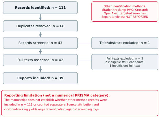
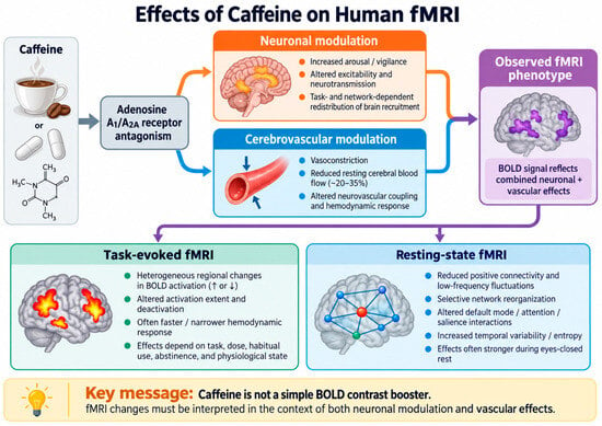
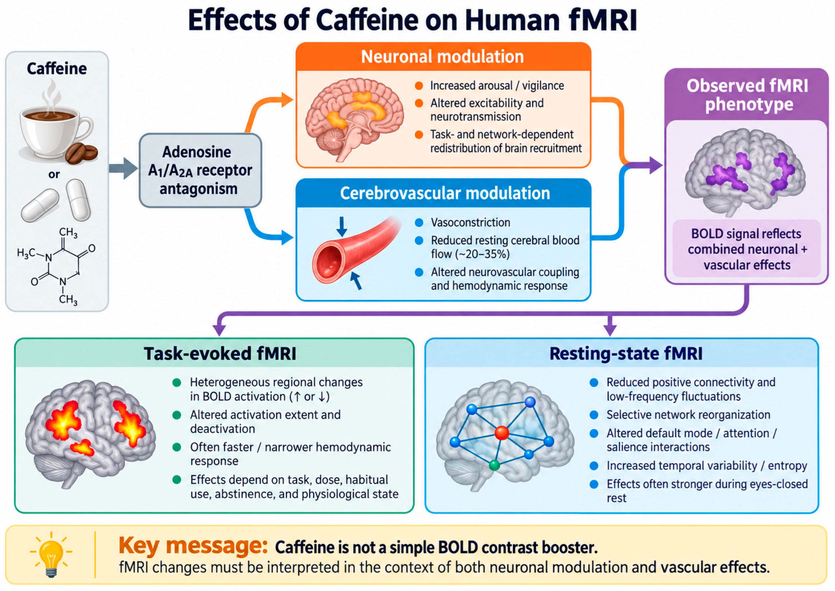

Disclaimer/Publisher’s Note: The statements, opinions and data contained in all publications are solely those of the individual author(s) and contributor(s) and not of MDPI and/or the editor(s). MDPI and/or the editor(s) disclaim responsibility for any injury to people or property resulting from any ideas, methods, instructions or products referred to in the content. |
© 2026 by the authors. Licensee MDPI, Basel, Switzerland. This article is an open access article distributed under the terms and conditions of the Creative Commons Attribution (CC BY) license.