


Abstract
Background/Objectives: Melatonin has been proposed as a potential strategy to improve female fertility, but existing evidence is dispersed across assisted reproduction, reproductive disorders, and in vitro studies. This study aimed to comprehensively evaluate the relationships of melatonin supplementation, in vitro melatonin treatment, and endogenous melatonin levels with female fertility. Methods: PubMed, Web of Science, and EMBASE were searched from inception through February 2026. Randomized and non-randomized intervention studies, observational studies, and in vitro studies involving human oocytes or embryos were included. Random-effects meta-analyses were performed, and certainty of evidence was assessed using GRADE. Results: Thirty-six publications were included. Among women undergoing ART, melatonin supplementation increased the clinical pregnancy rate (RR = 1.55, 95% CI: 1.23–1.94) and live birth (RR = 1.59, 95% CI: 1.01–2.51). Melatonin supplementation also improved selected ovarian and embryological outcomes. In vitro treatment increased fertilization (OR = 1.97, 95% CI: 1.40–2.76) and blastocyst formation (OR = 2.00, 95% CI: 1.33–3.03). Observational studies showed higher morning serum melatonin levels in women with PCOS (MD = 7.83 pg/mL, 95% CI: 3.56–12.11). The certainty of evidence ranged from very low to moderate. Conclusions: Melatonin supplementation may improve selected reproductive outcomes, including clinical pregnancy and live birth, although evidence for live birth remains limited. In vitro and observational findings further support a potential role of melatonin in female reproductive function, but its clinical value requires confirmation in high-quality studies.
1. Introduction
Female fertility is an important component of reproductive health [1]. Reproductive disorders such as polycystic ovary syndrome (PCOS), diminished ovarian reserve (DOR), and premature ovarian insufficiency (POI) can adversely affect female fertility [2,3,4,5]. When impaired fertility manifests as infertility, assisted reproductive technology (ART) provides an important treatment option [6]. However, the likelihood of achieving pregnancy and live birth remains influenced by multiple factors [7]. Various strategies are therefore being explored to restore fertility and improve the success of ART.
Melatonin is an endogenous hormone primarily secreted by the pineal gland during the night, serving as a physiological signal of darkness and contributing to the regulation of circadian rhythms and the sleep–wake cycle [8]. In addition to these chronobiological functions, melatonin has potent antioxidant properties [9]. Oxidative stress is among the key biological factors influencing female fertility. Excessive reactive oxygen species can disrupt redox homeostasis in oocytes and embryos, impair oocyte maturation, fertilization, and subsequent embryo development, and thereby reduce reproductive potential [10]. Melatonin can directly scavenge free radicals, enhance antioxidant defenses, and contribute to the maintenance of mitochondrial homeostasis [11,12]. These diverse physiological and cellular actions have prompted interest in melatonin supplementation as a potential strategy to support female reproductive health.
Evidence regarding melatonin and female fertility has emerged from assisted reproduction, reproductive disorders, and in vitro studies of human oocytes and embryos [13]. However, existing studies vary considerably in population characteristics, study designs, and outcomes. Previous systematic reviews and meta-analyses have mainly focused on assisted reproduction, specific reproductive disorders, or individual experimental settings, whereas evidence spanning different reproductive contexts has largely been summarized in narrative reviews, and systematic integration across different study designs and levels of evidence remains lacking [13,14,15].
Therefore, this systematic review and meta-analysis aimed to comprehensively evaluate the relationships of exogenous melatonin supplementation, in vitro melatonin treatment, and endogenous melatonin levels with female fertility, and to assess the potential role and clinical relevance of melatonin in female fertility.
2. Materials and Methods
2.1. Search Strategy
PubMed, Web of Science, and EMBASE were systematically searched from database inception to February 2026. Search terms related to melatonin and female fertility were used, including assisted reproductive technology (ART), in vitro fertilization (IVF), intracytoplasmic sperm injection (ICSI), intrauterine insemination (IUI), polycystic ovary syndrome (PCOS), premature ovarian insufficiency (POI), premature ovarian failure (POF), and diminished ovarian reserve (DOR). Detailed search strategies are provided in Table S1. Reference lists of included studies were also screened. This review was registered with PROSPERO (CRD420261484630).
2.2. Eligibility Criteria
2.2.1. Inclusion Criteria
Eligible studies included randomized controlled trials, non-randomized intervention studies, prospective cohort studies, case–control studies, cross-sectional studies, and in vitro studies using human oocytes or embryos derived from ART cycles. Eligible populations included women undergoing ART, women with PCOS, DOR, POF, or POI, and human oocytes or embryos exposed to melatonin in vitro. Studies were required to evaluate melatonin as an intervention, exposure, or biomarker and report at least one fertility-related, embryological, ovarian, or PCOS-related metabolic/endocrine outcome. Studies involving co-interventions were included only when the effect of melatonin could be evaluated separately.
2.2.2. Exclusion Criteria
Non-original studies, animal studies, mechanistic or cell-line studies, non-English publications, duplicate reports, studies without extractable data, and studies without a comparable control group were excluded.
2.3. Study Selection and Data Extraction
Study selection and data extraction were independently performed by two reviewers. Any disagreements were resolved through discussion or consultation with a third reviewer. After duplicate removal, titles and abstracts were screened, followed by full-text assessment. Extracted data included study characteristics, population, melatonin exposure or intervention, comparator, relevant outcomes, corresponding sample sizes and outcome data, and information required for quality assessment.
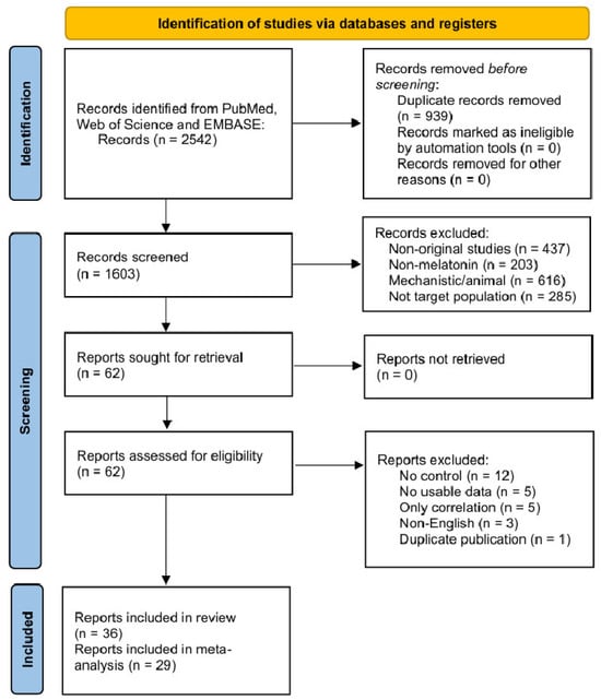
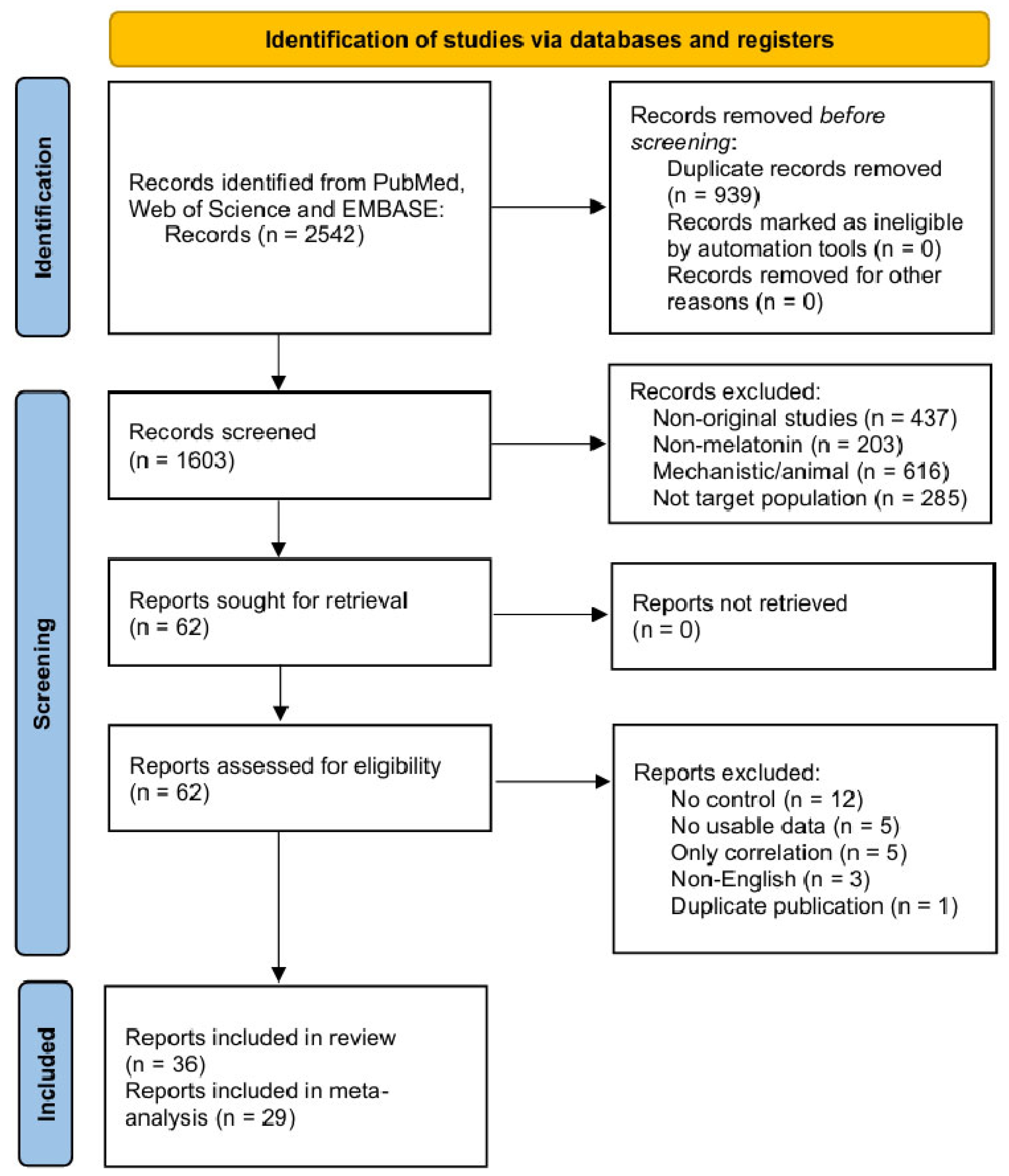
The study selection process was conducted and reported in accordance with the PRISMA 2020 statement and is presented in Figure 1.
Figure 1.
PRISMA flow diagram of the study selection process.
2.4. Assessment of Study Quality and Evidence Certainty
Study quality was independently assessed by two reviewers using design-specific tools: RoB 2.0 for RCTs [16]; ROBINS-I for non-randomized intervention studies [17]; the Newcastle–Ottawa Scale for observational studies [18,19]; and the QUIN tool for in vitro studies [20].
The overall certainty of evidence for key outcomes was assessed using GRADE, considering risk of bias, inconsistency, indirectness, imprecision, and publication bias [21].
2.5. Statistical Analysis
Statistical analyses were performed using R version 4.5.1. Continuous outcomes were pooled using mean differences (MDs), whereas dichotomous outcomes were pooled using risk ratios (RRs) or odds ratios (ORs). All effect estimates were reported with 95% confidence intervals (CIs). When continuous data were reported as medians with first and third quartiles, means and standard deviations were estimated using the method of Wan et al. [22]. For studies reporting means and 95% CIs without standard deviations, standard deviations were derived from the confidence intervals using the corresponding t distribution. For clinical outcomes, denominators followed the definitions used in the original studies and included participants/women, treatment cycles, or embryo-transfer cycles, as applicable. For in vitro studies, oocytes or embryos were used as the unit of analysis. Because donor-level data required to account for within-woman correlation were unavailable, cluster-adjusted estimates could not be reconstructed; therefore, unit-of-analysis characteristics and study-level results were additionally summarized descriptively (Table S5). For studies with multiple melatonin doses or concentrations, the 3 or 4 mg/day group for clinical studies and the 104 nmol/L group for in vitro studies were selected for the primary analysis to align with the exposure levels used in single-dose studies and improve comparability across studies.
Studies or outcomes were not quantitatively pooled when the available data were insufficient for effect estimation, outcome denominators could not be reliably determined, measurement units could not be reliably harmonized, outcome definitions were not sufficiently comparable, or fewer than two studies provided suitable data for quantitative synthesis. Such findings were retained in the narrative synthesis where appropriate.
Random-effects models were used because of anticipated clinical and methodological heterogeneity. Between-study variance was estimated using the restricted maximum likelihood (REML) method, and 95% CIs for pooled effects were calculated using the conventional Wald method. Between-study heterogeneity was assessed using Cochran’s Q test and the I2 statistic. Subgroup analyses were conducted for selected outcomes with notable heterogeneity according to study design or melatonin concentration. Funnel plots were examined for outcomes including at least seven studies and interpreted cautiously because most analyses included fewer than 10 studies. Leave-one-out sensitivity analyses were performed for key outcomes. Additional sensitivity analyses were performed by excluding studies judged to be at high risk of bias using RoB 2.0 or at serious or critical risk of bias using ROBINS-I, where sufficient studies remained for pooling.
3. Results
3.1. Study Selection and Characteristics
A total of 2542 records were identified through the initial search. After screening, 36 publications met the eligibility criteria and were included in the review. The study selection process is shown in Figure 1.
The included studies comprised randomized controlled trials, non-randomized intervention studies, observational studies, and in vitro studies using human oocytes or embryos. Study populations and experimental materials included women undergoing assisted reproductive technology, women with polycystic ovary syndrome, women with diminished ovarian reserve or premature ovarian failure, and human oocytes and embryos. Studies of exogenous melatonin mainly evaluated the effects of melatonin supplementation on assisted reproductive outcomes, ovarian function, and PCOS-related outcomes; in vitro studies assessed the effects of different melatonin concentrations on oocyte fertilization and embryo development; and observational studies investigated associations between endogenous melatonin levels and reproductive disorders. Detailed characteristics of the included studies are presented in Table S2, and the corresponding outcome-level extracted data are provided in Table S8.
3.2. Quality Assessment of Included Studies
The results of quality assessment varied across study designs. Risk-of-bias assessments of RCTs varied from low risk to high risk, with concerns primarily related to the randomization process, deviations from intended interventions, and selective reporting of results (Figure S1). Non-randomized intervention studies had a relatively higher risk of bias, mainly due to inadequate control of confounding and bias related to participant selection (Figure S2). Observational studies were generally of moderate quality, with the main limitations being insufficient comparability between groups and potential confounding (Table S3). In vitro studies generally showed a low or moderate risk of bias (Table S4).
3.3. Meta-Analysis Results
3.3.1. Clinical Effects of Exogenous Melatonin Supplementation
ART Population
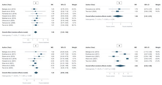
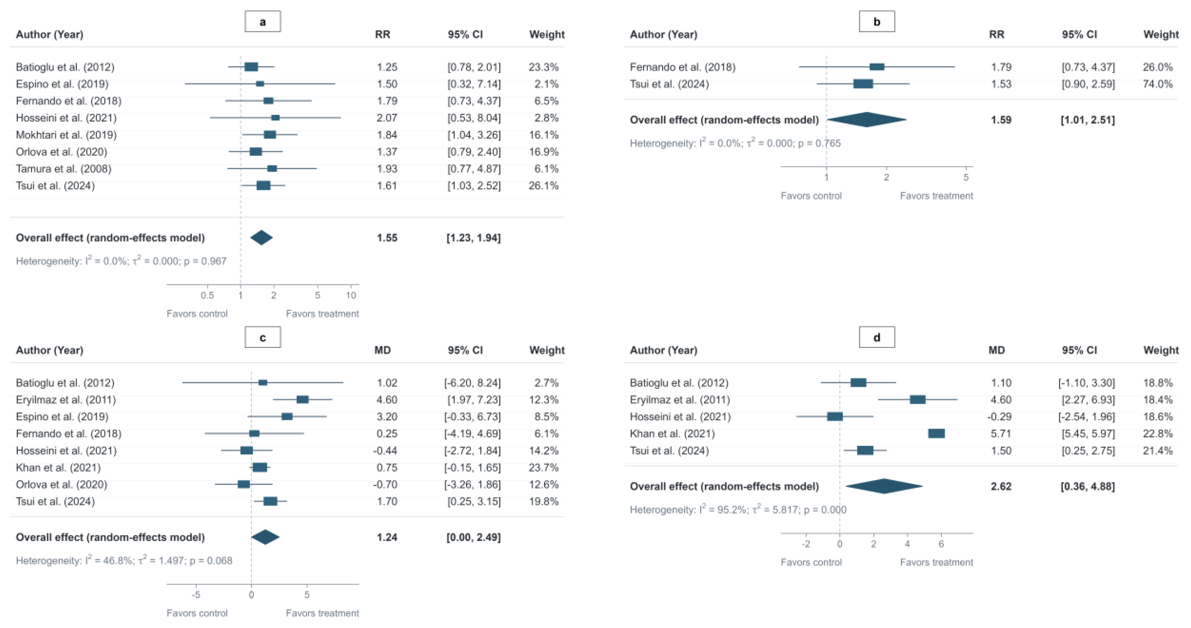
Among women undergoing ART, melatonin supplementation increased the clinical pregnancy rate (RR = 1.55, 95% CI: 1.23–1.94), biochemical pregnancy rate (RR = 1.61, 95% CI: 1.09–2.38) and live birth (RR = 1.59, 95% CI: 1.01–2.51). Melatonin supplementation also increased the number of mature oocytes (MD = 2.62, 95% CI: 0.36–4.88), and transferable embryos (MD = 4.51, 95% CI: 2.59–6.43), while no significant differences were found in the number of oocytes retrieved, follicular fluid estradiol concentration, fertilized oocytes, fertilization rate, good-quality embryos, or day 3 good-quality embryos (Table 1; Figure 2 and Figure S3). Miscarriage was not quantitatively pooled because the two studies reporting this outcome used different denominators. The two studies showed effects in opposite directions [23,24].
Table 1.
Meta-analysis of melatonin supplementation on ART outcomes.
Figure 2.
Forest plots of melatonin supplementation on ART outcomes: (a) clinical pregnancy rate; (b) live birth rate; (c) number of oocytes retrieved; (d) number of mature oocytes [23,24,25,26,27,28,29,30,31,32].
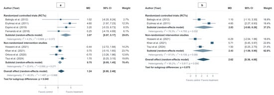
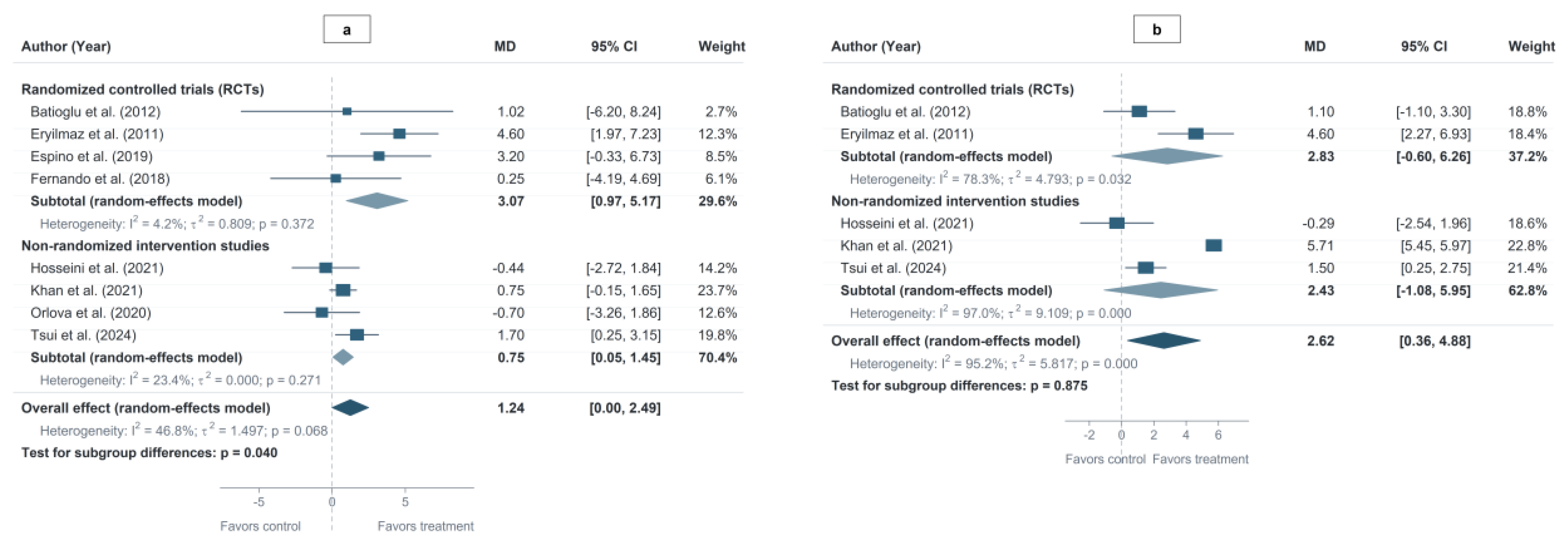
Subgroup analyses by study design showed a larger increase in oocytes retrieved in RCTs than in non-randomized intervention studies (p = 0.040), whereas no significant subgroup difference was observed for mature oocytes (p = 0.875) (Figure 3).
Figure 3.
Subgroup analyses by study design: (a) number of oocytes retrieved; (b) number of mature oocytes [23,24,25,26,27,28,29,32].
PCOS Population
Among women with PCOS, pooled analyses showed no significant effects of melatonin supplementation on total antioxidant capacity, fasting glucose, or lipid-related outcomes (Table 2; Figure S4).
Table 2.
Meta-analysis of melatonin supplementation on PCOS-related outcomes.
In addition to these outcomes, Akter et al. reported improvements in follicular development, ovulation, and clinical pregnancy [33]; Jamilian et al. and Alizadeh et al. reported improvements in hyperandrogenism-related outcomes [34,35]; and Shabani et al. reported improvements in sleep and psychological outcomes [36].
DOR/POF Population
Among women with DOR, pooled analyses showed that melatonin supplementation increased the number of mature oocytes (MD = 2.70, 95% CI: 0.86–4.54) and serum estradiol levels on the day of hCG administration (MD = 694.55 pg/mL, 95% CI: 401.65–987.44), whereas no significant effects were observed on total HMG dose or stimulation duration (Table 3; Figure S5). In addition, Sadeghpour et al. reported improvements in oocyte retrieval, fertilization, embryo quality, and biochemical pregnancy [37], while Jahromi et al. reported improvements in mature oocyte retrieval and good-quality embryos [38]. Among women with POF, Li et al. reported reductions in LH, FSH, and ROS levels and improved ovarian hormone secretion following melatonin treatment [39].
Table 3.
Meta-analysis of melatonin supplementation on DOR-related outcomes.
3.3.2. Effects of Melatonin on Oocytes and Embryos in In Vitro Studies
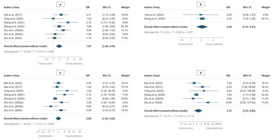
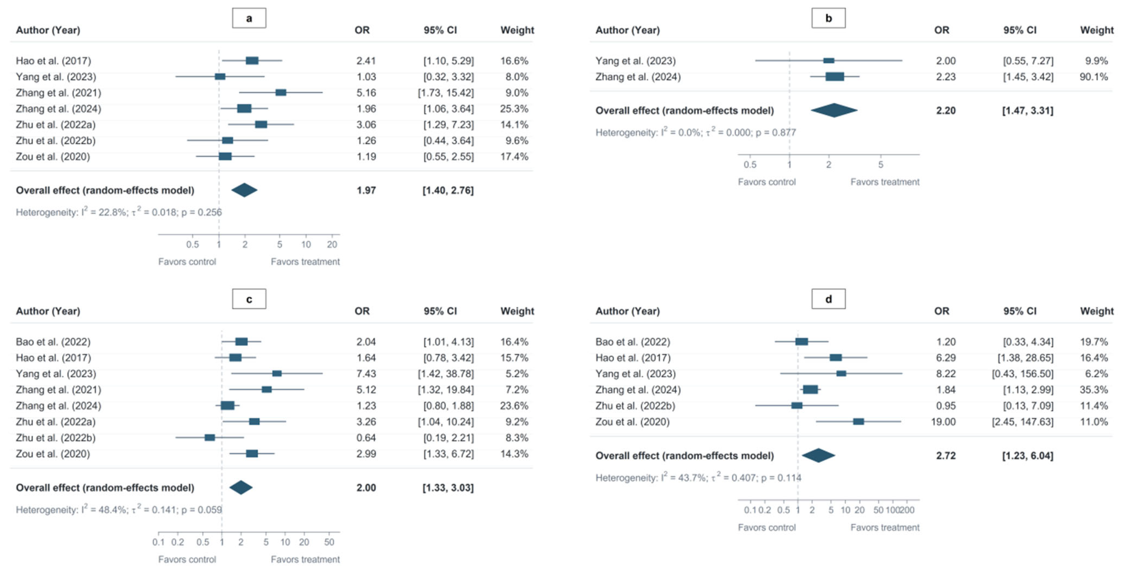
In vitro melatonin treatment increased fertilization, oocyte survival, embryo quality, and blastocyst-related outcomes, whereas no significant effects were observed on oocyte maturation or cleavage rates (Table 4; Figure 4 and Figure S6). One study not included in the quantitative meta-analysis reported that 1 nM melatonin increased nuclear maturation of GV- and MI-stage oocytes, whereas 106 nM reduced maturation [40].
Table 4.
Meta-analysis of melatonin treatment on oocyte and embryo developmental outcomes.
Figure 4.
Forest plots of melatonin treatment on oocyte and embryo developmental outcomes: (a) fertilization rate; (b) good-quality embryo rate; (c) blastocyst formation rate; (d) good-quality blastocyst rate [41,42,43,44,45,46,47,48].
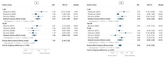
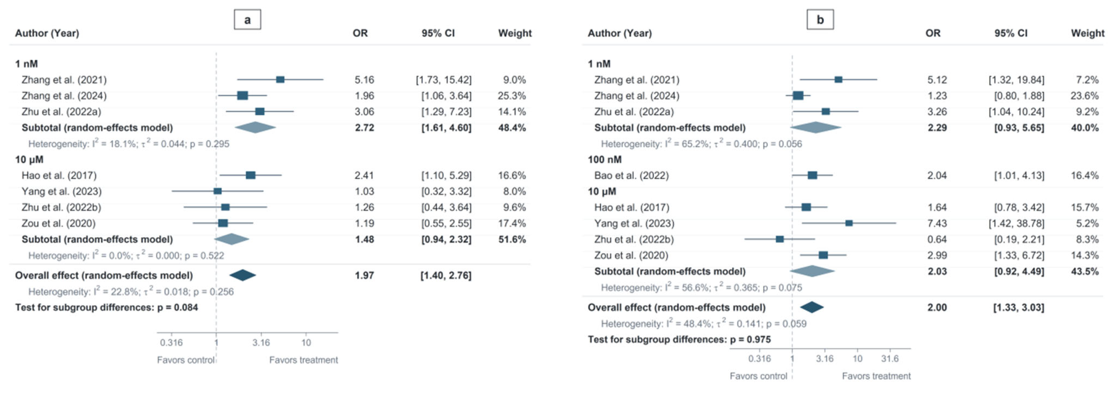
To explore the effects of different melatonin concentrations, subgroup analyses were further conducted (Figure 5). The results showed no significant differences in fertilization or blastocyst formation rates across melatonin concentrations.
Figure 5.
Subgroup analyses by melatonin concentration: (a) fertilization rate; (b) blastocyst formation rate [41,42,43,44,45,46,47,48].
Among the 10 in vitro studies, only seven reported donor/woman numbers for the experiments corresponding to the developmental outcomes included in this review, and only one explicitly reported sibling-oocyte allocation across experimental conditions for the main comparison. None reported statistical adjustment for within-woman clustering or pairing. Study-level effect estimates were therefore additionally presented descriptively alongside the unit-of-analysis characteristics in Table S5.
3.3.3. Association Between Endogenous Melatonin Levels and Fertility
Among women with PCOS, morning serum melatonin levels were higher than in controls (MD = 7.83 pg/mL, 95% CI: 3.56–12.11; I2 = 22.7%), whereas no significant difference was observed in nighttime serum melatonin levels (Table 5; Figure S7). Abduljabbar et al., which was not included in the quantitative synthesis because its reported measurement unit could not be reliably harmonized, also reported higher morning serum melatonin levels in women with PCOS than in controls [49].
Table 5.
Meta-analysis of endogenous melatonin levels in women with polycystic ovary syndrome.
In addition to serum melatonin levels, Shreeve et al. and Luboshitzky et al. reported higher urinary melatonin metabolite levels in women with PCOS [50,51]. Li et al. found lower follicular fluid melatonin concentrations, which were positively associated with day 3 good-quality embryo formation [52].
3.4. Sensitivity Analyses and Publication Bias
Leave-one-out sensitivity analyses showed stable pooled estimates for clinical pregnancy (RR range: 1.50–1.65), fertilization (OR range: 1.79–2.19), and blastocyst formation (OR range: 1.84–2.27). The statistical significance of the number of oocytes retrieved and mature oocytes varied after exclusion of some studies (Table S6).
Additional sensitivity analyses excluding studies at high, serious, or critical risk of bias showed that the associations with clinical pregnancy (RR = 1.73, 95% CI: 1.27–2.36) remained statistically significant. For the number of oocytes retrieved, the pooled estimate became statistically significant (MD = 1.00, 95% CI: 0.23–1.76), while heterogeneity decreased to 0%. In contrast, the association with the number of mature oocytes was no longer statistically significant (MD = 3.65, 95% CI: −0.47–7.78), with substantial heterogeneity remaining (I2 = 97.6%). Estimates for the number of fertilized oocytes remained nonsignificant. After these exclusions, too few studies remained to quantitatively synthesize the number of transferable embryos, the number of good-quality embryos, and biochemical pregnancy rate (Table S6).
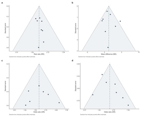
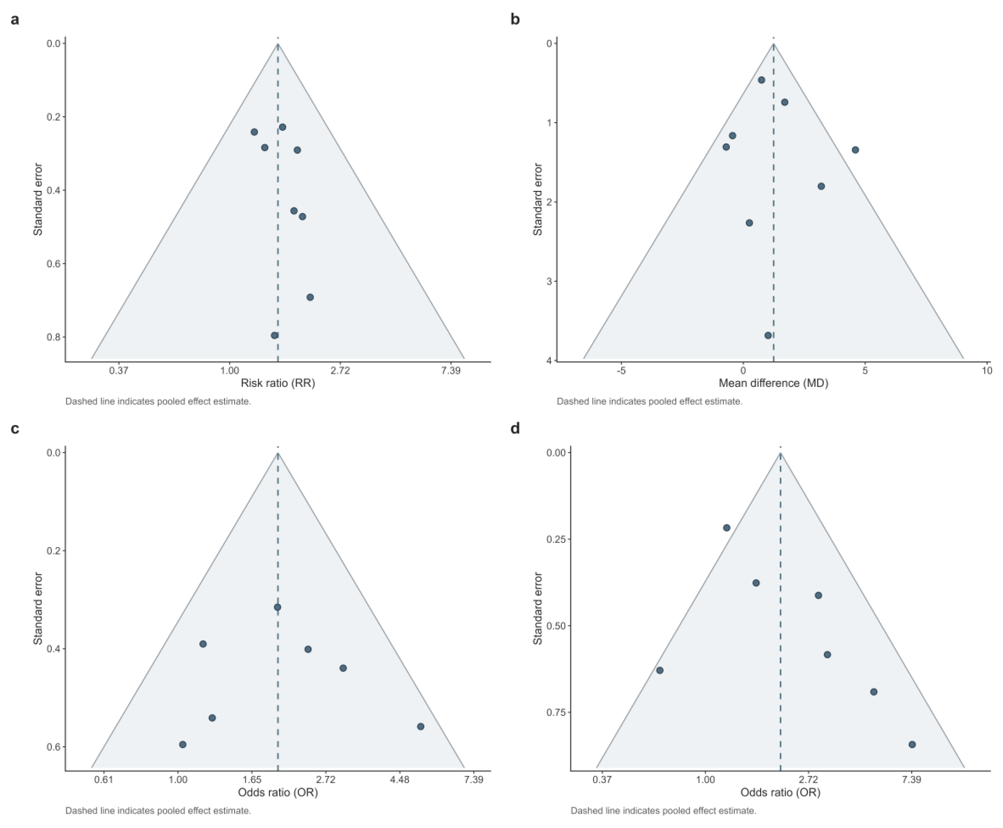
Funnel plots showed no obvious asymmetry for the key outcomes (Figure 6). However, given the limited number of included studies, publication bias cannot be completely ruled out.
Figure 6.
Funnel plots for assessment of potential publication bias in key outcomes: (a) clinical pregnancy rate; (b) number of oocytes retrieved; (c) fertilization rate; (d) blastocyst formation rate.
3.5. Certainty of Evidence
The certainty of evidence ranged from very low to moderate (Table S7). Evidence for clinical pregnancy from RCTs was rated as moderate, whereas the evidence for live birth was rated as low. Evidence for the other key outcomes was generally low or very low, mainly because of risk of bias, limited sample size, and heterogeneity.
4. Discussion
4.1. Main Findings
This study integrated evidence from exogenous clinical interventions, in vitro studies of human oocytes and embryos, and observational studies of endogenous melatonin. Overall, exogenous melatonin supplementation showed potential benefits for selected assisted reproductive outcomes; in vitro studies further supported direct effects of melatonin on human oocyte and embryo development; and altered endogenous melatonin levels in women with PCOS suggested that melatonin homeostasis may be related to female fertility.
In studies of exogenous melatonin supplementation, meta-analysis showed higher clinical and biochemical pregnancy rates with low between-study heterogeneity. A higher live birth rate was also observed; however, this finding should be interpreted cautiously because the certainty of evidence was low and the confidence interval was wide, with its lower bound close to the null. Melatonin supplementation was also associated with increases in the number of mature oocytes and transferable embryos. However, the evidence for these ovarian response and intermediate embryological outcomes should be interpreted cautiously, given the substantial heterogeneity observed for some outcomes and the loss of statistical significance for mature oocyte number after studies at high, serious, or critical risk of bias were excluded. Among women with DOR, melatonin supplementation increased serum estradiol levels on the day of hCG administration. Estradiol is primarily secreted by follicular granulosa cells, and this increase may reflect enhanced follicular endocrine activity during ovarian stimulation, although whether this indicates improved follicular developmental potential requires further evidence [53,54]. In contrast, pooled metabolic and oxidative stress-related outcomes in women with PCOS showed no clear improvement, and few studies directly assessed fertility outcomes; therefore, the reproductive benefit of melatonin supplementation in women with PCOS cannot yet be determined.
In vitro studies provided further evidence for direct effects of melatonin on oocytes and embryos. Meta-analysis showed that melatonin treatment increased fertilization rate and improved outcomes related to good-quality embryos and blastocysts, while no significant effects were observed on oocyte maturation or cleavage rates. This pattern suggests that the potential effects of melatonin may be more closely related to fertilization competence and subsequent embryo developmental potential rather than a general enhancement of development at all stages. Notably, these effects may be concentration dependent, as one included study reported a reduced oocyte maturation rate at 106 nM melatonin, indicating that higher concentrations do not necessarily confer greater developmental benefit. Previous studies have suggested that the potential effects of melatonin on oocyte and embryo development may involve multiple biological pathways. Melatonin may act through receptor-mediated signaling at the ovarian cell membrane as well as through receptor-independent pathways [55,56]. Melatonin can cross cell membranes and enter intracellular and subcellular compartments to scavenge reactive oxygen species and reduce oxidative damage, while also supporting cellular energy metabolism by maintaining mitochondrial membrane potential and ATP production [11,57]. These actions may help preserve redox and mitochondrial homeostasis in oocytes, thereby providing a more favorable intracellular environment for fertilization and early embryo development.
Melatonin is not only an exogenous intervention but also an endogenous component of female reproductive physiology [58,59]. In women with PCOS, morning serum melatonin levels were higher than in controls, whereas no significant difference was observed in nighttime serum melatonin levels, with substantial heterogeneity across studies. Nighttime urinary melatonin metabolite levels were also increased, while follicular fluid melatonin concentrations were reduced and positively associated with the day 3 good-quality embryo formation rate. Together, these findings may reflect altered temporal regulation of melatonin in PCOS rather than a uniform increase in melatonin levels; however, differences in sampling time, assay methods, and collection conditions limit a more definitive interpretation. The findings in follicular fluid further suggest that local melatonin status within the ovarian microenvironment may be related to oocyte and embryo developmental potential. However, studies directly comparing the reproductive relevance of circulating and local melatonin levels are currently lacking. Overall, the endogenous evidence indicates that melatonin may have physiological and pathological relevance to female reproduction beyond its potential value as an exogenous intervention.
4.2. Clinical Implications
The findings of this study suggest that melatonin may have potential relevance to female reproductive health, particularly in the context of assisted reproduction. Potential benefits were observed across both laboratory and clinical outcomes, including selected oocyte-related and embryo-related outcomes as well as biochemical pregnancy, clinical pregnancy, and live birth. However, given the limited number of studies and the overall certainty of the current evidence, routine melatonin supplementation for fertility treatment cannot yet be recommended.
An important unresolved issue is the relationship between oral melatonin dose and local exposure within the follicular microenvironment. Although clinical studies have shown that oral melatonin supplementation can increase follicular fluid melatonin concentrations [25,26], the relationship among oral dose, follicular exposure, and the concentrations associated with biological effects in vitro remains poorly defined. Establishing this dose–exposure–effect relationship may therefore be important for determining clinically appropriate dosing regimens.
The timing of supplementation may be equally important. Melatonin is not merely an antioxidant but also a physiological signal involved in the regulation of circadian rhythms and the sleep–wake cycle. Accordingly, the effects of supplementation may depend not only on the administered dose but also on its timing relative to the endogenous circadian rhythm. Inappropriate timing may alter normal circadian signaling and contribute to adverse effects such as drowsiness. Future clinical studies should therefore consider both dose and timing when optimizing melatonin supplementation.
Although PCOS has recently been renamed polyendocrine metabolic ovarian syndrome (PMOS), the studies included in this review enrolled participants according to the PCOS definitions and diagnostic criteria used at the time [60]. Therefore, we retained the term PCOS for these study populations rather than retrospectively reclassifying them as PMOS. Future studies should consider the updated PMOS framework when defining study populations and interpreting their comparability with previous PCOS-based evidence.
Given the heterogeneity of the populations studied, including women undergoing ART and those with DOR or PCOS, the effects of melatonin should not be assumed to be uniform across reproductive conditions. Altered endogenous melatonin status in disorders such as PCOS further suggests that reproductive phenotype, metabolic and endocrine characteristics, and endogenous melatonin status may all be relevant when identifying populations most likely to benefit from supplementation. Future studies should therefore explore more individualized supplementation strategies that incorporate these characteristics.
Overall, before melatonin can be recommended for routine use in assisted reproduction, higher-quality evidence is needed to establish the optimal dose and timing of administration and to identify the target populations most likely to benefit.
4.3. Strengths and Limitations
This study has several strengths. Compared with previous studies that mainly focused on a single population or outcome, we integrated evidence from clinical interventions, in vitro experiments, and observational studies, thereby evaluating the relationship between melatonin and female fertility across multiple levels, including clinical outcomes, oocyte and embryo development, and disease-related melatonin status. Design-specific quality assessment and GRADE were used to evaluate study quality and evidence certainty.
However, several limitations should be acknowledged. The small number of studies available for some outcomes limited the reliability of further subgroup analyses and publication bias assessment. Differences in study design, study populations, intervention regimens, and outcome definitions also increased heterogeneity and uncertainty in interpretation. For the in vitro studies, oocytes or embryos from the same woman may not have been statistically independent. Because donor-level data and the information required to account for within-woman correlation were unavailable, cluster-adjusted estimates could not be reconstructed. Consequently, the confidence intervals of the pooled in vitro estimates may be overly precise, and these results should be interpreted cautiously. For studies of endogenous melatonin, differences in sampling time, assay methods, collection conditions, and reported measurement units limited comparability across studies and, in some cases, precluded quantitative pooling. In addition, evidence from non-randomized, in vitro, and observational studies limited the overall certainty of evidence, and observational findings on endogenous melatonin do not permit causal inference. Sensitivity analyses further indicated that some findings were not fully robust to the exclusion of studies at high, serious, or critical risk of bias, particularly the number of mature oocytes.
Larger randomized trials with standardized protocols and core outcomes are needed to clarify the clinical value of melatonin supplementation. Greater standardization of experimental and measurement methods is also needed to improve the comparability of in vitro and endogenous melatonin studies.
5. Conclusions
The present study suggests that melatonin is associated with female fertility and that melatonin supplementation may improve selected reproductive outcomes, particularly clinical pregnancy in women undergoing assisted reproduction, with a potential benefit also observed for live birth, although evidence for this outcome remains limited. In vitro studies further support potential effects of melatonin on fertilization and early embryo development, while altered endogenous melatonin levels in women with PCOS suggest a possible role of melatonin homeostasis in female reproductive function. However, given the limited number of studies for some outcomes, the methodological limitations of the available evidence and its generally low certainty, current evidence is insufficient to support routine melatonin supplementation for fertility treatment, and its clinical value requires further evaluation in high-quality studies.
Supplementary Materials
The following supporting information can be downloaded at: https://www.mdpi.com/article/10.3390/nu18203431/s1, Figure S1: Risk of bias assessment of RCTs using RoB 2. (a) Risk-of-bias judgments for individual studies across domains; (b) summary of risk-of-bias judgments across domains; Figure S2: Risk of bias assessment of non-randomized studies using ROBINS-I. (a) Risk-of-bias judgments for individual studies across domains; (b) summary of risk-of-bias judgments across domains; Figure S3: Forest plots of melatonin supplementation on ART outcomes. (a) follicular fluid estradiol concentration; (b) fertilization rate; (c) number of fertilized oocytes; (d) number of good-quality embryos; (e) number of day 3 good-quality embryos; (f) number of transferable embryos; (g) biochemical pregnancy rate; Figure S4: Forest plots of melatonin supplementation on PCOS-related outcomes. (a) total antioxidant capacity; (b) fasting blood glucose; (c) triglycerides; (d) total cholesterol; (e) low-density lipoprotein cholesterol; (f) high-density lipoprotein cholesterol; Figure S5: Forest plots of melatonin supplementation on DOR-related outcomes. (a) duration of ovarian stimulation; (b) total hMG dose; (c) serum estradiol concentration on the day of hCG administration; (d) number of metaphase II oocytes retrieved; Figure S6: Forest plots of melatonin treatment on oocyte and embryo developmental outcomes. (a) oocyte survival rate; (b) oocyte maturation rate; (c) cleavage rate; (d) day 3 good-quality embryo rate; Figure S7: Forest plots of endogenous melatonin levels in women with polycystic ovary syndrome. (a) morning serum melatonin concentration; (b) nighttime serum melatonin concentration; Table S1: Database search strategies; Table S2: Characteristics of the included studies; Table S3: Quality assessment of observational studies using the Newcastle–Ottawa Scale (NOS); Table S4: Quality assessment of in vitro studies using the QUIN tool; Table S5: Unit-of-analysis characteristics and study-level results of in vitro studies; Table S6: Sensitivity analyses; Table S7. GRADE evidence profile for key outcomes; Table S8. Outcome-level data extraction of included studies.
Author Contributions
Conceptualization, H.Y. and X.H.; methodology, H.Y., B.W. and J.Y.; formal analysis, H.Y.; investigation, H.Y. and Y.H.; data curation, H.Y. and Y.H.; validation, H.W. and B.W.; visualization, H.Y. and Y.S.; writing—original draft preparation, H.Y.; writing—review and editing, H.Y., J.Y. and X.H.; supervision, J.Y. and X.H.; project administration, X.H.; funding acquisition, X.H. All authors have read and agreed to the published version of the manuscript.
Funding
This research was funded by the National Natural Science Foundation of China, grant numbers 82404284 and 82574109.
Institutional Review Board Statement
Not applicable.
Informed Consent Statement
Not applicable.
Data Availability Statement
All data analyzed in this study were obtained from previously published studies and are available within the article and its Supplementary Materials.
Conflicts of Interest
The authors declare no conflicts of interest. The funders had no role in the design of the study; in the collection, analyses, or interpretation of data; in the writing of the manuscript; or in the decision to publish the results.
Abbreviations
The following abbreviations are used in this manuscript:
| ART | Assisted reproductive technology |
| DOR | Diminished ovarian reserve |
| GRADE | Grading of Recommendations Assessment, Development and Evaluation |
| ICSI | Intracytoplasmic sperm injection |
| IUI | Intrauterine insemination |
| IVF | In vitro fertilization |
| MD | Mean difference |
| NOS | Newcastle–Ottawa Scale |
| PCOS | Polycystic ovary syndrome |
| POF | Premature ovarian failure |
| POI | Premature ovarian insufficiency |
| RCT | Randomized controlled trial |
| RoB 2 | Risk of Bias 2 |
| ROBINS-I | Risk Of Bias In Non-randomized Studies—of Interventions |
References
- Yang, J.; Pang, W.W.; Yu, G.; Antero, J.; Li, L.-J.; Huang, Z.; Zhang, C. Women’s health and female fertility: Current evidence and knowledge gaps in the Asia–Pacific region. Lancet Reg. Health West. Pac. 2025, 65, 101710. [Google Scholar] [CrossRef] [Scilit]
- Hu, S.; Xu, B.; Jin, L. Perinatal outcome in young patients with diminished ovarian reserve undergoing assisted reproductive technology. Fertil. Steril. 2020, 114, 118–124.e111. [Google Scholar] [CrossRef] [Scilit]
- Teede, H.J.; Tay, C.T.; Laven, J.; Dokras, A.; Moran, L.J.; Piltonen, T.T.; Costello, M.F.; Boivin, J.; Redman, L.M.; A. Boyle, J.; et al. Recommendations from the 2023 International Evidence-based Guideline for the Assessment and Management of Polycystic Ovary Syndrome. Fertil. Steril. 2023, 120, 767–793. [Google Scholar] [CrossRef] [Scilit]
- Webber, L.; Davies, M.; Anderson, R.; Bartlett, J.; Braat, D.; Cartwright, B.; Cifkova, R.; de Muinck Keizer-Schrama, S.; Hogervorst, E.; Janse, F.; et al. ESHRE Guideline: Management of women with premature ovarian insufficiency. Hum. Reprod. 2016, 31, 926–937. [Google Scholar] [CrossRef] [Scilit]
- Xie, M.; Li, J.; Bai, W. Sleep Disorders in Patients with PCOS After Ketogenic Diet Intervention: An Analysis of Influencing Factors and Development of Predictive Model. Br. J. Hosp. Med. 2026, 87, 50655. [Google Scholar] [CrossRef] [Scilit]
- Farquhar, C.M.; Bhattacharya, S.; Repping, S.; Mastenbroek, S.; Kamath, M.S.; Marjoribanks, J.; Boivin, J. Female subfertility. Nat. Rev. Dis. Primers 2019, 5, 7. [Google Scholar] [CrossRef] [Scilit]
- Gaskins, A.J.; Zhang, Y.; Chang, J.; Kissin, D.M. Predicted probabilities of live birth following assisted reproductive technology using United States national surveillance data from 2016 to 2018. Am. J. Obstet. Gynecol. 2023, 228, 557.e1–557.e10. [Google Scholar] [CrossRef] [Scilit]
- Zisapel, N. New perspectives on the role of melatonin in human sleep, circadian rhythms and their regulation. Br. J. Pharmacol. 2018, 175, 3190–3199. [Google Scholar] [CrossRef] [Scilit]
- Hardeland, R. Redox Biology of Melatonin: Discriminating Between Circadian and Noncircadian Functions. Antioxid. Redox Signal. 2022, 37, 704–725. [Google Scholar] [CrossRef] [Scilit]
- Hardy, M.L.M.; Day, M.L.; Morris, M.B. Redox Regulation and Oxidative Stress in Mammalian Oocytes and Embryos Developed In Vivo and In Vitro. Int. J. Environ. Res. Public Health 2021, 18, 11374. [Google Scholar] [CrossRef] [Scilit]
- García, J.J.; López-Pingarrón, L.; Almeida-Souza, P.; Tres, A.; Escudero, P.; García-Gil, F.A.; Tan, D.X.; Reiter, R.J.; Ramírez, J.M.; Bernal-Pérez, M. Protective effects of melatonin in reducing oxidative stress and in preserving the fluidity of biological membranes: A review. J. Pineal Res. 2014, 56, 225–237. [Google Scholar] [CrossRef] [Scilit]
- Reiter, R.J.; Mayo, J.C.; Tan, D.X.; Sainz, R.M.; Alatorre-Jimenez, M.; Qin, L. Melatonin as an antioxidant: Under promises but over delivers. J. Pineal Res. 2016, 61, 253–278. [Google Scholar] [CrossRef] [Scilit]
- Li, Q.; Zheng, T.; Chen, J.; Li, B.; Zhang, Q.; Yang, S.; Shao, J.; Guan, W.; Zhang, S. Exploring melatonin’s multifaceted role in female reproductive health: From follicular development to lactation and its therapeutic potential in obstetric syndromes. J. Adv. Res. 2025, 70, 223–242. [Google Scholar] [CrossRef] [Scilit]
- Jaiswar, S.; Bhise, M.; Shukla, P. Effects of Melatonin in Women with Polycystic Ovary Syndrome Undergoing ART: A Systematic Review. Antioxidants 2026, 15, 896. [Google Scholar] [CrossRef] [Scilit]
- Wu, Y.; Huang, W.; Tang, L.; Feng, Y.; Chen, H.; Pan, M.; Peng, J.; Li, C.; Wang, H. Melatonin improved the outcomes of women with ART: A systematic review and meta-analysis of randomized trials. Front. Reprod. Health 2025, 7, 1680984. [Google Scholar] [CrossRef] [Scilit]
- Sterne, J.A.C.; Savović, J.; Page, M.J.; Elbers, R.G.; Blencowe, N.S.; Boutron, I.; Cates, C.J.; Cheng, H.Y.; Corbett, M.S.; Eldridge, S.M.; et al. RoB 2: A revised tool for assessing risk of bias in randomised trials. BMJ 2019, 366, l4898. [Google Scholar] [CrossRef] [Scilit]
- Sterne, J.A.; Hernán, M.A.; Reeves, B.C.; Savović, J.; Berkman, N.D.; Viswanathan, M.; Henry, D.; Altman, D.G.; Ansari, M.T.; Boutron, I.; et al. ROBINS-I: A tool for assessing risk of bias in non-randomised studies of interventions. BMJ 2016, 355, i4919. [Google Scholar] [CrossRef] [Scilit]
- Wells, G.; Shea, B.; O’Connell, D.; Peterson, J.; Welch, V.; Losos, M.; Tugwell, P. The Newcastle-Ottawa Scale (NOS) for Assessing the Quality of Nonrandomised Studies in Meta-Analyses. Available online: https://ohri.ca/en/who-we-are/core-facilities-and-platforms/ottawa-methods-centre/newcastle-ottawa-scale (accessed on 16 September 2026).
- Herzog, R.; Álvarez-Pasquin, M.J.; Díaz, C.; Del Barrio, J.L.; Estrada, J.M.; Gil, Á. Are healthcare workers’ intentions to vaccinate related to their knowledge, beliefs and attitudes? A systematic review. BMC Public Health 2013, 13, 154. [Google Scholar] [CrossRef] [Scilit]
- Sheth, V.H.; Shah, N.P.; Jain, R.; Bhanushali, N.; Bhatnagar, V. Development and validation of a risk-of-bias tool for assessing in vitro studies conducted in dentistry: The QUIN. J. Prosthet. Dent. 2024, 131, 1038–1042. [Google Scholar] [CrossRef] [Scilit]
- Schünemann, H.J.; Brożek, J.; Guyatt, G.; Oxman, A. GRADE Handbook for Grading Quality of Evidence and Strength of Recommendations; The GRADE Working Group: Hamilton, ON, Canada, 2013. [Google Scholar]
- Wan, X.; Wang, W.; Liu, J.; Tong, T. Estimating the sample mean and standard deviation from the sample size, median, range and/or interquartile range. BMC Med. Res. Methodol. 2014, 14, 135. [Google Scholar] [CrossRef] [Scilit]
- Orlova, V.V.; Suslikova, L.V.; Orlova, O.A.; Dmytriienko, D.V.; Malysheva, I.V. Assisted reproductive technology efficiency improvement of tubal-peritoneal infertility. Wiad. Lek. 2020, 73, 2370–2377. [Google Scholar] [CrossRef] [Scilit]
- Tsui, K.H.; Li, C.J.; Lin, L.T. Melatonin supplementation attenuates cuproptosis and ferroptosis in aging cumulus and granulosa cells: Potential for improving IVF outcomes in advanced maternal age. Reprod. Biol. Endocrinol. 2024, 22, 138. [Google Scholar] [CrossRef] [Scilit]
- Fernando, S.; Wallace, E.M.; Vollenhoven, B.; Lolatgis, N.; Hope, N.; Wong, M.; Lawrence, M.; Lawrence, A.; Russell, C.; Leong, K.; et al. Melatonin in Assisted Reproductive Technology: A Pilot Double-Blind Randomized Placebo-Controlled Clinical Trial. Front. Endocrinol. 2018, 9, 545. [Google Scholar] [CrossRef] [Scilit]
- Espino, J.; Macedo, M.; Lozano, G.; Ortiz, Á.; Rodríguez, C.; Rodríguez, A.B.; Bejarano, I. Impact of Melatonin Supplementation in Women with Unexplained Infertility Undergoing Fertility Treatment. Antioxidants 2019, 8, 338. [Google Scholar] [CrossRef] [Scilit]
- Batiioǧlu, A.S.; Şahin, U.; Grlek, B.; Öztrk, N.; Ünsal, E. The efficacy of melatonin administration on oocyte quality. Gynecol. Endocrinol. 2012, 28, 91–93. [Google Scholar] [CrossRef] [Scilit]
- Eryilmaz, O.G.; Devran, A.; Sarikaya, E.; Aksakal, F.N.; Mollamahmutoğlu, L.; Cicek, N. Melatonin improves the oocyte and the embryo in IVF patients with sleep disturbances, but does not improve the sleeping problems. J. Assist. Reprod. Genet. 2011, 28, 815–820. [Google Scholar] [CrossRef] [Scilit]
- Hosseini, F.S.; Shamsipour, M.; Yazdekhasti, H.; Akbari-Asbagh, F.; Shahraki, Z.; Aghaee-Bakhtiari, S.H. The effect of oral melatonin supplementation on MT-ATP6 gene expression and IVF outcomes in Iranian infertile couples: A nonrandomized controlled trial. Naunyn-Schmiedeberg’s Arch. Pharmacol. 2021, 394, 1487–1495. [Google Scholar] [CrossRef] [Scilit]
- Mokhtari, F.; Asbagh, F.A.; Azmoodeh, O.; Bakhtiyari, M.; Almasi-Hashiani, A. Effects of melatonin administration on chemical pregnancy rates of polycystic ovary syndrome patients undergoing intrauterine insemination: A randomized clinical trial. Int. J. Fertil. Steril. 2019, 13, 225–229. [Google Scholar] [CrossRef] [Scilit]
- Tamura, H.; Takasaki, A.; Miwa, I.; Taniguchi, K.; Maekawa, R.; Asada, H.; Taketani, T.; Matsuoka, A.; Yamagata, Y.; Shimamura, K.; et al. Oxidative stress impairs oocyte quality and melatonin protects oocytes from free radical damage and improves fertilization rate. J. Pineal Res. 2008, 44, 280–287. [Google Scholar] [CrossRef] [Scilit]
- Khan, H.L.; Bhatti, S.; Abbas, S.; Kaloglu, C.; Qurat-ul-Ain Zahra, S.; Khan, Y.L.; Hassan, Z.; Turhan, N.Ö.; Aydin, H.H. Melatonin levels and microRNA (miRNA) relative expression profile in the follicular ambient microenvironment in patients undergoing in vitro fertilization process. J. Assist. Reprod. Genet. 2021, 38, 443–459. [Google Scholar] [CrossRef] [Scilit]
- Akter, S.; Banu, J.; Ishrat, S.; Rani, C.; Jahan, S.; Nazneen, S.; Jahan, N. Melatonin enhances ovarian response in infertile women with polycystic ovary syndrome: A randomized controlled trial. Bangladesh J. Med. Sci. 2023, 22, 850–858. [Google Scholar] [CrossRef] [Scilit]
- Jamilian, M.; Foroozanfard, F.; Mirhosseini, N.; Kavossian, E.; Aghadavod, E.; Bahmani, F.; Ostadmohammadi, V.; Kia, M.; Eftekhar, T.; Ayati, E.; et al. Effects of Melatonin Supplementation on Hormonal, Inflammatory, Genetic, and Oxidative Stress Parameters in Women With Polycystic Ovary Syndrome. Front. Endocrinol. 2019, 10, 273. [Google Scholar] [CrossRef] [Scilit]
- Alizadeh, M.; Karandish, M.; Jafarabadi, M.A.; Heidari, L.; Nikbakht, R.; Rezaei, H.B.; Mousavi, R. Metabolic and hormonal effects of melatonin and/or magnesium supplementation in women with polycystic ovary syndrome: A randomized, double-blind, placebo-controlled trial. Nutr. Metab. 2021, 18, 57. [Google Scholar] [CrossRef] [Scilit]
- Shabani, A.; Foroozanfard, F.; Kavossian, E.; Aghadavod, E.; Ostadmohammadi, V.; Reiter, R.J.; Eftekhar, T.; Asemi, Z. Effects of melatonin administration on mental health parameters, metabolic and genetic profiles in women with polycystic ovary syndrome: A randomized, double-blind, placebo-controlled trial. J. Affect. Disord. 2019, 250, 51–56. [Google Scholar] [CrossRef] [Scilit]
- Sadeghpour, S.; Ghasemnejad-Berenji, M.; Maleki, F.; Behroozi-Lak, T.; Bahadori, R.; Ghasemnejad-Berenji, H. The effects of melatonin on follicular oxidative stress and art outcomes in women with diminished ovarian reserve: A randomized controlled trial. J. Ovarian Res. 2025, 18, 5. [Google Scholar] [CrossRef] [Scilit]
- Jahromi, B.N.; Sadeghi, S.; Alipour, S.; Parsanezhad, M.E.; Alamdarloo, S.M. Effect of melatonin on the outcome of assisted reproductive technique cycles in women with diminished ovarian reserve: A double-blinded randomized clinical trial. Iran. J. Med. Sci. 2017, 42, 73–78. [Google Scholar]
- Li, Y.M.; Liu, H.L.; Sun, J.; Tian, Y.P.; Li, C.Z. Effect of melatonin on the peripheral T lymphocyte cell cycle and levels of reactive oxygen species in patients with premature ovarian failure. Exp. Ther. Med. 2016, 12, 3589–3594. [Google Scholar] [CrossRef] [Scilit]
- Wei, D.; Zhang, C.; Xie, J.; Song, X.; Yin, B.; Liu, Q.; Hu, L.; Hao, H.; Geng, J.; Wang, P. Supplementation with low concentrations of melatonin improves nuclear maturation of human oocytes in vitro. J. Assist. Reprod. Genet. 2013, 30, 933–938, Erratum in J. Assist. Reprod. Genet. 2014, 31, 623. [Google Scholar] [CrossRef] [Scilit]
- Bao, Z.J.; Li, G.D.; Wang, R.X.; Xue, S.G.; Zeng, Y.; Deng, S.L. Melatonin Improves Quality of Repeated-Poor and Frozen-Thawed Embryos in Human, a Prospective Clinical Trial. Front. Endocrinol. 2022, 13, 853999. [Google Scholar] [CrossRef] [Scilit]
- Zhu, Q.; Ding, D.; Yang, H.; Zou, W.; Yang, D.; Wang, K.; Zhang, C.; Chen, B.; Ji, D.; Hao, Y.; et al. Melatonin Protects Mitochondrial Function and Inhibits Oxidative Damage against the Decline of Human Oocytes Development Caused by Prolonged Cryopreservation. Cells 2022, 11, 4018. [Google Scholar] [CrossRef] [Scilit]
- Zhang, C.; Yang, D.; Ding, D.; Fan, Y.; Yang, H.; Wang, J.; Zou, H.; Rao, B.; Wang, Q.; Ye, T.; et al. Melatonin application during cryopreservation improves the development and clinical outcomes of human vitrified-warmed oocytes. Cryobiology 2024, 115, 104902. [Google Scholar] [CrossRef] [Scilit]
- Zhang, Z.; Mu, Y.; Ding, D.; Zou, W.; Li, X.; Chen, B.; Leung, P.C.K.; Chang, H.M.; Zhu, Q.; Wang, K.; et al. Melatonin improves the effect of cryopreservation on human oocytes by suppressing oxidative stress and maintaining the permeability of the oolemma. J. Pineal Res. 2021, 70, e12707. [Google Scholar] [CrossRef] [Scilit]
- Yang, D.; Mu, Y.; Wang, J.; Zou, W.; Zou, H.; Yang, H.; Zhang, C.; Fan, Y.; Zhang, H.; Zhang, H.; et al. Melatonin enhances the developmental potential of immature oocytes from older reproductive-aged women by improving mitochondrial function. Heliyon 2023, 9, e19366. [Google Scholar] [CrossRef] [Scilit]
- Zou, H.; Chen, B.; Ding, D.; Gao, M.; Chen, D.; Liu, Y.; Hao, Y.; Zou, W.; Ji, D.; Zhou, P.; et al. Melatonin promotes the development of immature oocytes from the COH cycle into healthy offspring by protecting mitochondrial function. J. Pineal Res. 2020, 68, e12621. [Google Scholar] [CrossRef] [Scilit]
- Hao, Y.; Zhang, Z.G.; Han, D.; Cao, Y.X.; Zhou, P.; Wei, Z.L.; Lv, M.R.; Chen, D. Gene expression profiling of human blastocysts from in vivo and ‘rescue IVM’ with or without melatonin treatment. Mol. Med. Rep. 2017, 16, 1278–1288. [Google Scholar] [CrossRef] [Scilit]
- Zhu, Q.; Wang, K.; Zhang, C.; Chen, B.; Zou, H.; Zou, W.; Xue, R.; Ji, D.; Yu, Z.; Rao, B.; et al. Effect of melatonin on the clinical outcome of patients with repeated cycles after failed cycles of in vitro fertilization and intracytoplasmic sperm injection. Zygote 2022, 30, 471–479. [Google Scholar] [CrossRef] [Scilit]
- Abduljabbar Hameed, A.; Sabah Ahmeid, M. Evaluation the serum level of melatonin in iraqi patients with polycystic ovarian syndrome. Med.-Leg. Update 2020, 20, 1434–1439. [Google Scholar]
- Luboshitzky, R.; Shen-Orr, Z.; Herer, P.; Nave, R. Urinary 6-sulfatoxymelatonin excretion in hyperandrogenic women with polycystic ovary syndrome: The effect of ethinyl estradiol-cyproterone acetate treatment. Gynecol. Endocrinol. 2003, 17, 441–447. [Google Scholar] [CrossRef] [Scilit]
- Shreeve, N.; Cagampang, F.; Sadek, K.; Tolhurst, M.; Houldey, A.; Hill, C.M.; Brook, N.; Macklon, N.; Cheong, Y. Poor sleep in PCOS; is melatonin the culprit? Hum. Reprod. 2013, 28, 1348–1353. [Google Scholar] [CrossRef] [Scilit]
- Li, H.; Liu, M.; Zhang, C. Women with polycystic ovary syndrome (PCOS) have reduced melatonin concentrations in their follicles and have mild sleep disturbances. BMC Womens Health 2022, 22, 79. [Google Scholar] [CrossRef] [Scilit]
- Hillier, S.G.; Parsons, J.H.; Margara, R.A.; Winston, R.M.L.; Crofton, M.E. Serum oestradiol and preovulatory follicular development before in-vitro fertilization. J. Endocrinol. 1984, 101, 113–118. [Google Scholar] [CrossRef] [Scilit]
- Hillier, S.G.; Whitelaw, P.F.; Smyth, C.D. Follicular oestrogen synthesis: The ‘two-cell, two-gonadotrophin’ model revisited. Mol. Cell. Endocrinol. 1994, 100, 51–54. [Google Scholar] [CrossRef] [Scilit]
- Niles, L.P.; Wang, J.; Shen, L.; Lobb, D.K.; Younglai, E.V. Melatonin receptor mRNA expression in human granulosa cells. Mol. Cell. Endocrinol. 1999, 156, 107–110. [Google Scholar] [CrossRef] [Scilit]
- Shi, F.; Li, H.; Wang, E.; Chen, Z.J.; Zhang, C. Melatonin reduces two-cell block via nonreceptor pathway in mice. J. Cell. Biochem. 2018, 119, 9380–9393. [Google Scholar] [CrossRef] [Scilit]
- He, C.; Wang, J.; Zhang, Z.; Yang, M.; Li, Y.; Tian, X.; Ma, T.; Tao, J.; Zhu, K.; Song, Y.; et al. Mitochondria Synthesize Melatonin to Ameliorate Its Function and Improve Mice Oocyte’s Quality under in Vitro Conditions. Int. J. Mol. Sci. 2016, 17, 939. [Google Scholar] [CrossRef] [Scilit]
- Brzezinski, A.; Seibel, M.M.; Lynch, H.J.; Deng, M.H.; Wurtman, R.J. Melatonin in human preovulatory follicular fluid. J. Clin. Endocrinol. Metab. 1987, 64, 865–867. [Google Scholar] [CrossRef] [Scilit]
- Reiter, R.J.; Tamura, H.; Tan, D.X.; Xu, X.Y. Melatonin and the circadian system: Contributions to successful female reproduction. Fertil. Steril. 2014, 102, 321–328. [Google Scholar] [CrossRef] [Scilit]
- Teede, H.J.; Khomami, M.B.; Morman, R.; Laven, J.S.E.; Joham, A.E.; Costello, M.F.; Patil, M.; Rees, D.A.; Berry, L.; Cree, M.G.; et al. Polyendocrine metabolic ovarian syndrome, the new name for polycystic ovary syndrome: A multistep global consensus process. Lancet 2026, 407, 2329–2339. [Google Scholar] [CrossRef] [Scilit]

Disclaimer/Publisher’s Note: The statements, opinions and data contained in all publications are solely those of the individual author(s) and contributor(s) and not of MDPI and/or the editor(s). MDPI and/or the editor(s) disclaim responsibility for any injury to people or property resulting from any ideas, methods, instructions or products referred to in the content. |
© 2026 by the authors. Licensee MDPI, Basel, Switzerland. This article is an open access article distributed under the terms and conditions of the Creative Commons Attribution (CC BY) license.