


Abstract
Background/Objectives: Medicinal and aromatic plants are important sources of phenolic compounds with antioxidant properties. This study evaluated the total phenolic content (TPC) and antioxidant activity of 19 commercially available medicinal herb and spice products of reported Mount Athos provenance and investigated the influence of extraction solvent on these properties. Methods: Extracts were prepared by ultrasound-assisted extraction using water, 50% aqueous methanol, or 50% aqueous acetone. TPC was determined using the Folin–Ciocalteu assay, while antioxidant activity was evaluated using DPPH, ABTS, FRAP, and a qualitative supercoiled plasmid DNA relaxation assay. Solvent effects were assessed using Friedman and post hoc Wilcoxon signed-rank tests, and associations between TPC and antioxidant activity were examined using correlation and regression analyses. Results: TPC and antioxidant activity varied considerably among botanical products and extraction conditions, with Origanum spp. generally exhibiting among the highest TPC values and strong antioxidant responses. Aqueous acetone yielded significantly higher TPC than aqueous methanol and water. Both aqueous organic solvents produced greater DPPH inhibition than water, whereas aqueous methanol produced greater ABTS activity than water; no significant solvent effect was observed for FRAP. TPC was positively associated with antioxidant activity, particularly ABTS and FRAP, explaining up to 78% of its variability. Several extracts also preserved the supercoiled plasmid DNA form under hydroxyl radical-generating conditions, although this observation requires quantitative confirmation. Conclusions: Medicinal herbs and spices of Athonite provenance exhibit considerable phytochemical and antioxidant variability. Extraction solvent influences phenolic recovery and antioxidant activity in an assay-dependent manner, with no single solvent being uniformly superior. These findings provide baseline data supporting further phytochemical characterization and quantitative evaluation of the antioxidant and DNA-protective properties of these botanical materials.
1. Introduction
Medicinal herbs and spices have long been used in traditional medicine, nutrition, and food preservation, owing to their characteristic aroma, flavor, and abundance of biologically active phytochemicals [1]. These secondary metabolites, which include phenolic compounds, terpenoids, alkaloids, and sulfur-containing compounds, contribute to the biological properties of medicinal plants and have attracted increasing interest as naturally occurring bioactive constituents with potential applications in functional foods and nutraceuticals [2]. Numerous studies have demonstrated that plant-derived phytochemicals possess antioxidant, anti-inflammatory, neuroprotective, antidiabetic, antihypertensive, cardioprotective, antimicrobial, anti-obesity, and anticancer activities, although the magnitude and mechanisms of these effects differ considerably among plant species and individual compounds [3,4,5,6,7,8,9,10,11,12].
Oxidative stress, resulting from an imbalance between reactive oxygen species (ROS) production and antioxidant defense systems, plays an important role in the pathogenesis of numerous chronic diseases, including cardiovascular disorders, neurodegenerative diseases, diabetes, cancer, and age-related conditions [13]. Endogenous antioxidant enzymes, including superoxide dismutase (SOD), catalase (CAT), glutathione peroxidase (GPX), thioredoxin (Trx), and glutaredoxin (Grx), constitute the primary cellular defense against oxidative damage [14,15,16]. Plant-derived phytochemicals may complement these endogenous defense mechanisms by scavenging reactive species, chelating transition metals, and modulating redox-sensitive pathways [17]. Although in vitro antioxidant activity cannot be directly extrapolated to in vivo efficacy because of differences in bioavailability and metabolism, antioxidant assays remain valuable screening tools for comparing botanical materials and identifying promising sources of bioactive compounds. Among the most widely used methods are the DPPH, ABTS, and ferric reducing antioxidant power (FRAP) assays, while total phenolic content (TPC) is commonly estimated using the Folin–Ciocalteu method [18,19].
The Mediterranean region represents one of the richest reservoirs of medicinal and aromatic plants worldwide because of its favorable climatic conditions, exceptional biodiversity, and long history of traditional plant cultivation and use. Numerous Mediterranean herbs have been reported to contain abundant phenolic compounds and exhibit considerable antioxidant potential [5]. Within this region, Mount Athos (Agion Oros), located on the Halkidiki peninsula in northern Greece, represents a distinctive ecological and cultural landscape characterized by geographical isolation, restricted human activity, and centuries of monastic cultivation practices [20]. These unique environmental conditions have contributed to the preservation of a diverse and largely undisturbed flora comprising numerous medicinal and aromatic plant species traditionally used by Athonite monastic communities for nutritional and medicinal purposes [20].
The long-standing monastic tradition of Mount Athos has also played an important role in preserving ethnobotanical knowledge. Historical evidence indicates that Athonite monasteries maintained herb gardens and infirmaries where medicinal plants were cultivated and used for the treatment of various ailments, whereas Byzantine pharmacological manuscripts preserved in monastic libraries document the traditional therapeutic applications of numerous plant species [21]. Consequently, Mount Athos may represent not only an area of exceptional biodiversity but also a valuable repository of traditional herbal knowledge.
Despite this unique botanical and ethnopharmacological heritage, phytochemical investigations of Athonite medicinal plants remain relatively limited. Previous studies have demonstrated that Laurus nobilis fruits collected from Mount Athos contain abundant phenolic acids and essential oil constituents with antioxidant and antimicrobial properties [22]. Likewise, comparative phytochemical investigations of Greek medicinal plants have shown considerable geographical variation in phenolic and terpenoid composition, suggesting that local environmental conditions substantially influence secondary metabolite biosynthesis [23]. Furthermore, phytochemical characterization of the Athonite clone Helleborus odorus subsp. cyclophyllus ‘Vatopaidi’ identified several bioactive metabolites with antioxidant and anti-inflammatory properties, further supporting the pharmaceutical and nutraceutical potential of Athonite plant resources [24].
Although numerous studies have evaluated the antioxidant properties of individual Mediterranean medicinal plants, comparative investigations characterizing multiple medicinal herbs and spices originating from Mount Athos remain scarce. Moreover, information regarding the influence of extraction solvent on phenolic recovery, antioxidant activity, and protection against oxidative DNA damage in these botanical materials is limited. Importantly, the present study was designed to characterize medicinal plants of Athonite provenance rather than to investigate the specific effects of geographical origin or local environmental and cultivation conditions on their phytochemical properties.
Therefore, the aim of the present study was to comparatively characterize 19 commercially available medicinal herb and spice samples originating from different locations on Mount Athos, Greece. Ultrasound-assisted aqueous, aqueous methanolic, and aqueous acetonic extracts were prepared, and their total phenolic content was determined using the Folin–Ciocalteu assay. Antioxidant activity was evaluated using complementary DPPH, ABTS, and FRAP assays together with a supercoiled plasmid DNA relaxation assay. In addition, the relationships between phenolic content and antioxidant activity were investigated using correlation and regression analyses. By providing a comparative characterization of these Athonite botanical materials, this study contributes new information regarding their phytochemical characteristics and antioxidant potential and provides baseline data for future studies investigating their potential applications as sources of bioactive ingredients for functional foods and nutraceuticals.
2. Materials and Methods
2.1. Plants Collection
Nineteen commercially available dried medicinal herb and spice products reported as originating from Mount Athos were obtained during 2025 from different commercial and monastic sources within Mount Athos ([Latitude: 40.2667° N, Longitude: 24.2333° E] shop, provided by Xenophontos monastery, Simonos Petras monastery, Iviron monastery produced by its Cell of Divine Ascension and Skete of Timiou Prodromou, and the Karyes Pharmacy). The available source information for each product is provided in Table S1. Because the investigated materials were commercially obtained rather than systematically collected by the investigators at their cultivation sites, precise geographical coordinates of cultivation were not available for all products. Accordingly, no single geographical coordinate was assigned to the investigated samples. The term “Mount Athos origin” used herein therefore refers to the reported provenance of the commercial botanical products and should not be interpreted as indicating a common or precisely documented cultivation location.
The specific plant part(s) examined for each herb and spice are provided in Table S1 of the Supplementary Material. All samples were stored at room temperature until extraction and subsequent analyses. The presence of endophytic fungi (endomycetes) in the investigated plant materials was not specifically assessed, as microbiological or mycological characterization was beyond the scope of the present study. In Table S1 of the Supplementary Material, the full list of herbs and spices used from Mount Athos is presented. In particular, the following commercially available medicinal herbs and spices were examined: Basil; Ocimum basilicum (Mount Athos; B1), Chamomile; Matricaria recutita (Mount Athos; C1), Lemon balm; Melissa officinalis (Mount Athos; LB1), Mint; Mentha sp. (Mount Athos; M1), Mountain tea; Sideritis sp. (Mount Athos; MT1), Oregano; Origanum sp. (Karyes Pharmacy; O1), Oregano; Origanum sp. (Simonos Petras monastery; O2), Oregano; Origanum sp. (Cell of Divine Ascension; O3), Oregano; Origanum sp. (Skete of Timiou Prodromou; O4), Rosemary; Rosmarinus officinalis (Mount Athos; R1), Sage; Salvia sp. (Karyes Pharmacy; S1), Sage; Salvia sp. (Simonos Petras monastery; S2), Sage; Salvia sp. (Cell of Divine Ascension; S3), Sage; Salvia sp. (Xenophontos monastery; S4), Spearmint; Mentha spicata (Mount Athos; SM1), Thyme; Thymus sp. (Karyes Pharmacy; T1), Thyme; Thymus serpyllum L. (Mount Athos; T2), Thyme; Thymus sp. (Cell of Divine Ascension; T3), Verbena; Aloysia citrodora (Mount Athos; V1).
2.2. Chemicals Used
Acetone, methanol and TAE Buffer 50× for Molecular Biology, were purchased from AppliChem GmbH (Darmstadt, Germany). Hydrochloric acid (HCl) and Folin–Ciocalteu reagent were purchased from Sigma Aldrich (Stenheim, Germany). Sodium acetate (CH3COONa) was purchased from Fischer Chemicals AG (Zurich, Switzerland). Calcium chloride anhydrous (CaCl2), acetic acid (CH3COOH) and potassium persulfate (K2S2O8) were purchased from Penta Chemicals (Prague, Czech Republic). 2,2-Diphenyl-1-picrylhydrazyl (DPPH) was purchased from TGI Life Sciences (West Sussex, UK). Gallic acid (3,4,5-Trihydroxybenzoic acid hydrate) was purchased from Fluorochem EU Limited (Cork, Ireland). Iron (II) sulfate heptahydrate (FeSO4·7H2O) and Iron (III) chloride hexahydrate (FeCl3·6H2O) were purchased from LabChem Inc. (Zelienople, PA, USA). Ampicillin, 2,2′-azino-bis-(3-ethylbenzothiazoline-6-sulphonic acid) (ABTS), and 2,4,6-Tris(2-pyridyl)-s-triazin (TPTZ) were purchased from Glentham Life Sciences (Corsham, UK). Agarose LE (Molecular biology grade) was purchased from Canvax (Valladolid, Spain). Gel Red Nucleic Acid Stain, 10,000× was purchased from Biotium Inc. (Fremont, CA, USA). DNA Loading Buffer (6×) was purchased from Takara Bio Europe (Saint-Germainen-Laye, France). Nucleospin Plasmid Purification kit (50 preps) was purchased from Macherey-Nagel (Hoerdt Cedex, France). pUC18 plasmid DNA (50 μg/vial; Cat. No. SD1162) was purchased from GenScript Biotech (Rijswijk, The Netherlands). FastGene 1 kb DNA Ladder Plus, 50 μg (100 lanes) was purchased from Nippon Genetics (Düren, Germany). Sodium carbonate anhydrous (Na2CO3) was purchased by Fisher Scientific (Pittsburgh, PA, USA). Hydrogen peroxide (H2O2) 50% v/v was purchased from BDH Chemicals (Vienna, Austria). Trypticasein Soy Broth (TSB) and Trypticasein Soy Agar (TSA) were purchased from Condalab (Madrid, Spain). All chemicals and reagents used were of analytical grade.
2.3. Preparation of Plant Extracts
Three different types of extracts from herbs and spices samples, were prepared, as follows: 0.4 g of plant sample was mixed with 20 mL (dilution 1:50) of 50% (v/v) aqueous methanol (methanol:dH2O 50:50 v/v) (methanolic extracts) or 50% (v/v) aqueous acetone (acetone:dH2O 50:50 v/v) (acetonic extracts) or distilled water (dH2O, aqueous extracts) and extraction was prepared by ultrasound sonication, incubating samples in an ultrasound waterbath TP 680 DH (Elma, Pforzheim, Germany) (frequency 80 kHz, power 40 W, temperature 50 °C, 1 h) [3]. After cooling samples for 15 min, extracts were filtrated by whatman No. 1 filter papers. The filtrate was collected and centrifuged at 4000 g for 5 min at room temperature (RT) by using an OHAUS Frontier 5000 Multi Pro refrigerated benchtop centrifuge, S71BR, FC5718R (Ohaus Corporation, Nänikon, Switzerland) and extracts (at a final concentration of 20 mg/mL) were stored at −80 °C previous to analysis.
For each commercial plant product, the dried plant material was homogenized before extraction. Three separate 0.4 g aliquots of the same homogenized product sample were independently subjected to the complete extraction procedure for each solvent (water, 50% aqueous methanol, and 50% aqueous acetone). These replicates therefore represent independent extraction replicates rather than independent biological samples or independent commercial lots. Each resulting extract was subsequently analyzed in duplicate in the corresponding analytical assays.
2.4. Total Phenolic Content (TPC) of Plant Extracts (Folin–Ciocalteu Assay)
The total phenolic content (TPC) of different plant extracts was measured using the Folin–Ciocalteu assay [25]. Briefly, in a tube containing 1 mL of diluted extract (1:200) with dH2O (namely 5 μL extract and 995 μL dH2O), 100 μL of Folin–Ciocalteu reagent and 300 μL of sodium carbonate solution (Na2CO3) (20% w/v) were added. After mixing, the tubes were incubated at 40 °C for 30 min in darkness. dH2O, mixed with Folin–Ciocalteu reagent and Na2CO3, was used as the blank sample. Absorbance measurements were carried out at 765 nm, by using a Rayleigh VIS-7220G spectrophotometer (Beijing Beifen-Ruili Analytical Instrument Co., Ltd., Beijing, China). For acquiring the standard calibration curve, 6 standard solutions of gallic acid with dH2O were prepared at concentrations of 1, 5, 10, 20, 30 and 40 mg/L. TPC was represented as mg of gallic acid equivalents (GAE)/gram (g) of dry weight (DW) of plant. Analytical measurements were performed in duplicate and were repeated at least 3 times by performing three independent extractions for each herb or spice plant.
2.5. Antioxidant Activity of Plant Extracts
2.5.1. DPPH Assay
The antioxidant activity of the plant extracts was evaluated using the DPPH radical-scavenging assay [26]. Briefly, 50 μL of each extract was added to 850 μL of methanol, followed by 100 μL of 600 μM DPPH solution. A radical control containing 900 μL of methanol and 100 μL of DPPH solution, without plant extract, was prepared under the same experimental conditions. Samples were incubated at room temperature in darkness for 15 min, after which absorbance was measured at 515 nm using a Rayleigh VIS-7220G spectrophotometer (Beijing Beifen-Ruili Analytical Instrument Co., Ltd., Beijing, China). dH2O was used as the spectrophotometric blank. DPPH radical-scavenging activity was calculated according to the following equation:
where As is the absorbance of the sample, and Ac the absorbance of the control sample. The results were expressed as percentage (%) of radical scavenging activity. Measurements were performed in duplicate for each independently prepared extraction replicate.
DPPH inhibition (%) = 100 × (1 − As/Ac),
2.5.2. ABTS Assay
The ABTS radical scavenging assay was also used [27]. The ABTS radical cation (ABTS•+) solution was prepared by mixing 7 mM ABTS solution with 2.45 mM potassium per-sulfate (K2S2O8) solution in darkness for 16 h at RT. The working solution was prepared by diluting the ABTS•+ solution with methanol at 1:80, in order to make an absorbance of 0.70 ± 0.02 at 734 nm. Each sample was prepared by adding 1 μL of each extract to 999 μL working solution (dilution 1:1000). Control sample contained 1 μL methanol, instead of extract. Methanol was used as a blank sample. After mixing, samples remained in darkness at RT, and absorbance at 734 nm was measured by using a Ray-leigh VIS-7220G spectrophotometer (Beijing Beifen-Ruili Analytical Instrument Co., Ltd., Beijing, China). The ABTS•+ radical scavenging capacity (% inhibition of oxidation) was calculated by the following formula:
where As is the absorbance of the sample, and Ac the absorbance of the control sample. The results were expressed as percentage (%) of radical scavenging activity. Measurements were performed in duplicate for each independently prepared extraction replicate.
100 × (1 − As/Ac),
Both DPPH and ABTS assays were performed at a single extract concentration and were intended as comparative screening assays. No concentration–response curves or IC50/EC50 values were determined, and no external reference antioxidant (e.g., Trolox or ascorbic acid) was included; accordingly, antioxidant activity was expressed as percentage radical inhibition rather than as Trolox-equivalent antioxidant capacity. Radical controls in which the plant extract was replaced by the corresponding assay solvent were included. Sample-specific blanks containing each plant extract without the corresponding radical reagent were not performed.
2.5.3. Ferric Reducing Antioxidant Power (FRAP) Assay
The FRAP assay was also conducted [28]. The FRAP reagent consisted of 25 mL of 300 mM sodium acetate (CH3COONa) buffer (pH 3.6), 2.5 mL of 10 mM TPTZ (2,4,6-tris(2-pyridyl)-s-triazine) solution in 40 mM HCl, and 2.5 mL of 20 mM FeCl3·6H2O solution (stored in darkness at RT), in a total volume of 30 mL. The reagent was freshly prepared and warmed to 37 °C before use. Undiluted or diluted extracts (1:100, 1:200 or 1:400) in a total volume of 200 μL were mixed with 800 μL of FRAP reagent. The blank sample consisted of 200 μL dH2O instead of extract. The mixture was incubated at 37 °C for 30 min, and the absorbance was measured at 595 nm, by using a Rayleigh VIS-7220G spectrophotometer (Beijing Beifen-Ruili Analytical Instrument Co., Ltd., Beijing, China). A calibration curve was prepared by using dilutions of FeSO4·7H2O 1M (0.05, 0.1, and 1 mM). Results were expressed as mmol of Fe2+ equivalents per gram of dry weight (mmol Fe2+/g DW). Analysis was performed in duplicate and was repeated at least three times.
2.5.4. Supercoiled Plasmid DNA Relaxation Assay
The protective effect of different plant extracts against oxidative damage to DNA was evaluated using a supercoiled plasmid DNA relaxation assay, based on the method described by Kalote and co-workers, 2013 [29], with brief modifications. For the transformation procedure, E. coli cells (collection from the Department of Food Science and Nutrition) (DFSN_B1) were cultured in TSB medium and incubated at 37 °C overnight (O/N). 1 mL of cells was added to 4 mL TSB and the culture was grown at 37 °C about 1 h, until the Optical Density at 600 nm (OD600) became 0.5–0.6. Cells were centrifuged at 4430 g for 1 min at 4 °C, resuspended in 1 mL ice-cold CaCl2 0.1 M, and transferred to an ice-cold Eppendorf tube. Cells were centrifuged at 11,000 g for 1 min at 4 °C, and resuspended in 0.5 mL ice-cold CaCl2 0.1 M, and centrifuged again. 100 μL cells were mixed with 1 μL pUC18 plasmid (10 ng/μL) (resuspended in dH2O), remained on ice for 30 min, heat-shocked at 42 °C for 1 min, and placed back on ice for 2 min. 500 μL TSB were added and cells were incubated at 37 °C for 30 min. Cells were then centrifuged at 11,000 g for 1 min at 4 °C, resuspended in 100 μL TSB and plated in 3 Petri dishes with TSA and ampicillin (100 μg/mL), at different volumes (10, 40, 50 μL cells respectively). Colonies were picked up, cultured in TSB with ampicillin (100 μg/mL), and plasmid DNA was extracted by using the Nucleospin Plasmid Purification kit (50 preps) (Macherey-Nagel, Hoerdt Cedex, France). Plasmid DNA was calculated by using a UV-9200/VIS 7220G Spectrophotometer LAMBDA 25 (Perkin-Elmer, Shelton, WA, USA) and was stored at −20 °C until use.
The extract concentration of 1 mg/mL was selected based on preliminary optimization experiments, which identified this concentration as suitable for consistently evaluating the protective effects of the extracts against hydroxyl radical-induced DNA damage under the experimental conditions employed. For the supercoiled plasmid DNA relaxation assay, 1 μg plasmid DNA was mixed with 50 mM H2O2, in the presence or not of 1 mg/mL of each plant extract and irradiated with a 300 W UV lamp in a BIOAIR Safemate 1.2 VISION (LDE0200) laminar flow (Bioair s.p.a., Milano, Italy) for 10 min, in order to generate hydroxyl radicals (OH.−) by photolysis. Negative control plasmid DNA samples with or without plant extract, without adding H2O2 and without irradiation, were also used. Reaction stopped with the addition of 6× DNA loading buffer, and DNA samples were electrophoresed in a 1% agarose gel (in 1× TAE), with 2× Gel Red, at 100 Volts for 40 min, in 0.5× TAE. 3 μL of FastGene 1 kb DNA Marker Plus (MWD1P) were also loaded. The gel was visualized under UV light using a gel documentation system (Quantum ST4 3026/WL/LC/26MX XPRESS, Vilber Lourmat, Collégien, Marne-la-Vallée, France). The ability of the ex-tracts to prevent conversion of supercoiled (SC) to relaxed circular or linear (R) form, was assessed. Analysis was repeated at least three times, and a representative gel is presented.
2.6. Statistical Analysis
The normality of total phenolic content (TPC) and antioxidant activity data obtained from each assay (ABTS, DPPH and FRAP) and extract type (aqueous, acetonic and methanolic) was evaluated using the Kolmogorov–Smirnov test. As several variables deviated significantly from normality (p < 0.05), non-parametric statistical approaches were employed to assess associations between phenolic content and antioxidant activity.
To evaluate the effect of extraction solvent on TPC and antioxidant activity, the three extraction conditions (water, 50% aqueous acetone, and 50% aqueous methanol) were compared using the Friedman test, treating the 19 botanical products as matched observations because each product was subjected to all three extraction procedures. When a significant overall solvent effect was identified, post hoc pairwise comparisons were performed using Wilcoxon signed-rank tests with Holm adjustment for multiple comparisons. Separate analyses were conducted for TPC, DPPH, ABTS, and FRAP. Statistical significance was set at p < 0.05.
The relationships between TPC and antioxidant activity measured by the ABTS, DPPH, and FRAP assays were evaluated using Spearman’s rank correlation coefficient (ρ) separately for each extract type (aqueous, acetonic, and methanolic). Correlation coefficients and corresponding p-values were calculated to determine the strength and significance of the associations. Results were considered statistically significant at p < 0.05.
To investigate the predictive effect of phenolic compounds on antioxidant activity, simple linear regression analyses were subsequently performed using TPC as the independent variable (X) and antioxidant activity as the dependent variable (Y). Separate regression models were constructed for each antioxidant assay according to the following equation:
where Y represents antioxidant activity measured by the ABTS, DPPH, or FRAP assay, B0 is the intercept, B1 is the regression coefficient, and ε is the residual error term.
Y = B0 + B1(TPC) + ε
The coefficient of determination (R2) was used to estimate the proportion of variation in antioxidant activity explained by TPC. Regression coefficients (B), standardized coefficients (β), 95% confidence intervals, and p-values were calculated. The predictive capacity of TPC was considered significant when p < 0.05.
The assumptions underlying the linear regression models were evaluated prior to interpretation. Linearity was assessed by visual inspection of scatterplots and residuals-versus-fitted-value plots. Normality of the residuals was evaluated using Q–Q plots and the Shapiro–Wilk test, while homoscedasticity was assessed by inspection of residuals-versus-fitted-value plots and the Breusch–Pagan test. Potentially influential observations were evaluated using Cook’s distance, leverage values, and externally studentized residuals. Observations with Cook’s distance > 4/n or absolute externally studentized residuals > 3 were examined in sensitivity analyses by refitting the corresponding models after their exclusion. No substantial violations of linearity, residual normality, or homoscedasticity were identified.
All statistical analyses were performed using STATISTICA 8 (StatSoft Inc., Tulsa, OK, USA). During the preparation of this manuscript, the authors used AI-assisted graphic design software ChatGPT (GPT-5.6 Sol, Open AI, San Francisco, CA, USA) to facilitate the creation of figure layouts and visual elements. In this regard, the authors reviewed, edited, and validated all generated content and assume full responsibility for the accuracy and integrity of the figures and the manuscript.
3. Results
3.1. Effect of Extraction Solvent on Total Phenolic Content (TPC) Recovery
The total phenolic content (TPC) of aqueous, 50% aqueous acetonic, and 50% aqueous methanolic extracts prepared from 19 medicinal herbs and spice samples obtained from different sources within Mount Athos was determined using the Folin–Ciocalteu assay. The results are presented in Table 1.
Table 1.
TPC of aqueous, acetonic and methanolic extracts from herbs and spices from Mount Athos, expressed as mg GAE/g DW ± SD. Values are expressed as mean ± SD from three independently prepared extraction replicates from the same homogenized commercial product sample; each extract was analyzed in duplicate *.
TPC varied considerably among plant samples and extraction solvents. Aqueous extracts ranged from 3.1 ± 2.2 mg GAE/g DW (rosemary; R1) to 155.1 ± 12.9 mg GAE/g DW (oregano; O3), acetonic extracts ranged from 30.8 ± 6.5 mg GAE/g DW (verbena; V1) to 137.9 ± 26.8 mg GAE/g DW (oregano; O4), whereas methanolic extracts ranged from 14.1 ± 7.2 mg GAE/g DW (mountain tea; MT1) to 89.7 ± 8.9 mg GAE/g DW (oregano; O3). Overall, oregano samples exhibited among the highest TPC values, whereas rosemary, mountain tea, and several sage samples showed comparatively lower values.
The Friedman test demonstrated a significant overall effect of extraction solvent on TPC [χ2(2) = 12.56, p = 0.0019]. Post hoc pairwise comparisons using the Wilcoxon signed-rank test with Holm adjustment for multiple comparisons showed that TPC was significantly higher in acetonic extracts than in methanolic extracts (W = 8.0, adjusted p = 0.0022) and aqueous extracts (W = 37.0, adjusted p = 0.0362). In contrast, no significant difference in TPC was observed between methanolic and aqueous extracts (W = 80.0, adjusted p = 0.5678). These findings indicate that, across the investigated botanical products, 50% aqueous acetone provided significantly greater overall phenolic recovery than either 50% aqueous methanol or water.
3.2. Antioxidant Activity of Different Plant Extracts
The antioxidant activity of the plant extracts was evaluated using four complementary in vitro assays, including DPPH, ABTS, FRAP, and supercoiled plasmid DNA relaxation.
3.2.1. Effect of Extraction Solvent on DPPH Radical Scavenging Activity
The DPPH radical-scavenging activity of the investigated extracts at the tested concentration is summarized in Table 2. The DPPH radical-scavenging activity of the investigated extracts at the tested concentration is summarized in Table 2. Radical-scavenging activity ranged from 22.0 ± 5.3% to 92.6 ± 9.9% for aqueous extracts, from 79.7 ± 1.0% to 89.9 ± 6.1% for acetonic extracts, and from 74.5 ± 8.1% to 91.5 ± 3.9% for methanolic extracts. Median DPPH inhibition across the 19 botanical products was 65.0%, 86.4%, and 85.6% for the aqueous, acetonic, and methanolic extracts, respectively.
Table 2.
DPPH radical-scavenging activity of aqueous, acetonic, and methanolic extracts from medicinal herbs and spices originating from Mount Athos, measured at a single extract concentration and expressed as percentage (%) inhibition. Values are expressed as mean ± SD from three independently prepared extraction replicates from the same homogenized commercial product sample; each extract was analyzed in duplicate *.
The Friedman test demonstrated a significant overall effect of extraction solvent on DPPH radical-scavenging activity [χ2(2) = 22.84, p < 0.001]. Post hoc pairwise comparisons using Wilcoxon signed-rank tests with Holm adjustment for multiple comparisons showed significantly higher DPPH inhibition for both acetonic (W = 2.0, adjusted p < 0.001) and methanolic extracts (W = 2.0, adjusted p < 0.001) compared with aqueous extracts. In contrast, no significant difference was observed between acetonic and methanolic extracts (W = 83.0, adjusted p = 0.629).
These findings indicate that, at the concentration tested, extraction with 50% aqueous acetone or 50% aqueous methanol resulted in greater DPPH radical-scavenging activity than extraction with water, whereas the two aqueous organic solvents did not differ significantly from each other. However, because DPPH activity was evaluated at a single extract concentration and most acetonic and methanolic extracts exhibited high inhibition values within a relatively narrow range, these findings should not be interpreted as demonstrating differences in relative antioxidant potency or as a quantitative ranking of the extracts.
3.2.2. Effect of Extraction Solvent on ABTS Radical Scavenging Activity
ABTS radical scavenging activity is presented in Table 3. ABTS radical-scavenging activity is presented in Table 3. Aqueous extracts exhibited scavenging activities ranging from 15.2 ± 3.8% to 97.8 ± 4.4%, acetonic extracts from 29.2 ± 7.5% to 98.3 ± 1.3%, and methanolic extracts from 27.5 ± 4.9% to 97.8 ± 4.5%. Median ABTS inhibition across the 19 botanical products was 40.0%, 66.7%, and 71.7% for the aqueous, acetonic, and methanolic extracts, respectively. Considerable variability among individual plant samples was observed within each extraction condition.
Table 3.
ABTS assay of aqueous, acetonic and methanolic extracts from herbs and spices from Mount Athos, expressed as % radical scavenging activity. Values are expressed as mean ± SD from three independently prepared extraction replicates from the same homogenized commercial product sample; each extract was analyzed in duplicate.
The Friedman test demonstrated a significant overall effect of extraction solvent on ABTS radical-scavenging activity [χ2(2) = 10.78, p = 0.0046]. Post hoc pairwise comparisons using Wilcoxon signed-rank tests with Holm adjustment for multiple comparisons showed that methanolic extracts exhibited significantly higher ABTS inhibition than aqueous extracts (W = 14.0, adjusted p = 0.0055). In contrast, the difference between acetonic and aqueous extracts was not statistically significant after correction for multiple comparisons (W = 45.0, adjusted p = 0.120), nor was the difference between acetonic and methanolic extracts (W = 48.0, adjusted p = 0.120). These findings indicate an overall solvent-dependent variation in ABTS radical-scavenging activity, with 50% aqueous methanol producing significantly greater activity than water, whereas no statistically significant pairwise difference was established for aqueous acetone.
3.2.3. Effect of Extraction Solvent on Ferric Reducing Antioxidant Power (FRAP)
FRAP values are shown in Table 4. FRAP values are presented in Table 4. Aqueous extracts exhibited values ranging from 0.0 ± 0.0 to 12.2 ± 2.1 mmol Fe2+ equivalents/g DW, acetonic extracts from 0.2 ± 0.1 to 12.5 ± 2.6 mmol Fe2+ equivalents/g DW, and methanolic extracts from 0.0 ± 0.0 to 9.3 ± 2.8 mmol Fe2+ equivalents/g DW. Median FRAP values across the 19 botanical products were 3.2, 5.9, and 5.6 mmol Fe2+ equivalents/g DW for the aqueous, acetonic, and methanolic extracts, respectively. Considerable variability in ferric-reducing capacity was observed among individual botanical products and extraction conditions.
Table 4.
FRAP assay of aqueous, acetonic and methanolic extracts from herbs and spices from Mount Athos, expressed as mmol Fe2+ equivalents/g DW. Values are expressed as mean ± SD from three independently prepared extraction replicates from the same homogenized commercial product sample; each extract was analyzed in duplicate.
The Friedman test showed no statistically significant overall effect of extraction solvent on FRAP values [χ2(2) = 2.21, p = 0.331]. Therefore, although acetonic and methanolic extracts exhibited numerically higher median FRAP values than aqueous extracts, these differences were not statistically significant. These findings indicate that, unlike TPC, DPPH, and ABTS, ferric-reducing antioxidant capacity did not differ significantly among the three extraction solvents across the investigated botanical products.
3.2.4. Qualitative Assessment of Plasmid DNA Under Oxidative Conditions
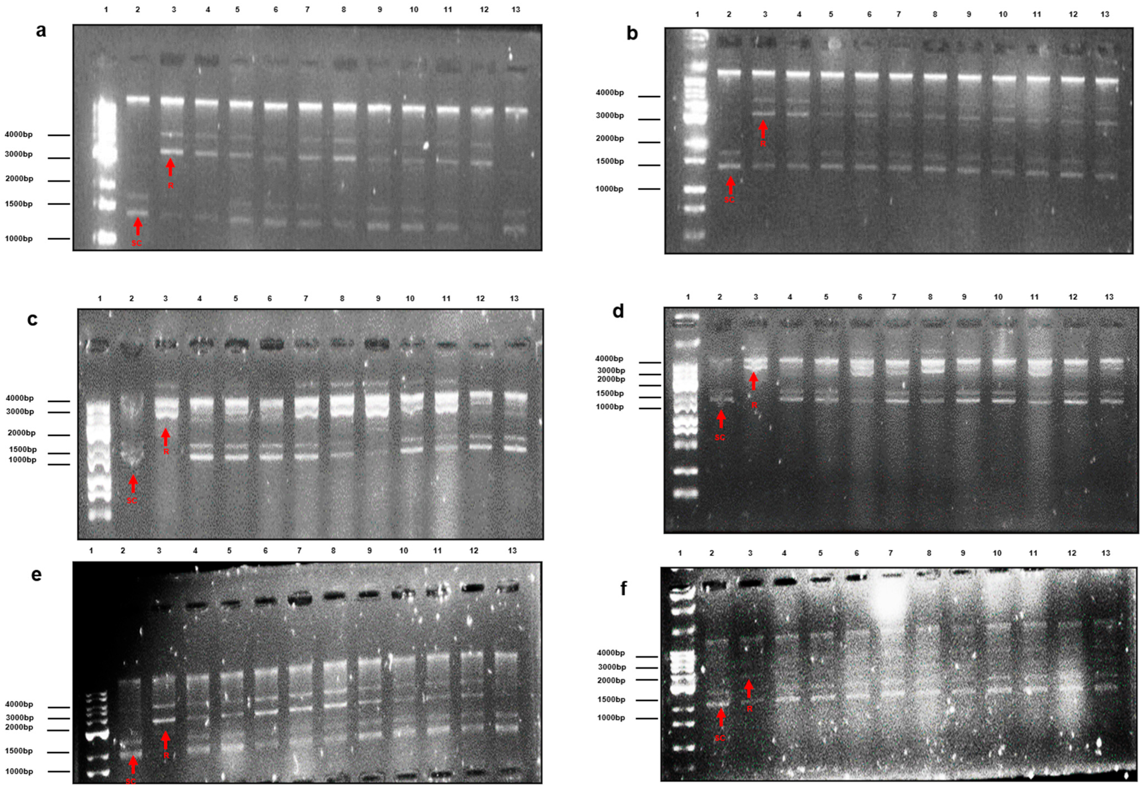
The effects of the extracts on hydroxyl radical-induced changes in plasmid DNA conformation were qualitatively evaluated using the supercoiled plasmid DNA relaxation assay (Figure 1). Exposure of plasmid DNA to H2O2 and UV irradiation resulted in extensive conversion of the supercoiled DNA form into relaxed or linear forms, consistent with oxidative DNA damage. In the presence of several plant extracts, preservation of the supercoiled DNA band was visually observed to varying degrees. Preservation of the supercoiled form was observed for nearly all acetonic extracts, all aqueous extracts except rosemary, and all methanolic extracts except the oregano sample O1. No apparent DNA damage was observed when plasmid DNA was incubated with the extracts in the absence of oxidative treatment. Because the assay was evaluated qualitatively, these observations should not be interpreted as quantitative estimates of DNA-protective efficacy.
Figure 1.
Qualitative assessment of the effects of Mount Athos plant extracts on hydroxyl radical-induced alterations in pUC18 plasmid DNA conformation. Lane 1: 1 kbp DNA ladder. Lane 2: Negative control (untreated plasmid DNA), showing the fast-migrating supercoiled (SC) and open circular (OS) forms. Lane 3: Positive control (plasmid DNA + 50 mM H2O2) after UV irradiation, showing DNA relaxation to a relaxed circular or linear form (R), due to oxidative stress. Lanes 4–13: Experimental groups (plasmid DNA + 50 mM H2O2 +aqueous ((a) Lanes 4–12, (b) Lanes 4–13), acetonic ((c) Lanes 4–12, (d) Lanes 4–13) and methanolic ((e) Lanes 4–12, (f) Lanes 4–13) extracts (1 mg/mL) ((a,c,e): B1, C1, LB1, M1, MT1, O1, O2, O3, O4. (b,d,f): R1, S1, S2, S3, S4, SM1, T1, T2, T3, V1) demonstrating varying degrees of preservation of the supercoiled band, and linear bands. Lane 13 (a,c,e): Extract control (plasmid DNA + 1 mg/mL aqueous (a), acetonic (c) or methanolic (e) extract O4 without H2O2 treatment and UV irradiation), confirming the absence of oxidative activity of the extract (SC: supercoiled form; R: relaxed circular or linear form).
3.3. Correlation Between Total Phenolic Content and Antioxidant Activity
Spearman rank correlation analysis demonstrated significant positive associations between TPC and antioxidant activity for most antioxidant assays (Table 5). Correlation coefficients ranged from ρ = 0.52 to 0.91 (p < 0.05), except for the association between aqueous TPC and DPPH activity, which was not statistically significant (ρ = 0.18, p = 0.468). The strongest correlation was observed between aqueous TPC and FRAP values (ρ = 0.91, p < 0.001), followed by methanolic TPC and ABTS activity (ρ = 0.80, p < 0.001).
Table 5.
Spearman’s rank correlation between TPC and antioxidant activity assessed by the ABTS, DPPH, and FRAP assays using aqueous, acetonic, and methanolic extracts.
3.4. Linear Regression Analysis
Linear regression analysis was performed to evaluate the predictive relationship between TPC and antioxidant activity (Table 6). Significant regression models were obtained for the ABTS and FRAP assays in most extract types. For the ABTS assay, TPC explained approximately 71% and 70% of the variability observed in aqueous and methanolic extracts, respectively (R2 = 0.71 and 0.70; p < 0.001). The strongest predictive relationship was observed for the FRAP assay in aqueous extracts (R2 = 0.78, p < 0.001). By contrast, the predictive models for DPPH activity were weaker, explaining 28–29% of the observed variability in acetonic and methanolic extracts, while no significant association was identified for aqueous extracts (R2 = 0.05, p = 0.376).
Table 6.
Linear regression analysis between TPC and antioxidant activity assessed by the ABTS, DPPH, and FRAP assays using aqueous, acetonic, and methanolic extracts.
Influence diagnostics identified several observations with elevated Cook’s distance and/or externally studentized residuals in individual models. Sensitivity analyses excluding these observations showed that the principal associations between TPC and ABTS or FRAP activity remained statistically significant and were generally similar or stronger in magnitude. The non-significant aqueous TPC–DPPH association also remained non-significant. The only substantive change was observed for the acetonic TPC–DPPH model, which was no longer statistically significant after exclusion of the influential V1 observation (R2 = 0.194, p = 0.068, compared with R2 = 0.281, p = 0.019 in the full model). Accordingly, this specific association should be interpreted cautiously. Overall, the sensitivity analyses supported the robustness of the principal regression findings, particularly those involving ABTS and FRAP.
4. Discussion
The present study comparatively characterized the total phenolic content (TPC) and antioxidant activity of 19 medicinal herbs and spice products of reported Mount Athos provenance. Considerable variation was observed among botanical products and extraction conditions, with Origanum spp. generally exhibiting among the highest TPC values and strong antioxidant responses. These findings are consistent with the recognized phytochemical richness of Mediterranean medicinal plants and their potential as sources of antioxidant constituents for functional foods and nutraceuticals [1,2,3,5,7,8,9,10,11,12,13,14,17]. The investigated species are known to contain diverse phenolic acids, flavonoids, terpenoids, and essential-oil constituents, including rosmarinic acid, carvacrol, thymol, carnosic acid, carnosol, salvianolic acid, verbascoside, neral, and geranial [6,30,31,32,33,34,35,36,37,38,39]. Their phytochemical profiles may vary according to genotype, geographical and environmental conditions, cultivation and harvesting practices, post-harvest processing, storage, and extraction methodology [3,4,40,41,42,43,44]. Because HPLC or LC–MS characterization was not performed, however, the antioxidant effects observed here cannot be attributed to specific individual constituents.
To our knowledge, this is the first study to systematically compare multiple medicinal herb and spice products obtained from different sources within Mount Athos. Previous phytochemical investigations have focused principally on individual Athonite species or specific botanical materials [22,23,24]. Mount Athos is recognized for its botanical diversity and conservation of endemic and threatened taxa [45,46], while its monastic tradition has contributed to the preservation of herbal and pharmaceutical knowledge [47]. These characteristics provide relevant context for investigating botanical products of Athonite provenance, although the present study was not designed to evaluate their historical use or to establish a specific environmental effect of Mount Athos.
The extraction solvent significantly influenced phenolic recovery. Fifty percent aqueous acetone yielded significantly higher TPC than both 50% aqueous methanol and water, whereas aqueous methanol and water did not differ significantly. This agrees with evidence that solvent polarity strongly affects recovery of phenolic compounds and that aqueous organic mixtures can efficiently extract phenolics from botanical matrices [42,43,44]. Water can promote swelling and penetration of plant tissues, whereas the organic component can improve solubilization of phenolics with intermediate polarity. Nevertheless, extraction efficiency varied among products, likely reflecting differences in phytochemical structure, polarity, glycosylation, concentration, and plant-matrix interactions [40,42,43,44]. Several aqueous extracts also retained appreciable TPC and antioxidant activity, consistent with the traditional consumption of many medicinal plants as aqueous infusions; however, these in vitro observations should not be extrapolated to biological or clinical efficacy.
Importantly, solvent effects on antioxidant activity were assay-dependent. Both aqueous acetone and aqueous methanol produced significantly greater DPPH inhibition than water, with no significant difference between the two aqueous organic solvents. For ABTS, only aqueous methanol produced significantly greater activity than water after correction for multiple comparisons, whereas FRAP did not differ significantly among extraction conditions. Thus, no single solvent was uniformly superior across all antioxidant endpoints. This pattern is consistent with the distinct physicochemical principles of DPPH, ABTS, and FRAP, and with previous evidence that extraction solvent and assay chemistry interact with the composition of complex botanical extracts [30,38,40,41,42,43,44]. Consequently, selection of an extraction solvent should depend on the botanical matrix and the analytical or technological objective rather than on an assumption of universal solvent superiority.
The DPPH and ABTS results require particular caution because both assays were performed at a single extract concentration. Most acetonic and methanolic extracts produced high DPPH inhibition values within a narrow range, suggesting near-saturating conditions and reduced discriminatory capacity. Several ABTS values also approached maximal inhibition, indicating that a ceiling effect may similarly have reduced discrimination among extracts. Therefore, the DPPH and ABTS measurements demonstrate radical-scavenging activity under the experimental conditions but should not be interpreted as quantitative rankings of antioxidant potency. Concentration–response analyses allowing determination of IC50 or EC50 values, or expression relative to a reference antioxidant such as Trolox, would provide more robust comparisons. The divergent patterns among DPPH, ABTS, and FRAP further emphasize that antioxidant capacity depends on both the qualitative phytochemical composition and the reaction mechanism of the analytical assay. Origanum spp., nevertheless, consistently showed high TPC and strong antioxidant responses, in agreement with previous evidence concerning the phenolic and terpenoid richness of Mediterranean oregano [30,38,40,41,42,43,44].
The qualitative supercoiled plasmid DNA relaxation assay provided complementary evidence concerning the behavior of the extracts under hydroxyl radical-generating conditions. Several extracts visually preserved the supercoiled DNA form following H2O2/UV exposure, which is compatible with a potential protective effect against oxidative DNA strand damage. However, because densitometric quantification of supercoiled, open-circular, and linear DNA forms was not performed, these observations should be considered preliminary and cannot be used to quantitatively compare DNA-protective efficacy. Confirmation will require densitometric and concentration–response analyses, appropriate reference antioxidants, and complementary cellular models of oxidative DNA damage.
TPC was positively associated with antioxidant activity, particularly ABTS and FRAP. TPC explained approximately 78% of the variation in FRAP activity in aqueous extracts and approximately 71% and 70% of ABTS variability in aqueous and methanolic extracts, respectively. These results support an important contribution of phenolic-rich constituents to the antioxidant properties of medicinal plant extracts [30,40,41,42,43,44]. The particularly strong relationship between TPC and FRAP may also partly reflect their shared dependence on electron-transfer reactions [30,42]. Nevertheless, the Folin–Ciocalteu assay is not entirely specific to phenolic compounds and can respond to other reducing constituents; therefore, TPC should be interpreted as an index of phenolic-rich reducing capacity rather than absolute phenolic abundance. The absence of chromatographic characterization further prevents identification of the compounds principally responsible for the observed activity.
Associations between TPC and DPPH were weaker than those observed for ABTS and FRAP, consistent with previous comparative investigations of Mediterranean medicinal plants [38,40,41,42,43,44]. This may reflect differences in the reactivity of individual phytochemicals toward the DPPH radical as well as the limited discriminatory capacity of the single-concentration measurements. The clustering of many organic extracts near the upper range of DPPH inhibition may have compressed the response and attenuated correlations with TPC. These relationships should therefore be interpreted cautiously and confirmed using concentration–response data.
Comparison with plants investigated in other geographical regions further highlights the substantial variability of medicinal-plant antioxidant profiles. Ocimum basilicum genotypes cultivated at ecologically contrasting locations in Türkiye exhibited significant differences in TPC and DPPH, ABTS, and FRAP activity [48]. Similarly, Salvia officinalis from different Tunisian habitats showed geographical variation in phenolic composition and antioxidant activity, including differences in rosmarinic acid, carnosol, and carnosic acid [49]. Geographical variability has also been reported for Thymus spp.; accessions from different regions of Iran differed substantially in phenolic content, essential-oil composition, and antioxidant activity [50], while T. vulgaris accessions originating from southern France and subsequently cultivated under common conditions in Austria showed marked differences in TPC and FRAP activity [51]. These findings provide relevant context for the variability observed among the Athonite products, although direct numerical comparison is limited by differences in genotype or chemotype, plant part, extraction procedure, concentration, assay methodology, and units of expression.
These geographical comparisons should not, however, be interpreted as evidence of a specific “Mount Athos effect.” The present study did not include corresponding species from other geographical regions analyzed under identical conditions, and therefore the contribution of Mount Athos environmental characteristics or traditional cultivation practices cannot be determined. Establishing such an effect would require comparison of corresponding species, preferably matched for genotype or chemotype, plant part, developmental stage, harvesting period, post-harvest treatment, extraction procedure, and analytical methodology. The present findings should therefore be interpreted as characterization of botanical products of reported Athonite provenance within the broader geographical, genetic, and phytochemical variability recognized for Mediterranean medicinal plants.
Several additional limitations should be acknowledged. The Folin–Ciocalteu assay is not fully specific to phenolic compounds, and HPLC or LC–MS profiling was not performed. All antioxidant assays were conducted in vitro and therefore cannot establish biological or clinical efficacy. The plasmid DNA assay was qualitative, while the single-concentration DPPH assay limited quantitative comparison of antioxidant potency. In addition, the investigated materials were commercial products rather than independently collected biological specimens; replicate extractions represented aliquots of the same homogenized products rather than independent plants or commercial lots, precluding assessment of biological, cultivation-site, and between-lot variability. Precise cultivation coordinates were unavailable for all products, and the presence of endomycetes was not assessed. Moreover, the absence of geographically matched comparator plants prevents attribution of the observed properties specifically to Mount Athos. An additional limitation concerns the DPPH and ABTS assays, which were performed at single extract concentrations and expressed as percentage inhibition. Several DPPH and ABTS measurements approached the upper range of the assays, which may have produced ceiling effects and reduced discriminatory capacity among extracts. Consequently, these results should be interpreted as screening measurements rather than quantitative estimates of antioxidant potency. Concentration–response analyses enabling determination of IC50 or EC50 values, or expression relative to standardized reference antioxidants such as Trolox, would provide more robust comparisons. Furthermore, sample-specific blanks containing each extract without the corresponding radical reagent were not included; therefore, potential contributions of extract color or turbidity to absorbance cannot be completely excluded. Although solvent/radical controls were included, no external positive antioxidant control was used. Future studies should incorporate sample-specific blanks, solvent-matched controls, appropriate reference antioxidants, and concentration–response analyses to improve analytical rigor and comparability.
Despite these limitations, the study extends the limited phytochemical evidence concerning botanical materials of Athonite provenance [22,23,24] and demonstrates that extraction solvent significantly affects phenolic recovery and selected antioxidant endpoints in an assay-dependent manner. Aqueous acetone provided greater TPC recovery, both aqueous organic solvents enhanced DPPH inhibition relative to water, aqueous methanol increased ABTS activity relative to water, and no significant solvent effect was observed for FRAP. Future investigations combining chromatographic and metabolomic profiling, standardized concentration–response antioxidant assays, quantitative DNA-protection methods, independent biological samples, geographically matched comparator plants, and appropriate cellular, animal, and ultimately human studies are required to determine the bioactive constituents, biological relevance, and potential functional-food or nutraceutical applications of these botanical resources.
5. Conclusions
This study provides comparative evidence on the phenolic content and antioxidant characteristics of 19 commercially available medicinal herb and spice products of reported Mount Athos provenance. Considerable variability was observed among botanical products, with Origanum spp. generally exhibiting among the highest TPC values and strong antioxidant responses. Extraction solvent significantly influenced phenolic recovery and selected antioxidant endpoints: aqueous acetone provided greater TPC recovery than aqueous methanol or water, both aqueous organic solvents produced greater DPPH inhibition than water, and aqueous methanol produced greater ABTS activity than water, whereas no significant solvent effect was observed for FRAP. Thus, no single extraction solvent was uniformly superior across all antioxidant assays.
The positive associations between TPC and antioxidant activity, particularly ABTS and FRAP, support an important contribution of phenolic-rich constituents to the antioxidant properties of these extracts. Several extracts also preserved the supercoiled plasmid DNA form under hydroxyl radical-generating conditions; however, this qualitative observation requires quantitative confirmation. Overall, these findings provide baseline phytochemical and antioxidant data for medicinal plants of Athonite provenance but should not be interpreted as demonstrating a specific “Mount Athos effect.” Further studies incorporating detailed phytochemical profiling, concentration–response antioxidant analyses, quantitative DNA-protection assays, and geographically matched comparator plants are warranted.
Supplementary Materials
The following supporting information can be downloaded at https://www.mdpi.com/article/10.3390/ijpb17090079/s1, Table S1: List of herbs and spices used from Mount Athos.
Author Contributions
Conceptualization, E.P. (Efthymios Poulios) and C.G.; methodology, E.P. (Efthymios Poulios), S.K.P. and E.P. (Evmorfia Psara); formal analysis, S.K.P., E.P. (Evmorfia Psara) and A.P.; investigation, E.P. (Efthymios Poulios), S.K.P. and E.P. (Evmorfia Psara); resources, E.P. (Efthymios Poulios), S.K.P. and A.P.; data curation, E.P. (Efthymios Poulios), A.P. and C.G.; writing—original draft preparation, E.P. (Efthymios Poulios), S.K.P. and A.P.; writing—review and editing, E.P. (Efthymios Poulios) and C.G.; visualization, E.P. (Efthymios Poulios) and C.G.; supervision, E.P. (Efthymios Poulios) and C.G.; project administration, E.P. (Efthymios Poulios) and C.G. All authors have read and agreed to the published version of the manuscript.
Funding
This research received no external funding.
Institutional Review Board Statement
Not applicable.
Informed Consent Statement
Not applicable.
Data Availability Statement
Data is available upon request on corresponding authors.
Acknowledgments
The authors acknowledge the use of the AI-assisted graphic design software ChatGPT (GPT-5.6 Sol, OpenAI, San Francisco, CA, USA) for the preparation and visualization of selected figures in this manuscript. AI use was limited to figure layout and graphical elements. All scientific content, data interpretation, figure concepts, and final figure verification were performed exclusively by the authors. AI was not used to generate, modify, or manipulate experimental data or original experimental images.
Conflicts of Interest
The authors declare no conflicts of interest.
References
- Jiang, T.A. Health Benefits of Culinary Herbs and Spices. J. AOAC Int. 2019, 102, 395–411. [Google Scholar] [CrossRef] [Scilit] [PubMed]
- Guerriero, G.; Berni, R.; Muñoz-Sanchez, J.A.; Apone, F.; Abdel-Salam, E.M.; Qahtan, A.A.; Alatar, A.A.; Cantini, C.; Cai, G.; Hausman, J.F.; et al. Production of Plant Secondary Metabolites: Examples, Tips and Suggestions for Biotechnologists. Genes 2018, 9, 309. [Google Scholar] [CrossRef] [Scilit] [PubMed]
- Poulios, E.; Giaginis, C.; Vasios, G.K. Current Advances on the Extraction and Identification of Bioactive Components of Sage (Salvia spp.). Curr. Pharm. Biotechnol. 2019, 20, 845–857. [Google Scholar] [CrossRef] [Scilit] [PubMed]
- Poulios, E.; Vasios, G.K.; Psara, E.; Antasouras, G.; Gialeli, M.; Pavlidou, E.; Tsantili-Kakoulidou, A.; Troumbis, A.Y.; Giaginis, C. Antioxidant Activity of Medicinal Plants and Herbs of North Aegean, Greece: Current Clinical Evidence and Future Perspectives. Nat. Prod. J. 2024, 14, 31–44. [Google Scholar] [CrossRef] [Scilit]
- Poulios, E.; Giaginis, C.; Vasios, G.K. Current State of the Art on the Antioxidant Activity of Sage (Salvia spp.) and Its Bioactive Components. Planta Med. 2020, 86, 224–238. [Google Scholar] [CrossRef] [Scilit] [PubMed]
- Cote, B.; Elbarbry, F.; Bui, F.; Su, J.W.; Seo, K.; Nguyen, A.; Lee, M.; Rao, D.A. Mechanistic Basis for the Role of Phytochemicals in Inflammation-Associated Chronic Diseases. Molecules 2022, 27, 781. [Google Scholar] [CrossRef] [Scilit] [PubMed]
- Naoi, M.; Maruyama, W.; Shamoto-Nagai, M. Disease-Modifying Treatment of Parkinson’s Disease by Phytochemicals: Targeting Multiple Pathogenic Factors. J. Neural Transm. 2022, 129, 737–753. [Google Scholar] [CrossRef] [Scilit] [PubMed]
- Džamić, A.M.; Matejić, J.S. Plant Products in the Prevention of Diabetes Mellitus. Mini-Rev. Med. Chem. 2022, 22, 1395–1419. [Google Scholar] [CrossRef] [Scilit] [PubMed]
- Kamyab, R.; Namdar, H.; Torbati, M.; Ghojazadeh, M.; Araj-Khodaei, M.; Fazljou, S.M.B. Medicinal Plants in the Treatment of Hypertension: A Review. Adv. Pharm. Bull. 2021, 11, 601–617. [Google Scholar] [CrossRef] [Scilit] [PubMed]
- Anand, A.; Srivastava, S.; Sharma, D.; Sridhar, S.B.; Tariq, M.; Koteswara Rao, G.S.N.; Mruthunjaya, K.; Manjula, S.N.; Sahu, R.; Hamida, M.B.B. Healing the Heart Naturally: The Role of Bioactive Phytocompounds in Cardiovascular Therapeutics. Phytomedicine 2026, 155, 158084. [Google Scholar] [CrossRef] [Scilit] [PubMed]
- Poulios, E.; Vasios, G.K.; Psara, E.; Giaginis, C. Medicinal Plants Consumption against Urinary Tract Infections: A Narrative Review of the Current Evidence. Expert Rev. Anti Infect. Ther. 2021, 19, 519–528. [Google Scholar] [CrossRef] [Scilit] [PubMed]
- Poulios, E.; Koukounari, S.; Psara, E.; Vasios, G.K.; Sakarikou, C.; Giaginis, C. Anti-Obesity Properties of Phytochemicals: Highlighting Their Molecular Mechanisms against Obesity. Curr. Med. Chem. 2024, 31, 25–61. [Google Scholar] [CrossRef] [Scilit] [PubMed]
- Guo, M.; Jin, J.; Zhao, D.; Rong, Z.; Cao, L.Q.; Li, A.H.; Sun, X.Y.; Jia, L.Y.; Wang, Y.D.; Huang, L.; et al. Research Advances on Anti-Cancer Natural Products. Front. Oncol. 2022, 12, 866154. [Google Scholar] [CrossRef] [Scilit] [PubMed]
- Juan, C.A.; Pérez de la Lastra, J.M.; Plou, F.J.; Pérez-Lebeña, E. The Chemistry of Reactive Oxygen Species (ROS) Revisited: Outlining Their Role in Biological Macromolecules (DNA, Lipids and Proteins) and Induced Pathologies. Int. J. Mol. Sci. 2021, 22, 4642. [Google Scholar] [CrossRef] [Scilit] [PubMed]
- Roy, Z.; Bansal, R.; Siddiqui, L.; Chaudhary, N. Understanding the Role of Free Radicals and Antioxidant Enzymes in Human Diseases. Curr. Pharm. Biotechnol. 2023, 24, 1265–1276. [Google Scholar] [CrossRef] [Scilit] [PubMed]
- Pei, J.; Pan, X.; Wei, G.; Hua, Y. Research Progress of Glutathione Peroxidase Family (GPX) in Redoxidation. Front. Pharmacol. 2023, 14, 1147414. [Google Scholar] [CrossRef] [Scilit] [PubMed]
- Poulios, E.; Roupaka, V.; Giaginis, C.; Galaris, D.; Spyrou, G. Implication of Thioredoxin 1 and Glutaredoxin 1 in H2O2-Induced Phosphorylation of JNK and p38 MAP Kinases. Curr. Mol. Med. 2025, 25, 305–319. [Google Scholar] [CrossRef] [Scilit] [PubMed]
- Ulewicz-Magulska, B.; Wesolowski, M. Total Phenolic Contents and Antioxidant Potential of Herbs Used for Medical and Culinary Purposes. Plant Foods Hum. Nutr. 2019, 74, 61–67. [Google Scholar] [CrossRef] [Scilit] [PubMed]
- Munteanu, I.G.; Apetrei, C. Analytical Methods Used in Determining Antioxidant Activity: A Review. Int. J. Mol. Sci. 2021, 22, 3380. [Google Scholar] [CrossRef] [Scilit] [PubMed]
- Strid, A.; Charalampidis, S.; Dimopoulos, P.; Raus, T. Flora of the Athos Peninsula: Plant Life of the Holy Mountain; Broken Hill Publishers Ltd.: Nicosia, Cyprus, 2025; pp. 566–588. ISBN 978-9925-35-333-0. [Google Scholar]
- Valiakos, E.; Marselos, M.; Sakellaridis, N.; Constantinidis, T.; Skaltsa, H. Ethnopharmacological Approach to the Herbal Medicines of the “Antidotes” in Nikolaos Myrepsos’ Dynameron. J. Ethnopharmacol. 2015, 163, 68–82. [Google Scholar] [CrossRef] [Scilit] [PubMed]
- Petkova, Z.; Stefanova, G.; Girova, T.; Antova, G.; Stoyanova, M.; Damianova, S.; Gochev, V.; Stoyanova, A.; Zheljazkov, V.D. Phytochemical Investigations of Laurel Fruits (Laurus nobilis). Nat. Prod. Commun. 2019, 14, 1934578X19868876. [Google Scholar] [CrossRef] [Scilit]
- Samanidis, I.; Krigas, N.; Athanasiadis, V.; Makrygiannis, I.; Mantiniotou, M.; Lalas, S.I. A Comparative Phytochemical Investigation of the Greek Members of the Genus Helichrysum Mill., with Emphasis on the Local Endemic Helichrysum amorginum Boiss. and Orph. Plants 2025, 14, 229. [Google Scholar] [CrossRef] [Scilit] [PubMed]
- Sarametidis, P.; Radovic, K.; Stylianou, M.; Kyriacou, M.; Koutsovoulou, K.; O’Brien, N.; Koffa, M.; Lazari, D.; Vougogiannopoulou, K. Helleborus odorus subsp. cyclophyllus: An Unexploited Source of Antioxidant, Antimicrobial, and Cytotoxic Bioactivity. Biology 2026, 15, 852. [Google Scholar] [CrossRef] [Scilit] [PubMed]
- Pérez, M.; Dominguez-López, I.; Lamuela-Raventós, R.M. The Chemistry Behind the Folin–Ciocalteu Method for the Estimation of (Poly)phenol Content in Food: Total Phenolic Intake in a Mediterranean Dietary Pattern. J. Agric. Food Chem. 2023, 71, 17543–17553. [Google Scholar] [CrossRef] [Scilit] [PubMed]
- Przybylski, P.; Konopko, A.; Łętowski, P.; Jodko-Piórecka, K.; Litwinienko, G. Concentration-Dependent HAT/ET Mechanism of the Reaction of Phenols with 2,2-Diphenyl-1-picrylhydrazyl (DPPH•) in Methanol. RSC Adv. 2022, 12, 8131–8136. [Google Scholar] [CrossRef] [Scilit] [PubMed]
- Re, R.; Pellegrini, N.; Proteggente, A.; Pannala, A.; Yang, M.; Rice-Evans, C. Antioxidant Activity Applying an Improved ABTS Radical Cation Decolorization Assay. Free Radic. Biol. Med. 1999, 26, 1231–1237. [Google Scholar] [CrossRef] [Scilit] [PubMed]
- Dudonné, S.; Vitrac, X.; Coutière, P.; Woillez, M.; Mérillon, J.M. Comparative Study of Antioxidant Properties and Total Phenolic Content of 30 Plant Extracts of Industrial Interest Using DPPH, ABTS, FRAP, SOD, and ORAC Assays. J. Agric. Food Chem. 2009, 57, 1768–1774. [Google Scholar] [CrossRef] [Scilit] [PubMed]
- Kalote, R.M.; Sanap, S.S.; Pawar, S.V. DNA Damage Protective Activity and Antioxidant Activity of Indian Medicinal Plants. J. Appl. Pharm. Sci. 2013, 3, 99–106. [Google Scholar] [CrossRef] [Scilit]
- Poulios, E.; Papadopoulou, S.K.; Psara, E.; Giaginis, C. Antioxidant, Antimicrobial, and Anticancer Activity of Basil (Ocimum basilicum). Antioxidants 2025, 14, 1469. [Google Scholar] [CrossRef] [Scilit] [PubMed]
- Dai, Y.L.; Li, Y.; Wang, Q.; Niu, F.J.; Li, K.W.; Wang, Y.Y.; Wang, J.; Zhou, C.Z.; Gao, L.N. Chamomile: A Review of Its Traditional Uses, Chemical Constituents, Pharmacological Activities and Quality Control Studies. Molecules 2023, 28, 133. [Google Scholar] [CrossRef] [Scilit] [PubMed]
- Petrisor, G.; Motelica, L.; Craciun, L.N.; Oprea, O.C.; Ficai, D.; Ficai, A. Melissa officinalis: Composition, Pharmacological Effects and Derived Release Systems—A Review. Int. J. Mol. Sci. 2022, 23, 3591. [Google Scholar] [CrossRef] [Scilit] [PubMed]
- Hudz, N.; Kobylinska, L.; Pokajewicz, K.; Horčinová Sedláčková, V.; Fedin, R.; Voloshyn, M.; Myskiv, I.; Brindza, J.; Wieczorek, P.P.; Lipok, J. Mentha piperita: Essential Oil and Extracts, Their Biological Activities, and Perspectives on the Development of New Medicinal and Cosmetic Products. Molecules 2023, 28, 7444. [Google Scholar] [CrossRef] [Scilit] [PubMed]
- El Menyiy, N.; Mrabti, H.N.; El Omari, N.; Bakili, A.E.; Bakrim, S.; Mekkaoui, M.; Balahbib, A.; Amiri-Ardekani, E.; Ullah, R.; Alqahtani, A.S.; et al. Medicinal Uses, Phytochemistry, Pharmacology, and Toxicology of Mentha spicata. Evid.-Based Complement. Altern. Med. 2022, 2022, 7990508. [Google Scholar] [CrossRef] [Scilit] [PubMed]
- Chrysargyris, A.; Tomou, E.M.; Goula, K.; Dimakopoulou, K.; Tzortzakis, N.; Skaltsa, H.; Sideritis, L. Essential Oils: A Systematic Review. Phytochemistry 2023, 209, 113607. [Google Scholar] [CrossRef] [Scilit] [PubMed]
- Sharifi-Rad, M.; Berkay Yılmaz, Y.; Antika, G.; Salehi, B.; Tumer, T.B.; Kulandaisamy Venil, C.; Das, G.; Patra, J.K.; Karazhan, N.; Akram, M.; et al. Phytochemical Constituents, Biological Activities, and Health-Promoting Effects of the Genus Origanum. Phytother. Res. 2021, 35, 95–121. [Google Scholar] [CrossRef] [Scilit] [PubMed]
- Nieto, G.; Ros, G.; Castillo, J. Antioxidant and Antimicrobial Properties of Rosemary (Rosmarinus officinalis L.): A Review. Medicines 2018, 5, 98. [Google Scholar] [CrossRef] [Scilit] [PubMed]
- Waheed, M.; Hussain, M.B.; Saeed, F.; Afzaal, M.; Ahmed, A.; Irfan, R.; Akram, N.; Ahmed, F.; Hailu, G.G. Phytochemical Profiling and Therapeutic Potential of Thyme (Thymus spp.): A Medicinal Herb. Food Sci. Nutr. 2024, 12, 9893–9912. [Google Scholar] [CrossRef] [Scilit] [PubMed]
- Bahramsoltani, R.; Rostamiasrabadi, P.; Shahpiri, Z.; Marques, A.M.; Rahimi, R.; Farzaei, M.H. Aloysia citrodora Paláu (Lemon verbena): A Review of Phytochemistry and Pharmacology. J. Ethnopharmacol. 2018, 222, 34–51. [Google Scholar] [CrossRef] [Scilit] [PubMed]
- Arceusz, A.; Wesolowski, M.; Konieczynski, P. Methods for Extraction and Determination of Phenolic Acids in Medicinal Plants: A Review. Nat. Prod. Commun. 2013, 8, 1821–1829. [Google Scholar] [CrossRef] [Scilit]
- Park, J.B. Identification and Quantification of a Major Antioxidant and Anti-Inflammatory Phenolic Compound Found in Basil, Lemon Thyme, Mint, Oregano, Rosemary, Sage, and Thyme. Int. J. Food Sci. Nutr. 2011, 62, 577–584. [Google Scholar] [CrossRef] [Scilit] [PubMed]
- Piluzza, G.; Bullitta, S. Correlations between Phenolic Content and Antioxidant Properties in Twenty-Four Plant Species of Traditional Ethnoveterinary Use in the Mediterranean Area. Pharm. Biol. 2011, 49, 240–247. [Google Scholar] [CrossRef] [Scilit] [PubMed]
- Rababah, T.M.; Ereifej, K.I.; Esoh, R.B.; Al-u’datt, M.H.; Alrababah, M.A.; Yang, W. Antioxidant Activities, Total Phenolics and HPLC Analyses of the Phenolic Compounds of Extracts from Common Mediterranean Plants. Nat. Prod. Res. 2011, 25, 596–605. [Google Scholar] [CrossRef] [Scilit] [PubMed]
- Rumpf, J.; Burger, R.; Schulze, M. Statistical Evaluation of DPPH, ABTS, FRAP, and Folin–Ciocalteu Assays to Assess the Antioxidant Capacity of Lignins. Int. J. Biol. Macromol. 2023, 233, 123470. [Google Scholar] [CrossRef] [Scilit] [PubMed]
- Karydas, A.; Kamari, G. Monitoring Six Local Endemic Taxa of the Mt. Athos and Assessment According to the IUCN Red List Categories and Criteria. Bot. Chron. 2019, 22, 195–208. Available online: https://www.researchgate.net/publication/338544929_Monitoring_six_local_endemic_taxa_of_the_Mt_Athos_and_assessment_according_to_the_IUCN_Red_List_categories_and_criteria_-_Bot_Chron_22_195-208 (accessed on 29 July 2026).
- Xofis, P.; Buckley, P.G.; Takos, I.; Mitchley, J. Long-Term Post-Fire Vegetation Dynamics in North-East Mediterranean Ecosystems: The Case of Mount Athos, Greece. Fire 2021, 4, 92. [Google Scholar] [CrossRef] [Scilit]
- Vacić, Z. Guidelines for the Preparation and Dosage of Medicines, Instructions for Their Use, and Methods of Application According to the Hilandar Medical Codex No. 517. Srp. Arh. Celok. Lek. 2025, 153, 208–212. [Google Scholar] [CrossRef] [Scilit]
- Zorlu, A.; Telci, İ.; Elmastaş, M.; Kaçar, O.; Aytaç, Z.; Genç, N.; Kayır, Ö. Phenolics and Antioxidant Capacity of Basil (Ocimum basilicum L.) Genotypes Across Locations and Developmental Stages. Plant Foods Hum. Nutr. 2026, 81, 57. [Google Scholar] [CrossRef] [Scilit] [PubMed]
- Ben Farhat, M.; Jordán, M.J.; Chaouech-Hamada, R.; Landoulsi, A.; Sotomayor, J.A. Variations in Essential Oil, Phenolic Compounds, and Antioxidant Activity of Tunisian Cultivated Salvia officinalis L. J. Agric. Food Chem. 2009, 57, 10349–10356. [Google Scholar] [CrossRef] [Scilit] [PubMed]
- Tohidi, B.; Rahimmalek, M.; Arzani, A. Essential Oil Composition, Total Phenolic, Flavonoid Contents, and Antioxidant Activity of Thymus Species Collected from Different Regions of Iran. Food Chem. 2017, 220, 153–161. [Google Scholar] [CrossRef] [Scilit] [PubMed]
- Chizzola, R.; Michitsch, H.; Franz, C. Antioxidative Properties of Thymus vulgaris Leaves: Comparison of Different Extracts and Essential Oil Chemotypes. J. Agric. Food Chem. 2008, 56, 6897–6904. [Google Scholar] [CrossRef] [Scilit] [PubMed]

Disclaimer/Publisher’s Note: The statements, opinions and data contained in all publications are solely those of the individual author(s) and contributor(s) and not of MDPI and/or the editor(s). MDPI and/or the editor(s) disclaim responsibility for any injury to people or property resulting from any ideas, methods, instructions or products referred to in the content. |
© 2026 by the authors. Licensee MDPI, Basel, Switzerland. This article is an open access article distributed under the terms and conditions of the Creative Commons Attribution (CC BY) license.