


Abstract
The causative agent of Lyme disease, Borrelia burgdorferi, poses significant health and economic problems in the United States and elsewhere. Developing borreliastatic or borreliacidal agents requires the use of solvent vehicles. Vehicle tolerance can affect the interpretation of antimicrobial screening tests. We examined the effects of dimethyl sulfoxide, sodium hydroxide, and ethanol on B. burgdorferi growth. Spirochaetes were exposed to vehicle concentrations ranging from 0.15% to 2.4% v/v. Cultures were evaluated with dark-field microscopy and a commercial cell viability assay. There were no significant decreases in cell counts or ATP-dependent luminescence with dimethyl sulfoxide. Sodium hydroxide was shown to be unsuitable as an unbuffered alkaline vehicle. Ethanol produced discordant ATP-dependent luminescence and cell-count endpoints at moderate concentrations, although detectable cell presence persisted through 1.2%. Differences between endpoints may be due to cellular stress from exposure. These results show that the identity and concentration of these vehicles differentially affect B. burgdorferi growth and ATP-dependent metabolic activity. The applicability of high-throughput methods may be affected by conditions that cause stress-induced changes. Future studies should explore how molecular effects relate to observed differences in cell counts and metabolic activity.
1. Introduction
Ticks continue to pose challenges to public health and the economy worldwide [1]. Borrelia burgdorferi is the causative agent of Lyme disease, which is primarily endemic to North America, northern Asia, and Europe [2]. An estimated 476,000 people are diagnosed and treated for Lyme disease annually in the United States [3]. The burden of Lyme disease and other tick-borne diseases has been increasing because of a variety of ecological and anthropogenic factors that have led to the expansion of tick geographical range and abundance [4]. Contributing factors include shifts in reservoir and reproductive host populations and changes in human activity [5]. Lyme disease commonly presents with erythema migrans, a localised expanding rash that develops at the site of an infected tick bite [6]. Treatment with oral doxycycline can usually resolve symptoms, but cases can progress to disseminated disease involving other organ systems [7,8]. Despite the general effectiveness of early antibiotic treatment, there is continued interest in developing novel antimicrobial strategies against B. burgdorferi. These strategies could offer another tool for human or companion animal treatments to supplement the narrow set of clinically relevant active ingredients. In parallel, novel active ingredients could be used in reservoir host-targeted interventions that reduce the prevalence of B. burgdorferi in host-seeking ticks [9].
Borrelia burgdorferi is an obligate host-associated spirochaete maintained in an enzootic cycle between vertebrate reservoir hosts and ticks [10]. In vitro work with B. burgdorferi is essential for studying aspects relevant to antimicrobial susceptibility. Spirochaete populations can be propagated in vitro using specialised growth media. Barbour-Stoenner-Kelly (BSK) culture medium and its variants have been adopted as the standard for maintaining Borrelia spp. populations under laboratory conditions [11]. This medium is CMRL-1066 tissue culture-based and supplemented with serum and other nutrients to support replication [12]. Species of Borrelia are particularly sensitive to environmental conditions. Small changes in conditions, such as salinity and redox state, can trigger physiological responses that affect metabolism and replication [13].
Environmental sensitivity is particularly relevant when studying cellular responses to candidate antimicrobials and other bioactive compounds. In these experiments, changes in viability or growth should be attributable to the active ingredient rather than to stress introduced by the experimental system, improving the physiological relevance of the assay. Active ingredients generally require delivery into culture using vehicles that address solubility or stability constraints. Although vehicle-associated effects can be accounted for by defining appropriate controls and applying normalisation techniques, the preferred approach is to use concentrations that are as biologically inert as possible and preserve conditions favourable to cell replication. Vehicle compounds are often assumed to be biologically inert at sufficiently low final concentrations. Studies in other microbial systems show that solvent exposure, even at low concentrations, can affect membrane physiology and gene expression with consequences for metabolic output and enzymatic activity [14]. Cell agglutination and biofilm formation can also be affected by different solvents, with inhibitory or promoting effects at low concentrations [15].
Vehicle suitability and tolerable concentrations vary among microbial species. Because B. burgdorferi is particularly sensitive to environmental conditions, even small vehicle-associated effects could alter growth or viability readouts. They may also confound the interpretation of antimicrobial activity due to synergism or other effects. At very low concentrations, dimethyl sulfoxide (DMSO) has been used under standard and modified B. burgdorferi culture conditions as a drug vehicle [16,17]. Dimethyl sulfoxide is relevant because it generally does not affect cell counts or indirect measures of viability at low concentrations in some microbial systems. This compound can also solubilise a broad range of poorly water-soluble small molecules without major changes to medium composition. This study aimed to define the gross effects of DMSO at low- to moderate-concentration ranges in B. burgdorferi cultures. Alongside DMSO, sodium hydroxide (NaOH) and ethanol (EtOH) were assessed as vehicles with different physicochemical properties that may be compatible with compounds that have different solubility requirements. Ethanol has been used in a prior B. burgdorferi study at low concentrations, though it requires further formal analysis [18]. The gross effects of NaOH are not characterised. We paired direct dark-field microscopy cell counts with BacTiter-Glo adenosine triphosphate (ATP)-dependent luminescence as endpoints, allowing vehicle effects to be assessed by both direct counts and an estimate of metabolic activity suitable for higher-throughput screening, with the primary objective of identifying the range of concentrations at which these chemicals may be used for in vitro drug screening.
2. Materials and Methods
2.1. Culture of Borrelia burgdorferi
Borrelia burgdorferi strain B31, clone 5A1 (BEI Resources NR-13251, Manassas, VA, USA) was propagated in BSK-IIB medium with 6% rabbit serum (Pel-Freez Biologicals 31125-5, Rogers, AR, USA) [12]. This clone was selected because it has a defined plasmid profile and retains a highly infectious phenotype in mammalian experimental models [19]. Spirochaetes were added to 500,000 cells/mL in sealed 1.5-mL tubes (three biological replicates) and incubated at 34 °C. The cultures were prepared from cells originating from individual volumes of stock cultures stored at −80 °C. Negative control tubes were also prepared for BSK-IIB and BSK-IIB + vehicles and incubated for the same duration as the experimental replicates.
Dimethyl sulfoxide, NaOH (1 N, equivalent to 1.0 M NaOH), and EtOH were added to the cultures on day zero to final concentrations of 0.15, 0.3, 0.6, 1.2, and 2.4% (v/v). For NaOH, these concentrations corresponded to molarities in solution of 1.5, 3.0, 6.0, 12.0 and 24.0 mM but are hereafter referred to by per cent v/v in solution. The pH with NaOH was measured with a calibrated pH meter at day six as an endpoint measure. We chose dimethyl sulfoxide because it is predicted to be tolerated at low concentrations by B. burgdorferi. In a prior test, we used NaOH in low concentrations (0.5% v/v maximum) to deliver IMPDH inhibitors with more complex solubility in BSK-II and found that cell viability was still present, as indicated by BacTiter-Glo (Promega Corporation, Madison, WI, USA), and that we could detect a difference in ATP-dependent luminescence in IMPDH inhibitor groups compared with NaOH-only controls (unpublished). Sodium hydroxide is therefore relevant to in vitro use but imposes distinct cellular stress when compared to EtOH, making EtOH a useful comparison in this test. Sodium hydroxide primarily alters environmental pH, whereas EtOH acts as an organic solvent with established concentration-dependent membrane-disrupting and antimicrobial effects. The concentration range is relevant because it allows delivery of concentrations used in in vitro compound testing without major disruptions to medium composition, and it aligns with tests in B. burgdorferi and other systems that commonly find these concentrations not harmful to cell counts before effects appear at higher ranges. Additionally, this range was hypothesised to reveal effects between the vehicles given the difference in physicochemical properties between the chemicals. Weaker effects were hypothesised to be seen at the lower bound of the chosen range, and stronger effects were predicted at the higher bound.
2.2. Quantifying the Metabolically Active Biomass as a Proxy for Viability and Cell Counts as an Indicator of Replication
Tubes were analysed six days after inoculation as an endpoint assessment. Luminescence was measured according to the standard protocol for the BacTiter-Glo cell viability assay [20]. The BacTiter-Glo reagent chemically lyses bacterial cells, releasing intracellular ATP. Light output from a luciferase-mediated luminescence reaction serves as an estimate of metabolically active biomass. In brief, cultures were gently vortexed and mixed with pipette action. Wells of an opaque 96-well plate were filled with a 100 μL culture aliquot. An equal volume of the BacTiter-Glo reagent was added to these wells. The mixture was homogenised with an orbital shaker and incubated for 5 min before luminescence was measured. Raw luminescence was adjusted by subtracting luminescence from BSK-IIB media and BSK-IIB with added vehicle for each concentration from the raw output values. Each biological replicate was analysed in four technical replicate wells. An ATP-only validation experiment was performed to ensure that EtOH did not affect the assay and showed that the presence of EtOH alone did not affect luminescence readout (File S1). Aliquots were frozen in glycerol to preserve the cross-sectional state of replication for counting while preventing the formation of damaging ice crystals. Each tube was counted three times. Spirochaete morphology was qualitatively assessed for the presence of pleomorphic forms.
2.3. Analysis
Analyses were performed using GraphPad Prism, Version 11 (GraphPad, Boston, MA, USA) [21]. Aligned scatter plots of cell counts and luminescence normalised to BSK-IIB-only groups (cell count or luminescence in cells grown with added vehicle divided by the corresponding value in cells grown in BSK-IIB only) were used to describe trends. Technical measurements were averaged within each biological replicate before presentation and quantitative analyses. Cell-count and luminescence values were analysed separately. Values were transformed as log10(x + 1), where x is the raw luminescence or cell count, to accommodate zero values of luminescence and counts and reduce observed skew.
The effects of vehicle identity and concentration were assessed using ordinary two-way ANOVA, with vehicle, concentration, and the vehicle × concentration interaction included as model terms. Concentration was treated as a categorical variable. The three independently prepared cultures within each vehicle-concentration combination were treated as biological replicates. Where a significant interaction was detected, concentration effects within each vehicle were examined using Tukey-adjusted simple-effects comparisons, with each vehicle treated as a separate family of comparisons. Model residuals were assessed for normality with the Shapiro–Wilk’s test and heteroscedasticity using the Spearman correlation between predicted values and absolute residuals. Separately, each vehicle-concentration combination was compared with the BSK-IIB-only control using Dunnett’s multiple-comparison test. The BSK-IIB-only control consisted of three independently prepared biological replicate cultures and was not included as a level in the two-way ANOVA because it represented a common untreated control rather than a vehicle-specific concentration group. This was instead considered with a separate ordinary one-way ANOVA for each endpoint, followed by Dunnett-adjusted comparisons with the BSK-IIB-only control. Homogeneity of variance was assessed with the Brown-Forsythe test.
Linear and rank-based associations between cell counts and luminescence were assessed with Pearson’s r and Spearman’s Rs, respectively (n = 45 biological-replicate treatment observations). A discrepancy analysis was used to assess differences between the two endpoints. Counts and luminescence were normalised to the mean of BSK-IIB-only controls. For each biological replicate, discrepancy (D) was calculated as normalised luminescence minus normalised cell count. Therefore, D = 0 indicated agreement between the normalised endpoints. Positive values indicated proportionally greater luminescence and negative values indicated proportionally greater counts. For each condition, a two-tailed, one-sample t-test was performed against the hypothetical mean of zero and gave an exploratory p value. Conditions in which both endpoints were zero across replicates were not subjected to the one-sample t-test because D had zero variance.
3. Results
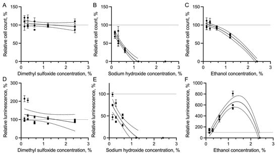
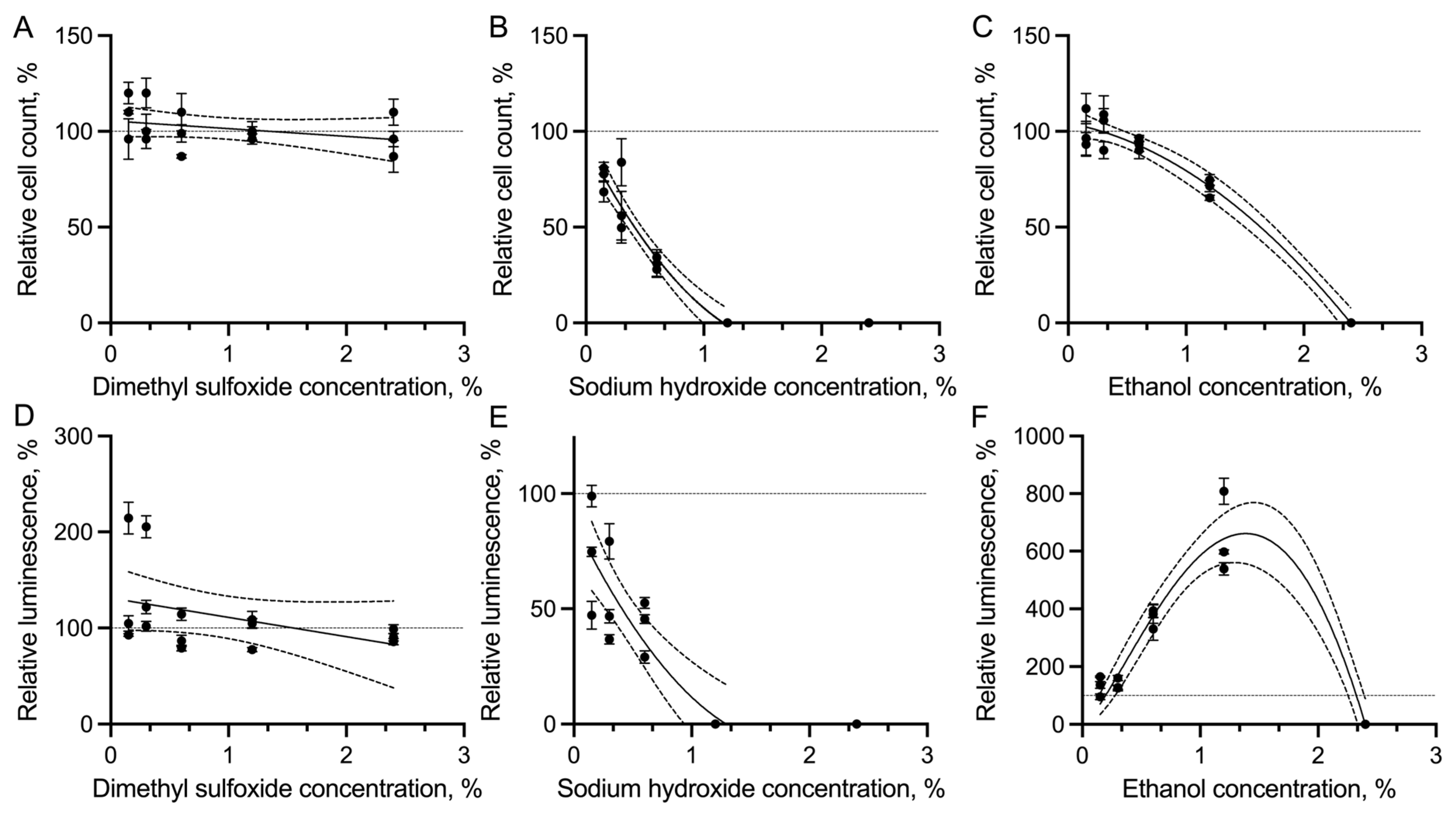
Relative cell counts and luminescence are shown in Figure 1. No significant effects were observed with DMSO across the tested concentration range. Stronger effects were observed with EtOH. Detectable cells were present through 1.2% EtOH, although cell density declined at the higher concentrations. No cells were detected at 2.4%. The highest sensitivity was observed with NaOH, which showed a dose-dependent reduction in growth. No cells were observed at 1.2% and 2.4%. At higher concentrations, reduced counts were also associated with visibly pleomorphic forms (primarily dynamic circular forms), suggesting cellular stress associated with NaOH exposure that did not occur in the presence of DMSO. The pH range of BSK-IIB with NaOH added was determined and was consistent with the predicted buffering capacity of BSK-II and its variants. The measured pH at each concentration was 8.01 (1.5 mM NaOH), 8.12 (3.0 mM), 8.15 (6.0 mM), 8.59 (12.0 mM), and 9.64 (24.0 mM) at the study endpoint. The pH of the control BSK-IIB was 7.7. More frequent and larger spirochaete aggregates were observed with EtOH at concentrations above 0.3%. Pleomorphic forms were not observed to the same extent as with NaOH-exposed spirochaetes.
Figure 1.
Effects of DMSO, NaOH, and EtOH on B. burgdorferi at day 6 of incubation. (A–C) Dark-field microscopy cell counts. Each point represents one culture tube counted three times for a technical measure of error (SEM, bars) around the point representing the mean. Relative cell count on the y-axis indicates the cell count in the vehicle-exposed group normalised to the average count of cells grown in BSK-IIB-only and expressed as a percentage of the control; (D–F) Luminescence was analysed in four technical replicates per culture. Descriptive curves (solid curves) are shown for DMSO (linear), NaOH (second-order polynomial), and EtOH (count: second-order polynomial, luminescence: beta growth with decay) with respective 95% confidence bands (dashed curves) about fitted estimates.
Vehicle responses differed according to concentration for both measured endpoints. For cell counts, two-way ANOVA identified a significant vehicle × concentration interaction: F (8,30) = 9133.22, p < 0.001. Significant main effects of vehicle identity (F (2,30) = 18,708.01, p < 0.001) and concentration (F (4,30) = 21,569.96, p < 0.001) were also detected. Given the significant interaction, concentration effects were examined within vehicles. No significant pairwise differences were detected among DMSO concentrations. For NaOH, cell counts did not differ between 0.15% and 0.30% (p = 0.142), but counts at 0.60% were lower than at 0.15% (p < 0.001) and 0.30% (p < 0.001). For EtOH, cell counts did not differ significantly among 0.15%, 0.30%, and 0.60%, whereas counts at 1.20% were lower than at 0.15% (p = 0.002), 0.30% (p = 0.001), and 0.60% (p = 0.018).
ATP-dependent luminescence similarly showed a significant vehicle × concentration interaction to cell counts (F (8,30) = 819.95, p < 0.001), with significant main effects of vehicle identity (F (2,30) = 1688.39, p < 0.001) and concentration (F (4,30) = 1788.36, p < 0.001). No significant pairwise differences were detected among DMSO concentrations. For NaOH, luminescence did not differ among concentrations from 0.15 to 0.60%. EtOH luminescence was significantly greater at 0.60% and 1.20% than at 0.15% and 0.30% (p < 0.001), while 0.60% and 1.20% did not significantly differ (p = 0.081). Residuals indicated that some heteroscedasticity remained after transformation (Rs cell count = 0.46, Rs luminescence = 0.38). Luminescence residuals did not deviate from normality (p = 0.28), but cell count residuals did (p = 0.008). These departures coincided with treatment combinations in which the biological replicates had zero detectable cell counts or background-adjusted luminescence.
In the separate comparison with the BSK-IIB-only control by one-way ANOVA (cell count F (15,32) = 14,043, p < 0.001; luminescence F (15,32) = 1221, p < 0.001) post hoc test, no DMSO concentration significantly differed from control for either endpoint. Sodium hydroxide reduced cell counts beginning at 0.15% (p = 0.0163), with stronger reductions at 0.3% and 0.6% (p < 0.001), while luminescence was significantly reduced at 0.30% (p = 0.0245) and 0.60% (p = 0.001). Cell counts in EtOH did not significantly differ before 1.2%. Counts at 1.2% were significantly lower (p = 0.002). ATP-dependent luminescence, in contrast, increased at 0.60% and 1.20% (p < 0.001). Homogeneity of variance was supported by Brown-Forsythe tests for both cell counts (p = 0.4766) and luminescence (p = 0.6144) for these analyses.
The zero-inclusive trends in relative ATP readouts showed a strong, significant ranked correlation with cell counts (Rs = 0.646, 95% CI 0.427 to 0.793, p < 0.001). However, a weak, non-significant linear relationship was shown with all vehicles considered (Pearson’s r = 0.261, 95% CI −0.036 to 0.5147, p = 0.084). The discrepancy between luminescence and cell count-derived estimates varied across vehicle-concentration groups as indicated by one-sample t-tests. Luminescence generally followed the cell count trend for DMSO and NaOH, as no significant discrepancies were identified at any concentrations. Exposure to EtOH, in contrast, produced greater discordance in endpoints. The discrepancy was not significant at 0.15% or 0.30% EtOH but was significant at 0.60% (D = 2.759; p = 0.005) and 1.20% (D = 5.779; p = 0.0188, Table A3). At these concentrations, ATP-dependent luminescence increased relative to controls while cell counts remained near controls at 0.60% and declined at 1.20%.
4. Discussion
Vehicle choice is critical when manipulating microbial systems in vitro. For a microorganism like B. burgdorferi, which is highly sensitive to environmental conditions, small changes in vitro can alter growth and metabolism. To better understand how B. burgdorferi reacts to additives in culture, we characterised the gross effects of DMSO, NaOH, and EtOH on growth across low-to-moderate concentrations in BSK-IIB medium using cell counts and ATP-dependent luminescence. No statistically detectable effect of DMSO on growth was observed, while NaOH suppressed growth and EtOH produced concentration-dependent effects at higher tested concentrations. This study provides an empirical basis for understanding solvent conditions in B. burgdorferi physiological assays.
The finding that DMSO did not lead to a reduction in cell counts or measured viability builds on a study that found DMSO did not harm replication at low concentrations when evaluating redox-active additives that quench toxic oxygen by-products in biphasic and standard BSK media [22]. Another study used DMSO in a high-throughput screen of small molecules against B. burgdorferi and did not find reductions in indirectly measured viability with BacTiter-Glo at 0.25%. This aligns with our results that expand on the concentration range that does not affect the results as measured with the BacTiter-Glo endpoint [23]. Dimethyl sulfoxide is widely used as a solvent in other microbial systems. In the yeast species Candida glabrata, DMSO reduces growth at and above 2.5% [24]. Some ecotoxicity assays report physiological effects with DMSO at EC50 values of 1–2% and may recommend final limiting concentration ranges ranging from 0.01% to 0.5% [25]. This range is more restrictive than indicated with B. burgdorferi in this study with this strain and medium, pending molecular corroboration.
Exposure to DMSO at sub-lethal concentrations in growth assays (e.g., 1–2.5% v/v in Escherichia coli) can still induce significant physiological changes. These effects may include changes in membrane properties and macromolecular composition. This shows that non-toxic dosing based on gross observation may not be truly inert [26,27]. Mechanistically, effects may differ by species, so it is not possible to predict effects from studies in other systems [15]. However, this evidence can be used as a foundation for future work to discern effects in B. burgdorferi. In microbial species that form biofilms, DMSO alone can inhibit or stimulate biofilm growth and may have synergistic effects with antimicrobials [28,29]. The in vivo relevance of biofilm-like and persister phenotypes in B. burgdorferi remains unresolved. Thus, it is still worth broadly considering these physiological effects as pleomorphic forms appear in vitro [30]. While these effects alone may not be relevant to growth, they may confound studies or become relevant when more variables are introduced into the system. So, while the DMSO concentrations in this study were evidently higher than those typically used in antimicrobial screening, it is possible that the operational dose range used in the antimicrobial study will need to be reduced to accommodate these effects. A future study with B. burgdorferi can determine physiological effects by examining the system with a molecular approach. Regardless, DMSO is the most operationally compatible tested vehicle.
Ethanol reduced cell density in this study, with cell counts low at 1.2% and absent at 2.4%. Mechanistically, EtOH can broadly stress cell envelopes and proteins. Ethanol also affects secondary metabolism, including altering oxidative stress responses and ROS-associated pathways [31,32]. Given the volatility of ethanol and the six-day incubation at 34 °C, some loss of EtOH (not quantified) from the liquid phase may have occurred, although the sealed culture tubes would be expected to limit evaporative loss given the use of high-quality tubes with gaskets. A future physiological study may include a chromatography component to measure the ethanol fraction remaining in the culture tubes throughout the experiment. Nevertheless, very low concentrations of EtOH have been shown to affect bacterial viability, recovery, and metabolic activity even after ethanol depletion [33]. Interestingly, the cell counts diverged from the ATP-dependent luminescence at concentrations up to 1.2%. This result may be consistent with shifts toward metabolically stressed states at concentrations below those lethal to most of the population [31].
Previous work has shown that B. burgdorferi responds to EtOH at lower concentrations, with EtOH acting as a chemorepellent and altering flagellar behaviour without detectable cell lysis at the concentrations tested [34]. These observations indicate that EtOH can alter B. burgdorferi physiology before causing overt loss of viability. This may be relevant to the divergence observed between cell counts and ATP-dependent luminescence. Borrelia has a highly specialised metabolism, and disruption of cellular processes can produce changes in behaviour before death [35]. Accordingly, ATP-dependent luminescence should be interpreted as an estimate of metabolically active biomass rather than a direct measure of cell number. Conversely, damaged cells may retain ATP for some period, and the assay signal can be influenced by differences in cell lysis. Therefore, more studies would be needed to confirm viability evidence with molecular validation. The discordance between luminescence and cell count at 0.6% and 1.2% may therefore reflect changes in ATP content per cell, but alternative assay-related mechanisms remain possible. Nonetheless, these findings support the use of EtOH concentrations below 0.6% v/v and support the range used by a prior study that used EtOH at 0.5% to assess the antimicrobial activity of phytochemicals and micronutrients against species of Borrelia in vitro [18].
Responses by bacteria to organic solvents may include membrane remodelling, efflux activation, and increased energetic demand. These mechanisms provide a framework for how solvent stress could transiently affect ATP-based readouts without a proportional increase in cell counts [36]. Solvents can alter membrane permeability or lysis efficiency, thereby increasing ATP recovery in molecular assays even if the true cell number remains unchanged [37]. Accordingly, these quantitative EtOH-associated effects may capture a phenotype associated with biological stress that induced a true increase in ATP output. The higher frequency and size of spirochaete aggregates observed with EtOH may reasonably provide an alternative explanation for the increased observed luminescence, as this can significantly interfere with the accuracy of counting fields with aggregates present. Support for this is also provided by the lack of increased luminescence observed in an ATP-only validation in EtOH-only trials and emphasises the need for accuracy in preparation and pipetting small volumes in these experiments. Follow-up studies should assess this mechanism directly and incorporate orthogonal validation when interpreting ATP-dependent luminescence in high-throughput drug-screening assays.
We assessed the potential of NaOH as an unbuffered alkaline vehicle, and it was shown to cause the largest effects on cell counts and viability. Detectable ATP-dependent luminescence persisted through 0.6% NaOH, consistent with our previous tests using IMPDH inhibitors delivered with 0.5% NaOH, which showed maintained viability with BacTiter-Glo and detectable differences between drug and NaOH-only controls (unpublished). Relatively low concentrations still impaired growth and induced morphological shifts to pleomorphic forms, indicative of a stress response. Borrelia burgdorferi is sensitive to pH changes, and pH is generally maintained between 7.4 and 7.8 to support growth. This indicates that unbuffered NaOH should not be used in BSK-IIB. Lower concentrations than those evaluated with these endpoints would require independent validation and may not disrupt the medium conditions. These concentrations are likely insufficient to use as a diluent for compatible active ingredients due to solubility constraints. Mechanistically, alkaline conditions can disrupt proton gradients and membrane potential. Direct exposure to alkalinity may cause cell lysis and death. This may also alter protein structure. Protein denaturation or degradation may be particularly relevant with bovine serum albumin and rabbit serum in the medium, potentially contributing to precipitation or altered nutrient availability. These effects were likely responsible for the stress-associated morphological changes before the complete loss of detectable spirochaetes at a threshold between 0.6% and 1.2% [38]. The observed morphological plasticity may represent a stress-associated state. Whether these forms possess persister characteristics was not assessed. The effects of shifts to slow-replicating or protective states may confound the relative interpretation of drug effects and be hidden when considering counts alone. Quantifying the morphological changes observed in more detail would be a useful future direction.
Limitations of this study can be addressed with future work. An important limitation is the use of a single B. burgdorferi strain in only one modified BSK formulation. Strain B31, clone 5A1, has a well-characterised genetic background and is infectious in mammalian models, making it useful for initial study [19]. These characteristics do not establish external validity as strains and isolates can differ in plasmid content and growth characteristics [39]. These factors may affect physiological responses to cellular stress and therefore warrant broader testing using other populations. It would also be useful to assess the impact of passage on spirochaete robustness to varying concentrations of these vehicles. Transcriptomic and other molecular analyses could provide a mechanistic assessment of vehicle-associated stress responses. Only DMSO, NaOH, and EtOH were tested. Other vehicles show distinct toxicity profiles across taxa and may behave differently, even within the same class of active ingredient [24]. It would therefore be relevant to assess additional options that may be improvements over what has been characterised.
The small biological sample size also limited the sensitivity for detecting more subtle vehicle-associated effects. However, the objective of the present study was to identify operationally relevant changes in growth that could influence testing outcomes. This was particularly evident with NaOH, where exposure to increasing concentrations resulted in marked reductions in cell counts and ATP-dependent luminescence. The study also identified a clear point at which no cells or viability indication were present with NaOH and EtOH. Prior studies have also performed similar in vitro assessments with this biological sample size with successful elucidation of effects [17,18]. By contrast, smaller dose-dependent effects may not have been detected, particularly with DMSO. Non-significant comparisons should therefore not be interpreted as evidence of complete biological inertness. Future studies may expand the biological sample size to evaluate these non-significant differences. These studies may also be performed to assess physiological effects over the duration of the experiment in the event of transient metabolic stress or growth inhibition/delay at different time points.
When considering the broader study of in vitro therapeutic investigations and the impact of this work, we must consider the state of the interface between clinical medicine and translational research. Although most cases of Lyme disease treated early resolve quickly and completely, there remains a strong rationale for investigating antimicrobials that target B. burgdorferi. In vitro studies have reported differential activity of antimicrobial compounds against stationary-phase populations and pleomorphic forms, although the clinical relevance of these compounds remains unresolved [40,41,42]. Novel compounds may also have applications outside of clinical medicine. Targeted delivery of antimicrobials to small mammals has reduced B. burgdorferi infection in both reservoir hosts and host-seeking ticks [43,44]. Such approaches would not address other tick-borne pathogens maintained in the same hosts and therefore would complement rather than replace broader anti-tick interventions. Therefore, B. burgdorferi-specific treatments may support contemporary public health efforts in the meantime, bridging the gap until an effective vaccine becomes available [45]. Whether compounds are intended for therapeutics or reservoir-targeted interventions, reliable in vitro evaluation requires vehicle conditions that do not independently affect B. burgdorferi physiology. The present study provides information to continue these studies accordingly.
5. Conclusions
We evaluated the effects of DMSO, NaOH, and EtOH on B. burgdorferi cell counts and viability. Practical recommendations are supported: no statistically detectable reduction in cell counts or ATP-dependent luminescence was observed with DMSO up to 2.4% v/v under the conditions tested, though the lowest concentrations needed for solubility are preferred. Ethanol may be used below 0.6% v/v. Use of unbuffered NaOH should be avoided as cell count and luminescence suppression were evident at very low concentrations. These recommendations are limited by these two measured endpoints in strain B31 clone 5A1. Possible molecular or physiological effects outside these endpoints and the day-six endpoint require further validation at these apparently compatible concentrations. Overall, this study expands our understanding of the identity and range of vehicles that may be used with B. burgdorferi in vitro.
Supplementary Materials
The following supporting information can be downloaded at: https://www.mdpi.com/article/10.3390/microbiolres17100208/s1, File S1: Biological replicate values for ATP-dependent luminescence and cell counts and ATP-only EtOH luminescence validation.
Author Contributions
E.L.S.: conceptualization, methodology, formal analysis, investigation, resources, data curation, writing—original draft preparation, writing—review and editing, visualization, supervision, project administration, funding acquisition; S.A.B.: methodology, software, validation, data curation, writing—review and editing; L.D.: methodology, data curation, writing—review and editing; P.P.: conceptualization, methodology, investigation, project administration, resources, writing—review and editing; G.X.: investigation, supervision, project administration, writing—review and editing; S.M.R.: writing—review and editing, investigation, resources, project administration, funding acquisition, supervision. All authors have read and agreed to the published version of the manuscript.
Funding
This study has been funded by a grant (2025) from the European Society of Clinical Microbiology and Infectious Diseases (Europäische Gesellschaft für klinische Mikrobiologie und Infektionskrankheiten) (ESCMID) to E.L.S. and the New England Center of Excellence in Vector-borne diseases—a cooperative agreement with the Centers for Disease Control and Prevention (CDC U01CK000661). Additional support was provided by the revenue fund of the Laboratory of Medical Zoology at the University of Massachusetts.
Institutional Review Board Statement
Not applicable.
Informed Consent Statement
Not applicable.
Data Availability Statement
All relevant data are found within the manuscript and Appendix A.
Acknowledgments
The authors thank Selasi Dankwa and Mary Elias-Gillette for support in this work. Their time and expertise are greatly appreciated. This manuscript represents research only. Any mention of active ingredient, product, or formulation does not constitute an endorsement for use by the authors or their affiliations.
Conflicts of Interest
The authors declare no conflicts of interest. The funders had no role in the design of the study; in the collection, analyses, or interpretation of data; in the writing of the manuscript; or in the decision to publish the results.
Abbreviations
The following abbreviations are used in this manuscript:
| ANOVA | Analysis of Variance |
| ATP | Adenosine triphosphate |
| BSK | Barbour-Stoenner-Kelly B. burgdorferi culture medium |
| DMSO | Dimethyl sulfoxide |
| EtOH | Ethanol |
| IMPDH | Inosine-5′-monophosphate dehydrogenase |
| NaOH | Sodium hydroxide |
| v/v | Volume per volume |
Appendix A
Expanded comparisons among vehicle–concentration groups and between measurement methods are shown in Table A1, Table A2 and Table A3. For Table A1 and Table A2, the matrix shows Tukey-adjusted p values from simple-effects comparisons of concentration within each vehicle following ordinary two-way ANOVA of log10(x + 1) transformed values. Each vehicle is treated as a separate family of comparisons. The ratios and 95% confidence intervals are back-transformed comparisons of the treatment to shared controls (cells grown in BSK-IIB only) from a separate ordinary one-way ANOVA with Dunnett adjustment. Because the analysis was performed on the transformed units, the ratios are of the geometric means of (x + 1). Table A3 shows the discrepancy analysis comparing endpoint measures. For each table, NS = Not significant. Dashes indicate that no comparison was made for that cell.
Table A1.
Multiple comparison analyses for B. burgdorferi cell counts.
Table A2.
Multiple comparison analyses for ATP-dependent luminescence as an indirect measure of viability.
Table A3.
Discrepancy analysis between ATP-dependent luminescence and cell counts, each normalised to the corresponding control.
References
- Medlock, J.M.; Hansford, K.M.; Bormane, A.; Derdakova, M.; Estrada-Peña, A.; George, J.-C.; Golovljova, I.; Jaenson, T.G.T.; Jensen, J.-K.; Jensen, P.M.; et al. Driving Forces for Changes in Geographical Distribution of Ixodes ricinus Ticks in Europe. Parasit. Vectors. 2013, 6, 1. [Google Scholar] [CrossRef] [Scilit] [PubMed]
- Helble, J.D.; McCarthy, J.E.; Hu, L.T. Interactions between Borrelia burgdorferi and Its Hosts across the Enzootic Cycle. Parasite Immunol. 2021, 43, e12816. [Google Scholar] [CrossRef] [Scilit] [PubMed]
- Kugeler, K.J.; Schwartz, A.M.; Delorey, M.J.; Mead, P.S.; Hinckley, A.F. Estimating the Frequency of Lyme Disease Diagnoses, United States, 2010–2018. Emerg. Infect. Dis. 2021, 27, 616–619. [Google Scholar] [CrossRef] [Scilit] [PubMed]
- Eisen, L.; Eisen, R.J. Changes in the Geographic Distribution of the Blacklegged Tick, Ixodes scapularis, in the United States. Ticks Tick-Borne Dis. 2023, 14, 102233. [Google Scholar] [CrossRef] [Scilit] [PubMed]
- Ostfeld, R.S.; Brunner, J.L. Climate Change and Ixodes Tick-Borne Diseases of Humans. Philos. Trans. R. Soc. B 2015, 370, 20140051. [Google Scholar] [CrossRef] [Scilit] [PubMed]
- Schwartz, A.M.; Hinckley, A.F.; Mead, P.S.; Hook, S.A.; Kugeler, K.J. Surveillance for Lyme Disease—United States, 2008–2015. MMWR Surveill. Summ. 2017, 66, 1–12. [Google Scholar] [CrossRef] [Scilit] [PubMed]
- Stupica, D.; Collinet-Adler, S.; Blagus, R.; Gomišček, A.; Cerar Kišek, T.; Ružić-Sabljić, E.; Velušček, M. Treatment of Erythema Migrans with Doxycycline for 7 Days versus 14 Days in Slovenia: A Randomised Open-Label Non-Inferiority Trial. Lancet Infect. Dis. 2023, 23, 371–379. [Google Scholar] [CrossRef] [Scilit] [PubMed]
- Stupica, D.; Lusa, L.; Ruzic-Sabljic, E.; Cerar, T.; Strle, F. Treatment of Erythema Migrans with Doxycycline for 10 Days Versus 15 Days. Clin. Infect. Dis. 2012, 55, 343–350. [Google Scholar] [CrossRef] [Scilit] [PubMed]
- Wormser, G.P.; Schwartz, I. Antibiotic Treatment of Animals Infected with Borrelia burgdorferi. Clin. Microbiol. Rev. 2009, 22, 387–395. [Google Scholar] [CrossRef] [Scilit] [PubMed]
- Caimano, M.J.; Drecktrah, D.; Kung, F.; Samuels, D.S. Interaction of the Lyme Disease Spirochete with Its Tick Vector. Cell. Microbiol. 2016, 18, 919–927. [Google Scholar] [CrossRef] [Scilit] [PubMed]
- Wang, G.; Iyer, R.; Bittker, S.; Cooper, D.; Small, J.; Wormser, G.P.; Schwartz, I. Variations in Barbour-Stoenner-Kelly Culture Medium Modulate Infectivity and Pathogenicity of Borrelia burgdorferi Clinical Isolates. Infect. Immun. 2004, 72, 6702–6706. [Google Scholar] [CrossRef] [Scilit] [PubMed]
- Replogle, A.J.; Sexton, C.; Young, J.; Kingry, L.C.; Schriefer, M.E.; Dolan, M.; Johnson, T.L.; Connally, N.P.; Padgett, K.A.; Petersen, J.M. Isolation of Borrelia miyamotoi and Other Borreliae Using a Modified BSK Medium. Sci. Rep. 2021, 11, 1926. [Google Scholar] [CrossRef] [Scilit] [PubMed]
- Gilmore, R.D., Jr.; Mbow, M.L.; Stevenson, B. Analysis of Borrelia burgdorferi gene expression during life cycle phases of the tick vector Ixodes scapularis. Microbes Infect. 2001, 3, 799–808. [Google Scholar] [CrossRef] [Scilit] [PubMed]
- Schalck, T.; Van den Bergh, B.; Michiels, J. Increasing Solvent Tolerance to Improve Microbial Production of Alcohols, Terpenoids and Aromatics. Microorganisms 2021, 9, 249. [Google Scholar] [CrossRef] [Scilit] [PubMed]
- Summer, K.; Browne, J.; Hollanders, M.; Benkendorff, K. Out of Control: The Need for Standardised Solvent Approaches and Data Reporting in Antibiofilm Assays Incorporating Dimethyl-Sulfoxide (DMSO). Biofilm 2022, 4, 100081. [Google Scholar] [CrossRef] [Scilit] [PubMed]
- Lynch, A.; Pearson, P.; Savinov, S.N.; Li, A.Y.; Rich, S.M. Lactate Dehydrogenase Inhibitors Suppress Borrelia burgdorferi Growth In Vitro. Pathogens 2023, 12, 962. [Google Scholar] [CrossRef] [Scilit] [PubMed]
- Siegel, E.L.; Rich, C.; Saravanan, S.; Pearson, P.; Xu, G.; Rich, S.M. Effects of Inosine-5′-Monophosphate Dehydrogenase (IMPDH/GuaB) Inhibitors on Borrelia burgdorferi Growth in Standard and Modified Culture Conditions. Microorganisms 2024, 12, 2064. [Google Scholar] [CrossRef] [Scilit] [PubMed]
- Goc, A.; Niedzwiecki, A.; Rath, M. In Vitro Evaluation of Antibacterial Activity of Phytochemicals and Micronutrients against Borrelia burgdorferi and Borrelia garinii. J. Appl. Microbiol. 2015, 119, 1561–1572. [Google Scholar] [CrossRef] [Scilit] [PubMed]
- Norris, S.J.; Howell, J.K.; Garza, S.A.; Ferdows, M.S.; Barbour, A.G. High- and Low-Infectivity Phenotypes of Clonal Populations of in Vitro-Cultured Borrelia burgdorferi. Infect. Immun. 1995, 63, 2206–2212. [Google Scholar] [CrossRef] [Scilit] [PubMed]
- Pothineni, V.R.; Wagh, D.; Babar, M.M.; Inayathullah, M.; Watts, R.E.; Kim, K.-M.; Parekh, M.B.; Gurjarpadhye, A.A.; Solow-Cordero, D.; Tayebi, L.; et al. Screening of NCI-DTP Library to Identify New Drug Candidates for Borrelia burgdorferi. J. Antibiot. 2017, 70, 308–312. [Google Scholar] [CrossRef] [Scilit] [PubMed]
- GraphPad Prism, version 11 for Mac; GraphPad Software: Boston, MA, USA, 2026. Available online: https://www.graphpad.com/ (accessed on 9 August 2026).
- Padgett, P.J.; Golden, M.J.; Pekala, D.J. Improving the Growth Response of Borrelia Species. Can. J. Microbiol. 1995, 41, 1031–1034. [Google Scholar] [CrossRef] [Scilit]
- Pothineni, V.; Wagh, D.; Babar, M.M.; Inayathullah, M.; Solow-Cordero, D.; Kim, K.-M.; Samineni, A.; Parekh, M.B.; Tayebi, L.; Rajadas, J. Identification of New Drug Candidates against Borrelia burgdorferi Using High-Throughput Screening. Drug Des. Devel. Ther. 2016, 2016, 1307–1322. [Google Scholar] [CrossRef] [Scilit] [PubMed]
- Liu, J.; Zhang, H.; Zhang, L.; Li, T.; Liu, N.; Liu, Q. Effect of Various Concentrations of Common Organic Solvents on the Growth and Proliferation Ability of Candida glabrata and Their Permissible Limits for Addition in Drug Susceptibility Testing. PeerJ 2023, 11, e16444. [Google Scholar] [CrossRef] [Scilit] [PubMed]
- Andrade-Vieira, L.F.; Bojic, C.; Santana Alvarenga, I.F.; De Carvalho, T.S.; Masfaraud, J.-F.; Cotelle, S. Ecotoxic Effects of the Vehicle Solvent Dimethyl Sulfoxide on Raphidocelis subcapitata, Daphnia magna and Brachionus calyciflorus. Chem. Ecol. 2022, 38, 471–483. [Google Scholar] [CrossRef] [Scilit]
- Tunçer Çağlayan, S.; Gurbanov, R. Modulation of Bacterial Membranes and Cellular Macromolecules by Dimethyl Sulfoxide: A Dose-Dependent Study Providing Novel Insights. Int. J. Biol. Macromol. 2024, 267, 131581. [Google Scholar] [CrossRef] [Scilit] [PubMed]
- Tunçer, S.; Gurbanov, R. Non-Growth Inhibitory Doses of Dimethyl Sulfoxide Alter Gene Expression and Epigenetic Pattern of Bacteria. Appl. Microbiol. Biotechnol. 2023, 107, 299–312. [Google Scholar] [CrossRef] [Scilit] [PubMed]
- Yahya, M.F.Z.R.; Alias, Z.; Karsani, S.A. Antibiofilm Activity and Mode of Action of DMSO Alone and Its Combination with Afatinib against Gram-Negative Pathogens. Folia Microbiol. 2018, 63, 23–30. [Google Scholar] [CrossRef] [Scilit] [PubMed]
- Knap, K.; Kwiecień, K.; Ochońska, D.; Reczyńska-Kolman, K.; Pamuła, E.; Brzychczy-Włoch, M. Synergistic Effect of Antibiotics, α-Linolenic Acid and Solvent Type against Staphylococcus aureus Biofilm Formation. Pharmacol. Rep. 2024, 76, 1456–1469. [Google Scholar] [CrossRef] [Scilit] [PubMed]
- Gindl, A.; Schötta, A.; Berent, S.; Markowicz, M.; Stockinger, H.; Thalhammer, F.; Stary, G.; Strobl, J. Persistent Lyme Disease with Cutaneous Borrelia Biofilm Formation. Br. J. Dermatol. 2022, 186, 1041–1043. [Google Scholar] [CrossRef] [Scilit] [PubMed]
- Sen, O.; Hinks, J.; Lin, Q.; Lin, Q.; Kjelleberg, S.; Rice, S.A.; Seviour, T. Escherichia coli Displays a Conserved Membrane Proteomic Response to a Range of Alcohols. Biotechnol. Bioguels Bioprod. 2023, 16, 147. [Google Scholar] [CrossRef] [Scilit] [PubMed]
- Cao, H.; Wei, D.; Yang, Y.; Shang, Y.; Li, G.; Zhou, Y.; Ma, Q.; Xu, Y. Systems-Level Understanding of Ethanol-Induced Stresses and Adaptation in E. coli. Sci. Rep. 2017, 7, 44150. [Google Scholar] [CrossRef] [Scilit] [PubMed]
- Chatterjee, I.; Somerville, G.A.; Heilmann, C.; Sahl, H.-G.; Maurer, H.H.; Herrmann, M. Very Low Ethanol Concentrations Affect the Viability and Growth Recovery in Post-Stationary-Phase Staphylococcus aureus Populations. Appl. Environ. Microbiol. 2006, 72, 2627–2636. [Google Scholar] [CrossRef] [Scilit] [PubMed]
- Shi, W.; Yang, Z.; Geng, Y.; Wolinsky, L.E.; Lovett, M.A. Chemotaxis in Borrelia burgdorferi. J. Bacteriol. 1998, 180, 231–235. [Google Scholar] [CrossRef] [Scilit] [PubMed]
- Holly, K.J.; Kataria, A.; Flaherty, D.P.; Groshong, A.M. Unguarded Liabilities: Borrelia burgdorferi’s Complex Amino Acid Dependence Exposes Unique Avenues of Inhibition. Front. Antibiot. 2024, 3, 1395425. [Google Scholar] [CrossRef] [Scilit] [PubMed]
- Luo, J.; Song, Z.; Ning, J.; Cheng, Y.; Wang, Y.; Cui, F.; Shen, Y.; Wang, M. The Ethanol-Induced Global Alteration in Arthrobacter Simplex and Its Mutants with Enhanced Ethanol Tolerance. Appl. Microbiol. Biotechnol. 2018, 102, 9331–9350. [Google Scholar] [CrossRef] [Scilit] [PubMed]
- Bui, H.T. N-Alkanol Stress-Induced Cell Envelope Injury of σE Promoter in Escherichia coli. Acad. J. Biol. 2022, 44, 91–104. [Google Scholar] [CrossRef] [Scilit]
- Carroll, J.A.; Garon, C.F.; Schwan, T.G. Effects of Environmental pH on Membrane Proteins in Borrelia burgdorferi. Infect. Immun. 1999, 67, 3181–3187. [Google Scholar] [CrossRef] [Scilit] [PubMed]
- Purser, J.E.; Norris, S.J. Correlation between Plasmid Content and Infectivity in Borrelia burgdorferi. Proc. Natl. Acad. Sci. USA 2000, 97, 13865–13870. [Google Scholar] [CrossRef] [Scilit] [PubMed]
- Di Domenico, E.G.; Cavallo, I.; Bordignon, V.; D’Agosto, G.; Pontone, M.; Trento, E.; Gallo, M.T.; Prignano, G.; Pimpinelli, F.; Toma, L.; et al. The Emerging Role of Microbial Biofilm in Lyme Neuroborreliosis. Front. Neurol. 2018, 9, 1048. [Google Scholar] [CrossRef] [Scilit] [PubMed]
- Sapi, E.; Kaur, N.; Anyanwu, S.; Luecke, D.; Datar, A.; Patel, S.; Rossi, M.; Stricker, R. Evaluation of in-vitro antibiotic susceptibility of different morphological forms of Borrelia burgdorferi. Infect. Drug Resist. 2011, 2011, 97–113. [Google Scholar] [CrossRef] [Scilit] [PubMed]
- Feng, J.; Wang, T.; Shi, W.; Zhang, S.; Sullivan, D.; Auwaerter, P.G.; Zhang, Y. Identification of Novel Activity against Borrelia burgdorferi Persisters Using an FDA Approved Drug Library. Emerg. Microbes Infect. 2014, 3, 1–8. [Google Scholar] [CrossRef] [Scilit] [PubMed]
- Dolan, M.C.; Zeidner, N.S.; Gabitzsch, E.; Dietrich, G.; Borchert, J.N.; Poché, R.M.; Piesman, J. A Doxycycline Hyclate Rodent Bait Formulation for Prophylaxis and Treatment of Tick-Transmitted Borrelia burgdorferi. Am. J. Trop. Med. Hyg. 2008, 78, 803–805. [Google Scholar] [CrossRef] [Scilit]
- Eisen, L. Rodent-Targeted Approaches to Reduce Acarological Risk of Human Exposure to Pathogen-Infected Ixodes Ticks. Ticks Tick-Borne Dis. 2023, 14, 102119. [Google Scholar] [CrossRef] [Scilit] [PubMed]
- Siegel, E.L.; Rich, S.M. The Lyme Disease Vaccine Paradox. J. Clin. Med. 2026, 15, 1634. [Google Scholar] [CrossRef] [Scilit] [PubMed]
Disclaimer/Publisher’s Note: The statements, opinions and data contained in all publications are solely those of the individual author(s) and contributor(s) and not of MDPI and/or the editor(s). MDPI and/or the editor(s) disclaim responsibility for any injury to people or property resulting from any ideas, methods, instructions or products referred to in the content. |
© 2026 by the authors. Licensee MDPI, Basel, Switzerland. This article is an open access article distributed under the terms and conditions of the Creative Commons Attribution (CC BY) license.