


Abstract
Fresh fruits may act as vehicles for microorganisms introduced during cultivation, harvest, and handling, especially in settings where crop production occurs close to animal farming. In this study, three Gram-negative strains incidentally recovered during the screening of lactic acid bacteria from Rangpur limes (Citrus × limonia) were identified and characterized. Phenotypic and biochemical analyses, rep-PCR, and 16S rRNA gene sequencing identified one strain as Escherichia albertii and two strains as Kosakonia cowanii. The Escherichia albertii and two Kosakonia cowanii strains were further evaluated for β-galactosidase production, antimicrobial susceptibility, hemolytic activity, biogenic amine production, and virulence-associated genes. The three studied strains were hemolytic, positive for β-galactosidase, showed presumptive amino acid decarboxylase activity, and demonstrated resistance to ampicillin, with intermediate susceptibility to nalidixic acid. Classical virulence genes such as eae, cva, iss, iroN, iut, and omp were not detected, whereas fim and hly were observed. These findings indicate that Rangpur limes cultivated at the animal–crop interface may harbor opportunistic and emerging bacteria of relevance to food safety and One Health surveillance.
1. Introduction
Fruits are natural foods widely recommended for daily consumption. The World Health Organization (WHO), for instance, recommends a daily intake of five servings (400 g) of fruits and vegetables for adults [1]. Together, these plant-based foods serve as primary sources of dietary fiber, vitamins, and essential minerals crucial for human nutrition. Citrus fruits, as an example, can provide up to 40% of the recommended daily intake of vitamin C. Furthermore, these plant foods contain a wide variety of non-nutrient compounds within their structures, such as flavonoids, polyphenols, phytoestrogens, and bioactive peptides, which add functional value to their consumption [2].
The ingestion of fruits as fresh or minimally processed remains the most common form of consumption. Although essential for maintaining the integrity of nutrients and bioactive compounds, this lack of thermal treatment does not ensure that these foods are free of microorganisms, including pathogens [3]. Consequently, fresh produce represents a significant vehicle for the transmission of foodborne diseases. While washing and disinfection with sodium hypochlorite are widely recommended, these practices are not universally effective. This limitation is particularly evident when fruits present natural openings or physical injuries that facilitate the internalization of pathogens, shielding them from surface sanitization measures [4]. Pre-harvest contamination of these crops is primarily driven by fertilized soils, poor-quality irrigation water, airborne particles, and direct or indirect contact with animal feces. During processing, contamination is frequently attributed to contaminated water, handling equipment, and the presence of biofilms on food contact surfaces [2,5,6].
In this context, effective surveillance of fresh produce is important for tracking potential pathogens and implementing preventive measures. Among emerging pathogens, we highlight Escherichia albertii, a microorganism belonging to the Enterobacteriaceae family, which is frequently confused with other phylogenetically related bacteria, such as enteropathogenic Escherichia coli (EPEC) or Shigella. These misidentifications lead to the underdiagnosis of E. albertii in gastrointestinal infections. Consequently, classical biochemical tests are insufficient for their identification, making the use of more advanced biomolecular techniques necessary for accurate diagnosis. E. albertii is frequently associated with wild and domestic birds and can cause gastroenteritis in humans, with contamination via avian feces being a probable route for human infection [7,8].
E. albertii is a Gram-negative, non-motile bacterial species within the genus Escherichia, closely related to E. coli. It was first recognized as a human pathogen after being isolated from children with diarrhea in Bangladesh, where it was initially misidentified as Hafnia alvei due to overlapping biochemical traits. In 2003, advances in genetic characterization led to its reclassification as a novel species, E. albertii, distinguished by features such as its inability to ferment lactose and its production of the virulence factor intimin, which often causes confusion with enteropathogenic E. coli [7].
E. albertii is considered an emerging foodborne and waterborne pathogen, associated primarily with watery diarrhea in children and immunocompromised individuals. Transmission is linked to contaminated water and food, particularly poultry products, and the organism has been frequently isolated from domestic and wild birds, suggesting that avian species may serve as a major reservoir [9]. It is interesting to point out that, to our knowledge, this is the first report on the isolation of E. albertii from fruits. However, it is clear that microbial presence on fruits can be associated with contact between fruits and birds, insects, farmers and workers. However, what is important is that pathogens generally associated with gastrointestinal tracts of animals, like E. albertii, can survive on the surface of citrus and present health problems for the consumers.
K. cowanii is a Gram-negative, motile, facultatively anaerobic bacterium belonging to the family Enterobacteriaceae [10]. Although often considered an environmental organism, K. cowanii has gained attention as an emerging plant pathogen. It has been implicated in the bacterial wilt of patchouli, where strain Pa82 demonstrated high virulence and carried genes associated with adherence, biofilm formation, and type VI secretion system-mediated pathogenicity. Additional reports identify K. cowanii as a causal agent of soybean disease, highlighting its agricultural relevance [10,11].
Beyond contamination of animal origin, the cultivation environment itself poses significant risks. In this context, we can highlight the presence of K. cowanii on fruits and vegetables as contamination. Frequently classified as a phytopathogen or environmental microorganism, recent studies have identified K. cowanii as the causative agent of severe human infections, such as bacteremia and cholecystitis without evidence of trauma, suggesting the ingestion of contaminated food as a probable route of colonization [12]. Similar to E. albertii, K. cowanii is notoriously difficult to be identified using traditional methods, as it is frequently misidentified as other closely related bacteria, such as the Pantoea or Enterobacter species [12,13].
Considering the close proximity of animal farming and crop cultivation on small properties, this study aims to report the identification and characterization of three bacterial strains, one E. albertii and two K. cowanii, isolated from Rangpur limes (Citrus × limonia) obtained from a property in the rural parts of the city of São Paulo region, Brazil. These strains, incidentally isolated during the screening for lactic acid bacteria (LAB), underwent a combined characterization comprising phenotypic, biochemical, and molecular analyses (16S rRNA gene sequencing). Additionally, the isolates were screened for virulence factors and antimicrobial resistance, contributing to the understanding of the role of fresh fruits as potential reservoirs for emerging pathogens within the One Health framework.
2. Materials and Methods
2.1. Screening and Isolation
Six rangpur lime samples were collected in aseptic conditions from two trees from the same farm, located in a rural suburban area of Sao Paulo, SP, Brazil and transported to the laboratory in refrigerated conditions, where all fruits were polled, aseptically weighed, and homogenized in 0.85% (w/v) saline solution in proportions 1:9 (w/v) using a Stomacher-type homogenizer (Smasher, AES Laboratoire, Ames, IA, USA) at 200 rpm for 2 min as the single research sample. Serial dilutions were performed from this initial solution. Aliquots of the dilutions were surface-plated on MRS agar (BD Difco, Detroit, MI, USA) (2% w/v). The plates were incubated at 37 °C for 24 h or 48 h under aerobic conditions. The resulting individual colonies were purified by streaking on MRS medium, incubated at 37 °C for 24 h, and subsequently stored at −20 °C in MRS broth supplemented with glycerol (20%, v/v) for long-term preservation.
2.2. Phenotypic Screening and Isolate Selection
The isolates were initially subjected to Gram staining and the catalase test. Although the primary goal was to obtain LAB (Gram-positive and catalase-negative), some isolates exhibited an atypical morphological and biochemical profile (Gram-negative rods and catalase-positive). Given the persistence of these isolates in the selective medium for LAB, they were retained for molecular identification and virulence characterization. Moreover, evaluated isolates were examined for their fermentative profile in modified MRS, where glucose was replaced respectively with lactose, sucrose or rhamnose. Studied isolates were cultured in modified MRS for 48 h at 37 °C and observed for changes in turbidity as evidence for bacterial growth.
2.3. Production of β-Galactosidase
β-galactosidase activity was assessed using ortho-nitrophenyl-β-D-galactopyranoside (ONPG) disks as the substrate. Bacterial strains were streaked onto BHI agar (2%) plates and incubated at 37 °C for 48 h to isolate individual colonies. For each strain, 3–4 colonies were suspended in 0.2 mL of sterile saline solution (0.85% NaCl) in test tubes, followed by the addition of an ONPG disk. The suspensions were incubated at 37 °C for up to 6 h. Samples were monitored regularly for the development of a yellow color, indicating the enzymatic hydrolysis of ONPG into ortho-nitrophenol by β-galactosidase [14]. All assays were performed in duplicate.
2.4. DNA Extraction, Genotyping by rep-PCR and Taxonomic Identification
For DNA extraction, the isolates were cultured in BHI broth at 37 °C for 24 h. Genomic DNA extraction was performed using the ZR Fungal/Bacterial DNA Kit (Zymo Research, Irvine, CA, USA), following the manufacturer’s instructions. The concentration and purity of the genetic material were evaluated by spectrophotometry using a NanoDrop equipment (Spectrostarnano, Ortenberg, Germany).
The differentiation of the isolates was performed using the rep-PCR technique with the (GTG)5 primer, following the protocol described by Choi et al. [15]. The amplification reaction was conducted in a Veriti 96 thermal cycler (Thermo Fisher Scientific, Waltham, MA, USA) under the following cycling conditions: initial denaturation at 95 °C for 5 min; followed by 30 cycles of denaturation at 95 °C for 30 s, annealing at 40 °C for 30 s, and extension at 65 °C for 8 min, with a final extension at 65 °C for 16 min. The resulting amplicons were separated by electrophoresis on a 1.5% (w/v) agarose gel, stained with SYBR® Safe DNA Gel Stain (Thermo Fisher Scientific), and visualized using a molecular imager (Bio-Rad, Hercules, CA, USA).
Taxonomic identification was performed by partial amplification of the 16S rRNA gene using the universal primers 27F (5′-AGA GTT TGA TCC TGG CTC AG-3′) and 1492R (5′-GGT TAC CTT GTT ACG ACT T-3′) [16]. The PCR reaction was carried out under the following conditions: initial denaturation at 95 °C for 7 min; 35 cycles of 95 °C for 1 min, 58 °C for 1.5 min, and 72 °C for 2.5 min; and a final extension at 72 °C for 5 min. The PCR products were analyzed on a 1% (w/v) agarose gel and purified. Sequencing was conducted at the Center for Human Genome Studies (ICB-USP, São Paulo, Brazil) using the Sanger method (BigDye Terminator v3.1 Cycle Sequencing Kit, Thermo Ficher Scientific) on an automated sequencer. The resulting sequences were edited and assembled using Sequencing Analysis 7.0 software and identified by comparison with the GenBank database (NCBI) using the BLASTn tool 2.15.0 (Basic Local Alignment Search Tool).
2.5. Safety Assessments
2.5.1. Antimicrobial Susceptibility
Antibiotic susceptibility was evaluated using the disk diffusion method against a panel of antimicrobial agents recommended for LAB and E. coli [17]: ampicillin (10 µg/disk), clindamycin (10 µg/disk), erythromycin (15 µg/disk), gentamicin (10 µg/disk), streptomycin (10 µg/disk), vancomycin (30 µg/disk), nalidixic acid (30 µg/disk), tetracycline (30 µg/disk), chloramphenicol (30 µg/disk), and kanamycin (30 µg/disk) (Cefar™, São Paulo, Brazil). Study cultures were inoculated (at approximately log 5 CFU/mL, final concentration) on BHI supplemented with 1% agar, and after depositing disks on the surface, plates were incubated for 24 h at 37 °C. These tests were performed on at least three independent occasions in duplicates. Results were expressed in millimeter inhibition zones, and appropriate SDs were calculated.
2.5.2. Gelatinase and Hemolytic Activity
To assess gelatinase production, 10 µL of the studied cultures were inoculated into tubes containing BHI broth supplemented with 3% gelatin. Cultures were incubated at 37 °C for 24 h, followed by refrigeration at 4 °C for 4 h. Medium liquefaction was considered a positive result for gelatin hydrolysis [18].
To assess hemolytic activity, 10 µL of the culture was spotted onto Tryptic Soy Agar (TSA) plates supplemented with 5% defibrinated sheep blood (LABORCLIN, Pinhais, Brazil). Plates were incubated at 37 °C for 24 h [18]. Strains from the culture collection of the ProBacLab (Faculty of Pharmaceutical Sciences, University of Sao Paulo, Sao Paulo, Brazil) were applied as positive (E. coli) and negative (Lactiplantibacillus plantarum) controls. The assays were performed in duplicate.
2.5.3. Biogenic Amines
Strains were sub-cultured daily for five consecutive days at 37 °C in modified BHI broth supplemented with 0.1% of each amino acid precursor: histidine, ornithine, lysine and tyrosine, respectively (Synth S.A., Volta Redonda, Brazil). Following this induction period, cultures were plated onto BHI agar (2%) supplemented with the respective precursors (1%), pyridoxal-5-phosphate (0.005%), and bromocresol purple (0.006%), with the pH adjusted to 5.3. Plates were incubated at 37 °C for 48 h. Since amino acid decarboxylation increases the pH of the medium, a positive result is indicated by a visible color change to purple [19]. Strains from the culture collection of the ProBacLab, were applied as positive (E. coli) and negative (Lpb. plantarum) controls. The assays were performed in duplicate.
2.5.4. Virulence Genes
DNA from strains studied was isolated as described before and was subjected to screening for the presence for virulence genes, as suggested by Teymurazov et al. [8], listed in Table 1. The PCR reaction was carried out under the conditions suggested by Teymurazov et al. [8] and generated amplicons evaluated on a 2% (w/v) agarose gels.
Table 1.
Primers and sequences used in this study. Records for the presence of targeted genes were indicated for the further identified and studied strains of E. albertii C4, K. cowanii C22 and C51.
3. Results and Discussion
3.1. Isolation and Taxonomic Identification
Microbial communities are a natural and persistent feature of tropical fruits, shaped by warm climates, high humidity, and diverse ecological interactions. The surfaces of fruits often harbor bacteria, yeast, and filamentous fungi, many of which originate from soil, irrigation water, insects, and handling during harvest and distribution. Studies highlight that contamination can occur at multiple points along the supply chain, with pre-harvest sources, including agricultural water and organic fertilizers, playing a major role in introducing microorganisms such as Salmonella, E. coli O157:H7, and norovirus [20].
As part of a research project focusing on screening for beneficial microbes with antioxidant properties, we have collected more than 160 microbial isolates from different tropical fruits. Moreover, emphasis was placed on Gram-positive and catalase-negative isolates. However, during the initial screening process, parallel attention was given to some Gram-negative isolates. In the preliminary evaluation for bacterial growth, it was recorded that some of the isolates were negative (no growth) in modified MRS, where glucose was replaced with lactose, sucrose or rhamnose, further identified as E. albertii; however, other initial isolates showed reasonable bacterial growth in the same modified MRS media, further identified as K. cowanii. Based on the performed repPCR, three isolates were selected, representing high frequency in microbial isolates obtained from rangpur lime samples. Further biochemical, physiological and biomolecular tests, including 16S rRNA, identified isolate C4 as E. albertii, and isolates C22 and C51 as K. cowanii, all with confidence higher than 99%. However, it is important to underline that 16S rRNA, even though it is considered standard in microbial taxonomy, can generate misunderstandings in some cases, especially for closely related species, as some of these belong to the Enterobacteriaceae. In further investigations, additional taxonomical approaches, including DAN-DNA hybridization and/or whole-genome sequencing, will be a logic addendum in the confirmation of identity of studied strains. Isolated not by principle objective, detected Gram-negative species were further investigated, driven by scientific curiosity. Further identification and safety evaluation of the obtained Gram-negative isolates was not part of the ecological study aiming to explore the presence of potential pathogens in the studied citrus, but coincidentally resulted in the isolation and identification of series of Gram-negative isolates, differentiated by repPCR and identified based on 16S rRNA as E. albertii and K. cowanii. However, with the objective of dipper analysis, not only with regard to reporting on the identity of these two potential pathogens obtained from studied fruits (on the surface or from the pulp) of citrus, but also the potential ecological adaptation to low pH of the citrus matrices and specificity of the presence of virulence factors, whole-genome sequencing and appropriate bioinformatic analyses will be next steps in exploring the pathogenicity of selected strains of E. albertii and K. cowanii.
The identification of E. albertii and K. cowanii on citrus fruit presents a serious food safety challenge. While the low intrinsic pH of citrus matrices (typically pH 2.0–4.0) is conventionally regarded as a robust barrier against bacterial survival, emerging evidence indicates that enteric and phytopathogenic bacteria can deploy stress-response mechanisms, including amino acid decarboxylase systems and protective biofilm formation, to withstand acidic microenvironments [21,22,23,24]. Consequently, relying solely on fruit acidity as a natural preservation barrier oversimplifies microbial resilience. From an epidemiological perspective, the presence of these species on fruit intended for fresh consumption (in natura) introduces distinct risks without the mitigation of a lethal processing kill-step: E. albertii represents a high-priority, emerging human pathogen equipped with the locus of enterocyte effacement island genes and Shiga toxin-encoding sequences [24,25,26], capable of causing severe gastrointestinal disease and systemic complications. Moreover, while K. cowanii was predominantly characterized as a plant pathogen responsible for tissue necrosis, it displayed opportunistic traits capable of causing nosocomial and soft-tissue infections in immunocompromised host populations [27,28,29]. Ultimately, the isolation of these organisms points to potential agricultural contamination, likely via irrigation water or soil amendments, and underscores the necessity for heightened post-harvest surveillance.
3.2. Biochemical Profile—β-Galactosidase
All evaluated strains (E. albertii C4, and K. cowanii C22 and C51) exhibited positive β-galactosidase activity (Table 2). The production of this enzyme is a widely recognized metabolic trait among many members of the Enterobacteriaceae family [30]. Beyond its taxonomic relevance, the ability to hydrolyze complex β-galactosides provides a significant ecological advantage. In the context of plant-associated environments, such as the surface or pulp of Citrus × limonia fruits, this enzymatic activity allows these opportunistic pathogens to exploit a broader range of available carbohydrates, thereby enhancing their survival, colonization, and persistence outside their primary host environments [31,32].
Table 2.
Biochemical and safety profile of E. albertii C4 and K. cowanii C22 and C51 strains.
β-galactosidase metabolism can be indirectly associated with pathogenicity in E. albertii because it reflects underlying genomic and phenotypic variability that may influence how the bacterium adapts to different hosts and environments. Although E. albertii is typically β-galactosidase-negative, some strains exhibit ONPG positivity or weak lactose related activity, suggesting alternative or previously uncharacterized metabolic pathways. This metabolic flexibility may enhance survival in the intestinal environment, where access to diverse carbohydrates supports persistence and colonization. Moreover, β-galactosidase-positive strains are under the risk of being misidentified as E. coli, delaying accurate diagnosis and potentially obscuring outbreak investigations. While β-galactosidase itself is not a virulence factor, its presence signals broader genomic diversity that can correlate with differences in virulence gene content, ecological fitness, and epidemiological behavior. Understanding these metabolic variations helps clarify strain-level pathogenic potential and improves diagnostic accuracy [7].
3.3. Safety Assessments
3.3.1. Antimicrobial Susceptibility
The antimicrobial susceptibilities of the studied E. albertii and K. cowanii strains are presented in Table 3. Since the initial isolation strategy targeted LAB, the isolates were challenged with a panel of antimicrobials recommended by EFSA for LAB safety assessment; however, antibiotics specific to E. coli were incorporated in the experiments as well. As expected for Gram-negative bacteria, all strains exhibited intrinsic resistance to vancomycin, erythromycin, and clindamycin [33]. This phenomenon is a well-documented characteristic of the Enterobacteriaceae family, attributed to the protective physical barrier provided by the outer membrane, which prevents the entry of large hydrophobic molecules like macrolides and glycopeptides [33,34]. Notably, the evaluated strains exhibited intermediate susceptibility to nalidixic acid. This is particularly concerning given that this antimicrobial is typically effective against Gram-negative bacteria.
Table 3.
Antimicrobial susceptibility profiles of E. albertii C4 and K. cowanii C22 and C51 strains.
From a One Health and food safety standpoint, a major concern was the absolute resistance to ampicillin across all tested isolates. While penicillin resistance in K. cowanii strains is generally intrinsic, typically driven by chromosomal AmpC β-lactamase production characteristic of the Enterobacter clade [35], the phenotype observed in E. albertii C4 is especially alarming. This ampicillin resistance strongly correlates with documented trends of high β-lactam resistance among Escherichia species prevalent in poultry settings [36,37]. However, as in our current study we have observed only phenotypic ampicillin resistance, a further study on the detection of genes specific to the resistance of these antibiotics will be an organic continuation of the current work, or an appropriate metagenomic analysis will be requested to clarify current observations.
Furthermore, all strains exhibited an intermediate susceptibility phenotype to nalidixic acid. It is interesting to point out that nalidixic acid is a first-generation quinolone, and reduced susceptibility to this agent is widely recognized as a primary indicator of early chromosomal mutations (such as in the gyrA or parC genes) leading to fluoroquinolone resistance [38]. The extensive historical use of quinolones (e.g., enrofloxacin) in the poultry industry creates a strong selective pressure [39]. The combined resistance to ampicillin and reduced susceptibility to nalidixic acid and streptomycin strongly support the hypothesis that these opportunistic pathogens originated from the adjacent poultry environment, subsequently contaminating the Citrus × limonia fruits [39,40].
All strains remained highly susceptible to broad-spectrum antimicrobials like gentamicin, tetracycline, and chloramphenicol. This suggests that they have not yet acquired complex multidrug-resistant plasmids, even though their presence at the agricultural interface remains a significant cross-contamination risk. It is relevant to underline that safety properties, including antibiotic resistance, are strain-specific and need to be explored individually.
3.3.2. Gelatinase and Hemolytic Activity
Gelatinase activity has not been widely documented as a core virulence trait in E. albertii, but its potential relevance can be inferred from broader bacterial pathogenesis research. In studied strains of E. albertii and K. cowanii, gelatinase tests generated positive results (Table 2). Gelatinases are extracellular metalloproteases capable of degrading gelatin, collagen, and other host protein substrates. In pathogens that possess them, these enzymes facilitate tissue invasion, nutrient acquisition, and evasion of host defenses. Although current genomic and phenotypic studies of E. albertii emphasize virulence factors such as intimin (eae), cytolethal distending toxin (cdt), and, in some strains, Shiga toxin, the species’ accessory genome shows considerable diversity, leaving open the possibility that proteolytic activities may contribute to virulence in specific lineages [41].
When present, gelatinase activity could enhance epithelial barrier disruption, promote colonization, or support biofilm formation—traits already observed in some E. albertii isolates. Because E. albertii remains under-characterized and often misidentified, further phenotypic screening is needed to determine whether gelatinase production occurs in particular strains and how it might influence pathogenicity.
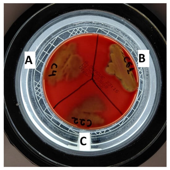
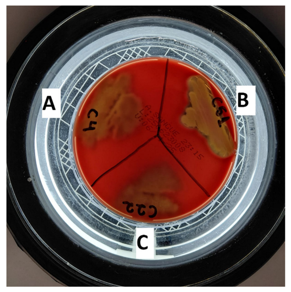
The production of hemolysins is a well-known virulence factor that facilitates bacterial invasion, iron acquisition, and evasion of the host immune system. In this study, E. albertii C4 and K. cowanii C22 exhibited α-hemolysis, characterized by the partial lysis of erythrocytes and the reduction of hemoglobin to methemoglobin. Most notably, K. cowanii C51 demonstrated strong β-hemolytic activity, characterized by a clear zone of complete erythrocyte lysis surrounding the colonies (Figure 1 and Table 2). The expression of β-hemolysins by environmental and opportunistic Enterobacteriaceae isolated from fresh produce is of significant concern, as these pore-forming toxins can cause severe tissue damage in the human gastrointestinal tract upon ingestion.
Figure 1.
Hemolytic activity of the isolated strains. (A) E. albertii C4: α-hemolysis; (B) K. cowanii C51: α-hemolysis; and (C) K. cowanii C22: β-hemolysis.
3.3.3. Presumptive Amino Acid Decarboxylase Activity
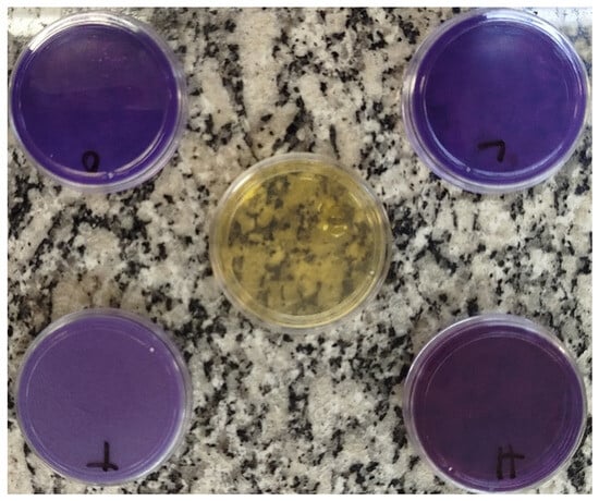
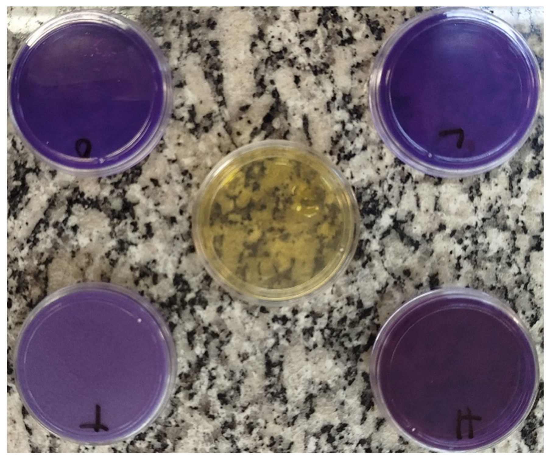
The assessment of biogenic amine production via presumptive amino acid decarboxylase activity is a crucial parameter in food safety, as the accumulation of these compounds can lead to severe toxicological effects in consumers. In the present study, all isolates (E. albertii C4, and K. cowanii C22 and C51) exhibited a highly positive decarboxylase activity for all tested precursor amino acids: histidine, tyrosine, lysine, and ornithine (Figure 2 and Table 2), a result obtained following experimental approaches recommended by Bover-Cid and Holzapfel [19]. Consequently, these strains demonstrate the intrinsic metabolic capacity and possible synthesize of histamine, tyramine, cadaverine, and putrescine, respectively.
Figure 2.
Plate assay for biogenic amine production in E. albertii C4. The purple color indicates medium alkalinization resulting from positive amino acid decarboxylation by the tested isolates.
From a public health standpoint, the simultaneous production of these four biogenic amines is a concerning issue. Histamine and tyramine are well-known triggers for foodborne intoxication and hypertensive crises, respectively [42]. This risk is further amplified by putrescine and cadaverine, which act as potent enhancers of histamine toxicity by inhibiting human intestinal diamine oxidases [43]. While the acidic nature of whole citrus fruits provides a restrictive environment, the presence of these potential pathogens becomes a severe chemical hazard if the fruits are minimally processed or used in beverages, creating conditions where heat-stable biogenic amines can rapidly accumulate and can persist in the final product [42,44]. Moreover, in the applied experimental protocol based on recommendations from Bover-Cid and Holzapfel [19], the detection of biogenic amine production by studied strains was followed by color changes in growth media as a consequence of changes in pH based on the decarboxylation of precursors (amino acids) and formation of related biogenic amines. Moreover, the production of biogenic amines needs to be evaluated not only from the point of view that strains expressing these virulence factors can produce and cumulate it in examined food matrix/s, but, furthermore, taking into consideration the fact that these strains can reach other food matrices or even gate access to human or other animals’ gut environments. The mentioned biogenic amine production assay is only a qualitative screening method. It is fact that a purple reaction in decarboxylase medium does not directly confirm the concentration of histamine, tyramine, putrescine, or cadaverine. Thus, further direct measurement by HPLC or LC–MS, preferably in a relevant citrus matrix, would be appropriate confirmation for concluding on whether mentioned strains will be in the condition to express such a negative effect on citruses.
Biogenic amines play a significant role in microbial physiology, and their control is essential when dealing with pathogenic microorganisms. These low-molecular-weight compounds, such as histamine, tyramine, putrescine, and cadaverine, are produced mainly through microbial decarboxylation of amino acids. In pathogens, excessive biogenic amine formation is undesirable because it can contribute to toxicity in food systems and result in public health outcomes. Controlling biogenic amine accumulation is therefore a key component of food safety management, requiring attention to raw material quality, microbial composition, and processing conditions. Strategies such as selecting amine-negative starter cultures, maintaining hygienic conditions, and applying preservatives or high-pressure treatments have been shown to reduce biogenic amine levels effectively [45].
Beyond their toxicological relevance, biogenic amines can also influence microbial virulence. Many bacteria use biogenic amine production as a stress-response mechanism, helping them survive in acidic environments or other hostile conditions. This adaptive advantage can indirectly enhance virulence by improving pathogen persistence and competitiveness in food matrices or host tissues. Some biogenic amines may also interact synergistically with other microbial metabolites, increasing toxicity or facilitating colonization. For example, cadaverine and putrescine can enhance histamine toxicity and may contribute to broader pathogenic effects. Understanding these mechanisms underscores why monitoring and controlling biogenic amines production is crucial not only for food quality but also for limiting the virulence potential of microbial contaminants [46].
3.3.4. Virulence Genes
Molecular screening for virulence factors revealed that the E. albertii (C4) and K. cowanii (C22 and C51) do not carry the classical genetic arsenal associated with highly invasive strains or severe avian pathogenicity. The genes eae (intimin), cva (colicin V), iss (increased serum survival), iroN, and iut (siderophore receptors), as well as the omp gene, were not detected in the evaluated isolates, although they were properly amplified in the control strains (E. coli ATCC 8739 and E. coli ATCC 35150). The absence of the eae gene in E. albertii strain is a particularly relevant finding, as it suggests that this E. albertii isolate possesses an atypical or essentially environmental profile, differentiating it from classical clinical strains responsible for attaching and effacing lesions.
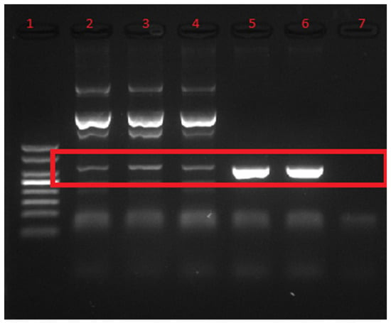
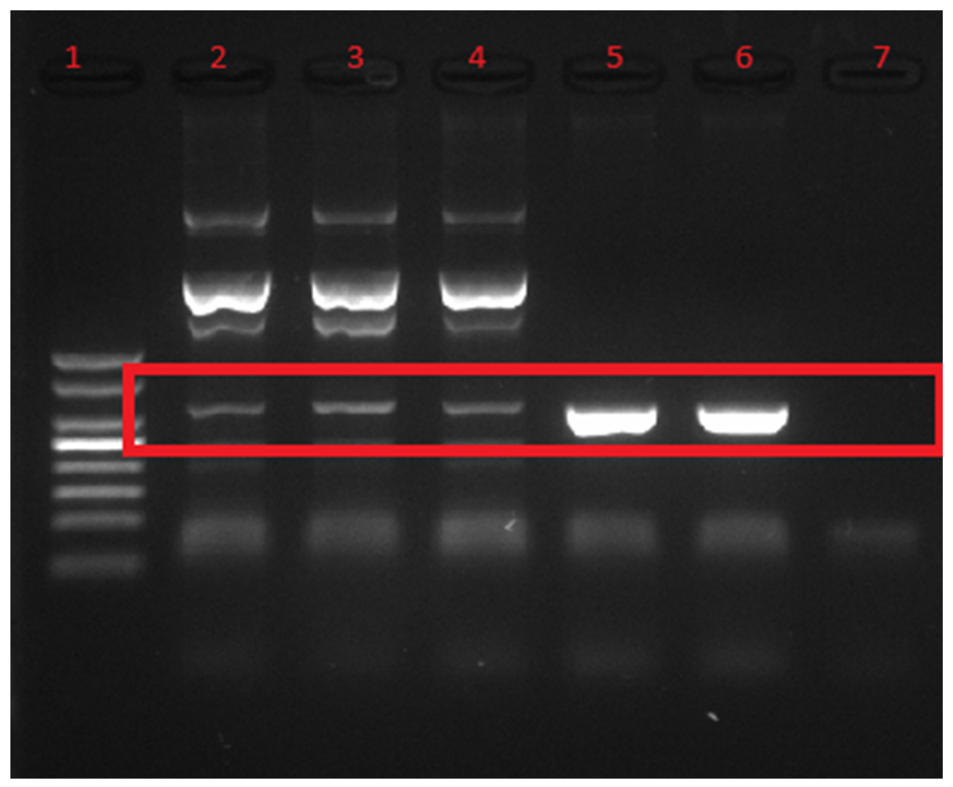
The detection of the fim gene in the isolates demonstrates the virulence of the studied strains when consumed by humans (Figure 3). The importance of fim is related to the adherence properties, and these properties merit further evaluation if they act in similar or different mechanisms for human/animal or plant environments and, furthermore, if there is potential for the persistence of these bacteria in the agricultural environment. This gene encodes type 1 fimbriae, crucial adhesion structures that enable bacterial attachment to abiotic surfaces and plant tissues, justifying the ability of these strains to colonize the crevices of Citrus × limonia and resist sanitization processes. Additionally, the amplification of a signal corresponding to the hly gene corroborates the α- or β-hemolytic activity observed phenotypically in vitro, indicating that, although lacking deep invasion genes, these microorganisms maintain opportunistic mechanisms of cytotoxicity. Moreover, crossing results from phenotypical and biomolecular expressions of virulence, such as homiletical activity, is important for the objective of obtaining better evidence for the evaluated virulence factor.
Figure 3.
Agarose gel electrophoresis (1.5%) showing the PCR amplification products of the fim virulence gene. The red rectangular box highlights the expected 331 bp amplicons. 1: 50 bp molecular weight marker; 2: K. cowanii strain C51; 3: K. cowanii strain C22; 4: E. albertii strain C4; 5 and 6: Positive control strains (E. coli ATCC 8739 and E. coli ATCC 35150); 7: negative control (PCR with water blank). Detection of fim gene by PCR can serve as the preliminary indication and further confirmation by gene sequencing will confirm obtained results.
E. albertii exhibits several virulence-associated traits, but hemolytic activity is not considered a defining or widespread characteristic of this emerging pathogen. Most studies describe E. albertii as lacking classical hemolysins typically found in other pathogenic Enterobacteriaceae. Instead, its virulence is primarily linked to attaching and effacing lesions mediated by the eae gene, the cytolethal distending toxin (cdt), and, in some strains, Shiga toxin (stx) [7].
Nevertheless, some isolates demonstrate cytotoxic effects on host cells, and limited reports suggest that certain strains may display weak or atypical hemolytic-like activity in vitro. These effects are generally attributed not to true hemolysins but to broader cellular damage caused by cdt-mediated DNA damage or by type III secretion system effectors [41].
Overall, while hemolysis is not a hallmark of E. albertii pathogenicity, the organism’s ability to damage epithelial cells, disrupt intestinal barriers, and induce inflammation functionally parallels the tissue injury outcomes observed in hemolysin-producing pathogens. Continued genomic and phenotypic surveillance is needed to determine whether hemolytic traits emerge in specific lineages or environmental contexts [25].
4. Conclusions
This study demonstrated the presence of E. albertii and K. cowanii on Rangpur limes collected from a rural production area with close proximity between fruit cultivation and poultry farming. Although the isolates did not harbor the full set of classical virulence markers investigated, they exhibited traits associated with environmental persistence and potential food safety relevance, including hemolytic activity, ampicillin resistance, reduced susceptibility to nalidixic acid, and the ability for potential production of biogenic amines. Taken together, these findings support the view that fresh fruits cultivated at animal–crop interfaces can possibly serve as reservoirs of opportunistic and emerging bacteria.
The objective of the current study was not to link the presence of E. albertii and K. cowanii to consequences of proximity to poultry farms and fruit trees, but to point out the fact that two mentioned pathogen species, previously reported in the literature as associated with poultry gut, can survive on the surface or even in the pulp of citrus fruits. The fact that we have detected these strains on citrus may be related to the proximity of poultry farms, but this is a secondary observation and merits further ecological investigations. Most important is that the evaluated pathogens mentioned can survive on citrus and, most probably, on other fruits or vegetables consumed in natura, and this can be considered as a potential risk for consumers. A pathogen that is considered strictly associated with animal gut but can be recorded on fruits and vegetables needs to be evaluated with more attention related to potential food safety.
At the same time, the results should be interpreted within the context of an exploratory study based on a limited number of isolates. Rather than indicating an immediate public health threat, these data reinforce the importance of microbiological surveillance, accurate identification of atypical isolates, and preventive practices in primary production systems. This work also expands current knowledge on the diversity of bacteria associated with citrus fruits and contributes to the broader discussion of food safety within the One Health framework.
Author Contributions
M.V.A.: Investigation, writing—original draft. G.S.S.: Investigation, writing. K.O.C.: Investigation, writing. K.L.O.d.S.S.: Investigation; E.M.F.L.: Writing—review and editing. S.D.T.: Conceptualization, supervision, project administration, writing—review and editing. All authors have read and agreed to the published version of the manuscript.
Funding
Sao Paulo Research Foundation (FAPESP) (grants 2023/05394-9; 2024/01721-8; 2024/13311-9), São Paulo, SP, Brazil; CAPES for the grants; Fundação para a Ciência e Tecnologia, Portugal for partial support to UID/05937/2025 (https://doi.org/10.54499/UID/05937/2025).
Institutional Review Board Statement
Not applicable.
Informed Consent Statement
Not Applicable.
Data Availability Statement
Data will be made available on request.
Acknowledgments
During the preparation of this manuscript, the authors used AI tools (ChatGPT (model GPT-4) and Grammarly 2.0) for improving the grammar and style of some sentences but not for generating content. After using these tools, the authors reviewed and edited the content as needed and took full responsibility for the content of the current article.
Conflicts of Interest
The authors declare no conflicts of interest.
References
- World Health Organization. Diet, Nutrition, and the Prevention of Chronic Diseases: Report of a Joint WHO/FAO Expert Consultation; WHO Technical Report Series; World Health Organization: Geneva, Switzerland, 2003; Volume 916. [Google Scholar]
- Septembre-Malaterre, A.; Remize, F.; Poucheret, P. Fruits and vegetables, as a source of nutritional compounds and phytochemicals: Changes in bioactive compounds during lactic fermentation. Food Res. Int. 2018, 104, 86–99. [Google Scholar] [CrossRef] [Scilit]
- Berger, C.N.; Sodha, V.S.; Shaw, R.K.; Teunis, P.F.; Frankel, G. Fresh fruit and vegetables as vehicles for the transmission of human pathogens. Environ. Microbiol. 2010, 12, 2385–2397. [Google Scholar] [CrossRef] [Scilit]
- Melo, J.; Quintas, C. Minimally processed fruits as vehicles for foodborne pathogens. AIMS Microbiol. 2023, 9, 1–19. [Google Scholar] [CrossRef] [Scilit]
- Steele, M.; Odumeru, J. Irrigation water as source of foodborne pathogens on fruit and vegetables. J. Food Prot. 2004, 67, 2839–2849. [Google Scholar] [CrossRef] [Scilit]
- Beuchat, L.R. Ecological factors influencing survival and growth of human pathogens on raw fruits and vegetables. Microbes Infect. 2002, 4, 413–423. [Google Scholar] [CrossRef] [Scilit]
- Muchaamba, F.; Barmettler, K.; Treier, A.; Stephan, R. Microbiology and Epidemiology of Escherichia albertii—An Emerging Elusive Foodborne Pathogen. Microorganisms 2022, 10, 875. [Google Scholar] [CrossRef] [Scilit]
- Teymurazov, M.G.; Kartsev, N.N.; Abaimova, A.A.; Ershova, O.N.; Sinyugina, A.A.; Volozhantsev, N.V.; Kislichkina, A.A.; Fursova, N.K. Cases of Isolation of Escherichia albertii Strains from Commercial Quails with Gastroenteritis in Russia. Microorganisms 2025, 13, 816. [Google Scholar] [CrossRef] [Scilit]
- Ahmed, M.; Hossain, I.; Hossain, D. Emerging pathogen alert: Global epidemiology of zoonotic potential of Escherichia albertii: A systematic review and meta-analysis. Int. J. Food Microbiol. 2026, 447, 111564. [Google Scholar] [CrossRef] [Scilit]
- Zhang, Y.; Wang, B.; Li, Q.; Huang, D.; Zhang, Y.; Li, G.; He, H. Isolation and Complete Genome Sequence Analysis of Kosakonia cowanii Pa82, a Novel Pathogen Causing Bacterial Wilt on Patchouli. Front. Microbiol. 2022, 12, 818228. [Google Scholar] [CrossRef] [Scilit]
- Krawczyk, K.; Borodynko-Filas, N. Kosakonia cowanii as the New Bacterial Pathogen Affecting Soybean (Glycine max Willd.). Eur. J. Plant Pathol. 2020, 157, 173–183. [Google Scholar] [CrossRef] [Scilit]
- Berinson, B.; Bellon, E.; Christner, M.; Both, A.; Aepfelbacher, M.; Rohde, H. Identification of Kosakonia cowanii as a rare cause of acute cholecystitis: Case report and review of the literature. BMC Infect. Dis. 2020, 20, 366. [Google Scholar] [CrossRef] [Scilit]
- Merlino, J.; Pillay, K.; Rizzo, S.; Baskar, S.R.; Seed, D.; Siarakas, S.; Hettiarachchi, R.; McKew, G.; Gray, T. Bacterial skin infection caused by a plant pathogen Kosakonia cowanii: Identification with the MALDI Biotyper sirius one and susceptibility testing. Access Microbiol. 2025, 7, 000923.v3. [Google Scholar] [CrossRef] [Scilit]
- Li, W.; Zhao, X.; Zou, S.; Ma, Y.; Zhang, K.; Zhang, M. Scanning assay of β-galactosidase activity. Appl. Biochem. Microbiol. 2012, 48, 603–607. [Google Scholar] [CrossRef] [Scilit]
- Choi, G.H.; Holzapfel, W.H.; Todorov, S.D. Diversity of the bacteriocins, their classification and potential applications in combat of antibiotic resistant and clinically relevant pathogens. Crit. Rev. Microbiol. 2023, 49, 578–597. [Google Scholar] [CrossRef] [Scilit]
- Felske, A.; Wolterink, A.; Van Lis, R.; Akkermans, A.D. Phylogeny of the main bacterial 16S rRNA sequences in Drentse A grassland soils (The Netherlands). Appl. Environ. Microbiol. 1998, 64, 871–879. [Google Scholar] [CrossRef] [Scilit]
- European Food Safety Authority (EFSA) Panel on Additives and Products or Substances used in Animal Feed (FEEDAP); Rychen, G.; Aquilina, G.; Azimonti, G.; Bampidis, V.; de Lourdes Bastos, M.; Bories, G.; Chesson, A.; Cocconcelli, P.S.; Flachowsky, G.; et al. Guidance on the characterisation of microorganisms used as feed additives or as production organisms. EFSA J. 2018, 16, e05206. [Google Scholar] [CrossRef] [Scilit]
- Colombo, M.; Nero, L.A.; Todorov, S.D. Safety profiles of beneficial lactic acid bacteria isolated from dairy systems. Braz. J. Microbiol. 2020, 51, 787–795. [Google Scholar] [CrossRef] [Scilit]
- Bover-Cid, S.; Holzapfel, W.H. Improved screening procedure for biogenic amine production by lactic acid bacteria. Int. J. Food Microbiol. 1999, 53, 33–41. [Google Scholar] [CrossRef] [Scilit]
- Strawn, L.K.; Schneider, K.R.; Danyluk, M.D. Microbial safety of tropical fruits. Crit. Rev. Food Sci. Nutr. 2011, 51, 132–145. [Google Scholar] [CrossRef] [Scilit]
- Bearson, S.; Bearson, B.; Foster, J.W. Acid stress responses in enterobacteria. FEMS Microbiol. Lett. 1997, 147, 173–180. [Google Scholar] [CrossRef] [PubMed]
- Brameyer, S.; Schumacher, K.; Kuppermann, S.; Jung, K. Division of labor and collective functionality in Escherichia coli under acid stress. Commun. Biol. 2022, 5, 327. [Google Scholar] [CrossRef] [Scilit]
- Capozzi, V.; Fiocco, D.; Amodio, M.L.; Gallone, A.; Spano, G. Bacterial stressors in minimally processed food. Int. J. Mol. Sci. 2009, 10, 3076–3105. [Google Scholar] [CrossRef] [Scilit]
- Brandal, L.T.; Tunsjø, H.S.; Ranheim, T.E.; Løbersli, I.; Lange, H.; Wester, A.L. Shiga toxin 2a in Escherichia albertii. J. Clin. Microbiol. 2015, 53, 1454–1455. [Google Scholar] [CrossRef] [Scilit]
- Gomes, T.A.T.; Ooka, T.; Hernandes, R.T.; Yamamoto, D.; Hayashi, T. Escherichia albertii Pathogenesis. EcoSal Plus 2020, 9, 10–1128. [Google Scholar] [CrossRef] [Scilit]
- Ooka, T.; Seto, K.; Kawano, K.; Kobayashi, H.; Etoh, Y.; Ichihara, S.; Kaneko, A.; Isobe, J.; Yamaguchi, K.; Horikawa, K.; et al. Clinical Significance of Escherichia albertii. Emerg. Infect. Dis. 2012, 18, 488–492. [Google Scholar] [CrossRef] [Scilit]
- Brady, C.; Cleenwerck, I.; Venter, S.; Coutinho, T.; De Vos, P. Taxonomic Evaluation of the Genus Enterobacter Based on multilocus Sequence Analysis (MLSA): Proposal to Reclassify E. nimipressuralis and E. amnigenus into Lelliottia gen. nov. as Lelliottia nimipressuralis comb. nov. and Lelliottia amnigena comb. nov., Respectively, E. gergoviae and E. pyrinus into Pluralibacter gen. nov. as Pluralibacter gergoviae comb. nov. and Pluralibacter pyrinus comb. nov., respectively, E. cowanii, E. radicincitans, E. oryzae and E. arachidis into Kosakonia gen. nov. as Kosakonia cowanii comb. nov., Kosakonia radicincitans comb. nov., Kosakonia oryzae comb. nov. and Kosakonia arachidis comb. nov., respectively, and E. turicensis, E. helveticus and E. pulveris into Cronobacter as Cronobacter zurichensis nom. nov., Cronobacter helveticus comb. nov. and Cronobacter pulveris comb. nov., Respectively, and Emended Description of the Genera Enterobacter and Cronobacter. Syst. Appl. Microbiol. 2013, 36, 309–319. [Google Scholar] [CrossRef] [Scilit]
- Carrión, V.J.; Perez-Jaramillo, J.; Cordovez, V.; Tracanna, V.; de Hollander, M.; Ruiz-Buck, D.; Mendes, L.W.; van Wezel, G.P.; Raaijmakers, J.M. Pathopatho-genomics of Kosakonia species causing bacterial wilt and soft rot. Phytopathol. 2018, 108, 1430–1441. [Google Scholar]
- Inoue, K.; Sugiyama, K.; Kosako, Y.; Sakazaki, R.; Tamura, K. Enterobacter cowanii sp. nov., a new species isolated from clinical specimens. Curr. Microbiol. 2020, 41, 417–420. [Google Scholar] [CrossRef] [Scilit]
- Leclerc, H.; Mossel, D.A.A.; Edberg, S.C.; Struijk, C.B. Advances in the Bacteriology of the Coliform Group: Their Suitability as Markers of Microbial Water Safety. Annu. Rev. Microbiol. 2001, 55, 201–234. [Google Scholar] [CrossRef] [Scilit]
- Vorholt, J.A. Microbial life in the phyllosphere. Nat. Rev. Microbiol. 2012, 10, 828–840. [Google Scholar] [CrossRef] [Scilit]
- De Mandal, S.; Jeon, J. Phyllosphere microbiome in plant health and disease. Plants 2023, 12, 3481. [Google Scholar] [CrossRef] [Scilit]
- Arzanlou, M.; Chai, W.C.; Venter, H. Intrinsic, adaptive and acquired antimicrobial resistance in Gram-negative bacteria. Essays Biochem. 2017, 61, 49–59. [Google Scholar] [CrossRef] [Scilit]
- Moxley, R.A. Family Yersiniaceae. In Veterinary Microbiology, 4th ed.; Scott, D., Kennedy, M., Chengappa, M.M., Wilkes, R., Eds.; Wiley: New York, NY, USA, 2022; pp. 88–99. [Google Scholar] [CrossRef] [Scilit]
- Fournier, C.; Nordmann, P.; de la Rosa, J.M.O.; Kusaksizoglu, A.; Poirel, L. KSA-1, a naturally occurring Ambler class A extended spectrum β-lactamase from the enterobacterial species Kosakonia sacchari. J. Glob. Antimicrob. Resist. 2024, 39, 6–11. [Google Scholar] [CrossRef] [Scilit]
- Hassan, J.; Utsho, K.S.; Karmakar, S.; Ali, W.; Awasthi, S.P.; Uyama, C.; Hatanaka, N.; Yamasaki, S.; Hinenoya, A. Occurrence and cross contamination of Escherichia albertii in retail chicken outlets in Bangladesh. Int. J. Food Microbiol. 2025, 431, 111081. [Google Scholar] [CrossRef] [Scilit]
- Li, Q.; Wang, H.; Xu, Y.; Zhang, C.; Liu, J.; Zhao, Z.; Jiang, S. Multidrug-resistant Escherichia albertii: Co-occurrence of β-lactamase and MCR-1 encoding genes. Front. Microbiol. 2018, 9, 258. [Google Scholar] [CrossRef] [Scilit]
- Hooper, D.C.; Jacoby, G.A. Mechanisms of drug resistance: Quinolone resistance. Ann. N. Y. Acad. Sci. 2015, 1354, 12–31. [Google Scholar] [CrossRef] [Scilit]
- Hricová, K.; Röderová, M.; Pudová, V.; Hanulík, V.; Halová, D.; Papoušková, A.; Bardoň, J. Quinolone-resistant Escherichia coli in Poultry Farming. Cent. Eur. J. Public Health 2017, 25, 163–167. [Google Scholar] [CrossRef] [Scilit]
- Alegbeleye, O.O.; Singleton, I.; Sant’Ana, A.S. Sources and contamination routes of microbial pathogens to fresh produce during field cultivation: A review. Food Microbiol. 2018, 73, 177–208. [Google Scholar] [CrossRef] [Scilit]
- Lima, M.P.; Yamamoto, D.; Santos, A.C.M.; Ooka, T.; Hayashi, T.; Gomes, T.A.T. Phenotypic characterization and virulence-related properties of Escherichia albertii strains isolated from children with diarrhea in Brazil. Pathog. Dis. 2019, 77, ftz014. [Google Scholar] [CrossRef] [Scilit]
- Ruiz-Capillas, C.; Herrero, A.M. Impact of biogenic amines on food quality and safety. Foods 2019, 8, 62. [Google Scholar] [CrossRef] [Scilit]
- Doeun, D.; Davaatseren, M.; Chung, M.S. Biogenic amines in foods. Food Sci. Biotechnol. 2017, 26, 1463–1474. [Google Scholar] [CrossRef] [Scilit]
- Feddern, V.; Mazzuco, H.; Fonseca, F.N.; de Lima, G.J.M.M. A review on biogenic amines in food and feed: Toxicological aspects, impact on health and control measures. Anim. Prod. Sci. 2019, 59, 608–618. [Google Scholar] [CrossRef] [Scilit]
- Saha Turna, N.; Chung, R.; McIntyre, L. A review of biogenic amines in fermented foods: Occurrence and health effects. Heliyon 2024, 10, e24501. [Google Scholar] [CrossRef] [Scilit]
- Tabanelli, G. Biogenic Amines and Food Quality: Emerging Challenges and Public Health Concerns. Foods 2020, 9, 859. [Google Scholar] [CrossRef] [Scilit]

Disclaimer/Publisher’s Note: The statements, opinions and data contained in all publications are solely those of the individual author(s) and contributor(s) and not of MDPI and/or the editor(s). MDPI and/or the editor(s) disclaim responsibility for any injury to people or property resulting from any ideas, methods, instructions or products referred to in the content. |
© 2026 by the authors. Licensee MDPI, Basel, Switzerland. This article is an open access article distributed under the terms and conditions of the Creative Commons Attribution (CC BY) license.