


Abstract
Pulmonary arterial hypertension (PAH) is an irreversible disease characterized by vascular and systemic inflammation, ultimately leading to right ventricular failure. There is a great need for adjunctive therapies to extend survival for PAH patients. The gut microbiome influences the host immune system and is a potential novel target for PAH treatment. We review the emerging preclinical and clinical evidence which strongly suggests that there is gut dysbiosis in PAH and that alterations in the gut microbiome may either initiate or facilitate the progression of PAH by modifying systemic immune responses. We also outline approaches to modify the intestinal microbiome and delineate some practical challenges that may impact efforts to translate preclinical microbiome findings to PAH patients. Finally, we briefly describe studies that demonstrate contributions of infections to PAH pathogenesis. We hope that this review will propel further investigations into the mechanisms by which gut dysbiosis impacts PAH and/or right ventricular function, approaches to modify the gut microbiome, and the impact of infections on PAH development or progression.
1. Introduction
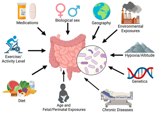
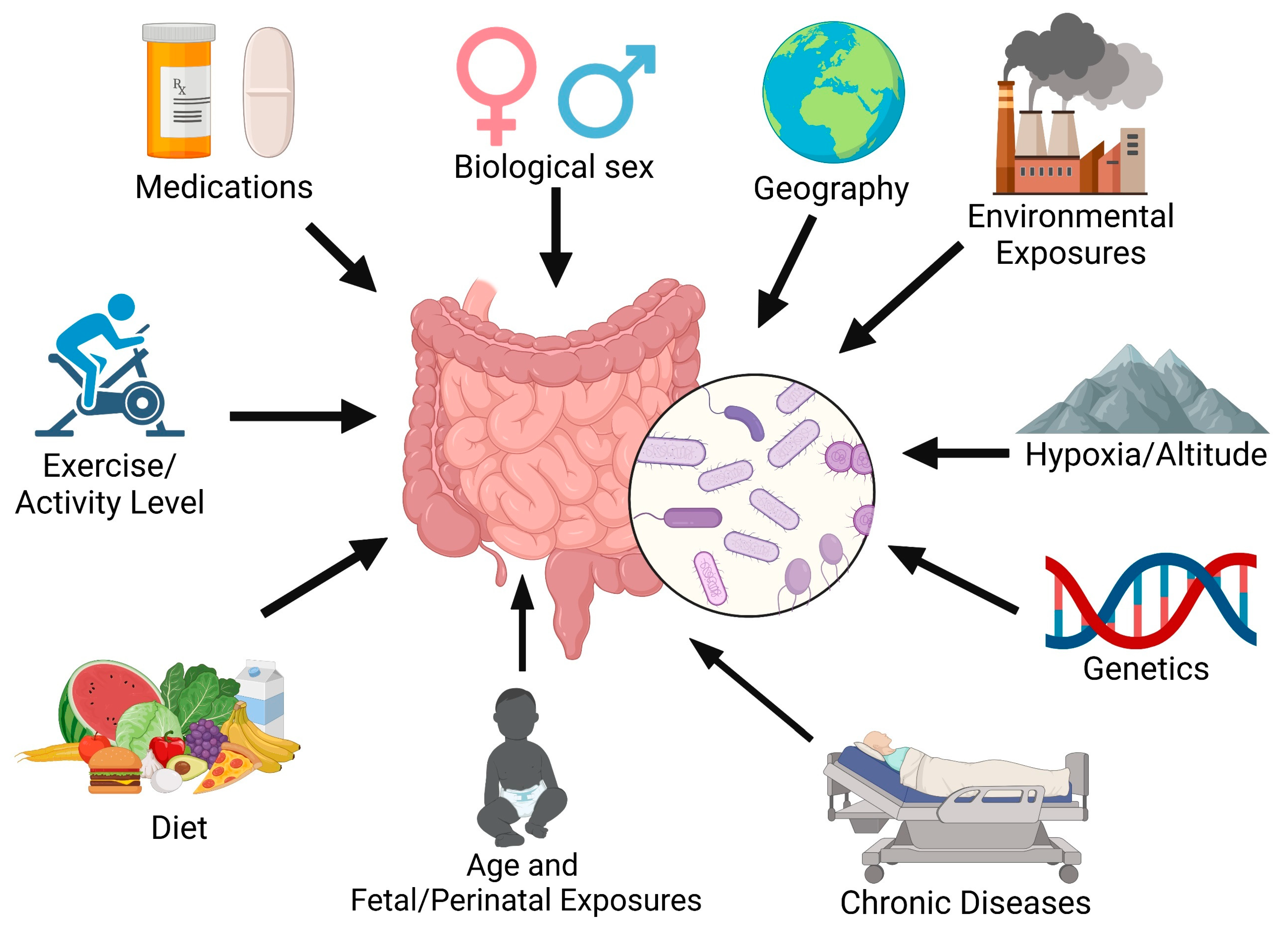
Vascular injury and inflammation are key drivers of pulmonary vascular remodeling in pulmonary arterial hypertension (PAH) [1,2,3,4,5]. Primarily known as a cardiopulmonary disease, growing evidence has implicated interorgan communication in PAH pathogenesis. We and others have observed the potential role of the intestinal microbiome in the onset and progression of PAH. The gastrointestinal system contributes to systemic inflammation as it contains about 70–80% of the body’s immune cells [6,7], along with trillions of microorganisms known as the gut microbiome [8]. The composition of the gut microbiome can be influenced by many factors, including environmental pollutants, medications, nutrient availability/diet, exercise, oxygen level, sex, age, and genetics (Figure 1) [9]. There is now evidence of the gut microbiome’s impact on many chronic diseases [10]. Clinical trials of targeted anti-inflammatory drugs to date have not clearly demonstrated benefits in PAH [11,12,13], partially due to enrollment size, trial design, and patient selection. Modulating a key immune organ, such as the intestine, to alter many inflammatory pathways may be an effective adjunctive approach to treating PAH.
Figure 1.
Factors influencing intestinal microbiome composition.
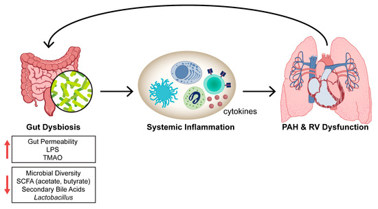
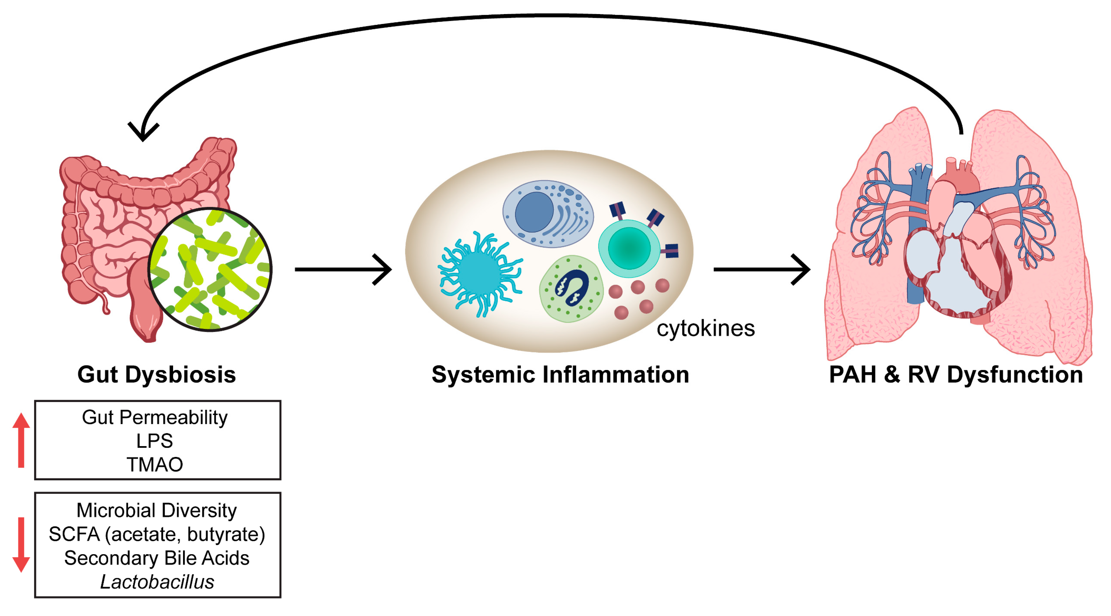
This review describes the experimental (Table 1) and clinical evidence (Table 2) which implicates the gut microbiota as key contributors to PAH pathogenesis. Additionally, we provide a brief overview of the effect of infections on PAH development and the gut microbiome. While there are five broad World Health Organization (WHO) groups of pulmonary hypertension (PH), which are defined by a mean pulmonary artery pressure > 20 mmHg [14], we primarily focus on WHO Group 1 PAH and Group 3 PH (PH secondary to lung disease) in this review. The current overarching hypothesis is that gut dysbiosis leads to increased systemic inflammation and the progression of PAH and/or right ventricular (RV) dysfunction (Figure 2).
Figure 2.
Working hypothesis linking gut dysbiosis to PAH and/or RV dysfunction. LPS: lipopolysaccharide; PAH: pulmonary arterial hypertension; RV: right ventricle; SCFA: short-chain fatty acids; TMAO: trimethylamine N-oxide.
2. Preclinical Studies of the Gut–Lung Axis in Pulmonary Hypertension
Multiple studies demonstrate that PH rodent models have alterations in the gut microbiome commonly referred to as gut dysbiosis [15,16,17,18,19,20,21,22,23] (Table 1). Monocrotaline (MCT) PAH rats have increased intestinal permeability as measured by fluorescein isothiocyanate (FITC)-dextran, lipopolysaccharide (LPS), and soluble CD14 in the blood [24] and disrupted intestinal morphology, with greater muscularis layer thickness and fibrosis and diminished villus length and goblet cell number [21,25]. Sugen hypoxia (SuHx) rats have an elevated fecal Firmicutes-to-Bacteroidetes ratio and fewer short-chain fatty acid (SCFA)-producing bacteria, such as those that are acetate- and butyrate-producing, with no change in lactate-producing bacteria [15]. The milder hypoxic rat PH model also has altered and distinct gut microbiota with more arginine and arginine-producing bacteria, Blautia and Bifidobacterium, and the trimethylamine N-oxide (TMAO) biosynthetic bacteria, Streptococcus [19]. Hypoxic mice have disrupted gut microbiome composition with increases in the genera Prevotella, Oscillospira, and Ruminococcus and decreases in Lactobacillus [26]. Alterations in the gut microbiome also occur in the large animal, bovine brisket disease PH model, with lower total volatile fatty acids and alpha diversity (richness and evenness of bacteria) in rumen fluid [27]. The specific intestinal microbiome differences observed in various PH rodent models are comprehensively reviewed elsewhere [28].
Several preclinical studies have modified the gut microbiome to assess its effects on PAH. Altering the microbiome with a broad-spectrum antibiotic cocktail (ampicillin, vancomycin, neomycin, and metronidazole) prior to SU5416 administration mitigates PAH in SuHx rats [29]. Additionally, diet impacts PAH pathogenesis and severity. For example, apolipoprotein E (ApoE) knockout mice given a Paigen (high fat, high cholesterol) diet develop PAH [30] compared to ApoE knockout mice fed normal chow. Double ApoE/IL1R1 knockout mice consuming a Paigen diet have even worse PAH, implicating the role of IL-1 in PAH pathogenesis [30]. Western diet increases right ventricular systolic pressure (RVSP) and RV myocardial lipid deposition and reduces RV function in mice [31]. Metformin decreases RVSP and RV lipid and ceramide accumulation [31]. Moreover, a high-soluble-fiber diet attenuates hypoxia-induced pulmonary vascular remodeling by increasing the abundance of SCFA-producing bacteria (Bacteroides, Anaerostripes, and Anaerocolumna), diminishing proinflammatory bacteria (Romboutsia, Mammalicoccus, Staphylococcus, Clostridioides, and Streptococcus), and reducing lung interstitial macrophages, dendritic cells, and nonclassical monocytes [32]. The serum metabolites, phosphatidylcholines, lysophosphatidylcholines, ceramides, and hexosylceramides, are lower, while propionylcarnitine and probetaine are greater, in high-soluble-fiber-fed mice compared to low-soluble-fiber-fed hypoxic mice [32]. Interestingly, treatment with a phosphodiesterase-5 inhibitor (tadalafil) or an endothelin pathway inhibitor (macitentan) in SuHx rats reduces the plasma levels of many phosphatidylcholines [33]. However, not all suspected advantageous dietary interventions have been effective in PAH. Although intermittent fasting augments RV function and extends survival in MCT rats, there is minimal effect on PAH severity [25], suggesting that more targeted dietary interventions may be needed to alter PAH severity.
One of the primary ways that the gut microbiota interact with their host is via metabolites, which are intermediate or end products of microbial metabolism [34]. The gut metabolites are generated from bacterial metabolism of dietary or other host substrates. Supplementation with the SCFA, butyrate, which is an endogenous histone deacetylase (HDAC) inhibitor, attenuates pulmonary vascular remodeling and accumulation of alveolar (CD68+) and interstitial (CD68+ and CD163+) lung macrophages in hypoxic PH [35]. Butyrate’s positive effects on pulmonary vascular remodeling have been corroborated in other unpublished studies [36,37] that suggest that butyrate also regulates endothelial cell inflammatory activation and migration. However, the improvement in PH with butyrate treatment appears to only be seen in prevention models but not when given after two and four weeks of hypoxic exposure [35].
Trimethylamine N-oxide (TMAO), derived from trimethylamine (TMA), is a bacterial metabolite generated by the breakdown of dietary choline, carnitine, and betaine. It is elevated in intermediate- and high-risk idiopathic PAH (IPAH) patients [38]. Administering TMAO to hypoxic mice worsens PH through macrophage secretion [38]. Treating hypoxic and MCT rodents with a structural analog of choline that inhibits TMAO synthesis, 3,3-dimethyl-1-butanol (DMB), reduces RV systolic pressure and pulmonary vascular thickness/muscularization and suppresses cytokine and chemokine signaling, with the strongest associations with Cxcl6 and Il6 [38,39]. While TMAO worsens PAH, long-term TMAO treatment of MCT rats may be beneficial to RV function by preserving fatty acid oxidation and decreasing pyruvate metabolism, thus preserving mitochondrial energy metabolism and mitigating the development of RV dysfunction in PAH [40]. The role of other microbial metabolites [34,40,41,42], such as amino acid metabolites and retinoic and bile acids in PAH and RV failure is not well-defined.
There are few publications investigating the outcomes of direct manipulation of the microbiome or supplementation of specific bacterial genera/species in PAH. Fecal transplantation from angiotensin-converting enzyme 2 (Ace2)-overexpressing mice, which have less hypoxia-induced PH, to wild-type hypoxic mice attenuates PH development [26,43]. Administration of Lactobacillus reuteri to postnatal growth-restricted pups exposed to hyperoxia mitigates PH severity and RV hypertrophy [44]. Lactobacillus rhamnosus supplementation two weeks after MCT injection does not affect pulmonary vascular remodeling but enhances RV function [45].
Other approaches to modulate the microbiome involve human umbilical cord blood-derived mesenchymal stem cells (MSCs), which are nonhematopoietic cells that can self-renew and secrete antibacterial peptides [46]. There is recent interest in their ability to regulate the gut microbiome and alleviate inflammatory bowel diseases [47,48,49]. Treatment with MSCs rebalances the gut microbiome (reducing the disease-associated and increasing the anti-inflammatory bacteria) and attenuates hypoxia- [17] and MCT-induced PH [50,51]. Most of the animal studies modifying the gut microbiota in PH thus far use prevention models, intervening prior to or immediately after providing a stimulus to generate PH. Promising indirect evidence supports a need to further explore the role of the gut microbiome in PH. In SuHx mice, treatment with Ang1-7 four weeks after hypoxia initiation mitigates PH, partially attenuates disease-associated changes in gut microbiota, and enhances the beneficial metabolites, butyric acid and tryptophan [52]. Administering irbesartan, an angiotensin II receptor blocker, 30 days after starting exposure to hypoxia, mitigates PH, partially normalizes the Firmicutes-to-Bacteroidetes ratio, increases the intestinal abundance of Lactobacillaceae and Lachnospiraceae, and decreases Prevotellaceae and Desulfovibrionaceae in high-altitude PH hypobaric hypoxia rats [22]. Future studies need to evaluate the efficacy of altering the gut microbiome after PAH is established. Due to the many potential environmental confounders in clinical microbiome research, animal studies are needed to establish a robust foundation for biotherapeutics targeting the host microbiome composition and its systemic effects on respiratory health. Lastly, the potential gut–brain–lung axis in PAH [21,26,43] should be further explored.
Table 1.
Summary of preclinical studies linking the gut microbiome to pulmonary hypertension.
3. Clinical Evidence of the Gut–Lung Axis in PAH
Clinical studies show that PAH patients have gut dysbiosis and a leaky gut with increased bacterial translocation from the intestinal lumen to systemic circulation [24] (Table 2). Kim et al. [54] completed one of the initial clinical studies describing the distinct gut microbiome composition in PAH by studying the fecal microbiome of 18 PAH patients and 12 age- and sex-matched healthy controls. Compared to controls, PAH patients have a distinct microbiome composition with lower alpha diversity, fewer bacteria associated with polysaccharide fermentation and SCFA production (Butyrivibrio crossotus, Bacteroides cellulosilyticus, Eubacterium siraeum, Bacteroides vulgatus, Akkermansia muciniphila), and more bacteria associated with the proinflammatory metabolites, TMA and TMAO [54]. PAH patients also have a disparate intestinal virome [54].
In a subsequent single-center pilot study of 20 PAH patients and 20 healthy controls cohabiting with PAH patients (20 matched pairs), Jose et al. [55] observed no difference in alpha- (within a specific sample) or beta- diversity (between samples). In the largest PAH microbiota study to date of 72 patients, Moutsoglou et al. [56] demonstrated that the gut microbiome is less diverse in PAH patients compared to healthy controls and family members residing in the same household. Gut microbiome diversity correlates with measures of pulmonary vascular disease (mean pulmonary artery pressure, pulmonary vascular resistance, and pulmonary arterial compliance), but not with RV function [56], suggesting that the alterations in the gut microbiome are not due to RV failure and intestinal congestion. PAH patients have reduced abundance of gut bacteria containing genes encoding for the production of anti-inflammatory metabolites, specifically SCFAs (Eubacterium ramulus, Firmicutes sp. coabundance gene 110, Coprococcus comes, Dorea longicatena, Bifidobacterium adolescentis, Gemmiger formicilis, Fusicatenibacter saccharivorans, Eubacterium hallii, Anaerostipes hadrus, Gordonibacter pamelaeae, Ruminococcus torques, Coprococcus catus, Coprococcus eutactus, and Blautia obeum) and secondary bile acids (Collinsella aerofaciens, Coprococcus eutactus, Anaerostipes hadrus, Eubacterium ramulus, Blautia obeum, Eubacterium hallii, Ruminococcus bicirculans, Ruminococcus torques, Eubacterium eligens, Fusicatenibacter saccharivorans, Roseburia faecis, Dorea longicatena, Coprococcus catus, and Roseburia hominis) and increased relative abundance of bacteria with genes encoding for the production of TMAO (Clostridium bolteae, Escherichia coli, and Klebsiella pneumoniae) [56]. Consistent with the above gut microbiome changes, PAH patients have relatively lower circulating levels of anti-inflammatory microbial metabolites (SCFA and bile acids) and a trend towards higher levels of the proinflammatory microbial metabolite TMAO. In a study of 35 patients with IPAH [38] and another of 124 PAH patients [39], TMAO levels are elevated in higher-risk patients, suggesting that TMAO is associated with worse outcomes. The role of the other intestinal microorganisms (fungi, protozoans, and archaea) in clinical PAH is not well-established.
Environmental effects, such as high altitude and hypoxia, may also alter the gut microbiome. A small study of six highlander PH patients living on the Tibetan plateau range and seven lowlander PH patients (residents of Shanghai) demonstrated that while there are overall divergent gut microbial signatures between PH patients and controls, altitude contributes to the gut microbiota differences [57]. TMA synthesis enzymes are enriched in lowlanders with PH, and there is no difference in TMA-producing microbiota between highlander controls and highlander PH patients [57].
Whether there is a causal relationship between the gut microbiome/metabolites and PAH is not yet determined in clinical studies. Mendelian randomization of data from the MiBioGen consortium, the largest genome-wide meta-analysis of intestinal microbiota [58], investigated the potential direct link between gut microbiota, metabolites, diet, and PAH [59]. The bacteria, Alistipes and Victivallis, correlate with increased PAH risk, while Coprobacter, Erysipelotrichaeae, Lachnospiraceae, and Ruminococcaceae protect against PAH [59]. However, SCFAs, TMAO, and dietary patterns are not causally associated with PAH in a Mendelian randomization analysis [59]. In another recent Mendelian randomization study, Su et al. identified 11 gut microbial taxa, including Bifidobacteriaceae, Eubacterium eligens group, and Sutterella, and 24 bacterial metabolites that are linked to PAH pathogenesis by regulating the expression of ITPR2, IDE, NRIP1, and IGF1 genes in lung tissue [60]. Limitations of these studies are that the design only evaluates the impact of genetic factors on intestinal microbiota abundance and the development of PAH, a small effect size with the use of single-nucleotide polymorphisms (SNPs) for metabolites and bacteria, Mendelian randomization being prone to false positives, and cohorts primarily of European ethnicity, restricting the generalizability of the findings. Thus, more clinical studies are needed to determine whether there is a casual connection between the intestinal microbiota/metabolites and PAH.
There are now emerging data showing that not only is the gut microbiome changed in PAH but also the airway microbiome composition may be distinct. In a study of PH patients of various etiologies (Group 1 PAH, PH due to lung disease, and chronic thromboembolic PH), Zhang et al. [61] observed higher alpha-diversity and increased Streptococcus, Lautropia, and Ralstonia in the airways of PH patients compared to reference controls. Intratracheal instillation of Streptococcus induced PH in rats [62]. A recent study identified disparate airway mycobiomes or fungal compositions between PH and healthy controls [63]. Further research should assess the microbiome/mycobiome in other parts of the body.
Table 2.
Summary of the clinical studies delineating the role of the gut microbiome in PAH.
4. Potential Approaches to Modulate the Gut–Lung Axis to Treat PAH
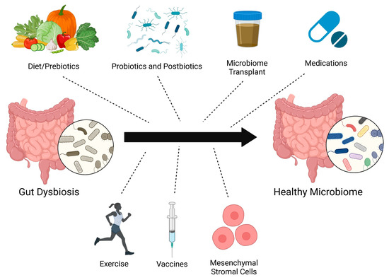
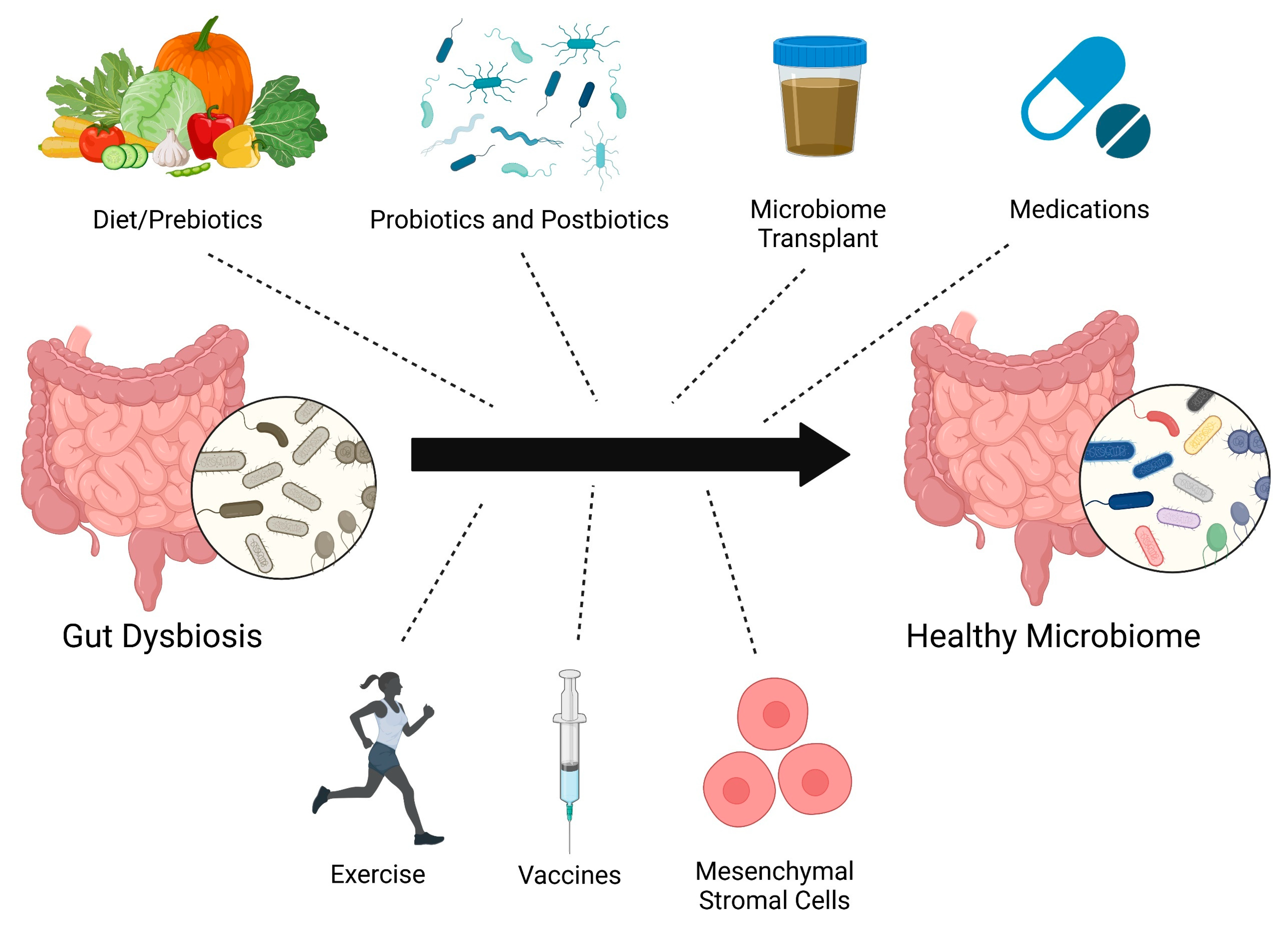
There are several different strategies to restructure the microbiome, including diet/prebiotics, probiotics, postbiotics [64] (inanimate microorganisms or their components), microbiota transplant, medications, vaccines [65], exercise [66], and mesenchymal stromal cell therapy [51,67] (Figure 3). There have not yet been many clinical trials investigating how altering the gut microbiome affects PAH. There is an ongoing study evaluating the impact of microbiota transplant from healthy controls to PAH patients [68]. Unfortunately, the SARS-CoV-2 pandemic impeded the enrollment and initiation of the trial assessing the use of chlorhexidine mouthwash and oral nitrate therapy in PH patients (NCT03787082). Challenges to translating preclinical findings to PAH patients include genetic factors, age, sex, concomitant chronic diseases, and different environmental exposures [69] (medications, diet, timing of eating, toxin use, chemicals in the environment/products, hygiene, air pollution, daylight exposure, etc.). Fetal/maternal or perinatal microbiota exposures may also complicate the efficacy of microbiota clinical trials. For example, supplementing omega-3 polyunsaturated fatty acids in the diet of pregnant rats improves RV systolic pressure and survival in pups exposed to hyperoxia at the time of birth [70]. Additionally, the timing and duration of gut microbiome modulation needed to confer benefits in PAH are unknown. Despite these possible confounders, the vast potential health benefits of altering the microbiome in PAH should be explored to a greater extent.
Figure 3.
Potential therapeutic approaches to restructure the gut microbiome.
5. Contribution of Infections in PAH Pathogenesis
Infectious agents, including bacteria, viruses, fungi, and parasites, can cause pulmonary arterial injury and inflammation by impacting vascular cells, leading to severe pulmonary vascular remodeling and PAH [71]. The pathophysiology and mechanisms by which infections lead to pulmonary vascular disease are more extensively reviewed elsewhere [72]. Infections in the setting of a dysregulated immune system may elevate susceptibility to PH development [73,74]. There is now growing interest in determining how host microbiota promote or resist infections [75].
A common global cause of PAH is infection by the intravascular parasite, Schistosoma mansoni. Schistosomiasis affects over 200 million people with about 1–10 million chronically infected people at risk of developing PAH [76,77]. Schistosomiasis cases occur in many regions worldwide, but the majority are observed in Africa and Asia, although longstanding epidemiological and socioeconomic challenges may underestimate its global impact. Schistosomiasis disrupts both gut and lung microbiota [53]. S. mansoni egg exposure decreases lung alpha diversity, mainly by impacting the relative abundance of the phylum, Ascomycota, while the pulmonary Firmicutes-to-Bacteroidetes ratio remains unchanged [53]. In contrast, Schistosomiasis increases gut microbiota alpha diversity and the Firmicutes-to-Bacteroidetes ratio [53], suggesting significant differences in lung and gut microbiome responses to Schistosomiasis infection.
HIV infection is also a well-recognized cause of PAH [78]. Host factors and geography contribute to the fecal microbiota disruptions that occur after HIV infection [79]. Geographic location has a greater effect on fecal microbiota composition than HIV infection status [79]. Interestingly, while HIV infection is known to disrupt gut epithelial barrier function, there are regional differences in immune activation with elevated soluble CD14 levels in HIV-infected individuals from all three regions studied (United States, Botswana, and Uganda), but there is increased intestinal fatty acid-binding protein in HIV-infected individuals from only the United States and Botswana [79]. Thus, distinct gut microbial alterations due to the host region are major confounders in microbiota studies.
Unquestionably, additional work is needed to unravel the precise mechanisms contributing to the complex interactions between infections and host microbiota. Future studies should further ascertain whether microbiome alterations after infection impact susceptibility to PAH.
6. Future Directions
There is a need to explore whether modifying the gut microbiome is effective in combating established PAH and to define the approaches (e.g., diet, gut metabolites, and/or specific microorganisms) that are advantageous in PAH. Moreover, future research should assess the potential role of the other gut microorganisms (fungi, viruses, archaea, protozoa, bacteriophages, etc.) in PAH pathogenesis. Additional knowledge gaps include how environmental exposures affect the gut microbiome, whether the microbiomes of other tissues (e.g., lung, skin, etc.) impact PAH pathogenesis, and the role of the microbiome in other PH subtypes.
7. Conclusions
Animal and clinical studies reveal an altered intestinal microbiota composition in PAH. Modulating the gut microbiome diminishes systemic inflammation and pulmonary immune cell infiltration. Additional research is needed to delineate whether restructuring the microbiome attenuates PAH after it is established and to explore the role of the gut and airway/lung microbiome in infection-associated PAH.
Author Contributions
Conceptualization, S.Z.P., T.T. and I.A.G.; resources, S.Z.P., T.T. and I.A.G.; writing—original draft preparation, S.Z.P., S.D.O., E.K.W., T.T. and I.A.G.; writing—review and editing, S.Z.P., S.D.O., E.K.W., T.T. and I.A.G.; visualization, S.Z.P.; supervision, S.Z.P. and I.A.G.; project administration, S.Z.P., S.D.O., E.K.W., T.T. and I.A.G.; funding acquisition, S.Z.P., S.D.O., E.K.W., T.T. and I.A.G. All authors have read and agreed to the published version of the manuscript.
Funding
This research was funded by National Institutes of Health grant number 5K01HL159037 (S.D.O.), 5K08HL168166 (S.Z.P.), and R01HL148712 (I.A.G.).
Institutional Review Board Statement
Not applicable.
Informed Consent Statement
Not applicable.
Data Availability Statement
No new data were created or analyzed in this study.
Acknowledgments
Figures were created with BioRender.com (accessed on 14 March 2025, Toronto, ON M5V 2J1 Canada) and assistance from Cynthia Faraday.
Conflicts of Interest
T.T. Served as a consultant for Merck, Gossamer Bio, Bayer, and Aerovate Therapeutics, and received research grants for clinical trials from Merck, Aria CV, Liquidia, and Tenax Therapeutics. S.Z.P. is on the speakers bureau for Merck.
References
- Rabinovitch, M.; Guignabert, C.; Humbert, M.; Nicolls, M.R. Inflammation and immunity in the pathogenesis of pulmonary arterial hypertension. Circ. Res. 2014, 115, 165–175. [Google Scholar] [CrossRef] [Scilit]
- Sweatt, A.J.; Hedlin, H.K.; Balasubramanian, V.; Hsi, A.; Blum, L.K.; Robinson, W.H.; Haddad, F.; Hickey, P.M.; Condliffe, R.; Lawrie, A.; et al. Discovery of Distinct Immune Phenotypes Using Machine Learning in Pulmonary Arterial Hypertension. Circ. Res. 2019, 124, 904–919. [Google Scholar] [CrossRef] [Scilit] [PubMed]
- Steiner, M.K.; Syrkina, O.L.; Kolliputi, N.; Mark, E.J.; Hales, C.A.; Waxman, A.B. Interleukin-6 overexpression induces pulmonary hypertension. Circ. Res. 2009, 104, 236–244, 228p following 244. [Google Scholar] [CrossRef] [Scilit] [PubMed]
- Soon, E.; Crosby, A.; Southwood, M.; Yang, P.; Tajsic, T.; Toshner, M.; Appleby, S.; Shanahan, C.M.; Bloch, K.D.; Pepke-Zaba, J.; et al. Bone morphogenetic protein receptor type II deficiency and increased inflammatory cytokine production. A gateway to pulmonary arterial hypertension. Am. J. Respir. Crit. Care Med. 2015, 192, 859–872. [Google Scholar] [CrossRef] [Scilit] [PubMed]
- Savai, R.; Pullamsetti, S.S.; Kolbe, J.; Bieniek, E.; Voswinckel, R.; Fink, L.; Scheed, A.; Ritter, C.; Dahal, B.K.; Vater, A.; et al. Immune and inflammatory cell involvement in the pathology of idiopathic pulmonary arterial hypertension. Am. J. Respir. Crit. Care Med. 2012, 186, 897–908. [Google Scholar] [CrossRef] [Scilit]
- Furness, J.B.; Kunze, W.A.; Clerc, N. Nutrient tasting and signaling mechanisms in the gut. II. The intestine as a sensory organ: Neural, endocrine, and immune responses. Am. J. Physiol. 1999, 277, G922–G928. [Google Scholar] [CrossRef] [Scilit]
- Castro, G.A.; Arntzen, C.J. Immunophysiology of the gut: A research frontier for integrative studies of the common mucosal immune system. Am. J. Physiol. 1993, 265, G599–G610. [Google Scholar] [CrossRef] [Scilit]
- Thenappan, T.; Khoruts, A.; Chen, Y.; Weir, E.K. Can intestinal microbiota and circulating microbial products contribute to pulmonary arterial hypertension? Am. J. Physiol. Heart Circ. Physiol. 2019, 317, H1093–H1101. [Google Scholar] [CrossRef] [Scilit]
- Van Hul, M.; Cani, P.D.; Petitfils, C.; De Vos, W.M.; Tilg, H.; El-Omar, E.M. What defines a healthy gut microbiome? Gut 2024, 73, 1893–1908. [Google Scholar] [CrossRef] [Scilit]
- Lynch, S.V.; Pedersen, O. The Human Intestinal Microbiome in Health and Disease. N. Engl. J. Med. 2016, 375, 2369–2379. [Google Scholar] [CrossRef] [Scilit]
- Trankle, C.R.; Canada, J.M.; Kadariya, D.; Markley, R.; De Chazal, H.M.; Pinson, J.; Fox, A.; Van Tassell, B.W.; Abbate, A.; Grinnan, D. IL-1 Blockade Reduces Inflammation in Pulmonary Arterial Hypertension and Right Ventricular Failure: A Single-Arm, Open-Label, Phase IB/II Pilot Study. Am. J. Respir. Crit. Care Med. 2019, 199, 381–384. [Google Scholar] [CrossRef] [Scilit] [PubMed]
- Toshner, M.; Church, C.; Harbaum, L.; Rhodes, C.; Villar Moreschi, S.S.; Liley, J.; Jones, R.; Arora, A.; Batai, K.; Desai, A.A.; et al. Mendelian randomisation and experimental medicine approaches to interleukin-6 as a drug target in pulmonary arterial hypertension. Eur. Respir. J. 2022, 59, 2002463. [Google Scholar] [CrossRef] [Scilit] [PubMed]
- Zamanian, R.T.; Badesch, D.; Chung, L.; Domsic, R.T.; Medsger, T.; Pinckney, A.; Keyes-Elstein, L.; D’Aveta, C.; Spychala, M.; White, R.J.; et al. Safety and Efficacy of B-Cell Depletion with Rituximab for the Treatment of Systemic Sclerosis-associated Pulmonary Arterial Hypertension: A Multicenter, Double-Blind, Randomized, Placebo-controlled Trial. Am. J. Respir. Crit. Care Med. 2021, 204, 209–221. [Google Scholar] [CrossRef] [Scilit] [PubMed]
- Maron, B.A. Revised Definition of Pulmonary Hypertension and Approach to Management: A Clinical Primer. J. Am. Heart Assoc. 2023, 12, e029024. [Google Scholar] [CrossRef] [Scilit]
- Callejo, M.; Mondejar-Parreño, G.; Barreira, B.; Izquierdo-Garcia, J.L.; Morales-Cano, D.; Esquivel-Ruiz, S.; Moreno, L.; Cogolludo, Á.; Duarte, J.; Perez-Vizcaino, F. Pulmonary Arterial Hypertension Affects the Rat Gut Microbiome. Sci. Rep. 2018, 8, 9681. [Google Scholar] [CrossRef] [Scilit]
- Hong, W.; Mo, Q.; Wang, L.; Peng, F.; Zhou, Y.; Zou, W.; Sun, R.; Liang, C.; Zheng, M.; Li, H.; et al. Changes in the gut microbiome and metabolome in a rat model of pulmonary arterial hypertension. Bioengineered 2021, 12, 5173–5183. [Google Scholar] [CrossRef] [Scilit]
- Luo, L.; Chen, Q.; Yang, L.; Zhang, Z.; Xu, J.; Gou, D. MSCs Therapy Reverse the Gut Microbiota in Hypoxia-Induced Pulmonary Hypertension Mice. Front. Physiol. 2021, 12, 712139. [Google Scholar] [CrossRef] [Scilit]
- Cao, W.; Wang, L.; Mo, Q.; Peng, F.; Hong, W.; Zhou, Y.; Sun, R.; Li, H.; Liang, C.; Zhao, D.; et al. Disease-associated gut microbiome and metabolome changes in rats with chronic hypoxia-induced pulmonary hypertension. Front. Cell Dev. Biol. 2024, 12, 1022181. [Google Scholar] [CrossRef] [Scilit]
- Luo, L.; Yin, H.; Gou, D. Gut Microbiota and Metabolome Changes in Three Pulmonary Hypertension Rat Models. Microorganisms 2023, 11, 472. [Google Scholar] [CrossRef] [Scilit]
- Chen, J.; Zhou, D.; Miao, J.; Zhang, C.; Li, X.; Feng, H.; Xing, Y.; Zhang, Z.; Bao, C.; Lin, Z.; et al. Microbiome and metabolome dysbiosis of the gut-lung axis in pulmonary hypertension. Microbiol. Res. 2022, 265, 127205. [Google Scholar] [CrossRef] [Scilit]
- Sharma, R.K.; Oliveira, A.C.; Yang, T.; Kim, S.; Zubcevic, J.; Aquino, V.; Lobaton, G.O.; Goel, R.; Richards, E.M.; Raizada, M.K. Pulmonary arterial hypertension-associated changes in gut pathology and microbiota. ERJ Open Res. 2020, 6, 00253-2019. [Google Scholar] [CrossRef] [Scilit] [PubMed]
- Nijiati, Y.; Maimaitiyiming, D.; Yang, T.; Li, H.; Aikemu, A. Research on the improvement of oxidative stress in rats with high-altitude pulmonary hypertension through the participation of irbesartan in regulating intestinal flora. Eur. Rev. Med. Pharmacol. Sci. 2021, 25, 4540–4553. [Google Scholar] [CrossRef] [Scilit]
- Adak, A.; Maity, C.; Ghosh, K.; Mondal, K.C. Alteration of predominant gastrointestinal flora and oxidative damage of large intestine under simulated hypobaric hypoxia. Z. Gastroenterol. 2014, 52, 180–186. [Google Scholar] [CrossRef] [Scilit] [PubMed]
- Ranchoux, B.; Bigorgne, A.; Hautefort, A.; Girerd, B.; Sitbon, O.; Montani, D.; Humbert, M.; Tcherakian, C.; Perros, F. Gut-Lung Connection in Pulmonary Arterial Hypertension. Am. J. Respir. Cell Mol. Biol. 2017, 56, 402–405. [Google Scholar] [CrossRef] [Scilit]
- Prisco, S.Z.; Eklund, M.; Moutsoglou, D.M.; Prisco, A.R.; Khoruts, A.; Weir, E.K.; Thenappan, T.; Prins, K.W. Intermittent Fasting Enhances Right Ventricular Function in Preclinical Pulmonary Arterial Hypertension. J. Am. Heart Assoc. 2021, 10, e022722. [Google Scholar] [CrossRef] [Scilit]
- Sharma, R.K.; Oliveira, A.C.; Yang, T.; Karas, M.M.; Li, J.; Lobaton, G.O.; Aquino, V.P.; Robles-Vera, I.; de Kloet, A.D.; Krause, E.G.; et al. Gut Pathology and Its Rescue by ACE2 (Angiotensin-Converting Enzyme 2) in Hypoxia-Induced Pulmonary Hypertension. Hypertension 2020, 76, 206–216. [Google Scholar] [CrossRef] [Scilit]
- Gaowa, N.; Panke-Buisse, K.; Wang, S.; Wang, H.; Cao, Z.; Wang, Y.; Yao, K.; Li, S. Brisket Disease Is Associated with Lower Volatile Fatty Acid Production and Altered Rumen Microbiome in Holstein Heifers. Animals 2020, 10, 1712. [Google Scholar] [CrossRef] [Scilit] [PubMed]
- Huang, L.; Zhang, H.; Liu, Y.; Long, Y. The Role of Gut and Airway Microbiota in Pulmonary Arterial Hypertension. Front. Microbiol. 2022, 13, 929752. [Google Scholar] [CrossRef] [Scilit]
- Sanada, T.J.; Hosomi, K.; Shoji, H.; Park, J.; Naito, A.; Ikubo, Y.; Yanagisawa, A.; Kobayashi, T.; Miwa, H.; Suda, R.; et al. Gut microbiota modification suppresses the development of pulmonary arterial hypertension in an SU5416/hypoxia rat model. Pulm. Circ. 2020, 10, 2045894020929147. [Google Scholar] [CrossRef] [Scilit]
- Lawrie, A.; Hameed, A.G.; Chamberlain, J.; Arnold, N.; Kennerley, A.; Hopkinson, K.; Pickworth, J.; Kiely, D.G.; Crossman, D.C.; Francis, S.E. Paigen diet-fed apolipoprotein E knockout mice develop severe pulmonary hypertension in an interleukin-1-dependent manner. Am. J. Pathol. 2011, 179, 1693–1705. [Google Scholar] [CrossRef] [Scilit]
- Brittain, E.L.; Talati, M.; Fortune, N.; Agrawal, V.; Meoli, D.F.; West, J.; Hemnes, A.R. Adverse physiologic effects of Western diet on right ventricular structure and function: Role of lipid accumulation and metabolic therapy. Pulm. Circ. 2019, 9, 2045894018817741. [Google Scholar] [CrossRef] [Scilit] [PubMed]
- Pakhomov, N.V.; Kostyunina, D.S.; Macori, G.; Dillon, E.; Brady, T.; Sundaramoorthy, G.; Connolly, C.; Blanco, A.; Fanning, S.; Brennan, L.; et al. High-Soluble-Fiber Diet Attenuates Hypoxia-Induced Vascular Remodeling and the Development of Hypoxic Pulmonary Hypertension. Hypertension 2023, 80, 2372–2385. [Google Scholar] [CrossRef] [Scilit]
- Mamazhakypov, A.; Weiß, A.; Zukunft, S.; Sydykov, A.; Kojonazarov, B.; Wilhelm, J.; Vroom, C.; Petrovic, A.; Kosanovic, D.; Weissmann, N.; et al. Effects of macitentan and tadalafil monotherapy or their combination on the right ventricle and plasma metabolites in pulmonary hypertensive rats. Pulm. Circ. 2020, 10, 2045894020947283. [Google Scholar] [CrossRef] [Scilit]
- Krautkramer, K.A.; Fan, J.; Bäckhed, F. Gut microbial metabolites as multi-kingdom intermediates. Nat. Rev. Microbiol. 2021, 19, 77–94. [Google Scholar] [CrossRef] [Scilit]
- Karoor, V.; Strassheim, D.; Sullivan, T.; Verin, A.; Umapathy, N.S.; Dempsey, E.C.; Frank, D.N.; Stenmark, K.R.; Gerasimovskaya, E. The Short-Chain Fatty Acid Butyrate Attenuates Pulmonary Vascular Remodeling and Inflammation in Hypoxia-Induced Pulmonary Hypertension. Int. J. Mol. Sci. 2021, 22, 9916. [Google Scholar] [CrossRef] [Scilit] [PubMed]
- 2022 Annual World Congress of the Pulmonary Vascular Research Institute. In Pulmonary Circulation; Wiley Periodicals LLC/on behalf of the Pulmonary Vascular Research Institute: Hoboken, NJ, USA, 2022; Volume 12.
- Pulgarin, A.; Alabdallat, M.; Methe, B.; Morris, A.; Al Ghouleh, I. Abstract 18631: Mechanistic Insight on the Protective Role of Microbiome-Derived Butyrate in Pulmonary Hypertension. Circulation 2023, 148, A18631. [Google Scholar] [CrossRef] [Scilit]
- Huang, Y.; Lin, F.; Tang, R.; Bao, C.; Zhou, Q.; Ye, K.; Shen, Y.; Liu, C.; Hong, C.; Yang, K.; et al. Gut Microbial Metabolite Trimethylamine. Am. J. Respir. Cell Mol. Biol. 2022, 66, 452–460. [Google Scholar] [CrossRef] [Scilit] [PubMed]
- Yang, Y.; Zeng, Q.; Gao, J.; Yang, B.; Zhou, J.; Li, K.; Li, L.; Wang, A.; Li, X.; Liu, Z.; et al. High-circulating gut microbiota-dependent metabolite trimethylamine N-oxide is associated with poor prognosis in pulmonary arterial hypertension. Eur. Heart J. Open 2022, 2, oeac021. [Google Scholar] [CrossRef] [Scilit]
- Videja, M.; Vilskersts, R.; Korzh, S.; Cirule, H.; Sevostjanovs, E.; Dambrova, M.; Makrecka-Kuka, M. Microbiota-Derived Metabolite Trimethylamine N-Oxide Protects Mitochondrial Energy Metabolism and Cardiac Functionality in a Rat Model of Right Ventricle Heart Failure. Front. Cell Dev. Biol. 2020, 8, 622741. [Google Scholar] [CrossRef] [Scilit]
- Levy, M.; Thaiss, C.A.; Elinav, E. Metabolites: Messengers between the microbiota and the immune system. Genes. Dev. 2016, 30, 1589–1597. [Google Scholar] [CrossRef] [Scilit]
- Roager, H.M.; Licht, T.R. Microbial tryptophan catabolites in health and disease. Nat. Commun. 2018, 9, 3294. [Google Scholar] [CrossRef] [Scilit] [PubMed]
- Oliveira, A.C.; Yang, T.; Li, J.; Sharma, R.K.; Karas, M.K.; Bryant, A.J.; de Kloet, A.D.; Krause, E.G.; Joe, B.; Richards, E.M.; et al. Fecal matter transplant from Ace2 overexpressing mice counteracts chronic hypoxia-induced pulmonary hypertension. Pulm. Circ. 2022, 12, e12015. [Google Scholar] [CrossRef] [Scilit] [PubMed]
- Wedgwood, S.; Warford, C.; Agvatisiri, S.R.; Thai, P.N.; Chiamvimonvat, N.; Kalanetra, K.M.; Lakshminrusimha, S.; Steinhorn, R.H.; Mills, D.A.; Underwood, M.A. The developing gut-lung axis: Postnatal growth restriction, intestinal dysbiosis, and pulmonary hypertension in a rodent model. Pediatr. Res. 2020, 87, 472–479. [Google Scholar] [CrossRef] [Scilit]
- Prisco, S.Z.; Blake, M.; Kazmirczak, F.; Moon, R.; Kremer, B.P.; Hartweck, L.M.; Kim, M.; Vogel, N.; Mendelson, J.B.; Moutsoglou, D.; et al. Lactobacillus Restructures the Micro/Mycobiome to Combat Inflammation-Mediated Right Ventricular Dysfunction in Pulmonary Arterial Hypertension. Circ. Heart Fail. 2024, e012524. [Google Scholar] [CrossRef] [Scilit]
- Alcayaga-Miranda, F.; Cuenca, J.; Khoury, M. Antimicrobial Activity of Mesenchymal Stem Cells: Current Status and New Perspectives of Antimicrobial Peptide-Based Therapies. Front. Immunol. 2017, 8, 339. [Google Scholar] [CrossRef] [Scilit] [PubMed]
- Yang, F.; Ni, B.; Liu, Q.; He, F.; Li, L.; Zhong, X.; Zheng, X.; Lu, J.; Chen, X.; Lin, H.; et al. Human umbilical cord-derived mesenchymal stem cells ameliorate experimental colitis by normalizing the gut microbiota. Stem Cell Res. Ther. 2022, 13, 475. [Google Scholar] [CrossRef] [Scilit]
- Ocansey, D.K.W.; Wang, L.; Wang, J.; Yan, Y.; Qian, H.; Zhang, X.; Xu, W.; Mao, F. Mesenchymal stem cell-gut microbiota interaction in the repair of inflammatory bowel disease: An enhanced therapeutic effect. Clin. Transl. Med. 2019, 8, 31. [Google Scholar] [CrossRef] [Scilit]
- Liu, A.; Li, C.; Wang, C.; Liang, X.; Zhang, X. Impact of Mesenchymal Stem Cells on the Gut Microbiota and Microbiota Associated Functions in Inflammatory Bowel Disease: A Systematic Review of Preclinical Evidence on Animal Models. Curr. Stem Cell Res. Ther. 2024, 19, 981–992. [Google Scholar] [CrossRef] [Scilit]
- Kim, K.C.; Lee, J.C.; Lee, H.; Cho, M.S.; Choi, S.J.; Hong, Y.M. Changes in Caspase-3, B Cell Leukemia/Lymphoma-2, Interleukin-6, Tumor Necrosis Factor-α and Vascular Endothelial Growth Factor Gene Expression after Human Umbilical Cord Blood Derived Mesenchymal Stem Cells Transfusion in Pulmonary Hypertension Rat Models. Korean Circ. J. 2016, 46, 79–92. [Google Scholar] [CrossRef] [Scilit]
- Muhammad, S.A.; Abbas, A.Y.; Saidu, Y.; Fakurazi, S.; Bilbis, L.S. Therapeutic efficacy of mesenchymal stromal cells and secretome in pulmonary arterial hypertension: A systematic review and meta-analysis. Biochimie 2020, 168, 156–168. [Google Scholar] [CrossRef] [Scilit]
- Abudukeremu, A.; Aikemu, A.; Yang, T.; Fang, L.; Aihemaitituoheti, A.; Zhang, Y.; Shanahaiti, D.; Nijiati, Y. Effects of the ACE2-Ang-(1-7)-Mas axis on gut flora diversity and intestinal metabolites in SuHx mice. Front. Microbiol. 2024, 15, 1412502. [Google Scholar] [CrossRef] [Scilit]
- Marinho, Y.; Villarreal, E.S.; Aboagye, S.Y.; Williams, D.L.; Sun, J.; Silva, C.L.M.; Lutz, S.E.; Oliveira, S.D. Schistosomiasis-associated pulmonary hypertension unveils disrupted murine gut-lung microbiome and reduced endoprotective Caveolin-1/BMPR2 expression. Front. Immunol. 2023, 14, 1254762. [Google Scholar] [CrossRef] [Scilit] [PubMed]
- Kim, S.; Rigatto, K.; Gazzana, M.B.; Knorst, M.M.; Richards, E.M.; Pepine, C.J.; Raizada, M.K. Altered Gut Microbiome Profile in Patients With Pulmonary Arterial Hypertension. Hypertension 2020, 75, 1063–1071. [Google Scholar] [CrossRef] [Scilit] [PubMed]
- Jose, A.; Apewokin, S.; Hussein, W.E.; Ollberding, N.J.; Elwing, J.M.; Haslam, D.B. A unique gut microbiota signature in pulmonary arterial hypertension: A pilot study. Pulm. Circ. 2022, 12, e12051. [Google Scholar] [CrossRef] [Scilit]
- Moutsoglou, D.M.; Tatah, J.; Prisco, S.Z.; Prins, K.W.; Staley, C.; Lopez, S.; Blake, M.; Teigen, L.; Kazmirczak, F.; Weir, E.K.; et al. Pulmonary Arterial Hypertension Patients Have a Proinflammatory Gut Microbiome and Altered Circulating Microbial Metabolites. Am. J. Respir. Crit. Care Med. 2023, 207, 740–756. [Google Scholar] [CrossRef] [Scilit]
- Dong, W.; Ma, L.; Huang, Q.; Yang, X.; Mei, Z.; Kong, M.; Sun, Z.; Zhang, Z.; Li, J.; Zou, J.; et al. Gut microbiome alterations in pulmonary hypertension in highlanders and lowlanders. ERJ Open Res. 2023, 9, 00617-2022. [Google Scholar] [CrossRef] [Scilit] [PubMed]
- Kurilshikov, A.; Medina-Gomez, C.; Bacigalupe, R.; Radjabzadeh, D.; Wang, J.; Demirkan, A.; Le Roy, C.I.; Raygoza Garay, J.A.; Finnicum, C.T.; Liu, X.; et al. Large-scale association analyses identify host factors influencing human gut microbiome composition. Nat. Genet. 2021, 53, 156–165. [Google Scholar] [CrossRef] [Scilit]
- Li, X.; Tan, J.S.; Xu, J.; Zhao, Z.; Zhao, Q.; Zhang, Y.; Duan, A.; Huang, Z.; Zhang, S.; Gao, L.; et al. Causal impact of gut microbiota and associated metabolites on pulmonary arterial hypertension: A bidirectional Mendelian randomization study. BMC Pulm. Med. 2024, 24, 185. [Google Scholar] [CrossRef] [Scilit]
- Su, L.; Wang, X.; Lin, Y.; Zhang, Y.; Yao, D.; Pan, T.; Huang, X. Exploring the Causal Relationship Between Gut Microbiota and Pulmonary Artery Hypertension: Insights From Mendelian Randomization. J. Am. Heart Assoc. 2025, 14, e038150. [Google Scholar] [CrossRef] [Scilit]
- Zhang, C.; Zhang, T.; Lu, W.; Duan, X.; Luo, X.; Liu, S.; Chen, Y.; Li, Y.; Chen, J.; Liao, J.; et al. Altered Airway Microbiota Composition in Patients With Pulmonary Hypertension. Hypertension 2020, 76, 1589–1599. [Google Scholar] [CrossRef] [Scilit]
- Zhang, C.; Zhang, T.; Xing, Y.; Lu, W.; Chen, J.; Luo, X.; Wu, X.; Liu, S.; Chen, L.; Zhang, Z.; et al. Airway delivery of Streptococcus salivarius is sufficient to induce experimental pulmonary hypertension in rats. Br. J. Pharmacol. 2023, 180, 2102–2119. [Google Scholar] [CrossRef] [Scilit] [PubMed]
- Zhang, C.; Zhong, B.; Jiang, Q.; Lu, W.; Wu, H.; Xing, Y.; Wu, X.; Zhang, Z.; Zheng, Y.; Li, P.; et al. Distinct airway mycobiome signature in patients with pulmonary hypertension and subgroups. BMC Med. 2025, 23, 148. [Google Scholar] [CrossRef] [Scilit]
- Salminen, S.; Collado, M.C.; Endo, A.; Hill, C.; Lebeer, S.; Quigley, E.M.M.; Sanders, M.E.; Shamir, R.; Swann, J.R.; Szajewska, H.; et al. The International Scientific Association of Probiotics and Prebiotics (ISAPP) consensus statement on the definition and scope of postbiotics. Nat. Rev. Gastroenterol. Hepatol. 2021, 18, 649–667. [Google Scholar] [CrossRef] [Scilit]
- Zimmermann, P. The immunological interplay between vaccination and the intestinal microbiota. NPJ Vaccines 2023, 8, 24. [Google Scholar] [CrossRef] [Scilit]
- Buys, R.; Avila, A.; Cornelissen, V.A. Exercise training improves physical fitness in patients with pulmonary arterial hypertension: A systematic review and meta-analysis of controlled trials. BMC Pulm. Med. 2015, 15, 40. [Google Scholar] [CrossRef] [Scilit]
- Castillo-Galán, S.; Parra, V.; Cuenca, J. Unraveling the pathogenesis of viral-induced pulmonary arterial hypertension: Possible new therapeutic avenues with mesenchymal stromal cells and their derivatives. Biochim. Biophys. Acta Mol. Basis Dis. 2025, 1871, 167519. [Google Scholar] [CrossRef] [Scilit] [PubMed]
- Moutsoglou, D.M. 2021 American Thoracic Society BEAR Cage Winning Proposal: Microbiome Transplant in Pulmonary Arterial Hypertension. Am. J. Respir. Crit. Care Med. 2022, 205, 13–16. [Google Scholar] [CrossRef] [Scilit]
- Ahn, J.; Hayes, R.B. Environmental Influences on the Human Microbiome and Implications for Noncommunicable Disease. Annu. Rev. Public Health 2021, 42, 277–292. [Google Scholar] [CrossRef] [Scilit] [PubMed]
- Zhong, Y.; Catheline, D.; Houeijeh, A.; Sharma, D.; Du, L.; Besengez, C.; Deruelle, P.; Legrand, P.; Storme, L. Maternal omega-3 PUFA supplementation prevents hyperoxia-induced pulmonary hypertension in the offspring. Am. J. Physiol. Lung Cell. Mol. Physiol. 2018, 315, L116–L132. [Google Scholar] [CrossRef] [Scilit]
- Marinho, Y.; Villarreal, E.S.; Loya, O.; Oliveira, S.D. Mechanisms of lung endothelial cell injury and survival in pulmonary arterial hypertension. Am. J. Physiol. Lung Cell. Mol. Physiol. 2024, 327, L972–L983. [Google Scholar] [CrossRef] [Scilit]
- Oliveira, S.D.; Almodóvar, S.; Butrous, G.; De Jesus Perez, V.; Fabro, A.; Graham, B.B.; Mocumbi, A.; Nyasulu, P.S.; Tura-Ceide, O.; Oliveira, R.K.F.; et al. Infection and pulmonary vascular diseases consortium: United against a global health challenge. Pulm. Circ. 2024, 14, e70003. [Google Scholar] [CrossRef] [Scilit]
- Cool, C.D.; Voelkel, N.F.; Bull, T. Viral infection and pulmonary hypertension: Is there an association? Expert. Rev. Respir. Med. 2011, 5, 207–216. [Google Scholar] [CrossRef] [Scilit] [PubMed]
- Pullamsetti, S.S.; Savai, R.; Janssen, W.; Dahal, B.K.; Seeger, W.; Grimminger, F.; Ghofrani, H.A.; Weissmann, N.; Schermuly, R.T. Inflammation, immunological reaction and role of infection in pulmonary hypertension. Clin. Microbiol. Infect. 2011, 17, 7–14. [Google Scholar] [CrossRef] [Scilit] [PubMed]
- Mizutani, T.; Ishizaka, A.; Koga, M.; Tsutsumi, T.; Yotsuyanagi, H. Role of Microbiota in Viral Infections and Pathological Progression. Viruses 2022, 14, 950. [Google Scholar] [CrossRef] [Scilit]
- Alves, J.L.; Gavilanes, F.; Jardim, C.; Fernandes, C.J.C.D.; Morinaga, L.T.K.; Dias, B.; Hoette, S.; Humbert, M.; Souza, R. Pulmonary arterial hypertension in the southern hemisphere: Results from a registry of incident Brazilian cases. Chest 2015, 147, 495–501. [Google Scholar] [CrossRef] [Scilit]
- Knafl, D.; Gerges, C.; King, C.H.; Humbert, M.; Bustinduy, A.L. Schistosomiasis-associated pulmonary arterial hypertension: A systematic review. Eur. Respir. Rev. 2020, 29, 190089. [Google Scholar] [CrossRef] [Scilit] [PubMed]
- Almodovar, S.; Cicalini, S.; Petrosillo, N.; Flores, S.C. Pulmonary hypertension associated with HIV infection: Pulmonary vascular disease: The global perspective. Chest 2010, 137, 6S–12S. [Google Scholar] [CrossRef] [Scilit]
- Rocafort, M.; Gootenberg, D.B.; Luévano, J.M.; Paer, J.M.; Hayward, M.R.; Bramante, J.T.; Ghebremichael, M.S.; Xu, J.; Rogers, Z.H.; Munoz, A.R.; et al. HIV-associated gut microbial alterations are dependent on host and geographic context. Nat. Commun. 2024, 15, 1055. [Google Scholar] [CrossRef] [Scilit]
Disclaimer/Publisher’s Note: The statements, opinions and data contained in all publications are solely those of the individual author(s) and contributor(s) and not of MDPI and/or the editor(s). MDPI and/or the editor(s) disclaim responsibility for any injury to people or property resulting from any ideas, methods, instructions or products referred to in the content. |
© 2025 by the authors. Licensee MDPI, Basel, Switzerland. This article is an open access article distributed under the terms and conditions of the Creative Commons Attribution (CC BY) license (https://creativecommons.org/licenses/by/4.0/).