


Neurol. Int., Volume 17, Issue 9 (September 2025) – 20 articles
Cover Story (view full-size image):
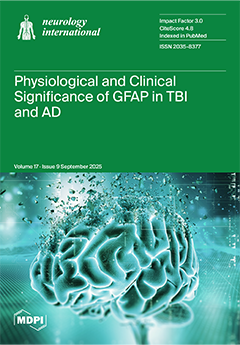
GFAP (glial fibrillary acid protein) is an intermediate filaments upregulated during astrogliosis in the brains of Alzheimer’s disease (AD) patients. Once GFAP is produced within the astrocytes, it navigates through peri-arterial and peri-venous pathways for emigrating into the systemic circulation. Fluctuations in GFAP in the blood can be used as an early indicator to forecast clinical severity and the prognosis of traumatic brain injury (TBI) and AD. Unfortunately, due to TBI- and AD-induced encumbrances blocking its emigration, the smooth sailing of GFAP from brain circulation into systemic circulation never transpires. Future research is needed to ascertain these impediments in the disease context before we can safely rely on GFAP for predicting disease-related changes in TBI and AD. View this paper
- Issues are regarded as officially published after their release is announced to the table of contents alert mailing list.
- You may sign up for e-mail alerts to receive table of contents of newly released issues.
- PDF is the official format for papers published in both, html and pdf forms. To view the papers in pdf format, click on the "PDF Full-text" link, and use the free Adobe Reader to open them.
Previous Issue
Next Issue