


Abstract
Pulmonary fibrosis (PF) is a progressive and often fatal interstitial lung disease for which the currently available pharmacological therapies remain largely limited to slowing disease progression rather than reversing established fibrosis. This limitation has stimulated increasing interest in innovative therapeutic platforms capable of modulating complex fibrotic pathways. In this context, exosomes—nanoscale extracellular vesicles—have emerged as promising cell-free nanocarriers due to their intrinsic biocompatibility, low immunogenicity, and ability to be engineered for targeted drug delivery. In this review, we provide a comprehensive overview of both natural and engineered exosome-based strategies for the diagnosis and treatment of pulmonary fibrosis. We summarize recent advances in exosome engineering, including ligand functionalization, glycoengineering, and therapeutic cargo loading, highlighting how these approaches may support the development of more targeted and potentially personalized nanotherapeutic strategies. We further discuss emerging hybrid delivery platforms, such as exosome–liposome chimeras and hydrogel-based depots, which may enhance pulmonary retention, improve therapeutic durability, and enable controlled drug release. Finally, we outline key challenges and opportunities for clinical translation, including large-scale manufacturing, regulatory considerations, and clinically relevant delivery routes such as inhalation-based administration. Collectively, this review provides a translational perspective on engineered exosomes as emerging nanotherapeutic platforms for pulmonary fibrosis.
1. Introduction
Pulmonary fibrosis (PF) encompasses a heterogeneous group of interstitial lung disorders characterized by excessive extracellular matrix (ECM) deposition, fibroblast proliferation, and irreversible distortion of alveolar architecture [1,2]. These include idiopathic pulmonary fibrosis (IPF), connective tissue disease-associated PF, occupational or environmental fibrotic lung disease, and post-injury fibrotic disorders. IPF, the most prevalent subtype, carries a median survival of only 3–5 years after diagnosis, underscoring the urgent need for more effective therapeutic strategies [3,4,5]. Patients with IPF frequently present with multiple comorbidities, including pulmonary hypertension, gastroesophageal reflux disease, obesity, emphysema, cardiovascular disease, and obstructive sleep apnea, all of which further complicate disease management and worsen clinical outcomes [6]. Although most patients experience a gradual decline in lung function, approximately 10–15% undergo rapid disease acceleration within months, often culminating in acute respiratory failure [7]. Accurate estimates of IPF incidence and prevalence remain challenging due to diagnostic complexity, frequent misclassification, and inconsistencies in clinical coding practices [8]. Together, IPF and its associated comorbidities substantially impair health-related quality of life and impose a considerable socioeconomic burden [9].
Current clinical diagnosis of PF relies on multidisciplinary evaluation integrating high-resolution computed tomography (HRCT), pulmonary function testing, clinical history, and in selected cases, histopathological assessment [10]. Despite advances in disease management, currently approved anti-fibrotic agents—pirfenidone and nintedanib—provide only modest clinical benefit and do not reverse established fibrosis [11,12]. Both agents are approved by the European Medicines Agency and the U.S. Food and Drug Administration and are recommended in international treatment guidelines [13,14]. In Phase 3 clinical trials, including ASCEND (NCT01366209) and CAPACITY (NCT00287729, NCT00287716), pirfenidone reduced the risk of disease progression or death compared with the placebo, while nintedanib demonstrated similar efficacy in the INPULSIS trials (NCT01335464, NCT01335477). Nevertheless, neither therapy halts disease progression in most patients, and treatment is frequently limited by gastrointestinal and hepatic adverse effects, poor tolerability, and high cost, leading to dose reduction or discontinuation in real-world settings [15,16,17]. As a result, current pharmacological approaches primarily delay functional decline rather than fundamentally altering the disease course.
Importantly, the limited efficacy of existing therapies reflects not only pharmacological constraints but also a fundamental mismatch between the multifactorial pathobiology of PF and single-target therapeutic paradigms. Fibrotic progression arises from the dynamic interplay of epithelial injury, immune dysregulation, fibroblast activation, and aberrant wound-healing responses, rendering isolated pathway inhibition insufficient for durable disease control [18,19]. This complexity has contributed to repeated translational failures, in which interventions showing promise in preclinical models fail to achieve meaningful clinical benefit. Furthermore, the lack of reliable biomarkers for early diagnosis, disease stratification, and treatment monitoring continues to hinder timely intervention and the development of personalized therapeutic strategies [20,21,22].
Collectively, these limitations have driven growing interest in alternative therapeutic modalities capable of addressing multiple pathological processes simultaneously. In this context, cell-free nanoscale systems—particularly extracellular vesicles (EVs) and exosomes (EXOs)—have emerged as attractive candidates, offering regenerative, immunomodulatory, and anti-fibrotic potential while avoiding many of the safety concerns associated with whole-cell transplantation [23,24,25].
EXOs are nanoscale extracellular vesicles (30–150 nm) originating from the endosomal compartment of most eukaryotic cells and serve as key mediators of intercellular communication under both physiological and pathological conditions [26,27,28]. Due to their endogenous origin, EXOs exhibit high biocompatibility, low intrinsic toxicity, and prolonged stability in circulation, partly through their ability to evade immune surveillance. These properties distinguish EXOs from many synthetic nanocarriers, whose clinical translation is often limited by rapid clearance, immunogenicity, or off-target toxicity [29,30,31].
Functionally, EXOs transport a diverse repertoire of bioactive cargos—including proteins, lipids, messenger RNAs, and non-coding RNAs such as microRNAs and long non-coding RNAs—that reflect the physiological state and identity of their parent cells [32,33,34]. Upon uptake by recipient cells, EXOs can reprogram cellular behavior by modulating gene expression, signaling pathways, and metabolic states, thereby contributing to tissue homeostasis or disease progression in a context-dependent manner [35,36,37]. These intrinsic signaling capabilities have positioned EXOs as promising therapeutic agents in regenerative medicine and immunomodulation, particularly in diseases characterized by chronic inflammation, fibrosis, and impaired tissue repair [38,39,40].
Compared with cell-based therapies, EXO-based strategies offer key safety and translational advantages, including reduced risk of immune rejection, uncontrolled engraftment, and tumorigenicity, while retaining beneficial paracrine signaling functions [41,42,43]. Their nanoscale size and amenability to bioengineering further support their utility as therapeutic and diagnostic platforms [44,45]. These features have stimulated growing interest in EXO-based strategies for PF, where preclinical studies suggest potential anti-fibrotic, immunomodulatory, and regenerative effects [46].
Importantly, however, not all EXOs are inherently therapeutic. Their biological effects are highly context-dependent and governed by factors such as cellular origin, disease stage, microenvironmental cues, and cargo composition. Without rigorous mechanistic insight and rational design, exosome-based interventions may inadvertently propagate pathological signaling or reproduce translational challenges previously encountered in nanomedicine. Accordingly, the successful clinical development of EXO-based therapeutics will require a shift from empirical application toward mechanism-driven engineering and precise functional characterization. In addition, therapeutic EXOs may also present potential safety concerns, including unintended activation of off-target signaling pathways, immune recognition following repeated administration, or variability arising from donor cell source and cargo heterogeneity. These considerations highlight the importance of rigorous safety evaluation, standardized characterization, and careful engineering design for clinical translation.
The rationale for applying EXO-based strategies to PF lies in their ability to modulate multiple interconnected pathological processes, including inflammation, fibroblast activation, and aberrant tissue remodeling [47,48,49,50,51]. In addition to their intrinsic biological activity, advances in exosome engineering have expanded their potential as targeted therapeutic delivery platforms.
These mechanistic advantages are supported by growing preclinical evidence demonstrating that EXOs derived from MSCs or macrophages can attenuate fibrotic pathology in experimental PF models, including reduced collagen deposition, partial restoration of alveolar architecture, and the suppression of pro-inflammatory cytokines such as TNF-α and IL-6 [52]. Beyond their intrinsic biological activity, EXOs also offer favorable delivery characteristics, including high biocompatibility, circulatory stability, and compatibility with both systemic and local administration routes [53,54]. Furthermore, advances in exosome engineering—including surface functionalization, therapeutic cargo loading, and hybridization with synthetic nanocarriers—have further expanded their potential as targeted anti-fibrotic delivery platforms (Table 1) [55,56,57].
Table 1.
Representative strategies for EXO engineering and their therapeutic relevance.
Despite rapid advances in the field, however, EXO-based research in PF remains conceptually fragmented. Diagnostic applications, therapeutic studies, and engineering strategies are often pursued in parallel but discussed in isolation, frequently without sufficient integration of disease biology, biomarker discovery, and translational constraints. As a result, promising experimental findings do not always converge toward clinically actionable strategies.
In this review, we seek to address this gap by proposing a unified framework that conceptualizes EXOs not merely as therapeutic carriers, but as disease-informed nanoplatforms that bridge biomarker identification, mechanistic insight, and the development of more targeted therapeutic strategies (Figure 1). We first summarize the biological characteristics and fibrogenic roles of EXOs in pulmonary disease, followed by an evaluation of their emerging utility as diagnostic and prognostic biomarkers. We then critically examine therapeutic EXOs derived from MSCs and other cellular sources, as well as engineered and hybrid EXO platforms designed to enhance targeting efficiency and anti-fibrotic efficacy. Finally, we discuss key translational considerations—including manufacturing scalability, delivery strategies, and regulatory challenges—to delineate which EXO-based approaches are most likely to achieve clinical relevance and which may require fundamental rethinking to overcome current limitations.
Figure 1.
Exosomes as disease-informed nanoplatforms in pulmonary fibrosis. In pulmonary fibrosis (PF), exosomes (EXOs) released from injured epithelial cells, activated fibroblasts, and immune cells propagate profibrotic signaling, amplify inflammation, and promote extracellular matrix deposition within the fibrotic lung microenvironment. At the same time, EXOs isolated from biofluids such as bronchoalveolar lavage fluid, blood, and saliva serve as accessible liquid-biopsy platforms carrying disease-relevant molecular signatures, including microRNAs, proteins, and other regulatory cargos. Through rational engineering strategies, such as cargo loading, surface functionalization, glycoengineering, and hybridization with synthetic carriers or biomaterials, EXOs can be repurposed into targeted anti-fibrotic nanotherapeutics. Collectively, EXOs bridge PF pathogenesis, biomarker discovery, and targeted therapeutic development, highlighting their potential as integrated platforms for diagnosis, treatment, and clinical translation.
2. EXOs in PF: Biology and Pathogenic Role
2.1. Biogenesis, Composition, and Cargo Sorting of EXOs
EXOs are nanoscale EVs (30–150 nm) generated through the endosomal pathway, and their biogenesis constitutes a tightly regulated process that fundamentally shapes vesicle composition and function [27]. EXO biogenesis proceeds through the endosomal trafficking pathway, in which endocytosed membrane components are processed through early and late endosomal compartments, ultimately leading to the formation of multivesicular bodies (MVBs) [60,61,62]. Within MVBs, membrane invagination gives rise to intraluminal vesicles (ILVs) that selectively incorporate proteins, lipids, and nucleic acids reflective of the cellular state. Upon the fusion of MVBs with the plasma membrane, ILVs are released into the extracellular space as EXOs (Figure 2). This process is governed by both endosomal sorting complexes required for transport (ESCRT)-dependent and ESCRT-independent mechanisms, involving ceramides and tetraspanins (e.g., CD9, CD63, and CD81), while Rab GTPases such as Rab27a/b and Rab11 regulate MVB trafficking and vesicle release [63,64,65,66,67,68,69,70]. Importantly, the molecular decisions made during EXO biogenesis provide a mechanistic basis for the marked heterogeneity observed in EXO cargo and downstream biological effects.
Figure 2.
Biogenesis pathways of exosomes (EXOs). Microvesicles are generated by direct outward budding and fission of the plasma membrane, whereas EXOs originate from the endosomal pathway through the formation of intraluminal vesicles (ILVs) within multivesicular endosomes (MVEs), followed by release into the extracellular space upon the fusion of MVEs with the plasma membrane. Adapted from Ref. [64].
The molecular composition of EXOs is partly conserved yet highly context-dependent. While EXOs characteristically express canonical surface markers such as CD63, CD81, and TSG101, their molecular cargo varies substantially according to the originating cell type and its physiological or pathological state [71,72,73]. EXOs transport a broad spectrum of functional biomolecules including structural proteins (e.g., actin and tubulin), heat shock proteins (e.g., Hsp70 and Hsp90), signaling molecules (e.g., STAT3 and kinases), enzymes, and adhesion-related proteins [74,75]. Notably, their nucleic acid cargo has attracted considerable attention, as EXOs selectively package messenger RNAs (mRNAs), microRNAs (miRNAs), long non-coding RNAs (lncRNAs), and circular RNAs (circRNAs), which can exert profound regulatory effects on gene expression in recipient cells [76,77,78].
The selective sorting of RNA into EXOs is mediated by sequence motifs, RNA-binding proteins such as hnRNPA2B1, and lipid raft-associated mechanisms, enabling targeted and functionally relevant cargo delivery [79,80,81]. By encapsulating and protecting these biomolecules, EXOs enhance their stability and bioavailability, thereby facilitating effective intercellular communication following uptake by recipient cells [82]. Consistent with this role, exosomal miRNAs and lncRNAs have been implicated in the pathogenesis of multiple lung diseases. For example, exosomal miR-21 derived from human bronchial epithelial cells exposed to cigarette smoke extract promotes fibroblast-to-myofibroblast differentiation in chronic obstructive pulmonary disease [83,84,85], while lncRNAs such as H19 and MEG3 have been reported to be upregulated in patients with IPF, highlighting their potential utility as disease-associated biomarkers [86]. In addition, exosomal protein cargos can exert potent biological effects, as demonstrated by vimentin-containing EXOs derived from metastatic lung cancer cells or end-stage lung cancer serum, which induce epithelial–mesenchymal transition in bronchial epithelial cells [87].
In the context of PF, however, EXO cargo has emerged as a critical determinant of pathological intercellular communication. EXOs released by injured alveolar epithelial cells or activated fibroblasts are frequently enriched in profibrotic cargos such as miR-21, miR-199a-5p, and TGF-β1, which collectively promote myofibroblast differentiation, ECM deposition, and inflammatory activation in neighboring cells. These observations provide a mechanistic basis for how chronic epithelial injury and aberrant repair responses propagate fibrotic signaling beyond the initially damaged regions of the lung.
Taken together, these findings underscore the context-dependent nature of exosome biology in PF, where EXO-mediated signaling may either exacerbate or mitigate fibrotic progression depending on the cellular source and cargo composition. EXOs released from injured epithelial cells, activated fibroblasts, or pro-inflammatory immune cells frequently propagate a fibrotic microenvironment by reinforcing profibrotic signaling loops among epithelial, stromal, and immune compartments [88,89,90]. In contrast, EXOs derived from MSCs, endothelial progenitor cells (EPCs), or immunomodulatory macrophages tend to exert anti-fibrotic effects through the delivery of regulatory miRNAs (e.g., miR-29, miR-let-7d, and miR-30), growth factors such as hepatocyte growth factor and keratinocyte growth factor, and immunomodulatory proteins that collectively suppress fibroblast activation and promote tissue repair [91,92,93,94,95].
This duality highlights a central concept: the biological outcome of EXO-mediated signaling is dictated not by the vesicle itself, but by its cellular origin, cargo composition, and disease context. Accordingly, a detailed understanding of EXO biogenesis, molecular composition, and cargo sorting mechanisms is essential not only for elucidating fibrotic pathogenesis, but also for the rational development of engineered EXOs as therapeutic agents and the identification of vesicle-based biomarkers that reflect disease stage and treatment response [4,61].
2.2. EXO-Mediated Regulation of PF
The ECM provides essential structural and biochemical cues that regulate tissue homeostasis, but its persistent accumulation is a defining pathological feature of PF [96]. In the fibrotic lung, unresolved epithelial injury and aberrant wound-healing responses lead to sustained myofibroblast activation and excessive ECM deposition, thereby establishing a self-perpetuating fibrotic niche [97]. These processes are governed by interconnected signaling networks, among which transforming growth factor-β (TGF-β)/Smad signaling serves as the canonical central fibrogenic axis, with the Wnt/β-catenin, MAPK, and inflammatory signaling pathways acting as interconnected modulatory networks (Figure 3) [98,99,100,101,102,103].
Figure 3.
EXO-mediated signaling hierarchy in pulmonary fibrosis. Schematic overview of the major signaling pathways involved in EXO-mediated regulation of pulmonary fibrosis. TGF-β/Smad signaling is presented as the central fibrogenic axis, with reciprocal crosstalk involving the Wnt/β-catenin, MAPK, and inflammatory signaling pathways. The figure further illustrates how distinct pro-fibrotic and anti-fibrotic EXO cargos influence fibroblast activation, myofibroblast differentiation, and extracellular matrix deposition.
Increasing evidence indicates that EXOs function as critical intermediaries within these signaling networks by enabling direct intercellular transfer of fibrogenic or anti-fibrotic cues [104,105]. In PF, chronic epithelial damage initiates sustained crosstalk among alveolar epithelial cells, fibroblasts, endothelial cells, and immune cells, a process that is increasingly recognized to be mediated by EXO-based communication [106,107]. Through the delivery of functional RNAs, proteins, and lipids, EXOs modulate recipient cell phenotypes and amplify or restrain fibrogenic signaling cascades within the lung microenvironment [59,108,109].
Pro-fibrotic EXOs are commonly derived from injured epithelial cells, activated fibroblasts, and pro-inflammatory macrophages [24,110]. For example, epithelial cell-derived EXOs released following TGF-β1 stimulation carry miR-21, miR-199a-5p, and Wnt pathway components that are readily internalized by lung fibroblasts [85]. These cargos activate Smad3-dependent transcription, promote α-smooth muscle actin (α-SMA) expression, and enhance ECM production, including collagen type I and fibronectin [111,112]. In parallel, fibroblast-derived EXOs under fibrotic conditions reinforce autocrine and paracrine activation loops, while EXOs from M1-like macrophages exacerbate fibrosis by transferring inflammatory cytokines and regulatory RNAs that activate NF-κB and STAT3 signaling in stromal and epithelial cells [113,114,115,116].
In contrast, EXOs secreted by therapeutic cell populations—such as MSCs, ADSCs, and immunomodulatory M2-like macrophages—exert pronounced anti-fibrotic effects [117,118]. These effects are consistently supported by histological evidence, including reduced collagen deposition and restoration of alveolar architecture observed by H&E, Gomori’s trichrome, and picrosirius red staining, as well as quantitative reductions in Ashcroft scores and lung hydroxyproline content (Figure 4) [50]. Mechanistically, therapeutic EXOs are enriched in miRNAs such as miR-29, miR-let-7d, miR-30, and miR-186, which suppress genes involved in ECM synthesis, fibroblast proliferation, and myofibroblast differentiation [119,120]. They also deliver proteins including hepatocyte growth factor, keratinocyte growth factor, and IL-10, thereby promoting epithelial repair, reducing oxidative stress, and fostering anti-inflammatory immune polarization [121,122].
Figure 4.
Therapeutic effects of EXO administration in an experimental PF model. (A) Schematic illustration of the experimental design for evaluating EXO therapeutic efficacy in pulmonary fibrosis. (B–D) Representative histological analyses of lung tissue following EXO treatment, including hematoxylin and eosin (H&E), Gomori’s trichrome, and picrosirius red staining, demonstrating attenuation of fibrotic remodeling and collagen deposition. (E) Quantitative assessment of pulmonary fibrosis using the Ashcroft scoring system. (F) Lung hydroxyproline quantification as a biochemical indicator of collagen accumulation. Reproduced with permission from Dinh et al. [50].
Beyond direct effects on fibroblasts and epithelial cells, EXOs modulate immune responses by regulating alveolar macrophage polarization and regulatory T-cell activity [123,124]. For instance, MSC-derived EXOs induce M2 macrophage polarization via let-7 family miRNAs, resulting in increased IL-10 production and the suppression of pro-inflammatory cytokines [125,126,127]. These immunomodulatory effects contribute to reduced neutrophil infiltration, improved vascular integrity, and enhanced resolution of lung injury [95]. Collectively, the ability of EXOs to concurrently regulate inflammation, epithelial dysfunction, and matrix remodeling positions them as uniquely suited mediators of fibrotic progression and resolution [1,59].
Importantly, the biological outcome of EXO-mediated signaling is highly context-dependent. Microenvironmental cues, disease stage, cellular origin, and recipient cell type collectively determine whether EXOs promote fibrogenesis or facilitate repair [128,129]. While this heterogeneity complicates therapeutic translation, it also presents an opportunity: rational control of EXO composition through selective isolation or bioengineering may enable the development of more effective and disease-adapted anti-fibrotic therapies. In summary, EXOs orchestrate complex intercellular signaling networks that shape the trajectory of PF, functioning both as drivers of disease pathogenesis and as promising agents for therapeutic intervention [88].
3. EXOs as Diagnostic and Prognostic Biomarkers
3.1. Biofluid-Derived EXOs in PF
The identification of non-invasive, sensitive, and disease-specific biomarkers remains a major unmet need in the early diagnosis and longitudinal monitoring of PF. In this context, EXOs isolated from accessible biological fluids—including bronchoalveolar lavage fluid (BALF), serum, plasma, sputum, and saliva—have emerged as a promising liquid biopsy platform, as they stably encapsulate molecular information that reflects the physiological and pathological state of their cells of origin [130]. Unlike freely circulating biomarkers, exosomal cargos are selectively sorted and protected by a lipid bilayer, rendering them resistant to enzymatic degradation and particularly suitable for clinical analysis [131,132,133].
Among the exosomal cargos, microRNAs (miRNAs) have attracted the greatest attention as PF-related biomarkers. Although miRNAs are detectable in multiple biofluids, they predominantly circulate in an exosome-associated form, which markedly enhances their stability and reproducibility in clinical samples [131,132,133]. As a result, disease-associated changes in exosomal miRNA profiles provide insights not only into fibrotic mechanisms, but also into diagnostic stratification and therapeutic prediction. In BALF-derived EXOs from patients with IPF, anti-epithelial miRNAs such as miR-200a-3p, miR-200b-3p, and miR-141-3p are consistently downregulated, whereas miR-320a-3p, miR-320b, and miR-22-3p are upregulated, reflecting epithelial injury and fibroblast activation [134].
In parallel, circulating EXOs from IPF patients exhibit elevated levels of profibrotic miRNAs including miR-21, miR-155, and miR-199a-5p, which correlate with disease severity and poor prognosis (Table 2) [135,136,137]. Conversely, reduced levels of anti-fibrotic miRNAs such as the miR-29 family, miR-200, and let-7d are associated with increased fibrotic burden and progressive lung function decline [138,139,140].
Table 2.
Representative microRNAs implicated in pulmonary fibrosis pathogenesis, diagnosis, and therapeutic modulation.
Beyond miRNAs, exosomal protein cargos have also been explored as complementary biomarkers. Increased expression of EXO-associated proteins such as CD63, TGF-β1, and fibronectin in serum or BALF has been proposed to reflect fibrotic activity and treatment responsiveness, highlighting the potential of multimodal exosomal signatures that integrate both nucleic acid and protein readouts [141,142,143].
BALF-derived EXOs are particularly informative in respiratory diseases because they directly reflect the local pulmonary microenvironment [144]. Recent studies demonstrate that BALF exosomal miR-22 and miR-142-3p can distinguish PF patients from healthy controls and may serve as indicators of alveolar epithelial damage and fibroblast activation [145,146]. Although BALF collection is semi-invasive and less suitable for routine screening, it remains a valuable resource for biomarker discovery and mechanistic validation [147,148,149]. In contrast, serum- and saliva-derived EXOs offer more practical options for repeated sampling and longitudinal monitoring, supporting their potential use in real-world clinical settings [150].
Collectively, these findings highlight the potential of biofluid-derived EXOs as diagnostic and prognostic biomarker candidates in PF. However, most currently reported EXO-based biomarker studies remain limited by preclinical evidence, small patient cohorts, methodological heterogeneity, and the lack of large-scale multicenter validation, which currently restrict their clinical applicability [151,152]. Standardized protocols for vesicle isolation, normalization, and cargo analysis will be essential to improve reproducibility across studies and platforms. Although the integration of exosomal profiling with multi-omics approaches may further improve biomarker discovery and disease monitoring, substantial clinical validation will be required before routine implementation in personalized PF management can be considered [153,154].
3.2. Exosomal microRNAs and Their Predictive Value
MicroRNAs (miRNAs) are small non-coding RNAs that regulate gene expression at the post-transcriptional level and play essential roles in cellular differentiation, apoptosis, immune regulation, and fibrotic remodeling [58,155]. When selectively packaged into EXOs, miRNAs are shielded from extracellular degradation, conferring high stability and reproducibility in biological fluids. In PF, accumulating evidence indicates that disease-associated exosomal miRNA signatures are closely linked to disease onset, progression, and treatment responsiveness, positioning them as promising predictive biomarkers rather than passive diagnostic readouts [156,157,158,159,160,161].
Among these candidates, exosomal miR-21 is the most extensively studied and is consistently associated with fibrotic severity and poor clinical outcomes. Elevated levels of miR-21-containing EXOs have been detected in serum and BALF from patients with IPF and correlate with disease progression. Mechanistically, miR-21 promotes fibrogenesis by suppressing SMAD7, thereby enhancing TGF-β/Smad2/3 signaling, driving myofibroblast differentiation and ECM accumulation [162,163]. Importantly, experimental inhibition of miR-21 not only reduces collagen deposition but also attenuates fibrotic remodeling in bleomycin-induced PF models, underscoring its functional relevance beyond biomarker association (Figure 5) [164].
Figure 5.
Therapeutic inhibition of exosomal miR-21 attenuates PF in a bleomycin-induced mouse model. (A) Quantification of lung collagen content following treatment with PBS, bleomycin plus control probe, or bleomycin plus miR-21 antisense probe. (B) Representative Western blot analysis of fibronectin, collagen type I alpha 1 (Col1A1), and α-smooth muscle actin (α-SMA) expression in lung tissue. (C) Histological evaluation of pulmonary fibrosis by hematoxylin and eosin (H&E) and Masson’s trichrome staining, demonstrating reduced collagen deposition and improved tissue architecture following miR-21 inhibition. (D) Immunohistochemical analysis of α-SMA expression in lung sections, indicating the suppression of myofibroblast activation after miR-21 antisense treatment. * p < 0.05 and ** p < 0.01 were considered statistically significant. Reproduced with permission from Liu et al. [164].
In a similar profibrotic context, miR-199a-5p is enriched in EXOs derived from fibrotic lungs and circulating biofluids. This miRNA exacerbates fibrotic remodeling by targeting caveolin-1 (CAV1), a negative regulator of TGF-β signaling, thereby amplifying profibrotic signal transduction [137,165,166]. In contrast, several exosomal miRNAs exert protective, anti-fibrotic effects and are consistently downregulated during PF progression. Members of the miR-29 family, particularly miR-29a and miR-29b, suppress the expression of collagen synthesis genes such as COL1A1 and COL3A1, and reduced levels of miR-29-containing EXOs are associated with increased fibrotic burden and accelerated lung function decline [88]. Additional anti-fibrotic candidates, including let-7d and miR-186, are similarly diminished in PF-derived EXOs and may reflect a loss of epithelial integrity and unresolved inflammatory signaling [147].
Beyond their mechanistic relevance, exosomal miRNAs exhibit substantial predictive value for clinical decision-making. Baseline serum levels of profibrotic miRNAs such as miR-21 and miR-155 have been proposed as predictors of responsiveness to anti-fibrotic therapies, including pirfenidone and nintedanib [167,168]. Moreover, longitudinal monitoring of exosomal miRNA dynamics during treatment has been suggested to provide early indicators of therapeutic efficacy, disease stabilization, or impending relapse, preceding conventional clinical or radiographic changes [169,170].
Despite these promising findings, several challenges must be addressed before exosomal miRNAs can be fully integrated into clinical practice. Technical variability in EXO isolation and miRNA quantification, lack of standardized normalization strategies, and limited validation in large, multicenter cohorts currently constrain their translational applicability [171,172]. Nevertheless, rapid advances in next-generation sequencing, digital PCR, and multi-omics integration are expected to accelerate the clinical implementation of exosomal miRNA-based predictive biomarkers in PF [173,174].
3.3. Challenges in Clinical Translation of EXO Biomarkers
Despite the growing interest in exosome (EXO)-based biomarkers for PF, their translation into routine clinical practice remains constrained by a range of technical, biological, and regulatory challenges [1]. Although EXOs offer distinct advantages—including molecular stability in biofluids, cell-type specificity, and the ability to reflect dynamic pathological processes—robust and standardized methodologies are still lacking for their reliable isolation, characterization, and quantitative analysis [1,4].
A major technical limitation lies in the absence of standardized EXO isolation protocols [175,176]. Ultracentrifugation, widely regarded as the gold standard, is labor-intensive, time-consuming, and poorly suited for high-throughput clinical workflows. Alternative approaches, such as polymer-based precipitation kits or size-exclusion chromatography, offer improved scalability but frequently suffer from low purity, co-isolation of non-exosomal EVs, or contamination with protein aggregates [175,177,178]. This platform-dependent variability leads to substantial inconsistencies in exosomal yield, composition, and downstream molecular analyses, thereby compromising reproducibility and cross-study comparability—both of which are essential for biomarker validation.
Combining orthogonal isolation methods—such as size-exclusion chromatography with downstream immunoaffinity capture—substantially improves EXO purity over single-step approaches [179,180]. At the field level, systematic adoption of MISEV guidelines and EV-TRACK-compliant reporting provides a practical and immediately implementable framework for harmonizing isolation practices across research centers [181,182].
Beyond technical considerations, the biological heterogeneity of EXOs presents an additional challenge [183]. In the fibrotic lung microenvironment, EXOs are released from multiple cell types, including alveolar epithelial cells, fibroblasts, endothelial cells, and immune cells, each contributing overlapping yet distinct cargo profiles [1,184]. Without reliable cell-of-origin markers or enrichment strategies, interpreting exosomal signals from complex biofluids such as plasma or BALF remains inherently ambiguous [4]. Furthermore, EXO cargo composition is dynamic and may shift in response to disease stage, therapeutic intervention, or comorbid conditions such as infection or malignancy, potentially confounding biomarker specificity and longitudinal interpretation [162,185].
Cell-type-specific immunoaffinity enrichment—using surface markers such as EpCAM for epithelial-derived or FAP for fibroblast-derived EXOs—enables the targeted isolation of diagnostically relevant subpopulations from complex biofluids [186,187]. Integration of single-vesicle analysis platforms (e.g., nano-flow cytometry) with machine learning-assisted cargo classification further holds promise for resolving EXO heterogeneity and improving biomarker specificity in PF [188,189].
Quantification and normalization of exosomal contents—particularly RNA species—represent another unresolved bottleneck. Currently, there is no consensus on endogenous reference controls for normalizing exosomal miRNA expression, and pre-analytical variables such as sample collection, processing, storage conditions, and freeze–thaw cycles can markedly influence the analytical outcomes [152,190,191]. In addition, the low abundance of certain diagnostically relevant exosomal miRNAs or proteins necessitates ultrasensitive detection platforms, which are not yet widely implemented in standard clinical laboratories [192].
Synthetic spike-in RNA controls and droplet digital PCR (ddPCR)—which enables absolute miRNA quantification independent of endogenous reference genes—offer robust and clinically transferable solutions to current normalization limitations [189,193,194]. For protein-level biomarkers, single-molecule array (SiMoa) technology extends detection sensitivity to the femtomolar range, addressing the analytical gap posed by low-abundance exosomal targets [195].
From a regulatory and translational perspective, EXO-based diagnostics must satisfy stringent criteria for analytical validity, clinical utility, and cost-effectiveness before clinical adoption [196]. Existing biomarker qualification frameworks do not yet fully accommodate the complexity of EVs, underscoring the need for dedicated regulatory guidance tailored to EV-based diagnostics [197]. Addressing these challenges will require coordinated efforts among academia, industry, and regulatory agencies to establish standardized reference materials, proficiency testing schemes, and harmonized clinical trial designs for EXO biomarker validation [197,198,199].
Alignment with established regulatory frameworks—including the FDA BEST resource and EMA qualification opinion process—alongside engagement with international consortia such as the ISEV Biomarker Task Force, provides a structured pathway for advancing EXO diagnostics toward clinical qualification [58,181,200]. Incorporating EXO biomarkers as exploratory co-endpoints within ongoing anti-fibrotic drug trials represents a pragmatic near-term strategy for generating the clinical evidence required for formal regulatory submission [201,202].
Taken together, while exosomal biomarkers hold substantial promise for transforming the diagnosis and monitoring of PF, clinical translation will require a staged approach: beginning with cross-laboratory standardization of isolation and quantification methods, followed by large-scale multicenter validation, and culminating in regulatory-grade biomarker qualification. Sustained interdisciplinary collaboration among researchers, clinicians, and regulatory agencies will be essential to advance EXO-based diagnostics from exploratory tools to actionable clinical assays in fibrotic lung disease.
4. EXO-Derived Therapeutics in PF
4.1. Natural Sources of Therapeutic EXOs
EXOs derived from naturally occurring cell sources have emerged as promising cell-free therapeutics for PF as they can recapitulate many of the paracrine effects of donor cells while avoiding the safety concerns associated with direct cell transplantation [203,204]. These vesicles inherently carry bioactive cargos—including microRNAs, proteins, lipids, and metabolites—that reflect the regulatory phenotype of their parent cells and enable multifaceted modulation of the fibrotic lung microenvironment [61,205,206,207]. Upon systemic or local administration, natural EXOs exert anti-fibrotic, immunomodulatory, and tissue-reparative effects in multiple preclinical PF models [204,208,209,210,211].
Among the natural sources, mesenchymal stem cell-derived EXOs (MSC-Exos) represent the most extensively investigated and validated platform for PF therapy [212,213,214]. MSC-Exos are enriched in anti-fibrotic microRNAs such as miR-29 family members, let-7 family members, miR-186, and miR-129, which collectively suppress collagen biosynthesis, inhibit TGF-β signaling, and attenuate fibroblast activation [215,216,217,218,219]. Consistent with these mechanisms, administration of MSC-Exos in bleomycin-induced PF models reduces lung fibrosis scores, decreases hydroxyproline content, preserves alveolar architecture, and improves survival [215,220,221].
Beyond their direct effects on fibroblasts and ECM remodeling, MSC-Exos exert potent immunomodulatory functions. They suppress pro-inflammatory cytokine production, promote macrophage polarization toward an anti-inflammatory M2 phenotype, and enhance regulatory T cell responses, thereby mitigating chronic inflammation—a central driver of fibrogenesis [95,222,223,224,225,226,227]. Supporting this concept, MSC-Exo-mediated M2 macrophage polarization has consistently been observed across inflammatory disease models, accompanied by reduced levels of pro-inflammatory cytokines and increased expression of IL-10 [225,228,229].
MSC-Exos also convey anti-fibrotic and cytoprotective signals through growth factors and regulatory proteins inherited from parent MSCs, including hepatocyte growth factor and IL-1 receptor antagonist (IL-1Ra), which inhibit immune cell-mediated lung injury, epithelial–mesenchymal transition (EMT), and collagen deposition [230,231,232,233]. In parallel, MSC-Exos display antioxidant properties by scavenging intracellular reactive oxygen species (ROS), preserving mitochondrial integrity, and enhancing intrinsic antioxidant defenses [234,235]. Collectively, these multifaceted mechanisms contribute to the robust anti-fibrotic efficacy of MSC-Exos observed across pulmonary and extrapulmonary fibrosis models [236].
In addition to MSCs, ADSCs and umbilical cord-derived MSCs have emerged as alternative EXO sources. ADSC-Exos offer advantages such as higher yield, easier harvest, and lower donor-site morbidity while exhibiting comparable anti-fibrotic efficacy in animal models [237,238]. Umbilical cord MSC-derived EXOs display low immunogenicity and high proliferative capacity, making them attractive candidates for allogeneic applications [239,240].
Beyond stem cells, immune cell-derived EXOs—particularly those from alternatively activated M2 macrophages—represent another promising therapeutic avenue [241]. M2-derived EXOs are enriched in anti-inflammatory and reparative cargos such as miR-223, miR-146a, IL-10, and arginase-1, which facilitate macrophage polarization, suppress neutrophil recruitment, and attenuate oxidative stress [242,243,244,245,246]. Notably, macrophage-derived EXOs carrying miR-142-3p have been shown to suppress TGF-β receptor signaling in alveolar epithelial cells and fibroblasts, thereby slowing fibrotic progression [247].
Endothelial progenitor cell-derived EXOs (EPC-Exos) have also demonstrated reparative potential in PF models by delivering pro-angiogenic factors and ECM-regulatory microRNAs that support vascular regeneration, stabilize the alveolar–capillary barrier, and indirectly suppress fibrotic remodeling [248,249,250]. Similarly, epithelial cell-derived EXOs—including those from alveolar type II (ATII) cells, human bronchial epithelial cells (HBECs), and human amniotic epithelial cells (hAECs)—exhibit intrinsic anti-fibrotic properties [251,252,253,254,255,256]. These EXOs inhibit TGF-β-induced myofibroblast differentiation, reduce cellular senescence, and suppress collagen production through coordinated protein and microRNA cargoes [251,252,253,254,255,256].
Given the diversity of therapeutic EXO sources, their translational potential varies considerably in terms of biological activity, scalability, and safety considerations. A comparative overview of the major therapeutic EXO sources relevant to PF therapy is summarized in Table 3.
Table 3.
Comparative translational characteristics of the major therapeutic EXO sources for pulmonary fibrosis.
Despite these advantages, several limitations remain in the therapeutic use of naturally derived EXOs including cargo heterogeneity, donor-to-donor variability, limited yield, and challenges in large-scale biomanufacturing [1,257,258,259]. EXO content and potency are further influenced by donor age, health status, and culture conditions [260,261,262]. To address these challenges, strategies such as standardized donor cell banks, optimized culture systems, and GMP-compliant purification protocols are actively being developed [263]. Collectively, the multimodal functionality, biocompatibility, and favorable safety profile of naturally sourced EXOs position them as compelling candidates for PF therapy, warranting continued validation through rigorous preclinical studies and early-phase clinical trials.
4.2. Engineered EXOs for Enhanced Anti-Fibrotic Activity
While naturally derived EXOs exhibit intrinsic anti-fibrotic and immunomodulatory properties, their therapeutic performance can be further optimized through bioengineering strategies that precisely control cargo composition, targeting specificity and delivery behavior [264,265]. Engineered EXOs address key limitations of native vesicles—including heterogeneous cargo loading, suboptimal biodistribution, and limited tissue selectivity—by introducing deliberate genetic, molecular, or structural modifications [266]. We therefore discuss engineered EXOs along an “input–design–output” logic: what to load (cargo), where to go (targeting), and how long to stay (formulation) (Figure 6).
Figure 6.
Input–design–output framework for engineering exosomes in pulmonary fibrosis therapy. This schematic illustrates an input–design–output strategy for exosome (EXO) engineering. Input involves loading therapeutic cargos (e.g., miRNAs, proteins, or drugs), design focuses on targeting modification to specific fibrotic lung cells, and output emphasizes formulation and delivery optimization to improve pulmonary retention and therapeutic durability. This framework highlights how engineered EXOs overcome the limitations of native vesicles in targeting and efficacy.
4.2.1. Genetic and Cargo Engineering Strategies
One widely explored approach involves the genetic modification of donor cells, such as MSCs, to enrich exosomal cargo with anti-fibrotic molecules [267]. MSCs engineered to overexpress miR-29b, let-7d, or miR-181b produce EXOs with enhanced capacity to suppress collagen synthesis, attenuate TGF-β/Smad signaling, and regulate inflammatory cytokine expression [72,215,268,269]. These engineered EXOs demonstrate superior therapeutic efficacy in preclinical PF models by reducing myofibroblast differentiation and promoting epithelial repair [270].
Complementary to genetic engineering, direct cargo-loading strategies enable the incorporation of exogenous therapeutic agents into pre-isolated EXOs. Techniques such as incubation, electroporation, sonication, extrusion, and freeze–thaw cycling have been employed to load RNAs, peptides, or small-molecule drugs targeting EMT, fibroblast proliferation, and ECM remodeling [266,270]. While passive loading approaches offer simplicity, they suffer from limited encapsulation efficiency, whereas active methods may compromise vesicle integrity or payload stability (Table 4) [271,272].
Table 4.
Representative strategies for engineering EXOs to enhance therapeutic cargo delivery.
4.2.2. Surface Engineering and Targeted Delivery
To improve tissue specificity and cellular uptake, surface engineering approaches have been developed, including enzymatic glycoengineering (EGE) and ligand functionalization [284]. Glycoengineering via glycosyltransferases involves attaching an unnatural sugar to the EXO surface using a glycosyltransferase and a modified glycosyl donor [285]. Subsequently, the EXO can be functionalized using click chemistry to link the modified sugar residue (e.g., with an azide) to a tagged molecule (e.g., with an alkyne) and to targeting ligands such as antibodies or peptides that recognize fibrotic tissues [286,287]. In the case of these EXO EGEs, research continues to attach modified sialic acid using sialyltransferases (sial-Ts) due to the complexity of the enzyme. For example, Kundu et al. employed sialyltransferase-mediated incorporation of azide-modified sialic acids onto EXOs, enabling subsequent functionalization with therapeutic proteins and imaging probes [285]. These EGE-modified EXOs exhibited efficient intracellular trafficking and perinuclear localization, suggesting enhanced payload delivery to target cells (Figure 7).
Figure 7.
Intracellular trafficking and subcellular localization of enzymatic glycoengineered (EGE) EXOs. (A) Confocal fluorescence images showing intracellular trafficking and organelle colocalization of EGE-modified EXOs following cellular uptake. (B) Visualization of intracellular delivery of both surface-modified glycans and encapsulated protein cargos. (C) Comparative analysis of EXO internalization pathways relative to canonical endocytic uptake. (D) Representative fluorescence images confirming efficient cellular internalization and intracellular distribution of glycoengineered EXOs. Reproduced with permission from Kundu et al. [285].
Alternative strategies exploit endogenous EV scaffold proteins. Zheng et al. genetically engineered parent cells to express glycosylated CD63 in combination with fucosyltransferases, generating EXOs with selective affinity for activated endothelial or dendritic cells [284]. In their study, parent cells were genetically engineered to co-express a glycosylation domain (inserted into the large extracellular loop of the scaffold protein CD63) and a corresponding fucosyltransferase (FUT7 or FUT9). This strategy enabled the production of functionalized EVs with high specificity for activated endothelial cells and dendritic cells, respectively.
4.2.3. Hybrid EXO Platforms and Combination Delivery
Hybrid EXO–liposome systems represent another powerful engineering strategy, combining the biological advantages of EXOs with the high drug-loading capacity and stability of synthetic liposomes [288,289]. As shown in Figure 8, the fabricated hybrid EXOs not only form slightly larger vesicles compared with liposomes, but also exhibit higher cellular internalization efficiency. These hybrid EXOs maintain a size comparable to that of liposomes while demonstrating enhanced internalization efficiency, thereby improving drug delivery efficiency and ultimately augmenting the therapeutic efficacy of drugs [290].
Figure 8.
Fabrication and cellular uptake of doxorubicin (DOX)-loaded exosome–liposome hybrid vesicles. (A) Schematic illustration of hybrid EXO preparation via membrane fusion between natural EXOs and synthetic liposomes. (B) Representative transmission electron microscopy (TEM) images of liposomes, native EXOs, and EXO–liposome hybrid vesicles. (C) Confocal fluorescence microscopy images comparing cellular internalization of Rhodamine B-labeled liposomes and hybrid EXOs. Reproduced with permission from Palakurthi et al. [290].
Hybrid vesicles can be fabricated through incubation, freeze–thaw cycling, or extrusion [291], and have been successfully applied to deliver complex cargos such as CRISPR/Cas9 systems with minimal vesicle damage [292]. Importantly, these systems enable the co-delivery of exosomal cargo and small-molecule anti-fibrotic agents or siRNAs via multiple administration routes, including intravenous, intranasal, or intratracheal delivery [293,294,295].
Beyond hybrid vesicles, engineered EXOs can be integrated into biomaterial-based delivery systems, such as hydrogels or nanoparticle depots, to achieve sustained local release and prolonged retention within fibrotic lung tissue [296,297]. Such composite platforms are particularly attractive for non-invasive administration strategies and for overcoming rapid clearance in the pulmonary microenvironment.
4.2.4. Toward Next-Generation EXO Therapeutics in PF
Looking forward, advances in multi-omics profiling and systems biology are enabling the rational design of personalized EXO therapeutics [281]. By integrating patient-specific transcriptomic and fibrotic signatures, EXOs can be engineered with tailored cargo compositions, targeting ligands, and release kinetics to address inter-individual heterogeneity in PF progression and treatment response [298].
Emerging technologies—including imaging-visible EXOs and stimulus-responsive biomaterials—are further expanding the potential for spatiotemporal control over EXO delivery and activity within fibrotic lungs [299,300]. Collectively, these innovations align with the broader vision of precision nanomedicine for fibrotic diseases.
Despite these advances, significant translational challenges remain, including scalable GMP manufacturing, consistency in engineering protocols, and regulatory classification of hybrid biologics [301,302]. Nevertheless, engineered and hybrid EXOs represent a versatile and modular platform that integrates biological intelligence with nanotechnological control, positioning them at the forefront of next-generation therapeutic strategies for PF.
4.3. Delivery Strategies and Routes of Administration
The therapeutic efficacy of EXO-based interventions in PF is governed not only by vesicle bioactivity but also by delivery route and formulation strategy, which critically influence biodistribution, local retention, and pharmacodynamic outcomes [303]. Given the anatomical complexity of the lung and the spatial heterogeneity of fibrotic lesions, rational delivery design is essential to ensure sufficient EXO accumulation at target sites while minimizing off-target exposure [304].
4.3.1. Systemic Versus Local Pulmonary Delivery
Intravenous administration remains the most commonly explored delivery route in preclinical PF studies due to its technical simplicity and systemic accessibility. Owing to the extensive pulmonary capillary network and first-pass filtration effect, IV-injected EXOs preferentially accumulate in the lung [305,306,307]. However, substantial off-target uptake by the liver, spleen, and kidneys often limits lung specificity and necessitates higher dosing [308]. To address this limitation, surface-engineered EXOs bearing targeting ligands for CD44, αvβ3 integrins, or fibrotic ECM components have been developed to enhance selective homing to activated fibroblasts or injured epithelium [304,309,310].
In contrast, local pulmonary delivery routes—including intratracheal and intranasal administration—enable the direct deposition of EXOs into the alveolar space, bypassing systemic circulation [311]. These approaches significantly improve pulmonary retention and therapeutic efficacy in bleomycin-induced fibrosis models while reducing systemic exposure [50,312]. Importantly, aerosolized or nebulized EXO formulations are under active investigation as non-invasive delivery platforms suitable for repeated or outpatient administration, highlighting their translational potential (Figure 9) [311].
Figure 9.
Comparison of intratracheal instillation and inhalation as pulmonary delivery routes for exosome-based therapeutics. Intratracheal instillation allows precise, region-specific dosing and is widely used in preclinical studies, whereas inhalation provides more physiological lung distribution, improved patient compliance, and higher clinical translatability for chronic pulmonary applications.
4.3.2. Biomaterial-Assisted and Sustained Delivery Platforms
Beyond direct administration, biomaterial-assisted delivery platforms have been developed to address the rapid clearance and limited retention of EXOs in vivo. Incorporation of EXOs into hydrogels, microparticles, or hybrid matrices enables sustained release, protects vesicles from enzymatic degradation, and prolongs local bioavailability within fibrotic tissues [296,313,314,315,316]. These approaches are particularly relevant in PF, where repeated dosing and long-term modulation of the fibrotic niche are often required.
Although most delivery strategies for PF focus on pulmonary routes, transdermal microneedle-based EXO delivery provides an illustrative example of how sustained, minimally invasive administration can be achieved for chronic fibrotic conditions. In a representative study, Song et al. developed a dissolvable microneedle patch incorporating EXOs, hepatocyte growth factor, and an anti-fibrotic agent, which enabled continuous systemic absorption and prolonged therapeutic exposure [317]. Mechanistically, this platform attenuated fibroblast activation and suppressed TGF-β1/SMAD3 signaling, highlighting how EXO-based combination systems can simultaneously modulate immune responses and fibrotic pathways (Figure 10). While demonstrated in a hepatic fibrosis model, this work underscores generalizable design principles—namely, controlled release, cargo co-delivery, and patient-friendly administration—that are highly relevant to the development of next-generation EXO delivery strategies for PF.
Figure 10.
In vitro anti-fibrotic effects of H@EV-H/G/N microneedle patch-derived extracts. (A–D) Transcriptomic profiling and gene set enrichment analysis demonstrating the modulation of anti-fibrotic and immunoregulatory pathways following treatment. (E) Schematic illustration of the proposed anti-fibrotic mechanism of H@EV-H/G/N microneedle patch treatment. (F–H) Fibroblast migration assays showing reduced cellular motility following treatment. (I,J) Quantitative analysis of fibrosis-related markers, including collagen type I (Col1) and α-smooth muscle actin (α-SMA), indicating suppression of myofibroblast activation and extracellular matrix production. * p < 0.05, ** p < 0.01, *** p < 0.001, and **** p < 0.0001 were considered statistically significant. Reproduced with permission from Song et al. [317].
4.3.3. Translational Considerations and Future Directions
Collectively, these delivery strategies illustrate that optimal EXO therapy for PF will likely require a tailored combination of administration route, formulation design, and disease stage. Local pulmonary delivery may be preferable for early or localized disease, whereas systemic or depot-based strategies may better address advanced or diffuse fibrosis. Moreover, integration of EXOs with smart biomaterials—such as stimulus-responsive hydrogels or inhalable dry powders—offers opportunities for the spatiotemporal control of release and improved patient compliance.
As EXO-based therapeutics progress toward clinical translation, systematic evaluation of delivery route-dependent pharmacokinetics, safety, and patient acceptability will be essential. Ultimately, delivery strategy optimization will be as critical as EXO engineering itself in determining the clinical success of EXO-based therapies for PF.
4.4. Preclinical and Clinical Evidence Supporting Therapeutic EXOs
Over the past decade, a growing body of preclinical evidence has established the therapeutic potential of exosome (EXO)-based interventions in PF. Studies employing diverse experimental models—including bleomycin-, silica-, and radiation-induced lung injury—have consistently demonstrated that both natural and engineered EXOs attenuate key pathological features of fibrosis, such as excessive ECM deposition, fibroblast activation, and epithelial–mesenchymal transition [1,90].
In bleomycin-induced PF models, systemic or intratracheal administration of MSC-Exos has been shown to reduce fibrotic burden, as evidenced by the decreased expression of profibrotic markers such as α-smooth muscle actin and collagen type I, preservation of alveolar architecture, and improvement in lung function [312,318]. These beneficial effects are mediated in part by exosomal microRNAs, including members of the miR-29 and let-7 families, which suppress TGF-β/Smad signaling and collagen biosynthesis, as well as by anti-inflammatory and cytoprotective proteins such as hepatocyte growth factor and TSG-6 [319,320,321]. Importantly, EXOs engineered to enrich these anti-fibrotic cargos have demonstrated superior efficacy in vivo, underscoring the value of rational vesicle modification for therapeutic optimization [319].
Beyond native vesicles, engineered and hybrid EXO platforms have further expanded the therapeutic landscape. Liposome–EXO hybrid systems, for example, have been reported to enhance drug retention, reduce fibrotic remodeling, and improve survival outcomes in experimental PF models [56]. These findings support the concept that multimodal cargo delivery and hybrid formulations can synergistically amplify anti-fibrotic effects beyond those achieved by unmodified EXOs alone.
Across multiple animal studies, EXO-based therapies have repeatedly exhibited favorable safety profiles. They are characterized by low immunogenicity, minimal systemic toxicity, and an absence of off-target organ injury, even following repeated administration [322,323,324]. This safety advantage distinguishes EXOs from cell-based therapies and many synthetic nanocarriers, particularly in the context of chronic lung diseases that may require long-term treatment.
Although clinical trials specifically targeting idiopathic PF remain limited, early human studies provide important proof-of-concept for the feasibility of EXO-based pulmonary therapy. Notably, a Phase I clinical trial (NCT04276987) evaluating aerosolized MSC-derived EXOs in patients with severe COVID-19-associated acute respiratory distress syndrome (ARDS) demonstrated excellent tolerability, absence of dose-limiting toxicity, and improvements in pulmonary inflammation and oxygenation [325,326]. While conducted in an acute lung injury setting, these results establish the safety and practicality of inhaled EXO delivery to the human lung—an essential prerequisite for future clinical translation in fibrotic lung diseases.
Collectively, accumulating preclinical and early clinical evidence supports the therapeutic promise of EXO-based interventions for PF (Table 5). With continued advances in bioengineering, delivery strategies, and regulatory alignment, EXO therapeutics are increasingly positioned to transition from experimental platforms toward clinically viable, cell-free treatments for fibrotic lung disorders.
Table 5.
Clinical trials of exosome-based therapies in pulmonary diseases (data retrieved from ClinicalTrials.gov; accessed September 2025.). Clinical trials were identified using the search terms exosome, PF, and exosome/ARDS. This table summarizes key trial characteristics, including disease indication, trial phase, extracellular vesicle (EV) source, route of administration, ClinicalTrials.gov identifier, and study completion date. Although no trials to date have been conducted specifically in IPF, these studies collectively demonstrate the clinical feasibility, safety, and translational momentum of exosome-based therapeutics in lung injury, inflammatory pulmonary disorders, and acute respiratory syndromes.
5. Future Perspectives and Translational Challenges
EXO-based therapies have emerged as a promising therapeutic strategy for PF, offering biological specificity, versatile cargo capacity, and unique intercellular communication properties [1]. The convergence of stem cell biology, nanomedicine, and materials science has enabled the development of engineered and hybrid EXOs that address several limitations associated with both conventional anti-fibrotic therapies and native EVs [327]. Despite these advances, the clinical translation of EXO-based therapeutics remains constrained by interconnected scientific, technical, and regulatory challenges. Compared with established nanodelivery platforms such as lipid nanoparticles (LNPs), polymeric nanoparticles, and synthetic extracellular vesicle mimetics, EXOs offer distinct biological advantages, including endogenous biocompatibility and inherent cell-communication capabilities [328,329]. However, these features are counterbalanced by challenges related to biological heterogeneity, limited manufacturing scalability, complex quality control requirements, and regulatory uncertainty. In contrast, synthetic nanoplatforms generally provide superior manufacturing reproducibility, compositional control, and more established translational pathways, although they may face limitations such as immunogenicity, reduced biological targeting fidelity, or lower compatibility with complex biomolecular cargo [330,331]. Accordingly, EXO-based therapeutics should be viewed not as universal replacements, but as complementary nanoplatforms whose clinical utility will depend on specific disease contexts and therapeutic objectives.
One of the most critical barriers is the lack of standardized manufacturing processes for EXO production, encompassing cell source selection, culture conditions, isolation strategies, and quality control parameters [205]. Batch-to-batch variability remains a major concern for clinical-grade EXO preparations, particularly given the sensitivity of EXO cargo to subtle changes in upstream processing [332]. Accordingly, the establishment of Good Manufacturing Practice (GMP)-compliant workflows, together with scalable and reproducible purification technologies such as tangential flow filtration and size-exclusion chromatography, has been identified as a prerequisite for regulatory approval and commercialization (Table 6) [333,334].
Table 6.
Key challenges and translational opportunities in exosome-based therapies for PF. This table summarizes the major scientific, technical, and regulatory barriers limiting the clinical translation of EXO-based therapies for PF, together with emerging strategies proposed to address each challenge and enable precision nanomedicine-based interventions.
In parallel, a comprehensive mechanistic understanding of EXO–target cell interactions within fibrotic lungs is still lacking. Although functional cargos—including miRNAs, long non-coding RNA (lncRNAs), and proteins—have been implicated in anti-fibrotic and immunomodulatory effects, the context-dependent uptake mechanisms, biodistribution kinetics, and downstream signaling pathways of EXOs in PF remain poorly defined [344,345]. Advanced analytical approaches, such as single-cell-resolved profiling, spatial omics, and in vivo EXO tracking, are therefore required to elucidate the spatiotemporal dynamics of EXO activity within the heterogeneous and mechanically altered fibrotic lung microenvironment [335].
Although EXO therapeutics have generally demonstrated favorable safety profiles in preclinical and early clinical studies, potential immunogenicity remains insufficiently explored. While EXOs are often considered less immunogenic than synthetic nanocarriers due to their endogenous origin, immune responses may still arise depending on donor cell source, purification quality, cargo composition, and administration frequency [346,347]. In particular, EXOs derived from allogeneic or xenogeneic sources may carry immunologically active membrane proteins or residual contaminants capable of triggering host immune recognition, especially following repeated administration [348]. Damage to vesicles during isolation or storage—particularly through ultracentrifugation or dead-end filtration—can induce aggregation or membrane disruption, which may trigger unintended immune responses [348,349]. Moreover, the differentiation status of donor cells has been suggested as a determinant of inherent immunogenicity, with less differentiated stem cell sources exhibiting lower immune activation potential [348,349,350]. These findings underscore the importance of both upstream cell selection and downstream processing in minimizing immunogenic risk.
Storage stability represents another practical but critical barrier to EXO translation. Conventional cryopreservation is commonly used for EXO storage, but repeated freeze–thaw cycles may induce vesicle aggregation, membrane disruption, cargo leakage, and progressive loss of biological activity [351,352]. To improve long-term preservation, alternative stabilization strategies such as lyophilization with cryoprotective excipients (e.g., trehalose or sucrose) have been explored to maintain vesicle integrity while facilitating storage, transport, and clinical handling [351,353]. In parallel, encapsulation-based stabilization approaches—including hydrogel immobilization, polymer-assisted formulations, or hybrid carrier integration—may further protect EXOs from structural degradation and enable controlled release under physiologically relevant conditions [346,354]. However, standardized preservation protocols and robust comparability studies remain limited, representing an important translational bottleneck for clinical deployment.
Rapid progress in precision EXO engineering has further expanded the therapeutic opportunities. Surface modification with disease-specific ligands, metabolic glycoengineering, and EXO–liposome hybridization strategies have been developed to enhance targeting specificity, cargo loading efficiency, and delivery control [292,355,356]. Collectively, these advances support the emerging paradigm of potentially personalized anti-fibrotic strategies, in which patient-specific molecular signatures guide EXO design, cargo selection, and administration routes, thereby addressing inter-individual heterogeneity in disease progression and therapeutic response [205,338,339,340].
However, the increasing structural and functional complexity of engineered EXO platforms also introduces substantial translational challenges. Manufacturing scalability remains a major obstacle, as advanced engineering workflows often involve multiple modification and purification steps that may compromise production efficiency and cost-effectiveness. In addition, batch-to-batch reproducibility remains difficult to ensure due to donor cell heterogeneity, cargo variability, and sensitivity to upstream processing conditions. These challenges are further compounded by regulatory uncertainty, as engineered EXOs occupy a complex classification space between biologics, cell-derived therapeutics, and nanomedicine-based delivery systems, necessitating rigorous standardization and quality control frameworks for clinical translation.
Delivery optimization represents another major translational bottleneck. While intravenous and inhalation routes have demonstrated efficacy in preclinical PF models, practical challenges associated with pulmonary administration remain substantial. In particular, aerosolization and nebulization processes may compromise EXO membrane integrity, induce vesicle aggregation, or reduce cargo stability, thereby affecting biological activity and delivery efficiency. Furthermore, the fibrotic lung presents additional barriers, including abnormal mucus accumulation, impaired mucus penetration, mucociliary clearance, enzymatic degradation, and rapid sequestration by alveolar macrophages, all of which may significantly reduce therapeutic retention at target sites [357,358,359,360]. To overcome these limitations, incorporation of EXOs into biomaterial-based carriers—including hydrogels, nanoparticles, and microneedle systems—has been proposed to improve vesicle stability, enhance pulmonary retention, protect cargo from premature degradation, and enable sustained or localized administration strategies [292,336,337].
In addition, technologies for EXO isolation and purification remain insufficiently robust for large-scale production, and these challenges are exacerbated by the inherent heterogeneity of EXO populations [361]. This issue is particularly relevant in PF, where EXOs originate from diverse cell types—including epithelial cells, fibroblasts, endothelial cells, and immune cells—each contributing distinct and sometimes opposing biological signals. Although ultracentrifugation remains widely used due to its simplicity and cost-effectiveness, it is prone to contamination, vesicle aggregation, and sample loss, especially when processing heterogeneous samples [362]. Microfluidic-based isolation platforms enable size- or marker-specific enrichment but are currently limited by device complexity, low throughput, and suboptimal recovery rates [363]. Consequently, no single isolation strategy is sufficient, and combinatorial or application-specific workflows optimized for distinct EXO subpopulations are likely required to achieve clinical-grade consistency [361].
From a regulatory perspective, frameworks governing EXO-based therapeutics are still evolving [339]. EXOs occupy a regulatory gray zone between biologics, cell-derived products, and drug delivery systems, complicating their classification, quality control, and approval pathways [197]. Early and continuous engagement with regulatory authorities to establish harmonized standards for identity, safety, potency, and efficacy—alongside well-designed clinical trials—will therefore be essential to accelerate clinical translation [302,341].
Although direct clinical evidence for EXO-based therapeutics in pulmonary fibrosis remains limited, early clinical studies in related respiratory conditions, including acute respiratory distress syndrome (ARDS), COVID-19-associated lung injury, and inflammatory pulmonary disorders, have provided initial evidence supporting the feasibility and safety of EXO-based interventions [342,343]. These emerging clinical experiences offer important translational insights, but robust disease-specific Phase II/III studies will be required to establish therapeutic efficacy, optimal dosing regimens, and long-term safety in PF patients. Interdisciplinary collaboration across pulmonology, pharmacology, bioengineering, and regulatory science will be critical to translating EXO technology from experimental innovation into approved anti-fibrotic therapy [364].
Looking ahead, accumulating safety data, ongoing clinical trials, and evolving regulatory frameworks suggest that EXO-based therapies are poised to enter the clinical arena. Future success will depend on continued interdisciplinary efforts to standardize manufacturing, deepen mechanistic understanding, and optimize delivery strategies tailored to patient-specific disease profiles. Moreover, convergence with emerging technologies—such as AI-guided EXO design, personalized EXO platforms, and 3D-bioprinted EXO-loaded matrices—has the potential to further expand the therapeutic possibilities. Together, these advances position EXO-based therapeutics as a promising and adaptable platform for precision medicine in PF.
6. Conclusions
EXO-based therapeutics have emerged as a transformative cell-free strategy for the treatment of PF. Accumulating preclinical and early clinical evidence demonstrates that both natural and engineered EXOs can effectively suppress fibroblast activation, reduce excessive collagen deposition, modulate aberrant immune responses, and promote the restoration of epithelial homeostasis. These multifaceted therapeutic effects are mediated by diverse bioactive cargos—including miRNAs, lncRNAs, and proteins—that collectively regulate key fibrogenic pathways such as TGF-β/Smad and Wnt/β-catenin while maintaining excellent biocompatibility and low immunogenicity.
Beyond their therapeutic potential, EXOs also represent a promising class of diagnostic and prognostic biomarkers for PF. Their stable, cell-specific molecular cargos—particularly circulating miRNAs such as miR-21, miR-29, and let-7d—closely reflect disease activity, progression, and therapeutic responsiveness. Integration of EXO profiling with multi-omics platforms and liquid biopsy technologies may therefore enable earlier diagnosis, real-time disease monitoring, and more precise patient stratification. From a medicinal chemistry and drug development perspective, EXOs should be viewed as modular and designable nanoplatforms that connect disease biology with controllable delivery, rather than as passive cell-derived vesicles.
Recent advances in EXO engineering, including ligand-based surface functionalization, metabolic glycoengineering, and hybridization with liposomes or biomaterial matrices, have further expanded their therapeutic versatility by improving targeting specificity, cargo stability, and sustained release. In parallel, optimization of delivery routes—most notably intravenous and inhalation-based administration—has enhanced pulmonary accumulation while preserving favorable safety profiles.
Despite these advances, several challenges must be addressed before widespread clinical translation can be achieved. These include the need for scalable GMP-compliant manufacturing, standardized isolation and characterization protocols, and robust potency assays to ensure batch-to-batch consistency. Moreover, a deeper mechanistic understanding of EXO–cell interactions, biodistribution, and fate within the complex fibrotic lung microenvironment remains essential for rational therapeutic design.
Taken together, EXO-based nanotherapeutics represent a promising frontier in PF management, uniquely combining biological intelligence with engineering versatility. Continued progress in bioengineering strategies, delivery technologies, and regulatory standardization is expected to accelerate their transition from experimental platforms to clinically translatable therapeutic strategies for fibrotic lung diseases.
Author Contributions
Conceptualization, J.M.L., C.-S.L. and H.S.H.; investigation, J.M.L., K.T.K., C.-S.L. and H.S.H.; data curation, J.M.L., C.-S.L. and H.S.H.; writing—original draft preparation, J.M.L., K.T.K., C.-S.L. and H.S.H.; writing—review and editing, C.-S.L. and H.S.H.; supervision, C.-S.L. and H.S.H.; project administration, C.-S.L. and H.S.H.; funding acquisition, C.-S.L. and H.S.H. All authors have read and agreed to the published version of the manuscript.
Funding
This research was supported by the Soonchunhyang University Research Fund. Additional support was provided by the Global-Learning & Academic Research Institution for Master’s·PhD students, and Postdocs (G-LAMP) Program of the National Research Foundation of Korea (NRF) through a Ministry of Education grant (No. RS-2025-25441283), and by the Korean government (MSIT) (RS-2024-00334037).
Institutional Review Board Statement
Not applicable.
Informed Consent Statement
Not applicable.
Data Availability Statement
No new data were created or analyzed in this study.
Acknowledgments
Figure 1, Figure 3, Figure 6 and Figure 9 in this study were generated with the assistance of ChatGPT (OpenAI; GPT-5.5) for visual concept development. The authors carefully reviewed, modified, and finalized all AI-assisted visual content. ChatGPT was also used to assist with English language editing; however, all scientific content, interpretations, and conclusions were independently verified and approved by the authors.
Conflicts of Interest
The authors declare no conflicts of interest.
References
- Yang, Y.; Liu, Y.; Chai, Y.; Liu, K.; Hu, W.; Zhao, K.; Zhu, Y.; Gao, P.; Huang, Q.; Zhang, C. Exosomes in pathogenesis, diagnosis, and treatment of pulmonary fibrosis. Front. Pharmacol. 2022, 13, 927653. [Google Scholar] [CrossRef] [Scilit] [PubMed]
- Luppi, F.; Kalluri, M.; Faverio, P.; Kreuter, M.; Ferrara, G. Idiopathic pulmonary fibrosis beyond the lung: Understanding disease mechanisms to improve diagnosis and management. Respir. Res. 2021, 22, 109. [Google Scholar] [CrossRef] [Scilit] [PubMed]
- Martinez, F.J.; Collard, H.R.; Pardo, A.; Raghu, G.; Richeldi, L.; Selman, M.; Swigris, J.J.; Taniguchi, H.; Wells, A.U. Idiopathic pulmonary fibrosis. Nat. Rev. Dis. Primers 2017, 3, 17074. [Google Scholar] [CrossRef] [Scilit]
- Yang, Y.; Huang, H.; Li, Y. Roles of exosomes and exosome-derived miRNAs in pulmonary fibrosis. Front. Pharmacol. 2022, 13, 928933. [Google Scholar] [CrossRef] [Scilit] [PubMed]
- Mei, Q.; Liu, Z.; Zuo, H.; Yang, Z.; Qu, J. Idiopathic pulmonary fibrosis: An update on pathogenesis. Front. Pharmacol. 2022, 12, 797292. [Google Scholar] [CrossRef] [Scilit]
- Raimundo, K.; Chang, E.; Broder, M.S.; Alexander, K.; Zazzali, J.; Swigris, J.J. Clinical and economic burden of idiopathic pulmonary fibrosis: A retrospective cohort study. BMC Pulm. Med. 2016, 16, 2. [Google Scholar] [CrossRef] [Scilit]
- Moss, B.J.; Ryter, S.W.; Rosas, I.O. Pathogenic mechanisms underlying idiopathic pulmonary fibrosis. Annu. Rev. Pathol. Mech. Dis. 2022, 17, 515–546. [Google Scholar] [CrossRef] [Scilit]
- Hutchinson, J.; Fogarty, A.; Hubbard, R.; McKeever, T. Global incidence and mortality of idiopathic pulmonary fibrosis: A systematic review. Eur. Respir. J. 2015, 46, 795–806. [Google Scholar] [CrossRef] [Scilit]
- Lee, A.S.; Mira-Avendano, I.; Ryu, J.H.; Daniels, C.E. The burden of idiopathic pulmonary fibrosis: An unmet public health need. Respir. Med. 2014, 108, 955–967. [Google Scholar] [CrossRef] [Scilit]
- Raghu, G.; Remy-Jardin, M.; Myers, J.L.; Richeldi, L.; Ryerson, C.J.; Lederer, D.J.; Behr, J.; Cottin, V.; Danoff, S.K.; Morell, F. Diagnosis of idiopathic pulmonary fibrosis. An official ATS/ERS/JRS/ALAT clinical practice guideline. Am. J. Respir. Crit. Care Med. 2018, 198, e44–e68. [Google Scholar] [CrossRef] [Scilit]
- Trachalaki, A.; Sultana, N.; Wells, A.U. An update on current and emerging drug treatments for idiopathic pulmonary fibrosis. Expert Opin. Pharmacother. 2023, 24, 1125–1142. [Google Scholar] [CrossRef] [Scilit]
- Aryal, S.; Nathan, S.D. An update on emerging drugs for the treatment of idiopathic pulmonary fibrosis. Expert Opin. Emerg. Drugs 2018, 23, 159–172. [Google Scholar] [CrossRef] [Scilit]
- Ma, J.; Li, G.; Wang, H.; Mo, C. Comprehensive review of potential drugs with anti-pulmonary fibrosis properties. Biomed. Pharmacother. 2024, 173, 116282. [Google Scholar] [CrossRef] [Scilit] [PubMed]
- Maher, T.M.; Molina-Molina, M.; Russell, A.-M.; Bonella, F.; Jouneau, S.; Ripamonti, E.; Axmann, J.; Vancheri, C. Unmet needs in the treatment of idiopathic pulmonary fibrosis―insights from patient chart review in five European countries. BMC Pulm. Med. 2017, 17, 124. [Google Scholar] [CrossRef] [Scilit]
- King, T.E., Jr.; Bradford, W.Z.; Castro-Bernardini, S.; Fagan, E.A.; Glaspole, I.; Glassberg, M.K.; Gorina, E.; Hopkins, P.M.; Kardatzke, D.; Lancaster, L. A phase 3 trial of pirfenidone in patients with idiopathic pulmonary fibrosis. N. Engl. J. Med. 2014, 370, 2083–2092. [Google Scholar] [CrossRef] [Scilit] [PubMed]
- Noble, P.W.; Albera, C.; Bradford, W.Z.; Costabel, U.; Glassberg, M.K.; Kardatzke, D.; King, T.E.; Lancaster, L.; Sahn, S.A.; Szwarcberg, J. Pirfenidone in patients with idiopathic pulmonary fibrosis (CAPACITY): Two randomised trials. Lancet 2011, 377, 1760–1769. [Google Scholar] [CrossRef] [Scilit]
- Richeldi, L.; Du Bois, R.M.; Raghu, G.; Azuma, A.; Brown, K.K.; Costabel, U.; Cottin, V.; Flaherty, K.R.; Hansell, D.M.; Inoue, Y. Efficacy and safety of nintedanib in idiopathic pulmonary fibrosis. N. Engl. J. Med. 2014, 370, 2071–2082. [Google Scholar] [CrossRef] [Scilit]
- Betensley, A.; Sharif, R.; Karamichos, D. A systematic review of the role of dysfunctional wound healing in the pathogenesis and treatment of idiopathic pulmonary fibrosis. J. Clin. Med. 2016, 6, 2. [Google Scholar] [CrossRef] [Scilit]
- Ma, Y.; Liu, Z.; Miao, L.; Jiang, X.; Ruan, H.; Xuan, R.; Xu, S. Mechanisms underlying pathological scarring by fibroblasts during wound healing. Int. Wound J. 2023, 20, 2190–2206. [Google Scholar] [CrossRef] [Scilit]
- Zheng, Z.; Peng, F.; Zhou, Y. Biomarkers in idiopathic pulmonary fibrosis: Current insight and future direction. Chin. Med. J. Pulm. Crit. Care Med. 2024, 2, 72–79. [Google Scholar] [CrossRef] [Scilit] [PubMed]
- Comes, A.; Sgalla, G.; Ielo, S.; Magrì, T.; Richeldi, L. Challenges in the diagnosis of idiopathic pulmonary fibrosis: The importance of a multidisciplinary approach. Expert Rev. Respir. Med. 2023, 17, 255–265. [Google Scholar] [CrossRef] [Scilit] [PubMed]
- Koudstaal, T.; Funke-Chambour, M.; Kreuter, M.; Molyneaux, P.L.; Wijsenbeek, M.S. Pulmonary fibrosis: From pathogenesis to clinical decision-making. Trends Mol. Med. 2023, 29, 1076–1087. [Google Scholar] [CrossRef] [Scilit]
- Gonda, A.; Kabagwira, J.; Senthil, G.N.; Wall, N.R. Internalization of exosomes through receptor-mediated endocytosis. Mol. Cancer Res. 2019, 17, 337–347. [Google Scholar] [CrossRef] [Scilit]
- Xie, L.; Zeng, Y. Therapeutic potential of exosomes in pulmonary fibrosis. Front. Pharmacol. 2020, 11, 590972. [Google Scholar] [CrossRef] [Scilit] [PubMed]
- Worthington, E.N.; Hagood, J.S. Therapeutic use of extracellular vesicles for acute and chronic lung disease. Int. J. Mol. Sci. 2020, 21, 2318. [Google Scholar] [CrossRef] [Scilit]
- Yang, B.; Chen, Y.; Shi, J. Exosome biochemistry and advanced nanotechnology for next-generation theranostic platforms. Adv. Mater. 2019, 31, 1802896. [Google Scholar] [CrossRef] [Scilit]
- Ebrahimi, F.; Kumari, A.; Ghadami, S.; Al Abdullah, S.; Dellinger, K. The Potential for Extracellular Vesicles in Nanomedicine: A Review of Recent Advancements and Challenges Ahead. Adv. Biol. 2024, 9, 2400623. [Google Scholar] [CrossRef] [Scilit]
- Simeone, P.; Bologna, G.; Lanuti, P.; Pierdomenico, L.; Guagnano, M.T.; Pieragostino, D.; Del Boccio, P.; Vergara, D.; Marchisio, M.; Miscia, S. Extracellular vesicles as signaling mediators and disease biomarkers across biological barriers. Int. J. Mol. Sci. 2020, 21, 2514. [Google Scholar] [CrossRef] [Scilit]
- Di Bella, M.A. Overview and update on extracellular vesicles: Considerations on exosomes and their application in modern medicine. Biology 2022, 11, 804. [Google Scholar] [CrossRef] [Scilit] [PubMed]
- Waqas, M.Y.; Javid, M.A.; Nazir, M.M.; Niaz, N.; Nisar, M.F.; Manzoor, Z.; Bhatti, S.A.; Hameed, S.; Khaliq, M.H. Extracellular vesicles and exosome: Insight from physiological regulatory perspectives. J. Physiol. Biochem. 2022, 78, 573–580. [Google Scholar] [CrossRef] [Scilit]
- Woith, E.; Fuhrmann, G.; Melzig, M.F. Extracellular vesicles—Connecting kingdoms. Int. J. Mol. Sci. 2019, 20, 5695. [Google Scholar] [CrossRef] [Scilit]
- Zhang, Y.; Liu, Y.; Liu, H.; Tang, W.H. Exosomes: Biogenesis, biologic function and clinical potential. Cell Biosci. 2019, 9, 19. [Google Scholar] [CrossRef] [Scilit] [PubMed]
- Simpson, R.J.; Lim, J.W.; Moritz, R.L.; Mathivanan, S. Exosomes: Proteomic insights and diagnostic potential. Expert Rev. Proteom. 2009, 6, 267–283. [Google Scholar] [CrossRef] [Scilit]
- Xu, C.; Jiang, C.; Li, Z.; Gao, H.; Xian, J.; Guo, W.; He, D.; Peng, X.; Zhou, D.; Li, D. Exosome nanovesicles: Biomarkers and new strategies for treatment of human diseases. MedComm 2024, 5, e660. [Google Scholar] [CrossRef] [Scilit]
- Guiot, J.; Struman, I.; Louis, E.; Louis, R.; Malaise, M.; Njock, M.-S. Exosomal miRNAs in lung diseases: From biologic function to therapeutic targets. J. Clin. Med. 2019, 8, 1345. [Google Scholar] [CrossRef] [Scilit]
- Horibe, S.; Tanahashi, T.; Kawauchi, S.; Murakami, Y.; Rikitake, Y. Mechanism of recipient cell-dependent differences in exosome uptake. BMC Cancer 2018, 18, 47. [Google Scholar] [CrossRef] [Scilit] [PubMed]
- Masyuk, A.I.; Masyuk, T.V.; LaRusso, N.F. Exosomes in the pathogenesis, diagnostics and therapeutics of liver diseases. J. Hepatol. 2013, 59, 621–625. [Google Scholar] [CrossRef] [Scilit] [PubMed]
- Guo, Y.; Liu, Q.; Yang, J.; Gao, Y.; Liu, Y. The role of stem cell-derived exosomes in regulating pyroptosis for disease therapy. Stem Cell Res. Ther. 2025, 16, 386. [Google Scholar] [CrossRef] [Scilit]
- Wei, W.; Ao, Q.; Wang, X.; Cao, Y.; Liu, Y.; Zheng, S.G.; Tian, X. Mesenchymal stem cell–derived exosomes: A promising biological tool in nanomedicine. Front. Pharmacol. 2021, 11, 590470. [Google Scholar] [CrossRef] [Scilit]
- Adelipour, M.; Lubman, D.M.; Kim, J. Potential applications of mesenchymal stem cells and their derived exosomes in regenerative medicine. Expert Opin. Biol. Ther. 2023, 23, 491–507. [Google Scholar] [CrossRef] [Scilit]
- Lu, G.; Su, X.; Wang, L.; Leung, C.-K.; Zhou, J.; Xiong, Z.; Wang, W.; Liu, H.; Chan, W.-Y. Neuroprotective effects of human-induced pluripotent stem cell-derived mesenchymal stem cell extracellular vesicles in ischemic stroke models. Biomedicines 2023, 11, 2550. [Google Scholar] [CrossRef] [Scilit]
- Sousa, P.; Lopes, B.; Sousa, A.C.; Moreira, A.; Coelho, A.; Alvites, R.; Alves, N.; Geuna, S.; Maurício, A.C. Advancements and insights in exosome-based therapies for wound healing: A comprehensive systematic review (2018–June 2023). Biomedicines 2023, 11, 2099. [Google Scholar] [CrossRef] [Scilit] [PubMed]
- Liu, Y.; Zheng, Y.; Yang, Y.; Liu, K.; Wu, J.; Gao, P.; Zhang, C. Exosomes in liver fibrosis: The role of modulating hepatic stellate cells and immune cells, and prospects for clinical applications. Front. Immunol. 2023, 14, 1133297. [Google Scholar] [CrossRef] [Scilit]
- Zeng, H.; Guo, S.; Ren, X.; Wu, Z.; Liu, S.; Yao, X. Current strategies for exosome cargo loading and targeting delivery. Cells 2023, 12, 1416. [Google Scholar] [CrossRef] [Scilit] [PubMed]
- Kim, H.; Jang, H.; Cho, H.; Choi, J.; Hwang, K.Y.; Choi, Y.; Kim, S.H.; Yang, Y. Recent advances in exosome-based drug delivery for cancer therapy. Cancers 2021, 13, 4435. [Google Scholar] [CrossRef] [Scilit]
- Li, D.; Li, D.; Wang, Z.; Li, J.; Shahzad, K.A.; Wang, Y.; Tan, F. Signaling pathways activated and regulated by stem cell-derived exosome therapy. Cell Biosci. 2024, 14, 105. [Google Scholar] [CrossRef] [Scilit]
- Lee, C.; Kim, M.; Han, J.; Yoon, M.; Jung, Y. Mesenchymal stem cells influence activation of hepatic stellate cells, and constitute a promising therapy for liver fibrosis. Biomedicines 2021, 9, 1598. [Google Scholar] [CrossRef] [Scilit] [PubMed]
- Tikhomirov, R.; Reilly-O’Donnell, B.; Catapano, F.; Faggian, G.; Gorelik, J.; Martelli, F.; Emanueli, C. Exosomes: From potential culprits to new therapeutic promise in the setting of cardiac fibrosis. Cells 2020, 9, 592. [Google Scholar] [CrossRef] [Scilit]
- Fakouri, A.; Razavi, Z.-S.; Mohammed, A.T.; Hussein, A.H.A.; Afkhami, H.; Hooshiar, M.H. Applications of mesenchymal stem cell-exosome components in wound infection healing: New insights. Burn. Trauma 2024, 12, tkae021. [Google Scholar] [CrossRef] [Scilit]
- Dinh, P.-U.C.; Paudel, D.; Brochu, H.; Popowski, K.D.; Gracieux, M.C.; Cores, J.; Huang, K.; Hensley, M.T.; Harrell, E.; Vandergriff, A.C. Inhalation of lung spheroid cell secretome and exosomes promotes lung repair in pulmonary fibrosis. Nat. Commun. 2020, 11, 1064. [Google Scholar] [CrossRef] [Scilit]
- Lv, K.; Wang, Y.; Lou, P.; Liu, S.; Zhou, P.; Yang, L.; Lu, Y.; Cheng, J.; Liu, J. Extracellular vesicles as advanced therapeutics for the resolution of organ fibrosis: Current progress and future perspectives. Front. Immunol. 2022, 13, 1042983. [Google Scholar] [CrossRef] [Scilit] [PubMed]
- Luan, X.; Sansanaphongpricha, K.; Myers, I.; Chen, H.; Yuan, H.; Sun, D. Engineering exosomes as refined biological nanoplatforms for drug delivery. Acta Pharmacol. Sin. 2017, 38, 754–763. [Google Scholar] [CrossRef] [Scilit] [PubMed]
- Li, J.; Wang, J.; Chen, Z. Emerging role of exosomes in cancer therapy: Progress and challenges. Mol. Cancer 2025, 24, 13. [Google Scholar] [CrossRef] [Scilit] [PubMed]
- Rohner, E.; Yang, R.; Foo, K.S.; Goedel, A.; Chien, K.R. Unlocking the promise of mRNA therapeutics. Nat. Biotechnol. 2022, 40, 1586–1600. [Google Scholar] [CrossRef] [Scilit]
- Wang, X.; Wan, W.; Lu, J.; Liu, P. Inhalable FN-binding liposomes or liposome-exosome hybrid bionic vesicles encapsulated microparticles for enhanced pulmonary fibrosis therapy. Int. J. Pharm. 2024, 656, 124096. [Google Scholar] [CrossRef] [Scilit]
- Sun, L.; Fan, M.; Huang, D.; Li, B.; Xu, R.; Gao, F.; Chen, Y. Clodronate-loaded liposomal and fibroblast-derived exosomal hybrid system for enhanced drug delivery to pulmonary fibrosis. Biomaterials 2021, 271, 120761. [Google Scholar] [CrossRef] [Scilit]
- Munagala, R.; Aqil, F.; Jeyabalan, J.; Gupta, R.C. Bovine milk-derived exosomes for drug delivery. Cancer Lett. 2016, 371, 48–61. [Google Scholar] [CrossRef] [Scilit]
- Lee, C.-S.; Lee, M.; Na, K.; Hwang, H.S. Stem cell-derived extracellular vesicles for cancer therapy and tissue engineering applications. Mol. Pharm. 2023, 20, 5278–5311. [Google Scholar] [CrossRef] [Scilit]
- Wang, Z.; Zhang, Y.; Li, X. Mitigation of oxidative stress in idiopathic pulmonary fibrosis through exosome-mediated therapies. Int. J. Nanomed. 2024, 19, 6161–6176. [Google Scholar] [CrossRef] [Scilit]
- Gurunathan, S.; Kang, M.-H.; Kim, J.-H. A comprehensive review on factors influences biogenesis, functions, therapeutic and clinical implications of exosomes. Int. J. Nanomed. 2021, 16, 1281–1312. [Google Scholar] [CrossRef] [Scilit]
- Xie, S.; Zhang, Q.; Jiang, L. Current knowledge on exosome biogenesis, cargo-sorting mechanism and therapeutic implications. Membranes 2022, 12, 498. [Google Scholar] [CrossRef] [Scilit]
- Boriachek, K.; Islam, M.N.; Möller, A.; Salomon, C.; Nguyen, N.T.; Hossain, M.S.A.; Yamauchi, Y.; Shiddiky, M.J. Biological functions and current advances in isolation and detection strategies for exosome nanovesicles. Small 2018, 14, 1702153. [Google Scholar] [CrossRef] [Scilit]
- Wang, Y.; Xiao, T.; Zhao, C.; Li, G. The regulation of exosome generation and function in physiological and pathological processes. Int. J. Mol. Sci. 2023, 25, 255. [Google Scholar] [CrossRef] [Scilit]
- Van Niel, G.; d’Angelo, G.; Raposo, G. Shedding light on the cell biology of extracellular vesicles. Nat. Rev. Mol. Cell Biol. 2018, 19, 213–228. [Google Scholar] [CrossRef] [Scilit] [PubMed]
- Krylova, S.V.; Feng, D. The machinery of exosomes: Biogenesis, release, and uptake. Int. J. Mol. Sci. 2023, 24, 1337. [Google Scholar] [CrossRef] [Scilit]
- Lee, Y.J.; Shin, K.J.; Chae, Y.C. Regulation of cargo selection in exosome biogenesis and its biomedical applications in cancer. Exp. Mol. Med. 2024, 56, 877–889. [Google Scholar] [CrossRef] [Scilit]
- Longo, A.; Manganelli, V.; Misasi, R.; Riitano, G.; Caglar, T.R.; Fasciolo, E.; Recalchi, S.; Sorice, M.; Garofalo, T. Extracellular Vesicles in the Crosstalk of Autophagy and Apoptosis: A Role for Lipid Rafts. Cells 2025, 14, 749. [Google Scholar] [CrossRef] [Scilit]
- Toribio, V.; Yanez-Mo, M. Tetraspanins interweave EV secretion, endosomal network dynamics and cellular metabolism. Eur. J. Cell Biol. 2022, 101, 151229. [Google Scholar] [CrossRef] [Scilit] [PubMed]
- Blanc, L.; Vidal, M. New insights into the function of Rab GTPases in the context of exosomal secretion. Small GTPases 2018, 9, 95–106. [Google Scholar] [CrossRef] [Scilit]
- Jin, H.; Tang, Y.; Yang, L.; Peng, X.; Li, B.; Fan, Q.; Wei, S.; Yang, S.; Li, X.; Wu, B. Rab GTPases: Central coordinators of membrane trafficking in cancer. Front. Cell Dev. Biol. 2021, 9, 648384. [Google Scholar] [CrossRef] [Scilit] [PubMed]
- Zheng, T.; Xu, S.; Xu, J. A review of the roles of specialized extracellular vesicles, migrasomes, and exosomes in normal cell physiology and disease. Med. Sci. Monit. Int. Med. J. Exp. Clin. Res. 2023, 29, e940118–940111. [Google Scholar] [CrossRef] [Scilit] [PubMed]
- Liu, M.; Wen, Z.; Zhang, T.; Zhang, L.; Liu, X.; Wang, M. The role of exosomal molecular cargo in exosome biogenesis and disease diagnosis. Front. Immunol. 2024, 15, 1417758. [Google Scholar] [CrossRef] [Scilit]
- Gurunathan, S.; Kang, M.-H.; Jeyaraj, M.; Qasim, M.; Kim, J.-H. Review of the isolation, characterization, biological function, and multifarious therapeutic approaches of exosomes. Cells 2019, 8, 307. [Google Scholar] [CrossRef] [Scilit]
- Loric, S.; Denis, J.A.; Desbene, C.; Sabbah, M.; Conti, M. Extracellular vesicles in breast cancer: From biology and function to clinical diagnosis and therapeutic management. Int. J. Mol. Sci. 2023, 24, 7208. [Google Scholar] [CrossRef] [Scilit] [PubMed]
- Feng, S.; Lou, K.; Zou, X.; Zou, J.; Zhang, G. The potential role of exosomal proteins in prostate cancer. Front. Oncol. 2022, 12, 873296. [Google Scholar] [CrossRef] [Scilit]
- Luo, Y.-W.; Liu, C.-G.; Kirby, J.A.; Chu, C.; Zang, D.; Chen, J. The Emerging Role of Extracellular Vesicle-Derived lncRNAs and circRNAs in Tumor and Mesenchymal Stem Cells: The Biological Functions and Potential for Clinical Application. Cancers 2025, 17, 2186. [Google Scholar] [CrossRef] [Scilit] [PubMed]
- Li, J.; Wang, A.; Guo, H.; Zheng, W.; Chen, R.; Miao, C.; Zheng, D.; Peng, J.; Wang, J.; Chen, Z. Exosomes: Innovative biomarkers leading the charge in non-invasive cancer diagnostics. Theranostics 2025, 15, 5277. [Google Scholar] [CrossRef] [Scilit]
- Yildirim, D.T.; Yildirim, A.B.; Salzet, M.; Bertelli, M.; Beccari, T.; Prakash, S.; Pascucci, L.; Dundar, M. Messaging malignancy: Tumour-derived exosomes at the nexus of immune escape, vascular remodelling and metastatic competence. EuroBiotech J. 2025, 9, 216–237. [Google Scholar] [CrossRef] [Scilit]
- Villarroya-Beltri, C.; Gutiérrez-Vázquez, C.; Sánchez-Cabo, F.; Pérez-Hernández, D.; Vázquez, J.; Martin-Cofreces, N.; Martinez-Herrera, D.J.; Pascual-Montano, A.; Mittelbrunn, M.; Sánchez-Madrid, F. Sumoylated hnRNPA2B1 controls the sorting of miRNAs into exosomes through binding to specific motifs. Nat. Commun. 2013, 4, 2980. [Google Scholar] [CrossRef] [Scilit]
- Wei, H.; Chen, Q.; Lin, L.; Sha, C.; Li, T.; Liu, Y.; Yin, X.; Xu, Y.; Chen, L.; Gao, W. Regulation of exosome production and cargo sorting. Int. J. Biol. Sci. 2021, 17, 163. [Google Scholar] [CrossRef] [Scilit]
- De Gassart, A.; Géminard, C.; Février, B.; Raposo, G.; Vidal, M. Lipid raft-associated protein sorting in exosomes. Blood 2003, 102, 4336–4344. [Google Scholar] [CrossRef] [Scilit] [PubMed]
- Li, Y.; Yin, Z.; Fan, J.; Zhang, S.; Yang, W. The roles of exosomal miRNAs and lncRNAs in lung diseases. Signal Transduct. Target. Ther. 2019, 4, 47. [Google Scholar] [CrossRef] [Scilit]
- Wu, D.m.; Deng, S.h.; Liu, T.; Han, R.; Zhang, T.; Xu, Y. TGF-β-mediated exosomal lnc-MMP2-2 regulates migration and invasion of lung cancer cells to the vasculature by promoting MMP2 expression. Cancer Med. 2018, 7, 5118–5129. [Google Scholar] [CrossRef] [Scilit] [PubMed]
- Alipoor, S.D.; Mortaz, E.; Garssen, J.; Movassaghi, M.; Mirsaeidi, M.; Adcock, I.M. Exosomes and exosomal miRNA in respiratory diseases. Mediat. Inflamm. 2016, 2016, 5628404. [Google Scholar] [CrossRef] [Scilit] [PubMed]
- Xu, H.; Ling, M.; Xue, J.; Dai, X.; Sun, Q.; Chen, C.; Liu, Y.; Zhou, L.; Liu, J.; Luo, F. Exosomal microRNA-21 derived from bronchial epithelial cells is involved in aberrant epithelium-fibroblast cross-talk in COPD induced by cigarette smoking. Theranostics 2018, 8, 5419. [Google Scholar] [CrossRef] [Scilit] [PubMed]
- Poulet, C.; Njock, M.-S.; Moermans, C.; Louis, E.; Louis, R.; Malaise, M.; Guiot, J. Exosomal long non-coding RNAs in lung diseases. Int. J. Mol. Sci. 2020, 21, 3580. [Google Scholar] [CrossRef] [Scilit]
- Rahman, M.A.; Barger, J.F.; Lovat, F.; Gao, M.; Otterson, G.A.; Nana-Sinkam, P. Lung cancer exosomes as drivers of epithelial mesenchymal transition. Oncotarget 2016, 7, 54852. [Google Scholar] [CrossRef] [Scilit]
- Negrete-García, M.C.; de Jesús Ramos-Abundis, J.; Alvarado-Vasquez, N.; Montes-Martínez, E.; Montaño, M.; Ramos, C.; Sommer, B. Exosomal micro-RNAs as intercellular communicators in idiopathic pulmonary fibrosis. Int. J. Mol. Sci. 2022, 23, 11047. [Google Scholar] [CrossRef] [Scilit]
- Song, L.; Peng, J.; Guo, X. Exosomal lncRNA TCONS_00064356 derived from injured alveolar epithelial type II cells affects the biological characteristics of mesenchymal stem cells. Life Sci. 2021, 278, 119568. [Google Scholar] [CrossRef] [Scilit]
- Brigstock, D.R. Extracellular Vesicles in Organ Fibrosis: Mechanisms, Therapies, and Diagnostics. Cells 2021, 10, 1596. [Google Scholar] [CrossRef] [Scilit]
- Jahangard, Y.; Monfared, H.; Moradi, A.; Zare, M.; Mirnajafi-Zadeh, J.; Mowla, S.J. Therapeutic effects of transplanted exosomes containing miR-29b to a rat model of Alzheimer’s disease. Front. Neurosci. 2020, 14, 564. [Google Scholar] [CrossRef] [Scilit]
- Zhu, L.; Xu, Y.; Wang, J.; Zhang, Y.; Zhou, J.; Wu, H. Mesenchymal stem cells-derived exosomes carrying microRNA-30b confer protection against pulmonary fibrosis by downregulating Runx1 via Spred2. Mol. Genet. Genom. 2024, 299, 33. [Google Scholar] [CrossRef] [Scilit]
- Drak Alsibai, K.; Meseure, D. Tumor microenvironment and noncoding RNAs as co-drivers of epithelial–mesenchymal transition and cancer metastasis. Dev. Dyn. 2018, 247, 405–431. [Google Scholar] [CrossRef] [Scilit]
- Cheng, X.; Jiang, M.; Long, L.; Meng, J. Potential roles of mesenchymal stem cells and their exosomes in the treatment of COVID-19. Front. Biosci. Landmark 2021, 26, 948–961. [Google Scholar] [CrossRef] [Scilit]
- Wang, S.; Lei, B.; Zhang, E.; Gong, P.; Gu, J.; He, L.; Han, L.; Yuan, Z. Targeted therapy for inflammatory diseases with mesenchymal stem cells and their derived exosomes: From basic to clinics. Int. J. Nanomed. 2022, 17, 1757–1781. [Google Scholar] [CrossRef] [Scilit] [PubMed]
- White, E.S. Lung extracellular matrix and fibroblast function. Ann. Am. Thorac. Soc. 2015, 12, S30–S33. [Google Scholar] [CrossRef] [Scilit] [PubMed]
- Booth, A.J.; Hadley, R.; Cornett, A.M.; Dreffs, A.A.; Matthes, S.A.; Tsui, J.L.; Weiss, K.; Horowitz, J.C.; Fiore, V.F.; Barker, T.H. Acellular normal and fibrotic human lung matrices as a culture system for in vitro investigation. Am. J. Respir. Crit. Care Med. 2012, 186, 866–876. [Google Scholar] [CrossRef] [Scilit] [PubMed]
- Moses, H.L.; Roberts, A.B.; Derynck, R. The discovery and early days of TGF-β: A historical perspective. Cold Spring Harb. Perspect. Biol. 2016, 8, a021865. [Google Scholar] [CrossRef] [Scilit]
- Verrecchia, F.; Mauviel, A. Transforming growth factor-β signaling through the Smad pathway: Role in extracellular matrix gene expression and regulation. J. Investig. Dermatol. 2002, 118, 211–215. [Google Scholar] [CrossRef] [Scilit]
- Kim, B.-G.; Malek, E.; Choi, S.H.; Ignatz-Hoover, J.J.; Driscoll, J.J. Novel therapies emerging in oncology to target the TGF-β pathway. J. Hematol. Oncol. 2021, 14, 55. [Google Scholar] [CrossRef] [Scilit]
- Smith, A.L.; Robin, T.P.; Ford, H.L. Molecular pathways: Targeting the TGF-β pathway for cancer therapy. Clin. Cancer Res. 2012, 18, 4514–4521. [Google Scholar] [CrossRef] [Scilit]
- Tian, J.; He, H.; Lei, G. Wnt/β-catenin pathway in bone cancers. Tumor Biol. 2014, 35, 9439–9445. [Google Scholar]
- MacDonald, B.T.; Tamai, K.; He, X. Wnt/β-catenin signaling: Components, mechanisms, and diseases. Dev. Cell 2009, 17, 9–26. [Google Scholar]
- Dent, P.; Yacoub, A.; Fisher, P.B.; Hagan, M.P.; Grant, S. MAPK pathways in radiation responses. Oncogene 2003, 22, 5885–5896. [Google Scholar] [CrossRef] [Scilit]
- Calzolari, A.; Raggi, C.; Deaglio, S.; Sposi, N.M.; Stafsnes, M.; Fecchi, K.; Parolini, I.; Malavasi, F.; Peschle, C.; Sargiacomo, M. TfR2 localizes in lipid raft domains and is released in exosomes to activate signal transduction along the MAPK pathway. J. Cell Sci. 2006, 119, 4486–4498. [Google Scholar] [CrossRef] [Scilit]
- Chambers, R.C.; Mercer, P.F. Mechanisms of alveolar epithelial injury, repair, and fibrosis. Ann. Am. Thorac. Soc. 2015, 12, S16–S20. [Google Scholar] [CrossRef] [Scilit]
- Confalonieri, P.; Volpe, M.C.; Jacob, J.; Maiocchi, S.; Salton, F.; Ruaro, B.; Confalonieri, M.; Braga, L. Regeneration or repair? The role of alveolar epithelial cells in the pathogenesis of idiopathic pulmonary fibrosis (IPF). Cells 2022, 11, 2095. [Google Scholar] [CrossRef] [Scilit]
- Li, M.; Jiang, M.; Meng, J.; Tao, L. Exosomes: Carriers of pro-fibrotic signals and therapeutic targets in fibrosis. Curr. Pharm. Des. 2019, 25, 4496–4509. [Google Scholar] [CrossRef] [Scilit] [PubMed]
- Mori, T.; Giovannelli, L.; Bilia, A.R.; Margheri, F. Exosomes: Potential next-generation nanocarriers for the therapy of inflammatory diseases. Pharmaceutics 2023, 15, 2276. [Google Scholar] [CrossRef] [Scilit]
- Zanotti, S.; Gibertini, S.; Blasevich, F.; Bragato, C.; Ruggieri, A.; Saredi, S.; Fabbri, M.; Bernasconi, P.; Maggi, L.; Mantegazza, R. Exosomes and exosomal miRNAs from muscle-derived fibroblasts promote skeletal muscle fibrosis. Matrix Biol. 2018, 74, 77–100. [Google Scholar] [CrossRef] [Scilit] [PubMed]
- Wu, W.; Wang, X.; Yu, X.; Lan, H.-Y. Smad3 signatures in renal inflammation and fibrosis. Int. J. Biol. Sci. 2022, 18, 2795. [Google Scholar] [CrossRef] [Scilit] [PubMed]
- Meng, X.-m.; Nikolic-Paterson, D.J.; Lan, H.Y. TGF-β: The master regulator of fibrosis. Nat. Rev. Nephrol. 2016, 12, 325–338. [Google Scholar] [CrossRef] [Scilit] [PubMed]
- Jiang, W.; Xiong, Y.; Li, X.; Yang, Y. Cardiac fibrosis: Cellular effectors, molecular pathways, and exosomal roles. Front. Cardiovasc. Med. 2021, 8, 715258. [Google Scholar] [CrossRef] [Scilit]
- Yao, M.-Y.; Zhang, W.-H.; Ma, W.-T.; Liu, Q.-H.; Xing, L.-H.; Zhao, G.-F. microRNA-328 in exosomes derived from M2 macrophages exerts a promotive effect on the progression of pulmonary fibrosis via FAM13A in a rat model. Exp. Mol. Med. 2019, 51, 1–16. [Google Scholar] [CrossRef] [Scilit] [PubMed]
- Wang, L.; Chen, Q.; Yu, Q.; Xiao, J.; Zhao, H. Cigarette smoke extract-treated airway epithelial cells-derived exosomes promote M1 macrophage polarization in chronic obstructive pulmonary disease. Int. Immunopharmacol. 2021, 96, 107700. [Google Scholar] [CrossRef] [Scilit]
- O’Neill, H.C.; Quah, B.J. Exosomes secreted by bacterially infected macrophages are proinflammatory. Sci. Signal. 2008, 1, pe8. [Google Scholar] [CrossRef] [Scilit]
- Dong, Z.; Fu, Y.; Cai, Z.; Dai, H.; He, Y. Recent advances in adipose-derived mesenchymal stem cell-derived exosomes for regulating macrophage polarization. Front. Immunol. 2025, 16, 1525466. [Google Scholar] [CrossRef] [Scilit]
- Arabpour, M.; Saghazadeh, A.; Rezaei, N. Anti-inflammatory and M2 macrophage polarization-promoting effect of mesenchymal stem cell-derived exosomes. Int. Immunopharmacol. 2021, 97, 107823. [Google Scholar] [CrossRef] [Scilit]
- Wang, S.; Liu, T.; Nan, N.; Lu, C.; Liang, M.; Wang, S.; Wang, H.; He, B.; Chen, X.; Xu, X. Exosomes from human umbilical cord mesenchymal stem cells facilitates injured endometrial restoring in early repair period through miR-202-3p mediating formation of ECM. Stem Cell Rev. Rep. 2023, 19, 1954–1964. [Google Scholar] [CrossRef] [Scilit]
- Yang, Y.; Wang, J.; Zhang, Y.; Hu, X.; Li, L.; Chen, P. Exosomes derived from mesenchymal stem cells ameliorate renal fibrosis via delivery of miR-186-5p. Hum. Cell 2022, 35, 83–97. [Google Scholar] [CrossRef] [Scilit]
- Casado-Díaz, A.; Quesada-Gómez, J.M.; Dorado, G. Extracellular vesicles derived from mesenchymal stem cells (MSC) in regenerative medicine: Applications in skin wound healing. Front. Bioeng. Biotechnol. 2020, 8, 146. [Google Scholar] [CrossRef] [Scilit]
- Wu, S.; Zhou, Z.; Li, Y.; Jiang, J. Advancements in diabetic foot ulcer research: Focus on mesenchymal stem cells and their exosomes. Heliyon 2024, 10, e37031. [Google Scholar] [CrossRef] [Scilit]
- Anel, A.; Gallego-Lleyda, A.; de Miguel, D.; Naval, J.; Martínez-Lostao, L. Role of exosomes in the regulation of T-cell mediated immune responses and in autoimmune disease. Cells 2019, 8, 154. [Google Scholar] [CrossRef] [Scilit] [PubMed]
- Lv, K.; Liang, Q. Macrophages in sepsis-induced acute lung injury: Exosomal modulation and therapeutic potential. Front. Immunol. 2025, 15, 1518008. [Google Scholar] [CrossRef] [Scilit]
- Ren, W.; Hou, J.; Yang, C.; Wang, H.; Wu, S.; Wu, Y.; Zhao, X.; Lu, C. Extracellular vesicles secreted by hypoxia pre-challenged mesenchymal stem cells promote non-small cell lung cancer cell growth and mobility as well as macrophage M2 polarization via miR-21-5p delivery. J. Exp. Clin. Cancer Res. 2019, 38, 62. [Google Scholar] [CrossRef] [Scilit]
- Ti, D.; Hao, H.; Tong, C.; Liu, J.; Dong, L.; Zheng, J.; Zhao, Y.; Liu, H.; Fu, X.; Han, W. LPS-preconditioned mesenchymal stromal cells modify macrophage polarization for resolution of chronic inflammation via exosome-shuttled let-7b. J. Transl. Med. 2015, 13, 308. [Google Scholar] [CrossRef] [Scilit] [PubMed]
- Zhao, Y.; Pan, S.; Wu, X. Human umbilical cord mesenchymal stem cell-derived exosomes inhibit ovarian granulosa cells inflammatory response through inhibition of NF-κB signaling in polycystic ovary syndrome. J. Reprod. Immunol. 2022, 152, 103638. [Google Scholar] [CrossRef] [Scilit]
- Dai, W.; Dong, Y.; Han, T.; Wang, J.; Gao, B.; Guo, H.; Xu, F.; Li, J.; Ma, Y. Microenvironmental cue-regulated exosomes as therapeutic strategies for improving chronic wound healing. NPG Asia Mater. 2022, 14, 75. [Google Scholar] [CrossRef] [Scilit]
- Wu, Q.; Zhou, L.; Lv, D.; Zhu, X.; Tang, H. Exosome-mediated communication in the tumor microenvironment contributes to hepatocellular carcinoma development and progression. J. Hematol. Oncol. 2019, 12, 53. [Google Scholar] [CrossRef] [Scilit]
- Pastor, L.; Vera, E.; Marin, J.M.; Sanz-Rubio, D. Extracellular vesicles from airway secretions: New insights in lung diseases. Int. J. Mol. Sci. 2021, 22, 583. [Google Scholar] [CrossRef] [Scilit]
- Ahmad, P.; Estrin, N.; Farshidfar, N.; Zhang, Y.; Miron, R.J. Isolation methods of exosomes derived from dental stem cells. Int. J. Oral Sci. 2025, 17, 50. [Google Scholar] [CrossRef] [Scilit] [PubMed]
- Li, Y.; Zhao, W.; Wang, Y.; Wang, H.; Liu, S. Extracellular vesicle-mediated crosstalk between pancreatic cancer and stromal cells in the tumor microenvironment. J. Nanobiotechnol. 2022, 20, 208. [Google Scholar] [CrossRef] [Scilit]
- Gallo, A.; Tandon, M.; Alevizos, I.; Illei, G.G. The majority of microRNAs detectable in serum and saliva is concentrated in exosomes. PLoS ONE 2012, 7, e30679. [Google Scholar] [CrossRef] [Scilit]
- Kaur, G.; Maremanda, K.P.; Campos, M.; Chand, H.S.; Li, F.; Hirani, N.; Haseeb, M.; Li, D.; Rahman, I. Distinct exosomal miRNA profiles from BALF and lung tissue of COPD and IPF patients. Int. J. Mol. Sci. 2021, 22, 11830. [Google Scholar] [CrossRef] [Scilit] [PubMed]
- Kwon, O.-S.; Kim, K.-T.; Lee, E.; Kim, M.; Choi, S.-H.; Li, H.; Fornace, A.J., Jr.; Cho, J.-H.; Lee, Y.-S.; Lee, J.-S. Induction of MiR-21 by stereotactic body radiotherapy contributes to the pulmonary fibrotic response. PLoS ONE 2016, 11, e0154942. [Google Scholar] [CrossRef] [Scilit]
- Eissa, M.G.; Artlett, C.M. The microRNA miR-155 is essential in fibrosis. Non-Coding RNA 2019, 5, 23. [Google Scholar] [CrossRef] [Scilit]
- Lino Cardenas, C.L.; Henaoui, I.S.; Courcot, E.; Roderburg, C.; Cauffiez, C.; Aubert, S.; Copin, M.-C.; Wallaert, B.; Glowacki, F.; Dewaeles, E. miR-199a-5p Is upregulated during fibrogenic response to tissue injury and mediates TGFbeta-induced lung fibroblast activation by targeting caveolin-1. PLoS Genet. 2013, 9, e1003291. [Google Scholar] [CrossRef] [Scilit]
- Xiao, J.; Meng, X.-M.; Huang, X.R.; Chung, A.C.; Feng, Y.-L.; Hui, D.S.; Yu, C.-M.; Sung, J.J.; Lan, H.Y. miR-29 inhibits bleomycin-induced pulmonary fibrosis in mice. Mol. Ther. 2012, 20, 1251–1260. [Google Scholar] [CrossRef] [Scilit] [PubMed]
- Liu, B.; Jiang, T.; Hu, X.; Liu, Z.; Zhao, L.; Liu, H.; Liu, Z.; Ma, L. Downregulation of microRNA-30a in bronchoalveolar lavage fluid from idiopathic pulmonary fibrosis patients. Mol. Med. Rep. 2018, 18, 5799–5806. [Google Scholar] [CrossRef] [Scilit]
- Chilosi, M.; Caliò, A.; Rossi, A.; Gilioli, E.; Pedica, F.; Montagna, L.; Pedron, S.; Confalonieri, M.; Doglioni, C.; Ziesche, R. Epithelial to mesenchymal transition-related proteins ZEB1, β-catenin, and β-tubulin-III in idiopathic pulmonary fibrosis. Mod. Pathol. 2017, 30, 26–38. [Google Scholar] [CrossRef] [Scilit]
- Logozzi, M.; Orefice, N.S.; Di Raimo, R.; Mizzoni, D.; Fais, S. The importance of detecting, quantifying, and characterizing exosomes as a new diagnostic/prognostic approach for tumor patients. Cancers 2023, 15, 2878. [Google Scholar] [CrossRef] [Scilit]
- Moon, P.-G.; Lee, J.-E.; Cho, Y.-E.; Lee, S.J.; Chae, Y.S.; Jung, J.H.; Kim, I.-S.; Park, H.Y.; Baek, M.-C. Fibronectin on circulating extracellular vesicles as a liquid biopsy to detect breast cancer. Oncotarget 2016, 7, 40189. [Google Scholar] [CrossRef] [Scilit]
- Garg, P.; Pareek, S.; Kulkarni, P.; Horne, D.; Salgia, R.; Singhal, S.S. Exploring the potential of TGFβ as a diagnostic marker and therapeutic target against cancer. Biochem. Pharmacol. 2025, 231, 116646. [Google Scholar] [CrossRef] [Scilit]
- Liu, Z.; Yan, J.; Tong, L.; Liu, S.; Zhang, Y. The role of exosomes from BALF in lung disease. J. Cell. Physiol. 2022, 237, 161–168. [Google Scholar] [CrossRef] [Scilit]
- Zareba, L.; Szymanski, J.; Homoncik, Z.; Czystowska-Kuzmicz, M. Evs from balf—Mediators of inflammation and potential biomarkers in lung diseases. Int. J. Mol. Sci. 2021, 22, 3651. [Google Scholar] [CrossRef] [Scilit] [PubMed]
- Su, G.; Ma, X.; Wei, H. Multiple biological roles of extracellular vesicles in lung injury and inflammation microenvironment. BioMed Res. Int. 2020, 2020, 5608382. [Google Scholar] [CrossRef] [Scilit]
- Yang, T.; Wang, J.; Zhao, J.; Liu, Y. Current and prospective applications of exosomal microRNAs in pulmonary fibrosis. Int. J. Mol. Med. 2022, 49, 37. [Google Scholar] [CrossRef] [Scilit] [PubMed]
- Kuse, N.; Kamio, K.; Azuma, A.; Matsuda, K.; Inomata, M.; Usuki, J.; Morinaga, A.; Tanaka, T.; Kashiwada, T.; Atsumi, K. Exosome-derived microRNA-22 ameliorates pulmonary fibrosis by regulating fibroblast-to-myofibroblast differentiation in vitro and in vivo. J. Nippon Med. Sch. 2020, 87, 118–128. [Google Scholar] [CrossRef] [Scilit] [PubMed]
- Njock, M.-S.; Guiot, J.; Henket, M.A.; Nivelles, O.; Thiry, M.; Dequiedt, F.; Corhay, J.-L.; Louis, R.E.; Struman, I. Sputum exosomes: Promising biomarkers for idiopathic pulmonary fibrosis. Thorax 2019, 74, 309–312. [Google Scholar] [CrossRef] [Scilit]
- Nonaka, T.; Wong, D.T. Saliva-exosomics in cancer: Molecular characterization of cancer-derived exosomes in saliva. Enzymes 2017, 42, 125–151. [Google Scholar]
- Lai, J.J.; Chau, Z.L.; Chen, S.Y.; Hill, J.J.; Korpany, K.V.; Liang, N.W.; Lin, L.H.; Lin, Y.H.; Liu, J.K.; Liu, Y.C. Exosome processing and characterization approaches for research and technology development. Adv. Sci. 2022, 9, 2103222. [Google Scholar] [CrossRef] [Scilit]
- Witwer, K.W.; Buzás, E.I.; Bemis, L.T.; Bora, A.; Lässer, C.; Lötvall, J.; Nolte-‘t Hoen, E.N.; Piper, M.G.; Sivaraman, S.; Skog, J. Standardization of sample collection, isolation and analysis methods in extracellular vesicle research. J. Extracell. Vesicles 2013, 2, 20360. [Google Scholar] [CrossRef] [Scilit]
- Di Sario, G.; Rossella, V.; Famulari, E.S.; Maurizio, A.; Lazarevic, D.; Giannese, F.; Felici, C. Enhancing clinical potential of liquid biopsy through a multi-omic approach: A systematic review. Front. Genet. 2023, 14, 1152470. [Google Scholar] [CrossRef] [Scilit] [PubMed]
- Youssef, E.; Palmer, D.; Fletcher, B.; Vaughn, R. Exosomes in precision oncology and beyond: From bench to bedside in diagnostics and therapeutics. Cancers 2025, 17, 940. [Google Scholar] [CrossRef] [Scilit]
- Hwang, H.S.; Lee, C.-S. Exosome-integrated hydrogels for bone tissue engineering. Gels 2024, 10, 762. [Google Scholar] [CrossRef] [Scilit] [PubMed]
- Jiang, Z.; Zhang, L.; Shen, J. MicroRNA: Potential biomarker and target of therapy in acute lung injury. Hum. Exp. Toxicol. 2020, 39, 1429–1442. [Google Scholar] [CrossRef] [Scilit] [PubMed]
- Das, K.; Rao, L.V.M. The role of microRNAs in inflammation. Int. J. Mol. Sci. 2022, 23, 15479. [Google Scholar] [CrossRef] [Scilit]
- Tili, E.; Michaille, J.J.; Croce, C.M. Micro RNA s play a central role in molecular dysfunctions linking inflammation with cancer. Immunol. Rev. 2013, 253, 167–184. [Google Scholar] [CrossRef] [Scilit]
- Chen, J.-j.; Yang, G.; Yan, Q.-q.; Zhao, J.; Li, S. Exosome-encapsulated microRNAs as promising biomarkers for Alzheimer’s disease. Rev. Neurosci. 2019, 31, 77–87. [Google Scholar] [CrossRef] [Scilit]
- Joyce, D.P.; Kerin, M.J.; Dwyer, R.M. Exosome-encapsulated microRNAs as circulating biomarkers for breast cancer. Int. J. Cancer 2016, 139, 1443–1448. [Google Scholar] [CrossRef] [Scilit]
- Soccio, P.; Moriondo, G.; Lacedonia, D.; Tondo, P.; Quarato, C.M.I.; Foschino Barbaro, M.P.; Scioscia, G. EVs-miRNA: The new molecular markers for chronic respiratory diseases. Life 2022, 12, 1544. [Google Scholar] [CrossRef] [Scilit]
- Makiguchi, T.; Yamada, M.; Yoshioka, Y.; Sugiura, H.; Koarai, A.; Chiba, S.; Fujino, N.; Tojo, Y.; Ota, C.; Kubo, H. Serum extracellular vesicular miR-21-5p is a predictor of the prognosis in idiopathic pulmonary fibrosis. Respir. Res. 2016, 17, 110. [Google Scholar] [CrossRef] [Scilit] [PubMed]
- Song, X.; Liu, F.; Chen, M.; Zhu, M.; Zheng, H.; Wang, W.; Chen, D.; Li, M.; Chen, S. MiR-21 regulates skeletal muscle atrophy and fibrosis by targeting TGF-beta/SMAD7-SMAD2/3 signaling pathway. Heliyon 2024, 10, e33062. [Google Scholar] [CrossRef] [Scilit]
- Liu, G.; Friggeri, A.; Yang, Y.; Milosevic, J.; Ding, Q.; Thannickal, V.J.; Kaminski, N.; Abraham, E. miR-21 mediates fibrogenic activation of pulmonary fibroblasts and lung fibrosis. J. Exp. Med. 2010, 207, 1589–1597. [Google Scholar] [CrossRef] [Scilit] [PubMed]
- Zhang, P.-X.; Cheng, J.; Zou, S.; D’Souza, A.D.; Koff, J.L.; Lu, J.; Lee, P.J.; Krause, D.S.; Egan, M.E.; Bruscia, E.M. Pharmacological modulation of the AKT/microRNA-199a-5p/CAV1 pathway ameliorates cystic fibrosis lung hyper-inflammation. Nat. Commun. 2015, 6, 6221. [Google Scholar] [CrossRef] [Scilit]
- Messner, C.J.; Schmidt, S.; Özkul, D.; Gaiser, C.; Terracciano, L.; Krähenbühl, S.; Suter-Dick, L. Identification of miR-199a-5p, miR-214-3p and miR-99b-5p as fibrosis-specific extracellular biomarkers and promoters of HSC activation. Int. J. Mol. Sci. 2021, 22, 9799. [Google Scholar] [CrossRef] [Scilit] [PubMed]
- Zhou, Z.; Xie, Y.; Wei, Q.; Zhang, X.; Xu, Z. Revisiting the role of MicroRNAs in the pathogenesis of idiopathic pulmonary fibrosis. Front. Cell Dev. Biol. 2024, 12, 1470875. [Google Scholar] [CrossRef] [Scilit]
- Wang, S.; Yu, H.; Liu, S.; Liu, Y.; Gu, X. Regulation of idiopathic pulmonary fibrosis: A cross-talk between TGF-β signaling and MicroRNAs. Front. Med. 2024, 11, 1415278. [Google Scholar] [CrossRef] [Scilit]
- Rodríguez-Martínez, A.; de Miguel-Pérez, D.; Ortega, F.G.; García-Puche, J.L.; Robles-Fernández, I.; Exposito, J.; Martorell-Marugan, J.; Carmona-Sáez, P.; Garrido-Navas, M.d.C.; Rolfo, C. Exosomal miRNA profile as complementary tool in the diagnostic and prediction of treatment response in localized breast cancer under neoadjuvant chemotherapy. Breast Cancer Res. 2019, 21, 21. [Google Scholar] [CrossRef] [Scilit]
- Manna, I.; Iaccino, E.; Dattilo, V.; Barone, S.; Vecchio, E.; Mimmi, S.; Filippelli, E.; Demonte, G.; Polidoro, S.; Granata, A. Exosome-associated miRNA profile as a prognostic tool for therapy response monitoring in multiple sclerosis patients. FASEB J. 2018, 32, 4241–4246. [Google Scholar] [CrossRef] [Scilit]
- Chorley, B.N.; Atabakhsh, E.; Doran, G.; Gautier, J.-C.; Ellinger-Ziegelbauer, H.; Jackson, D.; Sharapova, T.; Yuen, P.S.; Church, R.J.; Couttet, P. Methodological considerations for measuring biofluid-based microRNA biomarkers. Crit. Rev. Toxicol. 2021, 51, 264–282. [Google Scholar] [CrossRef] [Scilit]
- De Guire, V.; Robitaille, R.; Tetreault, N.; Guerin, R.; Menard, C.; Bambace, N.; Sapieha, P. Circulating miRNAs as sensitive and specific biomarkers for the diagnosis and monitoring of human diseases: Promises and challenges. Clin. Biochem. 2013, 46, 846–860. [Google Scholar] [CrossRef] [Scilit]
- Gupta, G.; Goyal, A.; Ilma, B.; Rekha, M.; Nayak, P.P.; Kaur, M.; Khachi, A.; Goyal, K.; Rana, M.; Rekha, A. Exosomal miRNAs as biomarkers and therapeutic targets in silicosis-related lung fibrosis. Mol. Biol. Rep. 2025, 52, 585. [Google Scholar] [CrossRef] [Scilit] [PubMed]
- Ghafouri-Fard, S.; Shoorei, H.; Dong, P.; Poornajaf, Y.; Hussen, B.M.; Taheri, M.; Dilmaghani, N.A. Emerging functions and clinical applications of exosomal microRNAs in diseases. Non-Coding RNA Res. 2023, 8, 350–362. [Google Scholar] [CrossRef] [Scilit]
- Zhang, Y.; Bi, J.; Huang, J.; Tang, Y.; Du, S.; Li, P. Exosome: A review of its classification, isolation techniques, storage, diagnostic and targeted therapy applications. Int. J. Nanomed. 2020, 15, 6917–6934. [Google Scholar] [CrossRef] [Scilit]
- Hon, K.W.; Abu, N.; Ab Mutalib, N.-S.; Jamal, R. Exosomes as potential biomarkers and targeted therapy in colorectal cancer: A mini-review. Front. Pharmacol. 2017, 8, 583. [Google Scholar] [CrossRef] [Scilit] [PubMed]
- Yang, X.-X.; Sun, C.; Wang, L.; Guo, X.-L. New insight into isolation, identification techniques and medical applications of exosomes. J. Control. Release 2019, 308, 119–129. [Google Scholar] [CrossRef] [Scilit]
- Böing, A.N.; Van Der Pol, E.; Grootemaat, A.E.; Coumans, F.A.; Sturk, A.; Nieuwland, R. Single-step isolation of extracellular vesicles by size-exclusion chromatography. J. Extracell. Vesicles 2014, 3, 23430. [Google Scholar] [CrossRef] [Scilit] [PubMed]
- Bellotti, C.; Lang, K.; Kuplennik, N.; Sosnik, A.; Steinfeld, R. High-grade extracellular vesicles preparation by combined size-exclusion and affinity chromatography. Sci. Rep. 2021, 11, 10550. [Google Scholar] [CrossRef] [Scilit]
- Kapoor, K.S.; Harris, K.; Arian, K.A.; Ma, L.; Zancanela, B.S.; Church, K.A.; McAndrews, K.M.; Kalluri, R. High throughput and rapid isolation of extracellular vesicles and exosomes with purity using size exclusion liquid chromatography. Bioact. Mater. 2024, 40, 683–695. [Google Scholar] [CrossRef] [Scilit]
- Welsh, J.A.; Goberdhan, D.C.; O’Driscoll, L.; Buzas, E.I.; Blenkiron, C.; Bussolati, B.; Cai, H.; Di Vizio, D.; Driedonks, T.A.; Erdbrügger, U. Minimal information for studies of extracellular vesicles (MISEV2023): From basic to advanced approaches. J. Extracell. Vesicles 2024, 13, e12404. [Google Scholar] [CrossRef] [Scilit]
- Consortium, E.-T.; Van Deun, J.; Mestdagh, P.; Agostinis, P.; Akay, Ö.; Anand, S.; Anckaert, J.; Martinez, Z.A.; Baetens, T.; Beghein, E. EV-TRACK: Transparent reporting and centralizing knowledge in extracellular vesicle research. Nat. Methods 2017, 14, 228–232. [Google Scholar] [CrossRef] [Scilit]
- Dhar, R.; Gorai, S.; Devi, A.; Muthusamy, R.; Alexiou, A.; Papadakis, M. Decoding of exosome heterogeneity for cancer theranostics. Clin. Transl. Med. 2023, 13, e1288. [Google Scholar] [CrossRef] [Scilit]
- Zhu, J.; Zhang, J.; Ji, X.; Tan, Z.; Lubman, D.M. Column-based technology for CD9-HPLC immunoaffinity isolation of serum extracellular vesicles. J. Proteome Res. 2021, 20, 4901–4911. [Google Scholar] [CrossRef] [Scilit] [PubMed]
- Guiot, J.; Moermans, C.; Henket, M.; Corhay, J.-L.; Louis, R. Blood biomarkers in idiopathic pulmonary fibrosis. Lung 2017, 195, 273–280. [Google Scholar] [CrossRef] [Scilit] [PubMed]
- Shen, X.; Yang, Y.; Chen, Y.; Zhou, C.; Zhao, X.; Li, N.; Lou, C.; Huang, Y.; Tian, D.; Shen, Y. Evaluation of EpCAM-specific exosomal lncRNAs as potential diagnostic biomarkers for lung cancer using droplet digital PCR. J. Mol. Med. 2022, 100, 87–100. [Google Scholar] [CrossRef] [Scilit]
- Yang, P.; Luo, Q.; Wang, X.; Fang, Q.; Fu, Z.; Li, J.; Lai, Y.; Chen, X.; Xu, X.; Peng, X. Comprehensive analysis of fibroblast activation protein expression in interstitial lung diseases. Am. J. Respir. Crit. Care Med. 2023, 207, 160–172. [Google Scholar] [CrossRef] [Scilit] [PubMed]
- Lee, Y.-J.; Chae, S.; Choi, D. Monitoring of single extracellular vesicle heterogeneity in cancer progression and therapy. Front. Oncol. 2023, 13, 1256585. [Google Scholar] [CrossRef] [Scilit]
- Paproski, R.J.; Pink, D.; Sosnowski, D.L.; Vasquez, C.; Lewis, J.D. Building predictive disease models using extracellular vesicle microscale flow cytometry and machine learning. Mol. Oncol. 2023, 17, 407–421. [Google Scholar] [CrossRef] [Scilit]
- Koritzinsky, E.H.; Street, J.M.; Star, R.A.; Yuen, P.S. Quantification of exosomes. J. Cell. Physiol. 2017, 232, 1587–1590. [Google Scholar] [CrossRef] [Scilit]
- Barreiro, K.; Dwivedi, O.P.; Valkonen, S.; Groop, P.H.; Tuomi, T.; Holthofer, H.; Rannikko, A.; Yliperttula, M.; Siljander, P.; Laitinen, S. Urinary extracellular vesicles: Assessment of pre-analytical variables and development of a quality control with focus on transcriptomic biomarker research. J. Extracell. Vesicles 2021, 10, e12158. [Google Scholar] [CrossRef] [Scilit] [PubMed]
- Yu, J.; Zhou, R.; Liu, S.; Zheng, J.; Yan, H.; Su, S.; Chai, N.; Segal, E.; Jiang, C.; Guo, K. Electrochemical Biosensors for the Detection of Exosomal microRNA Biomarkers for Early Diagnosis of Neurodegenerative Diseases. Anal. Chem. 2025, 97, 5355–5371. [Google Scholar] [CrossRef] [Scilit]
- Campomenosi, P.; Gini, E.; Noonan, D.M.; Poli, A.; D’Antona, P.; Rotolo, N.; Dominioni, L.; Imperatori, A. A comparison between quantitative PCR and droplet digital PCR technologies for circulating microRNA quantification in human lung cancer. BMC Biotechnol. 2016, 16, 60. [Google Scholar] [CrossRef] [Scilit] [PubMed]
- Hindson, C.M.; Chevillet, J.R.; Briggs, H.A.; Gallichotte, E.N.; Ruf, I.K.; Hindson, B.J.; Vessella, R.L.; Tewari, M. Absolute quantification by droplet digital PCR versus analog real-time PCR. Nat. Methods 2013, 10, 1003–1005. [Google Scholar] [CrossRef] [Scilit]
- Wei, P.; Wu, F.; Kang, B.; Sun, X.; Heskia, F.; Pachot, A.; Liang, J.; Li, D. Plasma extracellular vesicles detected by Single Molecule array technology as a liquid biopsy for colorectal cancer. J. Extracell. Vesicles 2020, 9, 1809765. [Google Scholar] [CrossRef] [Scilit]
- Lawrence, S.R.; Shah, K.M. Prospects and current challenges of extracellular vesicle-based biomarkers in cancer. Biology 2024, 13, 694. [Google Scholar] [CrossRef] [Scilit]
- Verma, N.; Arora, S. Navigating the Global Regulatory Landscape for Exosome-Based Therapeutics: Challenges, Strategies, and Future Directions. Pharmaceutics 2025, 17, 990. [Google Scholar] [CrossRef] [Scilit]
- Cheng, K.; Kalluri, R. Guidelines for clinical translation and commercialization of extracellular vesicles and exosomes based therapeutics. Extracell. Vesicle 2023, 2, 100029. [Google Scholar] [CrossRef] [Scilit]
- Yadav, A.; Xuan, Y.; Sen, C.K.; Ghatak, S. Standardized reporting of research on exosomes to ensure rigor and reproducibility. Adv. Wound Care 2024, 13, 584–599. [Google Scholar] [CrossRef] [Scilit]
- Cagney, D.N.; Sul, J.; Huang, R.Y.; Ligon, K.L.; Wen, P.Y.; Alexander, B.M. The FDA NIH Biomarkers, EndpointS, and other Tools (BEST) resource in neuro-oncology. Neuro-Oncol. 2018, 20, 1162–1172. [Google Scholar] [CrossRef] [Scilit] [PubMed]
- Califf, R.M. Biomarker definitions and their applications. Exp. Biol. Med. 2018, 243, 213–221. [Google Scholar] [CrossRef] [Scilit] [PubMed]
- Amur, S.; Frueh, F.W.; Lesko, L.J.; Huang, S.-M. Integration and use of biomarkers in drug development, regulation and clinical practice: A US regulatory perspective. Biomark. Med. 2008, 2, 305–311. [Google Scholar] [CrossRef] [Scilit]
- Xie, H.; Gao, Y.M.; Zhang, Y.C.; Jia, M.W.; Peng, F.; Meng, Q.H.; Wang, Y.C. Low let-7d exosomes from pulmonary vascular endothelial cells drive lung pericyte fibrosis through the TGFβRI/FoxM1/Smad/β-catenin pathway. J. Cell. Mol. Med. 2020, 24, 13913–13926. [Google Scholar] [CrossRef] [Scilit]
- Kletukhina, S.; Mutallapova, G.; Titova, A.; Gomzikova, M. Role of mesenchymal stem cells and extracellular vesicles in idiopathic pulmonary fibrosis. Int. J. Mol. Sci. 2022, 23, 11212. [Google Scholar] [CrossRef] [Scilit]
- Fujita, Y. Extracellular vesicles in idiopathic pulmonary fibrosis: Pathogenesis and therapeutics. Inflamm. Regen. 2022, 42, 23. [Google Scholar] [CrossRef] [Scilit] [PubMed]
- Keshtkar, S.; Kaviani, M.; Soleimanian, S.; Azarpira, N.; Asvar, Z.; Pakbaz, S. Stem cell-derived exosome as potential therapeutics for microbial diseases. Front. Microbiol. 2022, 12, 786111. [Google Scholar] [CrossRef] [Scilit]
- Wozniak, A.L.; Adams, A.; King, K.E.; Dunn, W.; Christenson, L.K.; Hung, W.-T.; Weinman, S.A. The RNA binding protein FMR1 controls selective exosomal miRNA cargo loading during inflammation. J. Cell Biol. 2020, 219, e201912074. [Google Scholar] [CrossRef] [Scilit]
- Tan, J.L.; Lau, S.N.; Leaw, B.; Nguyen, H.P.; Salamonsen, L.A.; Saad, M.I.; Chan, S.T.; Zhu, D.; Krause, M.; Kim, C. Amnion epithelial cell-derived exosomes restrict lung injury and enhance endogenous lung repair. Stem Cells Transl. Med. 2018, 7, 180–196. [Google Scholar] [CrossRef] [Scilit] [PubMed]
- Harrell, C.R.; Fellabaum, C.; Jovicic, N.; Djonov, V.; Arsenijevic, N.; Volarevic, V. Molecular Mechanisms Responsible for Therapeutic Potential of Mesenchymal Stem Cell-Derived Secretome. Cells 2019, 8, 467. [Google Scholar] [CrossRef] [Scilit]
- Xu, C.; Hou, L.; Zhao, J.; Wang, Y.; Jiang, F.; Jiang, Q.; Zhu, Z.; Tian, L. Exosomal let-7i-5p from three-dimensional cultured human umbilical cord mesenchymal stem cells inhibits fibroblast activation in silicosis through targeting TGFBR1. Ecotoxicol. Environ. Saf. 2022, 233, 113302. [Google Scholar] [CrossRef] [Scilit]
- Royce, S.G.; Patel, K.P.; Mao, W.; Zhu, D.; Lim, R.; Samuel, C.S. Serelaxin enhances the therapeutic effects of human amnion epithelial cell-derived exosomes in experimental models of lung disease. Br. J. Pharmacol. 2019, 176, 2195–2208. [Google Scholar] [CrossRef] [Scilit] [PubMed]
- Chen, F.; Li, X.; Zhao, J.; Geng, J.; Xie, J.; Xu, B. Bone marrow mesenchymal stem cell-derived exosomes attenuate cardiac hypertrophy and fibrosis in pressure overload induced remodeling. Vitr. Cell. Dev. Biol. Anim. 2020, 56, 567–576. [Google Scholar] [CrossRef] [Scilit]
- Bernardo, M.E.; Fibbe, W.E. Mesenchymal stromal cells: Sensors and switchers of inflammation. Cell Stem Cell 2013, 13, 392–402. [Google Scholar] [CrossRef] [Scilit]
- Ahmadi, M.; Rezaie, J. Ageing and mesenchymal stem cells derived exosomes: Molecular insight and challenges. Cell Biochem. Funct. 2021, 39, 60–66. [Google Scholar] [CrossRef] [Scilit]
- Wan, X.; Chen, S.; Fang, Y.; Zuo, W.; Cui, J.; Xie, S. Mesenchymal stem cell-derived extracellular vesicles suppress the fibroblast proliferation by downregulating FZD6 expression in fibroblasts via micrRNA-29b-3p in idiopathic pulmonary fibrosis. J. Cell. Physiol. 2020, 235, 8613–8625. [Google Scholar] [CrossRef] [Scilit]
- Ji, Z.; Wang, C. Mesenchymal stem cell-derived exosomal mir-21-5p inhibits YAP1 expression and improves outcomes in myocardial infarction. BMC Cardiovasc. Disord. 2024, 24, 547. [Google Scholar] [CrossRef] [Scilit] [PubMed]
- Zhou, J.; Lin, Y.; Kang, X.; Liu, Z.; Zhang, W.; Xu, F. microRNA-186 in extracellular vesicles from bone marrow mesenchymal stem cells alleviates idiopathic pulmonary fibrosis via interaction with SOX4 and DKK1. Stem Cell Res. Ther. 2021, 12, 96. [Google Scholar] [CrossRef] [Scilit] [PubMed]
- Chen, P.-Y.; Qin, L.; Barnes, C.; Charisse, K.; Yi, T.; Zhang, X.; Ali, R.; Medina, P.P.; Yu, J.; Slack, F.J. FGF regulates TGF-β signaling and endothelial-to-mesenchymal transition via control of let-7 miRNA expression. Cell Rep. 2012, 2, 1684–1696. [Google Scholar] [CrossRef] [Scilit]
- Barbetta, C.; Bonomi, F.; Lepri, G.; Furst, D.E.; Randone, S.B.; Guiducci, S. Mesenchymal Stem-Cell-Derived Exosomes and MicroRNAs: Advancing Cell-Free Therapy in Systemic Sclerosis. Cells 2025, 14, 1018. [Google Scholar] [CrossRef] [Scilit]
- Yang, J.; Hu, H.; Zhang, S.; Jiang, L.; Cheng, Y.; Xie, H.; Wang, X.; Jiang, J.; Wang, H.; Zhang, Q. Human umbilical cord mesenchymal stem cell-derived exosomes alleviate pulmonary fibrosis in mice by inhibiting epithelial-mesenchymal transition. J. South. Med. Univ. 2020, 40, 988–994. [Google Scholar]
- Purghè, B.; Manfredi, M.; Ragnoli, B.; Baldanzi, G.; Malerba, M. Exosomes in chronic respiratory diseases. Biomed. Pharmacother. 2021, 144, 112270. [Google Scholar] [CrossRef] [Scilit]
- Yi, Y.-F.; Fan, Z.-Q.; Liu, C.; Ding, Y.-T.; Chen, Y.; Wen, J.; Jian, X.-H.; Li, Y.-F. Immunomodulatory effects and clinical application of exosomes derived from mesenchymal stem cells. World J. Stem Cells 2025, 17, 103560. [Google Scholar] [CrossRef] [Scilit]
- Yan, L.; Li, J.; Zhang, C. The role of MSCs and CAR-MSCs in cellular immunotherapy. Cell Commun. Signal. 2023, 21, 187. [Google Scholar] [CrossRef] [Scilit] [PubMed]
- Teng, L.; Maqsood, M.; Zhu, M.; Zhou, Y.; Kang, M.; Zhou, J.; Chen, J. Exosomes derived from human umbilical cord mesenchymal stem cells accelerate diabetic wound healing via promoting M2 macrophage polarization, angiogenesis, and collagen deposition. Int. J. Mol. Sci. 2022, 23, 10421. [Google Scholar] [CrossRef] [Scilit] [PubMed]
- Ong, H.S.; Riau, A.K.; Yam, G.H.-F.; Yusoff, N.Z.B.M.; Han, E.J.; Goh, T.-W.; Lai, R.C.; Lim, S.K.; Mehta, J.S. Mesenchymal stem cell exosomes as immunomodulatory therapy for corneal scarring. Int. J. Mol. Sci. 2023, 24, 7456. [Google Scholar] [CrossRef] [Scilit]
- Wu, L.; Zhang, L.; Huang, M.; Wu, Y.; Jin, S.; Zhang, Y.; Gan, X.; Yu, T.; Yu, G.; Zhang, J. Mesenchymal stem cell-derived exosomes: Emerging as a promising cell-free therapeutic strategy for autoimmune hepatitis. Biomolecules 2024, 14, 1353. [Google Scholar] [CrossRef] [Scilit]
- Song, W.-J.; Li, Q.; Ryu, M.-O.; Ahn, J.-O.; Bhang, D.H.; Jung, Y.C.; Youn, H.-Y. TSG-6 released from intraperitoneally injected canine adipose tissue-derived mesenchymal stem cells ameliorate inflammatory bowel disease by inducing M2 macrophage switch in mice. Stem Cell Res. Ther. 2018, 9, 91. [Google Scholar] [CrossRef] [Scilit] [PubMed]
- Zhao, S.; Mi, Y.; Guan, B.; Zheng, B.; Wei, P.; Gu, Y.; Zhang, Z.; Cai, S.; Xu, Y.; Li, X. Tumor-derived exosomal miR-934 induces macrophage M2 polarization to promote liver metastasis of colorectal cancer. J. Hematol. Oncol. 2020, 13, 156. [Google Scholar] [CrossRef] [Scilit]
- Zhao, Y.; Song, W.; Yuan, Z.; Li, M.; Wang, G.; Wang, L.; Liu, Y.; Diao, B. Exosome derived from human umbilical cord mesenchymal cell exerts immunomodulatory effects on B cells from SLE patients. J. Immunol. Res. 2023, 2023, 3177584. [Google Scholar] [CrossRef] [Scilit]
- Chen, Y.; Liu, X.; Tong, Z. Mesenchymal stem cells in radiation-induced pulmonary fibrosis: Future prospects. Cells 2022, 12, 6. [Google Scholar] [CrossRef] [Scilit]
- Vats, A.; Chaturvedi, P. The regenerative power of stem cells: Treating bleomycin-induced lung fibrosis. Stem Cells Cloning Adv. Appl. 2023, 16, 43–59. [Google Scholar] [CrossRef] [Scilit] [PubMed]
- Harrell, C.R.; Djonov, V.; Volarevic, V. The cross-talk between mesenchymal stem cells and immune cells in tissue repair and regeneration. Int. J. Mol. Sci. 2021, 22, 2472. [Google Scholar] [CrossRef] [Scilit] [PubMed]
- Harrell, C.R.; Jovicic, N.; Djonov, V.; Arsenijevic, N.; Volarevic, V. Mesenchymal stem cell-derived exosomes and other extracellular vesicles as new remedies in the therapy of inflammatory diseases. Cells 2019, 8, 1605. [Google Scholar] [CrossRef] [Scilit]
- Xia, C.; Dai, Z.; Jin, Y.; Chen, P. Emerging antioxidant paradigm of mesenchymal stem cell-derived exosome therapy. Front. Endocrinol. 2021, 12, 727272. [Google Scholar] [CrossRef] [Scilit]
- Yang, J.; Liu, X.-X.; Fan, H.; Tang, Q.; Shou, Z.-X.; Zuo, D.-M.; Zou, Z.; Xu, M.; Chen, Q.-Y.; Peng, Y. Extracellular vesicles derived from bone marrow mesenchymal stem cells protect against experimental colitis via attenuating colon inflammation, oxidative stress and apoptosis. PLoS ONE 2015, 10, e0140551. [Google Scholar] [CrossRef] [Scilit]
- Jiang, W.; Tan, Y.; Cai, M.; Zhao, T.; Mao, F.; Zhang, X.; Xu, W.; Yan, Z.; Qian, H.; Yan, Y. Human umbilical cord MSC-derived exosomes suppress the development of CCl4-induced liver injury through antioxidant effect. Stem Cells Int. 2018, 2018, 6079642. [Google Scholar] [CrossRef] [Scilit]
- Kunze, K.N.; Burnett, R.A.; Wright-Chisem, J.; Frank, R.M.; Chahla, J. Adipose-derived mesenchymal stem cell treatments and available formulations. Curr. Rev. Musculoskelet. Med. 2020, 13, 264–280. [Google Scholar] [CrossRef] [Scilit]
- Wu, B.; Feng, J.; Guo, J.; Wang, J.; Xiu, G.; Xu, J.; Ning, K.; Ling, B.; Fu, Q.; Xu, J. ADSCs-derived exosomes ameliorate hepatic fibrosis by suppressing stellate cell activation and remodeling hepatocellular glutamine synthetase-mediated glutamine and ammonia homeostasis. Stem Cell Res. Ther. 2022, 13, 494. [Google Scholar] [CrossRef] [Scilit]
- Wang, C.; Hu, X.; Liu, Y.; Xiao, Y.; Jiang, P.; Lin, Y.; Liu, X.; Zhang, Z.; Li, L.-c.; Qi, Z. Immunological Safety Evaluation of Exosomes Derived from Human Umbilical Cord Mesenchymal Stem Cells in Mice. Stem Cells Int. 2025, 2025, 9986368. [Google Scholar] [CrossRef] [Scilit]
- Chen, D.; Chen, Z.; Yuan, J.; Chen, G.; Chen, Y.; He, K.; Hu, Y.; Ye, L.; Yang, Y. Research landscape and trends of human umbilical cord mesenchymal stem cell-derived exosomes. Stem Cell Res. Ther. 2025, 16, 259. [Google Scholar] [CrossRef] [Scilit] [PubMed]
- Zhu, Z.; Zhang, X.; Lin, X.; Wang, Y.; Han, C.; Wang, S. Research advances and application progress on miRNAs in exosomes derived from M2 macrophage for tissue injury repairing. Int. J. Nanomed. 2025, 20, 1543–1560. [Google Scholar] [CrossRef] [Scilit]
- Xing, Y.; Sun, X.; Dou, Y.; Wang, M.; Zhao, Y.; Yang, Q.; Zhao, Y. The Immuno-Modulation Effect of Macrophage-Derived Extracellular Vesicles in Chronic Inflammatory Diseases. Front. Immunol. 2021, 12, 785728. [Google Scholar] [CrossRef] [Scilit]
- Peng, X.; He, F.; Mao, Y.; Lin, Y.; Fang, J.; Chen, Y.; Sun, Z.; Zhuo, Y.; Jiang, J. miR-146a promotes M2 macrophage polarization and accelerates diabetic wound healing by inhibiting the TLR4/NF-κB axis. J. Mol. Endocrinol. 2022, 69, 315–327. [Google Scholar] [CrossRef] [Scilit] [PubMed]
- Zhang, W.; Zhou, R.; Liu, X.; You, L.; Chen, C.; Ye, X.; Liu, J.; Liang, Y. Key role of exosomes derived from M2 macrophages in maintaining cancer cell stemness. Int. J. Oncol. 2023, 63, 126. [Google Scholar] [CrossRef] [Scilit]
- He, X.; Dong, Z.; Cao, Y.; Wang, H.; Liu, S.; Liao, L.; Jin, Y.; Yuan, L.; Li, B. MSC-derived exosome promotes M2 polarization and enhances cutaneous wound healing. Stem Cells Int. 2019, 2019, 7132708. [Google Scholar] [CrossRef] [Scilit] [PubMed]
- Jiao, Y.; Zhang, T.; Liu, M.; Zhou, L.; Qi, M.; Xie, X.; Shi, X.; Gu, X.; Ma, Z. Exosomal PGE2 from M2 macrophages inhibits neutrophil recruitment and NET formation through lipid mediator class switching in sepsis. J. Biomed. Sci. 2023, 30, 62. [Google Scholar] [CrossRef] [Scilit] [PubMed]
- Guiot, J.; Cambier, M.; Boeckx, A.; Henket, M.; Nivelles, O.; Gester, F.; Louis, E.; Malaise, M.; Dequiedt, F.; Louis, R. Macrophage-derived exosomes attenuate fibrosis in airway epithelial cells through delivery of antifibrotic miR-142-3p. Thorax 2020, 75, 870–881. [Google Scholar] [CrossRef] [Scilit]
- Wu, X.; Liu, Z.; Hu, L.; Gu, W.; Zhu, L. Exosomes derived from endothelial progenitor cells ameliorate acute lung injury by transferring miR-126. Exp. Cell Res. 2018, 370, 13–23. [Google Scholar] [CrossRef] [Scilit]
- Terriaca, S.; Fiorelli, E.; Scioli, M.G.; Fabbri, G.; Storti, G.; Cervelli, V.; Orlandi, A. Endothelial Progenitor Cell-Derived Extracellular Vesicles: Potential Therapeutic Application in Tissue Repair and Regeneration. Int. J. Mol. Sci. 2021, 22, 6375. [Google Scholar] [CrossRef] [Scilit]
- Zeng, C.-Y.; Xu, J.; Liu, X.; Lu, Y.-Q. Cardioprotective roles of endothelial progenitor cell-derived exosomes. Front. Cardiovasc. Med. 2021, 8, 717536. [Google Scholar] [CrossRef] [Scilit]
- Kadota, T.; Fujita, Y.; Araya, J.; Watanabe, N.; Fujimoto, S.; Kawamoto, H.; Minagawa, S.; Hara, H.; Ohtsuka, T.; Yamamoto, Y. Human bronchial epithelial cell-derived extracellular vesicle therapy for pulmonary fibrosis via inhibition of TGF-β-WNT crosstalk. J. Extracell. Vesicles 2021, 10, e12124. [Google Scholar] [CrossRef] [Scilit]
- Feng, Z.; Jing, Z.; Li, Q.; Chu, L.; Jiang, Y.; Zhang, X.; Yan, L.; Liu, Y.; Jiang, J.; Xu, P. Exosomal STIMATE derived from type II alveolar epithelial cells controls metabolic reprogramming of tissue-resident alveolar macrophages. Theranostics 2023, 13, 991. [Google Scholar] [CrossRef] [Scilit] [PubMed]
- Lin, F.; Zeng, Z.; Song, Y.; Li, L.; Wu, Z.; Zhang, X.; Li, Z.; Ke, X.; Hu, X. YBX-1 mediated sorting of miR-133 into hypoxia/reoxygenation-induced EPC-derived exosomes to increase fibroblast angiogenesis and MEndoT. Stem Cell Res. Ther. 2019, 10, 263. [Google Scholar] [CrossRef] [Scilit] [PubMed]
- Cui, H.; Banerjee, S.; Xie, N.; Ge, J.; Liu, R.-M.; Matalon, S.; Thannickal, V.J.; Liu, G. MicroRNA-27a-3p is a negative regulator of lung fibrosis by targeting myofibroblast differentiation. Am. J. Respir. Cell Mol. Biol. 2016, 54, 843–852. [Google Scholar] [CrossRef] [Scilit] [PubMed]
- Hu, H.; Xu, Z.; Li, C.; Xu, C.; Lei, Z.; Zhang, H.-T.; Zhao, J. MiR-145 and miR-203 represses TGF-β-induced epithelial-mesenchymal transition and invasion by inhibiting SMAD3 in non-small cell lung cancer cells. Lung Cancer 2016, 97, 87–94. [Google Scholar] [CrossRef] [Scilit]
- Venugopal, S.K.; Jiang, J.; Kim, T.-H.; Li, Y.; Wang, S.-S.; Torok, N.J.; Wu, J.; Zern, M.A. Liver fibrosis causes downregulation of miRNA-150 and miRNA-194 in hepatic stellate cells, and their overexpression causes decreased stellate cell activation. Am. J. Physiol.-Gastrointest. Liver Physiol. 2010, 298, G101–G106. [Google Scholar] [CrossRef] [Scilit]
- Martinez-Zalbidea, I.; Wagner, G.; Bergendahl, N.; Mesfin, A.; Puvanesarajah, V.; Hitzl, W.; Schulze, S.; Wuertz-Kozak, K. CRISPR-dCas9 Activation of TSG-6 in MSCs Modulates the Cargo of MSC-Derived Extracellular Vesicles and Attenuates Inflammatory Responses in Human Intervertebral Disc Cells In Vitro. Cell. Mol. Bioeng. 2025, 18, 83–98. [Google Scholar] [CrossRef] [Scilit]
- Lener, T.; Gimona, M.; Aigner, L.; Börger, V.; Buzas, E.; Camussi, G.; Chaput, N.; Chatterjee, D.; Court, F.A.; Portillo, H.A.d. Applying extracellular vesicles based therapeutics in clinical trials–an ISEV position paper. J. Extracell. Vesicles 2015, 4, 30087. [Google Scholar] [CrossRef] [Scilit]
- Jafari, D.; Malih, S.; Eini, M.; Jafari, R.; Gholipourmalekabadi, M.; Sadeghizadeh, M.; Samadikuchaksaraei, A. Improvement, scaling-up, and downstream analysis of exosome production. Crit. Rev. Biotechnol. 2020, 40, 1098–1112. [Google Scholar] [CrossRef] [Scilit]
- Sourki, P.A.; Pourfathollah, A.A.; Kaviani, S.; Zomorrod, M.S.; Ajami, M.; Wollenberg, B.; Multhoff, G.; Dezfouli, A.B. The profile of circulating extracellular vesicles depending on the age of the donor potentially drives the rejuvenation or senescence fate of hematopoietic stem cells. Exp. Gerontol. 2023, 175, 112142. [Google Scholar] [CrossRef] [Scilit]
- Cao, J.; Wang, B.; Tang, T.; Lv, L.; Ding, Z.; Li, Z.; Hu, R.; Wei, Q.; Shen, A.; Fu, Y. Three-dimensional culture of MSCs produces exosomes with improved yield and enhanced therapeutic efficacy for cisplatin-induced acute kidney injury. Stem Cell Res. Ther. 2020, 11, 206. [Google Scholar] [CrossRef] [Scilit]
- Willis, G.R.; Kourembanas, S.; Mitsialis, S.A. Toward exosome-based therapeutics: Isolation, heterogeneity, and fit-for-purpose potency. Front. Cardiovasc. Med. 2017, 4, 63. [Google Scholar] [CrossRef] [Scilit]
- Chen, H.; Li, Q. Recent advances in scalable exosome production: Challenges and innovations. Chin. J. Plast. Reconstr. Surg. 2025, 7, 149–163. [Google Scholar] [CrossRef] [Scilit]
- Sadeghi, S.; Tehrani, F.R.; Tahmasebi, S.; Shafiee, A.; Hashemi, S.M. Exosome engineering in cell therapy and drug delivery. Inflammopharmacology 2023, 31, 145–169. [Google Scholar] [CrossRef] [Scilit]
- Malekian, F.; Shamsian, A.; Kodam, S.P.; Ullah, M. Exosome engineering for efficient and targeted drug delivery: Current status and future perspective. J. Physiol. 2023, 601, 4853–4872. [Google Scholar] [CrossRef] [Scilit] [PubMed]
- Huang, L.; Wu, E.; Liao, J.; Wei, Z.; Wang, J.; Chen, Z. Research advances of engineered exosomes as drug delivery carrier. ACS Omega 2023, 8, 43374–43387. [Google Scholar] [CrossRef] [Scilit]
- Nasiri, H.; Manoochehrabadi, T.; Eskandari, F.; Majidi, J.; Gholipourmalekabadi, M. Genetic modification of mesenchymal stem cells (MSCs): Novel strategy to expand their naïve applications in critical illness. Mol. Biol. Rep. 2025, 52, 501. [Google Scholar] [CrossRef] [Scilit]
- Asl, B.J.; Monjezi, S.; Orak, G.; Ghaffari, F.; Bavarsad, S.S.; Dinarvand, N.; Khedri, A. The Up-Regulation of miR-146a and miR-29b via Exosomes Protects Against Liver Fibrosis by Inhibiting the TGF-β/Smad3c Signaling Pathway. Hepat. Mon. 2024, 24, e144095. [Google Scholar]
- Park, J.; Jeong, S.; Park, K.; Yang, K.; Shin, S. Expression profile of microRNAs following bone marrow-derived mesenchymal stem cell treatment in lipopolysaccharide-induced acute lung injury. Exp. Ther. Med. 2018, 15, 5495–5502. [Google Scholar] [CrossRef] [Scilit]
- Long, Y.; Yang, B.; Lei, Q.; Gao, F.; Chen, L.; Chen, W.; Chen, S.; Ren, W.; Cao, Y.; Xu, L. Targeting senescent alveolar epithelial cells using engineered mesenchymal stem cell-derived extracellular vesicles to treat pulmonary fibrosis. ACS Nano 2024, 18, 7046–7063. [Google Scholar] [CrossRef] [Scilit] [PubMed]
- Johnsen, K.B.; Gudbergsson, J.M.; Skov, M.N.; Pilgaard, L.; Moos, T.; Duroux, M. A comprehensive overview of exosomes as drug delivery vehicles—Endogenous nanocarriers for targeted cancer therapy. Biochim. Biophys. Acta (BBA) Rev. Cancer 2014, 1846, 75–87. [Google Scholar] [CrossRef] [Scilit] [PubMed]
- Sutaria, D.S.; Badawi, M.; Phelps, M.A.; Schmittgen, T.D. Achieving the promise of therapeutic extracellular vesicles: The devil is in details of therapeutic loading. Pharm. Res. 2017, 34, 1053–1066. [Google Scholar] [CrossRef] [Scilit]
- Yano, J.; Hirabayashi, K.; Nakagawa, S.-i.; Yamaguchi, T.; Nogawa, M.; Kashimori, I.; Naito, H.; Kitagawa, H.; Ishiyama, K.; Ohgi, T. Antitumor activity of small interfering RNA/cationic liposome complex in mouse models of cancer. Clin. Cancer Res. 2004, 10, 7721–7726. [Google Scholar] [CrossRef] [Scilit]
- Bahadorani, M.; Nasiri, M.; Dellinger, K.; Aravamudhan, S.; Zadegan, R. Engineering exosomes for therapeutic applications: Decoding biogenesis, content modification, and cargo loading strategies. Int. J. Nanomed. 2024, 19, 7137–7164. [Google Scholar] [CrossRef] [Scilit]
- Haney, M.J.; Klyachko, N.L.; Zhao, Y.; Gupta, R.; Plotnikova, E.G.; He, Z.; Patel, T.; Piroyan, A.; Sokolsky, M.; Kabanov, A.V. Exosomes as drug delivery vehicles for Parkinson’s disease therapy. J. Control. Release 2015, 207, 18–30. [Google Scholar] [CrossRef] [Scilit]
- Lamichhane, T.N.; Jeyaram, A.; Patel, D.B.; Parajuli, B.; Livingston, N.K.; Arumugasaamy, N.; Schardt, J.S.; Jay, S.M. Oncogene knockdown via active loading of small RNAs into extracellular vesicles by sonication. Cell. Mol. Bioeng. 2016, 9, 315–324. [Google Scholar] [CrossRef] [Scilit]
- Fuhrmann, G.; Serio, A.; Mazo, M.; Nair, R.; Stevens, M.M. Active loading into extracellular vesicles significantly improves the cellular uptake and photodynamic effect of porphyrins. J. Control. Release 2015, 205, 35–44. [Google Scholar] [CrossRef] [Scilit]
- Chen, H.; Wang, L.; Zeng, X.; Schwarz, H.; Nanda, H.S.; Peng, X.; Zhou, Y. Exosomes, a new star for targeted delivery. Front. Cell Dev. Biol. 2021, 9, 751079. [Google Scholar] [CrossRef] [Scilit] [PubMed]
- Sato, Y.T.; Umezaki, K.; Sawada, S.; Mukai, S.-a.; Sasaki, Y.; Harada, N.; Shiku, H.; Akiyoshi, K. Engineering hybrid exosomes by membrane fusion with liposomes. Sci. Rep. 2016, 6, 21933. [Google Scholar] [CrossRef] [Scilit] [PubMed]
- Wahlgren, J.; Karlson, T.D.L.; Brisslert, M.; Vaziri Sani, F.; Telemo, E.; Sunnerhagen, P.; Valadi, H. Plasma exosomes can deliver exogenous short interfering RNA to monocytes and lymphocytes. Nucleic Acids Res. 2012, 40, e130. [Google Scholar] [CrossRef] [Scilit]
- Premchandani, T.; Tatode, A.; Taksande, J.; Umekar, M.; Qutub, M.; Hussain, U.M.; Singanwad, P. Engineered exosomes as smart drug carriers: Overcoming biological barriers in CNS and cancer therapy. Drugs Drug Candidates 2025, 4, 19. [Google Scholar] [CrossRef] [Scilit]
- Higginbotham, J.N.; Zhang, Q.; Jeppesen, D.K.; Scott, A.M.; Manning, H.C.; Ochieng, J.; Franklin, J.L.; Coffey, R.J. Identification and characterization of EGF receptor in individual exosomes by fluorescence-activated vesicle sorting. J. Extracell. Vesicles 2016, 5, 29254. [Google Scholar] [CrossRef] [Scilit]
- Koh, E.; Lee, E.J.; Nam, G.-H.; Hong, Y.; Cho, E.; Yang, Y.; Kim, I.-S. Exosome-SIRPα, a CD47 blockade increases cancer cell phagocytosis. Biomaterials 2017, 121, 121–129. [Google Scholar] [CrossRef] [Scilit]
- Zheng, W.; He, R.; Liang, X.; Roudi, S.; Bost, J.; Coly, P.M.; van Niel, G.; Andaloussi, S.E. Cell-specific targeting of extracellular vesicles through engineering the glycocalyx. J. Extracell. Vesicles 2022, 11, e12290. [Google Scholar] [CrossRef] [Scilit]
- Kundu, S.; Guo, J.; Islam, M.S.; Rohokale, R.; Jaiswal, M.; Guo, Z. A New Strategy to Functionalize Exosomes via Enzymatic Engineering of Surface Glycans and its Application to Profile Exosomal Glycans and Endocytosis. Adv. Sci. 2025, 12, 2415942. [Google Scholar] [CrossRef] [Scilit]
- Wang, M.; Altinoglu, S.; Takeda, Y.S.; Xu, Q. Integrating protein engineering and bioorthogonal click conjugation for extracellular vesicle modulation and intracellular delivery. PLoS ONE 2015, 10, e0141860. [Google Scholar] [CrossRef] [Scilit]
- McKay, C.S.; Finn, M. Click chemistry in complex mixtures: Bioorthogonal bioconjugation. Chem. Biol. 2014, 21, 1075–1101. [Google Scholar] [CrossRef] [Scilit] [PubMed]
- Piffoux, M.; Silva, A.K.; Wilhelm, C.; Gazeau, F.; Tareste, D. Modification of extracellular vesicles by fusion with liposomes for the design of personalized biogenic drug delivery systems. ACS Nano 2018, 12, 6830–6842. [Google Scholar] [CrossRef] [Scilit] [PubMed]
- Liu, A.; Yang, G.; Liu, Y.; Liu, T. Research progress in membrane fusion-based hybrid exosomes for drug delivery systems. Front. Bioeng. Biotechnol. 2022, 10, 939441. [Google Scholar] [CrossRef] [Scilit] [PubMed]
- Palakurthi, S.S.; Shah, B.; Kapre, S.; Charbe, N.; Immanuel, S.; Pasham, S.; Thalla, M.; Jain, A.; Palakurthi, S. A comprehensive review of challenges and advances in exosome-based drug delivery systems. Nanoscale Adv. 2024, 6, 5803–5826. [Google Scholar] [CrossRef] [Scilit]
- Lu, M.; Huang, Y. Bioinspired exosome-like therapeutics and delivery nanoplatforms. Biomaterials 2020, 242, 119925. [Google Scholar] [CrossRef] [Scilit] [PubMed]
- Lin, Y.; Wu, J.; Gu, W.; Huang, Y.; Tong, Z.; Huang, L.; Tan, J. Exosome–liposome hybrid nanoparticles deliver CRISPR/Cas9 system in MSCs. Adv. Sci. 2018, 5, 1700611. [Google Scholar] [CrossRef] [Scilit]
- Chen, Y.; Douanne, N.; Wu, T.; Kaur, I.; Tsering, T.; Erzingatzian, A.; Nadeau, A.; Juncker, D.; Nerguizian, V.; Burnier, J.V. Leveraging nature’s nanocarriers: Translating insights from extracellular vesicles to biomimetic synthetic vesicles for biomedical applications. Sci. Adv. 2025, 11, eads5249. [Google Scholar] [CrossRef] [Scilit]
- Peng, H.; Li, Y.; Ji, W.; Zhao, R.; Lu, Z.; Shen, J.; Wu, Y.; Wang, J.; Hao, Q.; Wang, J. Intranasal administration of self-oriented nanocarriers based on therapeutic exosomes for synergistic treatment of Parkinson’s disease. ACS Nano 2022, 16, 869–884. [Google Scholar] [CrossRef] [Scilit]
- Lv, Q.; Cheng, L.; Lu, Y.; Zhang, X.; Wang, Y.; Deng, J.; Zhou, J.; Liu, B.; Liu, J. Thermosensitive exosome–liposome hybrid nanoparticle-mediated chemoimmunotherapy for improved treatment of metastatic peritoneal cancer. Adv. Sci. 2020, 7, 2000515. [Google Scholar] [CrossRef] [Scilit]
- Hu, W.; Wang, W.; Chen, Z.; Chen, Y.; Wang, Z. Engineered exosomes and composite biomaterials for tissue regeneration. Theranostics 2024, 14, 2099. [Google Scholar] [CrossRef] [Scilit]
- Wang, X.; Cheng, W.; Su, J. Research progress of extracellular vesicles-loaded microneedle technology. Pharmaceutics 2024, 16, 326. [Google Scholar] [CrossRef] [Scilit]
- Beetler, D.J.; Di Florio, D.N.; Bruno, K.A.; Ikezu, T.; March, K.L.; Cooper, L.T., Jr.; Wolfram, J.; Fairweather, D. Extracellular vesicles as personalized medicine. Mol. Asp. Med. 2023, 91, 101155. [Google Scholar] [CrossRef] [Scilit] [PubMed]
- Yang, H.; Wang, W.; Xiao, J.; Yang, R.; Feng, L.; Xu, H.; Xu, L.; Xing, Y. ROS-responsive injectable hydrogels loaded with exosomes carrying miR-4500 reverse liver fibrosis. Biomaterials 2025, 314, 122887. [Google Scholar] [CrossRef] [Scilit] [PubMed]
- Ma, N.; Wu, C.; Meng, Z. In vivo imaging and tracking of exosomes for theranostics. J. Innov. Opt. Health Sci. 2021, 14, 2130005. [Google Scholar] [CrossRef] [Scilit]
- Ding, Y.; Luo, Q.; Que, H.; Wang, N.; Gong, P.; Gu, J. Mesenchymal stem cell-derived exosomes: A promising therapeutic agent for the treatment of liver diseases. Int. J. Mol. Sci. 2022, 23, 10972. [Google Scholar] [CrossRef] [Scilit] [PubMed]
- Wang, C.K.; Tsai, T.H.; Lee, C.H. Regulation of exosomes as biologic medicines: Regulatory challenges faced in exosome development and manufacturing processes. Clin. Transl. Sci. 2024, 17, e13904. [Google Scholar] [CrossRef] [Scilit]
- Diwan, R.; Bhatt, H.N.; Beaven, E.; Nurunnabi, M. Emerging delivery approaches for targeted pulmonary fibrosis treatment. Adv. Drug Deliv. Rev. 2024, 204, 115147. [Google Scholar] [CrossRef] [Scilit]
- Choi, H.; Choi, Y.; Yim, H.Y.; Mirzaaghasi, A.; Yoo, J.-K.; Choi, C. Biodistribution of exosomes and engineering strategies for targeted delivery of therapeutic exosomes. Tissue Eng. Regen. Med. 2021, 18, 499–511. [Google Scholar] [CrossRef] [Scilit] [PubMed]
- Morishita, M.; Takahashi, Y.; Nishikawa, M.; Takakura, Y. Pharmacokinetics of exosomes—An important factor for elucidating the biological roles of exosomes and for the development of exosome-based therapeutics. J. Pharm. Sci. 2017, 106, 2265–2269. [Google Scholar] [CrossRef] [Scilit] [PubMed]
- Pelizzo, G.; Silvestro, S.; Avanzini, M.A.; Zuccotti, G.; Mazzon, E.; Calcaterra, V. Mesenchymal stromal cells for the treatment of interstitial lung disease in children: A look from pediatric and pediatric surgeon viewpoints. Cells 2021, 10, 3270. [Google Scholar] [CrossRef] [Scilit]
- Braun, R.K.; Chetty, C.; Balasubramaniam, V.; Centanni, R.; Haraldsdottir, K.; Hematti, P.; Eldridge, M.W. Intraperitoneal injection of MSC-derived exosomes prevent experimental bronchopulmonary dysplasia. Biochem. Biophys. Res. Commun. 2018, 503, 2653–2658. [Google Scholar] [CrossRef] [Scilit]
- Mirzaaghasi, A.; Han, Y.; Ahn, S.-H.; Choi, C.; Park, J.-H. Biodistribution and pharmacokinectics of liposomes and exosomes in a mouse model of sepsis. Pharmaceutics 2021, 13, 427. [Google Scholar] [CrossRef] [Scilit]
- Gong, L.; Zhou, H.; Zhang, S.; Wang, C.; Fu, K.; Ma, C.; Zhang, Y.; Peng, C.; Li, Y. CD44-targeting drug delivery system of exosomes loading forsythiaside a combats liver fibrosis via regulating NLRP3-mediated pyroptosis. Adv. Healthc. Mater. 2023, 12, 2202228. [Google Scholar] [CrossRef] [Scilit]
- Zhang, H.; Xing, J.; Sun, M.; Cui, Y.; Li, Y.; Hu, S.; Wang, X.; Jiang, H.; Zhang, Y.; Jiang, X. Engineered exosomes for targeted microRNA delivery to reverse liver fibrosis. Biomaterials 2026, 324, 123510. [Google Scholar] [CrossRef] [Scilit]
- Azhdari, M.H.; Goodarzi, N.; Doroudian, M.; MacLoughlin, R. Molecular Insight into the Therapeutic Effects of Stem Cell-Derived Exosomes in Respiratory Diseases and the Potential for Pulmonary Delivery. Int. J. Mol. Sci. 2022, 23, 6273. [Google Scholar] [CrossRef] [Scilit] [PubMed]
- Xu, X.; Wang, Y.; Luo, X.; Gao, X.; Gu, W.; Ma, Y.; Xu, L.; Yu, M.; Liu, X.; Liu, J. A non-invasive strategy for suppressing asthmatic airway inflammation and remodeling: Inhalation of nebulized hypoxic hUCMSC-derived extracellular vesicles. Front. Immunol. 2023, 14, 1150971. [Google Scholar] [CrossRef] [Scilit]
- Zou, Z.; Li, H.; Xu, G.; Hu, Y.; Zhang, W.; Tian, K. Current knowledge and future perspectives of exosomes as nanocarriers in diagnosis and treatment of diseases. Int. J. Nanomed. 2023, 18, 4751–4778. [Google Scholar] [CrossRef] [Scilit]
- Sunkara, S.P.; Kar, N.R.; Kareemulla, S.; Sarma, K.N.; Thool, K.U.; Katual, M.K.; Kondrapu, P. Recent Advancement in Exosome-Inspired Lipid Nanovesicles for Cell-Specific Drug Delivery. Int. J. Pharm. Investig. 2024, 14, 1085–1095. [Google Scholar] [CrossRef] [Scilit]
- Jansen, E.M.; van der Koog, L.; Elferink, R.A.; Rafie, K.; Nagelkerke, A.; Gosens, R.; Frijlink, H.W.; Hinrichs, W.L. Stabilized Extracellular Vesicle Formulations for Inhalable Dry Powder Development. Small 2026, 22, 2411096. [Google Scholar] [CrossRef] [Scilit]
- Cheng, G.; Zhu, D.; Huang, K.; Caranasos, T.G. Minimally invasive delivery of a hydrogel-based exosome patch to prevent heart failure. J. Mol. Cell. Cardiol. 2022, 169, 113–121. [Google Scholar] [CrossRef] [Scilit]
- Song, Z.; Lu, S.; Zhang, X.; Wang, H.; Yao, Q.; Bian, L.; Wu, Z.; Yang, T.; Wu, J.; Liu, D. Multilayer drug-release microneedles loaded with functional exosomes constitute a multidimensional therapeutic system for the treatment of liver injury. Adv. Compos. Hybrid Mater. 2025, 8, 169. [Google Scholar] [CrossRef] [Scilit]
- Mansouri, N.; Willis, G.R.; Fernandez-Gonzalez, A.; Reis, M.; Nassiri, S.; Mitsialis, S.A.; Kourembanas, S. Mesenchymal stromal cell exosomes prevent and revert experimental pulmonary fibrosis through modulation of monocyte phenotypes. JCI Insight 2019, 4, e128060. [Google Scholar] [CrossRef] [Scilit]
- Yuan, R.; Dai, X.; Li, Y.; Li, C.; Liu, L. Exosomes from miR-29a-modified adipose-derived mesenchymal stem cells reduce excessive scar formation by inhibiting TGF-β2/Smad3 signaling. Mol. Med. Rep. 2021, 24, 758. [Google Scholar] [CrossRef] [Scilit] [PubMed]
- Xu, S.; Liu, C.; Ji, H.-L. Concise review: Therapeutic potential of the mesenchymal stem cell derived secretome and extracellular vesicles for radiation-induced lung injury: Progress and hypotheses. Stem Cells Transl. Med. 2019, 8, 344–354. [Google Scholar] [CrossRef] [Scilit]
- Usunier, B.; Brossard, C.; L’Homme, B.; Linard, C.; Benderitter, M.; Milliat, F.; Chapel, A. HGF and TSG-6 Released by Mesenchymal Stem Cells Attenuate Colon Radiation-Induced Fibrosis. Int. J. Mol. Sci. 2021, 22, 1790. [Google Scholar] [CrossRef] [Scilit]
- Xu, W.; Jieda, X.; Wu, Y.; Du, F.; Ma, L.; Luo, L.; Liu, D.; Guo, L.; Liu, J.; Dong, W. Safety, Efficacy and Bio-Distribution Analysis of Exosomes Derived from Human Umbilical Cord Mesenchymal Stem Cells for Effective Treatment of Bronchopulmonary Dysplasia by Intranasal Administration in Mice Model. Int. J. Nanomed. 2025, 20, 2521–2553. [Google Scholar] [CrossRef] [Scilit] [PubMed]
- Zhang, H.; Wang, S.; Sun, M.; Cui, Y.; Xing, J.; Teng, L.; Xi, Z.; Yang, Z. Exosomes as smart drug delivery vehicles for cancer immunotherapy. Front. Immunol. 2023, 13, 1093607. [Google Scholar] [CrossRef] [Scilit]
- Zheng, W.; Zhu, T.; Tang, L.; Li, Z.; Jiang, G.; Huang, X. Inhalable CAR-T cell-derived exosomes as paclitaxel carriers for treating lung cancer. J. Transl. Med. 2023, 21, 383. [Google Scholar] [CrossRef] [Scilit]
- Gobin, J.; Muradia, G.; Mehic, J.; Westwood, C.; Couvrette, L.; Stalker, A.; Bigelow, S.; Luebbert, C.C.; Bissonnette, F.S.-D.; Johnston, M.J. Hollow-fiber bioreactor production of extracellular vesicles from human bone marrow mesenchymal stromal cells yields nanovesicles that mirrors the immuno-modulatory antigenic signature of the producer cell. Stem Cell Res. Ther. 2021, 12, 127. [Google Scholar] [CrossRef] [Scilit]
- Zhu, Y.-G.; Shi, M.-m.; Monsel, A.; Dai, C.-x.; Dong, X.; Shen, H.; Li, S.-k.; Chang, J.; Xu, C.-l.; Li, P. Nebulized exosomes derived from allogenic adipose tissue mesenchymal stromal cells in patients with severe COVID-19: A pilot study. Stem Cell Res. Ther. 2022, 13, 220. [Google Scholar] [CrossRef] [Scilit] [PubMed]
- Jiang, H.; Kumarasamy, R.V.; Pei, J.; Raju, K.; Kanniappan, G.V.; Palanisamy, C.P.; Mironescu, I.D. Integrating engineered nanomaterials with extracellular vesicles: Advancing targeted drug delivery and biomedical applications. Front. Nanotechnol. 2025, 6, 1513683. [Google Scholar] [CrossRef] [Scilit]
- De Jong, O.G.; Kooijmans, S.A.; Murphy, D.E.; Jiang, L.; Evers, M.J.; Sluijter, J.P.; Vader, P.; Schiffelers, R.M. Drug delivery with extracellular vesicles: From imagination to innovation. Acc. Chem. Res. 2019, 52, 1761–1770. [Google Scholar] [CrossRef] [Scilit] [PubMed]
- Elsharkasy, O.M.; Nordin, J.Z.; Hagey, D.W.; de Jong, O.G.; Schiffelers, R.M.; Andaloussi, S.E.; Vader, P. Extracellular vesicles as drug delivery systems: Why and how? Adv. Drug Deliv. Rev. 2020, 159, 332–343. [Google Scholar] [CrossRef] [Scilit]
- Hou, X.; Zaks, T.; Langer, R.; Dong, Y. Lipid nanoparticles for mRNA delivery. Nat. Rev. Mater. 2021, 6, 1078–1094. [Google Scholar] [CrossRef] [Scilit]
- Mitchell, M.J.; Billingsley, M.M.; Haley, R.M.; Wechsler, M.E.; Peppas, N.A.; Langer, R. Engineering precision nanoparticles for drug delivery. Nat. Rev. Drug Discov. 2021, 20, 101–124. [Google Scholar] [CrossRef] [Scilit]
- Korchak, J.A.; Wiest, E.F.; Zubair, A.C. How do we assess batch-to-batch consistency between extracellular vesicle products? Transfusion 2023, 63, 279–287. [Google Scholar] [CrossRef] [Scilit]
- Humbert, C.; Cordier, C.; Drut, I.; Hamrick, M.; Wong, J.; Bellamy, V.; Flaire, J.; Bakshy, K.; Dingli, F.; Loew, D. GMP-Compliant Process for the Manufacturing of an Extracellular Vesicles-Enriched Secretome Product Derived from Cardiovascular Progenitor Cells Suitable for a Phase I Clinical Trial. J. Extracell. Vesicles 2025, 14, e70145. [Google Scholar] [CrossRef] [Scilit]
- Teixeira, F.G.; Panchalingam, K.M.; Anjo, S.I.; Manadas, B.; Pereira, R.; Sousa, N.; Salgado, A.J.; Behie, L.A. Do hypoxia/normoxia culturing conditions change the neuroregulatory profile of Wharton Jelly mesenchymal stem cell secretome? Stem Cell Res. Ther. 2015, 6, 133. [Google Scholar] [CrossRef] [Scilit]
- Liu, Q.; Huang, J.; Xia, J.; Liang, Y.; Li, G. Tracking tools of extracellular vesicles for biomedical research. Front. Bioeng. Biotechnol. 2022, 10, 943712. [Google Scholar] [CrossRef] [Scilit] [PubMed]
- Mu, S.; Chang, H.; Qu, F. Fabrication and Characterization of Microneedle Patches for Loading and Delivery of Exosomes. J. Vis. Exp. JoVE 2024, 209, e67109. [Google Scholar] [CrossRef] [Scilit] [PubMed]
- Zhang, Y.; Yan, W.; Wu, L.; Yu, Z.; Quan, Y.; Xie, X. Different exosomes are loaded in hydrogels for the application in the field of tissue repair. Front. Bioeng. Biotechnol. 2025, 13, 1545636. [Google Scholar] [CrossRef] [Scilit]
- Koh, H.; Kim, H.; Kang, S.; Yoo, T. Exosome-based drug delivery: Translation from Bench to Clinic. Pharmaceutics 2023, 15, 2042. [Google Scholar] [CrossRef] [Scilit] [PubMed]
- Wang, C.; Xu, M.; Fan, Q.; Li, C.; Zhou, X. Therapeutic potential of exosome-based personalized delivery platform in chronic inflammatory diseases. Asian J. Pharm. Sci. 2023, 18, 100772. [Google Scholar] [CrossRef] [Scilit]
- Nija, R.; Nithya, T. Pulmonary fibrosis and exosomes: Pathways to treatment. Mol. Biol. Rep. 2025, 52, 749. [Google Scholar] [CrossRef] [Scilit]
- Li, Q.; Li, Y.; Shao, J.; Sun, J.; Hu, L.; Yun, X.; Liuqing, C.; Gong, L.; Wu, S. Exploring Regulatory Frameworks for Exosome Therapy: Insights and Perspectives. Health Care Sci. 2025, 4, 299–309. [Google Scholar] [CrossRef] [Scilit]
- Camussi, G.; Lötvall, J. The importance of controlled clinical trials with extracellular vesicles. J. Extracell. Vesicles 2023, 12, e12347. [Google Scholar] [CrossRef] [Scilit] [PubMed]
- Asham, H.; Jafari, N.; Mohamadrezapour, E.; Baghi, H.B.; Eslami, H.; Entezari-Maleki, T. Safety and efficacy of extracellular vesicles in individuals with cancer; A systematic review. BioImpacts BI 2024, 15, 30501. [Google Scholar] [CrossRef] [Scilit]
- Lau, N.C.H.; Yam, J.W.P. From exosome biogenesis to absorption: Key takeaways for cancer research. Cancers 2023, 15, 1992. [Google Scholar] [CrossRef] [Scilit]
- Chen, Y.; Li, M.; Yang, J.; Chen, Y.; Wang, J. Engineerable mesenchymal stem cell-derived extracellular vesicles as promising therapeutic strategies for pulmonary fibrosis. Stem Cell Res. Ther. 2025, 16, 367. [Google Scholar] [CrossRef] [Scilit]
- Herrmann, I.K.; Wood, M.J.A.; Fuhrmann, G. Extracellular vesicles as a next-generation drug delivery platform. Nat. Nanotechnol. 2021, 16, 748–759. [Google Scholar] [CrossRef] [Scilit] [PubMed]
- El Andaloussi, S.; Mäger, I.; Breakefield, X.O.; Wood, M.J. Extracellular vesicles: Biology and emerging therapeutic opportunities. Nat. Rev. Drug Discov. 2013, 12, 347–357. [Google Scholar] [CrossRef] [Scilit] [PubMed]
- Xia, Y.; Zhang, J.; Liu, G.; Wolfram, J. Immunogenicity of extracellular vesicles. Adv. Mater. 2024, 36, 2403199. [Google Scholar] [CrossRef] [Scilit]
- Kou, M.; Huang, L.; Yang, J.; Chiang, Z.; Chen, S.; Liu, J.; Guo, L.; Zhang, X.; Zhou, X.; Xu, X. Mesenchymal stem cell-derived extracellular vesicles for immunomodulation and regeneration: A next generation therapeutic tool? Cell Death Dis. 2022, 13, 580. [Google Scholar] [CrossRef] [Scilit]
- Wood, K.J.; Issa, F.; Hester, J. Understanding stem cell immunogenicity in therapeutic applications. Trends Immunol. 2016, 37, 5–16. [Google Scholar] [CrossRef] [Scilit]
- Bosch, S.; de Beaurepaire, L.; Allard, M.; Mosser, M.; Heichette, C.; Chrétien, D.; Jegou, D.; Bach, J.-M. Trehalose prevents aggregation of exosomes and cryodamage. Sci. Rep. 2016, 6, 36162. [Google Scholar] [CrossRef] [Scilit]
- Ahmadian, S.; Jafari, N.; Tamadon, A.; Ghaffarzadeh, A.; Rahbarghazi, R.; Mahdipour, M. Different storage and freezing protocols for extracellular vesicles: A systematic review. Stem Cell Res. Ther. 2024, 15, 453. [Google Scholar] [CrossRef] [Scilit]
- Charoenviriyakul, C.; Takahashi, Y.; Nishikawa, M.; Takakura, Y. Preservation of exosomes at room temperature using lyophilization. Int. J. Pharm. 2018, 553, 1–7. [Google Scholar] [CrossRef] [Scilit]
- Brennan, M.Á.; Layrolle, P.; Mooney, D.J. Biomaterials functionalized with MSC secreted extracellular vesicles and soluble factors for tissue regeneration. Adv. Funct. Mater. 2020, 30, 1909125. [Google Scholar] [CrossRef] [Scilit]
- Chanda, D.; Otoupalova, E.; Hough, K.P.; Locy, M.L.; Bernard, K.; Deshane, J.S.; Sanderson, R.D.; Mobley, J.A.; Thannickal, V.J. Fibronectin on the surface of extracellular vesicles mediates fibroblast invasion. Am. J. Respir. Cell Mol. Biol. 2019, 60, 279–288. [Google Scholar] [CrossRef] [Scilit]
- Lim, G.T.; You, D.G.; Han, H.S.; Lee, H.; Shin, S.; Oh, B.H.; Kumar, E.P.; Um, W.; Kim, C.H.; Han, S. Bioorthogonally surface-edited extracellular vesicles based on metabolic glycoengineering for CD44-mediated targeting of inflammatory diseases. J. Extracell. Vesicles 2021, 10, e12077. [Google Scholar] [CrossRef] [Scilit] [PubMed]
- Yan, X.; Sha, X. Nanoparticle-mediated strategies for enhanced drug penetration and retention in the airway mucosa. Pharmaceutics 2023, 15, 2457. [Google Scholar] [CrossRef] [Scilit]
- Cao, J.; Lv, G.; Wei, F. Engineering exosomes to reshape the immune microenvironment in breast cancer: Molecular insights and therapeutic opportunities. Clin. Transl. Med. 2024, 14, e1645. [Google Scholar] [CrossRef] [Scilit] [PubMed]
- Masterson, C.H.; McCarthy, S.D.; O’Toole, D.; Laffey, J.G. The role of cells and their products in respiratory drug delivery: The past, present, and future. Expert Opin. Drug Deliv. 2020, 17, 1689–1702. [Google Scholar] [CrossRef] [Scilit] [PubMed]
- Parada, N.; Romero-Trujillo, A.; Georges, N.; Alcayaga-Miranda, F. Camouflage strategies for therapeutic exosomes evasion from phagocytosis. J. Adv. Res. 2021, 31, 61–74. [Google Scholar] [CrossRef] [Scilit]
- Popowski, K.; Lutz, H.; Hu, S.; George, A.; Dinh, P.-U.; Cheng, K. Exosome therapeutics for lung regenerative medicine. J. Extracell. Vesicles 2020, 9, 1785161. [Google Scholar] [CrossRef] [Scilit] [PubMed]
- Li, P.; Kaslan, M.; Lee, S.H.; Yao, J.; Gao, Z. Progress in exosome isolation techniques. Theranostics 2017, 7, 789. [Google Scholar] [CrossRef] [Scilit] [PubMed]
- Yang, F.; Liao, X.; Tian, Y.; Li, G. Exosome separation using microfluidic systems: Size-based, immunoaffinity-based and dynamic methodologies. Biotechnol. J. 2017, 12, 1600699. [Google Scholar] [CrossRef] [Scilit]
- Guan, J.; Meng, F.; Wang, C.; Zhang, B.; Chen, J.; Han, J. Recent advances in engineered exosome-based therapies for ocular vascular disease. J. Nanobiotechnol. 2025, 23, 526. [Google Scholar] [CrossRef] [Scilit] [PubMed]
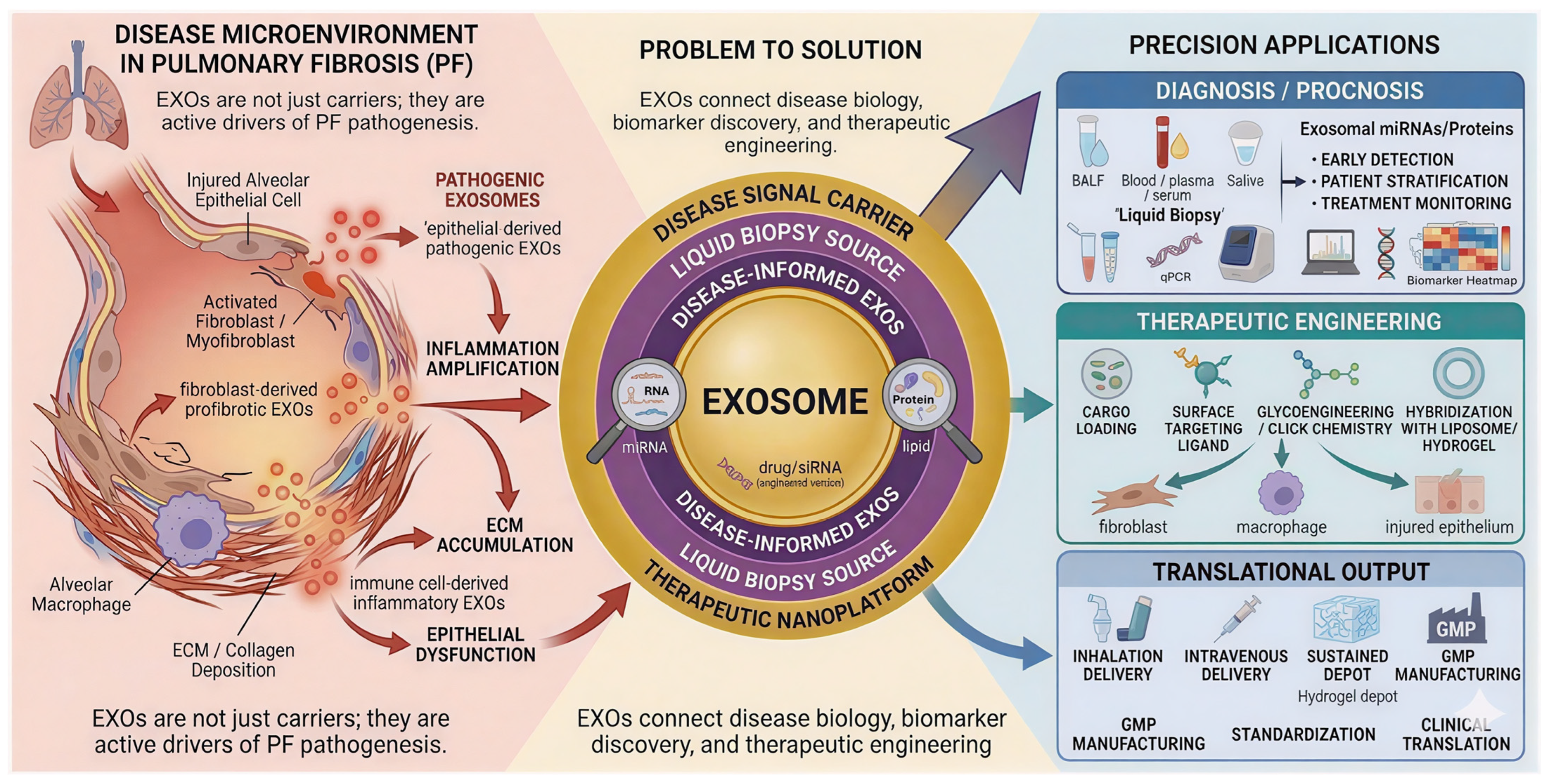
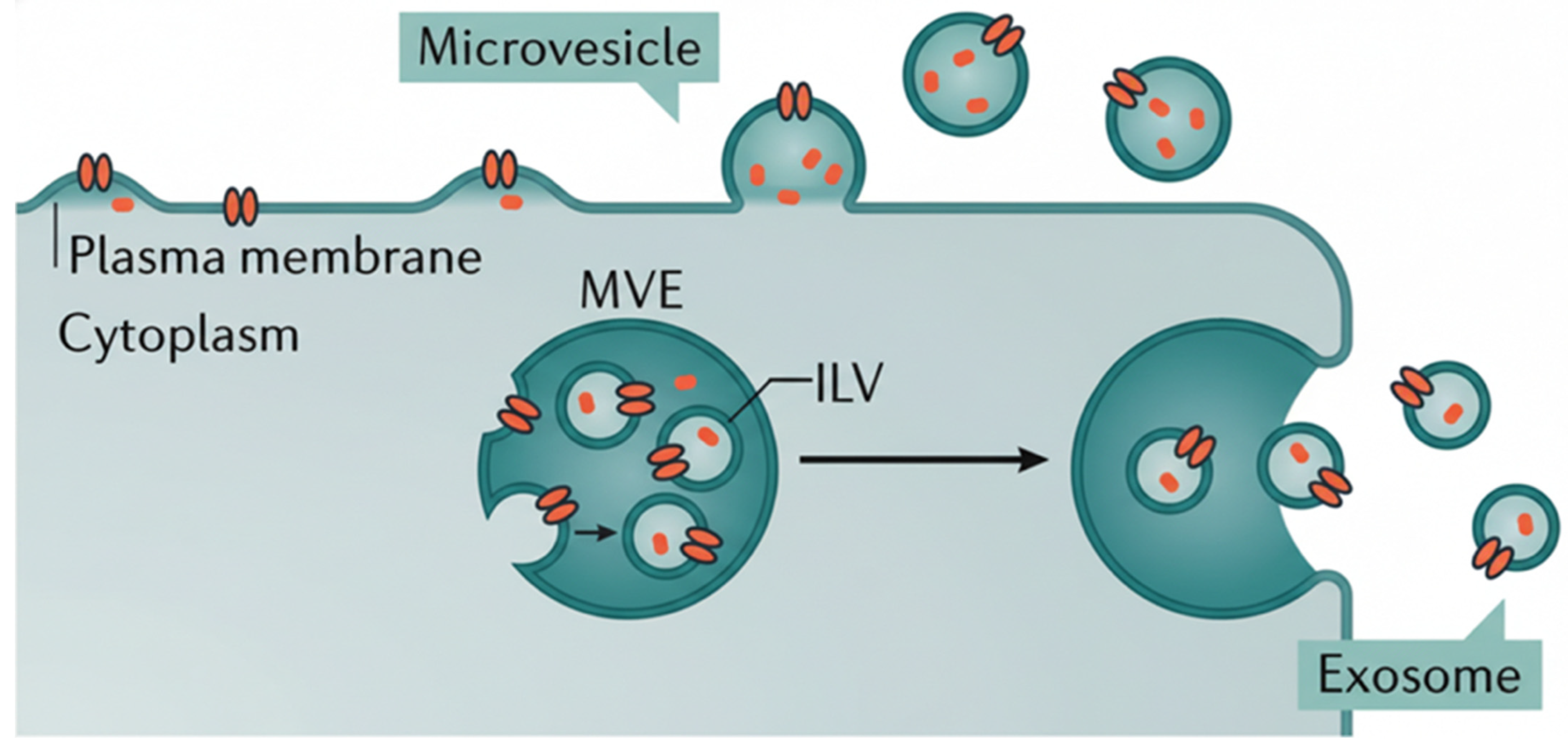
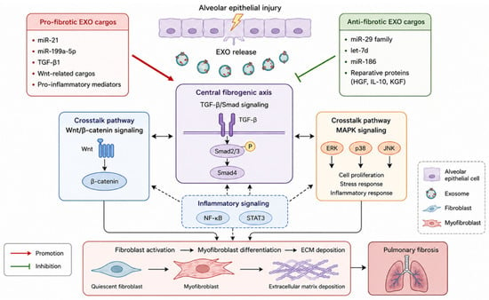
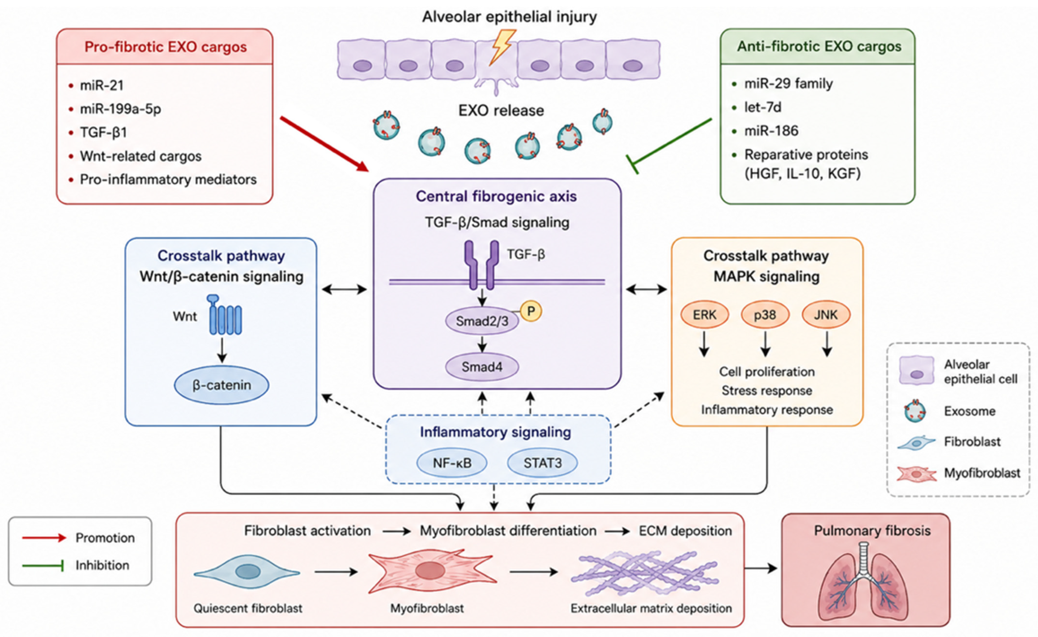
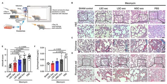
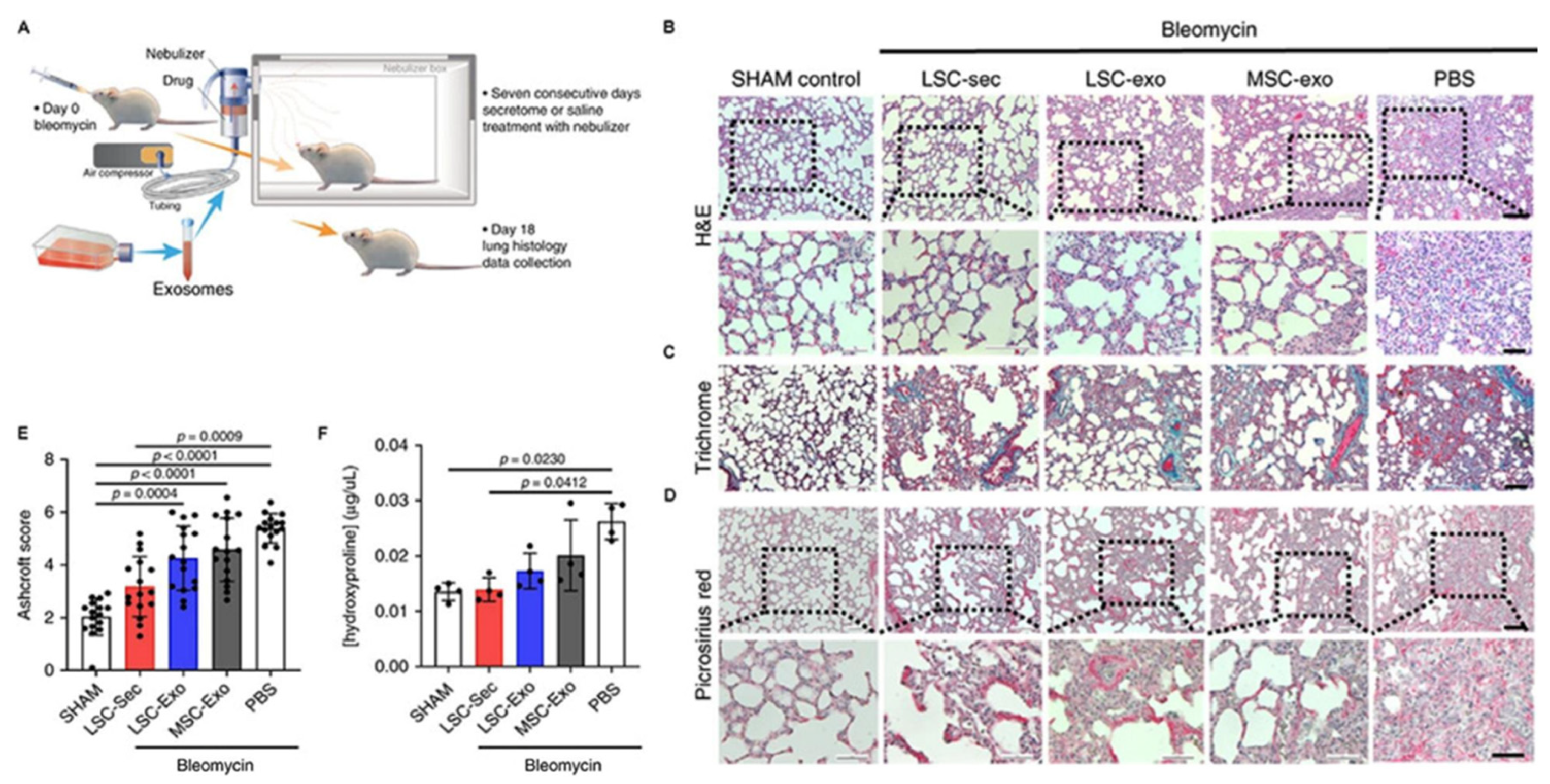
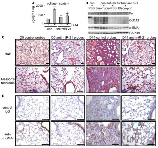
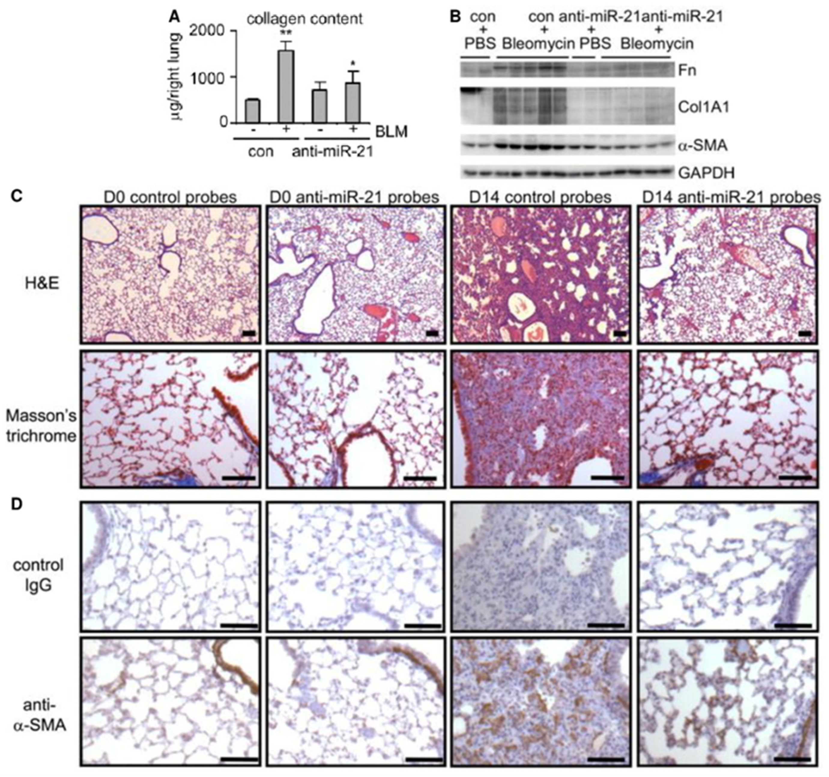
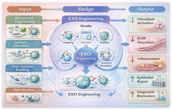
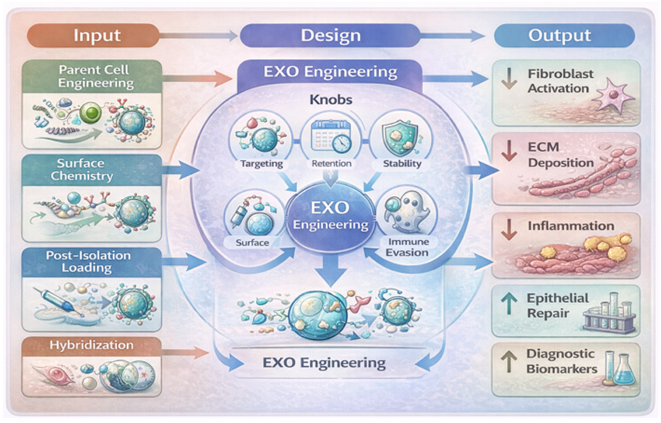
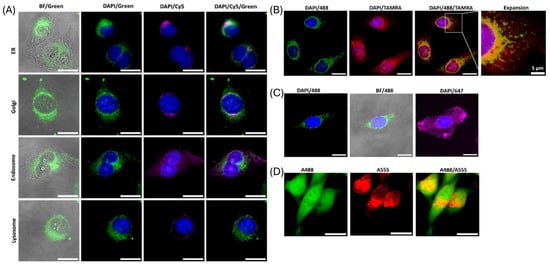
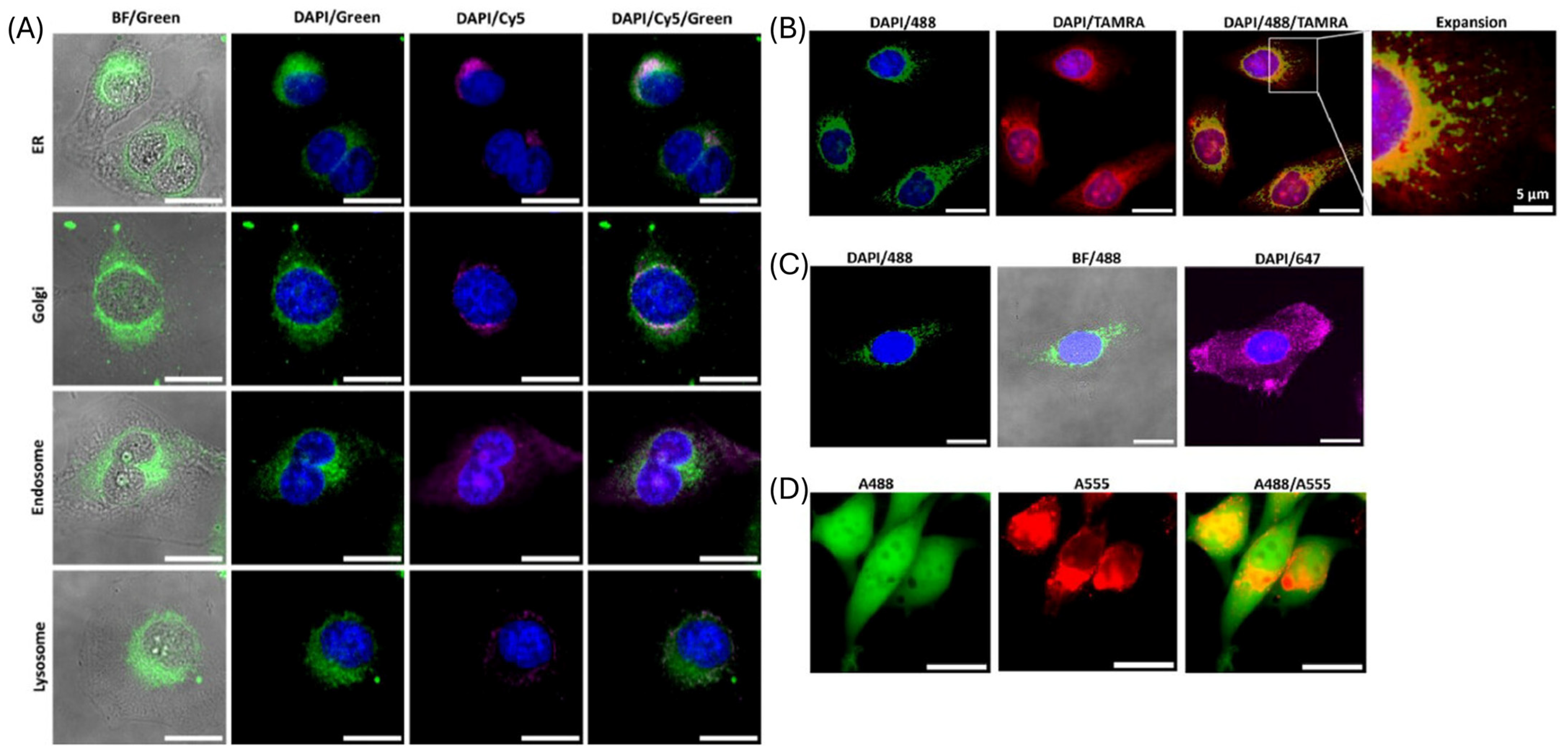
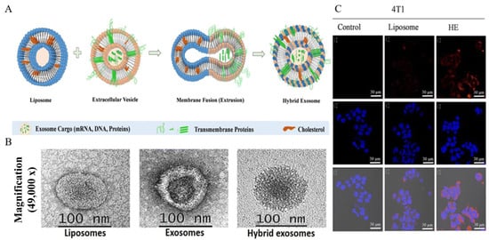
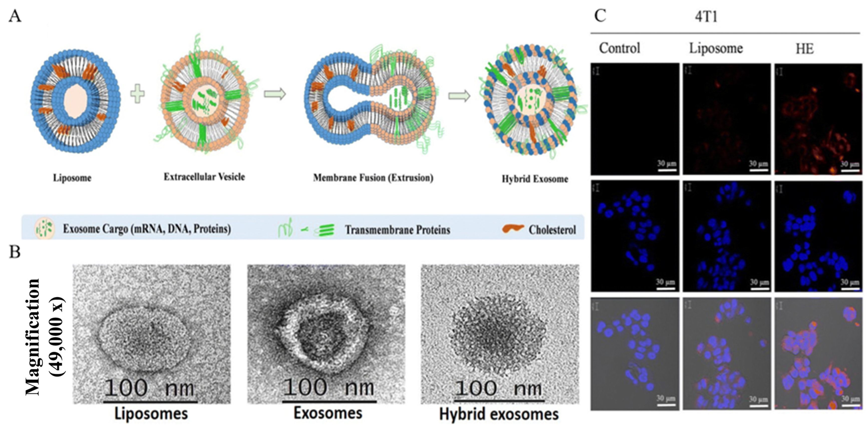
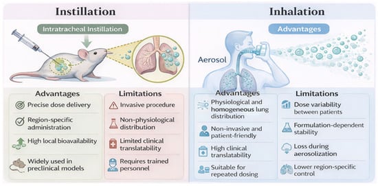
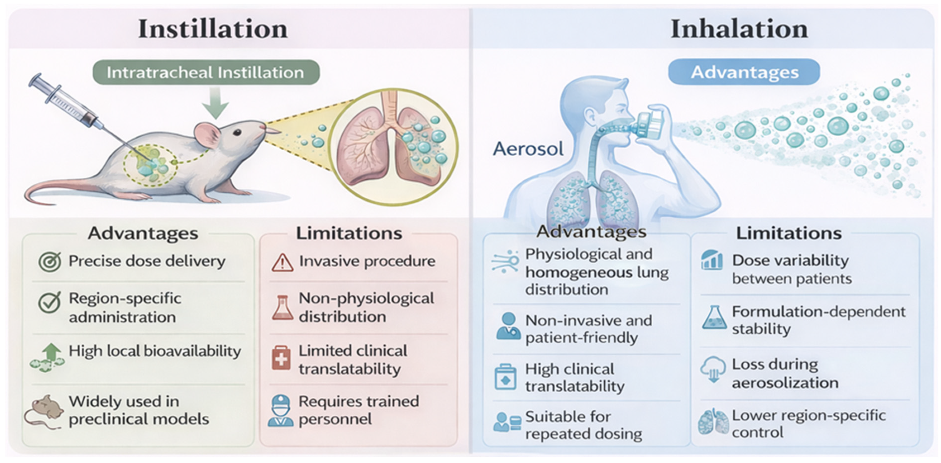
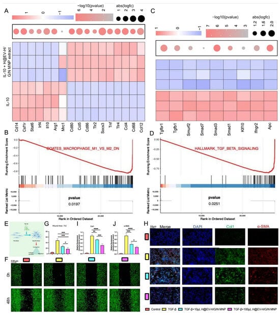
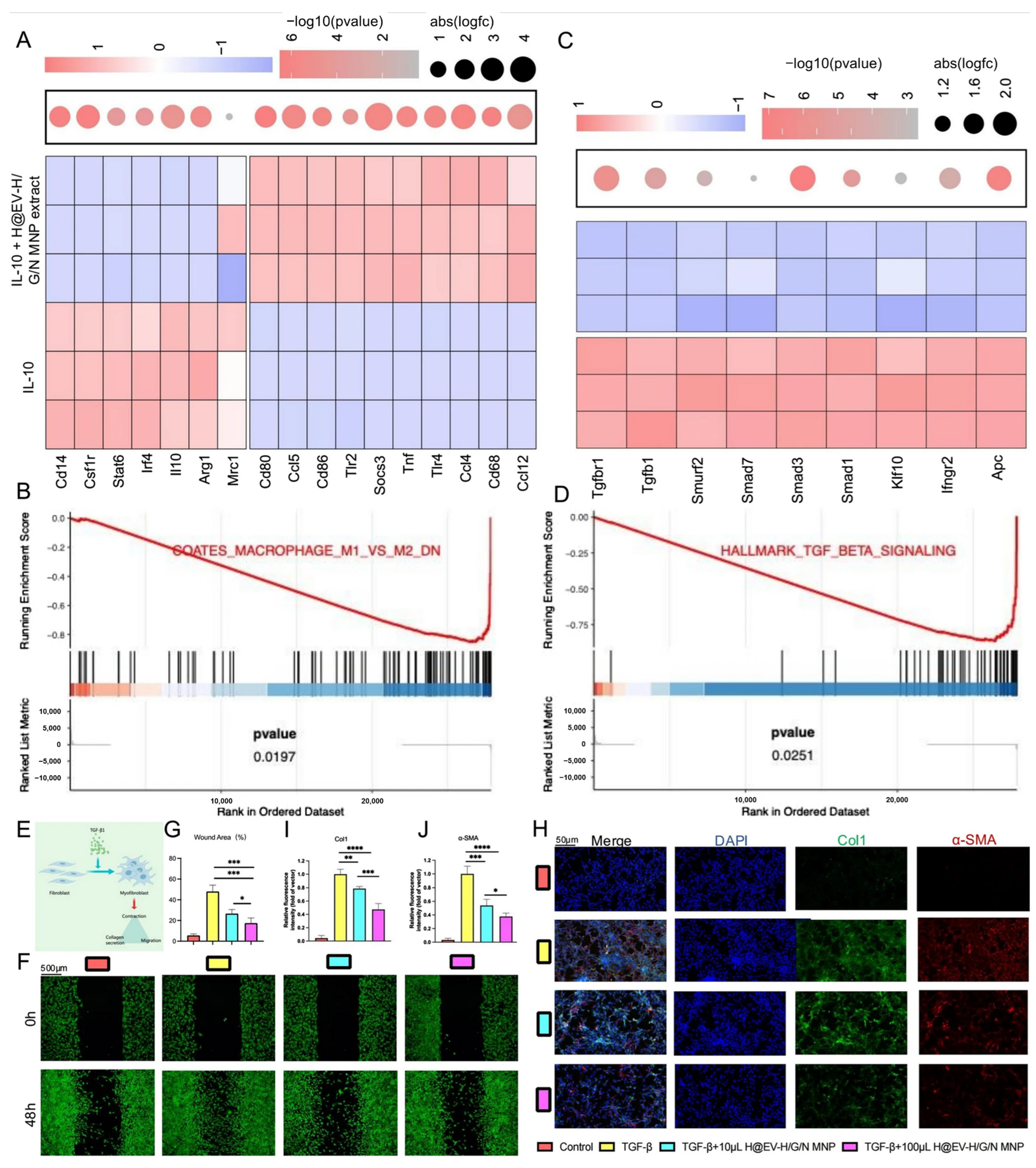

Disclaimer/Publisher’s Note: The statements, opinions and data contained in all publications are solely those of the individual author(s) and contributor(s) and not of MDPI and/or the editor(s). MDPI and/or the editor(s) disclaim responsibility for any injury to people or property resulting from any ideas, methods, instructions or products referred to in the content. |
© 2026 by the authors. Licensee MDPI, Basel, Switzerland. This article is an open access article distributed under the terms and conditions of the Creative Commons Attribution (CC BY) license.